Embed Size (px)
Citation preview
ABSTRACT BOOKOral & E-Poster Presentations

Athens, Greece, March 22-24, 201810th International Congress of Internal Medicinee-Abstract BookAbstracts are published under all authors’ acceptance to be included in all official congresspublications/material. © All rights reserved.
Organizers:
INSTITUTE OF INTERNAL MEDICINE & HEPATOLOGY
DEPARTMENT OF MEDICINE & RESEARCH LABORATORYOF INTERNAL MEDICINE,
UNIVERSITY OF THESSALY MEDICAL SCHOOL,LARISSA, GREECE
Director: Professor G.N. Dalekos
10th International Congressof Internal Medicine
Abstract BookOral & E-Poster Presentations
Endorsed by:
FOUNDATION FOR THE DEVELOPMENT OF INTERNAL MEDICINE IN EUROPE
Participating scientific authorities:
EUROPEAN SOCIETY FOR SWALLOWING DISORDERS
HELLENIC ASSOCIATION FOR THE STUDY OF DIABETIC FOOT
HELLENIC ASSOCIATION FOR THE STUDY OF THE LIVER
HELLENIC DIABETES ASSOCIATION
HELLENIC SEPSIS STUDY GROUP
HELLENIC SOCIETY OF INTERNAL MEDICINE
HELLENIC STROKE ORGANIZATION
Under the auspices of:
UNIVERSITY OF THESSALY MEDICAL SCHOOL, LARISSA, GREECE
March 22-24, 2018, Athens, Greece
4
10th International Congress of Internal MedicineMarch 22-24, 2018, Athens, Greece
ORGANIZATIONOrganizers:
INSTITUTE OF INTERNAL MEDICINE & HEPATOLOGY
DEPARTMENT OF MEDICINE & RESEARCH LABORATORY OFINTERNAL MEDICINE, UNIVERSITY OF THESSALY MEDICAL SCHOOL,LARISSA, GREECEDirector: Professor G.N. Dalekos
ORGANIZING COMMITTEEPresident: G.N. DalekosMembers: K. Krapis C. Mandros K.P. Makaritsis G. Ntaios
SCIENTIFIC COMMITTEE & FACULTY MEMBERS A. AlexopoulouR. Al-Shahi SalmanG. AndrikopoulosQ. AnsteeM. BlankY. de BoerA. BoultonE. BoutatiR. BulbuliaM. BurnierH.R. van BuurenL. CamposM.-D. CappelliniJ.-M. Cavaillon L. ChristouF. CicciaP. ClaveE. CostaG.L. DaikosG.N. DalekosS. De VitaM. DeutschG.D. DimitriadisG. DimopoulosΜ.-Α. DimopoulosS. DourakisS. EngelterA. FloreaniR. GansΝ.K. GatselisS. Georgiadou
V. GeorgouliasE.J. Giamarellos-BourboulisA. GiannoukasS. GiannouliP. GiovasC. GogosL. GuillevinC. HadjichristodoulouS.J. HadziyannisSh. HamdyE. HatziagelakiC. HézodeG. HirschfieldP. InvernizziE. JudeN. KarachaliouS. KaratapanisP. KardasΝ.L. KatsilambrosP. van de KerkhofG. KolovouJ. KoskinasA. KotsakisV. KotsisP.-L. LakatosP. LamperticoM. LenziR. LiberalJ. Lopez-MirandaV. LougarisΚ.P. Makaritsis
P.M. MannucciS. ManolakopoulosM.N. ManoussakisA.M. MarraF. MenichettiY. MetaxasE. MichouG. Mieli-VerganiD. MikhailidisI. MitroulisM. NeteaP. NilssonG. NtaiosS.E. OrfanosP. PapanagiotouN. PapanasG.V. PapatheodoridisV. PapavasileiouA. ParesP. Perez-MartinezG. PetrikkosG. PoulakouM. PsichogiouY. RenaudineauΕ.I. RigopoulouΚ. RitisI. RužaE. SaloustrosD. SamonakisE.C. SandsetM. Schina
C. SchrammD. SereniP.P. SfikakisP. SkendrosM. SpeletasK. SpengosA. StefosN. TentolourisM. TraunerC. TriantosΕ.V. TsianosE.A. TsochatzisM. TumbarelloS. UnalP. VardasD. VassilopoulosM. VavοuranakisΚ. VemmosD. VerganiC.-D. VizzaP.G. VlachoyiannopoulosM. VoulgarelisH. WedemeyerF. WeidanzD. WerringC. YurdaydinΚ. ZachouA. Zanchetti
5
Endorsed by:FOUNDATION FOR THE DEVELOPMENT OF INTERNAL MEDICINEIN EUROPE
Participating scientific authorities:
EUROPEAN SOCIETY FOR SWALLOWING DISORDERS
HELLENIC ASSOCIATION FOR THE STUDY OF DIABETIC FOOT
HELLENIC ASSOCIATION FOR THE STUDY OF THE LIVER
HELLENIC DIABETES ASSOCIATION
HELLENIC SEPSIS STUDY GROUP
HELLENIC SOCIETY OF INTERNAL MEDICINE
HELLENIC STROKE ORGANIZATION
Under the auspices of: UNIVERSITY OF THESSALY MEDICAL SCHOOL, LARISSA, GREECE
10th International Congress of Internal MedicineMarch 22-24, 2018, Athens, Greece
6
10th International Congress of Internal MedicineMarch 22-24, 2018, Athens, Greece
CONTENTS
Oral Presentations ............................................................................................... 7- 56
Thursday, March 22, 2018
Cardiovascular Diseases (OP01-OP09) ..................................................................................... 8-16
Gastroenterology/Hepatology (A) (OP10-OP18) ...................................................................... 17-25
Friday, March 23, 2018
Gastroenterology/Hepatology (Β) (OP19-OP27) ...................................................................... 26-35
Internal Medicine/Miscellaneous (OP28-OP36) ........................................................................ 36-44
Saturday, March 24, 2018
Diabetes Mellitus (OP37-OP42) ................................................................................................ 45-50
Infections/Immunology (OP43-OP48) ....................................................................................... 51-56
E-Posters ............................................................................................................. 57-263
Gastroenterology/Hepatology (ePP001- ePP027) .................................................................... 58-84
Rheumatology (ePP028- ePP041) ............................................................................................ 85-98
Nephrology (ePP042- ePP061) .............................................................................................. 99-121
Diabetes Mellitus / Endocrinology (ePP062- ePP082) .......................................................... 122-142
Hematology / Oncology (ePP83-ePP122) ............................................................................ 143-182
Cardiovascular Diseases (ePP123-ePP141) ........................................................................ 183-201
Infectious Diseases (ePP142-ePP185) ................................................................................. 202-245
Miscellaneous (ePP186-ePP203) ......................................................................................... 246-263
Index .................................................................................................................... 265-278

7
10th International Congress of Internal MedicineMarch 22-24, 2018, Athens, Greece
Oral Presentations
8
ORAL PRESENTATIONS - CARDIOVASCULAR DISEASES - Thursday, March 22, 2018 (OP01-OP09)
OP01
HEART FAILURE WITH MID-RANGE EJECTION FRACTION: THE UNWANTED MIDDLE CHILD FINDS A UNIQUE PLACE AMONG PATIENTS WITH NON-VALVULAR ATRIAL FIBRILLATION
-DATA FROM THE MISOAC-AF REGISTRY
Athanasios Samaras, George Fotos, George Dividis, Eleni Paschou, Ioannis Goulas,
Eleni Thomaidou, Dimitra Vasdeki, Evropi Forozidou, Paraskevi Tsoukra, Eleni Kotsi,
Anastasios Kartas, George Giannakoulas, Haralambos Karvounis, Apostolos Tzikas
1st Department of Cardiology, AHEPA University Hospital, Thessaloniki, Greece
Background: Heart failure (HF) often coexists with atrial fibrillation (AF) increasing the risk for throm-boembolic events. Data associated with the newly defined group of patients with heart failure and mid-range ejection fraction(HFmrEF) in the substrate of AF are currently lacking.
Aim: To determine the differences of HFmrEF patients’ characteristics, rate and rhythm control among the rest of HF patients in a non-valvular AF (NVAF) dataset.
Methods: Patients with HF were classified in three groups according to their ejection fraction(EF)[HF with reduced EF:<40%(HFrEF),HF with mid-range EF:40-49% EF(HFmrEF),HF with preserved EF:>=50%(HFpEF)]. All the patients had coexisting NVAF.
Results: 307 NVAF-HF patients were studied (HFpEF: 52%, HFmrEF: 19%, HFrEF: 29%). When compared with HFrEF patients, HFmrEF patients were associated with higher frequencies of fe-male gender (38% vs 23%, p=0.043), prior episodes of lower gastrointestinal bleeding (14% vs 3%, p=0.025) and palpitations as their main symptom at admission (19% vs 6%, p= 0.015), while lower frequencies were observed at the use of implantable cardioverter defibrillator (6% vs 14%, p=0.048) and the coexistence of dilated cardiomyopathy (2% vs 15%, p=0.009). In comparison to the HFpEF patients, HFmrEF patients were more likely to be men (62% vs 39%, p=0.003), younger [mean age: 74(+- 10) vs 77(+-9), p=0.03)],with higher risk of bleeding [HASBLED>=3: 45% vs 27%, p=0.038] and increased coexistence of ischemic cardiomyopathy (7% vs 1%, p=0.019) and coronary artery disease (66% vs 44%, p=0.005). Paroxysmal AF was more frequently observed at HFpEF compared to HFmrEF patients (37% vs 22%, p=0.047), although more of the last had at least one attempt of prior electrical cardioversion (17% vs 7%, p=0.035). HFmrEF patients received at discharge more regularly calcium channel blockers (22% vs 6%, p=0.01) and less often amiodarone (6% vs 25%, p=0.007) compared with HFrEF patients and more frequently beta blockers (96% vs 80%, p=0.002) compared with HFpEF patients.
Conclusions: Among the AF community, HFmrEF patients have distinct differences with the rest of the HF patients, which are related to their special characteristics, comorbidities and rate and rhythm control strategies. Their unique identity demands further studies on the subject.
9
ORAL PRESENTATIONS - CARDIOVASCULAR DISEASES - Thursday, March 22, 2018 (OP01-OP09)
OP02
WHAT DO WE KNOW ABOUT OUR PATIENTS WITH MYOCARDIAL INFARCTION WITH NON-OBSTRUCTIVE CORONARY ARTERIES?
1Carmen Collado Moreno, 2Virginia Pérez Vázquez, 3Irene Valiente Alemán,
1Ramón Fernando García González, 1Francisco José Morales Ponce
1Cardiology, Puerto Real Universitary Hospital, Cádiz, Spain 2Internal Medicine, Puerto Real Universitary Hospital, Cádiz, Spain
3Critical Care Medicine, Puerto Real Universitary Hospital, Cádiz, Spain
Background/Aim: Myocardial Infarction and Non-Obstructive Coronary Arteries (MINOCA) is an important subtype of MI type 1 where epicardial vessel shows a stenosis < 50%. The aim of this study is to describe prevalence, clinical features, management and prognosis of patients diagnosed with MINOCA.
Methods: A restrospective observational study of patients admitted to Intensive Care Unit with diag-nosis of acute MI was performed between January 1st 2007 and December 31 st 2016. Data were extracted from the ARIAM Andalucia registry. All patients underwent coronary angyography. Patients with MI and no were diagnosed as MINOCA. Follow-up data were obtained from DIRAYA.
Results: A total of 1676 patients with diagnosis of acute MI were registered but only 54 patients fulfilled inclussion criteria for MINOCA disease. The prevalence of MINOCA was 3.2 % with a mean age of 60.59. MINOCA was more frequent in women than in men. Most frequent cardiovascular risk factors were hypertension (55.6%), smoking (51.9%) and hyperlipemia (35.2%). Median risk score GRACE was 123±32.68 and CRUSADE 27.4±16.07. Left ventricular ejection fraction (LVEF) was preserved in 59.39%, mildly reduced in 24.1%, moderately reduced in 11.1% and severely reduced in 5.5% patients. Preferred treatments at discharge were aspirin (96%), statins (86%), ACEI/ARBs (80%), beta-bloquers (70%) and DAPT (64%). One patient died at 3-month follow-up (1.85%).
Conclusions: MINOCA disease has an important prevalence and short-term mortality. Diagnostic work-up of these patients is often suboptimal so its management is not completely accurate. There-fore, more studies on aetiology and management of MINOCA are needed.
10
ORAL PRESENTATIONS - CARDIOVASCULAR DISEASES - Thursday, March 22, 2018 (OP01-OP09)
OP03
QUEST OF EMBOLIC STROKE SOURCE(S) IN ELDERLY PATIENTS: A CASE REPORT REMINDING THAT ATRIAL FIBRILLATION IS NOT ALWAYS THE ANSWER
1Numan Khan, 1Joshua Kirby, 1Bethany Spencer-Lane, 1Stephanie Oade, 1,2Vasileios Papavasileiou
1Stroke Service, Department of Neurosciences, Leeds Teaching Hospitals NHS Trust, UK 2School of Medicine, Faculty of Medicine and Health, University of Leeds, UK
Background/Aims: Cardioembolism is one of the commonest aetiologies of Acute Ischaemic Stroke (AIS); 9.3-38% in large case series (Schneck MJ et al, Cardioembolic stroke, Medscape 2015). Data suggest that although Atrial Fibrillation (AF) is the most widely recognised source in patients aged >60 years, isolated AF is present in only 22% (Arboix A. Curr Cardiol Rev. 2010). The aim of this case report is to highlight the non-AF related sources of cardioembolism.
Case Report: A 81-year old man with a history of previous MI was admitted with an acute left frontal lobe cortical infarct. His work-up revealed non-significant carotid atherosclerosis bilaterally and unremarkable blood tests. His ECG and 11-hour cardiac rhythm monitoring did not reveal AF; prolonged monitoring not feasible due to cognitive problems. His transthoracic cardiac ultracound showed a hypermobile mass attached to the mitral valve in keeping with a vegetation and left ventri-cle (LV) ejection fraction of 25-35%. The absence of an LV thrombus was confirmed with a contrast study. The patient did not meet any other major or 3 minor Duke criteria for infective endocarditis. Regarding marantic endocarditis lupus anticoagulant and antiphospholipid antibodies were negative and there were no signs of disseminated intravascular coagulation, malignancy or systemic lupus erythematosus. A transoesophageal cardiac ultrasound and cardiac MRI could not be performed due to minimal cooperation. Despite the patient’s advanced age, AF was not among the 2 cardioembolic sources identified as probably related to his recent stroke.
11
ORAL PRESENTATIONS - CARDIOVASCULAR DISEASES - Thursday, March 22, 2018 (OP01-OP09)
OP04
20-YEAR TRENDS OF CHARACTERISTICS AND OUTCOMES OF STROKE PATIENTS WITH ATRIAL FIBRILLATION
1George Ntaios, 1Dimitrios Sagris, 2Fotios Gioulekas, 3Petros Galanis, 4Christianna Pardali, 4Anastasia Vemmou, 5Eleni Koroboki, 6Vasileios Papavasileiou, 5Sofia Vassilopoulou,
4Efstathios Manios, 1Konstantinos Makaritsis, 5Konstantinos Spengos, 5Dimos-Dimitrios Mitsikostas,
7Haralambos Milionis, 4Konstantinos Vemmos
1Department of Medicine, Larissa University Hospital, School of Medicine, University of Thessaly, Larissa, Greece
2Sub-directorate of Informatics, Larissa General University Hospital, Larissa, Greece 3Centre for Health Services, Management and Evaluation Faculty of Nursing,
University of Athens, Athens, Greece 4Department of Clinical Therapeutics, Alexandra Hospital, National and Kapodistrian
University of Athens, Athens, Greece 5Department of Neurology, Aeginition Hospital, National and Kapodistrian
University of Athens, Athens, Greece 6Comprehensive Stroke Centre, Salford Royal NHS Foundation Trust, Manchester Academic
Health Sciences Centre, Salford Royal Foundation Trust, Manchester M6 8HD, United Kingdom 7Department of Medicine, Ioannina University Hospital, School of Medicine,
University of Ioannina, Ioannina, Greece
Objective: We aimed to report the overall, age- and sex-specific secular trends of characteristics and outcomes of patients with acute ischemic stroke (AIS) and atrial fibrillation (AF) between 1993 and 2012 in the Athens Stroke Registry.
Methods: We used Joinpoint regression analysis to calculate the average annual percent changes and 95% confidence intervals.
Results: Among 3314 stroke patients, 1044 (31.5%) had AF. Between 1993-2012, there was: aver-age annual percentage reduction (AAPR) (0.8%, 95%CI:-1.5%;0.0%) in the proportion of AF-pa-tients among all AIS patients; AAPI (7.1%, 95%CI:5.4%;8.9%) in the proportion of newly-diagnosed AF-patients among all AF-patients; AAPR (2.9%, 95%CI:-2.7;-3.2%) in the proportion of previous-ly-known AF-patients among all AF-patients; AAPR (2.4%, 95%CI:-1.2;-3.6%) in the proportion of previously-known AF-patients not receiving any antithrombotic treatment at admission; AAPI (6.4%, 95%CI:1.2;11.9%) in the proportion of previously-known AF-patients on anticoagulants at admission; AAPI (2.3%, 95%CI: -0.4;5.0%) in the proportion of previously-known AF-patients on aspirin at ad-mission; AAPI in the proportion of AF-patients who were prescribed anticoagulant (3.5% for patients with mRS<4; 7.2% for mRS:4-5) and AAPR in the proportion of AF-patients who were prescribed aspirin (5.8% for mRS<4; 1.6% for mRS:4-5) or no antithrombotic at discharge (7.1% for mRS<4 and 5.3% for mRS:4-5); AAPR in stroke recurrences (5.8%, 95%CI:-8.6;-3.0%), cardiovascular events (6.5%, 95%CI:-8.3;-4.7%) and deaths (7.9%, 95%CI:-9.2;-6.5%).
Conclusions: Between 1993-2012, the proportion of AF-patients on proper antithrombotic treat-ment and the rate of newly-diagnosed AF increased significantly. Rates of stroke recurrence, cardio-vascular events and mortality reduced significantly.
12
ORAL PRESENTATIONS - CARDIOVASCULAR DISEASES - Thursday, March 22, 2018 (OP01-OP09)
OP05
CLOSURE OF PATENT FORAMEN OVALE VS. MEDICAL THERAPY IN PATIENTS WITH CRYPTOGENIC STROKE OR TRANSIENT ISCHEMIC ATTACK: UPDATED SYSTEMATIC
REVIEW AND META-ANALYSIS
1George Ntaios, 2Vasileios Papavasileiou, 1Dimitrios Sagris, 1Konstantinos Makaritsis,
3Konstantinos Vemmos, 4, 5Thorsten Steiner, 6Patrik Michel
1Department of Medicine, University of Thessaly, Larissa, Greece 2Stroke Service, Department of Neurosciences, Leeds Teaching Hospitals NHS Trust and Medical
School, University of Leeds, Leeds, UK 3Hellenic Cardiovascular Research Society, Athens, Greece
4Department of Neurology, Klinikum Frankfurt Höchst, Frankfurt, Germany 5Department of Neurology, Heidelberg University, Heidelberg, Germany
6Stroke Center, Lausanne University Hospital, Lausanne, Switzerland
Background: Previous systematic reviews and meta-analyses compared the efficacy and safety of patent foramen ovale(PFO) closure vs. medical treatment in patients with cryptogenic ischemic stroke or transient ischemic attack(TIA). Recently, new evidence from randomized trials became available.
Methods: We searched PubMed until 24/9/2017 for randomized trials comparing PFO closure with medical treatment in patients with cryptogenic stroke/TIA using the items: “stroke or cerebrovascular accident or TIA” and “patent foramen ovale or paradoxical embolism” and “trial or study”.
Results: Among 851 potentially eligible articles, 5 were eligible for the meta-analysis. In 3627 pa-tients (1829 in the PFO-closure arm and 1798 in the medical treatment), there was statistically signif-icant difference in ischemic stroke recurrence (2.02% vs. 4.17% respectively, odds-ratio (OR):0.43, 95% confidence intervals (CI): 0.21-0.90, relative-risk-reduction (RRR):50.53%, absolute-risk-reduc-tion (ARR):2.15%, number-needed-to-treat (NNT):46.5), whereas there was no statistically signifi-cant difference in TIA (3.09% vs. 3.80% respectively, OR:0.80, 95%CI:0.53-1.19) or death (0.71% vs. 0.88% respectively, OR:0.73, 95%CI:0.34-1.56). New-onset atrial fibrillation occurred more fre-quently in the PFO-closure arm (4.75% vs. 0.94% respectively, OR:5.15, 95%CI:2.18-12.15, rela-tive-risk-increase:403.1%, absolute-risk-increase:3.8%, NNT:26.2) and resolved in 72% of cases within 45 days, whereas the rates of myocardial infarction (0.33% vs. 0.27% respectively, OR:1.22, 95%CI:0.25-5.91) and any serious adverse events (27.97% vs. 27.20% respectively, OR:1.07, 95%CI:0.92-1.25) were similar.
Conclusions: In patients with cryptogenic stroke/TIA and PFO who have their PFO closed, ischemic stroke recurrence is less frequent compared to patients receiving medical treatment and atrial fibril-lation is more frequent but mostly transient. There is no difference in TIA or death or myocardial infarction.
13
ORAL PRESENTATIONS - CARDIOVASCULAR DISEASES - Thursday, March 22, 2018 (OP01-OP09)
OP06
ACUTE HEART FAILURE (AHF) IN A PATIENT WITH LOWER URINARY TRACT INFECTION (LUTI): A SECONDARY REVERSE TAKOTSUBO CASE REPORT
1,2Anastasia Nouli, 1Aikaterini Dimitra, 2Eleni Tsakiri, 3Dimosthenis Stavrianakis, 1,2,3Nikitas Moschos
1Cardiology Department, General Hospital of Rhodes, Rhodes, Greece 2Intensive Coronary Care Unit, General Hospital of Rhodes, Rhodes, Greece
3Cardiac Catheterization Laboratory, General Hospital of Rhodes, Rhodes, Greece
Background: Takotsubo cardiomyopathy is typified by a transient systolic dysfunction of the apical segments of the left ventricle (LV) -apical ballooning- mimicking myocardial infarction in the absence of obstructive coronary artery disease (CAD), possibly due to acute catecholaminergic myocardial stunning. Reverse takotsubo, a variant form in which the basal and midventricular segments of the LV are akinetic, occurs in a minority of patients. Some Takotsubo cases occur in patients already hospitalized for another medical condition. In these patients, sudden activation of the sympathetic nervous system or a rise in catecholamines precipitates an acute Takotsubo syndrome as a compli-cation of the primary condition. Such cases are diagnosed as secondary Takotsubo syndrome.
Case Report: A 73 yo male was hospitalized due to LUTI with pyrexia and elevated inflammation indexes. The second day of hospitalization he complained of chest pain and developed pulmonary edema with low blood pressure. The echocardiography revealed low EFLV=25%, the troponine lev-els increased and he was transferred to the ICCU. After several days, the patient was stabilized and underwent a coronarography, which was free of obstructive CAD. The left ventriculogram showed a reverse Takotsubo pattern, with hypercontractility of the apical and akinesia of the basal and mid-ventricular segments. The patient got discharged with treatment for heart failure (b-blockers, ACEI, MRA). A month later a cardiac MRI was performed, which showed normal EFLV=60% and no signs of myocardial necrosis.
14
ORAL PRESENTATIONS - CARDIOVASCULAR DISEASES - Thursday, March 22, 2018 (OP01-OP09)
OP07
COMPARISON OF SAFETY OUTCOMES BETWEEN PCSK9 INHIBITORS AND STANDARD OF CARE: SYSTEMATIC REVIEW AND META-ANALYSIS OF 47 RANDOMISED CONTROLLED
TRIALS
1Karagiannaki Anastasia, 2Kalliopi Perlepe, 2Makaritsis Konstantinos, 2Ntaios George
1General Hospital of Thessaloniki Agios Pavlos, Thessaloniki, Greece 2Department of Medicine, University of Thessaly, Larissa, Greece
Background: Recent trials of monoclonal antibodies to proprotein convertase subtilizin/kexin type 9 (PCSK9) reported robust low-density lipoprotein cholesterol reductions.
Objective: To assess the safety of PSCK9 inhibitors in published trials of PCSK9 inhibitors.
Methods: We performed a systematic review and meta-analysis of randomized controlled trials of PSCK9 inhibitors vs. standard-of-care (SOC). The search items in PubMed and Web of Science were: (Proprotein convertase subtilisin/kexin type 9 inhibitor OR PSCK9 inhibitor OR alirocumab OR bococizumab OR evolocumab) AND (prospective OR randomized OR trial). Safety outcomes assessed included haemorrhagic stroke, new-onset diabetes mellitus (DM) or worsening of pre-existing diabetes, cancer, myalgia, CK> x3 the upper limit of normal (ULN), liver transaminase > x3ULN or ALT>3 ULN, cataract and neurocognition.
Results: There was no statistically significant difference between PCSK9 inhibitors and SOC in DM (HR: 1.03,95%CI: 0.80-1.33 in 12 studies and 43,122 patients); cancer (HR: 1.10,95%CI: 0.67-1.82 in 8 studies and 6,650 patients), cataract (HR: 0.94,95%CI: 0.78-1.13 in 1 study and 27,525 patients), neurocognition (HR: 1.14,95%CI: 0.75-1.73 in 16 studies and 46,492 patients), high CK levels (HR: 0.82,95%CI: 0.69-0.97 in 28 studies and 54,364 patients), myalgia (HR: 0.95,95%CI: 0.71-1.27 in 19 studies and 22,864 patients), higher liver transaminase levels (HR: 0.78,95%CI: 0.61-1.01 in 25 studies and 27,181 patients), haemorrhagic stroke (HR: 1.16, 95%CI: 0.68-1.98 in 1 study and 27,564 patients).
Conclusions: There was no major statistical difference between PSCK-9 inhibitors and SOC for the safety parameters of cancer, diabetes mellitus, cataract, CK, liver transaminases, myalgia, neuro-cognition and haemorrhagic stroke.
15
ORAL PRESENTATIONS - CARDIOVASCULAR DISEASES - Thursday, March 22, 2018 (OP01-OP09)
OP08
CHARACTERISTICS AND TEN-YEAR PROGNOSIS OF PATIENTS TREATED WITH ASPIRIN PRIOR TO A FIRST-EVER ACUTE ISCHEMIC STROKE. DATA FROM THE ‘ATHENS STROKE
OUTCOME PROJECT’
1, 2Haralampos Milionis, 1Fotios Barkas, 2,3George Ntaios, 2,4Eleni Koromboki,
2,5Vasileios Papavasileiou, 2Kostantinos Vemmos
1Department of Internal Medicine, School of Medicine, University of Ioannina, Ioannina, Greece 2Hellenic Stroke Organization, Athens, Greece
3Department of Internal Medicine, School of Medicine, University of Thessaly, Greece 4First Department of Neurology, National and Kapodistrian University of Athens, Athens, Greece
5Stroke Service, Department of Neurosciences, Leeds Teaching Hospitals NHS Trust and School of Medicine, Faculty of Medicine and Health, University of Leeds, Leeds, UK
Aims: To assess the clinical characteristics andthe ten-year prognosis of patients treated with aspir-inprior to a first-ever acute ischemic non-cardioembolic stroke (AINCS).
Methods: This was a retrospective study involving a total of 1,782 patients hospitalized due to a first-ever AINCS; of those, 14% (n=257) were treated with aspirin (ASA group) for elective purposes, while 1,525 patients received no antiplatelets (non-ASA group) prior to the index event. Primary endpoints included: time tostroke recurrence, composite cardiovascular event (stroke recurrence, myocardial infarction, unstable angina, coronary revascularization, peripheral atherosclerotic artery diseases, or sudden death), and all-cause mortality.
Results: Subjects pre-treated with aspirin had higher composite cardiovascular event-free survival (log rank test: 4.79, p=0.029) and 10-year mortality (log rank test: 6.8, p=0.012) compared with the non-ASA group, while there was no difference in time to stroke recurrence (log-rank test: 0.039, p=0.84). Age (hazard ratio, HR 1.07; 95%CI: 1.04-1.09, p<0.001), stroke severity on admission (HR: 1.05; 95%CI: 1.03-1.08, p<0.001), heart failure (HR 2.32; 95%CI: 1.24-4.36, p<0.01) and peripheral artery disease (HR 2.25; 95%CI: 1.15-4.39, p<0.05) were associated with higher 10-year mortality in the ASA group. Of note, the rate of cardiovascular events was higher among male patients (HR 2.27; 95%CI: 1.04-4.98, p <0.05) but lower among those treated for dyslipidemia prior to the index stroke (HR0.58; 95% CI: 0.36-0.93, p <0.05).
Conclusions: This study indicates that elective treatment with aspirin prior to an AINCS does not affect long-term stroke recurrence but increases mortality and cardiovascular morbidity.
16
OP09
A CASE REPORT OF EMBOLIC STROKE OF UNDETERMINED SOURCE: SHOULD ANTIPHOSPHOLIPID SYNDROME TESTING BE PERFORMED IN PATIENTS WITH KNOWN
ACTIVE MALIGNANCY?
1Bethany Spencer-Lane, 1Stephanie Oade, 1Marissa Minns, 1,2Vasileios Papavasileiou
1Stroke Service, Department of Neurosciences, Leeds Teaching Hospitals NHS Trust, UK 2School of Medicine, Faculty of Medicine and Health, University of Leeds, UK
Background/Aim: Studies demonstrate a higher prevalence of anti-cardiolipin antibodies (ACLAs) in patients with malignancies compared to controls (22 versus 3%) (Zuckerman et al. 1995). Amongst cancer patients, ACLAs positive individuals are at significantly higher risk of thromboembolic events (Zuckerman et al. 1995). Recent studies that compare direct oral anticoagulants versus antiplatelets in patients with embolic strokes of undetermined source have not included ACLAs as a mandatory investigation in patients with active malignancy. The aim of this case report is to highlight the need to consider screening patients with active malignancy and acute cryptogenic ischemic stroke for ACLAs as this may identify patients that have an indication for anticoagulation.
Case Report: A 75-year old woman with a recently excised invasive ductal breast carcinoma pre-sented with expressive aphasia and reduced right-hand coordination shortly after her first radio-therapy session. Brain MRI confirmed an acute posterior left middle cerebral artery cortical infarction and established silent bilateral cerebellar infarcts and right corona radiata / peri-Rolandic cortex infarct. MRA head and neck showed mild atherosclerotic changes at the carotid bifurcations bilater-ally but normal posterior circulation and intracranial arteries. A 7-day cardiac rhythm monitor and a transthoracic cardiac ultrasound did not reveal major cardioembolic sources or a patent foramen ovale. Antiphospholipid screen revealed ACLAs and b2-Glycoprotein IgG antibodies more than 8 times higher than the upper normal limit; titres unchanged at 3 months. The patient was diagnosed with antiphospholipid syndrome and anticoagulated with warfarin; INR target 2.0-3.0. She has not suffered any thromboembolic events 13 months later.
ORAL PRESENTATIONS - CARDIOVASCULAR DISEASES - Thursday, March 22, 2018 (OP01-OP09)

17
OP10
THE ROLE OF NEWER DAAs IN THE TREATMENT OF CHRONIC HEPATITIS C PATIENTS WITH GENOTYPE 5
George Ntetskas, Vasilios Papastergiou, Aikaterini Chasiou, Eustathia Anastasiou,
Despoina Papanikola, Anna Katsili, Filippos Papakonstantinou, Nikitas Katsilis,
Chrysostomos Psellas, Stylianos Karatapanis
1st Department of Internal Medicine, General Hospital of Rhodes, Rhodes, Greece
Background: Chronic hepatitis C patients harboring genotype 5 are rare in our country apart from a region in south Greece. We have already published our experience in treating these patients with the conventional therapy. The international experience on treating these patients with newer direct acting antiviral (DAAs) drugs is limited.
Aim: We report our experience on CHC patients with genotype 5, treated with DAAs
Methods: Twenty CHC patients with genotype 5, were included in the present study (M/F=3/15, mean age 68.3). All patients have had advanced fibrosis and/or cirrhosis as was assessed by tran-sient elastography (Fibroscan). All cirrhotic patients (15/20) included in the study had compensated cirrhosis. 7/20 patients had received double conventional treatment in the past. Fifteen patients were treated using sofosbuvir 400mg/day plus ribavirin (according to the patient’s weight: <75 kg 1000mg/day, >75 Kg 1200 mg/day) for 12 weeks, and five received ledipasvir/sofosbuvir plus ribavirin for 12 weeks. HCV-RNA in the serum was tested 12 weeks after the end of therapy.
Results: All patients had negative HCV-RNA at the end of therapy (EOT). Sustained virological re-spond (SVR) was noted in 17/17 patients who were tested 12 weeks after the end of therapy (15/17 had received sofosbuvir plus ribavirin). No significant adverse events were noticed during therapy (3 patients developed anemia). Conclusions: Our results indicate that the newer DAAs proved very efficacious in CHC patients (genotype 5) with advanced fibrosis and/or cirrhosis. The combination of sofosbuvir plus ribavirin resulted in high SVR rate.
ORAL PRESENTATIONS - GASTROENTEROLOGY/HEPATOLOGY (A) - Thursday, March 22, 2018 (OP10-OP18)
18
OP11
ASSESSMENT OF HEALTH RELATED QUALITY OF LIFE IN GREEK PATIENTS WITH PRIMARY BILIARY CHOLANGITIS USING THE PBC-40 QUESTIONNAIRE
M. Bakarozi, K. Galanis, S. Gabeta, K. Zachou, Nikolaos Κ. Gatselis, George N. Dalekos,
Eirini I. Rigopoulou
Department of Medicine and Research Laboratory of Internal Medicine, Medical School, University of Thessaly, Larissa, Greece
Background: Primary biliary cholangitis (PBC) is a chronic, autoimmune cholestatic liver disease with a complex clinical phenotype. Quality of life of PBC patients can be frequently impaired by symptoms such as fatigue and itch, which can occur at any point in the disease course. PBC-40 questionnaire, originally validated in UK PBC patient, is considered a useful tool for evaluation of health related quality of life (HRQoL) in PBC. Aim: Our aim is to assess HRQoL in Greek PBC pa-tients by using PBC-40 and SF-36 questionnaires.
Materials and methods: The PBC-40 questionnaire was translated to Greek using the forward-backward method. Both questionnaires were self-administrated to 60 PBC (95% females) patients up to now, attending our outpatient Liver clinic. The internal consistency was assessed by using Cronbach’s α coefficient. Data regarding diagnosis, laboratory characteristics and therapy were col-lected. Results: The average PBC-40 score was 74.23±22.16; the highest mean domain score was seen for fatigue (22.94±8.57) and the lowest for itch (2.91±2.64). The overall internal consistency of the PBC -40 was 0.94. Reliability was above 0.7 for all the domains of PBC-40, except for the domains “fatigue” (α=0.68) and “social” (α=0.69). High correlations were seen between PBC-40 and SF-36 concerning the domains “fatigue”, “social” and “emotional” (p<0.001).
Conclusions: This is the first validation of PBC-40 in Greek PBC patients. From our preliminary data, PBC-40 seems to be an efficient and valid questionnaire in assessing HRQoL in PBC. More work is needed to establish PBC-40 as a valuable scientific tool for Greek PBC patients.
ORAL PRESENTATIONS - GASTROENTEROLOGY/HEPATOLOGY (A) - Thursday, March 22, 2018 (OP10-OP18)
19
OP12
DETERIORATION OF SUBCLINICAL ATHEROSCLEROSIS MARKERS DURING AND AFTER PEGYLATED INTERFERON AND RIBAVIRIN TREATMENT FOR CHRONIC HEPATITIS C
1Alexandra Alexopoulou, 2George Georgiopoulos, 1Sopia Pouriki, 1Ilianna Mani, 1Larisa Vasilieva,
2Angeliki Laina, 1Dimitrios Zampetas, 2Kimon Stamatelopoulos, 1Spyros P Dourakis
12nd Department of Internal Medicine and Research Laboratory, Medical School, National and Kapodistrian University of Athens, “Hippokration” Hospital, Athens, Greece
2Vascular Laboratory, Department of Clinical Therapeutics, National and Kapodistrian University of Athens, “Alexandra” University Hospital, Athens, Greece
Background/Aim: Despite the increased incidence of adverse cardiovascular events in chronic hepatitis C (CHC) patients under pegylated-interferon-ribavirin (PR), the prospective association of PR-therapy with vascular indices has not been studied so far. In this study, we assessed the changes in vascular markers of subclinical atherosclerosis during and after PR-treatment.
Methods: All participants who completed PR, were prospectively evaluated across 3 visits (base-line-T1, end-of-treatment-T2 and 24-weeks-post-treatment-T3. Vascular function measurements in-cluded: peripheral and central-systolic (SBP) and diastolic-blood-pressure (DBP); flow-mediated, endothelium-dependent-vasodilation (FMD) (%); carotid-femoral-pulse-wave-velocity (PWV)(m/s); augmentation-index (AI%) and time-for-return-of-reflected-waves (TR)(miliseconds); intima-media-thickness (IMT) of the carotid artery.
Results: 26 consecutive patients [46.2% male, age 45 (31.7-53.5)], non-smokers with CHC and 51 healthy-controls were enrolled. Significant un-adjusted changes in demographic, hemodynamic and vascular variables were not observed at baseline and across the follow-up period in both groups. In contrast, a significant temporal effect of treatment was established in terms of vascular parameters in CHC patients by linear mixed model analysis. More specifically, antiviral treatment was associated with unfavorable changes in FMD and PWV between T3 and T1 compared to controls (P=0.015 and P=0.017, respectively). This effect was independent of age, gender and longitudinal changes in SBP and waist circumference. Moreover, HCV patients presented increased AI and a trend for early de-crease in TR concomitant to treatment administration (between T2 and T1) as compared to controls (P=0.043 and P=0.077, respectively), after adjusting for the above parameters.
Conclusions: Surrogate markers of subclinical atherosclerosis deteriorate in patients with CHC dur-ing PR. This deterioration continues at least 6 months post-treatment.
ORAL PRESENTATIONS - GASTROENTEROLOGY/HEPATOLOGY (A) - Thursday, March 22, 2018 (OP10-OP18)

20
OP13
EVALUATION OF TRANSIENT ELASTOGRAPHY IN THE DIAGNOSIS OF CYSTIC FIBROSIS LIVER DISEASE
1Sophia Pouriki, 1Theodoros Alexopoulos, 1Alexandra Alexopoulou, 1Larisa Vasilieva,
2Filia Diamantea, 2Maria Gioka, 2Alexandra Nakou, 1Ilianna Mani, 1Dimitrios Zampetas,
1Spyros P. Dourakis
12nd Department of Internal Medicine and Research Laboratory, Medical School, National and Kapodistrian University of Athens, “Hippokration” Hospital, Athens, Greece
2Third Respiratory Medicine Department, “Sismanogleio” General Hospital, Athens, Greece
Background/Aim: In cystic fibrosis (CF), liver disease (CFLD) is the third leading cause of mortality. As liver biopsy has been considered inconsistent in diagnosis of CFLD, a combination of modalities were utilized in the conventional Debray criteria (DC). More recently, transient elastography (TE) and non-invasive-liver-fibrosis-biomarkers (NILFB) were included in the New criteria (NC). In this study, we aimed to evaluate transient elastography (TE) and NILFB for the diagnosis of CFLD in adults.
Methods: Longitudinal data were collected from a cohort of genetically confirmed CF patients. CFLD was diagnosed by both DC and NC.
Results: 62 patents with CF, [56.5% male, age at enrollment 25 (22-31) years], were prospectively followed-up for 33 (28-36) months. TE was performed in all the patients. 16 (25.8%) and 26 (41.9%) met the classical DB and NC, respectively. Patients with CFLD had higher liver stiffness than those without, by both DC and NC (P=0.03 and P<0.001, respectively). ALP and INR correlated with TE (Spearman’s r=0.413, P=0.001 and r=0.532, P<0.001, respectively). Patients with an abnormal Fi-broscan (>6.8 kPa) had higher AST (P=0.007), γGT (P=0.005), ALP (P<0.001), INR (P=0.001), APRI (P=0.04) and FIB-4 (P=0.003) vs those with ≤6.8 kPa. TE has an optimal specificity and positive-predictive-value to detect CFLD by NC but lags sensitivity.
Conclusions: The cutoff value of 6.8 kPa by TE is highly accurate to detect true CFLD according to the NC but it lags sensitivity probably due to the patchy histologic nature of the disease. TE is more accurate than NILFB to detect CFLD.
ORAL PRESENTATIONS - GASTROENTEROLOGY/HEPATOLOGY (A) - Thursday, March 22, 2018 (OP10-OP18)
21
ORAL PRESENTATIONS - GASTROENTEROLOGY/HEPATOLOGY (A) - Thursday, March 22, 2018 (OP10-OP18)
OP14
EVALUATION OF THE NEW CRITERIA IN THE DIAGNOSIS OF CYSTIC FIBROSIS LIVER DISEASE
1Sophia Pouriki, 1Theodoros Alexopoulos, 1Alexandra Alexopoulou, 1Larisa Vasilieva,
2Filia Diamantea, 2Maria Gioka, 2Alexandra Nakou, 1Dimitrios Zampetas, 1Ilianna Mani,
1Spyros P Dourakis
12nd Department of Internal Medicine and Research Laboratory, Medical School, National and Kapodistrian University of Athens, “Hippokration” Hospital, Athens, Greece
2Third Respiratory Medicine Department, “Sismanogleio” General Hospital, Athens, Greece
Backround/Aim: In cystic fibrosis (CF), liver disease (CFLD) is the third leading cause of mortality. As liver biopsy has been considered inconsistent in diagnosis of CFLD, a combination of modalities were utilized in the conventional Debray criteria (DC). More recently, transient elastography and noninvasive-liver-fibrosis-biomarkers [AST/ALT ratio (AAR), FIB-4 index and APRI] were included in the New criteria (NC). In current study, we aimed to evaluate the NC for the diagnosis of CFLD.
Methods: Longitudinal data were collected from a cohort of genetically confirmed CF patients. CFLD was diagnosed by both DC and NC. RESULTS: 62 patents with CF, [56.5% male, median age 25 (22-31) years], were prospectively followed-up for 33 (28-36) months. 16 (25.8%) patients met the classical DC for CFLD. According to NC, 26 (41.9%) had CFLD. AST (P=0.001), ALP (P=0.002), g-GT (P=0.002), INR (P=0.023), liver stiffness (P<0.001), AAR (P=0.035), FIB4 (P<0.001) and APRI (P=0.001) were higher in CFLD vs no CFLD patients. 13 (50%) of patients who were classified as CFLD according to the NC had evidence of diffuse liver disease/cirrhosis in imaging and all of them had at least one additional parameter. From the 13 (50%) patients with no evidence of diffuse liver disease, 38.4%, 30.8% and 30.8% had 2, 3 and 4 of the four sets of parameters, respectively, clas-sifying them as CFLD.
Conclusions: The NC are able to identify 16.1% more CFLD patients compared to historical ones. The multiple non-invasive biomarkers incorporating in New criteria may enhance the ability to detect CFLD.
22
ORAL PRESENTATIONS - GASTROENTEROLOGY/HEPATOLOGY (A) - Thursday, March 22, 2018 (OP10-OP18)
OP15
CORRELATION BETWEEN VITAMIN D RECEPTOR SNP APAI AND LIVER FIBROSIS IN HCV PATIENTS FROM THRACE, GREECE
1Angeliki Anna Beka, 2Theodora Mylopoulou, 2Vasilis Papadopoulos, 2Ioannis Karakasiliotis,
1Penelope Mavromara, 2Konstantinos Mimidis, 2Stavroula Veletza
1Department of Molecular Biology and Genetics, Democritus University of Thrace, Alexandroupolis, Greece
2Department of Medicine, Democritus University of Thrace, Alexandroupolis, Greece
Background: Chronic hepatitis C is a leading cause of liver fibrosis, cirrhosis and hepatocellular carcinoma, worldwide. In addition, vitamin D deficiency as well as certain single nucleotide polymor-phisms (SNP) in the Vitamin D Receptor (VDR) gene, which result to its compromised affinity to D3, have also been associated with liver fibrosis.
Aim: To associate VDR ApaI (rs7975232) genotype of HCV mono-infected patients from Thrace-Greece with liver fibrosis progression.
Methods: Sixty-seven HCV viremic patients (Caucasian, Male: 46, Average Age: 51 years, HCV gen-otype1: 36, HCV genotype3: 22) were studied. Patients were genotyped for ApaI SNP (rs7975232, G/T) using PCR and ApaI digestion. To assess fibrosis stage, patients underwent transient elastog-raphy (FibroScan), resulting into classification into four groups: F0-1 (n=25), F2 (n=10), F3 (n=8) and F4/cirrhotic (n=24).
Results: Patients were grouped in low (F0-1, F2/n=35) and high (F3, F4/n=32) fibrosis. ApaI (rs7975232) genotype GG was negatively associated with liver fibrosis (Fisher exact test, P=0.032). Excluding patients with FibroScan F2 and F3 did not alter the results (Fisher exact test, P=0.016) and statistical significance between TT and TG genotype and advanced fibrosis/cirrhosis was re-ported (P=0.006). Checking for presence or absence of G no significant relation to fibrosis was found (Fisher exact test, P=0.218).
Conclusions: These preliminary results suggest that VDR ApaI genotype GG is linked with low fi-brosis progression, in contrast to the TT and TG genotypes that are linked to advanced fibrosis and cirrhosis. Further work is in progress to confirm the above-mentioned results.
23
ORAL PRESENTATIONS - GASTROENTEROLOGY/HEPATOLOGY (A) - Thursday, March 22, 2018 (OP10-OP18)
OP16
THE PREVALENCE OF VIRAL HEPATITIS INDEXES (HAV, HBV, HCV) AND HIV IN PWID AND FOLLOW A LOCAL SUBSTITUTION PROGRAM (OKANA)
Georgios Ntetskas, Vasilios Papastergiou, Aikaterini Chasiou, Maria Stampori, Efstathia Anastasiou,
Anna Katsili, Chrysostomos Psellas, Filippos Papakonstantinou, George Karlis, Stelios Karatapanis
1st Department of Internal Medicine, General Hospital of Rhodes, Rhodes, Greece
Background: The persons who inject drugs (PWID) represent a high risk group for viral hepatitis and HIV.
Aim: To investigate the epidemiological characteristics and the prevalence of viral hepatitis indexes (A, B and C) and HIV in PWID who follow a local substitution program.
Methods: In this study, all PWID persons who followed a local substitution program were included (N=323, M/F=261/62, men age 35.6 years, range 22-59). All persons were tested for anti-HAV (IgG, IgM), HBsAg, anti-HBc, anti-HBs, HBeAg, anti-Hbe, anti-HCV, anti-HIV (EIA-3). All positive samples for anti-HCV were furtherly tested for HCV-RNA (with PCR) and for HCV genotype.
Results: In total, there were tested 305/323 persons. HBsAg was positive in 3/305 (0.98%), anti-HBc (+) in 60/305 (19.67 %), and 52/60 were also positive for anti-HBs. Anti-HBe were found positive in 29/305 (9.50%). Anti-HAV-IgG were detected in 73/305 (23.9%), and only 3/305 (0.98%) persons found positive for HIV. Anti-HCV (+) were found in 160/305 (52.45%) cases. From the 160 cases found positive for anti-HCV the 132/160 (82.5%) had HCV-RNA positive (with PCR). Regarding to the HCV genotypes we had :genotype-1 52/132 (39.3 %), genotype 2/3 45/132 (34.0%), and geno-type-4 35/132 (26.5%).
Conclusion: In this local population of PWID who follow a substitution program, the high prevalence of HCV was confirmed as expected, however the prevalence of HBV and HIV were low. We had an increase in the prevalence of HCV genotypes 1 and 4 as compared with historical controls.
24
ORAL PRESENTATIONS - GASTROENTEROLOGY/HEPATOLOGY (A) - Thursday, March 22, 2018 (OP10-OP18)
OP17
CLINICAL PROFILE OF CHOLANGITIC ABSCESS IN COMPARISON TO LIVER ABSCESS: A STUDY FROM A TERTIARY CENTER FROM INDIA
1V.S. Srikanth, 2Rama P Venu, 1M. Gopala Krishna Pillai
1Department of General Medicine-Amrita Institute of Medical Sciences and Research Centre (AIMS), Kochi, India
2Department of Gastroenterology - Amrita Institute of Medical Sciences and Research Centre (AIMS), Kochi, India
Introduction: Cholangitic liver abscess is an extremely rare disease. This is the first study which details about the clinical features of cholangitic liver abscess in the world as far as our knowledge.
Methods: We have conducted the present study on 2,08,486 patients who have got admitted during in the hospital during the year January 2013-June 2017. The data was collected from the discharge summary of the patient admitted with diagnosis of cholangitic abscess and liver abscess. All the col-lected data was analyzed windows excel
Results: Prevalence of 6 cases per 1,00,000, age distribution with minimum age 45 years and maxi-mum age 74 years with a mean age of 54 years, gender distribution showed 8 males and 5 females. Geographical distribution -costal area. Mean duration of hospital stay was 20 days with minimum 6 days and maximum 45 days. Mean readmission within 20 days and 2 admissions with 6month. 1-mortality. Presenting complain -fever and severe right hypochondrial pain (9), jaundice with weight loss (4). Type 2 diabetes was the main comorbid conditions. The mean liver enlargement of 15 cm. The right quadrant mainly involved and segment 5 and 7 were involved. The mean LFT showed of all the patient showed BilirubinTotal (4), Bilirubin Direct (2), Protein (6), albumin (2.6), globulin (3.5), AST (85), ALT (84), ALP (338). The mean hemogram analysis showed WBC (15), Neutrophil (81%), Lymphocyte (11%), Monocyte (5%), Platelet (274). MPV (8), Inflammatory marker analysis showed CRP (219), neutrophil-lymphocyte ratio (NLR) (11), platelets-lymphocyte ratio (PLR) (34).
Conclusion: We conclude from this study that cholangitic abscess and liver abscess though have similar presentation, they have a totally different etiology and plan of management. Only if they are diagnosed correctly they can be managed appropriately and the recurrence can be prevented.
25
ORAL PRESENTATIONS - GASTROENTEROLOGY/HEPATOLOGY (A) - Thursday, March 22, 2018 (OP10-OP18)
OP18
UTILITY OF NEUTROPHIL-LYMPHOCYTE RATIO (NLR), PLATELET-LYMPHOCYTE RATIO (PLR) AND MEAN PLATELET VOLUME (MPV) AS DIAGNOSTIC AND PROGNOSTIC
MARKERS IN LIVER ABSCESS IN A TERTIARY CARE CENTER IN INDIA
1V.S. Srikanth, 1M. Gopala Krishna Pillai, 2Senthilvel Vasudevan
1Department of General Medicine-Amrita Institute of Medical Sciences and Research Centre (AIMS), Kochi, India
2Department of Biostatistics Amrita Institute of Medical Sciences and Research Centre (AIMS), Kochi, India
Introduction: The objective of our study is to evaluate the indicative potential of NLR, PLR and MPV to be used as diagnostic and prognostic markers in patients with liver abscess.
Methods: The study was conducted by screening 2,08,486 patients who got admitted as in patients during the period January2013 - June2017. The data collected were analyzed for NLR, PLR, MPV and CRP. Control group were taken to find the normal cut off value of NLR, PLR and MPV. Inclusion criteria: Patients of all age group with liver abscess USG/CT proven. Exclusion criteria: All cases other than liver abscess and other source of infection.
Result: Liver Abscess revealed male preponderance. The incidence was highest in coastal area Ernakulum 15 cases, Alaphuza and Kottayam 13 cases each. Study revealed, NLR (p - value 0.000) & PLR ( p -value 0.001) were statistically significant when compared to CRP. As per ROC analysis, CRP is first and PLR and NLR stands next. This indicates PLR is next best marker to CRP. MPV was statistically insignificant to be used as prognostic marker.
Conclusion: We conclude that NLR, PLR are better and cost-effective predictors and prognostic marker of liver diseases as compared to CRP which is an expensive investigation. These ratios of NLR and PLR can be interpreted from a peripheral smear at the primary health care level in rural part of the country where testing of inflammatory markers like CRP are not available. This simple and cost-effective test will reduce the financial burden and better patient care in early identification and monitoring the prognosis.
26
ORAL PRESENTATIONS - GASTROENTEROLOGY/HEPATOLOGY (B) - Friday, March 23, 2018 (OP19-OP027)
OP19
HEALTH RELATED QUALITY OF LIFE IN GREEK PATIENTS WITH CHRONIC HEPATITIS B
3Panagiotis Kalmoukos, 2Maria Sarigianni, 3Ioanna Papagiouvanni, 1Eleni Gigi
1Hepatology Clinic, 2nd Medical Department, “Ippokratio” General Hospital of Thessaloniki, Aristotle University of Thessaloniki, Thessaloniki, Greece
2Postdoctoral research Hepatology Clinic, 2nd Medical Department, “Ippokratio” General Hospital of Thessaloniki, Aristotle University of Thessaloniki, Thessaloniki, Greece
3Hepatology Clinic, 2nd Medical Department, “Ippokratio” General Hospital of Thessaloniki, Aristotle University Thessaloniki, Thessaloniki, Greece
Background: Health related quality of life (HRQoL) in patients with chronic hepatitis B in Greece is sparsely reported.
Aim: The aim of the present study was to evaluate the HRQoL of patients with chronic hepatitis B (CHB) using 3 different questionnaires: EQ5D (3-level), Short form 36 (SF36) and Chronic Liver Disease Questionnaire (CLDQ).
Methods: All consecutive patients with CHB referred to the Hepatology Outpatient Clinic of 2nd Department of Internal Medicine of Aristotle University of Thessaloniki, Greece, during the last 6 months were recruited. Data were analyzed with SPSS (IBM SPSS Statistics, Version 22.0. Armonk, NY:IBM Corp).
Results: Fifty eight (30 males, 52%) out of 66 patients agreed to participate (response rate 85%). The participants had similar characteristics with those who refused participation. All dimensions of HRQoL estimated with EQ-5D and SF36 were similar to those of the general population. Except of the anxiety/depression dimension estimated with EQ5D which showed higher proportion of patients had anxiety/depression problems (60% vs 11%). The CLDQ results were similar to those previously reported. Several HRQoL dimensions (mobility, role limitations-Physical health, EQ-VAS) were dif-ferent according to sex. No correlation was found between any HRQoL dimension to fibrosis stage, treatment or HBsAg positivity. However, there was a significant correlation between worry estimated with CLDQ and HBV-DNA positivity (p=0.046).
Conclusion: The HRQoL in patients with CHB is similar to the general population with the excep-tion of anxiety/depression. More than half of the patients with CHB experience anxiety/depression problems.
The present study was supported by Asklipios Grant Foundation.
27
ORAL PRESENTATIONS - GASTROENTEROLOGY/HEPATOLOGY (B) - Friday, March 23, 2018 (OP19-OP027)
OP20
THE PREVALENCE OF OCCULT HEPATITIS B INFECTION IN BLOOD DONOR POPULATION
1Eumorfia Vamvaka, 1Evangelos Aggelou, 2Georgios Ntetskas, 2Aikaterini Chasiou, 2Dimitra Kozikopoulou, 2Evangelos Papaoikonomou, 2Filippos Papakonstantinou, 2Nikitas Katsilis,
2Ourania Kariki, 2Stylianos Karatapanis
1Transfusion Unit, General Hospital of Rhodes, Rhodes, Greece 21st Department of Internal Medicine, General Hospital of Rhodes, Rhodes, Greece
Introduction: Occult hepatitis B virus (HBV) infection (OBI) refers to the presence of HBV DNA in the absence of detectable hepatitis B surface antigen. The prevalence of OBI varies according to the different endemicity of HBV infection, cohort characteristics, and sensitivity and specificity of the methods used for detection.
Aim: To investigate the prevalence of occult hepatitis B (OBI) in blood donor population in the last five years.
Methods: 34.928 blood samples were tested from 21.447 blood donors volunteers since 1/1/2012 till 31/12/2016. Serology test was performed using the chemiluminscence technique (CMIA, Architect). The discriminatory NAT was performed using the PCR, Cobas-TaqScreen-MPX and the Procleix-Ul-trio-Plus (sensitivity to HBV-DNA=2.3 and 2.1 mIU/ml respectively). All negative samples to HBsAg, anti-HCV, HIV Ag/Ab with positive NAT and negative discriminatory test were tested for anti-HBc, anti-HBs, HBeAg and anti-HBe.
Results: In total, there were found 60/34.628 (0.17%) cases with HBsAg (+). Among them 37/60 were NAT (+) with discriminatory test for HBV-DNA (dHBV) positive and 23/59 NAT negative. Anoth-er 29 samples found NAT (+) and HBsAg (-). Among them 10/29 were dHBV (+). The remaining 19 samples were positive to HBV antibodies and considered probable OBI with the following serology profile (1 anti-HBc+, anti-HBs+, and anti-HBe+, 7 anti-HBc (+) and anti-HBs (+), and 1 with anti-HBc (+) and anti-HBe (+).
Conclusions: In a large blood donor population, the percentage of occult hepatitis B proved very low (0.02%), however its presence has not to be ignored. Early identification of OBI using molecular techniques is mandatory.
28
ORAL PRESENTATIONS - GASTROENTEROLOGY/HEPATOLOGY (B) - Friday, March 23, 2018 (OP19-OP027)
OP21
TREATMENT OF HCV WITH DIRECT ACTING ANTI-VIRALS (DAAs)
1Andreas Kapatais, 2Solon Solomou, 1Panagiotis Labropoulos, 1Evaggelia Ouranou, 1Becator Latsis,
3Μargarita Theodorou, 3Αpostolos Toliopoulos, 4Mary Pavlou, 5Κonstantina Karamanli,
6Theofani Karaoulani
1Internal Medicine-Hepatological Department, General Hospital Western Attiki -General Hospital “Agios Panteleimon”, Nikaia, Pireaus, Greece
21st Department of Internal Medicine, General Hospital “Agios Panteleimon”- General Hospital Western Attiki, Nikaia, Pireaus, Greece
3Biochemical Medicine-Hepatological Department, General Hospital Western Attiki-General Hospital “Agios Panteleimon”, Nikaia, Pireaus, Greece
4Head Clinic Nurse MSc in the Internal Medicine-Hepatological Department, General Hospital Western Attiki -General Hospital “Agios Panteleimon”, Nikaia, Pireaus, Greece
5Clinic Nurse in the Internal Medicine-Hepatological Department, General Hospital Western Attiki -General Hospital “Agios Panteleimon”, Nikaia, Pireaus, Greece
6Biopathology Medicine-Hepatological Department, General Hospital Western Attiki-General Hospital “Agios Panteleimon”, Nikaia, Pireaus, Greece
Background: Treatment of CHC patients with DAAs has increased significantly in the last years in Greece. Studies show excellent safety profile with SVR 90-100%.
Aim: Τo present the new treatment options especially in patients with advanced liver disease and serious complications.
Methods: CHC patients treated with DAAs and completed their treatment. The stage of liver disease was determined by elastography. Patient tracking was performed by laboratory and clinical testing.
Results: 75 patients, 64% men (28-84) BMI 27 ± 0.3, including: a) 66% Greeks b) 29% East Euro-peans c) 3% Balkans and 2% others. 23% (16) patients had diabetes, 37% (26) were smokers, 27% (19) had alcohol abuse, 24% (17) had psychiatric problems. 78.5% (55) patients had failed previ-ous PEG IFN-RIB therapy. The main source of infection was blood transfusion and drug use (75%), while in 25% the cause remained unknown. PCR HCV-RNA was 3.90 x 10⁶ ± 9.64 x 10⁵ IU / ml. The genotypes were 7% G1a, 28% G1b, 5% G2, 45% G3 and 15% G4. 65% were F4 by Metavir score and 35% F3. Patients were treated with: 3D ± R x 12 weeks (n = 25), 2D + R x 12w (n = 12), SOF + daclatasvir ± R x 12w (n = 7), Peg + R + sofosbuvir (SOF) x 12w (n = 9), SOF + R x 12w (n = 10), SOF + simeprevir ± R x 12w (n = 7), ledipasvir+sofosbuvir ± R x 12w (n = 5). All patients completed their treatment with SVR 98%.
Conclusions: The treatment of CHC patients with second-generation DAAs was extremely tolerable and without complications. All antiviral drugs (DAAs) used to treat patients with CHC were extremely safe and effective, administered according to international guidelines.
29
ORAL PRESENTATIONS - GASTROENTEROLOGY/HEPATOLOGY (B) - Friday, March 23, 2018 (OP19-OP027)
OP22
HUMAN BETA-DEFENSIN-1 IS A HIGHLY PREDICTIVE MARKER OF MORTALITY IN PATIENTS WITH ACUTE-ON-CHRONIC LIVER FAILURE
Ilianna Mani, Alexandra Alexopoulou, Larisa Vasilieva, Emilia Hadziyannis, Danai Agiasotelli,
Theodoros Alexopoulos, Dimitrios Zampetas, Spyros P. Dourakis
2nd Department of Internal Medicine and Research Laboratory, Medical School, National and Kapodistrian University of Athens, Hippokration Hospital, Athens, Greece
Background/Aim: Human beta-defensin-1 (hBD-1) is a natural antimicrobial peptide expressed in the epithelia of multiple tissues including the digestive tract. Acute-on-chronic-liver-failure (ACLF) is defined as an acute deterioration of liver disease with high mortality in patients with cirrhosis.
Methods: 125 patients were divided into 3 groups: 39 with ACLF, 46 with acute-decompensation-without-ACLF (AD) and 40 with decompensated cirrhosis without acute event (DC). 15 healthy indi-viduals were used as controls.
Results: Serum hBD-1 levels were higher in ACLF compared to AD (P<0.001) and more elevated in AD compared to DC-group (P<0.001). In contrast, CRP values showed a blunt association with se-verity of liver disease. Controls demonstrated lower values compared to cirrhotics (P<0.001). Serum hBD-1 levels were not correlated with CRP (r=0.224). In ROC curve, the value of 30.635 ng/ml was associated with the best prediction of 60-day-mortality in ACLF-group (c-statistic 0.931). CRP was less accurate in predicting mortality (c-statistic 0.792). In AD-group neither serum hBD-1 nor CRP seemed to be associated with poor survival. In DC-group serum hBD-1 and CRP were equally ac-curate markers in predicting mortality (c-statistics 0.800 and 0.808, respectively). In ACLF-group, pa-tients with high hBD-1 had a poor prognosis compared to those with low values (log-rank P=0.001). In multivariate cox regression analysis only serum hBD-1 and MELD score emerged as independent predictors of mortality in ACLF group.
Conclusions: High serum hBD-1 was detected at presentation in patients with ACLF who died during the 60-day-follow-up period. Serum hBD-1 is an accurate predictor of short-term mortality in patients with ACLF.
30
OP23
MOVING FROM EARLY TO MODERATE OR ADVANCED BIOCHEMICAL DISEASE STAGE DURING FOLLOW-UP IS ASSOCIATED WITH AN INCREASING RISK OF CLINICAL EVENTS
IN PRIMARY BILIARY CHOLANGITIS PATIENTS (PBC)
1 *Nikolaos K. Gatselis, 2 *Jorn C. Goet, 1Kalliopi Zachou, 2Willem J. Lammers, 3Harry L.A. Janssen, 4Gideon Hirschfield, 5Christophe Corpechot, 6,7 Keith D. Lindor, 8Pietro Invernizzi, 9Marlyn J. Mayo,
10Pier M. Battezzati, 11Annarosa Floreani, 12Albert Pares, 1Vasiliki Lygoura, 13Frederik Nevens, 14Andrew L. Mason, 15Kris V. Kowdley, 16Cyriel Y. Ponsioen, 17Tony Bruns, 18Douglas Thorburn,
19Xavier Verhelst, 2Maren H. Harms, 2Henk R. van Buuren, 2Bettina E. Hansen, 1George N. Dalekos - on behalf of the Global PBC Study Group
*both authors contributed equally
1Department of Medicine and Research Laboratory of Internal Medicine, School of Medicine, University of Thessaly, Larissa, Greece
2 Gastroenterology and Hepatology, Erasmus University Medical Center, Rotterdam, Netherlands 3Toronto Centre for Liver Disease, Toronto General Hospital, University of Toronto, Toronto,
Canada 4 NIHR Biomedical Research Unit and Centre for Liver Research, University of Birmingham,
Birmingham, United Kingdom 5Centre de Reference des Maladies Inflammatoires des VoiesBiliaires, Hopital Saint-Antoine, Paris,
France 6Department of Gastroenterology and Hepatology, Mayo Clinic, Rochester, MN, USA
7Arizona State University, Phoenix, AZ, USA 8Department of Medicine and Surgery, University of Milan-Bicocca, Milan, Italy
9Digestive and Liver diseases, UT Southwestern Medical Center, Dallas, TX, USA 10Department of Health Sciences, Universita degli Studi di Milano, Milan, Italy
11Department of Surgery, Oncology and Gastroenterology, University of Padua, Padua, Italy 12Liver Unit, Hospital Clinic, CIBERehd, IDIBAPS, University of Barcelona, Barcelona, Spain
13Department of Hepatology, University Hospitals Leuven, KU Leuven, Leuven, Belgium 14Division of Gastroenterology and Hepatology, University of Alberta, Edmonton, AB, Canada
15Liver Care Network, Swedish Medical Center, Seattle, WA, USA 16Department of Gastroenterology and Hepatology, Academic Medical Center, Amsterdam,
Netherlands 17Department of Gastroenterology and Hepatology, University of Jena, Jena, Germany 18The Sheila Sherlock Liver Centre, The Royal Free Hospital, London, United Kingdom 19Department of Gastroenterology and Hepatology, Ghent University Hospital, Belgium
Background/Aim: Nowadays the majority of PBC patients are diagnosed at an early biochemical disease stage. We aimed to investigate the transition rates to biochemical moderate or advanced disease and the impact of these transitions on event-free survival.
Methods: Patients data were obtained from the Global PBC Study Group database. Patients with an early biochemical stage (normal albumin and bilirubin) at baseline were included. At every visit patients who transited from biochemical early- to moderate- (abnormal bilirubin or albumin) or ad-vanced stage (abnormal bilirubin and albumin) were identified. We used a composite endpoint of cirrhotic decompensation, development of hepatocellular carcinoma, liver transplantation or death. Cox-proportional hazard analyses were used to assess the impact of transition on event-free survival with time until the first transition as a time-dependent covariate.
ORAL PRESENTATIONS - GASTROENTEROLOGY/HEPATOLOGY (B) - Friday, March 23, 2018 (OP19-OP027)
31
ORAL PRESENTATIONS - GASTROENTEROLOGY/HEPATOLOGY (B) - Friday, March 23, 2018 (OP19-OP027)
Results: We included 2039 PBC patients (92% female, median follow-up 7.7 years) of whom 481 (24%) transited to moderate disease and sequentially 123 (6%) to advanced disease. Transition rates to moderate disease at 1, 3, 5 year(s) were 6%, 11% and 17%, and 12%, 20% and 24% for transition from moderate to advanced disease. During follow-up 296 patients experienced a clinical event. Once patients reached a moderate disease stage, the event-free survival rate at 1, 3 and 5 year(s) were 98%, 94% and 91%. For transition to advanced disease stage these rates were 73%, 52% and 40%. Biochemical transition during follow-up from early to moderate disease [time-depend-ent HR 1.5 (1.1-2.1)] and from moderate to advanced disease [time-dependent HR 10.6 (7.6-15.1)] were associated with a higher probability of a clinical event.
Conclusion: Almost one out of five patients presenting with a mild PBC will progress towards a more severe disease stage within 5 years. This was associated with a high increasing risk of a clinical event, underlining the importance of clinical surveillance even in early stage PBC presenting patients.
32
ORAL PRESENTATIONS - GASTROENTEROLOGY/HEPATOLOGY (B) - Friday, March 23, 2018 (OP19-OP027)
OP24
VALIDATION OF THE ULTRASOUND B-MODE SUBJECTIVE ESTIMATION OF LIVER STEATOSIS BY MAGNETIC RESONANCE PROTON DENSITY FAT FRACTION (MRI-PDFF)
AND STEATOTEST™
1Pavlos Zoumboulis, 2Aggeliki Meritsi, 3Anastasia Thanopoulou, 4Panos Koussis, 1Ilias Gatos,
1Ioannis Theotokas, 2Spyros Dourakis, 5Emanuel Manesis
1Diagnostic Echotomography S.A., Kifissia, Athens, Greece 22nd Department of Medicine, Athens University, School of Medicine, Hippokration General
Hospital, Athens, Greece 3Diabetic Outpatient Department (D-OPD), 2nd Department of Medicine, Hippokration General
Hospital (HGH), Athens, Greece 4Biomedicine Group of Healthcare Companies S.A., Athens, Greece
5Department of Internal Medicine, Athens University School of Medicine, Athens, Greece
Background: Grading of liver steatosis is traditionally estimated by Ultrasound (US) B-Mode com-parison to right kidney echogenicity. The method is subjective and operator-dependent.
Aims: To compare B-Mode steatosis scores, estimated by two experienced radiologists, with data given by MRI-PDFF and Steatotest™ in a large number of diabetic patients studied prospectively.
Methods: Aixplorer® apparatus was used for US B-Mode measurements. The MRI PDFF fat was quantified in a 1.5 T GE Healthcare apparatus with IDEAL-IQ software. Liver fat was analyzed as total liver fat (TLF) in grams, or as percentage of liver volume (%LF). For Steatotest™, directions by Biopredictive SA were followed. MRI-PDFF and Steatotest™, were kindly provided charge-free by Biomedicine and Biopredictive.
Results: One-hundred diabetic patients were studied [males 60.3%; median (IQR) age 61.6 (56.4-66.5) years; known diabetes 9.5 (4.0-15.0) years; BMI 30.6 (27.5-34.6)]. The overall TLF was 159.0 (61.9-305.3) grams; the %LF 8.5% (4.0-15.0) and Steatotest 0.50 (0.33-0.61). Liver steatosis scores by B-Mode corresponding to %LF were: grade-I: 5.4%, -II:35.1%, -III: 47.3%, -IV: 12.2%. B-Mode grading was highly related to TLF and %LF by Kruskal-Wallis testing (P<0.0001 for both variables). The AUC for steatosis detection, in decreasing sequence, was for: %LF 0.794±0.069 (P=0.0001); TLF 0.774±0.069 (P=0.0004); Steatotest 0.696±0.068 (P=0.0107); B-Mode 0.682±0.070 (P=0.0174). The TLF, %LF and Steatotest did not differ significantly in the corresponding B-Mode steatosis scores given by the two operators.
Conclusions: Subjective grading of liver steatosis by B-Mode ultrasound, are reliable and reflect the actual degree of liver fat, if performed by experienced operators.
33
ORAL PRESENTATIONS - GASTROENTEROLOGY/HEPATOLOGY (B) - Friday, March 23, 2018 (OP19-OP027)
OP25
A DEEP LEARNING APPROACH TO THE SIGNIFICANT LIVER FIBROSIS BINARY CLASSIFICATION PROBLEM USING PATIENT’S GENDER AND SHEAR WAVE
ELASTOGRAPHIC MEASUREMENT
1,2Athanasios Angelakis, 2Ilias Gatos, 2Ioannis Theotokas, 2Eleni Panteleakou, 2Aikaterini Kanavaki,
2Alexandros Soultatos, 3Irene Vafiadis, 4Emanuel Manesis, 2Pavlos Zoumpoulis
1Department of Mathematics, National Technical University of Athens, Athens, Greece 2Diagnostic Echotomography S.A., Athens, Greece
3Department of Medicine, University of Athens, Athens, Greece 4Euroclinic Athens, Athens, Greece
Background: Conventional methods such as: hematologic tests e.g. FIBROMETER®, strain, shear wave elastographic (SWE) stiffness cut-off values e.g. FIBROSCANTM, Supersonic Imagine, and parameters derived from image analysis e.g. color clustering of SWE, are used for the significant liver fibrosis binary classification (SLFBC) problem. The AUC of the above methods is: 0.82, 0.82, 0.84 and 0.87 accordingly.
Aim: The aim of this study was to design and tune a deep neural network (DNN) in order to automate classification of a patient’s liver fibrosis stage (F≤F1, F≥F2) with high accuracy, sensitivity, specificity and AUC.
Methods: Our data set consisted of 103 liver biopsy validated patients with chronic liver disease from which 52 were F≤F1 and 51 were F≥F2. Input parameters for our model were: {patient gender} and SWE measurement of right lobe (SWE-RL). We designed a DNN with two hidden layers and used the 10-fold cross validation (CV) technique in order to manage underfitting, overfitting, bias and variance issues.
Results: A 10-fold CV provided the following results: Accuracy: 0.9364+/-0.0914, Sensitivity: 0.909, Specificity: 0.9583, AUC: 0.9317 with 95% CI [0.733 - 1.0].
Conclusions: We proposed a different approach using deep learning on SLFBC problem. Our pre-liminary results demonstrate improved accuracy, sensitivity, specificity and AUC (all > 0.90) com-pared to conventional diagnostic methods. Our DNN model could be used as a decision support tool to assist clinicians on their medical report. Larger data sets are required to further validate our algorithm.

34
ORAL PRESENTATIONS - GASTROENTEROLOGY/HEPATOLOGY (B) - Friday, March 23, 2018 (OP19-OP027)
OP26
A DEEP LEARNING APPROACH TO THE NON-ALCOHOLIC FATTY LIVER DISEASE BINARY CLASSIFICATION PROBLEM USING PATIENT’S GENDER AND FEATURES DERIVED FROM
B-MODE ULTRASOUND IMAGES
1,2Athanasios Angelakis, 2Ilias Gatos, 3Irene Vafiadis, 4Emanuel Manesis, 5Thanasis Loupas,
2Pavlos Zoumpoulis
1Department of Mathematics, National Technical University of Athens, Athens, Greece 2Diagnostic Echotomography S.A., Athens, Greece
3Department of Medicine, University of Athens, Athens, Greece 4Euroclinic Athens, Athens, Greece
5Philips Ultrasound, Greater Seattle Area, USA
Background: Different approaches have been developed using machine learning techniques on features derived from US B-mode image analysis in order to automate classification of a patient’s liver steatosis stage. All of these approaches fail to achieve all scores: accuracy, AUC, sensitivity and specificity, greater than 0.90.
Aim: The aim of this study was to define an algorithm using deep neural network (DNN) in order to automate classification of a patient’s liver steatosis stage (S=S0, S≥S1).
Methods: Our dataset consisted of 63 subjects from which 27 were healthy (S0) and 36 were liver biopsy validated NAFLD patients (S≥S1). Five B-mode images in different speeds of sound {1420, 1480, 1540, 1600, 1660} m/s containing parts of liver parenchyma (LP) and right kidney (RK) cortex were acquired by a radiologist. Features extracted from radiologist’s selected ROIs of LP and RK: lateral sharpness (LS), lateral speckle size (LSS), echogenicity of LP (EL) and RK (ERK) accord-ingly. Input parameters for our model were: {gender, max(LS), min(LSS), EL, ERK}. We designed a DNN with two hidden nodes and used the 10-fold cross validation (CV) technique in order to manage bias and variance issues.
Results: A 10-fold CV provided the following results: Accuracy: 0.9683+/-0.0898, Sensitivity: 0.9474, Specificity: 1.000, AUC: 0.963 with 95% CI [0.800 - 1.0].
Conclusions: We proposed a new approach on NAFLD assessment using deep learning. Our re-sults demonstrate improved scores (all > 0.94) compared to other methods. Our DNN model could be used as a decision support tool to assist clinicians on their medical report.
35
ORAL PRESENTATIONS - GASTROENTEROLOGY/HEPATOLOGY (B) - Friday, March 23, 2018 (OP19-OP027)
OP27
DETECTION AND SIGNIFICANCE OF PD-1.3 SNP (RS11568821) AND IL28B SNP (RS12979860) IN PATIENTS WITH CURRENT OR PAST HEPATITIS B VIRUS (HBV)
INFECTION
1Asterios Saitis, 1Nikolaos K. Gatselis, 1Kalliopi Azariadi, 1Kalliopi Zachou, 2George K. Koukoulis, 1George N. Dalekos
1Department of Medicine and Research Laboratory of Internal Medicine, School of Medicine, Thessaly University, Larissa, Greece
2Department of Pathology, School of Medicine, Thessaly University, Larissa, Greece
Background: Controversy exists regarding the role of programmed cell death-1 (PD-1) and inter-leukin-28B (IL28B) in HBV infection.
Aim: To establish in-house assays for genotyping PD-1.3 (rs11568821) and IL28B (rs12979860) single nucleotide polymorphisms (SNPs) and investigate their impact on HBV infection susceptibility and interferon-treatment responses.
Methods: 208 patients with chronic hepatitis B, 100 with chronic HBV infection (inactive carriers) and 93 with spontaneous HBV clearance (past infection) were included. Two subsets of 78 and 14 interferon-treated patients were further analysed for the impact of IL28B and PD-1.3 SNPs on interferon-treatment responses, respectively.
Results: Whole-blood and serum samples were used for SNPs genotyping by end-point allelic dis-crimination analysis. The results were verified by direct sequencing and/or PCR-restriction fragment polymorphism (RFLP) analysis. IL28B genotypes (CC, CT, TT) were successfully determined in total group using serum samples, while PD-1.3 genotypes (GG, GA, AA) were determined in 120 subjects using whole-blood due to low-quality genomic DNA in serum samples. No significant difference was found regarding the frequency of IL28B and PD-1.3 genotypes between different groups with cur-rent or past HBV infection (p=0.283 and p=0.725, respectively). IL28B CC genotype was associated with higher treatment response rates (p<0.05), while no association was found in regards to PD-1.3 genotypes (P=0.396).
Conclusions: PD-1.3 (rs11568821) and IL28B (rs12979860) SNPs were successfully genotyped by in-house assays and found no association with genetic susceptibility to HBV infection in our Greek population. However, IL28B SNPs are related to different response to interferon treatment. Further studies with higher number of patients are needed in order to draw general conclusions.
This study has been supported by Asklepios GILEAD Grants (Greece)

36
ORAL PRESENTATIONS - INTERNAL MEDICINE/MISCELLANEOUS - Friday, March 10, 2017 (OP28-OP036)
OP28
THE KNOWLEDGE OF SELECTED CANCER RISK FACTORS AND PREVENTION METHODS IN POLISH OUTPATIENTS
Marta Kurczewska-Michalak, Przemyslaw Kardas
Department of Family Medicine, Medical University of Lodz, Lodz, Poland
Background: Mortality connected with cancer increase nowadays, due to demographic changes and increasing number of health risks leading to cancer. The primary cancer prevention is based on avoiding exposure to identified cancer risk factors. Therefore, the awareness of basic health hazards is necessary.
Objectives: The purpose of this study was to assess the knowledge of selected cancer risk factors and prevention methods in Polish outpatients.
Methods: This was a survey involving Polish primary care patients. The study was based on an original questionnaire designed for this purpose and was distributed among primary care patients in either hardcopy (among primary care patients in the urban settings of the city of Lodz) or over the Internet.
Results: As many as 612 respondents took part in the study, of which women accounted for nearly 2/3 (56.9%). Tobacco smoking is a well-known cancer risk factor, what was admitted by 97.2% of re-spondents. Respondents were aware that smoking cause the lung cancer (88.8%), laryngeal cancer (39.1%), oropharyngeal caner (16.5%), and others. Second-hand smoke exposure in the workplace was reported by 18.3% of the respondents, more often by those with secondary education then with university degree (28.6% vs 14.7%). Second-hand smoke exposure was strongly connected with the age, the older the person the more often smoke exposure was reported (4.8% vs 16.0% vs 22.0%).
Conclusions: Tobacco smoking is a well-known cancer risk factor. Second-hand smoke exposure remains a serious problem in Poland despite a total ban on smoking in the workplace and public places.

37
ORAL PRESENTATIONS - INTERNAL MEDICINE/MISCELLANEOUS - Friday, March 10, 2017 (OP28-OP036)
OP29
EFFECTS OF PALMITIC ACID ON THE EXPRESSION OF GROWTH AND MYOGENIC REGULATORY FACTORS IN CARDIAC MYOTUBES
1Orjona Taso, 2Evangelos Zevolis, 2Athanasios Moustogiannis, 2Michael Koutsilieris,
2Philippou Anastassios
1Department of Neurology Physiology and Pharmacology, Division of Biosciences, Faculty of Life Sciences, University College London, London, UK
2Department of Experimental Physiology, Medical School of Athens, National and Kapodistrian University of Athens, Athens, Greece
Background: Fatty acid metabolism is a vital mechanism for generating energy in the heart. How-ever, excessive uptake of fatty acids can have negative effects on cardiomyocytes, since lipotoxicity induces impairment in energy metabolism and eventually cell apoptosis (Chen, Y et al., 2016; Li, Y et al., 2013).
Aim: We investigated the effects of the most abundant systemic fatty acid, palmitic acid (PA), on the expression of anabolic and myogenic regulatory factors (MRFs) in cardiomyoblasts during their differentiation, in vitro.
Methods: H9C2 cardiomyocytes were cultured in growth medium supplemented with 10% FBS till they reached ~80% confluence, and then were switched into differentiation medium (2% horse serum) for 3 days. Differentiated myotubes were subsequently treated with 0.5 mM of PA for 48 hours, while fresh media was provided every 24hours. Treatments were performed in triplicate and non-treated cells served as controls. Q-Real-time PCR was used to monitor the mRNA expression changes of the IGF-1 isoforms (IGF-1Ea and IGF-1Eb) and MRFs (MyoD, MyoG and MRF4), while phosphorylation of ERK1/2 was determined by immunoblotting. Results PA reduced significantly (p<0.05) the expression of the IGF-1 isoforms and MRFs in a time dependent manner, while it re-sulted in decreased activation of ERK1/2 in the differentiating cardiomyocytes.
Conclusions: The decreased phosphorylation levels of ERK1/2 in combination with the down-regu-lation of myogenic and anabolic factors after PA administration may indicate a drive towards inhibition of differentiation and atrophy of cardiomyotubes, potentially due to lipid overload and a consequent dysregulation of their metabolic/anabolic pathways (Ghosh and Rodrigues, 2006).
38
ORAL PRESENTATIONS - INTERNAL MEDICINE/MISCELLANEOUS - Friday, March 10, 2017 (OP28-OP036)
OP30
ASSOCIATION OF BONE MINERAL DENSITY WITH VITAMIN B12 LEVELS IN PATIENTS AGED 65 YEARS AND OVER
Hanife Şerife Aktaş, Sema Ucak
Internal medicine, Umraniye Educatıon and Research Hospital, Istanbul, Turkey
Background/Aim: With the ageing world population, osteoporosis becomes a major health issue. Vitamin and mineral deficiencies are the preventable causes of osteoporosis; and data regarding the effects of vit B12 deficiency on BMD remain conflicting. Consequently, we aimed to investigate the effects of vit B12 deficiency, which is common in patients older than 65 years of age, on osteoporo-sis.
Methods: This prospective study was performed on 118 patients aged 65 and over who were ad-mitted to the Internal Medicine outpatient clinics of the Umraniye Education and Research Hospital. Patients were divided into 3 groups based on their vit B12 levels: vit- B12 levels lower than 200pg/mL (group 1), between 200-300pg/mL (group 2) and over 300pg/mL (group 3). BMDs obtained from the total hip, femoral neck and lumbar spine regions were compared between groups. Serum Ca, foliate, thyroid stimulating hormone (TSH) and free T4 (FT4) levels were also compared between groups.
Results: There was a statistically significant difference between groups regarding femur neck BMD(p<0.001) .
Conclusion: We suppose that, vit-B12 levels above 300pg/ L would also protect the individuals from the osteoporotic hip fractures besides preventing neuropsychiatric symptoms, in elderly people.
39
ORAL PRESENTATIONS - INTERNAL MEDICINE/MISCELLANEOUS - Friday, March 10, 2017 (OP28-OP036)
OP31
MULTIPLE OSSEOUS AND SOFT TISSUE LESIONS: REMEMBER NON-SECRETORY MULTIPLE MYELOMA
1,2Eirini Apostolidou, 2,3Stella Arelaki, 1Themistoklis Gkousios, 1Akif Chοusko Mechmet,
4Savas Deftereos, 1,2Konstantinos Ritis, 1,2Panagiotis Skendros
1First Department of Internal Medicine, University General Hospital of Alexandroupolis, Democritus University of Thrace, Alexandroupolis, Greece
2Laboratory of Molecular Hematology, Medical School, Democritus University of Thrace, Alexandroupolis, Greece
3Department of Pathology, University General Hospital of Alexandroupolis, Democritus University of Thrace, Alexandroupolis, Greece
4Department of Radiology, University General Hospital of Alexandroupolis, Democritus University of Thrace, Alexandroupolis, Greece
Background: Multiple myeloma (MM) is characterized by plasma cells proliferation resulting in monoclonal immunoglobulin (M-protein). The predominant types are IgG, IgA and light-chains, com-prising the 90% of MM, while the rest consists of IgD, IgM, IgE, and non-secretory types (1-5%). Regarding IgE MM, less than 50 cases have been reported worldwide. Although its clinical features are similar to the common types of MM, the IgE M-protein presents in trace amounts, resulting in a small or undetectable spike in electrophoresis.
Case Report: We describe a 60-year-old woman presented with pathologic fractures in left femur and arm. Additionally, CT scan revealed a soft tissue tumor in left iliac infiltrating the surrounding bone. Guided biopsy revealed a predominant population of plasma cells of varying degrees of dif-ferentiation positive for CD138, CD38, IgE and λ-light chain, leading to diagnosis of IgE MM. The patient had normal serum calcium, hemoglobin and creatinine value. Serum electrophoresis iden-tified no M-protein spike, while serum IgG and IgM concentration was decreased. There was no Bence-Jones proteinuria. Concentration of IgE was normal. During disease progression, multiple soft-tissue plasmatocytomas developed in different parts of her body. The patient is under chemo-therapy with bortezomib, thalidomide and dexamethazone. Additionally, targeted radiotherapy has been performed. Over the last 9 months, no serious adverse events were observed.
Conclusion: Despite the absence of circulating M-protein, anemia and hypercalcemia, when there is high index of clinical suspicion for MM, bone lesion biopsy and extensive diagnostic work-up is necessary to rule-out non-secretory, rare cases of MM including IgE type.
40
ORAL PRESENTATIONS - INTERNAL MEDICINE/MISCELLANEOUS - Friday, March 10, 2017 (OP28-OP036)
OP32
BILATERAL FACIAL NERVE PALSY IN A PATIENT WITH CHRONIC HEPATITIS B INFECTION: CASE REPORT
Antreas Ioannou, Eleni Papanicolaou, Antreas Tofarides, Panagiotis Dimitriou, Chrysostomos Zintilis, George Panos
Internal Medicine Department, Nicosia General Hospital, University of Cyprus Medical School, Nicosia, Cyprus
Backround/Aim: Facial diplegia or bilateral facial nerve palsy is an extremely rare condition (1 case per million), caused in the majority of patients by an underlying condition. The broad differential di-agnosis of facial diplegia causes a major diagnostic challenge.
Case report: We report the case of a 71-year-old patient, with past medical history of chronic HBV infection (treated with Tenofovir), with sudden onset of bilateral facial nerve palsy and mild right hypoglossal nerve paresis. Laboratory and imaging investigation was without significant findings. Cerebrospinal fluid examination (CSF) showed increased protein (69mg/dL) and mild CSF leuko-cytosis (39/μL), with negative further CSF investigation regarding infections or CNS malignancies. Facial nerve conduction studies and electromyography showed significant denervation and recent peripheral neuropathy of the V, VII and XII cranial nerves. The diagnosis of Guilain-Barré syndrome variant was the most likely and the patient recovered significantly after intravenous immunoglobulin administration.
Conclusion: The possibility of an association of the cranial nerve palsies and the chronic HBV infec-tion cannot be excluded, since unilateral cranial nerve palsy and HBV / HDV infections have been described in literature.

41
ORAL PRESENTATIONS - INTERNAL MEDICINE/MISCELLANEOUS - Friday, March 10, 2017 (OP28-OP036)
OP33
RHABDOMYOLYSIS DIAGNOSED IN THE DEPARTMENT OF INTERNAL MEDICINE
1 Gloria Perez-Vazquez, 2 Luis Garcia-Aragon, 1Eva Maria Lozano-Loaiza, 1Carolina Luque-Amado,
3Esther Rubio-Muñoz, 2Juan Antonio Jimenez-Casas
1Internal Medicine Unit, Hospital Punta Europa, Algeciras, Spain 2Primary Care, Area Gestion Sanitaria Campo Gibraltar, Spain
3Nephrology Unit, Hospital La Linea, La Linea de la Concepcion, Spain
Background/Objectives: Rhabdomyolysis is the destruction of muscle fibers with the transfer of toxines to the systemic circulation. It is characterized by hyperkalemia, metabolic acidosis and acute kidney failure. The objective of this study is to know the casuality of rhabdomyolysis to characterize the presence of risk factors, complications, and treatment efficacy.
Methods: All cases of diagnosed rhabdomyolysis in our hospital in the last five years were retro-spectively reviewed. Epidemiological and analytical data, toxins, symptom prevalence, kidney sup-port necessity, exitus data were collected.
Results: We found 27 cases of diagnosed acute rhabdomyolysis in our database, most of them at-tended in the Internal Medicine Unit (52%) and in the Critical Care Unit (33%). 23 cases (85%) were men; average age 61±21 years; mean length-of-stay 14±14 days. The main risk factors observed were alcohol consumption (22%), drugs of abuse (7%), myotoxicity due to drugs (18%), immobility (41%), direct muscle injury (52%), hypothermia (11%), intense muscular activity (26%), infections (78%), sepsis (33%). Symptoms and signs: fever (48%); dehydration (59%), arterial hypotension (52%). Median CPK was 5140 UI/L. 29% of the patients presented arrythmia. Kidney failure was evidenced in 70% cases, haemodialysis was needed in 11% cases, hepatic failure in 18% cases. Eight patients died during the admission (30%).
Conclusions: These data let us to assert that rhabdomyolysis concerns young men, due to direct muscle injury, immobility and, lesser, because drugs and alcohol. The most frequent clinical sign was reversible kidney failure, and it is important to remember that is a serious complication in immobilism and traumatisms.
42
ORAL PRESENTATIONS - INTERNAL MEDICINE/MISCELLANEOUS - Friday, March 10, 2017 (OP28-OP036)
OP34
RELATIONSHIP BETWEEN PROGNOSTIC AND ORGAN FAILURE SCORING SYSTEMS AND NUTRITION SCORES IN GERIATRIC PATIENTS IN INTENSIVE CARE UNIT
1Rıdvan Sivritepe, 1Sema Uçak, 2Okcan Basat, 1Oğuzhan Kara, 1Ecem Sevim, 1Damla Ortaboz,
3Arzu Baygül
1Department of Internal Medicine, University of Health Sciences Umraniye Education and Research Hospital, Istanbul, Turkey
2Department of Internal Medicine, University of Health Sciences Gaziosmanpasa Taksim Education and Research Hospital, Istanbul, Turkey
3Department of Biostatistics and Medical Informatics, Beykent University, Istanbul, Turkey
Background/Aim: The frequency of geriatric patients is increasing in intensive care units (ICU). De-termination of nutritional status and treatment goal is very important for intensive care follow-up and treatment. In our study, the relationship between prognostic scoring systems and nutritional scores were investigated in geriatric patients in ICU.
Methods: Forty-five geriatric patients who were admitted to Internal Medicine ICU were included in the study. Patients with malignancies were not included in the study. The nutritional status of the pa-tients was evaluated by mini nutritional assessment (MNA) and nutrition risk screening 2002 (NRS-2002) test. Prognostic and organ failure scoring systems such as SOFA, MODS, SAPS II, APACHE II and GCS were calculated.
Results: Of the 45 patients who participated in the study, 53.3% were female and 46.7% were male. The mean age was 75.6. According to MNA score, malnutrition was detected in 18 of 45 patients and malnutrition risk was found in 22. According to NRS-2002, all patients were at risk of malnutrition. There was a negative, moderate correlation between MNA and simplified acute physiology score to predict hospital mortality (SAPS II), a weak, negative correlation with multiple organ dysfunction score (MODS), and a positive, moderate, statistically significant correlation with GCS. A positive, moderate correlation was found in between NRS-2002 and APACHE II, SAPS II, MODS, SOFA. A negative, moderate, statistically significant correlation was observed between GCS and NRS-2002.
Conclusions: Our study has shown that malnutrition may have an important effect on organ failure and prognosis in patients in ICU. As nutritional status worsens in intensive care patients, the prog-nostic scores used in patients worsen.
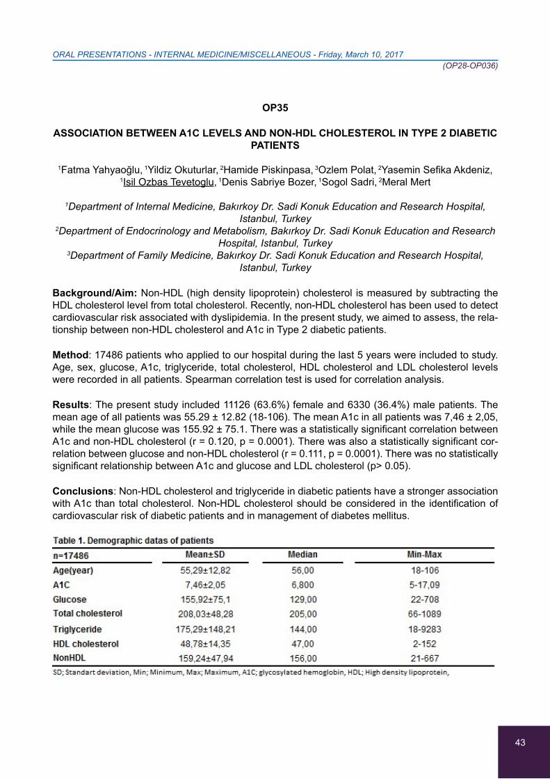
43
ORAL PRESENTATIONS - INTERNAL MEDICINE/MISCELLANEOUS - Friday, March 10, 2017 (OP28-OP036)
OP35
ASSOCIATION BETWEEN A1C LEVELS AND NON-HDL CHOLESTEROL IN TYPE 2 DIABETIC PATIENTS
1Fatma Yahyaoğlu, 1Yildiz Okuturlar, 2Hamide Piskinpasa, 3Ozlem Polat, 2Yasemin Sefika Akdeniz,
1Isil Ozbas Tevetoglu, 1Denis Sabriye Bozer, 1Sogol Sadri, 2Meral Mert
1Department of Internal Medicine, Bakırkoy Dr. Sadi Konuk Education and Research Hospital, Istanbul, Turkey
2Department of Endocrinology and Metabolism, Bakırkoy Dr. Sadi Konuk Education and Research Hospital, Istanbul, Turkey
3Department of Family Medicine, Bakırkoy Dr. Sadi Konuk Education and Research Hospital, Istanbul, Turkey
Background/Aim: Non-HDL (high density lipoprotein) cholesterol is measured by subtracting the HDL cholesterol level from total cholesterol. Recently, non-HDL cholesterol has been used to detect cardiovascular risk associated with dyslipidemia. In the present study, we aimed to assess, the rela-tionship between non-HDL cholesterol and A1c in Type 2 diabetic patients.
Method: 17486 patients who applied to our hospital during the last 5 years were included to study. Age, sex, glucose, A1c, triglyceride, total cholesterol, HDL cholesterol and LDL cholesterol levels were recorded in all patients. Spearman correlation test is used for correlation analysis.
Results: The present study included 11126 (63.6%) female and 6330 (36.4%) male patients. The mean age of all patients was 55.29 ± 12.82 (18-106). The mean A1c in all patients was 7,46 ± 2,05, while the mean glucose was 155.92 ± 75.1. There was a statistically significant correlation between A1c and non-HDL cholesterol (r = 0.120, p = 0.0001). There was also a statistically significant cor-relation between glucose and non-HDL cholesterol (r = 0.111, p = 0.0001). There was no statistically significant relationship between A1c and glucose and LDL cholesterol (p> 0.05).
Conclusions: Non-HDL cholesterol and triglyceride in diabetic patients have a stronger association with A1c than total cholesterol. Non-HDL cholesterol should be considered in the identification of cardiovascular risk of diabetic patients and in management of diabetes mellitus.
44
ORAL PRESENTATIONS - INTERNAL MEDICINE/MISCELLANEOUS - Friday, March 10, 2017 (OP28-OP036)
OP36
CLINICAL PROFILE OF HYPONATREMIA IN TERTIARY CARE CENTER IN INDIA: RETROSPECTIVE HOSPITAL BASED OBSERVATIONAL STUDY
1Akash Thomas, 1V.S. Srikanth, 1Senthilvel Vasudevan, 2Ranjitha
1Department of General Medicine-Amrita Institute of Medical Sciences and Research Centre (AIMS,) Kochi, India
2Department of Biostatistics Amrita Institute of Medical Sciences and Research Centre, (AIMS), Kochi, India
Background: Hyponatremia is the common of electrolyte abnormality. Hyponatremia is associated with altered sensorium, seizures, falls and cognitive dysfunction. Even mild hyponatremia could lead to severe complications and prolonged hospital stays. Understanding the emerging trends in manifestations and treatment of hyponatremia will help in efficient management of hyponatremia and allied comorbidities.
Methods: We have conducted the present study on 980 patients who have got admitted in AIMS, Kochi, Kerala. They were categorized based on serum sodium level under 3 groups of Hyponatremia (Mild, Moderate and Severe). The data collected were analyzed for Clinical presentations, Severity, Etiology of hyponatremia.
Results: Hyponatremia was observed predominantly in the age group >70yrs (37.3%), with male predisposition (63%). Altered sensorium is the most common presentation of hyponatremia. The number of patients with disorientation is more in the moderate hyponatremia (64%) compared to severe hyponatremia (20%). SIADH was observed as leading cause of hyponatremia. Respiratory causes pneumonia, asthma, OAD were the predominant causes of SIADH, followed by Dilutional hyponatremia and Drug Induced Hyponatremia. Of the various types of carcinoma, lung and geni-tourinary cancer were the main causes of SIADH. The infections associated with hyponatremia were UTI (68%), chest Infection (15%) and cellulitis (14%). 42% population with hyponatremia had Diabe-tes Mellitus, of which 64% had peripheral neuropathy and 10% had complications like diabetic foot and necrotizing fasciitis.
Conclusion: This study summarizes the varied presentations of hyponatremia, its causes and co-morbidities which will provide better understanding of hyponatremia. Also aid the physician in diag-nosing the precise cause of hyponatremia.
45
ORAL PRESENTATIONS - DIABETES MELLITUS - Saturday, March 24, 2018 (ΟP37-OP42)
OP37
AN OBESE 17 YEAR OLD PATIENT PRESENTING WITH DIABETIC KETOACIDOSIS (DKA) AS THE FIRST MANIFESTATION OF DIABETES TYPE 2 (DM2)
Haris Constantinou, Angelos Alexi, Magdalini Adamantou, Chrysostomos Louka, Marios Petrou,
Dania Rodotheou, Georgos Olympios, Andreas Stylianou
Department of Internal Medicine, Larnaca General Hospital, Larnaca, Cyprus
Backround: DKA is the most serious hyperglycemic emergency in patients with diabetes and is present in 25-40% of children and adolescents with newly diagnosed DM and in 5-25% as a first manifestation in children with DM2.
Case Report: A 17 year old patient presented to the Emergency Department with a 2 days history of severe, diffuse, sudden onset abdominal pain. He reported symptoms of upper respiratory tract infection and a 6 week history of polyuria and polydipsia. He was morbidly obese, acanthosis nigri-gans was observed at the maxillary region. The haematological results showed elevated glucose levels (590 mg/dl), reduced potassium levels (2,8 mmol/l), elevated white blood cells (18.850 with 90% neutrophils). Glucose (+++) and ketones (+++) were found in the urine analysis. The ABGs confirmed severe DKA (pH:7,11, HCO3:9,7, Lactic acid:1,2). The HbA1c was found elevated (11,8%) and confirmed the diagnosis of diabetes. The anti-GAD, anti-IAA and anti-IA2A were found negative. We measured c-peptide (0, 6 min) within normal range and the glucagon and dexamethasone sup-pression test were normal. The serum and cortisol levels as well as serum metanephrines, VMA and TSH were found within the normal limits.
Conclusions: DKA has long been considered as a key clinical feature of DM1. However, in recent years, an increasing number of ketoacidosis cases without precipitating cause have also been re-ported in children and adolescents who are usually obese, with strong family history of DM2 (Ketosis Prone Diabetes, KPD). We present an interesting case with possible diagnosis of KPD.
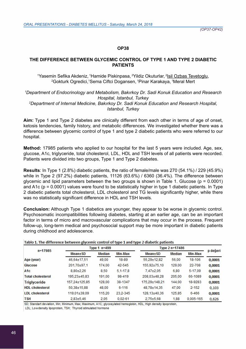
46
ORAL PRESENTATIONS - DIABETES MELLITUS - Saturday, March 24, 2018 (ΟP37-OP42)
OP38
THE DIFFERENCE BETWEEN GLYCEMIC CONTROL OF TYPE 1 AND TYPE 2 DIABETIC PATIENTS
1Yasemin Sefika Akdeniz, 1Hamide Piskinpasa, 2Yildiz Okuturlar, 2Isil Ozbas Tevetoglu,
2Gokturk Ogredici,1Sema Ciftci Dogansen, 1Pinar Karakaya, 1Meral Mert
1Department of Endocrinology and Metabolism, Bakırkoy Dr. Sadi Konuk Education and Research Hospital, Istanbul, Turkey
2Department of Internal Medicine, Bakırkoy Dr. Sadi Konuk Education and Research Hospital, Istanbul, Turkey
Aim: Type 1 and Type 2 diabetes are clinically different from each other in terms of age of onset, ketosis tendencies, family history, and metabolic differences. We investigated whether there was a difference between glycemic control of type 1 and type 2 diabetic patients who were referred to our hospital.
Method: 17985 patients who applied to our hospital for the last 5 years were included. Age, sex, glucose, A1c, triglyceride, total cholesterol, LDL, HDL and TSH levels of all patients were recorded. Patients were divided into two groups, Type 1 and Type 2 diabetes.
Results: In Type 1 (2.8%) diabetic patients, the ratio of female/male was 270 (54.1%) / 229 (45.9%) while in Type 2 (97.2%) diabetic patients, 11126 (63.6%) / 6360 (36.4%). The difference between glycemic and lipid parameters between the two groups is shown in Table 1. Glucose (p = 0.0001) and A1c (p = 0.0001) values were found to be statistically higher in type 1 diabetic patients. In Type 2 diabetic patients total cholesterol, LDL cholesterol and TG levels significantly higher, while there was no statistically significant difference in HDL and TSH levels.
Conclusion: Although Type 1 diabetics are younger, they appear to be worse in glycemic control. Psychosomatic incompatibilities following diabetes, starting at an earlier age, can be an important factor in terms of micro and macrovascular complications that may occur in the process. Frequent follow-up, long-term medical and psychosocial support may be more important in diabetic patients during childhood and adolescence.
47
ORAL PRESENTATIONS - DIABETES MELLITUS - Saturday, March 24, 2018 (ΟP37-OP42)
OP39
ASSESSMENT OF THE LINK BETWEEN VITAMIN D AND METABOLIC SYNDROME COMPONENTS
1Hande Atalay, 2Yıldız Okuturlar, 3Banu Boyuk, 3Seher Irem Cetin, 2Irem Kırac Utku, 2Esra Demir
1Department of Internal Medicine, SBU Fatih Sultan Mehmet Training and Research Hospital, Istanbul, Turkey
2Department of Internal Medicine, SBU Bakırkoy Sadi Konuk Training and ResearchHospital, Istanbul, Turkey
3Department of Internal Medicine, SBU Gaziosmanpasa Taksim Training and Research Hospital, Istanbul, Turkey
Background: Vitamin D deficiency is one of the most common health problems worldwide. Growing evidence have shown that vitamin D likely plays a role in the development of Metabolic syndrome (MetS). We aimed to define relationship between vitamin D serum concentration and components of MetS.
Methods: In this cross-sectional study, 248 with MetS and 106 healthy participants were recruited by consecutive random sampling. We used IDF criteria for MetS diagnosis. Serum 25(OH) D was measured with an enzyme immunoassay kit in a single laboratory using the same lab assay.Serum 25-hydroxyvitamin D (25(OH) D) and MetS components were determined.
Results: 25(OH)D levels were lower than 25ng/mL in the patient and control groups (23,65±12,95 ng/ml vs. 16,06±10,73 ng/ml, p<0,01). We found that serum 25(OH) D (ng/ml) levels of control group were significantly lower than the patient group (p<0, 01). In our study, 25(OH) D levels indicated negative correlation with waist circumference, systolic and diastolic pressure (r=-0,120, p=0.05; r=-0,164, p=0.01; r=-0,197, p=0.002) but we did not found any significant correlation of other MetS components.
Conclusion: Serum 25(OH) D concentration was only associated with waist circumference and hy-pertension among the metabolic syndrome components. Low vitamin D levels of control group and the link between vitamin D and metabolic syndrom components suggest us the presence of invisible part of the iceberg for Turkish population. Larger controlled studies are required to confirm these findings. Keywords: Metabolic syndrome, MetS components, vitamin D.
Table. Relationship between vitamin D and other variables
r PBMI (kg/m2)BMI -0,202 0,001**Waist Circumference (cm) -0,120 0,05*Systolic pressure (mmHg) -0,164 0,01**Diastolic pressure (mmHg) -0,197 0,002**
BMI: Body mass index Statistical signifance: *p<0,05, ** p<0,01
48
ORAL PRESENTATIONS - DIABETES MELLITUS - Saturday, March 24, 2018 (ΟP37-OP42)
OP40
SEVERE HYPOGLYCEMIA REQUIRING MEDICAL ASSISTANCE IN PATIENTS WITH DIABETES IS ASSOCIATED WITH SIMULTANEOUS PROLONGATION OF QTC INTERVAL
1Maria Mylona, 2Georgios Anastasiadis, 2Vasilios Vazgiourakis, 2Kanelina Zerva,
2Konstantinos Tsakalis, 1Stavros Liatis
11st Department of Propaedeutic & Internal Medicine, Laiko General Hospital, Athens University Medical School, Athens, Greece
2Department of Cardiology, Laiko Hospital, Athens, Athens, Greece
Objectives: Severe hypoglycemia is considered a state, that may lead to sudden death as a result of cardiac arrhythmia. Aim of our study was to explore the association between severe hypoglycemic events requiring medical assistance and QT interval, in diabetic patients.
Methods: Nine clinics, in five cities of Greece, participated in a 16-month prospective survey of doc-umented iatrogenic hypoglycemia at the emergency departments (ED). According to the protocol, a 12-lead ECG was obtained simultaneously or immediately after the management of hypoglycemia and no later than 30 minutes after the administration of glucose. The patient- ECGs were compared to those from a control group, matched for age and gender. QT and RR intervals were measured blindly by three independent cardiologists, while QTc was calculated according to the Bazett formula. QTc measurements of≥440msec were considered abnormally prolonged. Patients receiving medica-tions possibly affecting the QTc intervaland those with hypokalaemia (serum potassium < 3.5mEq/l) were excluded from the analysis.
Results: 295 episodes of iatrogenic hypoglycemia in 294 diabetic patients were identified and 223 ECGs were obtained. 46 ECGs were excluded from the analysis, due to the presence of the above mentioned criteria.177 ECGs from hypoglycemic patients were analyzed (mean age 72.7±15.7 years, 48.6% women, 9% with type 1 diabetes) and compared to 91 controls. Mean QTc interval was significantly prolonged in patients compared to controls (440.4±45.1 msec vs. 413.9±32.5msec, p<0.001).
Conclusions: In patients with diabetes, severe iatrogenic hypoglycemia requiring medical assist-ance is associated with a both statistically and clinically significant prolongation of QTc interval.
49
ORAL PRESENTATIONS - DIABETES MELLITUS - Saturday, March 24, 2018 (ΟP37-OP42)
OP41
PREVALENCE AND GEOGRAPHICAL DISTRIBUTION OF TYPE 1 DIABETES MELLITUS IN CHILDREN AND YOUNG ADULTS IN GREECE
1Maria Mylona, 1Konstantinos Makrilakis, 1Georgios Dafoulas, 2Chara Kani, 1Petros P. Sfikakis,
1Stavros Liatis
11st Department of Propaedeutic Internal Medicine, Diabetes Center, National and Kapodistrian University of Athens Medical School, Laiko General Hospital, Athens, Greece
2Medicines Department, National Organization for Health Care Services Provision, Athens, Greece
Objectives: Type 1 diabetes mellitus (T1DM) is a heterogeneous disorder characterized by au-toimmune destruction of pancreatic beta cells, leading to absolute insulin deficiency. Its prevalence displays wide geographic differences. Herein, we aimed to identify all children and young adults with T1DM in Greece and to further explore the geographical distribution of the disease.
Methods: The electronic prescription database of the National Organization for Health Care Serv-ices Provision was used. Individuals < 35 years old, who received at least two fully-reimbursable insulin prescriptions with an ICD-10 code of E10 (insulin-dependent diabetes), between June 1st, 2014 and May 31st 2015 were identified. The size of the total Greek population was defined accord-ing to the 2011 census of the Greek population by the National Statistics Agency.
Results: The study population consisted of 4,081,093 individuals, accounting for 95% of the Greek population equal or younger than 35 years old. There were 6,183 cases with type 1 diabetes, cor-responding to a prevalence of 1.51‰. A slight male predominance was observed (53.9%). There were no remarkable geographical differences, with highest prevalence, however, being observed in the Aegean. In the Attica area, which represents almost 50% of the total Greek population, the prevalence was 1.56 ‰.
Conclusions: This is the first study to investigate the prevalence of T1DM in the total population of Greek children and young adults. A low prevalence of 1.51‰ was observed, without north-to-south differences. However, a moderately higher prevalence was revealed in the islands of the Aegean sea, but not in Crete.
50
ORAL PRESENTATIONS - DIABETES MELLITUS - Saturday, March 24, 2018 (ΟP37-OP42)
OP42
CLINICAL PROFILE OF DIABETIC PATIENTS WITH ATRIAL FIBRILLATION
Eleni Paschou, Athanasios Samaras, Georgios Fotos, Georgios Dividis, Elina Thomaidou, Ioannis Goulas, Dimitra Vasdeki, Europi Forozidou, Paraskeui Tsoukra, Anastasios Kartas,
Haralampos Karvounis, Apostolos Tzikas, Georgios Giannakoulas
1st Cardiology department, AHEPA University Hospital, Aristotle University of Thessaloniki, Thessaloniki, Greece
Background: There is a gap in evidence for the incidence of comorbidities among diabetic patients with atrial fibrillation (AF) and according to the underlying antidiabetic treatment.
Aim: To determine the differences of various comorbidities in diabetic versus vs non-diabetic pa-tients with AF and among diabetic patients according to their antidiabetic treatment.
Methods: The study included consecutive patients who were hospitalized to the cardiology depart-ment with any diagnosis and coexisting AF from December 2016 to May 2017. A comparison was made between diabetic and non-diabetic patients for various cardiovascular risk factors, such as hypertension (HT), dyslipidemia (DSL), as well as comorbidities, such as vascular disease (VD) and chronic kidney disease (CKD). The diabetic patients were furtherly divided into 3 subgroups accord-ing to the underlying antidiabetic treatment: patients treated with antidiabetic pills, patients treated with insulin and patients treated with both pills and insulin.
Results: 741 patients were studied (mean age 73.02 years, 45.5% women) 34.5% of whom were dia-betic. Diabetic patients had more frequently a history of HT (p=0.001), DSL (p=0.001), VD (p<0.001), stroke (p=0.008) and myocardial infarction (p=0.029), angina (p=0.015) and CKD (p<0.001) com-pared to non-diabetic patients. Patients who received both antidiabetic pills and insulin showed lower frequency of HT compared to those who received only antidiabetic pills and those under insulin (p=0.023).
Conclusions: AF patients with diabetes mellitus are under a greater risk for developing HT, DSL, VD, and CKD compared to non-diabetic patients. The combination of antidiabetic pills and insulin may have a protective effect against HT in diabetic patients with AF.
51
ORAL PRESENTATIONS - INFECTIONS/IMMUNOLOGY - Saturday, March 24, 2018 (ΟP43-OP48)
OP43
XPNPEP2 PROMOTER HAPLOTYPE AND ANGIOEDEMA ATTACKS
Athanasia-Marina Peristeri, Gerasimina Tsinti, Eirini Sevdali, Anastasios E. Germenis, Matthaios Speletas
Department of Immunology & Histocompatibility, University of Thessaly, Faculty of Medicine, Larissa, Greece
Background: A life-threating side effect of angiotensin I-converting enzyme inhibitors (ACEi) is bradykinin-mediated angioedema (ACEi-angioedema). Recently, La Corte et al have shown that a functional haplotype in the 5’-regulatory region of XPNPEP2 gene, encoding aminopepetidase (APP), is significantly associated with both a reduced plasma of APP activity and an increased prevalence of ACEi-angioedema. The aim of our study was to confirm this finding in a Greek cohort of patients with bradykinin-mediated angioedemas.
Methods: Nineteen patients with hereditary angioedema due to C1-inhibitor deficiency (HAE-CINH) (male/female: 10/9, mean age: 45.5 years, range: 25-86), 14 with bradykinin-mediated angioedema attacks of unknown origin (Unknown Angioedema, male/female: 10/4, mean age: 54 years, range: 40-82) and 29 healthy controls (male/female: 18/11, mean age: 51.5, range: 21/90) were enrolled. Two patients with HAE-CINH and 6 with unknown angioedema reported attacks related to the admin-istration of ACEi. The detection of the three single nucleotide polymorphisms (SNPs) c.-2399C>A, c.-1612G>T, and c.-393G>A, which define the ATG-haplotype, was performed by allele-specific PCR for the first SNP and by PCR-RFLP for the rest and confirmed by sequencing.
Results: The prevalence of ATG haplotype in HAE-CINH, Unknown Angioedema and healthy con-trols was 31.5%, 7.0% and 24.1%, respectively. Interestingly, no Greek patient exhibiting ACEi-an-gioedema displayed the ATG haplotype. The allele frequency of c.-2399C>A was 21.4%, 5.6% and 20.0%, of c.-1612G>T was 36.0%, 16.7% and 25.0% and of c.-393G>A was 5.0%, 11.0% and 5.0% for HAE-CINH, Unknown Angioedema and healthy controls, respectively.
Conclusions: The ATG haplotype is not associated with the ACEi-angioedema in a Greek cohort. Obviously, the increased prevalence and the effect of ATG haplotype in HAE-CINH needs further investigation.

52
ORAL PRESENTATIONS - INFECTIONS/IMMUNOLOGY - Saturday, March 24, 2018 (ΟP43-OP48)
OP44
FAMILIAL MEDITERRANEAN FEVER (FMF): A SINGLE GREEK CENTER EXPERIENCE
1Dimitrios Sagris, 1Asterios Saitis, 1Nikolaos K. Gatselis, 2Panagiotis Skendros, 1Eirini I. Rigopoulou,
1Konstantinos P. Makaritsis, 1Kalliopi Zachou, 2Konstantinos Ritis, 1George N. Dalekos
1Department of Medicine and Research Laboratory of Internal Medicine, Medical School, Thessaly University, Larissa, Greece
2First Department of Internal Medicine, Democritus University of Thrace, Alexandroupolis, Greece
Background/Aim: FMF is a hereditary autoinflammatory disease, caused by mutations in the Medi-terranean Fever gene (MEFV) encoding pyrin protein. Aim of the study was to evaluate the clinical characteristics and the spectrum of MEFV alterations in a cohort of patients diagnosed in our Depart-ment.
Methods: We retrospectively reviewed 46 FMF patients, tested for MEFV alteration in exon-2 and exon-10 by non-isotopic RNase-cleavage assay and subsequent sequence analysis.
Results: The patients (23 females) had a mean±SD age of 37.3±17.8 years, (disease duration: 58.9±98.6 months). Among all 46-patients, 38 (82.6%) presented fever, 30 (65.2%) abdominal pain, 15 (32.6%) joint involvement, 9 (19.6%) peritonitis, 9 (19.6%) pleuritis, 4 (8.7%) pericarditis, 3 (6.5%) rash, 2 (4.3%) infertility, 2 (4.6%) severe liver involvement, 2 (4.3%) periaortitis/retroperitoneal fibrosis and 1 (2.2%) meningitis. One patient presented with end-stage disease. Mutations/polymorphisms were detected in 39/46 (84.8%) [homozygotes 10/46, compound heterozygotes 8/46, heterozygotes 17/46, polymorphisms 4/46]. The mutations identified were R202Q (37%), M694V (21.7%), E148Q (8.7%), V726A (8.7%), M694I (6.5 %), E230K (4.3%), M680I (4.3%), E167D (4.3%), F479L (4.3%) and K695R (2.2%). Apart from one patient who died at the time of diagnosis due to amyloidosis, all patients were treated initially with colchicine with response in 44/45 (97.7%). One patient needed several immunomodulatory agents including canakinumab to achieve remission.
Conclusions: High-index of clinical suspicion even when the clinical phenotype of the disease is not complete along with molecular analysis is mandatory to achieve timely FMF diagnosis and initiate treatment in order to prevent the late complications of the disease.
53
ORAL PRESENTATIONS - INFECTIONS/IMMUNOLOGY - Saturday, March 24, 2018 (ΟP43-OP48)
OP45
INFLAMMATION SUSCEPTIBILITY BIOMARKER FREQUENCIES IN SOUTH EASTERN EUROPEAN CAUCASIANS (TNF-Α, IL6 AND CRP)
Evgenia Georgiadi, Alexandra Petraina, Martha-Spyridoula Katsarou, Maria Papasavva, Maria Lagiou, Thodoris Kalogridis, Nikolaos Drakoulis
Research Group of Clinical Pharmacology and Pharmacogenomics, Department of Pharmaceutical Technology, Faculty of Pharmacy, School of Health Sciences, National and Kapodistrian University
of Athens, Athens, Greece
Inflammation is a complicated, well-orchestrated process by a variety of cells and chemical mediators such as TNF-a, IL-6 and CRP. The less common A allele of TNF-a gene polymorphism rs1800629 seems to increase TNF expression levels. The mutated C allele of IL-6 gene polymorphism rs1800795 and T allele of CRP gene polymorphism rs1205 are related to lower levels of IL-6 and CRP, respec-tively. A frequency distribution analysis of rs1800629, rs1800795 and rs1205 polymorphisms in a South Eastern European Caucasian (SEC) population sample was performed. DNA from buccal swabs of 852 non related SEC was collected and analysed. Gene distribution for polymorphism rs1800629 was G:G=81.8%, G:A=17.4% and A:A=0.8. The wild-type G allele frequency was 90.5%. Frequencies for rs1800795 were G:G=54.8%, G:C=38.7% and C:C=6,5%. The wild-type G allele fre-quency was 74.2%. The frequencies for rs1205 were C:C=47.7%, C:T=41.9% and T:T =10.4%. The wild-type C allele frequency was 68.6%. The frequency distribution of all analysed polymorphisms of the SEC population differs significantly when compared with other populations (SEC v/s Europeans, Africans, East Asians and South Asians for rs1800629, v/s global, Europeans, Africans, East Asians and South Asians for rs1800795 and v/s Africans and East Asians for rs1205).
Although not statistically significant, the investigated SEC population has great similarity to the glo-bal population regarding the allele distribution of rs1800629 and to the European and South Asian population regarding the allele distribution of rs1205. Inflammation is affected by multiple factors, however, the genotyping analysis of polymorphisms may play a significant role in susceptibility of inflammation.
54
ORAL PRESENTATIONS - INFECTIONS/IMMUNOLOGY - Saturday, March 24, 2018 (ΟP43-OP48)
OP46
ATTITUDE OF GREEK DIABETIC PATIENTS VS. GENERAL POPULATION SUBJECTS TOWARDS COMMON VACCINES: THEORY AND PRACTICE, DIFFERENCES AND
SIMILARITIES
1Ioannis Kyriazis,2Dimitra Latsou,3Kleoniki Georgousi, 3Dimitra Stefani, 3Emmanouil Kalafatis, 3Panagiotis Koutsouris, 3Eftychia Pouliou,3Andreas Marketos, 3Ioannis Tsamis,
3Ioannis Kachrimanidis, 3Fotios Tatakis, 1Theodora Lappa, 3Moyssis Lelekis
1Diabetes Outpatient Clinic , KAT General Hospital , Attica , Greece2Scientific Associate of Diabetes Outpatient Clinic , KAT General Hospital Attica Greece
& University of Peloponnese, Greece
3Internal Medicine Department , KAT General Hospital , Attica , Greece
Aim: To study the attitude of Greek diabetic patients (pts) vs. subjects of the general population to-wards vaccination against common pathogens.
Materials/Methods: 120 pts (group A) and 275 subjects of the general population without diabetes (group B) were included in the study. All were asked to fill in a specific questionnaire concerning their practice on vaccination against influenza and pneumococcus.
Results: 45% of group A vs. 29% of group B, had positive attitude towards flu vaccination, p=0.003). However, when comparing group A with a subgroup of group B with at least one risk factor- indication for vaccination (population at risk), no such difference was found (p=0.409). Furthermore, 31,7% of group A and 20,4% of group B stated that they are regularly vaccinated every year against influenza (p=0.015). However there was no difference between group A and the subgroup (p=0.344). 30% of group A and 23,3% of group B stated that they were vaccinated during the last vaccination period (p=0.157). However significantly greater percentage of the population at risk were in fact vaccinated (43,1% vs 30%, p=0.039). In both groups (A and B) low rates of vaccination against pneumococcus were recorded (14,2% vs 12,4%, p=0.623). The same was true when population at risk was evalu-ated (p= 0.761).
Conclusions: Although in the high risk group, overall our diabetic pts and general population sub-jects are not sufficiently vaccinated against both influenza and pneumococcus. Greater effort is needed to convince all subjects belonging to risk groups on the necessity of vaccination.
55
ORAL PRESENTATIONS - INFECTIONS/IMMUNOLOGY - Saturday, March 24, 2018 (ΟP43-OP48)
OP47
DEVELOPMENT OF PYELONEPHRITIS IN PATIENTS ADMITTED WITH ACUTE ISCHEMIC STROKE REFLECTS STROKE SEVERITY AND DOES NOT AFFECT OUTCOME
Marianthi Papagianni, Konstantinos Tziomalos, Stavroula Kostaki, Stella-Maria Angelopoulou,
Konstantinos Christou, Danai-Thomais Kostourou, Dimitris Milonas, Eleftheria Ztriva,
Christos Savopoulos, Apostolos I. Hatzitolios
First Propedeutic Department of Internal Medicine, Medical School, Aristotle University of Thessaloniki, “AHEPA” Hospital, Thessaloniki, Greece
Background: Patients admitted with acute ischemic stroke have increased risk for pyelonephritis. However, there are limited data regarding the effect of pyelonephritis on the outcome of this popula-tion.
Aim: To evaluate the association between pyelonephritis and acute ischemic stroke outcome.
Methods: We prospectively studied 922 consecutive patients admitted with acute ischemic stroke (42.2% males, age 79.6±6.9 years). The severity of stroke was assessed at admission with the Na-tional Institutes of Health Stroke Scale (NIHSS). The outcome was assessed with dependency rates at discharge (modified Rankin scale 2-5) and in-hospital mortality.
Results: During hospitalization, 34 patients developed pyelonephritis (3.7% of the total study popu-lation). Patients who developed pyelonephritis had higher rates of dependency at discharge than patients who did not develop pyelonephritis (85.2% vs. 59.9%, p<0.05). Independent risk factors for dependency were age (relative risk (RR) 1.08, 95% confidence interval (CI) 1.04-1.12, p<0.001), history of ischemic stroke (RR 1.89, 95% CI 1.22-2.94, p<0.005) and NIHSS at admission (RR 1.49, 95% CI 1.39-1.62, p<0.001). Patients who developed pyelonephritis had similar rates of in-hospital mortality than patients who did not develop pyelonephritis (8.8% vs. 9.5%, p=NS). Independent risk factors for in-hospital mortality were diastolic blood pressure at admission (RR 1.05, 95% CI 1.03-1.07, p<0.001) and NIHSS at admission (RR 1.20, 95% CI 1.16-1.25, p<0.001).
Conclusions: In patients admitted with acute ischemic stroke, the development of pyelonephritis reflects the severity of stroke and is not independently associated with the outcome.

56
ORAL PRESENTATIONS - INFECTIONS/IMMUNOLOGY - Saturday, March 24, 2018 (ΟP43-OP48)
OP48
REPORTING PATTERNS OF LEISHMANIA INFECTION IN WESTERN GREECE: A 5-YEAR EXPERIENCE
1Karolina Akinosoglou, 2Maria Panoutsakou, 2Christina-Panagiota Koutsouri, 1Charalambos Gogos
1Department of Internal Medicine and Infectious Diseases, University Hospital of Patras, Patras, Greece
2Department of Internal Medicine, University Hospital of Patras, Patras, Greece
Background: Leishmania species represents an emerging pathogen in developed countries in the context of increasing population of immunosuppressed patients.
Aim: We aimed to explore patterns of Leishmania infection during the last five years in Western Greece.
Methods: This was a single centre, retrospective, observational study conducted at a Greek tertiary university hospital, representing a referral centre for Leishmania cases in Western Greece. Compu-terized searches were used to identify patients using a list of ICD-10 codes suggesting cutaneous or visceral Leishmaniasis. Available data from respective files were analysed accordingly. Results shown as median (range).
Results: Nineteen patients with a diagnosis of Leishmaniasis [n=18 (95%) visceral, 1 (5%) cutane-ous] were identified through electronic searches from January 2012 to September 2017. Median age of patients was 22 (2-52) years, 12 (63%) were male, 3 (16%) reported previous canine contact while in only 3 (16%) immunosuppression was observed. Fever of 39 (38-39) was the most commonly presenting symptom (100%), followed by rigors (42%) and lymphadenopathy (37%). Splenomegaly and/or hepatomegaly was present in 63 and 36% of patients respectively. Diagnosis was made in approximately 12 (1-240) days upon fever presentation through rK39 Ag detection 17 (90%), bone marrow biopsy 3 (16%) or both 2 (11%). Fever subsided in 2 (1-4) days upon treatment initiation. All patients recovered following common therapeutic schemes of liposomal Amphotericin B in accord-ance with international guidelines, and discharged 8 (5-21) days later.
Conclusions: Identification of Leishmania cases requires high clinical suspicion, in view of non-specific clinical manifestations and observations of delayed diagnosis.

57
10th International Congress of Internal MedicineMarch 22-24, 2018, Athens, Greece
e-Posters
58
e-POSTERS - Gastroenterology/Hepatology(ePP001-ePP027)
ePP01
EVALUATION OF VASCULAR ENDOTHELIAL GROWTH FACTOR AS A PROGNOSTIC MARKER IN INOPARABLE HEPATOCELLULAR CARCINOMA
Georgios Tassopoulos, Vasilios Heras, Andreas Kapouralos, Anastasios Stefanopoulos,
Panagiotis Heras
Department of Internal Medicine, General Hospital of Argos, Argos, Greece
Background: Hepatocellular carcinoma (HCC) ranks as the seventh most common malignancy in males and ninth most common in females. HCV infection is a major risk factor for HCC. Greece has high prevalence of HCV and rising rates of HCC.
Methods: This was a prospective study evaluating the prognostic value of vascular endothelial growth factor (VEGF) in 75 inoperable hepatocellular carcinoma patients treated with at least one session of transarterial hemoembolization (TACE). VEGF was measured before and after interva-tion.
Results: Partial response was achieved in 22 patients (29%) while, 36 patients (48%) had stable disease. Time to disease progression was 6.2 months. Pretreatment serum VEGF level was of prog-nostic value.
Conclusion: Pretreatment VEGF could be used as a prognostic marker for inoperable HCC treated with TACE.

59
e-POSTERS - Gastroenterology/Hepatology(ePP001-ePP027)
ePP02
ATYPICAL PRESENTATIONS OF HEPATOCELLULAR CARCINOMA: 2 CASE REPORTS
1Ioannis Kontaxakis, 1Konstantinos Karatarakis, 1Anthi Chalkiadaki, 1Antonis Mpitsakis,
1Maria Leontaki, 1Georgios Fytrolakis, 2Nikolaos Chrysanthos, 3Maria Rovithi
1Department of Internal Medicine, Agios Nikolaos General Hospital, Agios Nikolaos, Crete, Greece 2Gastrointestinal Unit, Agios Nikolaos General Hospital, Agios Nikolaos, Crete, Greece
3Medical Oncology Unit, Agios Nikolaos General Hospital, Agios Nikolaos, Crete, Greece
Background: Hepatocellular carcinoma (HCC) represents the third cause of cancer-related mortal-ity worldwide with a steadily increased incidence, strongly associated with chronic liver disease. We here present two cases of atypical clinical presentations of HCC.
Case Report 1: A 70-year old man was referred to our hospital due to deteriorating dyspnoea. CT thorax done on an outpatient basis was suggestive of pulmonary embolism and revealed a solitary liver lesion. Further work up with MRI showed a 17.8 x 14.3 x 15.5 cm right liver lobe lesion with imaging features characteristic of HCC. Tumor tissue invasion was noted in the inferior vena cava and, following the anatomical course of the vein, extended into the right heart atrium. Serum alpha fetoprotein levels (AFP) were increased (167 ng/mL, normal range: 0.9- 8.8). Echocardiography con-firmed the presence of tissue in the atrium and pulmonary embolism was subsequently attributed to neoplastic emboli. Serologic tests showed that the patient suffered from chronic active hepatitis B.
Case Report 2: An 86-year old man presented with fatigue and a palpable, progressively growing, painless mass over the left scapula. Subsequent biopsy revealed histopathologic markers indicative of metastatic infiltration from HCC. Diagnosis was later supported by a significantly elevated serum AFP level and the presence of multiple liver lesions in the context of a cirrhotic liver, due to alcohol abuse. Both opted for Best Supportive Care.
Conclusion: In patients with known risk factors, the physician should be alert for potential HCC de-velopment, that might present with less anticipated clinical manifestations.
60
e-POSTERS - Gastroenterology/Hepatology(ePP001-ePP027)
ePP03
A DEEP LEARNING APPROACH TO THE SIGNIFICANT LIVER FIBROSIS BINARY CLASSIFICATION PROBLEM USING GENDER, MORPHOLOGIC AND HEMODYNAMIC
MEASUREMENTS DERIVED FROM B-MODE ULTRASOUND IMAGES
1,2Athanasios Angelakis, 2Ilias Gatos, 2Ioannis Theotokas, 2Eleni Panteleakou, 2Aikaterini Kanavaki,
2Alexandros Soultatos, 3Irene Vafiadis, 4Emanuel Manesis, 2Pavlos Zoumpoulis
1Department of Mathematics, National Technical University of Athens, Athens, Greece 2Diagnostic Echotomography S.A., Athens, Greece
3Department of Medicine, National and Kapodistrian University of Athens, Athens, Greece 4Euroclinic Athens, Athens, Greece
Background: Conventional methods such as: hematologic tests e.g. FIBROMETER®, strain, shear wear elastographic (SWE) stiffness cut-off values e.g. FIBROSCANTM, SuperSonic Imagine SWE, and parameters derived from image analysis e.g. color clustering of SWE, are used for the significant liver fibrosis binary classification (SLFBC) problem. The AUC of the above methods is: 0.82, 0.82, 0.84 and 0.87 accordingly.
Aim: The aim of this study was to design and tune a deep neural network (DNN) in order to automate classification of a patient’s liver fibrosis stage (F≤F1, F≥F2).
Methods: Our data set consisted of 199 liver FIBROSCANTM validated patients with chronic liver disease from which 68 were F≤F1 and 131 were F≥F2. Input parameters for our model were: {patient gender}, B-Mode morphologic longitudinal diameter (LD) measurements: {right lobe, caudate lobe, left lobe, spleen} and one hemodynamic parameter {portal vein LD}. We designed a DNN with two hidden layers and used the 10-fold cross validation (CV) technique in order to manage underfitting, overfitting, bias and variance issues.
Results: A 10-fold CV provided the following results: Accuracy: 0.9648+/-0.0594, Sensitivity: 0.9696, Specificity: 0.9552, AUC: 0.9591 with 95% CI [0.78 - 1.00].
Conclusions: We proposed a different approach using deep learning on SLFBC problem. Our pre-liminary results demonstrate improved accuracy, sensitivity, specificity and AUC (all > 0.95) com-pared to conventional diagnostic methods. Our DNN model could be used as a decision support tool to assist clinicians on their medical report. Larger data sets are required to further validate our algorithm.
61
e-POSTERS - Gastroenterology/Hepatology(ePP001-ePP027)
ePP04
NUTRITION IN CHRONIC LIVER DISEASE: WHAT IS KNOWN AND HOW TO IMPLEMENT IT IN EVERYDAY CLINICAL PRACTICE FOR VALID NUTRITIONAL INTERVENTIONS
Dimitra Kagiafa
Clinical Nutrition Department, Hippocratio General Hospital of Athens, Athens, Greece
Background: Due to altered substrate utilization and extend body fluid transports, the cirrhotic pa-tient needs to adapt to a special diet. However, due to different disease progression and co-morbid-ities, the appropriate diet varies substantially. At the same time, the risk for undernutrition is as high as to be considered “the norm” among these patients.
Objectives: This review aims to exhibit the most rigid nutritional guidelines for chronic liver disease, as well as to highlight new assessment techniques that allow a more accurate diagnosis of the pa-tient‘s nutritional/ hydration status.
Methods: Review of the latest ASPEN and ESPEN guidelines on chronic liver disease and review of the recent (since 2007) research on relevant assessment techniques.
Results: The energy needs of the cirrhotic patient are usually highly increased, whereas protein needs are only moderately increased. The type of the administered aminoacids also matters, e.g. branched-chain aminoacids have a therapeutic effect in encephalopathy. Non-protein calories should mainly come from complex, fiber-rich carbohydrates and considerably less from sugars and fats. Ad-ditionally, when portal hypertension or ascites arise, the cirrhotic patient should utterly change to a low-sodium diet. Lastly, an efficient nutritional intervention should also tackle anorexia, dysphagia, anemia etc. As for the most promising assessment techniques, bio-electric impedance vector analy-sis stands out, as more precise and highly applicable to bed-side patients.
Conclusions: The general recommendation for low-sodium diet cannot suit all patients with chronic liver disease. And no nutritional intervention can be as much beneficial, as when proper assessment techniques have been preceded.

62
e-POSTERS - Gastroenterology/Hepatology(ePP001-ePP027)
ePP05
ANTIPHOSPHOLIPID SYNDROME AND CHRONIC HEPATITIS C VIRUS: COEXISTENCE OR A RARE CLINICAL HCV MANIFESTATION
1Maria Palatianou, 1Christina Ellina, 2Christiana Kaliouli, 3Maria Karagianni, 1Evgenia Papadopoulos,
4Evangelia Kalagiakou, 4Kalliopi Vallianatou, 1Elias Skopelitis, 1Christos Drakoulis
1Division of Hepatology, Second Internal Medicine Department, General Hospital of Nikaia “Agios Panteleimon”, Piraeus, Greece
2Immunology Laboratory, General Hospital of Nikaia “Agios Panteleimon”, Piraeus, Greece 3Histopathology Laboratory, General Hospital of Nikaia “Agios Panteleimon”, Piraeus, Greece
4Hematology Laboratory, General Hospital of Nikaia “Agios Panteleimon”, Piraeus, Greece
Background: Hepatitis C virus (HCV) has been associated with some systemic autoimmune diseas-es (Sjögren syndrome, rheumatoid arthritis, and systemic lupus erythematosus) but its association with the antiphospholipid syndrome (APS) is controversial. APS is an autoimmune disease charac-terized by arterial or venous thrombosis and often by multiple fetal losses and (usually moderate) thrombocytopenia. It is diagnosed with the presence of antiphopholipid antibodies (aPLs) (mainly anticardiolipin antibodies [aCLs] and lupus anticoagulant [LA]).
Case report: A 73-year-old man was admitted to Department of Internal Medicine of the Gener-al Hospital of Nikaia with sudden loss of vision. He was diagnosed with HCV genotype 1b (viral load=260.000 IU/ml) in 2013. The patient was diabetic and had developed vein thrombosis. Tempo-ral artery biopsy did not show giant cell arteritis. Initial biochemical tests showed normal liver function but blood count revealed a moderate low platelet number (118.000 to 144.000/μL the past 5 years, n = 150.000-450.000/μL). Transient elastography (FibroScan) produced a measurement of 7.2 kPa. Tests for the presence of aPLs were positive for IgM aCL (39MPL, n < 13 MPL) but negative for LA (35.7 sec, n = 31-44 sec). He was treated with a standard combination of interferon (IFN) and riba-virin (RBV) and with anticoagulants. The patient performed a follow-up examination a year later that turn out negative for IgM aCL.
Conclusion: Although the association of HCV with APS needs further clarifications the negative result for IgM anticardiolipin antibody after treatment of HCV could be an indication that APS consti-tutes an extrahepatic manifestation of HCV.
63
e-POSTERS - Gastroenterology/Hepatology(ePP001-ePP027)
ePP06
SUCCESSFUL TREATMENT OF CHRONIC HEPATITIS B AND D WITH PEGYLATED-INTERFERON PLUS TENOFOVIR
1Maria Papavdi, 2Nikolaos Papadopoulos, 1Spilios Manolakopoulos, 1Melanie Deutsch
12nd Academic Department of Internal Medicine, Medical School National and Kapodistrian University of Athens, Hippokration Hospital, Athens, Greece
21st Department of Internal Medicine, 417 Army Share Fund Hospital of Athens, Athens, Greece
Background/Aim: Interferon-based regimen has been used to treat hepatitis D virus (HDV) super-infection on top of hepatitis B virus (HBV); however, viral relapse is frequent after stopping therapy.
Case report: A 32-year-old Caucasian male with HBeAg negative chronic hepatitis B (HBV) and hepatitis delta (HDV) co-infection, presented with a persistent elevation of serum alanine aminotrans-ferase (ALT), low levels of HBV DNA (402 IU/ml) and HDV RNA levels of 1042 IU/ml. He received combination therapy of pegylated-interferon α-2a plus tenofovir. Decline of ALT levels, undetectable HBV DNA and HDV RNA were noted 15 months of therapy, while he was still HBsAg positive. After almost 3 years of combination treatment, the patient has decided, by himself, to discontinue treat-ment and has been lost from follow-up. He came in outpatient liver clinic for re-evaluation 1.5 years later: ALT levels were in normal range accompanying by HBsAg loss and undetectable HBV DNA / HDV RNA levels.
Conclusions: Pegylated interferon is the most widely used drug for the treatment of chronic hepa-titis D but only approximately 25% of patients maintain a sustained viral response after 1 year of treatment. The best marker of therapeutic success would be the clearance of HBsAg, but this data is rare in clinical practice.
64
e-POSTERS - Gastroenterology/Hepatology(ePP001-ePP027)
ePP07
WILSON’S DISEASE UNVEILED AFTER CESAREAN SECTION IN A 17-YEAR-OLD FULL TERM PREGNANT PATIENT
1Ioannis Andrianopoulos, 1Xanthi Zikou, 1Maria Saranti, 1Athanasios Papathanasiou,
1Dimos Charalambidis, 2Gerasimos Baltayiannis, 1Vasilios Koulouras
1Intensive Care Unit, University Hospital of Ioannina, Ioannina, Greece 2Gastroenterology Department, University Hospital of Ioannina, Ioannina, Greece
Aim: A rare presentation of a rare disease.
Case report: A 17 years old 34-weeks pregnant patient was admitted in the O&G department of a district hospital with a 3-day history of pruritus, nausea and lower limb edema, with slightly raised aminotransferases (AST 67u/L, ALT 24u/L) and prolonged INR (1.82). Due to her condition, a c-section was performed. Twenty-four hours later, she became jaundiced with a total Bilirubin of 11mg/dl and with thrombocytopenia (96000/μL) and two days later, the patient was transferred to our ICU for further management and monitoring. On admission, she was hemodynamically stable with no signs of encephalopathy. A thorough clinical examination revealed no signs of chronic liver disease and the initial laboratory tests revealed: a Coombs-negative hemolytic anemia (Hb 9g/dl), raised total bilirubin (13mg/dl), and slightly raised aminotrasferases. A provisional diagnosis of acute fatty liver of pregnancy was made and a thorough diagnostic work up was undertaken to rule out other possible diagnoses. Imaging revealed hepatosplenomegaly with no other abnormal findings. Serological tests for viral and autoimmune causes of hepatitis were negative. In the next few day bilirubin continued to rise with a peak total bilirubin of 45 mg/dl, but with no significant change in INR and aminotrans-ferase levels. Serum Ceruloplasmin levels were low (16 mg/dl), 24h urine copper levels were raised (80μg/24h)a nd a diagnosis of Wilson’s disease was suspected. The patient was transferred to a liver transplant unit, where the diagnosis of Wilson’s disease was established. She was started on D-penicillamine and gradually improved.
Conclusion: Wilson’s disease needs to be considered in pregnant patients with acute liver failure.
65
e-POSTERS - Gastroenterology/Hepatology(ePP001-ePP027)
ePP08
A YOUNG WOMAN WITH OVARIAN HYPERSTIMULATION SYNDROME AND LARGE VOLUME ASCITES - A CASE REPORT
Ourania Kariki, Despoina Diakonikola, Efstathia Anastasiou, Aikaterini Chasiou, Nikitas Katsilis,
Georgios Ntetskas, Filippos Papakonstantinou, Dimitra Kozikopoulou, Evangelos Papaoikonomou,
Stylianos Karatapanis
1st Department of Internal Medicine, General Hospital of Rhodes, Rhodes, Greece
Background/Aim: The ovarian hyperstimulation syndrome is a rare iatrogenic complication follow-ing infertility treatments such as assisted reproduction technology. The syndrome occurs after the administration of gonadotropins and it is fully manifested several days after the oocyte retrieval or assisted ovulation. The main characteristic of the syndrome is the enlargement of the ovaries due to multiple cysts and the acute loss of fluids to the extravascular space. The complications of the syndrome include ascites, hemoconcentration, hypovolemia and electrolyte imbalances. We present a woman with severe ovarian hyperstimulation syndrome manifested with huge ascites.
Case report: A 31-year-old woman was transferred from a private clinic to our hospital because of suspected ovarian hyperstimulation syndrome (OHSS). She was at the 8th day of the ovarian stimulation stage. The patient complained of epigastric pain and vomiting. The imaging tests showed extensive accumulation of fluid in the abdomen and small bilateral pleural effusions. Oliguria was noticed as well as deterioration of the renal function. The ascitic fluid was classified as exudate (SAAG<1.1). A pelvic ultrasound showed bilateral enlargement of the ovaries, suggestive of OHSS. The patient was treated with intravenous administration of human albumin, small molecular weight heparin and daily removals of ascitic fluid (20 liters totally in 5 days). One week after hospitalization her medical condition had progressively improved.
Conclusion: The ovarian hyperstimulation syndrome as a rare complication of assisted reproduc-tion technology requires a multidisciplinary team in order to be successfully managed, especially in its severe manifestation (ascites, pleural effusions and renal dysfunction).
66
e-POSTERS - Gastroenterology/Hepatology(ePP001-ePP027)
ePP09
NEW ONSET OF WILSON’S DISEASE PRESENTED WITH ASCITES A FEW DAYS AFTER DELIVERY-A CASE REPORT
Georgios Ntetskas, Aikaterini Chasiou, Ioannis Familias, Dimitra Kozikopoulou, Ourania Kariki,
Filippos Papakonstantinou, George Karlis, Efstathia Anastasiou, Evangelos Papaoikonomou,
Stylianos Karatapanis
1st Department of Internal Medicine, General Hospital of Rhodes, Rhodes, Greece
Background: Wilson’s disease is a genetic disorder in which copper accumulates in the body. This disease may have various manifestations depending on the age of diagnosis and the affected or-gans. Neurological symptoms, liver involvement and the Kayser-Fleisher ring are the more charac-teristic findings of the disease. Wilson’s disease may be asymptomatic or with atypical findings for many years.
Aim: To present a woman who developed ascites a few days after delivery and diagnosed with Wilson’s disease.
Case presentation: A 27-year-old pregnant woman had a history of proeclamsia during her first pregnancy and was to a close monitoring during the last weeks of her second pregnancy. Four days after labor she developed significant abdominal swelling due to the presence of ascites. The exami-nation of ascitic fluid showed findings consistent with the presence of portal hypertension (serum albumin - ascites albumin: SAAG>1.1). The patient underwent a thorough investigation to diagnose the cause of liver involvement (viral hepatitis, autoimmune hepatitis etc). An examination by oculist revealed the presence of Kayser-Fleisher rings in the cornea of the eyes. Serum ceruloplasmine levels and 24h urine copper measurement showed findings consistent with the presence of Wilson’s disease. The patient started treatment with trientine, and her condition showed gradual but signifi-cant improvement over a period of 3 months.
Conclusions: Wilson’s disease has to be always in the differential diagnosis in a patient with liver involvement and especially in younger patients.

67
e-POSTERS - Gastroenterology/Hepatology(ePP001-ePP027)
ePP10
A RARE CASE OF AUTOIMMUNE POLYENDOCRINE SYNDROME TYPE 3 COMPLICATED WITH AUTOIMMUNE HEPATITIS
1Maria Palatianou, 2Charalambos Tsentidis, 2Dimitrios Ioannidis, 1Christina Ellina,
1Dimitrios Charitos, 3Christiana Kaliouli, 4Maria Karagianni, 5Nikolaos Naoumis, 1Larisa Karasavidou,
1Christos Drakoulis
1Division of Hepatology, Second Internal Medicine Department, General Hospital of Nikaia “Agios Panteleimon”, Piraeus, Greece
2Department of Endocrinology, General Hospital of Nikaia “Agios Panteleimon”, Piraeus, Greece 3Immunology Laboratory, General Hospital of Nikaia “Agios Panteleimon”, Piraeus, Greece
4Histopathology Laboratory, General Hospital of Nikaia “Agios Panteleimon”, Piraeus, Greece 5Hormone Laboratory, General Hospital of Nikaia “Agios Panteleimon”, Piraeus, Greece
Background: Autoimmune polyendocrine syndrome 3 (APS3) is a rare autoimmune disease de-fined by the presence of a thyroid autoimmune disease (TAD) (excluding Addison’s disease) and other autoimmune diseases (type 1 diabetes mellitus, chronic atrophic or pernicious anaemia and vitiligo, alopecia or myasthenia gravis).
Case report: A 62-year-old male, obese with type 2 diabetes mellitus, was admitted to the Gen-eral Hospital of Nikaia-Piraeus for monitoring of the APS. The patient had a history of subclinical hypothyroidism, generalized vitiligo and autoimmune hepatitis depicted in the permanent abnormal hepatic enzymes values, the presence of cholestasis and lymphocytosis. The clinical examination revealed thyroid-associated ophthalmopathy (unilateral exophthalmos-left eye). Examination ex-cluded the presence of Addison’s disease and type 1 diabetes mellitus. The patient underwent an esophagogastroduodenoscopy (EGDS) that did not reveal any pathological findings. High levels of high levels of AST, ALT, γ-GT and ALP were detected. A liver biopsy was conducted and a diagnosis of autoimmune hepatitis was performed. In view of the above, the type 3 autoimmune polyendocrine syndrome was defined.
Conclusion: Up until now, only 2 other cases of APS-3 complicated with autoimmune hepatitis have been described so far. To our knowledge, this is the first reported case of endocrine ophthalmopathy, vitiligo and autoimmune hepatitis progressing to cirrhosis. Treatment required a multidisciplinary therapeutic approach.
68
e-POSTERS - Gastroenterology/Hepatology(ePP001-ePP027)
ePP11
MULTIPLE, LARGE PYOGENIC ABSCESSES TREATED CONSERVATIVELY: A CASE REPORT
Elena Tsokola, Afroditi Roumpou, Maria Koutroumpi, Ioanna Papaioannou, Christos Lampropoulos
Department of Internal Medicine, Argolidos General Hospital, Naflpion, Greece
Background/Aim: Pyogenic liver abscesses are a rare cause of admission. They may be caused by hematogenous dissemination, ascending cholangitis or cysts’ superinfection. Treatment includes antibiotics (cephalosporins or quinolones plus metronidazole and/or aminoglycosides) and surgical intervention (aspiration, drainage or resection), when indicated: age >55 years, size ≥5cm, involve-ment of left or both lobes, duration of symptoms more than 7 days. Conservative treatment usually fails with high mortality (45-95%), except from solitary or small abscesses. An unusual case of a patient with multiple, large abscesses of left lobe treated conservatively is described.
Case Report: An 85-year old lady presented with fever and abdominal pain. Her medical history in-cluded hypertension and cholecystectomy. Abdominal ultrasound revealed multiple, left lobe, cavities (6.2, 5.6 and 3.26cm). Laboratory investigation showed increased WBC (17.800/μL), ESR (100/1h), glucose (184mg/dL), urea (71mg/dL), creatinine (2.2mg/dL), SGOT (73IU/L), SGPT (58IU/L) and CRP [16.3mg/dL, (<0.5)]. Abdominal CT depicted multiple cavities of left liver lobe with irregular, thick borders and faint enhancement after contrast administration. Antibodies against echinococcus, Entamoeba Histolytica and blood cultures were negative. The patient denied any surgical interven-tion. A combined regimen was started (IV ciprofloxacin 400mgx2, metronidazole 500mgx3) with ad-dition of ampicillin/sulbactam (12gr/day) and amikasin (1gr/day) because of failure. Fever was sus-tained for two weeks with gradual remission. The patient was discharged afebrile ten days later with ciprofloxacin and metronidazole for four weeks. Her blood tests were normal. Her follow-up revealed gradual reduction of abscesses’ size (less than 2cm).
69
e-POSTERS - Gastroenterology/Hepatology(ePP001-ePP027)
ePP12
SAFETY, EFFICIENCY AND BIOCHEMICAL RESPONSE OF HCV THERAPY BY DIRECT ACTING ANTI-VIRALS (DAAS)
1Andreas Kapatais, 2Solon Solomou, 1Panagiotis Labropoulos, 1Evaggelia Ouranou, 1Becator Latsis,
3Μargarita Theodorou, 3Αpostolos Toliopoulos, 4Mary Pavlou, 5Κonstantina Karamanli, 6Theofani Karaoulani
1Internal Medicine-Hepatological Department, General Hospital Western Attiki -General Hospital “Agios Panteleimon”, Nikaia, Pireaus, Greece
21st Department of Internal Medicine, General Hospital “Agios Panteleimon”- General Hospital Western Attiki, Nikaia, Pireaus, Greece
3Biochemical Medicine-Hepatological Department, General Hospital Western Attiki-General Hospital “Agios Panteleimon”, Nikaia, Pireaus, Greece
4Head Clinic Nurse MSc in the Internal Medicine-Hepatological Department, General Hospital Western Attiki -General Hospital “Agios Panteleimon”, Nikaia, Pireaus, Greece
5Clinic Nurse in the Internal Medicine-Hepatological Department, General Hospital Western Attiki -General Hospital “Agios Panteleimon”, Nikaia, Pireaus, Greece
6Biopathology Medicine-Hepatological Department, General Hospital Western Attiki-General Hospital “Agios Panteleimon”, Nikaia, Pireaus, Greece
Background: Patients with HCV infection have a low response to combination therapy with PEG IFN-RBV with a high rate of undesirable side effects and discontinuation of therapy.
Aim: To investigate the safety, efficacy and biochemical response (normalization of hepatic bio-chemistry) of HCV infection therapy with DAAs
Methods: Treatment with DAAs, confirmation SVR by PCR and analysis of biochemical markers at different time points.
Results: Sample of 51 patients (24 M / 27 W), mean age 45.5 years. 70.6% of patients had META-VIR Stage IV fibrosis and 29.4% Stage III. Frequent genotype: 1b (44.0%), INR: mean 1.24, creati-nine 0.87. 5.9% received Sofosbuvir and Ribavirin, 25.5% Daclatasvir -Sofosbuvir, 23.5% Simepre-vir- Sofosbuvir, 17.7% Ledipasvir- Sofosbuvir, 21.6% Ombitasvir/Paritaprevir/Ritonavir/Dasasbuvir, 5.8% Ombitasvir/Paritaprevir/Ritonavir/ Ribavirin. Overall response (SVR) was 94.1% without dis-continuation of treatment and no serious undesirable side effects. The values of ALT-AST were significantly reduced from baseline to end of treatment (p <0.001) and from the end of treatment up to 24 weeks after treatment (p <0.01). The mean reduction from the baseline of up to three months after treatment was for ALT 56.2 IU / ml and for AST 61.1 IU / ml. Three patients (5.9%), two men (49 and 52 years) with genotype 4 (Daclatasvir-Sofosbuvir) and 1b (Sofosbuvir and Ribavirin) and one female (47 years) with genotype 1b (Ledipasvir- Sofosbuvir) did not achieve SVR, all were with compensated cirrhosis.
Conclusions: Treatment of HCV infection with DAAs is safe, effective without side effects and dis-continuation of medication with clear biochemical benefit.

70
e-POSTERS - Gastroenterology/Hepatology(ePP001-ePP027)
ePP13
OVERLAP OF IGG4-ASSOCIATED CHOLANGITIS AND PRIMARY BILIARY CIRRHOSIS
1Maria Palatianou, 1Dimitrios Charitos, 1Maria Agrogianni, 2Christiana Kaliouli, 3Maria Karagianni,
1Kontantinos Giannakas, 1Athanasia Farmaki, 1Aikaterini Vordoni, 4Dimitra Rontogianni, 1Christos Drakoulis
1Division of Hepatology, Second Internal Medicine Department, General Hospital of Nikaia “Agios Panteleimon”, Piraeus, Greece
2Immunology Laboratory, General Hospital of Nikaia “Agios Panteleimon”, Piraeus, Greece 3Histopathology Laboratory, General Hospital of Nikaia “Agios Panteleimon”, Piraeus, Greece
4Histopathology Laboratory, “Evaggelismos” General Hospital, Athens, Greece
Background: Primary Biliary Cirrhosis (PBC) is an autoimmune liver disease of unknown aetiology distinct from IgG4-associated cholangitis (IAC), which is classified as a biliary manifestation of IgG4-related disease (IgG4-RD). Up until today only a few cases of overlapping PBC and IAC have been reported.
Case report: A 79-year-old woman was admitted to the Department of Hepatology of the General Hospital of Nikaia with abnormal liver function tests: elevated ALP (382 IU/L, n=40-125 IU/L), γ-GT (1105 IU/L, n=5-55 IU/L) and AST (58 IU/L, n=10-40 IU/L). Glucose was borderline high (105 mg/dL, n=70-100 mg/dL), albumin below normal (2.9 g/dL, n=3.4-5 g/dL) and extremely elevated CRP (165.7 mg/L, n<3 mg/L). Blood count tests revealed low haemoglobin (11.3 g/dL, n=11.8-17.8 g/dL) and hematocrit (34.1%, n=37.7-47.9%) and a high platelet count (387 K/μL, n=150-350 K/μL). Tumor marker tests came back negative. The patient had elevated IgG4 concentration (573 mg/dL, n=8-140mg/dL) and a normal total IgG level (1350 mg/dL) and was positive for the ANA test (titer >1:640, n<1:80), the anti-CENP-B antibody, the AMA (1:160, n<1:40) and the anti-M2 (83.9 U/ml, n<15 U/ml). Liver biopsy stained with Masson’s trichrome stain showed fibrosis and cholestasis. MRCP images revealed: a) two large gallstones within the intrapancreatic portion of the bile duct causing obstruction and b) a small subcapsular lesion occupying segment VIII.
Conclusion: We reported a rare case of IAC-overlapping with PBC in a 79-year-old woman. When liver enzymes are elevated it is recommended to perform immunodiagnostics test for the detection of IgG4-RD.

71
e-POSTERS - Gastroenterology/Hepatology(ePP001-ePP027)
ePP14
Α COMPARATIVE ANALYSIS OF METAPLASIA RATES IN PATIENTS WITH H. PYLORI RELATED CHRONIC GASTRITIS AND GASTRITIS TYPE A
1Nikolaos Chrysanthos, 2Euaggelia Anagnostopoulou, 2Stamatia Patsavela, 3Achilleas Melios, 4Pinelopi Korkolopoulou
1Gastrointestinal Department, General Hospital of Agios Nikolaos, Agios Nikolaos, Crete 2Gastrointestinal Department, General Hospital of Chania, Chania, Crete
3Anatomopathological Department, General Hospital of Chania, Chania, Crete 4Anatomopathological Department, Medical School of Athens, Athens, Greece
Background: In clinical practice, atrophic gastritis related to hp infection and type A gastritis (AG-A) represents the two more common forms of chronic gastritis. Intestinal metaplasia requires patient’s surveillance in order to detect early dysplastic lesions. It is established that incomplete metaplasia predispose earlier in dysplastic areas of the gastric mucosa.
Aim: To assess metaplasia incidence and type in patients with AG-A in comparison with metaplasia found in patients with hp related atrophic gastritis. Patients
Methods: It is a prospective study which included 66 patients with hp related chronic gastritis [34 (51.5%) men / 32 (48.5%) women] and 38 patients with type A atrophic gastritis [14 (36.8%) men and 24 (73.2%) women]. Mean age of the study population was 58 years old. All patients underwent an upper GI endoscopy with biopsies. Data were analyzed with SPSS 21.0.
Results: Patients with autoimmune gastritis presents significant more frequent intestinal metapla-sia in comparison with patients with hp related atrophic gastritis [19/38 (50%) vs 19/66 (28.8%), p: 0.03]. However both teams have a higher relative risk for metaplasia in comparison with the general population [RR: 2.474 95%CI L:1.079-H:.]. Patients with type A chronic gastritis presents significant more frequent incomplete metaplasia in comparison with hp related atrophic gastritis [4/38 (10.5%) vs 1/66 (1.5%), p<0.04].
Conclusion: Patients with type A chronic gastritis must be under surveillance in shorter periods in comparison with patients with hp related chronic gastritis because they present more frequent in-complete intestinal metaplasia. Patients with hp infection should benefit of treatment.
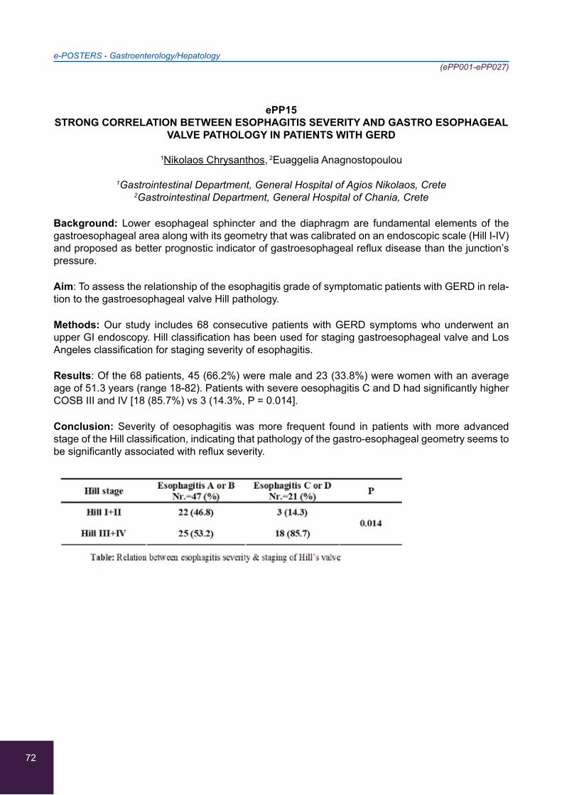
72
e-POSTERS - Gastroenterology/Hepatology(ePP001-ePP027)
ePP15STRONG CORRELATION BETWEEN ESOPHAGITIS SEVERITY AND GASTRO ESOPHAGEAL
VALVE PATHOLOGY IN PATIENTS WITH GERD
1Nikolaos Chrysanthos, 2Euaggelia Anagnostopoulou
1Gastrointestinal Department, General Hospital of Agios Nikolaos, Crete 2Gastrointestinal Department, General Hospital of Chania, Crete
Background: Lower esophageal sphincter and the diaphragm are fundamental elements of the gastroesophageal area along with its geometry that was calibrated on an endoscopic scale (Hill I-IV) and proposed as better prognostic indicator of gastroesophageal reflux disease than the junction’s pressure.
Aim: To assess the relationship of the esophagitis grade of symptomatic patients with GERD in rela-tion to the gastroesophageal valve Hill pathology.
Methods: Our study includes 68 consecutive patients with GERD symptoms who underwent an upper GI endoscopy. Hill classification has been used for staging gastroesophageal valve and Los Angeles classification for staging severity of esophagitis.
Results: Of the 68 patients, 45 (66.2%) were male and 23 (33.8%) were women with an average age of 51.3 years (range 18-82). Patients with severe oesophagitis C and D had significantly higher COSB III and IV [18 (85.7%) vs 3 (14.3%, P = 0.014].
Conclusion: Severity of oesophagitis was more frequent found in patients with more advanced stage of the Hill classification, indicating that pathology of the gastro-esophageal geometry seems to be significantly associated with reflux severity.

73
e-POSTERS - Gastroenterology/Hepatology(ePP001-ePP027)
ePP16
MALABSORPTION SYNDROME AS A FORM OF PRESENTATION OF MIXED CONNECTIVE TISSUE DISEASE
1Gloria Perez-Vazquez, 1Eva Maria Lozano-Loaiza, 1Carolina Luque-Amado, 2Esther Rubio-Muñoz,
3Luis Garcia-Aragon, 3Juan Antonio Jimenez-Casas
1Internal Medicine Unit, Hospital Punta Europa, Algeciras, Spain 2Nephrology Unit, Hospital La Linea, La Linea de la Concepcion, Spain
3Primary Care, Area de Atencion Sanitaria Campo de Gibraltar, Gibraltar, Spain
A 78-year-old woman admitted to Internal Medicine with chronic diarrhea of 6 months with lower limb disability, retrosternal pain, asthenia and palpitations. Blood test with hypokalemia and severe hypomagnesemia, acute renal failure, normocytic normocytic anemia, high ESR, total protein defi-cit, albumin and vitamin D. ECG with supraventricular paroxysmal tachycardia in relation to severe hypokalemia. Chest radiography, ultrasound and abdominal MRI, colonoscopy, echocardiogram and octreoscan were normal.
The autoimmunity study revealed ANA positive with mottled pattern, Ac AntiRNP, Ac AntiSM and Anti SS-A / Ro positive. Recover renal function and correct metabolic disorders, start oral corticosteroid treatment with striking clinical improvement, finding a real asymptomatic date.
The definitive diagnosis of the mixed connective tissue disease is complex, since the manifestations of the disease are usually presented sequentially, and may depend on criteria of several connective tissue diseases but with a different prognosis than the rest. Therefore, it is important not to forget this entity when it comes to patients with multiple clinical manifestations carried over time. The initial clinical manifestations of nonspecific alterations to joint, mucocutaneous, cardiovascular, pleuro-pulmonary, gastrointestinal, neurological or renal manifestations. Within the gastrointestinal tract, malabsorption due to bacterial overgrowth and enteropathy loses proteins and alterations that occur frequently.
74
e-POSTERS - Gastroenterology/Hepatology(ePP001-ePP027)
ePP17
ACUTE PANCREATITIS DUE TO SEVERE HYPERTRIGLYCERIDEMIA. A CASE REPORT
1Antonios Mpitsakis, 1Anthoula Chalkiadaki, 1George Fytrolakis, 1John Kontaxakis, 1Konstantina Theofanous, 1Maria Leontaki, 2Maria Rovithi, 3Nikolaos Chrysanthos
1Pathological Department, General Hospital of Agios Nikolaos, Agios Nikolaos, Greece 2Department of Oncology, General Hospital of Agios Nikolaos, Agios Nikolaos, Greece
3Gastrointestinal Department, General Hospital of Agios Nikolaos, Agios Nikolaos, Greece
Background: Acute recurrent pancreatitis induced by hypertriglyceridemia is a rare condition. Usu-ally serum triglyceride levels more of 1000mg/dl represents the causal factor. Clinical course and outcome presents no differences with other causes. Several treatment modalities have been pro-posed such as insulin and heparin administration or plasmapheresis.
Case report: We report a rare case of a 40 years old male patient with obesity and hyperlipidemia who has been admitted in our department with fever, epigastric pain, belching and bloating. He presented with 103 pulses/min, temperature: 37,6, blood pressure 155/93mmHg and a respiratory rate of 28/min. Laboratory findings reveals: wbc: 14.7k/μL, Hb:16.3 gr/dl, plt:183 k/μL, pt:13.2sec, INR:1, aptt:26.7sec, dbil:1 tbil:1.3, CK:30, LDH:230, SGOT:90, SGPT:450, ALP:88, γGT:45iu/L, Gluc:252mg/dl urea:19,8mg/dl, creat:0,61mg/dl, Trigl:5940mg/dl, K:4,5mEq/L, Na:127mEq/L Ca:9,2mg/dl Alb:4,2gr/dl, Amylase:541iu/L, Lipase:1550,4u/L, CRP:6.2mg/dl. Blood Gas: Ph: 7.45, pCo2:29.9, pO2:72.2, sO2%:95.4. Abdominal ultrasound reveals fatty liver infiltration without fo-cal lesions, gall-blader and bile-duct without stones. Abdominal x-ray typically with sentinel loop. We perform a computer tomography which reveals pancreatic enlargement with peripancreatic fat stranding and a small left pleural collection. Initial treatment of the patient included analgesics narco-tis and intravenous fluids. Moreover, insulin administration along with heparin results in a significant clinical improvement of the patient in only 48 hours. Also, laboratory tests prove this with amylase levels of 157iu/L and lipase of 225u/L. Triglyceride levels’ reduction was also impressive with 713mg/dl in 48h.
Conclusions: Patients with hypertriglyceridemia induced acute pancreatitis is an urgent incident. Insulin and heparin seems to be an effective rescue therapy.

75
e-POSTERS - Gastroenterology/Hepatology(ePP001-ePP027)
ePP18
EXTREMELY HIGH CA 19-9 LEVELS, IS IT ALWAYS CANCER?
Athanasia Apsemidou, Georgios Kalopitas, Konstantinos Kalogiannis, Ioanna-Maria Mouskeftara,
Georgios Pilianidis, Nikolaos Papaioannou, Nikolaos Petridis
Internal Medicine Department, Papanikolaou General Hospital, Thessaloniki, Greece
Background/Aim: Carbohydrate antigen 19-9 (Ca19-9) is a common serum-based marker for di-agnosis and follow up of pancreatic and other gastrointestinal malignancies. According to the litera-ture, a CA19-9 value higher than 1,000 U/ml has almost 100 % specificity for pancreatic carcinoma. Nevertheless, few cases of benign disease including chronic pancreatitis, cirrhosis, cholangitis and choledocholithiasis with elevated CA19-9 levels have also been reported.
Case report: A 45-year-old male patient presented to our Emergency Department with a 4-week history of abdominal and left hip pain. Except for past alcohol abuse his medical history was other-wise unremarkable. Clinical and laboratory examination indicated diabetic ketoacidosis, which was treated properly and an extremely elevated CA 19-9 serum level (1200 U/ml). Additionally, antibodies for diabetes mellitus type 1 were negative. Abdominal CT scan and MRI demonstrated pancreatic le-sions suspicious for pancreatic cancer, enlarged peripancreatic lymph nodes and osteolytic lesion in the left hip. Suspicion of metastatic pancreatic cancer was raised and a PET scan was performed but no metabolic activity was detected. CA 19-9 levels were checked two weeks later and found within normal range. Symptomatology and pancreatic lesions were attributed to past acute pancreatitis and osteonecrotic lesion to alcoholism history.
Conclusions: Elevated serum CA 19-9 levels should be interpreted cautiously as a diagnostic mark-er.CA 19-9 by itself is insufficient for diagnosing pancreatic cancer and should be evaluated accord-ing to clinical context and imaging findings.
76
e-POSTERS - Gastroenterology/Hepatology(ePP001-ePP027)
ePP19
SIADH SECONDARY TO ACUTE INTERMITTENT PORPHYRIA, A DIAGNOSTIC CHALLENGE THAT CAN NOT BE DELAYED
Magdalena Martín-Pérez, Jose María Pérez de Navarro, Ylenia Avellaneda-López
Internal Medicine, Hospital Punta Europa, Algeciras, Spain
Case Report: A 30-year-old woman with persistent abdominal pain of 1 week of evolution, going to the hospital emergency department for treatment with NSAIDs, benzodiazepines, spasmolytics and opiates with no response to oral and intravenous administration. Physical exploration without find-ings.
Analytical study highlights hyponatremia: 116ng / ml, with decreased plasma osmolarity, sodium in urine of 146 mEq / l, urinary osmolality of 447 mOsm / kg with renal function and normal urinary sedi-ment. Negative pregnancy test. Rx were normal. Abdominal CT with no significant retroperitoneal adenopathy, jejunum discreetly distended without evident cause. Gynecological assessment without findings. Surgery rules out acting on your part. In the presence of hypoosmolar hyponatremia with normal extracellular volume, we consider the initial differential diagnosis of SIADH, hypothyroidism or adrenal insufficiency. Start treatment with water restriction and hypertonic serum with very slight improvement. It is ruled out in analytical thyroid and adrenal pathology, continuing the etiology of SIADH without result until we receive a result of porphyrins in urine that are positive. In the interval between the availability of the results and the haematin for treatment, the patient developed motor neuropathy with proximal symmetric muscle weakness in the upper extremities that required admis-sion to the ICU.
Conclusions: This case should remind us of the diagnostic possibility of an acute intermittent por-phyria in the face of recurrent abdominal pain with hyponatremia, which should be diagnosed as soon as possible since delays in the initiation of treatment may lead to a fatal evolution or irreversible neurological damage.
77
e-POSTERS - Gastroenterology/Hepatology(ePP001-ePP027)
ePP20
EOSINOPHILIC ENTERITIS PRESENTED WITH IRON RESISTANCE IRON DEFICIENCY ANEMIA
2,3Xin Ning Ng, 1,2,3Khai Jing Ng, 1,2,3Ning-Sheng Lai, 1,2,3Kuang-Yung Huang, 1,2,3Chien-Hsueh Tung,
1,2,3Ming-Chi Lu, 1,2,3Bao-Bao Hsu
1Division of Rheumatology, Immunology and Allergy of Buddhist Dalin Tzu Chi Hospital, Chiayi, Taiwan
2Department of Internal Medicine of Buddhist Dalin Tzu Chi Hospital, Chiayi, Taiwan 3Tzu Chi University, Hualien, Taiwan
Background: Iron deficiency anemia is a global health problem and prevalent medical condition encountered in everyday clinical practice. Female are more commonly affected. The cause of iron deficiency should always be investigated especially in young male as it could be caused by serious diseases. Evaluation by clinical history, urinalysis and serological tests for celiac disease, gastros-copy and colonoscopy are the key diagnostic tools for investigation. We must insist on diagnostic evaluation in the absence of response to oral iron, or if the anemia is severe or clinical suspicion of important disease persists. Repeat endoscopic studies should be deliberated in many cases and random mucosal biopsies are strongly suggested even if endoscopically identified mucosal lesions are absent.
Case report: We describe a 40-year-old male presenting iron resistance iron deficiency anemia, chronic watery diarrhea and unintentional weight loss. Laboratory studies showed microcytic anemia and peripheral eosinophilia (total eosinophil count: 3708). He was under 200mg iron supplement when he presented to our clinic. However, Iron level was 17ug/dL, TIBC 408ug/dL, and Ferritin level 1.7ng/mL. Esophagogastroduodenoscopy and colonoscopy were performed with random biopsy, revealing eosinophilic infiltration in the duodenum and terminal ileum. The patient was eventually treated with oral prednisolone and elimination diets with complete resolution of symptoms.
Conclusion: This case highlights the diagnostic challenge of a condition that presents with common symptoms and apparently normal investigations. These findings should motivate and encourage the clinician to consider rarer diagnoses and pay special attention to seemingly unimportant clues such as peripheral eosinophilia.
78
e-POSTERS - Gastroenterology/Hepatology(ePP001-ePP027)
ePP21
AUTOIMMUNE HEPATITIS IN AN ELDERLY TURKISH MAN
1Firuze Midi Kursat, 1Hakan Kursat, 1Betül Erişmiş, 2Aliye Soylu, 1Yıldız Okuturlar
1Department of Internal Medicine, Bakirkoy Dr. Sadi Konuk Education and Research Hospital, Istanbul, Turkey
2Department of Gastroenterology, Bakirkoy Dr Sadi Konuk Education and Research Hospital, Istanbul, Turkey
Backgound: Autoimmune hepatitis (AIH) is an uncommon chronic progressive inflammatory dis-ease of the liver, characterised by hypergammaglobulinaemia, circulating autoantibodies and the histological change of interface hepatitis, which is responsive to immunosuppressive therapy in the majority of cases.
Case Description: A 78-year-old man presented to our outpatient clinic with jaundice. He had known hypertension and coronary heart disease. He had no history of smoking, drinking alcohol, using any herbal medicine. On examination he was icteric, he had no lymphadenopathy or hepatosplenom-egaly. His liver panel showed AST 1039U/L, ALT 1134U/L, ALP 183U/L, GGT 167U/L, total bilirubin 9,8g/dL, direct bilirubin 6.5mg/Dl, albumin 3 g/dL and INR 1.39. He tested negative for HBsAg, anti-HBs, anti-HBc total, HBV DNA by PCR, anti-HCV, HCV RNA by PCR. ANA was positive at 1:1320 and ASMA was positive at 1:3200 dilution but tests for AMA, anti-M2, anti-LKM 1 were all negative. Serum IgG was 2700mg/dL. Ultrasonography of abdomen revealed coarse ecotexture of liver with irregular. Percutaneous liver biopsy was planned but the patient did not accept it. We diagnosed him as type-1 AIH according to AIH scoring system and treated with 40 mg prednisolone per day with reduction of the dose. After 15 days treatment, hepatic tests were in normal range and he remained in remission at 3-month follow-up.
Discussion: Diagnosis of AIH often seems to be delayed in the elderly, presumably because AIH is erroneously considered to be a disease of young age. AIH should be considered in the older patient to avoid delayed initiation of immunosuppressive therapy.

79
e-POSTERS - Gastroenterology/Hepatology(ePP001-ePP027)
ePP22
ACUTE LIVER FAILURE FOLLOWING PHYSICAL EXHAUSTION
1Ioannis Andrianopoulos, 1Maria Saranti, 1Xanthi Zikou, 1Eleonora Kostanti, 1Aggeliki Giamarelou,
2Gerasimos Baltayiannis, 1Vasilios Koulouras
1Intensive Care Unit, University Hospital of Ioannina, Ioannina, Greece 2Gastroenterology Department, University Hospital of Ioannina, Ioannina, Greece
Case report: We present a rare case of acute liver failure. A 28-year-old albanian male patient was brought in by ambulance, after being found drowsy and exhausted. A 5-6 hour walk in a hot day had preceded as he tried to pass the borders on foot according to witnesses. He was admitted in our ICU drowsy but easily rousable, hypotensive, and tachycardic. Due to hypovolemia, he was started immediately on iv fluids, vasopressors and antibiotics. Laboratory tests revealed acute kidney injury with serum creatinine of 2.4 mg/dl, rhabdomyolysis, raised aminotrasnferases with prolonged INR. The patient gradually improved as his blood pressure stabilized and he became more alert. How-ever, the laboratory test values continued to worsen with a rise in the aminotransferase levels (AST 4800 u/L, ALT 6600 u/L), CK levels (peak value 59940 units/L), INR (peak level 4) and development of jaundice (peak total bilirubin 13 mg/dl). Serology testing for Hepatitis A, B,C, HIV,HSV,EBV,CMV, leptospirosis, autoimmune screening as well as blood and urine cultures were negative, ceruloplas-min levels were normal and ultrasound of the abdomen showed mild hepatomegaly with no other abnormal findings. When the patient recovered, he gave no significant past medical history and denied taking any medication, herbs, illicit drugs or any mushrooms. Conservative treatment with iv fluids was continued and a few days later the laboratory values started to improve until discharge to the Gastroenterology ward.
Conclusion: This is an interesting case of acute liver failure, as only few cases of acute liver injury secondary to physical exhaustion have been reported.
80
e-POSTERS - Gastroenterology/Hepatology(ePP001-ePP027)
ePP23
AN INTERESTING CASE OF A PATIENT WITH CHRONIC PANCREATITIS PRESENT WITH ANEMIA
Andreas Stylianou, Haris Constantinou, Angelos Adamantou, Angelos Alexi, Sofia Christodoulou,
Dania Rodotheou, Georgos Olympios
Department of Internal Medicine, Larnaca General Hospital, Larnaca, Cyprus
Background/Aim: Aneurysm of the splenic artery is uncommon and constitutes about 60% of the visceral aneurysms. We report a case of a patient with past medical history of chronic alcoholic pan-creatitis and chronic HBV infection who had been hospitalized for anemia.
Case report: Α 55-year-old patient with multiple hospitalizations due to chronic alcoholic pancrea-titis, presented to the hospital with generalized weakness and fatigue. The blood tests showed an Hb:5.6 mg/dl. The gastroscopy revealed an active hemorrhage from the ampulla of Vater. Computed tomography of the abdomen demonstrated a pancreatic pseudocyst detectable on previous imaging, while digital angiography showed stagnation of the contrast agent inside the pseudocyst which com-municated with the splenic artery. Ulcers of the splenic artery which were illustrated through the way of the pseudocyst were consistent with aneurysm. Embolization of the splenic artery was considered as the treatment of choice since the patient was at high risk. The first attempt was unsuccessful while it revealed a pseudoaneurysm in the middle area of the splenic artery. A new successful attempt was made a week later.
Conclusions: Aneurysm of the splenic artery is uncommon and the angiography confirms the diag-nosis. Pre-existing conditions related to the formation of these aneurysms are fibrodysplasia, portal vein hypertension with splenomegaly and an increased number of pregnancies. Other uncommon causes like the patient’s described above, are inflammatory diseases (chronic pancreatitis) and my-cotic aneurysms. The embolization consists the treatment of choice in high risk patients.
81
e-POSTERS - Gastroenterology/Hepatology(ePP001-ePP027)
ePP24
OLMESARTAN-INDUCED ENTEROPATHY: A CASE REPORT
Maria Tataridou, Sofia Flouda, Eirini Sveroni, Vasiliki Argiana, Fotios Kavallieratos, Anna Pavlidou,
Vasilios German, Nikolaos Papadopoulos, George Aloizos
1st Department of Internal Medicine, 417 Army Share Fund Hospital of Athens, Athens, Greece
Background/Aim: We present a case of severe malabsorption in a hypertensive woman treated with olmesartan.
Case Report: A 80-year-old Caucasian woman with hypertension, without any history of cardio-vascular disease, diabetes, and gastrointestinal disease, presented with chronic diarrhea, nausea and anorexia resulting in weight loss. The patient was treated with olmesartan (20 mg/d) until that time. Physical examination revealed muscle weakness and signs of hypocalcemia. Laboratory ex-ams showed severe malabsorption as demonstrated by severe hypocalcemia, hypokalemia, hypo-phosphatemia, hypoalbuminemia and anemia. Olmesartan was stopped, and parenteral nutrition, electrolyte correction and intravenous vitamin D supplementation were initiated. The patient under-went endoscopy which revealed aphthous ulcers in the terminal ileum. She was put on treatment with budesonide awaiting the biopsy results. Patients’ status rapidly improved in the next 10 days, and she was discharged with instructions for oral electrolyte and mineral supplementation. Biopsy showed chronic nonspecific ileitis and chronic inflammation accompanying by villous atrophy.
Conclusions: Olmesartan has been associated with sprue like enteropathy. This case report presents a case of severe malabsorption due to olmesartan, treated successfully with discontinu-ation of the offending drug and a short scheme of budesonide. Olmesartan induced enteropathy should always be included in the differential diagnosis of chronic diarrhea in hypertensive patients treated with this agent.
82
e-POSTERS - Gastroenterology/Hepatology(ePP001-ePP027)
ePP25
RECURRENT NON-VARICEAL UPPER GASTROINTESTINAL BLEEDING (NVUGIB) CAUSED BY SIMULTANEOUS EXTRAVASATION OF TWO DIFFERENT VESSELS TREATED WITH
EMBOLISM
1Dimitrios Moschovis, 2Vassilios E Nikolaou, 3Maria Agrogianni, 1Eirini Zacharopoulou,
1Chrysoula Veimou, 1Georgios Kechagias, 1Iosif Internos, 1Vasilios Vamvakousis,
2Christos Drakoulis, 1Maria Tzouvala
1Gastroenterology Department, General Hospital of Nikea Piraeus “Ag. Panteleimon”, Piraeus, Greece
2Radiology Department, General Hospital of Nikea Piraeus “Ag. Panteleimon”, Piraeus, Greece 32nd Department of Internal Medicine, General Hospital of Nikea Piraeus “Ag. Panteleimon”,
Piraeus, Greece
Background: Acute NVUGIB is a common condition with significant morbidity and mortality, com-monly from peptic ulcers. Endoscopic criteria determinate the type of treatment. A second attempt is recommended at rebleeding following index endoscopy with endoscopic hemostasis if indicated. In case of failure trans-arterial embolization (TAE) or surgery may be considered.
Case Report: A 92 years old woman in anticoagulants hemodynamic stable was admitted to the ER due to hematochezia and melena. Few hours later a new episode of hematochesia with decrease of Hemoglobin led to gastroscopy. A deep ulcer in the anterior duodenal bulb wall with clean base (Forrest IIc) was observed without red blood in the stomach. Patient received RBC transfusion and remained homodynamic stable. The following fifth and sixth day afterwards, hematochesia was re-ported with decrease of Hemoglobin. Upper endoscopy revealed active bleeding from the ulcer at the duodenal bulb (ForrestIb) treated with endoscopic methods. Three days later a new episode of hematochesia and reduction of Hemoglobin lead to another upper endoscopy where an adherent clot was observed on the duodenal bulbe’s ulcer with active bleeding. Two hemostatic clips plus di-luted epinephrine controlled the hemorrhage. However, 12 hours later angiography was performed because of a large volume hematochesia. Dysplastic vessels in the bulb without extravasation but extravasation in the distal area of 2nd part of duodenum were responsible for bleeding they were both successfully treated with embolism.
Conclusions: TAE is very effective method of control of NVUGIB particularly in rare cases of active bleeding from multiple sites.
83
e-POSTERS - Gastroenterology/Hepatology(ePP001-ePP027)
ePP26
THE MOST COMMON CAUSES OF GASTROINTESTINAL BLEEDING ALONG WITH THEIR ASSOCIATED SEVERITY
1David Symeonidis, 2Alkistis Kalessis, 1Eyridiki Soumpourou, 1George Aygoyleas, 1Stavros Antonopoulos
1B Pathology Department, Tzaneio General Hospital of Piraeous, Athens, Greece 2Cardiology Department Tzaneio General Hospital, Greece
Background: Depending on its location, gastrointestinal bleedings are classified into upper and lower ones.
Objective: To underline the most common causes of gastrointestinal bleeding along with their as-sociated severity.
Methods: 176 patients that underwent upper or lower gastrointestinal tract endoscopy during the period 2014-2016 were included while a complete medical history was obtained.
Results: 58% presented with an upper gastrointestinal tract bleeding, 26% with a lower tract bleed-ing and 16% were found to have both upper and lower tract bleeding. The antiplatelet and antico-agulant therapies, were the most common causes, with a 58% of the patients having received either antiplatelet or anticoagulant therapy. Active GI bleeding during endoscopy was found in 73% of pa-tients under any type of anti-clotting medication and in 84% of patients receiving NSAIDs. Upper GI tract bleeding was more common in males (69% of males presented with an upper GI tract bleeding whereas 41% of females presented with a lower GI tract bleeding). Finally, 53% of men and 82% of women were over 75 while 47% of men and 18% of women were under 75.
Conclusion: The use of any type of anti-clotting medication was considered as the most common cause of gastrointestinal bleeding whereas the role of antiplatelet agents was underlined. The low rate of use of newer anticoagulant agents (NOACs) was also noted. Another important finding worth mentioning is the fact that in a large proportion of patients receiving antiplatelet therapy, there was active bleeding at the time of endoscopy.
84
e-POSTERS - Gastroenterology/Hepatology(ePP001-ePP027)
ePP27
COMPARATIVE STUDY BETWEEN SEROLOGICAL AND HISTOPATHOLOGICAL FINDINGS FOR COELIAC DISEASE AND PREVALENCE OF COELIAC DISEASE IN A MEDITERRANEAN
POPULATION OF MALE YOUNG ADULTS
1Dafni Koumoutsea, 2+Dimitrios Zokaris (in memoriam), 1,2,3Andreas Nakos, 3Eraclis Eracleous,
4Panagiota Thalassinou, 5Marios Tabakopoulos, 6Dimitrios Krentiris, 7Nikolaos Filiotis,
8Giovanni Apostolo Teodossiu, 1Dimitrios Tabakopoulos
1Sector of Internal Medicine and Division of Gastroenterology 401 General Army Hospital of Athens, Athens, Greece
2Medical Sector of Hellenic Force in Cyprus (ELDYK), Cyprus 3Department of Internal Medicine, ‘’Evangelismos’’ Hospital of Paphos, Paphos, Cyprus
4Second Department of Internal Medicine, “Hippokrateion” Hospital of Athens, Athens, Greece 5Department of Internal Medicine Naval Hospital of Crete, Greece
6Health Center of Salamina, Salamina, Greece 7Department of Digestive and Endoscopic Surgery University Hospital
of Catanzaro ‘’Magna Graecia’’, Italy 8Department of Digestive and Endoscopic Surgery University Hospital of Bari ‘’Aldo Moro’’, Italy
Aim: This study was conducted to draw the attention of clinicians concerning the reliability, sensitiv-ity and specificity of established serological testing versus jejunal biopsy for coeliac disease (CD) as well as to implement an established serological screening procedure for CD and to estimate the prevalence of CD in a Mediterranean population of male young adults.
Materials-Methods: Serum samples were collected from 5739 military recruits, mean age 22 years (range 18-26 years). All the samples were tested for IgA-serum levels to exclude IgA deficiency. Sam-ples with total IgA within the normal range were tested for IgA antibodies against native human-tissue transglutaminase (anti-tTG). Samples that were anti-tTG positive were tested for IgA antiendomysial antibodies (EMA). All antibody positive subjects were asked to undergo jejunal biopsy procedure. Results: Results of both tests (anti-tTG and EMA abs) were concordant. Of the 5739 subjects 35 (0,61%) had positive tissue transglutaminase (t-TG) and endomysial antibody (EMA) tests. Of the 35 subjects with positive antibody assays 33 subjects agreed to undergo biopsy and 31 (0,54%) had evidence of CD on biopsy. Of the 31 subjects with evidence of CD on biopsy 27 were considered to be asymptomatic and 4 were presented with a subclinical course.
Conclusions: The minimum prevalence of CD among male young adults in southern Greece and Cyprus is estimated to be 0,54%. The presence of serum tissue transglutaminase and endomysial antibodies is predictive and indicative of CD which allow better selection of patient for jejunal biopsy findings (villous atrophy, crypts hyperplasia and intraepithelial lymphocytes).
85
e-POSTERS - Rheumatology(ePP028-ePP041)
ePP28
A CASE OF POSSIBLE PRIMARY CATASTROPHIC ANTIPHOSPHOLIPID SYNDROME
1Karmen Tasiopoulou, 2Irina Krasovskagia, 3Christiana Anastasiadou, 4Konstantinos Alexandris,
5Aggelos Megalopoulos
1Nephrology Department, “Papanicolaou” General Hospital, Thessaloniki, Greece 21st Department of Internal Medicine, “Papageorgiou” General Hospital, Thessaloniki, Greece
3Vascular Surgery Department, “KAT” Hospital, Athens, Greece 4Vascular Surgery Department, “Hippokrateio” General Hospital, Thessaloniki, Greece
5Vascular Surgery Department, “Papanicolaou” General Hospital, Thessaloniki, Greece
Backround: Catastrophic antiphospholipid syndrome (CAPS) is a rare, lifethreatening condition than differs considerably from the simple/classic antiphospholipid syndrome (APS) in several as-pects, viz, the rapid development of multiorgan failure (within a week) following precipitating factors, infection, trauma including minor surgery like biopsy, lupus flares, anticoagulation problems ,malig-nancies.The multiorgan failure predominantely results from small vessel occlusion although large vessel occlusion can occur (deep vein thrombosis, arterial occlusion causing stroke or peripheral ischemia). It can be primary or secondary to immune disease, particulary lupus.
Case presentation: A 37 years old woman was admitted in the Vascular Surgery Department be-cause of critical ischemia of the left limb and significant occlusion of polpiteal artery on angiography, wheareas there was an usuccessfull attempt of angioplasty. Because of the prolonged aPTT there was a strong suspicion of APS and Tinzparine was started. Three days after she displayed fever, respiratory distress and worsening renal function with proteinuria. In conjunction with high levels of anticardiolipin antibodies, positive LAC, presence of cryoglobulins and negative serology for lupus and vasculitis primary CAPS was suspected and patient started plasmapheresis, pulses of methyl-prednisolone followed by oral administration, cyclophosphamide and warfarin, with significant clini-cal and serologic improvement. Twenty-five days after a succesfull angioplasty was performed with Tizaparine use before and after the procedure. Eighteen hours later she displayed acute respiratory distress, hemodynamic colapsus and she died within one hour probably due to massive pulmonary embolism (family refused autopsy).
Conclusions: Primary CAPS is a rare severe condition with high mortality even with early diagnosis and treatment initiation.
86
e-POSTERS - Rheumatology(ePP028-ePP041)
ePP29
BEHÇET’S DISEASE PRESENTING AS RECURRENT ORCHEO-EPIDYDIMITIS IN A PATIENT PREVIOUSLY MISDIAGNOSED AS JUVENILE RHEUMATOID ARTHRITIS
Panagiotis Oikonomou, Katerina Argyraki, Maria Meliou, Spyridon Kloudas, Frosso Konstantinou,
Ioanna Kokkinaki, Aggelos Pefanis
Department of Internal Medicine, ‘’Sotiria’’ General and Chest Diseases Hospital, Athens, Greece
Background: Behçet’s Disease (BD) is a rare systemic vasculitis, occurring mainly in Asia, Middle East and Mediterranean, characterized by recurrent, typically self-limiting, attacks of acute inflam-mation, manifestating with aphthous stomatitis with genitourinary, eye, skin, gastrointestinal, nerv-ous, vascular and joint involvement.
Aim: Presentation of a BD case, previously misdiagnosed as juvenile Rheumatoid Arthritis (JRA) in childhood, fully developed in adulthood.
Case report: An 18-years-old male with history of childhood-onset uveitis (diagnosed as JRA, treat-ed with adalimumab) presented with recurrent testicular pain, fever and diarrhea. On examination, tenderness of testicle and epididymis, rebound tenderness in lower abdomen, painful splenomegaly, oral painful-aphthous ulcers (>3 times last 6 months) and acneiform back lesions were noted. Sig-nificant laboratory findings: elevated inflammation markers, hyperglobulinemia. Scrotal ultrasonog-raphy: Enlargment and increased blood flow of right epididymis. Patient was treated empirically with antibiotics based on abdomen CT findings indicative of inflammatory bowel disease (IBD). GI endoscopy revealed aphthous ulcerations of colon and atrophy of intestinal villi. After remission of initial symptoms, erythema nodosum-like lesions appeared on both knees with concomitant painful pustules at the sites of venipuncture (pathergy phenomenon). Lesion biopsy revealed plasma and lymphocyte infiltration and granulomatous inflammation with deep ulceration. Meeting the criteria of BS, supported by HLA-B51 positivity, adalimumab, azathioprine and colchicine were administered with subsequent remission of skin lesions and aphthous ulcers in MR enterography.
Conclusions: This is a unique case of BD, presenting as uveitis in childhood, while full-blown dis-ease developed in late adolescence. Atypical presentation, mimicking acute orcheo-epididymitis and IBD, delayed diagnosis.

87
e-POSTERS - Rheumatology(ePP028-ePP041)
ePP30
ATYPICAL PRESENTATION OF FAMILIAL MEDITERRANEAN FEVER AT ADVANCED AGE
1,2Christina Flourou, 1,3Aram Katsios, 4,5Savvas Psarelis, 1,2Antreas Tofarides, 1,5Chrysostomos Zintilis
1Internal Medicine Department, Nicosia General Hospital, Nicosia, Cyprus,2Aristotle University of Thessaloniki, Larnaca, Cyprus
3Charles University, Prague, Nicosia, Cyprus4Rheumatological department, Nicosia General Hospital, Nicosia, Cyprus
5National and Kapodistrian University of Athens, Nicosia, Cyprus
Background: Familial Mediterranean Fever is the most common autoinflammatory disorder. Even though it is inherited by autosomal recessive manner, it is referred that this disorder manifests in heterozygotes as well. Usual age of manifestation is at an age of less than 20 years at a percentage of 65-90%, however at a percentage of less than 0.5% it can manifest at an age of greater than 50.
Case report: A 58 year-old woman, Greek-Cypriot, β-heterozygous anemia was admitted due to fe-ver over the last 20 days. Blood tests showed microcytic hypochromic anemia and elevated levels of ESR 75 mm/1h, CRP 115 mg/L and serum amyloid 68.8 mg/dl. Negative immunological tests. At the CT scan was observed pericardial effusion 1.4cm, pleural effusions and hepatomegaly 22.3cm. Bi-opsy of the rectum and congo red stain was positive. Additionally, the genetic control for TRAPS and CAPS syndrome was negative, but the FMF test was positive with heterozygoty in exon 2 - E148Q. The patient was administered Colchicine 2mg/daily with gradual reduction of pericardial effusion.
Conclusion: Genetic variations of the exon 2 are related with mild non specific inflammatory mani-festations. Pericardial effusion in Familial Mediterranean Fever could present in less than 1% in homozygote cases and in a lesser percentage in case of heterozygotes. In cases of inflammatory syndromes and secondary amyloidosis independent of age, autoinflammatory disorders must be excluded.

88
e-POSTERS - Rheumatology(ePP028-ePP041)
ePP31
MYOPERICARDIAL INVOLVEMENT IN A CHURG STRAUSS SYNDROME PATIENT
1Pantelis Toskas, 2Styliani Vakrou, 2Christos Kapelios, 3Maria Mylona
11st Cardiology Department, University of Athens Medical School, “Hippokration” Hospital, Athens, Greece
2Department of Cardiology, “Laiko” Hospital, Athens, Greece 31st Department of Propaedeutic & Internal Medicine, “Laiko” General Hospital, Athens University
Medical School, Athens, Greece
Background/Aim: Churg-Strauss syndrome (CSS) is a necrotizing vasculitis of small vessels char-acterized by upper and lower airway disease followed by peripheral eosinophilia and multiple organ involvement. We present an interesting case of a woman with CSS and myopericardial disease. Cardiac involvement is evident in 40-50% of CSS cases and carries poor prognosis. Some of the features of myocardial CSS involvement can be evaluated by echocardiography however cardiac MRI is advocated as the gold-standard method with crucial role in the prompt recognition and man-agement of such patients.
Case description: A Caucasian 45-year old woman, with a kwown history of CSS under no medi-cation, presented with dyspnea NYHA III of recent onset. Her chest-X-ray revealed bilateral pleural effusions and the ECG showed sinus tachycardia, left ventricular hypertrophy and diffuse horizon-tal ST segment depression. Blood analysis was remarkable for leukocytosis, severe eosinophilia (=15,000/μl), abnormal troponin and BNP levels. Initial transthoracic echocardiogram (TTE) re-vealed EF=45%, hypokinetic inferior and inferolateral and mid/apical anteroseptal segments with marked hyperechogenicity of inferior and inferolateral wall involving both the subendocardial and subepicardial segments. The patient received 1 gr methylprednizolone/24hrs x 3days with clinical improvement. Repeat TTE, 5 days later, demonstrated improved EF=55% and slightly less infiltra-tion. Cardiac MRI was performed and all three facets of the inflammatory process namely acute/chronic inflammation and healing were evident in various degrees with signs of fibrosis. The patient was discharged receiving, metoprolol, ramipril and monthly cyclophosphamide IV. Her global systolic function remains normal.
89
e-POSTERS - Rheumatology(ePP028-ePP041)
ePP32
BACTERIAL OVERGROWTH SYNDROME WITH MALABSORPTION DUE TO INTESTINAL PSEUDO-OBSTRUCTION IN A CASE OF LIMITED SYSTEMIC SCLEROSIS
1Alkiviadis Ioannou, 1Charalampos Giannopoulos, 1Nikolaos Alexiou, 1Konstantina Gkizeli,
1Eirini Skoura, 1Tatiana Tataridoy, 1Georgios Maikantis, 1Oksana Tsatov, 1Marina Koupetori,
2Stilianos Anastasiadis, 2Despina Papadeli, 1Stiliani Sybardi
11st Department of Internal Medicine, “Thriasio” General Hospital, Elefsina, Greece 2Gastroenterology Department, “Thriasio” General Hospital, Elefsina, Greece
Background: Intestinal bacterial overgrowth syndrome is a clinical entity, which can be derived from intestinal motility dysfunction.
Aim: Case report of first diagnosed limited systemic sclerosis with over 2 years persistent intestinal symptomatology.
Case report: A 79 years old female with history of essential hypertension was admitted because of chronic diarrhea and significant weight loss. History of multiple hospitalizations due to similar symp-tomatology and recent colonoscopy with colorectal adenoma (biopsies negative for malignancy). Physical examination: BP 140/80 mmHG,T:36.3oC,SatO2: 98%,HR: 75/min, dehydration. Labor: se-vere hypokalemia, hypocalcemia, hypomagnesemia.
Results: Clinical and laboratory improvement after parenteral K, Mg, Ca substitution. CT-Abdomen: small-intestinal dilated loops with air fluid levels. Labor: Stool analysis: fat, undigested food compo-nents. Stool culture/ova and parasites examination: negative Cl. Difficile (Ag, Tox): negative. Thy-roid function normal. Hormones: Chromogranin A, Gastrin, Glucagon, VIP, Calcitonin, Thyroglobulin: normal. Serology: (-) anti-tTG, anti-EMA anti DGP. (+) ΑΝΑ (1:320), anti-ΕΝΑ, anti-SCL-70 anti-RNP, anti-Ro, anti-Sm. Gastroscopy: atrophic gastric mucosa, atrophic duodenal folds (biopsy: atrophic villi, hyperplastic crypts, lemphocytes proliferation of lamina propria). Oesophageal-PH-manometry: incomplete relaxation of the lower esophangeal sphincter. Echocardiogram: negative for pulmonary hypertension. Under the diagnosis bacterial overgrowth syndrom with malabsorption due to limited systemic sclerosis, therapy with rifaximine and prokinetic agents. Gradual recession of the diarrhoic syndrome, and the patient was referred to a rheumatology clinic.
Conclusions: Intestinal motility disorders caused by systemic sclerosis can lead to malabsorption due to bacterial overgrowth, and subsequently to chronic diarrhea. In this case, considering the absence of the typical manifestations of systemic sclerosis, the diagnosis was based on serologic testing.

90
e-POSTERS - Rheumatology(ePP028-ePP041)
ePP33
AN INTERESTING CASE OF GIANT CELL ARTERITIS IN AN ASYMPTOMATIC 72-YEAR-OLD PATIENT
Angeliki Tsifi, Sevastianos Chatzidavid, Stavroula-Panagiota Lontou, Maria Triantafyllou,
Marina Skouloudi, Elpida Mastrogianni
1st Department of Internal Medicine, “Laikon” General Hospital of Athens, Athens, Greece
Background: Giant cell arteritis (GCA) is a common vasculitis especially in elderly, female patients. It mainly involves medium to large size vessels and expresses itself with classic symptoms, such as headache, jaw claudication, visual disturbance, fever or even acute visual loss. Asymptomatic cases often cause diagnostic problems and underline the importance of including GCA in the differential diagnosis of anemia and high inflammatory markers in the elderly population.
Case Report: The present study reports an interesting case of a 72-year-old female who was ad-mitted to our hospital for further investigation of her anemia and elevated C-reactive protein and erythrocyte sedimentation rate. The patient did not present any of the classic symptoms of giant cell arteritis. However, the lack of explanation for her anemia and elevated inflammatory markers prompted a biopsy of her right temporal artery. The result was consistent with the diagnosis of giant cell arteritis. The patient was immediately commenced on oral prednisone, thus avoiding potential irreversible complications and additionally she was referred to a rheumatologist. The inflammation markers decreased and her blood tests normalized.
Conclusion: Although anemia and elevated inflammatory markers are infrequent initial manifesta-tions of giant cell arteritis, physicians should always include it in their differential diagnosis in the eld-erly population, mainly because the disease could ultimately have disastrous effects on the patient’s vision and health.

91
e-POSTERS - Rheumatology(ePP028-ePP041)
ePP34
NEUROLEPTIC SYNDROME
1Maria Rodriguez-Mora, Daniel Ramirez-Yesa, 1Virginia Perez-Vazquez, 1Maria Jose Tinoco-Bernal,
2Gloria Perez-Vazquez, 3Luis Garcia-Aragon
1Internal Medicine Unit, Hospital Universitario Puerto Real, Puerto Real, Spain 2Internal Medicine Unit, Hospital Punta Europa, Algeciras, Spain
3Primary Care, Area Gestion Sanitaria Campo de Gibraltar, Gibraltar, Spain
Case report: 25-year-old patient with a deep autistic spectrum disorder is admitted to Psychiatry Department for psychomotor agitation and at the fourth day of admission started with fever of 39.6oC and muscular stiffness. Initially treated with haloperidol because of the agitation and antibiotics due to tooth infection. Blood analysis revealed CPK elevation (up to 30000 U/L) with an elevation of acute phase reactants (PCR and VSG), plus acute renal failure. The patient was treated with dant-rolene started after blood and urine culture extractions, with the suspicion of malignant neuroleptic syndrome, and the antibiotic spectrum is increased. Despite the start of dantrolene 2 mg/kg every 8 hours during 15 days, the fever remains between 37.5-39 degrees every day, and appearance of edema in both arms. Due to lack of peripheral venous access, a central venous catheter was chan-neled, later with appearance of late complication as deep venous thrombosis of upper right arm con-firmed by Doppler ultrasound and anticoagulation is started. Central venous catheter tip is cultured and multi-sensitive Klebsiella Pneumoniae is isolated. Rest of blood and urine cultures are negative. Echocardiography and abdominal ultrasound without abnormalities. Dantrolene is removed from the treatment after 15 days, and two days later that bromocriptine is added to treat the fever. Within the 48 hours after the start of bromocriptine, the fever disappears. The patient was discharged after 7 days without fever, maintaining a progressively decreasing dosis of bromocriptine for 2 months at home, until totally finishing the treatment.
92
e-POSTERS - Rheumatology(ePP028-ePP041)
ePP35
SYSTEMIC LUPUS ERYTHEMATOSUS FLARE IN A PREGNANT WOMAN WITH HISTORY OF ARTHRITIS ASSOCIATED WITH POSSIBLE NON-PRIMARY CMV INFECTION
1Sofia Pitsigavdaki, 1Dafni Korela, 1Stamatis Karakonstantis, 1Evgenia Emmanouilidou,
1Melina Kavousanaki, 1Despina Galani, 1Kalliopi Milaki, 1Evangelos Thalassinos,
2Georgios Papazoglou, 1Afroditi Koufogianni, 1Charalampos Lydakis
12nd Internal Medicine Department, “Venizelio Pananio” General Hospital, Heraklion, Greece 2Emergency Department, “Venizelio Pananio” General Hospital, Heraklion, Greece
Background: Pregnancy is associated with a high rate of systemic lupus erythematosus (SLE) flares ranging from 25-60%. Early diagnosis and management is essential for managing an unevent-ful pregnancy.
Case Report: A 29-year-old female at 16 weeks gestation with history of seronegative rheumatoid arthritis in remission with no current treatment and Hashimoto’s thyroiditis, presented with fever up to 39oC for two weeks and sore throat. She had been prescribed cefuroxime without response. Virol-ogy testing before pregnancy indicated latency for CMV infection (positive IgG). Clinical examination revealed multiple erythematous macules on the trunk and aphthous stomatitis, without palpable lym-phadenopathy. Laboratory test showed CRP:2mg/dl, ESR:88mm/hr and lymphocytopenia:400/μl. Blood, urine, vaginal cultures proved sterile and abdominal ultrasonography was normal. She pro-gressively developed arthritis of the proximal interphalangeal, carpometacarpal joints and reported arthralgias at both knees and shoulders with morning stiffness. CMV IgM titers were positive sug-gesting reactivation or reinfection. Testing for EBV, Toxoplasma, Parvo, HSV and Coxsackie were negative. Immunology results showed ANA:1/1.280, anti-ds-DNA:816IU/ml, positive anti-SS-A(Ro) and hypocomplementemia. Renal, liver function and urine sediment were normal. A rheumatologist was consulted and the patient was diagnosed with SLE flare during pregnancy. She was treated with prednisolone and hydroxychloroquine with gradual remission of fever, arthritis and rash. During the next five months, there were no complications and she gave birth to a healthy girl.
Conclusion: Infections commonly trigger SLE flares. The clinical distinction between infection and lupus is challenging. Whether SLE promoted CMV reactivation or CMV was the trigger for the SLE flare is unclear.
93
e-POSTERS - Rheumatology(ePP028-ePP041)
ePP36
PATIENT WITH POEMS SYNDROME PRESENTING WITH VEIN DEEP THROMBOSIS
1Virginia Perez-Vazquez, 2Gloria Perez-Vazquez, 3Luis Garcia-Aragon, 1Juan-Bosco Lopez-Saez,
2Carolina Luque-Amado, 1Eva Lozano-Loaiza
1Internal Medicine Unit, Hospital Universitario Puerto Real, Puerto Real, Spain 2Internal Medicine Unit, Hospital Punta Europa, Algeciras, Spain
3Primary Care, Area Gestion Sanitaria Campo de Gibraltar, Gibraltar, Spain
Case report: 60-year-old female with a history of 6 years of disabling peripheral neuropathy treated with corticosteroids whithout improvement. She was admitted into our Hospital with bilateral numb-ness in the distal lower limbs, followed by a progressive loss of strength. Physical examination revealed hypertrichosis and mild edema of the lower limbs, hepatosplenomegaly and lymphaden-opathy, with sclerosis on fingers and hands. Neurological examination revealed moderate muscle weakness and mild sensory disturbance of both lower limbs, the Achilles tendon reflex was absent bilaterally. Laboratory testing showed moderate renal faillure, CRP 2.2 mg⁄dL, ESR 30 mm⁄h and elevated levels of ACTH and cortisol. Monoclonal IgG lambda-type protein was detected by immu-noelectrophoresis.
The results of autoimmune and peripheral neuropathy autoantibodies were negative. Plasma VEGF levels were markedly elevated 609 pg ⁄mL. Transthoracic echocardiography revealed normal left ventricular volumes and ejection fraction, mild pericardial effusion, sever tricuspid regurgitation, and pulmonary hypertension. Body CT-Scan showed pericardial and pleural effusion, hepatosplenom-egaly with ascites, osteosclerotic lesions with an expansive lesion at the posteromedial border of the left iliac crest with periosteal reaction. Electromiyography confirmed chronic inflammatory demyeli-nating polyneuropathy. The findings are consistent with previous reports of bone lesions in patients with POEMS syndrome, confirmed by the biopsy of the lesion with an infiltration of plasma cells.
Conclusions: POEMS is a multisystemic disease that is difficult to diagnose due to its low incidence and prevalence, the fact that the appearance of signs and symptoms does not occur simultaneously. Our patient showed favorable responde to melphalam and dexametasone.

94
e-POSTERS - Rheumatology(ePP028-ePP041)
ePP37
CHRONIC DIARRHEA AS DEBUT OF MIXED CONJECTIVE TISSUE DISEASE
1Esperanza Miranda-Sancho, 1Jose-Manuel Gil-Gascon, 1Gloria Perez-Vazquez,
2Luis Garcia-Aragon
1Internal Medicine Unit, Hospital Punta Europa, Algeciras, Spain 2Primary Care, Area Gestion Sanitaria Campo de Gibraltar, Spain
Case report: 78-year-old woman with arterial hypertension was taken to the hospital due to su-praventricular tachycardia with secondary hemodynamic angina and hypokalemia. She presented chronic diarrhea for several months with associated weight loss, without identifying the origin of diarrhea, with all the normal complementary study (TAC body, gastroscopy, opaque enema, celiac disease, stool culture and Clostridium difficile toxin), being high with the diagnoses of hypokalemia secondary to chronic diarrhea syndrome not affiliated. The physical examination in our hospitali-zation was normal. Evolutionary analysis revealed: urea 169 -> 25 mg/dl, creatinine 3.62 -> 0.54 mg/dl, potassium 1.5 -> 4.5 mmol/L, total protein 5.2 g/dl, Hb 12.1 g/dl, leukocytes 15200/dl (92% neutrophils), fibrinogen 687 mg/dl, 25-OH-Vitamin D 4 -> 9, ANA 1/640 (homogeneous standard), anti-RNP Ac positive. Other normal (Ac antiphospholipids, TSH, trypsin in faeces, serology Giardia lambdia, cytomegalovirus, toxoplasma, HIV, HCV and HBV). The remaining complementary tests were anodyne (chest X-ray, abdomen ultrasound, abdomen MRI, octreoscan and colonoscopy). The evolution was favorable, normalizing the deficit analytical parameters due to malabsorption after hydroelectrolitic replenishment and systemic corticosteroids, with the disappearance of diarrhea and recovery of body weight.
Conclusion: We consider this case interesting because malabsorption is a rare (and described) presentation of a mixed connective tissue disease.
95
e-POSTERS - Rheumatology(ePP028-ePP041)
ePP38
FUO AND ARTERIAL THROMBOSIS, IS IT LUPUS?
Ifigeneia Kassotaki, Aggeliki Kypraki, Despoina-Eleni Arna, Asimina Koulouridi,
Sevasti-Alexandra Maragou, Afroditi Koufogianni, Emmanouil Tzagarakis, Kyriaki Petraki,
Melina Kavousanaki, Charalampos Lydakis
B Internal Medicine Department, “Venizeleio” General Hospital, Heraklion, Greece
Background/Aim: Fever of unknown origin is a common reason of hospitalization and a demanding diagnostic medical field. Herein, we present a case of persistent fever in a young woman, triggered by acute myocarditis.
Case report: A 33 year’s old woman with a medical history of deep vein thrombosis, preeclampsia and type 1 diabetes mellitus was admitted to the cardiology department due to acute myocarditis, with positive Coxsackie IgM antibodies. She gradually improved; however, on the third week of hospitalization, fever and pancytopenia occurred. From the physical examination, palpable spleen and inflammation of the right forearm (on the intravenous catheter site, used in the coronary angi-ography) were noted. Upper limb Doppler ultrasound revealed thrombosis of the right radial artery. Blood tests revealed pancytopenia and ESR 68. Blood and urine cultures were negative. Immuno-logic profile suggested of positive ANA (1:1280), positive ENA screen and anti-Sm, anti-RNP mildly positive. Also, low levels of C3 and C4 complement and positive direct Coombs test. Thrombophilia screening came positive for anticardiolipin (aCL) antibody, and anti-beta-2glycoprotein I antibody (anti-B2GPI). Diagnostic criteria of both lupus and antiphospholipid syndrome were fulfilled, so the patient was treated with LMWH bridging therapy, till acenocoumarol-achieved therapeutic levels, and prednisolone. Fever subsided quickly after corticosteroid therapy initiation. The patient was dis-charged and referred to a rheumatology department, with the diagnosis of SLE and APS, possibly triggered by a viral myocarditis.
Conclusions: SLE is a common cause of persistent fever, and must be diagnosed promptly, espe-cially in cases of major thrombotic events.
96
e-POSTERS - Rheumatology(ePP028-ePP041)
ePP39
A RARE CASE OF A PATIENT ACTUALLY TREATED FOR SARCOIDOSIS THAT PROVED TO BE PRIMARY SJOGREN’S SYNDROME
Georgios Kalopitas, Nikolaos Petridis, Eleni Setsidou, Konstantinos Kalogiannis,
Konstantinos Geronatsios, Georgios Pilianidis
1st Department of Internal Medicine, General Hospital of Thessaloniki “Georgios Papanikolaou”, Thessaloniki, Greece
Background: Sjogren’s syndrome is an autoimmune chronic inflammatory disease affecting the exocrine glands that is mainly characterized by diminished salivary and lacrimal gland function. Sjogren’s syndrome may appear as an individual disease (primary SS) or as secondary along with another disease such as Rheumatoid arthritis and others. Sjogren’s syndrome rarely might also have non-exocrine gland involvement
Case Report: The patient is a 62-year-old male who presented to his primary care physician with a two-month history of gradually worsening fatigue and dyspnea on exertion. He was then examined by a cardiologist and a respiratory medicine specialist and treated for sarcoidosis. The initial diagno-sis was based on chest X-ray findings, chest computed tomography scan findings and bronchoscopy results. Two months later the patient presented in the emergency department with fever and short-ness of breath. On admission to the hospital he had hypoxemia and elevated WBC, ESR, CRP. He was treated for a respiratory tract infection. His personal medical history was reviewed and he had a thorough laboratory investigation and a CT scan repeated. He was found to have a strongly positive anti-Ro antibody titer, positive Schirmer’s test and proteinuria along with interstitial lung disease. Sal-ivary gland biopsy was also performed. Primary Sjogren’s Syndrome diagnosis was established.
Conclusion: Sjogren’s syndrome is one of the most common autoimmune diseases which mainly af-fects salivary and lacrimal gland function. It may have serious complications such as interstitial lung disease or proteinuria and we should always consider it when a new complex symptom develops.
97
e-POSTERS - Rheumatology(ePP028-ePP041)
ePP40
MORPHOLOGICAL CHANGES OF NASAL MUCOSA IN CHILDREN WITH GENERALIZED NASAL POLYPOSIS: REVIEW OF LITERATURE
1Georgios Karabelias, 2Maria Korodima, 3Maria Marmara, 1,4Aikaterini Drylli
1 ENT Department, Penteli Children’s Hospital, Athens, Greece2 ENT Department, Navy Hospital of Athens, Athens, Greece
3ENT, Lamia, Greece4Audiology-Neurotology, Medical School, National and Kapodistrian University of Athens,
Athens, Greece
Background: Nasal polyps are soft, pink or pearl white outgrowths usually connected with nasal mucosa by flaccid crus. In children, generalized nasal polyposis is uncommon and mostly character-izes Cystic Fibrosis (CF) as a result of the inflammatory-bioelectric formation of nasal mucosa which was proposed by Bernstein in 1994.
Objectives/Aim: To describe the morphological changes of nasal mucosa in children with general-ized nasal polyposis.
Methods: A retrospective systematic review was conducted of articles, by using appropriate search terms such as [(generalized nasal polyposis) AND children], on an initial search of medical literature data base (PubMed).
Results: 4 articles describe CF as an inherited multisystemic disorder that results in a general-ized dysfunction of exocrine glands and presented in children with generalized nasal polyposis. A light- and electron microscopical examination of histological sections reveal under a thick layer of respiratory epithelium with a high proportion of goblet cells, the seromucous glands display abnor-mal morphological structures with wide mucous cells and cystic dilatation. The glandular cells show inhomogeneous glandular droplets in the supranuclear cell portion. The nucleus contains dispersed chromatin as a sign of increased activity and the structures of the Golgi apparatus are clearly detect-able.
Conclusions: Nasal polyposis was mentioned in the Hippocrates’s notes from 4th century B.C. and was confirmed by 3rd century B.C. inscription on the King Sabur’s tombstone. However, generalized nasal polyposis is a rare disease in children and required a histological examination to diagnosed CF.
98
e-POSTERS - Rheumatology(ePP028-ePP041)
ePP41
SINONASAL FINDINGS IN GRANULOMATOSIS WITH POLYANGIITIS (GPA): REVIEW OF THE LITERATURE
1Georgios Karabelias, 1,2Aikaterini Drylli, 3Eleni Paschou, 4Nikolaos Sabanis
1 ENT Department, Penteli Children’s Hospital, Athens, Greece2 Audiology-Neurotology, Medical School, National and Kapodistrian University of Athens,
Athens, Greece3 General Medicine Department, Health Center Vyrona, Athens, Greece
4 Nephrology Department, General Hospital Livadeias, Viotia, Greece
Background: Granulomatosis with polyangiitis (GPA), formerly Wegener granulomatosis, is a multi-system, necrotizing, medium/small-vessel granulomatous vasculitis, that also occurs in the sinona-sal cavity.
Objectives / Aim: To describe the imaging characteristics, distribution, and location of sinonasal pathology in patients with GPA.
Methods: A retrospective systematic review was conducted, of English language articles, by using appropriate search terms, which reported the findings specific to sinonasal disease in patients with GPA.
Results: A total of 14 articles were identified on an initial search of medical literature data base (PubMed). Commonly reported symptoms included headache/facial pain, nasal obstruction, chronic congestion, nasal discharge, nasal ulcerations and epistaxis. Computed tomography and magnetic resonance imaging findings in patients with GPA were: mucosal thickening, bone destruction, and septal erosion. Nasal endoscopy, nasal biopsy, erythrocyte sedimentation rate, and autoimmune antibody levels such as anti-neutrophil-cytoplasmatic-antibody (ANCA), anti-peroxidase-antibody (anti-PR3) and serum IgG4 levels were performed in all patients with evidence of sinonasal involve-ment. Bony structure erosion was independent of ANCA status or systemic involvement. ANCA were usually positive in patients with GPA.
Conclusions: In patients, GPA is often present as sinonasal disease. The diagnosis ultimately relies on a constellation of radiographic findings (septal erosion, mucosal thickening, and bone changes), laboratory values, and accurate clinical history. A high index of suspicion should be maintained, es-pecially in younger age groups, followed by repeated ANCA testing, biopsy, and imaging where indi-cated. Histological diagnosis remains the gold standard in patients with persistent isolated sinonasal involvement although is accurately identified by PET.

99
e-POSTERS - Nephrology(ePP042-ePP061)
ePP42
HEPATORENAL POLYCYSTOSIS
1Gloria Perez-Vazquez, 2Virginia Perez-Vazquez, 3Luis Garcia-Aragon, 4Esther Rubio-Martin, 1Carolina Luque-Amado, 1Eva Lozano-Loaiza
1Internal Medicine Unit, Hospital Punta Europa, Algeciras, Spain 2Internal Medicine Unit, Hospital Universitario Puerto Real, Puerto Real, Spain
3Primary Care, Area Gestion Sanitaria Campo de Gibraltar, Gibraltar, Spain 4Nephrology Unit, Hospital Punta Europa, Algeciras, Spain
60-year-old patient is admitted to Internal Medicine Department with a constitucional syndrome for study. She had loss 10 kg with nausea and intake decrease and in last 24 hours started with dysp-noea (capillary saturation 88% at FiO2 21%). Abdominal perimeter was increased and present jaun-dice. Blood test was realized finding renal insufficiency with clearance creatinine CKD EPI 34.77 ml/min/m2 (phase IIIb A1) and Bilirubin 4.2 mg/dl, DB 3.6 mg/dl. In thoracoabdominal-CT Scan findings were compatible with polycystic kidney and liver disease and diaphragm was located at scapular lev-el. The angio-CT scan was without thromboembolic disease and ecocardiography without disease. The final diagnosis was rectrictive lung disease treated with domiciliary chronic oxygenotherapy and polycystic disease. The patient was proposed to double transplant, kidney and lung, and was admit-ted in waiting list.
The family study was realized finding two sisters with the same diagnosis so the final diagnosis was Hepatorenal polycystosis.
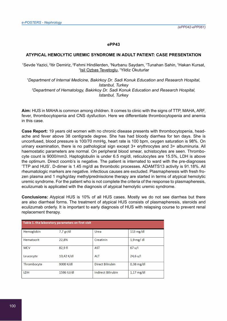
100
e-POSTERS - Nephrology(ePP042-ePP061)
ePP43
ATYPICAL HEMOLYTIC UREMIC SYNDROME IN ADULT PATIENT: CASE PRESENTATION
1Sevde Yazici, 2Itir Demiriz, 2Fehmi Hindilerden, 1Nurbanu Saydam, 1Tunahan Sahin, 1Hakan Kursat, 1Isil Ozbas Tevetoglu, 1Yildiz Okuturlar
1Department of Internal Medicine, Bakirkoy Dr. Sadi Konuk Education and Research Hospital, Istanbul, Turkey
2Department of Hematology, Bakirkoy Dr. Sadi Konuk Education and Research Hospital, Istanbul, Turkey
Aim: HUS in MAHA is common among children. It comes to clinic with the signs of TTP, MAHA, ARF, fever, thrombocytopenia and CNS dysfuction. Here we differentiate thrombocytopenia and anemia in this case.
Case Report: 19 years old women with no chronic disease presents with thrombocytopenia, head-ache and fever above 38 centigrade degree. She has had bloody diarrhea for ten days. She is unconfused, blood pressure is 100/70 mmHg, heart rate is 100 bpm, oxygen saturation is 98%. On urinary examination, there is no pathological sign except 3+ erythrocytes and 3+ albuminuria. All haemostatic parameters are normal. On peripheral blood smear, schistocytes are seen. Thrombo-cyte count is 9000/mm3. Haptoglobulin is under 6.5 mg/dl, reticulocytes are 15.5%, LDH is above the optimum. Direct coomb’s is negative. The patient is internated to ward with the pre-diagnoses ‘TTP and HUS’. D-dimer is 1.45 mg/dl as thrombotic processes. ADAMTS13 activity is 91.18%. All rheumatologic markers are negative. infectious causes are excluded. Plasmapheresis with fresh fro-zen plasma and 1 mg/kg/day methylprednisolone therapy are started in terms of atypical hemolytic uremic syndrome. For the patient who is not complete the criteria of the response to plasmapheresis, eculizumab is applicated with the diagnosis of atypical hemolytic uremic syndrome.
Conclusions: Atypical HUS is 10% of all HUS cases. Mostly we do not see diarrhea but there are also diarrheal forms. The treatment of atypical HUS consists of plasmapheresis, steroids and eculizumab orderly. It is important to early diagnosis of HUS with relapsing course to prevent renal replacement therapy.

101
e-POSTERS - Nephrology(ePP042-ePP061)
ePP44
AN EVALUATION OF OUR ACUTE RENAL FAILURE CASES ADMITTED FROM THE EMERGENCY DEPARTMENT
Bahar Ozdemir, Selda Tas, Kubilay Karaboyun, Gokturk Ogredici, Nadiye Sever, Yildiz Okuturlar
Bakirkoy Dr. Sadi Konuk Education and Research Hospital, Department of Internal Medicine, Instabul, Turkey
Aim: Acute renal failure (ARF) is a syndrome characterized by a rapid decrease in hours or weeks in glomerular filtration rates and the accumulation of nitrogen breakdown products. We analyzed 50 patients presenting to the emergency department and admitted to the internal diseases clinic.
Materials and Methods: We retrospectively analyzed data from 50 patients presenting to the emer-gency department of a research and training hospital and admitted to the internal diseases depart-ment.
Results: 25 patients were female and 25 were male, with a mean age of 67.6 years. Thirty-two patients were aged over 65. ARF was present in 28 patients, 14 of whom had chronic kidney fail-ure (CKF). Acidosis was present in 20 patients, nine of whom had CKF-based ARF (four hyperpo-tassemic), nine had ARF (three hyperpotassemic) and two had CKF (one hyperpotassemic). Six of the 28 patients were admitted for dialysis with acute dialysis indication. Three of the patients admit-ted for dialysis were enrolled on a routine dialysis program; two died and one was referred to the intensive care unit. Infection was involved in the etiology of 10 of the 28 ARF cases, hypervolemia in six and hypovolemia in 12.
Conclusion: Since the great majority of cases of community-acquired ARF are secondary to a de-crease in volume, potentially reversible causes are present in 90% of cases presenting to the emer-gency department. The most common causes of ARF-related mortality in the presence of dialysis were sepsis and cardiopulmonary failure. Well-designed studies with large patient groups are now needed.
102
e-POSTERS - Nephrology(ePP042-ePP061)
ePP45
ACUTE KIDNEY INJURY DUE TO ACYCLOVIR IN A 30-YEAR-OLD PATIENT
Stavroula-Panagiota Lontou, Angeliki Tsifi, Marina Mantzourani, Sevastianos Chatzidavid,
Marina Skouloudi, Maria Triantafyllou, Maria Tampaki, Michael Samarkos
1st Department of Internal Medicine, “Laikon” General Hospital of Athens, Athens, Greece
Background: Acyclovir is an antiviral drug generally well tolerated. However, in certain cases it can induce acute kidney injury within 24-48 hours of its administration, as indicated by a rapid rise in the serum creatinine. Hemodialysis may be needed in severe cases, although an accurate and rapid diagnosis and a treatment involving intravenus fluids and furosemide are of extreme importance and often can reverse the drug’s nephrotoxicity.
Case Report: The present study reports an interesting case of a 30-year-old male, with no known health problems, who was admitted to our hospital due to fever, intense headache with vomiting and sensitivity to light. After undergoing a brain CT scan and a lumbar puncture, whose results were indicative of viral meningitis, he was started on treatment with intravenous (iv) acyclovir. The pa-tient gradually improved, but his renal function began to worsen and eventually his creatinine levels reached a peak of 8.35 mg/dl over the following 4 days. The iv acyclovir was immediately discon-tinued. Aggressive hydration and diuretics such as furosemide were given to the patient in order to increase his urine output over 150 ml/hr and his renal function normalized over the following week.
Conclusion: This case reminds the physicians once again of the importance of a close clinical and laboratory monitoring of the hospitalized patients under treatment with acyclovir, which in turn may prevent any unfortunate side effects and at the same time protect the patients’ renal function.
103
e-POSTERS - Nephrology(ePP042-ePP061)
ePP46
PERITONEAL ULTRAFILTRATION: AN INCREASINGLY DEVELOPING ALTERNATIVE FOR THE MANAGEMENT OF REFRACTORY CONGESTIVE HEART FAILURE
1Carmen Collado Moreno, 2Virginia Pérez Vázquez, 3Pedro Luis Quirós Ganga,
1Ramón Fernando García González, 1Francisco José Morales Ponce
1Cardiology, Puerto Real Universitary Hospital, Cádiz, Spain 2Internal Medicine, Puerto Real Universitary Hospital, Cádiz, Spain
3Nephrology, Puerto Real Universitary Hospital, Cádiz, Spain
Background: Refractory congestive heart failure (RCHF) is a major cause of mortality and morbidity in the world. The aim of peritoneal ultrafiltration (UF) is to improve quality of life in patients with RCHF avoiding frequently hospital readmission and restoring their personal autonomy.
Methods: 15 patients meeting the inclusion criteria were included in the peritoneal dialysis program. Men 73,3 % with a mean age of 66,2 years. Ultrafiltration with PD was tried in three other patients in whom peritoneal catheter was placed but during the maduration periode two of them died for cardiac causes and other one did not perform PD due to abdominal mechanical problems. Once included, the tenckoff catheter was placed and maturation technique was carried out for two weeks. Subse-quently they underwent exchanges of icodextrin +/- dextrose according to the presence of renal disease. Our patients completed the Short Form-36 Health Survey (SF-36) before and after they underwent PD ultrafiltration so we could analyze how their quality of life improved.
Results: Outcomes of this program after a year of follow-up (Table 1).
Conclusions: Peritoneal ultrafiltation helps patients with refractory congestive heart failure to im-prove their quality of life avoiding frequent hospital readmissions and restoring their personal au-tonomy.
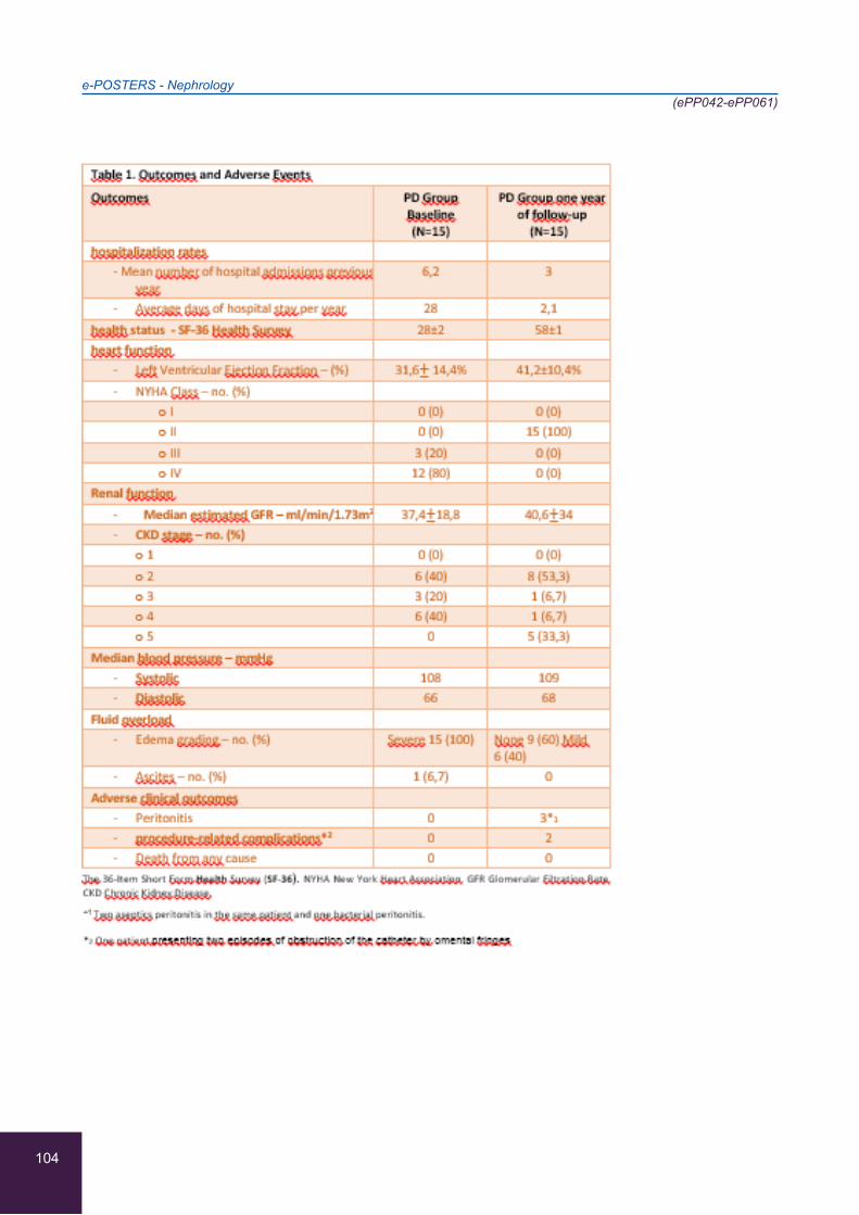
104
e-POSTERS - Nephrology(ePP042-ePP061)
105
e-POSTERS - Nephrology(ePP042-ePP061)
ePP47
A CASE REPORT: CHRONIC PHIMOSIS AS A CAUSE OF RENAL REPLACEMENT THERAPY
1Liga Jursevica, 1Liga Smala, 2Linda Kucane, 3Aivars Petersons
1Internal medicine, Pauls Stradins Clinical University hospital, Riga, Latvia, University of Latvia 2Unit of Nephrology, Pauls Stradins Clinical University Hospital, Riga, Latvia
3Head of Hospital and Nephrology Department, Pauls Stradins Clinical University Hospital, Riga, Latvia
Background: True phimosis is inability to retract the foreskin. This clinical case represents that chronic, untreated phimosis may be a cause of terminal kidney failure.
Case report: A 23-year-old man with phimosis was admitted to Emergency ward in a critical general condition. He had severe dyspnea, Glasgow coma scale 12 points. His creatinine level was 1443 (44 - 97 mkmol/ml) and glomerulal filtration rate (GFR) 4 ml/min/1,73 m2. Because of renal failure and hy-perhydration patient had anasarca with pulmonary edema and secondary dilatation cardiomiopathy with ejection fraction 20%, severe anaemia and arterial hypertension. CT showed hydronephrosis in both kidneys grade 2 - 3 and thin kidney parenchyma. Patient admitted that he had problems with urination for several years and developed progressive shortness of breath in last months. Circumci-sion was performed and patient spent one week in intensive care unit with continuous haemodialy-sis. MRI of heart was performed to exclude untreated heart pathology and showed no pathological changes. 3 months later patient was discharged and is on peritoneal dialysis.
Conclusion: Phimosis may be a cause of chronic kidney desease which leads to chronical renal replacement therapy.
106
e-POSTERS - Nephrology(ePP042-ePP061)
ePP48
SJÖGREN’S SYNDROME AND ACUTE RENAL FAILURE
1Carolina Luque-Amado, 1Eva Lozano-Loaiza, 1Gloria Perez-Vazquez, 2Esther Rubio-Martin,
3Juan-Antonio Jimenez-Casas, 3Luis Garcia-Aragon
1Internal Medicine Unit, Hospital Punta Europa, Algeciras, Spain 2Nephrology Unit, Hospital Punta Europa, Algeciras, Spain
3Primary Care, Area Gestion Sanitaria Campo de Gibraltar, Spain
56-year-old woman with primary Sjögren’s syndrome diagnosed in 1996 had undergone xerostomia, xerophthalmia, asthenia, non-erosive polyarthritis in hands with functional limitation, lymphopenia, hyperganmaglobulinemia, anemia and hypocomplementemia, on treatment with indomethacin and corticotherapy. She has referred pain after dental implant for 3 months, is associated with edema, difficulty in eating and dyspeptic symptoms. An increase in prednisone at 30 mg/d and indomethacin at 50 mg/8 h was decided. Two days later he went to Emergency for marked asthenia, decreased diuresis and dark urine. Upon examination, tachycardia and marked hypoventilation of pulmonary bases were noted. Analytical hemoglobin of 9.2 mg/dL, Leukocytes 6200/dl and Platelets 125000/dl was performed. Creatinine 2.9 mg / dl, Potassium 6.7 mmol/L and metabolic acidosis. Chest x-ray with bilateral pleural effusion. Echocardiogram with moderate pericardial effusion with telesystolic right atrial collapse and LVEF 70%. Rheumatoid factor 113 mg/dl, complement C4 1.6 mg/dl, C3 69 mg/dl. ANA positive, Anti Ro positive. The patient suffers renal failure requiring hemodialysis and is prescribed methylprednisolone and cyclophosphamide. In the renal biopsy, the glomeruli were ob-served with massive occlusion of the capillary lumen by proteinaceous PAS material. With positive immunofluorescence for IgM, C3 Kappa and lambda and negative for fibrin.
This together the positive determination to 50% of cryoglobulins in blood made us reach the diagno-sis; Cryoglobulinemic glomerulopathy.
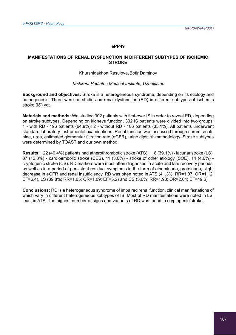
107
e-POSTERS - Nephrology(ePP042-ePP061)
ePP49
MANIFESTATIONS OF RENAL DYSFUNCTION IN DIFFERENT SUBTYPES OF ISCHEMIC STROKE
Khurshidakhon Rasulova, Botir Daminov
Tashkent Pediatric Medical Institute, Uzbekistan
Background and objectives: Stroke is a heterogeneous syndrome, depending on its etiology and pathogenesis. There were no studies on renal dysfunction (RD) in different subtypes of ischemic stroke (IS) yet.
Materials and methods: We studied 302 patients with first-ever IS in order to reveal RD, depending on stroke subtypes. Depending on kidneys function, 302 IS patients were divided into two groups: 1 - with RD - 196 patients (64.9%); 2 - without RD - 106 patients (35.1%). All patients underwent standard laboratory-instrumental examinations. Renal function was assessed through serum creati-nine, urea, estimated glomerular filtration rate (eGFR), urine dipstick-methodology. Stroke subtypes were determined by TOAST and our own method.
Results: 122 (40.4%) patients had atherothrombotic stroke (ATS), 118 (39.1%) - lacunar stroke (LS), 37 (12.3%) - cardioembolic stroke (CES), 11 (3.6%) - stroke of other etiology (SOE), 14 (4.6%) - cryptogenic stroke (CS). RD markers were most often diagnosed in acute and late recovery periods, as well as in a period of persistent residual symptoms in the form of albuminuria, proteinuria, slight decrease in eGFR and renal insufficiency. RD was often noted in ATS (41.3%; RR=1.07; OR=1.12; EF=6.4), LS (39.8%; RR=1.05; OR=1.09; EF=5.2) and CS (5.6%; RR=1.98; OR=2.04; EF=49.6).
Conclusions: RD is a heterogeneous syndrome of impaired renal function, clinical manifestations of which vary in different heterogeneous subtypes of IS. Most of RD manifestations were noted in LS, least in ATS. The highest number of signs and variants of RD was found in cryptogenic stroke.
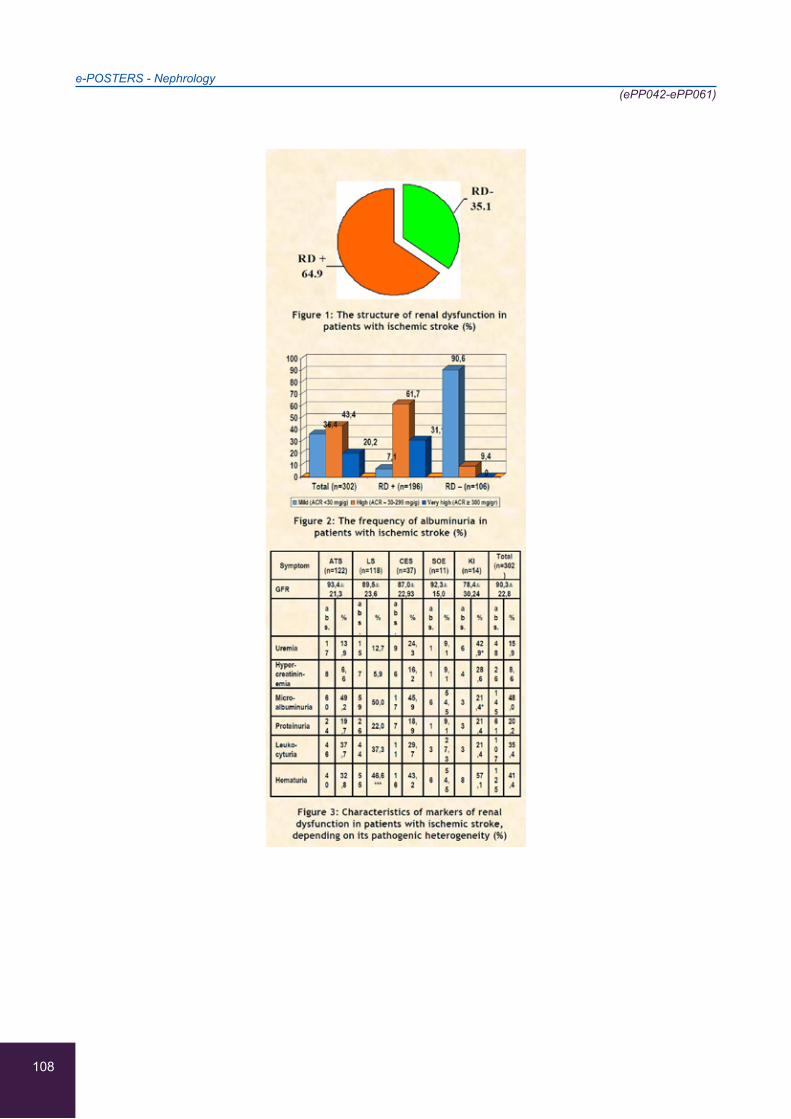
108
e-POSTERS - Nephrology(ePP042-ePP061)
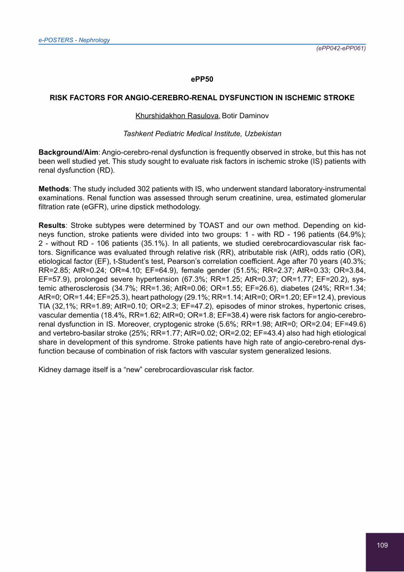
109
e-POSTERS - Nephrology(ePP042-ePP061)
ePP50
RISK FACTORS FOR ANGIO-CEREBRO-RENAL DYSFUNCTION IN ISCHEMIC STROKE
Khurshidakhon Rasulova, Botir Daminov
Tashkent Pediatric Medical Institute, Uzbekistan
Background/Aim: Angio-cerebro-renal dysfunction is frequently observed in stroke, but this has not been well studied yet. This study sought to evaluate risk factors in ischemic stroke (IS) patients with renal dysfunction (RD).
Methods: The study included 302 patients with IS, who underwent standard laboratory-instrumental examinations. Renal function was assessed through serum creatinine, urea, estimated glomerular filtration rate (eGFR), urine dipstick methodology.
Results: Stroke subtypes were determined by TOAST and our own method. Depending on kid-neys function, stroke patients were divided into two groups: 1 - with RD - 196 patients (64.9%); 2 - without RD - 106 patients (35.1%). In all patients, we studied cerebrocardiovascular risk fac-tors. Significance was evaluated through relative risk (RR), atributable risk (AtR), odds ratio (OR), etiological factor (EF), t-Student’s test, Pearson’s correlation coefficient. Age after 70 years (40.3%; RR=2.85; AtR=0.24; OR=4.10; EF=64.9), female gender (51.5%; RR=2.37; AtR=0.33; OR=3.84, EF=57.9), prolonged severe hypertension (67.3%; RR=1.25; AtR=0.37; OR=1.77; EF=20.2), sys-temic atherosclerosis (34.7%; RR=1.36; AtR=0.06; OR=1.55; EF=26.6), diabetes (24%; RR=1.34; AtR=0; OR=1.44; EF=25.3), heart pathology (29.1%; RR=1.14; AtR=0; OR=1.20; EF=12.4), previous TIA (32,1%; RR=1.89; AtR=0.10; OR=2.3; EF=47.2), episodes of minor strokes, hypertonic crises, vascular dementia (18.4%, RR=1.62; AtR=0; OR=1.8; EF=38.4) were risk factors for angio-cerebro-renal dysfunction in IS. Moreover, cryptogenic stroke (5.6%; RR=1.98; AtR=0; OR=2.04; EF=49.6) and vertebro-basilar stroke (25%; RR=1.77; AtR=0.02; OR=2.02; EF=43.4) also had high etiological share in development of this syndrome. Stroke patients have high rate of angio-cerebro-renal dys-function because of combination of risk factors with vascular system generalized lesions.
Kidney damage itself is a “new” cerebrocardiovascular risk factor.
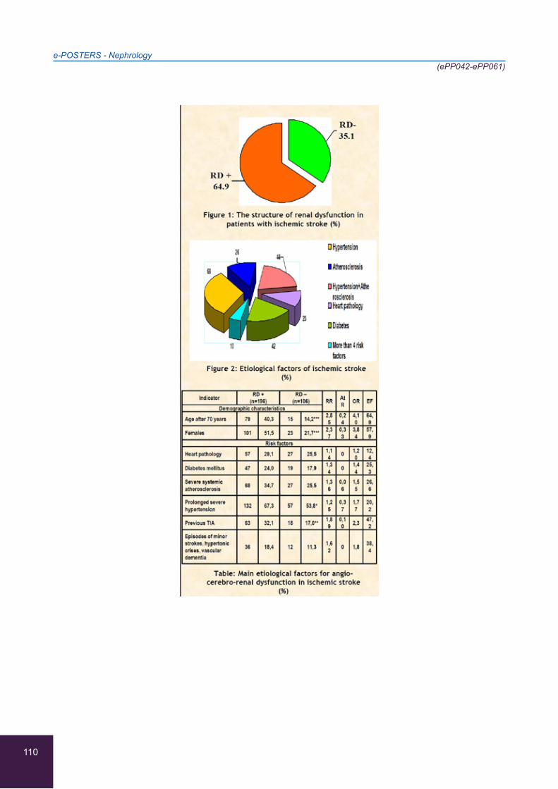
110
e-POSTERS - Nephrology(ePP042-ePP061)

111
e-POSTERS - Nephrology(ePP042-ePP061)
ePP51
CORRELATION OF MITRAL, AORTAL, TRICUSPID CARDIAC VALVE CALCIFICATIONS WITH NUTRITIONAL, INFLAMMATORY AND OTHER BIOCHEMICAL PARAMETERS IN
HEMODIALYSIS PATIENTS IN GREECE
1Petrini Plytzanopoulou, 1Olga Ellina, 1Panayiotis Politis, 3Ioanna Kokores, 1Georgios Tserkezis,
1Christoforos Parisis, 2Penelope Paraskevopoulou, 2Meropi Gika
1Dialysis Unit, “Konstantopouleio” General Hospital, Athens, Greece 2Biochemical department, “Konstantopouleio” General Hospital, Athens, Greece
3Department of Economics, University Piraeus, Piraeus, Greece
Background: Cardiac valve calcification (CVC) is a very common disorder in hemodialysis patients and it has been recognized as an important risk factor for cardiovascular morbidity and mortality.
Aim: To investigate the factors associated with (CVC) in hemodialysis patients.
Methods: In 42 patients (aged 72.97±11.65, duration of dialysis 4.8±4.1 years), Valve Calcifica-tions (VC) were detected by using bidimensional echocardiography. Biochemical parameters such as CRP, PAI-1, IL6 ferritin, transferrin, total proteins albumin, SGA score, PTH, 25(OH)D, calcium, phosphorus, total cholesterol, HDL, LDL, triglycerides were measured and investigated in relation to VC. All correlations were performed by using Pearson, Spearman, Partial Coefficient tests. Ordered logistic regression analysis was conducted to estimate the relative importance of the pre-mentioned risk factors.
Results: Regression analysis showed that total cholesterol decreased the risk in both mitral (OR=0.81, p=0.04) and aortal calcification (OR=0.71, p=0.01). HDL tended to increase the risk in mitral (OR=1.16, p=0.08) and in aortal (OR=1.25, p=0.04), whereas LDL also increased the risk in both valves (mitral OR=1.24, p=0.05, aortal OR=1.46, p=0.01). On the contrary, concerning tricus-pid calcification, HDL reduced the risk (OR=0.65, p=0.03) and LDL also tended to reduce the risk (OR=0.73, p=0.08). Total proteins, and malnutrition increased significantly the risk in mitral and aor-tal calcification.
Conclusions: Malnutrition, elevated serum levels of total proteins, ferritin, CRP seemed to be ag-gravating risk factors for mitral and aortal calcification. Total cholesterol presented a rather protective effect against mitral and aortal calcification, whereas lipoprotein fractions tend to appear a contradic-tory effect on various valve calcifications.
112
e-POSTERS - Nephrology(ePP042-ePP061)
ePP52
RELATIONSHIP BETWEEN VASCULAR, VALVE CALCIFICATION AND LEFT VENTRICULAR HYPERTROPHY IN PATIENTS WITH CHRONIC DIALYSIS TREATMENT
1Merita Rroji, 1Saimir Seferi, 2Majlinda Cafka, 1Nereida Spahia, 1Erjola Likaj, 1Alma Idrizi,
1Myftar Barbullushi
1Department of Nephrology, UHC “Mother Tereza”, Tirana, Albania 2Department of Cardiology, UHC “Mother Tereza”, Tirana, Albania
Introduction: Vascular calcifications (VC), cardiac valve calcification (CVC) and left ventricular hypertrophy (LVH) are frequently observed in chronic kidney disease (CKD) patients. The aim of this study was to investigate the relationship between VC, CVC and LVH in CKD patients.
Methods: We performed a cross-sectional study in 85 stable prevalent hemodialysis patients and 41 PD patients being in RRT for more than 6 months during December 2012-March 2013. Two-dimensional echocardiography was performed by an experienced cardiologist who was blinded to all clinical details of patients. Plain X-ray images of lateral lumbar spine from all subjects were studied to obtain images of the lower abdominal aorta using semiquantitative scores as described by Kaup-pila et al.
Results: Left ventricular hypertrophy was present in 86.7%, concentric hypertrophy was found in 64 (50.7%) and eccentric hypertrophy in 46 patients (36.7%). Among the 126 patients, 70.5% of the patients have one valve calcified and 38.3% of the patients have both valve calcified. Eighty-seven patients (69.4%) were identified with aortic abdominal calcification, and the mean Kauppila score was 4.91 ± 4.05. The mean Kauppila score significantly associated presence of CVC (r= 0.6, p=0.03). Logistic regression analysis found Kauppila score as an independent risk factor associated with LVMI (odds ratio [OR], 1.07; 95% confidence interval (CI), 1.01 to 1.29; P = 0.001).
Conclusion: Vascular, valve calcifications are highly prevalent in dialysis patients and independ-ently associated with LVMI. Vascular calcifications and LVM interrelated together and both contribute to CV morbidity in dialysis populations.
113
e-POSTERS - Nephrology(ePP042-ePP061)
ePP53
UNILATERAL RENAL AGENESIS AND THE AWARENESS OF MOSTYN EMBREY SYNDROME
Aamir Al Mosawi
Baghdad Medical City, Baghdad, Iraq
Background/Aim: Mostyn Embrey syndrome is a rare disorder associated with unilateral renal agenesis and malformations of the female reproductive tract. Delayed diagnosis is associated with serious diagnostic difficulties that may lead to inappropriate management including harmful surgery. The aim of this paper is to describe the first case of this rare syndrome in Iraq and to make a recom-mendation that pediatricians, and pediatric nephrologists need to be aware of this syndrome to avoid unexpected presentations which may lead to delayed diagnosis and some times to inappropriate management.
Case report: An 18-years old female presented with acute abdominal pain and poor urine output associated with retention of urine; about three years after the onset of menses. The pain was radiat-ing to the back and pelvis. Her menstrual cycles started at the age of fifteen, and were lasting seven days. Cycles were associated with normal flow. The first physician the patient consulted considered the diagnosis of uterine tumor and the need for hysterectomy, but the family of the patient consulted an other physician. Vaginal exam was performed and showed bulging left vaginal wall. The diagnosis of hematocolpos was made. A clotted blood was drained by trans- vaginal approach. Abdominal MRI showed left renal agenesis, and two uterine bodies, cervices, and vaginas with normal myometrium and endometrium (uterine didelphys), and normal ovaries.
Conclusion: A greater awareness of this syndrome by pediatricians and pediatric nephrologists should contribute to making early accurate diagnosis and early appropriate management before puberty.
114
e-POSTERS - Nephrology(ePP042-ePP061)
ePP54
THE MENTAL HEALTH AND HAEMODIALYSIS
1Evangelia Zacharioglou, 1Andonis Vithoulkas, 1Andonia Avgoustinou, 1Anna Likouresi,
2Tsvetan Bankov, 1Irini Katsarou
1Renal Unit, General Hospital Zakynthos, Greece2Philologist
Chronic, end-stage renal failure is a non-curable health disease. Mental health contributes to the quality of life of these patients.
Method: The research was aimed at the estimation of mental health’s level of patients in Haemo-dialysis Unit. A proper questionnaire, known as SF 36 (Medical Outcome Study short form Health Survey, 36 items), was delivered, adjusted to facilitate elderly patients
Patients: we studied 26 patients: 15 males and 11 females, aged 38 to 94 years old.
Results: Patients have adjustment difficulties regarding the nature of the disease, such as a per-sistent sense of fear and anxiety. Concerning the social attitude and communication they exhibit weakness to respond to family obligations and non-participation in social activities. Women appear much more stressed about family responsibilities. All patients experienced great anxiety upon join-ing in dialysis. Approximately 92% of patients are not keen on friendships, 27% of them indicate pain during the initial puncture and two patients had pain throughout the session. In addition, 54% of them experiences anxiety, during every session of haemodialysis. At the end of dialysis, they feel too much physical fatigue. Before haemodialysis, seven patients had a regular workday; two are still working. Meanwhile, they experience very lower levels of energy and vitality. In conclusion, the qual-ity of life in every chronic disease depends on the specific nature of the disease and its treatment. The assessment of mental health is an essential tool and a basic endpoint of the followed therapeutic schedule.
115
e-POSTERS - Nephrology(ePP042-ePP061)
ePP55
ACUTE RENAL FAILURE AND SEVERE LACTIC ACIDOSIS WITH METFORMIN: A CASE REPORT
1Elena Tsokola, 1Afroditi Roumpou, 2Tamta Sirbilantze, 1Ioanna Papaioannou, 2Christina Kordali,
2Andreas Poulas, 1Christos Lampropoulos
1Department of Internal Medicine, Argolidos General Hospital, Nafplion, Greece 2Department of Internal Medicine, Argolidos General Hospital, Argos, Greece
Background/Aim: Metformin is a rare cause of lactic acidosis in patients with predisposing factors for acidosis. Purpose of this presentation is to present a case of severe lactic acidosis in a lady, after metformin administration.
Case Report: A female patient, 70 years old, presented with vomits and abdominal pain the last 5 days. The first day she had revisited the emergency department without any clinical or laboratory findings. Her medical hispory included dementia and diabetes mellitus under metformin/vildagliptin. The patient was disoriented, afebrile, tachypnoic, oliguric with diffuse abdominal tenderness. A blood gas analysis was performed with severe lactic acidosis (>15mmol/Lt), pΗ=6.84, PC02=7mmHg, p02=133mmHg, glucose=57mg/dl, HC03<3mmol/Lt. In laboratory examination, there were creatinine=5.3mg/dl, urea=152mg/dl, WBC=17.000μL, ESR=43mm/h, Ht=37.3%, Na=133mmol/Lt, K=4.4mmol/dl. Bicarbonates 4.8% (4fl), fluids (3lt), dopamine (diuretic dose) and furosemide 160mg were administered with no improvement and the patient performed hemodialysis. Blood and urine cultures were negative. Meropenem was administered in case of severe sepsis. During the first day of admission, ischemic lesions of ECG were noticed which subsided after intravenous glyceryl trini-trate administration. Ultrasounds of heart and abdomen, thoracic x-ray and abdominal CT were nor-mal. Low-grade fever was noticed during the first day while she remained afebrile for the rest of hos-pitalization. Nevertheless, due to persistent lactic acidosis and low bicarbonates despite renal failure improvement, she submitted herself to 3 hemodialyses. There was a gradual improvement of mental condition and the patient was discharged after 8 days with urea=59mg/dl and creatinine=1.6mg/dl. In her follow-up, 20 days later, creatinine was 1mg/dl.
116
e-POSTERS - Nephrology(ePP042-ePP061)
ePP56
SECONDARY ACUTE KIDNEY FAILURE A SCLERODERMIC CRISIS
1Gloria Perez-Vazquez, 2Esther Rubio-Muñoz, 3Juan Antonio Jimenez-Casas, 1Eva Maria Lozano-Loaiza, 1Carolina Luque-Amado, 3Luis García-Aragon
1Internal Medicine Unit, Hospital Punta Europa, Algeciras, Spain 2Nephrology Unit, Hospital La Linea, La Linea de la Concepcion, Spain
3Primary Care, Area Gestion Sanitaria Campo de Gibraltar, Gibraltar, Spain
Case Report: A 48-year-old male admitted to the Nephrology Service with abdominal pain, diarrheic bowel movements, and acute oligoanuric renal failure with progressive worsening of renal function requiring renal replacement therapy. As a personal history, a diffuse systemic scleroderma of recent diagnosis by skin biopsy and fulfilling criteria according to the ACR/EULAR 2013 classification, with polyarticular, esophageal, pulmonary and cutaneous involvement. The examination of the abdo-men was depressible with pain on palpation without signs of peritoneal irritation, accelerated transit. Blood analysis hemoglobin 10.1g/dl, hematocrit 30%, leukocytes 13200/dl, platelets 84000/dl. The smear is compatible with microangiopathic hemolytic anemia. Urea 291mg/dl, creatinine 8.51mg/dl, normal ions, total proteinY4.2g/dl, albumin 2.5g/dl, LDH 982U/L, haptoglobin 108md/dl, ADAMTS negative, IgG 722, IgM 42, IgA 33. Negative immunological study. Systematic urine and sediment proteins 500mg/dl, erythrocytes 2010/ul, ratio Alb/Cr 333mg/g. Abdominal ultrasound with kidneys with a discrete increase in cortical echogenicity, although with preservation of cortico-medullary dif-ferentiation. Renal Doppler records flow with normal wave morphology and preserved resistance indices. Echocardiogram shows severe global chronic pericardial effusion with mild tamponade data. MRI-scan revealed gliotic-like lesions as sequelae of vasculitis due to systemic scleroderma. Initial diagnosis of renal scleroderma crisis with normal blood pressure levels, treated with angiotensin-converting enzyme inhibitors, corticosteroids and cyclophosphamide. The risk factors of the renal crisis are the use of corticosteroids at high doses, diffuse and rapidly progressive skin involvement, and the winter months.
Conclusion: This case is interesting because of normotension presentation, and remember that corticosteroids should be used with great caution in scleroderma crisis.

117
e-POSTERS - Nephrology(ePP042-ePP061)
ePP57
COLYMICIN-INDUCED ACQUIRED BARTTER-LIKE SYNDROME: A DIAGNOSTIC CHALLENGE OF A RARE ENTITY
1Paraskevi Papaioannou, 2Ioannis Gavalas, 1Sotiria Kotsanti, 1Aspasia Koudouna, 1Paraskevi Kontrafouri, 1Aikaterini Bitsani, 1Maria Mylona
11st Department of Propaedeutic & Internal Medicine, “Laiko” General Hospital, Athens University Medical School, Athens, Greece
2Nephrology Department and Renal Transplantation Unit, “Laiko” General Hospital, Athens, Greece
Introduction/Aim: Acquired Bartter-like syndrome (BLS), a very rare condition, characterized by hypokalemic metabolic alkalosis, hypocalcemia, hypomagnesemia and normal kidney function, can be induced by diuretics or antibiotics. Herein, we present a rare case of colimicin- induced BLS in a young female patient with thrombotic thrombocytopenic purpura (TTP) and bacteremia. Acquired BLS -though a rare entity- should be suspected in patients with sustained electrolyte disturbances and certain antibiotic treatments such as colimicin.
Case description: A 30-year old Caucasian woman, with a history of class III obesity and TTP, was transferred to our clinic after a two-week hospitalization in our ICU department due to TTP relapse. She was diagnosed with deep vein thrombosis of her right leg and concomitant severe cellulitis. She was febrile and Acinetobacter baumanii was isolated from two blood cultures. She was treated with Meropenem 2 grx3IV and Colimicin 4.5x10⁶x2 IV. From the 3rd day of antibiotic treatment, the patient presented with sustained hypokalemic metabolic alkalosis, hypocalcemia and hypomagnesemia. Serum creatinine, sodium and chloride levels were within the normal limits, while urine analysis re-vealed hypercalciuria and significant renal potassium loss without natriuresis. Renal ultrasonography and abdomen CT scan were also normal. Serum renin and aldosterone levels were high. Electrolyte levels were difficulty kept within normal ranges with IV supplementation. Since colimicin was thought to be the cause of the patient’ s syndrome, it was discontinued after 12 days of IV administration. The patient’ s electrolyte levels returned to normal from the second day of colimicin cessation.
118
e-POSTERS - Nephrology(ePP042-ePP061)
ePP58
AN APPARENT DIAGNOSIS OF ALCOHOL INTOXICATION? NOT SO FAST!
1Christos Pleros, 2Aggeliki Machaira, 2Irini Lagoudaki, 2Loukia Kalogeraki
1Nephrology Department, General Hospital of Chania, Chania, Greece 22nd Unit, Internal Medicine Department, General Hospital of Chania, Chania, Greece
Background: Humans lack the ability to rapidly metabolize D-lactate anions, one of the main prod-ucts of bacterial metabolism. D-lactic acidosis has been well documented in ruminants overfed with grain, often resulting in death of the animals. This rare form of acidosis can occur in humans with the short bowel syndrome when provided with a large carbohydrate load. Acidosis and encephalopathy dominate the clinical picture and can be life threatening in the presence of AKI or CKD.
Case report: A 70-year-old woman presented shortly after midnight with symptoms of inebriation. Her history included hypothyroidism, cholecystectomy and a jejunoileal bypass 40 years ago. Apart from tachypnea, confusion, ataxia and slurred speech, her physical examination was normal. A CT scan of the brain revealed no abnormalities. Arterial blood gas analysis showed a severe high-anion-gap metabolic acidosis with normal lactate levels, and serum chemistry was normal except for mild hypokalemia. She reported having eaten a lot of halva (confection made from tahini or semolina) after dinner and denied consumption of alcohol, drugs and other possible toxins. D-lactic acidosis was diagnosed after excluding all other possible causes of high-anion-gap metabolic acidosis. The patient remained fasted and potassium with bicarbonate were administered i.v. Vancomycin was administered orally to suppress intestinal bacterial overgrowth. Within 18 hours her clinical condition normalized completely.
Conclusion: Routine lactic acid assays measure only L-lactate. Thus, the diagnosis of D-lactic aci-dosis is often one of exclusion and requires a high index of clinical suspicion based on a patient’s history and clinical presentation.
119
ePP59
A RARE CASE OF MERCURY TOXICITY AFTER ABDOMINAL LAVAGE WITH MERCURY SOLUTION
1Tasiopoulou Karmen, 2Krasovskagia Irina, 1Tsantekidou Ellada, 1Pashou Stiliani
1Nephrology Department, “Papanicolaou” General Hospital, Thessaloniki, Greece 21st Department Of Internal Medicine, “Papageorgiou” General Hospital, Thessaloniki, Greece
Backround: Acute mercury toxicity is usually due to accidental ingestion or suicidal attempt. In the past, mercury solutions were used in cancer treatment. Renal injury is due to direct tubular injury, glomerulonephritis or interstitial nephritis.
Case presentation: A 32 old female was transfered in the ICU from the Cancer Hospital because of oliguric acute kidney injury and respiratory distress. Five days before she underwent surgery for ovarian cyst removal which was like to be teratoma (not confirmed by pathologist). During surgery, because of cyst rupture the abdomen was instiled with mercury solution (SUBLIME 1/1000). After excluding obvious causes of AKI (hemorhage, sepsis, drugs) mercury levels were ordered and found extremely high (950μg/l,nr,10). CVVHD was initiated together with chelating therapy (Dimercaptol for 10 days, followed by Dimercaptosuccinic acid-DMSA for 14 days). After her transfer in the Nephrol-ogy Department she continued on line HDF for one month together with another two 10 day courses of DMSA. Blood and urine mercury levels progresivelly decreased. Renal biopsy shown interstitial nephritis and her renal function completely recovered after 50 days. Three months later she was admitted in the Neurology Department because of seizures wich was estimated to be related to mercury toxicity despite noral blood and urine levels. The seizures resolved within three months with valproic acid.
Conclusions: Mecury toxicity after instilation is very rare but lifethreatening condition and for this reason, according to expert opinion this use should be avoided.
e-POSTERS - Nephrology(ePP042-ePP061)
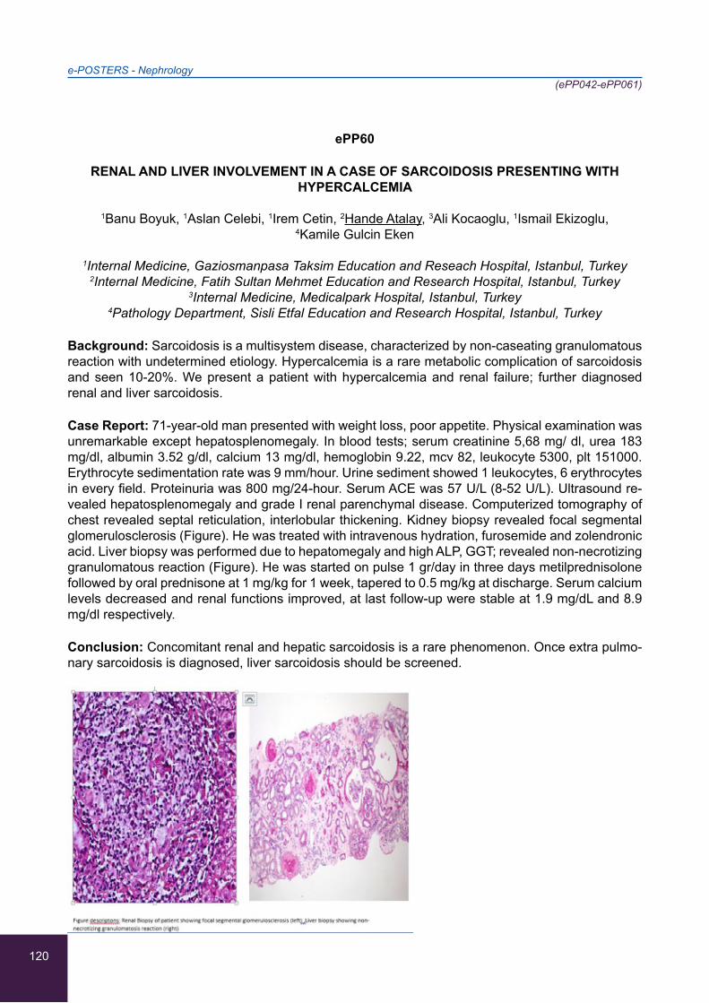
120
ePP60
RENAL AND LIVER INVOLVEMENT IN A CASE OF SARCOIDOSIS PRESENTING WITH HYPERCALCEMIA
1Banu Boyuk, 1Aslan Celebi, 1Irem Cetin, 2Hande Atalay, 3Ali Kocaoglu, 1Ismail Ekizoglu, 4Kamile Gulcin Eken
1Internal Medicine, Gaziosmanpasa Taksim Education and Reseach Hospital, Istanbul, Turkey 2Internal Medicine, Fatih Sultan Mehmet Education and Research Hospital, Istanbul, Turkey
3Internal Medicine, Medicalpark Hospital, Istanbul, Turkey 4Pathology Department, Sisli Etfal Education and Research Hospital, Istanbul, Turkey
Background: Sarcoidosis is a multisystem disease, characterized by non-caseating granulomatous reaction with undetermined etiology. Hypercalcemia is a rare metabolic complication of sarcoidosis and seen 10-20%. We present a patient with hypercalcemia and renal failure; further diagnosed renal and liver sarcoidosis.
Case Report: 71-year-old man presented with weight loss, poor appetite. Physical examination was unremarkable except hepatosplenomegaly. In blood tests; serum creatinine 5,68 mg/ dl, urea 183 mg/dl, albumin 3.52 g/dl, calcium 13 mg/dl, hemoglobin 9.22, mcv 82, leukocyte 5300, plt 151000. Erythrocyte sedimentation rate was 9 mm/hour. Urine sediment showed 1 leukocytes, 6 erythrocytes in every field. Proteinuria was 800 mg/24-hour. Serum ACE was 57 U/L (8-52 U/L). Ultrasound re-vealed hepatosplenomegaly and grade I renal parenchymal disease. Computerized tomography of chest revealed septal reticulation, interlobular thickening. Kidney biopsy revealed focal segmental glomerulosclerosis (Figure). He was treated with intravenous hydration, furosemide and zolendronic acid. Liver biopsy was performed due to hepatomegaly and high ALP, GGT; revealed non-necrotizing granulomatous reaction (Figure). He was started on pulse 1 gr/day in three days metilprednisolone followed by oral prednisone at 1 mg/kg for 1 week, tapered to 0.5 mg/kg at discharge. Serum calcium levels decreased and renal functions improved, at last follow-up were stable at 1.9 mg/dL and 8.9 mg/dl respectively.
Conclusion: Concomitant renal and hepatic sarcoidosis is a rare phenomenon. Once extra pulmo-nary sarcoidosis is diagnosed, liver sarcoidosis should be screened.
e-POSTERS - Nephrology(ePP042-ePP061)
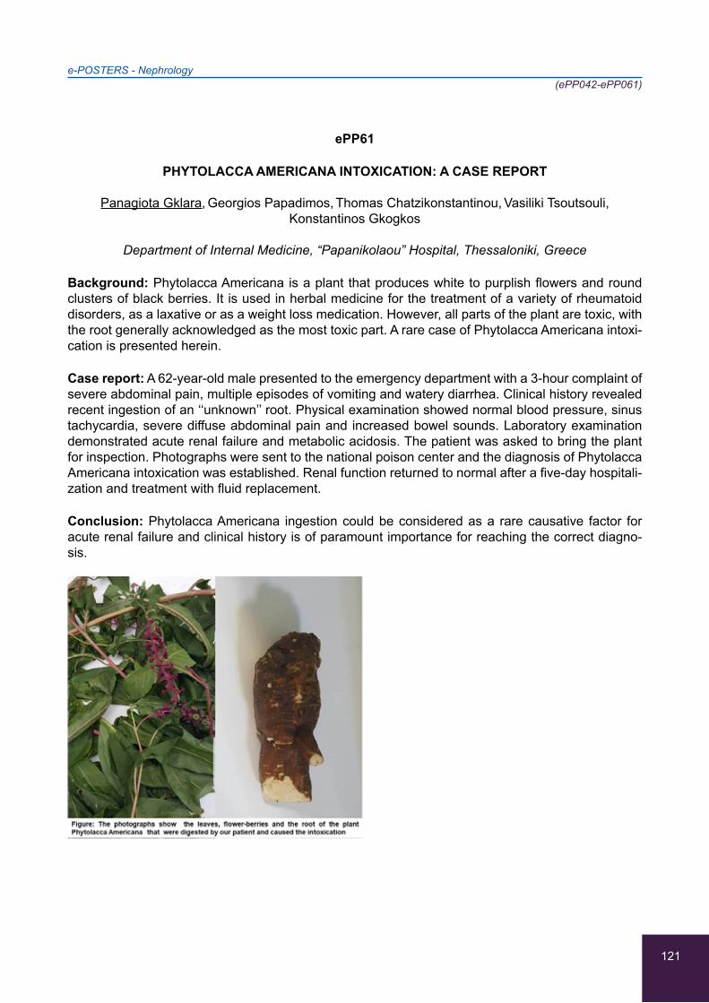
121
ePP61
PHYTOLACCA AMERICANA INTOXICATION: A CASE REPORT
Panagiota Gklara, Georgios Papadimos, Thomas Chatzikonstantinou, Vasiliki Tsoutsouli,
Konstantinos Gkogkos
Department of Internal Medicine, “Papanikolaou” Hospital, Thessaloniki, Greece
Background: Phytolacca Americana is a plant that produces white to purplish flowers and round clusters of black berries. It is used in herbal medicine for the treatment of a variety of rheumatoid disorders, as a laxative or as a weight loss medication. However, all parts of the plant are toxic, with the root generally acknowledged as the most toxic part. A rare case of Phytolacca Americana intoxi-cation is presented herein.
Case report: A 62-year-old male presented to the emergency department with a 3-hour complaint of severe abdominal pain, multiple episodes of vomiting and watery diarrhea. Clinical history revealed recent ingestion of an ‘‘unknown’’ root. Physical examination showed normal blood pressure, sinus tachycardia, severe diffuse abdominal pain and increased bowel sounds. Laboratory examination demonstrated acute renal failure and metabolic acidosis. The patient was asked to bring the plant for inspection. Photographs were sent to the national poison center and the diagnosis of Phytolacca Americana intoxication was established. Renal function returned to normal after a five-day hospitali-zation and treatment with fluid replacement.
Conclusion: Phytolacca Americana ingestion could be considered as a rare causative factor for acute renal failure and clinical history is of paramount importance for reaching the correct diagno-sis.
e-POSTERS - Nephrology(ePP042-ePP061)
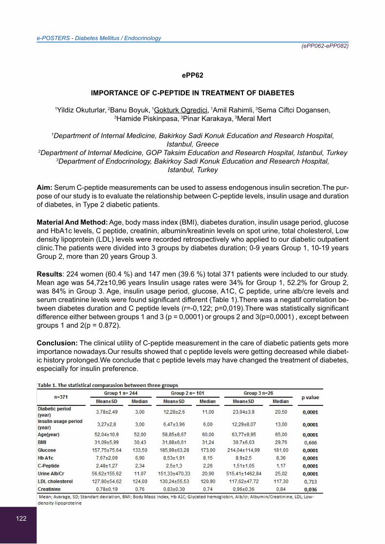
122
ePP62
IMPORTANCE OF C-PEPTIDE IN TREATMENT OF DIABETES
1Yildiz Okuturlar, 2Banu Boyuk, 1Gokturk Ogredici, 1Amil Rahimli, 3Sema Ciftci Dogansen,
3Hamide Piskinpasa, 3Pinar Karakaya, 3Meral Mert
1Department of Internal Medicine, Bakirkoy Sadi Konuk Education and Research Hospital, Istanbul, Greece
2Department of Internal Medicine, GOP Taksim Education and Research Hospital, Istanbul, Turkey 3Department of Endocrinology, Bakirkoy Sadi Konuk Education and Research Hospital,
Istanbul, Turkey
Aim: Serum C-peptide measurements can be used to assess endogenous insulin secretion.The pur-pose of our study is to evaluate the relationship between C-peptide levels, insulin usage and duration of diabetes, in Type 2 diabetic patients.
Material And Method: Age, body mass index (BMI), diabetes duration, insulin usage period, glucose and HbA1c levels, C peptide, creatinin, albumin/kreatinin levels on spot urine, total cholesterol, Low density lipoprotein (LDL) levels were recorded retrospectively who applied to our diabetic outpatient clinic.The patients were divided into 3 groups by diabetes duration; 0-9 years Group 1, 10-19 years Group 2, more than 20 years Group 3.
Results: 224 women (60.4 %) and 147 men (39.6 %) total 371 patients were included to our study. Mean age was 54,72±10,96 years Insulin usage rates were 34% for Group 1, 52.2% for Group 2, was 84% in Group 3. Age, insulin usage period, glucose, A1C, C peptide, urine alb/cre levels and serum creatinine levels were found significant different (Table 1).There was a negatif correlation be-tween diabetes duration and C peptide levels (r=-0,122; p=0,019).There was statistically significant difference either between groups 1 and 3 (p = 0,0001) or groups 2 and 3(p=0,0001) , except between groups 1 and 2(p = 0.872).
Conclusion: The clinical utility of C-peptide measurement in the care of diabetic patients gets more importance nowadays.Our results showed that c peptide levels were getting decreased while diabet-ic history prolonged.We conclude that c peptide levels may have changed the treatment of diabetes, especially for insulin preference.
e-POSTERS - Diabetes Mellitus / Endocrinology(ePP062-ePP082)
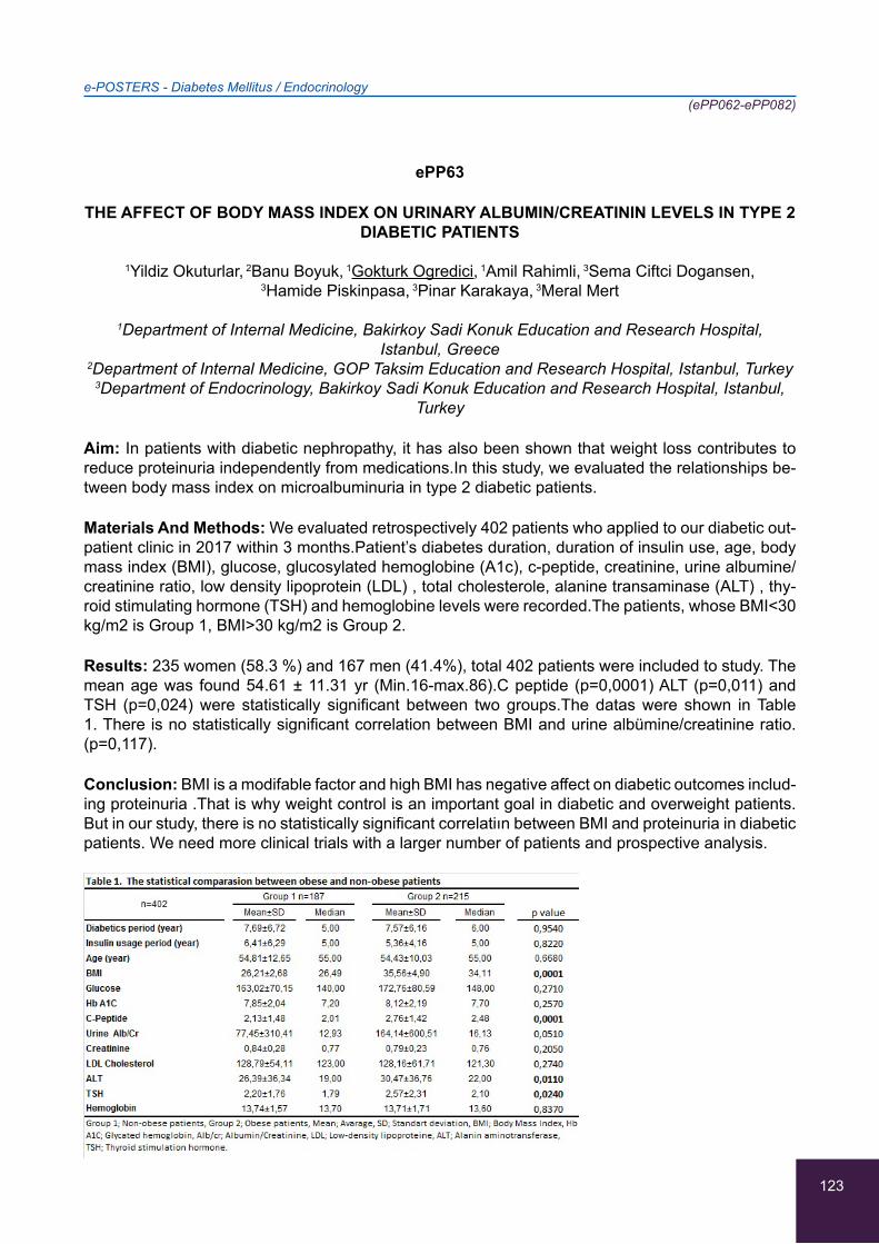
123
ePP63
THE AFFECT OF BODY MASS INDEX ON URINARY ALBUMIN/CREATININ LEVELS IN TYPE 2 DIABETIC PATIENTS
1Yildiz Okuturlar, 2Banu Boyuk, 1Gokturk Ogredici, 1Amil Rahimli, 3Sema Ciftci Dogansen,
3Hamide Piskinpasa, 3Pinar Karakaya, 3Meral Mert
1Department of Internal Medicine, Bakirkoy Sadi Konuk Education and Research Hospital, Istanbul, Greece
2Department of Internal Medicine, GOP Taksim Education and Research Hospital, Istanbul, Turkey 3Department of Endocrinology, Bakirkoy Sadi Konuk Education and Research Hospital, Istanbul,
Turkey
Aim: In patients with diabetic nephropathy, it has also been shown that weight loss contributes to reduce proteinuria independently from medications.In this study, we evaluated the relationships be-tween body mass index on microalbuminuria in type 2 diabetic patients.
Materials And Methods: We evaluated retrospectively 402 patients who applied to our diabetic out-patient clinic in 2017 within 3 months.Patient’s diabetes duration, duration of insulin use, age, body mass index (BMI), glucose, glucosylated hemoglobine (A1c), c-peptide, creatinine, urine albumine/creatinine ratio, low density lipoprotein (LDL) , total cholesterole, alanine transaminase (ALT) , thy-roid stimulating hormone (TSH) and hemoglobine levels were recorded.The patients, whose BMI<30 kg/m2 is Group 1, BMI>30 kg/m2 is Group 2.
Results: 235 women (58.3 %) and 167 men (41.4%), total 402 patients were included to study. The mean age was found 54.61 ± 11.31 yr (Min.16-max.86).C peptide (p=0,0001) ALT (p=0,011) and TSH (p=0,024) were statistically significant between two groups.The datas were shown in Table 1. There is no statistically significant correlation between BMI and urine albümine/creatinine ratio.(p=0,117).
Conclusion: BMI is a modifable factor and high BMI has negative affect on diabetic outcomes includ-ing proteinuria .That is why weight control is an important goal in diabetic and overweight patients.But in our study, there is no statistically significant correlatiın between BMI and proteinuria in diabetic patients. We need more clinical trials with a larger number of patients and prospective analysis.
e-POSTERS - Diabetes Mellitus / Endocrinology(ePP062-ePP082)
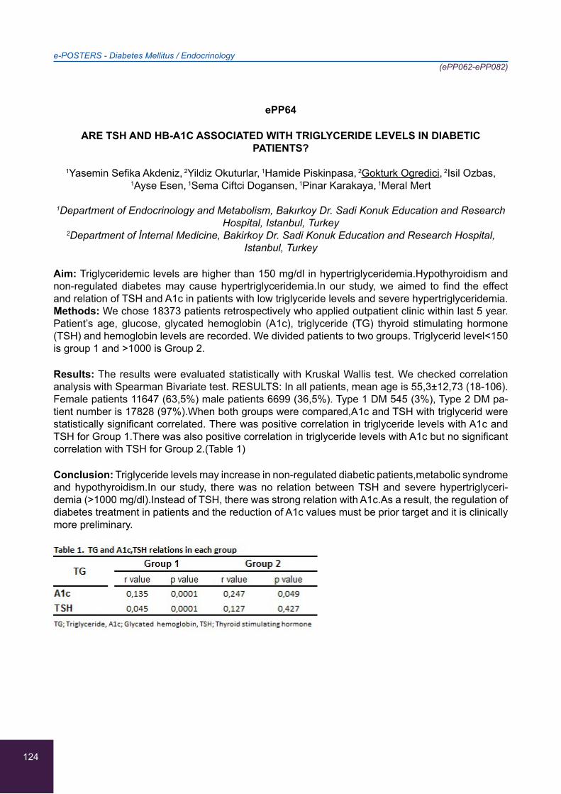
124
ePP64
ARE TSH AND HB-A1C ASSOCIATED WITH TRIGLYCERIDE LEVELS IN DIABETIC PATIENTS?
1Yasemin Sefika Akdeniz, 2Yildiz Okuturlar, 1Hamide Piskinpasa, 2Gokturk Ogredici, 2Isil Ozbas,
1Ayse Esen, 1Sema Ciftci Dogansen, 1Pinar Karakaya, 1Meral Mert
1Department of Endocrinology and Metabolism, Bakırkoy Dr. Sadi Konuk Education and Research Hospital, Istanbul, Turkey
2Department of İnternal Medicine, Bakirkoy Dr. Sadi Konuk Education and Research Hospital, Istanbul, Turkey
Aim: Triglyceridemic levels are higher than 150 mg/dl in hypertriglyceridemia.Hypothyroidism and non-regulated diabetes may cause hypertriglyceridemia.In our study, we aimed to find the effect and relation of TSH and A1c in patients with low triglyceride levels and severe hypertriglyceridemia. Methods: We chose 18373 patients retrospectively who applied outpatient clinic within last 5 year. Patient’s age, glucose, glycated hemoglobin (A1c), triglyceride (TG) thyroid stimulating hormone (TSH) and hemoglobin levels are recorded. We divided patients to two groups. Triglycerid level<150 is group 1 and >1000 is Group 2.
Results: The results were evaluated statistically with Kruskal Wallis test. We checked correlation analysis with Spearman Bivariate test. RESULTS: In all patients, mean age is 55,3±12,73 (18-106). Female patients 11647 (63,5%) male patients 6699 (36,5%). Type 1 DM 545 (3%), Type 2 DM pa-tient number is 17828 (97%).When both groups were compared,A1c and TSH with triglycerid were statistically significant correlated. There was positive correlation in triglyceride levels with A1c and TSH for Group 1.There was also positive correlation in triglyceride levels with A1c but no significant correlation with TSH for Group 2.(Table 1)
Conclusion: Triglyceride levels may increase in non-regulated diabetic patients,metabolic syndrome and hypothyroidism.In our study, there was no relation between TSH and severe hypertriglyceri-demia (>1000 mg/dl).Instead of TSH, there was strong relation with A1c.As a result, the regulation of diabetes treatment in patients and the reduction of A1c values must be prior target and it is clinically more preliminary.
e-POSTERS - Diabetes Mellitus / Endocrinology(ePP062-ePP082)
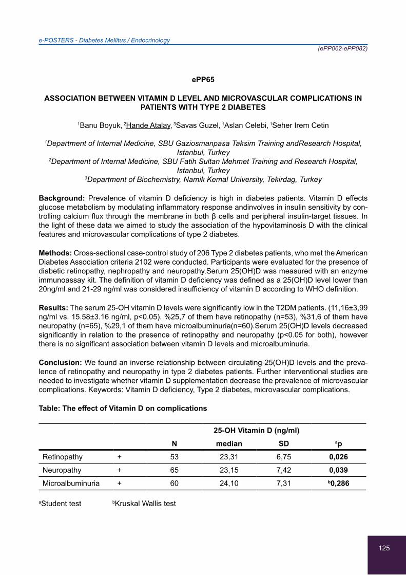
125
ePP65
ASSOCIATION BETWEEN VITAMIN D LEVEL AND MICROVASCULAR COMPLICATIONS IN PATIENTS WITH TYPE 2 DIABETES
1Banu Boyuk, 2Hande Atalay, 3Savas Guzel, 1Aslan Celebi, 1Seher Irem Cetin
1Department of Internal Medicine, SBU Gaziosmanpasa Taksim Training andResearch Hospital, Istanbul, Turkey
2Department of Internal Medicine, SBU Fatih Sultan Mehmet Training and Research Hospital, Istanbul, Turkey
3Department of Biochemistry, Namik Kemal University, Tekirdag, Turkey
Background: Prevalence of vitamin D deficiency is high in diabetes patients. Vitamin D effects glucose metabolism by modulating inflammatory response andinvolves in insulin sensitivity by con-trolling calcium flux through the membrane in both β cells and peripheral insulin-target tissues. In the light of these data we aimed to study the association of the hypovitaminosis D with the clinical features and microvascular complications of type 2 diabetes.
Methods: Cross-sectional case-control study of 206 Type 2 diabetes patients, who met the American Diabetes Association criteria 2102 were conducted. Participants were evaluated for the presence of diabetic retinopathy, nephropathy and neuropathy.Serum 25(OH)D was measured with an enzyme immunoassay kit. The definition of vitamin D deficiency was defined as a 25(OH)D level lower than 20ng/ml and 21-29 ng/ml was considered insufficiency of vitamin D according to WHO definition.
Results: The serum 25-OH vitamin D levels were significantly low in the T2DM patients. (11,16±3,99 ng/ml vs. 15.58±3.16 ng/ml, p<0.05). %25,7 of them have retinopathy (n=53), %31,6 of them have neuropathy (n=65), %29,1 of them have microalbuminuria(n=60).Serum 25(OH)D levels decreased significantly in relation to the presence of retinopathy and neuropathy (p<0.05 for both), however there is no significant association between vitamin D levels and microalbuminuria.
Conclusion: We found an inverse relationship between circulating 25(OH)D levels and the preva-lence of retinopathy and neuropathy in type 2 diabetes patients. Further interventional studies are needed to investigate whether vitamin D supplementation decrease the prevalence of microvascular complications. Keywords: Vitamin D deficiency, Type 2 diabetes, microvascular complications.
Table: The effect of Vitamin D on complications
25-OH Vitamin D (ng/ml)N median SD ap
Retinopathy + 53 23,31 6,75 0,026Neuropathy + 65 23,15 7,42 0,039Microalbuminuria + 60 24,10 7,31 b0,286
aStudent test bKruskal Wallis test
e-POSTERS - Diabetes Mellitus / Endocrinology(ePP062-ePP082)
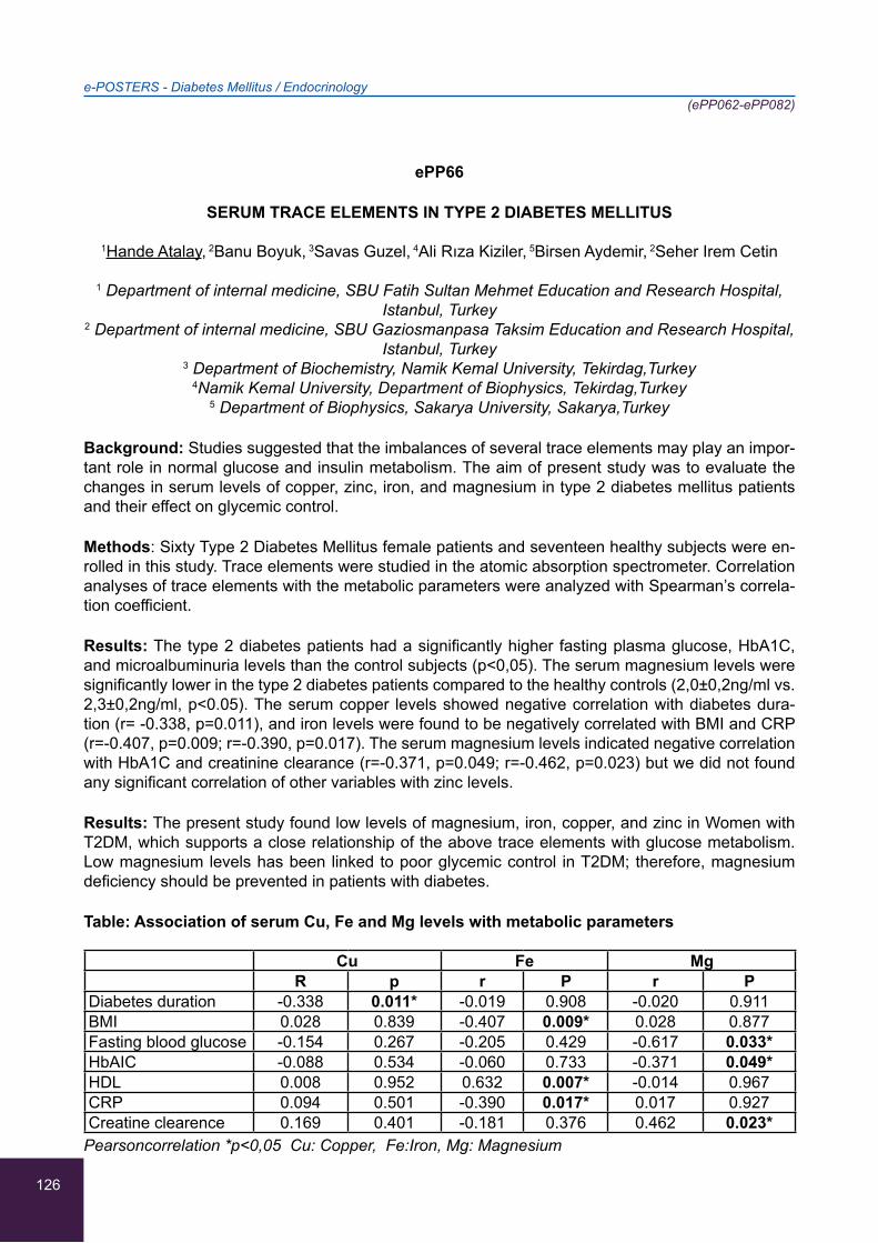
126
ePP66
SERUM TRACE ELEMENTS IN TYPE 2 DIABETES MELLITUS
1Hande Atalay, 2Banu Boyuk, 3Savas Guzel, 4Ali Rıza Kiziler, 5Birsen Aydemir, 2Seher Irem Cetin
1 Department of internal medicine, SBU Fatih Sultan Mehmet Education and Research Hospital, Istanbul, Turkey
2 Department of internal medicine, SBU Gaziosmanpasa Taksim Education and Research Hospital, Istanbul, Turkey
3 Department of Biochemistry, Namik Kemal University, Tekirdag,Turkey 4Namik Kemal University, Department of Biophysics, Tekirdag,Turkey
5 Department of Biophysics, Sakarya University, Sakarya,Turkey
Background: Studies suggested that the imbalances of several trace elements may play an impor-tant role in normal glucose and insulin metabolism. The aim of present study was to evaluate the changes in serum levels of copper, zinc, iron, and magnesium in type 2 diabetes mellitus patients and their effect on glycemic control.
Methods: Sixty Type 2 Diabetes Mellitus female patients and seventeen healthy subjects were en-rolled in this study. Trace elements were studied in the atomic absorption spectrometer. Correlation analyses of trace elements with the metabolic parameters were analyzed with Spearman’s correla-tion coefficient.
Results: The type 2 diabetes patients had a significantly higher fasting plasma glucose, HbA1C, and microalbuminuria levels than the control subjects (p<0,05). The serum magnesium levels were significantly lower in the type 2 diabetes patients compared to the healthy controls (2,0±0,2ng/ml vs. 2,3±0,2ng/ml, p<0.05). The serum copper levels showed negative correlation with diabetes dura-tion (r= -0.338, p=0.011), and iron levels were found to be negatively correlated with BMI and CRP (r=-0.407, p=0.009; r=-0.390, p=0.017). The serum magnesium levels indicated negative correlation with HbA1C and creatinine clearance (r=-0.371, p=0.049; r=-0.462, p=0.023) but we did not found any significant correlation of other variables with zinc levels.
Results: The present study found low levels of magnesium, iron, copper, and zinc in Women with T2DM, which supports a close relationship of the above trace elements with glucose metabolism. Low magnesium levels has been linked to poor glycemic control in T2DM; therefore, magnesium deficiency should be prevented in patients with diabetes.
Table: Association of serum Cu, Fe and Mg levels with metabolic parameters
Cu Fe MgR p r P r P
Diabetes duration -0.338 0.011* -0.019 0.908 -0.020 0.911BMI 0.028 0.839 -0.407 0.009* 0.028 0.877Fasting blood glucose -0.154 0.267 -0.205 0.429 -0.617 0.033*HbAIC -0.088 0.534 -0.060 0.733 -0.371 0.049*HDL 0.008 0.952 0.632 0.007* -0.014 0.967CRP 0.094 0.501 -0.390 0.017* 0.017 0.927Creatine clearence 0.169 0.401 -0.181 0.376 0.462 0.023*
Pearsoncorrelation *p<0,05 Cu: Copper, Fe:Iron, Mg: Magnesium
e-POSTERS - Diabetes Mellitus / Endocrinology(ePP062-ePP082)
127
ePP67
QUALITY OF LIFE IN PATIENTS WITH DIABETES MELLITUS
1Vasilki Sionti, 2Giorgos Papageorgiou, 3Dimitrios Peschos, 4George Charalambous,
5Evangelia Kotrotsiou, 6Victoria Zakopoulou, 7Mary Gouva
1 Postgraduate Program “Primary Health Care”, University of Thessaly, Larissa, Greece2 Derviziana Health Center, General Hospital of Ioannina G. Hatzikosta, Ioannina, Greece
3 Laboratory of Physiology, Faculty of Medicine, School of Health Sciences, University of Ioannina, Ioannina, Greece
4 School of Health Sciences, Frederick University, Nicosia, Cyprus5 Research Laboratory of Care, Technological Educational Institute of Thessaly, Larissa, Greece
6 Department of Speech and Language Therapy, Technological Educational Institute of Epirus, Ioannina, Greece
7 Research Laboratory Psychology of Patients Families and Health Professionals, Technological Educational Institute of Epirus, Ioannina, Greece
Background: Several studies indicate that the presence of a chronic disease is strongly correlated with the emergence of depression and anxiety symptoms and that psychological support to patients may improve their quality of life Objectives To investigate certain social and psychological param-eters and to compare them with basic demographic information, such as the gender, the education and occupation of patients suffering from diabetes mellitus (DM) Design -
Methods: A cross-sectional study of 100 diabetic patients. Psychological factors were assessed with questionnaires, including the Short-Form Health Survey (SF-36), the Life Orientation Test-Re-vised (LOT-R) and the Cardiac Anxiety Questionnaire (CAQ). The associations of psychological with socio-demographic factors were assessed through logistic regression analysis Results Women patients had higher levels of heart-focused anxiety psychopathology than men and therefore women tend to avoid activities that burden the heart. Men patients had a significantly higher index of physical functionality than women. For the patients with elementary education, lower levels of mental health (SF-36-MCS) were observed. Retirees presented the lowest vitality (SF-36-VT) and general health (SF-36-GH) values.
Conclusions: The chronicity of the disease aggravates the psychopathology of the patients thereby creating adverse impact not only on health but also on efforts for compliance. A personalized ap-proach by the health professionals could contribute in addressing the psychological factors that ac-company DM and improve their quality of life.
e-POSTERS - Diabetes Mellitus / Endocrinology(ePP062-ePP082)
128
ePP68
COPING BEHAVIOR IN PATIENTS WITH DIBETES MELLITUS
Anastasios Stefanopoulos, Georgios Tassopoulos, Maroulio Hera, Andreas Kapouralos,
Ilias Georgopoulos, Panagiotis Heras
Department of Internal Medicine, General Hospital of Argos, Greece
The aim of this study was the evaluation of coping behavior in patients with Diabetes Mellitus (DM).
Patients And Methods: In our study 60 (42 male and 18 female age: mean ± SD 62,4± 13.4) pa-tients were entered. As an instrument, the F.Helm questionnaire was used.
Results: It was revealed the prevalence of the following forms of coping strategies: behavioral -altru-ism, “active avoidance” (23%). “distraction”(51%), emotional optimism (48%), “repression of emo-tions “(23%), cognitive - presence of self control and dissimulation (23%). Using diabetes mellitus as a model has shown the prevalence of emotional adaptive coping mechanisms over maladaptive in patients with psychosomatic disorders that allow estimating positively their adaptation abilities.
Conclusion: From the result of our study, it seems necessary to apply cognitive-behavioral psycho-therapy in the system of treatment methods of patients with diabetes mellitus to facilitate the acquir-ing of more constructive forms of coping behavior.
e-POSTERS - Diabetes Mellitus / Endocrinology(ePP062-ePP082)

129
ePP69
EDUCATION AND PSYCHOSOCIAL ADAPTATION OF DIABETIC PATIENTS
Anastasios Stefanopoulos, Georgios Tassopoulos, Maroulio Hera, Andreas Kapouralos,
Ilias Georgopoulos, Panagiotis Heras
Department of Internal Medicine, General Hospital of Argos, Greece
The aim of this study was to investigate the impact of the education on diabetic patient’s self-image and the impact of the education on diabetic patients social environment relationship.
Patients And Methods: 280 (160 men and 120 women, age 15-52 years) patients suffering from Diabetes Mellitus entered the study experimental group (E, n=140) and control group (K, n=140)all matched regarding age and educational level. The patients of E group differed in regard to their pre-vious education; this group underwent the Greek Program “Learning to live with Diabetes Mellitus” over 3 months. Both groups answered questionnaire specifically designed to assess self-image of diabetic patients and relationship with the social environment.
Results: The education significantly improved self-image in E group when compared to K group (p<0.02). There was no significant difference in education between E and K group with regard to the social relationship (p>0.02).
Conclusions: The education has an important contribution in establishing self-management ap-proach in which patients assume responsibility for their behavior, for changing their environment and for planning their future. For successful diabetic patients’ psychosocial adaptation and social partici-pation, it is necessary that the whole society provides more resources for psychosocial support.
e-POSTERS - Diabetes Mellitus / Endocrinology(ePP062-ePP082)
130
ePP70
TYPE D PERSONALITY, PSYCHOLOGICAL DISTRESS AND QUALITY OF LIFE IN PATIENTS WITH DIAABETES MELITUS
Anastasios Stefanopoulos, Georgios Tassopoulos, Maroulio Hera, Andreas Kapouralos,
Ilias Georgopoulos, Panagiotis Heras
Department of Internal Medicine, General Hospital of Argos, Greece
Introduction: Studies have shown that distressed personality (Type D ) is associated with Diabetes Mellitus (DM) and have prognostic implications. The aim of this study was to assess Psychological distress and Quality of life (QoL) in patients with DM.
Patients And Methods: In this cross-sectional study, 100 (54 males, 46 females) first time diag-nosed (3-12 weeks) DM patients (WHO criteria ) were selected. Type D personality was assessed with Distress Scale (DS-14). Psychological distress (Anxiety, Depression) was measured with Hos-pital Anxiety and Depression Scale (HADS) while QoL was assessed with WHO Quality of life scale (WHOQOL-BREF).
Results: 59% of DM patients were identified with Type D personality traits (27 males,32 females ). Type D personality was identified as significant predictor of impaired QoL [OR: 1.06 95% CI (1.02-1.11) p≤0.01], Depression [OR:1.11 95% CI (1.06-1.16), p≤0.01] and Anxiety [OR 1.09 95%(1.04-1.14) p≤0.01]. The results also revealed that Type D DM patients have impaired QoL (mean=73,3[SD=15.6] versus 85.9 [SD 17.3], respectively t=3.8 p≤0.01), elevated levels of Anxiety and Depression com-pared to non type D patients.
Conclusions: This study highlighted the importance of Type D personality in research on DM as a determinant of adverse psychological outcomes. It also emphasized the need for routinely psycho-logical assessment of DM patients along with medical treatment to improve their QoL and rehabilita-tion.
e-POSTERS - Diabetes Mellitus / Endocrinology(ePP062-ePP082)
131
ePP71QUALITY OF LIFE IN PATIENTS WITH DIABETES MELLITUS AFTER PSYCOLOGICAL
REHABILITATION
Anastasios Stefanopoulos, Georgios Tassopoulos, Maroulio Hera, Andreas Kapouralos,
Ilias Georgopoulos, Panagiotis Heras
Department of Internal Medicine, General Hospital of Argos, Greece
The aim of this study was to assess Quality of Life (QoL) in Diabetes Mellitus patients undergoing the program of psychological rehabilitation.
Patients And Methods: 32 DM patients after treatment participated in the program of social, psy-chological, juridical support for DM patients and their relatives. Patients completed SF-36 question-naire at baseline in three months after the program start.
Results: Before the program SF-36 QoL parameters related to psychological functioning were worse than the ones related to physical well-being. In 3 months after the program start the improvement of all QoL variables was observed with the significant increase of general health (change from 28.2 to 41.2 ; p=0.020). Significant improvements in role functioning (from 52.5 to 68.3 p=0,027), pain (from 45.2 to 26.1; p=0.05), sleep disturbances (from 59.5 to 36,6; p=0.05) were identified. Other QoL pa-rameters had a marked tendency to improvement.
Conclusions: QoL assessment is the sensitive method of evolution of the effectiveness of rehabili-tation programs for DM patients. The program of psychosocial rehabilitation of these patients nd their relatives appeared to be effective. Improvement of different QoL parameters during the program was revealed.
e-POSTERS - Diabetes Mellitus / Endocrinology(ePP062-ePP082)
132
ePP72
SLEEP DISTURBANCES AND DEPRESSION IN PATIENTS WITH DIABETES MELLITUS
Anastasios Stefanopoulos, Georgios Tassopoulos, Maroulio Hera, Andreas Kapouralos, Ilias Georgopoulos, Panagiotis Heras
Department of Internal Medicine, General Hospital of Argos, Greece
The aim of this study is to estimate the degree of sleep disturbances, as well the possible contribut-ing factors in patients with diabetes mellitus (DM).
Patients and methods: 41 patients concluded in our study. 28 were female and 13 male with mean age of 51 (range 29-74). These patients underwent a clinical psychiatric interview according to the diagnostic criteria of ICD 10, The degree of sleep disturbances during the month prior to patients evaluation was assessed through the Athens Insomnia Scale (AIS) and psychopathology was as-sessed through the Montgomery Asberg Depression Rating Scale (MADRS).
Results: There was a strong correlation between the score of the AIS and that of the MADRS for all 41 patients (r: 0.40,p<0.01). 13 of our patients complained of insomnia. These patients scored higher on the MADRS than the ones without sleep difficulties (z:-3.032, p=0.002).
Conclusions: The result of our study suggests that sleep disturbance (insomnia) in patients with DM is a probable indicator of depression. Furthermore, these data suggest that insomnia in these patients is one of the factors mediating the association between depression and impairment in their functioning.
e-POSTERS - Diabetes Mellitus / Endocrinology(ePP062-ePP082)

133
ePP73
TELEMEDICAL MONITORING OF DIABETIC RETINOPATHY
1Rafaella Datseri, 1Nikolaos Papakonstantinou, 2Christos Tsagkaris, 3Dorothea Scordilis
1Comenius University Faculty of Medicine, Bratislava, Slovakia 2Faculty of Medicine, School of Health Science, University of Crete, Heraklion, Greece
3Faculty of Medicine, Medical University of Sofia, Sofia, Bulgaria
Introduction: Diabetic retinopathy is the leading preventable cause of blindness in people under 60 years of age.Annual screening examination is necessary to prevent manifestation of the microan-giopathy, control its progress and prevent ultimate blindness. Due to the limited number of ophthal-mologists available, Teleophthalmology has therefore been deployed to fill in the gap in an attempt to recreate the clinical encounter at a distance for patients residing in areas characterized as remote or secluded.
Aim: The aim of this article is to provide a database of hardware and software solutions currently available on the market, which are eligible for remote screening and follow-up of diabetic retinopathy in both a real-time and a store-and-forward setting.
Materials and Methods: We reviewed current literature, peer reviews, tech specs derived from each manufacturer’s official page, while also acquiring relative hands-on experience.
Results: Both real-time and Store-and-forward settings seem to cater for the needs of the screening programme, each in their own terms. Phone-docked fundus cameras seem to be the most viable so-lution when on the go and on a limited budget. The best image quality though is provided by portable fundus cams. Complementary software facilitates a smoother user experience. Messaging services and cloud service act as the key mediators between the patient and specialist(s).
Conclusion: The use of portable tech facilitates timely and convenient access to retinopathy screen-ing for patients with diabetes experiencing barriers to accessing eye care. Further research should be conducted in terms of automated evaluation and scoring as well as self-evaluation and monitor-ing.
e-POSTERS - Diabetes Mellitus / Endocrinology(ePP062-ePP082)
134
ePP74
ACQUIRED CHYLOMICRONEMIA-INDUCED ACUTE PANCREATITIS
Paraskevi Kontrafouri, Eleni Kourti, Anastasia Kopsaftopoulou, Kostas Reppas, Charis Efraimi,
Maria Mylona
1st Department of Propaedeutic & Internal Medicine, Laiko General Hospital, Athens University Medical School, Athens, Greece
Backround/Aim: Pancreatitis is an acute inflammatory response of the pancreas characterized by abdominal pain and elevated pancreatic enzymes. A serum triglyceride level of more than 2000 mg / dl is a rare, but well-known identifiable risk factor. The clinical course and routine management of hypertriglyceridemia-induced pancreatitis is similar to other causes. A thorough family history is im-portant, as is the identification of secondary causes of hypertriglyceridemia. The mainstay of therapy includes dietary restriction of fatty meal and fibric acid derivatives. Alcoholism itself effectuates ac-quired hypertriglyceridemia, since it causes an increase in the synthesis of triglycerides (TGs) and TG-rich lipoproteins in the liver, while it stimulates lipolysis in fatty tissue resulting in a larger supply of fatty acids to the liver. Herein we present a case of an alcoholic patient presenting with acquired chylomicronemia-induced acute pancreatitis.
Case Description: A 48-year old Greek man, with a history of chronic consumption of ethyl alcohol(>50g/d) , was admitted to our clinic due to progressively worsening of abdominal pain for the last 24 hours and two episodes of vomiting. Similar signs have been reported in the recent past. Blood chemistry revealed severely elevated TGs (Tg=3500 mg/dl)and transaminasemia. Hepatome-galy and fatty liver disease were found in the upper abdomen ultrasound, whereas the CT scan findings were compatible with chronic pancreatitis (calcification of the pancreatic duct). Fenofibrate 145mg/d and ω3 fatty acids were initiated along with standard pancreatitis treatment. The patient showed outstanding clinical and laboratory response and was discharged after six days of hospitali-zation.
e-POSTERS - Diabetes Mellitus / Endocrinology(ePP062-ePP082)

135
ePP75
IT IS NOT ALWAYS A MATTER OF GENES: A CASE REPORT OF AN ELDERLY PATIENT WITH UNTREATED FAMILIAL HYPERCHOLESTEROLEMIA
Paraskevi Kontrafouri, Eleni Kourti, Anastasia Kopsaftopoulou, Kostas Reppas, Charis Efraimi,
Maria Mylona
1st Department of Propaedeutic & Internal Medicine, Laiko General Hospital, Athens University Medical School, Athens, Greece
Introduction/Aim: Familial hypercholesterolemia (FH) is a common genetic disorder, characterized by elevated levels of low density lipoprotein cholesterol (LDL-C) and early cardiovascular manifesta-tions. It is distinguished in the heterozygous FH which is the most common type, with a frequency of 1/300 in the general population and in the homozygous FH which is more rare with a frequency of 1/10^6 and manifestations of the disease in early childhood.Men present complications of the disease during the 4th-5th decade of their life, while women a decade earlier. Herein we present an interesting case of a man with non-treated FH, who underwent a transient ischemic attack in a quite advanced age, in order to highlight the cardioprotective role of running a healthy life despite the pres-ence of a genetic predisposition.
Case Description: A 84-year old Greek patient, fit enough despite his age, with a history of hy-pertension under medication, was admitted to our clinic due to hypoesthesia of the right upper and lower limb which lasted less than 20 minutes. Similar symptoms were reported a month before. No findings were revealed from the brain CT scan. Physical examination was unremarkable apart from a systolic murmur in the right carotid artery. From blood chemistry, elevated levels of total cholesterol (T-Chol=270mg/dl) and LDL-C=220mg/dl with normal serum TG levels, were found. Right carotid artery stenosis (~45%) was found in Doppler ultrasound. Treatment with a high-dose statin (rosuv-astatin 20mg/d) and an antiplatelet agent (salospir 100mg) was initiated; the patient was discharged in 5 days.
e-POSTERS - Diabetes Mellitus / Endocrinology(ePP062-ePP082)

136
ePP76
FACTORS AFFECTING SURVIVAL IN PALLIATIVE CARE PATIENTS
1Mehmet Yürüyen, 1Isil Ozbas Tevetoglu, 1Yeliz Tekmen, 2Ozlem Polat, 3Musa Cirak,
1Arzu Ozdemir Kayalar, 4Savas Bayrak, 1Yıldız Okuturlar
1Department of Internal Medicine Palliative Care, Bakirkoy Dr. Sadi Konuk Education and Research Hospital, Istanbul, Turkey
2Department of Family Medicine, Bakirkoy Dr. Sadi Konuk Education and Research Hospital, Istanbul, Turkey
3Department of Neurosurgery, Bakirkoy Dr. Sadi Konuk Education and Research Hospital,Istanbul, Turkey
4Department of General Surgery, Bakirkoy Dr. Sadi Konuk Education and Research Hospital, Istanbul, Turkey
Aim: The aim of this study is to investigate the survival of patients in a palliative care center and to investigate the factors that influence it.
Method: Patients hospitalized in palliative care center between August 1, 2015 and July 30, 2017 were assessed for mortality status through the official death notification system on September 2017. Demographic characteristics, comorbid conditions, nutritional conditions and pressure wounds and laboratory tests results were taken from all the patients at first hospitalisation.
Results: 343 patients were included in the study, 319 (93%) were eligible. The mean age of the pa-tients (n = 140 women, 44%) was 71 ± 15.7 (median age 73, min 19, max 99), the mean number of comorbidities was 2.3 ± 1.04. At the end of follow-up, 82.4% of the patients died. The median follow-up period was 43 days (95% Confidence Interval (CI): 32.92 - 53.08) in the overall survival analysis. According to regression analysis, it is shown that statistically significant factors related to survival are malignancy (p = 0.017, Hazard ratio (HR): 1.74, 95% CI: 1.10-2.73), mean platelet volume (MPV) (95% CI: 1.03 - 1.07) and uric acid (p = 0.042, HR: 1.08, 95% CI: 1.00 - 1.17).
Conclusion: While the most effective comorbid disease is malignancy on survival in palliative care patients, the neutrophil / lymphocyte ratio, MPV and uric acid are most important laboratory param-eters.
e-POSTERS - Diabetes Mellitus / Endocrinology(ePP062-ePP082)
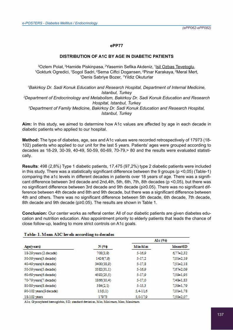
137
ePP77
DISTRIBUTION OF A1C BY AGE IN DIABETIC PATIENTS
3Ozlem Polat, 2Hamide Piskinpasa, 2Yasemin Sefika Akdeniz, 1Isil Ozbas Tevetoglu,
1Gokturk Ogredici, 1Sogol Sadri, 2Sema Ciftci Dogansen, 2Pinar Karakaya, 2Meral Mert,
1Denis Sabriye Bozer, 1Yildiz Okuturlar
1Bakirkoy Dr. Sadi Konuk Education and Research Hospital, Department of Internal Medicine, Istanbul, Turkey
2Department of Endocrinology and Metabolism, Bakirkoy Dr. Sadi Konuk Education and Research Hospital, Istanbul, Turkey
3Department of Family Medicine, Bakirkoy Dr. Sadi Konuk Education and Research Hospital, Istanbul, Turkey
Aim: In this study, we aimed to determine how A1c values are affected by age in each decade in diabetic patients who applied to our hospital.
Method: The type of diabetes, age, sex and A1c values were recorded retrospectively of 17973 (18-102) patients who applied to our unit for the last 5 years. Patients’ ages were grouped according to decades as 18-29, 30-39, 40-49, 50-59, 60-69, 70-79,> 80 and the results were evaluated statisti-cally.
Results: 498 (2,8%) Type 1 diabetic patients, 17,475 (97,2%) type 2 diabetic patients were included in this study. There was a statistically significant difference between the 9 groups (p <0,05) (Table-1) comparing the a1c levels in different decades in patients over 18 years of age. There was a signifi-cant difference between 3rd decade and 2nd,4th, 5th, 6th, 7th, 8th decades (p <0,05), but there was no significant difference between 3rd decade and 9th decade (p≥0.05). There was no significant dif-ference between 4th decade and 8th and 9th decade, but there was a significant difference between 4th and others. There was no significant difference between 5th decade, 6th decade, 7th decade, 8th decade and 9th decade (p≥0.05). The results are shown in Table 1.
Conclusion: Our center works as refferal center. All of our diabetic patients are given diabetes edu-cation and nutrition education. Also appointment priority to elderly patients that leads the chance of close follow-up, leading to more strict controls on A1c goals.
e-POSTERS - Diabetes Mellitus / Endocrinology(ePP062-ePP082)

138
ePP78
PAPILLARY THYROID CARCINOMA IN PATIENT WITH PRIMARY HYPERPARATHYROIDISM
1Hamide Piskinpasa, 1Sema Ciftci Dogansen, 1Sefika Yasemin Akdeniz, 2Fatmagul Kusku Cabuk,
3Cevher Akarsu, 4Yildiz Okuturlar, 4Gokturk Ogredici, 1Pinar Karakaya, 1Meral Mert
1Department of Endocrinology and Metabolism, Bakirkoy Dr. Sadi Konuk Education and Research Hospital, Istanbul, Turkey
2Department of General Surgery, Bakirkoy Dr. Sadi Konuk Education and Research Hospital, Istanbul, Turkey
3Department of Pathology, Bakirkoy Dr. Sadi Konuk Education and Research Hospital,Istanbul, Turkey
4Department of Internal Medicine, Bakirkoy Dr. Sadi Konuk Education and Research Hospital, Istanbul, Turkey
Introduction: Primary hyperparathyroidism (PHPT) is present in up to 0,5% of the general popula-tion. Papillary thyroid carcinoma (PTC) constitutes more than 70% of thyroid malignancies. Coexist-ence of PHPT and PTC is a rare condition. We report a case of a 69 years old PHPT confirmed by 99mTc-MIBI scintigraphy, hypercalcemia and elevated serum parathyroid hormone. PTC was diag-nosed postoperatively by histopathology.
Case Report: 69 years old male patient admitted to our clinic with an enlarged, painless nodular mass on the neck. At laboratory examination; he had elevated serum calcium 11,6 mg/dl (normal range:8,4-10,4), low serum phosphate 1,6 mg/dl (normal range 2,5-45) and elevated PTH:138 pg/ml (normal range 15-56). USG of the neck displayed multinoduler goiter with a 42*25 mm hypoechoic nodule near the lower pole of the right lobe and another 22*13 mm hypoechoic solid nodule at the superior of the left tyroid gland and 16*14 mm solid cystic nodule at the inferior of the left thyroid gland. The parathyroid 99mTc-MIBI scan demonstrated parathyroid adenoma at the right inferior and left superior pole of the tyhroid. US guided FNAB of the two nodules in left thyroid gland and right inferior nodules were benign The patient underwent total thyroidectomy and removal of right inferior and left superior parathyroid adenoma. Pathologic examination confirmed left superior parathyroid adenoma and follicular variant PTC.
Discussion: PTC with PHPT has been reported in 2.3-4.3% of patients undergoing surgery for PHPT. Therefore a comprehensive preoperative thyroid workup, including FNAB, should be per-formed in all patient with PHPT.
e-POSTERS - Diabetes Mellitus / Endocrinology(ePP062-ePP082)
139
ePP79
THYROID PAPILLARY CARCINOMA IN A TOXIC THYROID NODULE
1Hamide Piskinpasa, 1Sema Ciftci Dogansen, 1Sefika Yasemin Akdeniz, 2Ahmet Cem Dural,
3Fatmagul Kusku Cabuk, 4Yıldız Okuturlar, 4Isıl Ozbas Tevetoglu, 1Pinar Karakaya, 1Meral Mert
1Department of Endocrinology and Metabolism, Bakirkoy Dr. Sadi Konuk Education and Research Hospital, Istanbul, Turkey
2Department of General Surgery, Bakirkoy Dr. Sadi Konuk Education and Research Hospital, Istanbul, Turkey
3Department of Pathology, Bakirkoy Dr. Sadi Konuk Education and Research Hospital,Istanbul, Turkey
4Department of Internal Medicine, Bakirkoy Dr. Sadi Konuk Education and Research Hospital, Istanbul, Turkey
Introduction: Papillary Thyroid cancer (PTC) is the most common malignant endocrine tumour worldwide. The prevalence of malignancy in toxic adenomas varies between 1 and 8% in different series (most, corresponding to <2.5%). We present a case of toxic adenoma with the diagnosis of papillary thyroid carcinoma.
Case Report: 50-year-old male patient was admitted with the complaints of sweating, palpitation, tremor, and weight loss for three months. Thyroid function tests revealed hyperthyroidism with el-evated free triiodothyronine (FT3) of 6.27 pg/mL (normal: 2-4.4 pg/mL), free thyroxine (FT4) of 2.4 ng/dL (normal: 0.93-1.7 ng/dL), and suppressed thyroid-stimulating hormone (TSH) of <0.01 μIU/mL (normal: 0.27-4.20 μIU/mL). Thyroid peroxidase, thyroglobulin antibody and TSH receptor antibody levels were negative. Our ultrasonographic evaluation was consistent with a hypoechoic nodule of 20x12x18 mm in size in the right inferior thyoroid pole, having regular margins, dense millimetric mi-crocalcifications the central area. Technetium 99m scan showed tracer activity in a right nodule with suppression of extranodular tissue (Figure1). He was initially treated with methimazole. Fine needle aspiration biopsy (FNAB) revealed that the thyroid nodule was benign. The patient underwent right thyroid lobectomy. Pathology report revealed classical variant of PTC.
Conclusion: The frequency of PTC is increasing due to the increased frequency of use of diagnostic methods of thyroid nodules. We present a case with a rare association of hot nodules and papillary thyroid carcinoma, with a benıgn cytology on FNAB. Diagnosis of hot nodules does not exclude the possibility of thyroid carcinoma.So, hyperfunctioning thyroid nodules warrants careful evaluation and appropriate therapy.
e-POSTERS - Diabetes Mellitus / Endocrinology(ePP062-ePP082)

140
ePP80
PHASE ALLOCATION AND EFFICIENT TECHNIQUE FOR EARLY REVEALING OF CHOLELITHASIS
1Marchuk Yuliya Fedorivna, 2Dilbar Mohammed Saleem, 2Aneeta George, 2Vishnu Dhanarajan
1Department of Clinical Immunology, Allergology and endocrinology, Bukovinian State Medical University, Chernivtsi, Ukraine
2Department of Internal Medicine, Bukovinian State Medical University, Chernivtsi, Ukraine
Taking into consideration the great importance of disorders of metabolism by Diabetes mellitus and Cholelithiasis, it is of great value the investigation of interrelations of their pathological processes.
The aim of our investigation was to establish peculiarities of phase data disorders of bile in patients with chronic acalculous cholecystitis combined with diabetes mellitus type II.Detailed clinical investi-gation was carried out with 15 patients with chronic acalculous cholecystitis combined with diabetes mellitus type 2 (1st main group), 10 patients with diabetes mellitus (2nd group), 10 patients with chronic acalculous cholecystitis (3rd group) and 10 almost healthy patients. Correlative square of phase allocation of laser picture of bile S=0,09. Correlative square of phase allocation of laser pic-ture of bile with combined pathology was extremely decreased and is S=0,055. From achieved data of laser pictures of bile in patients of all groups and healthy people we could say that the most in-formative diagnostic value data for revealing gallstone disease were dispersion, asymmetry, excess, which characterized phase allocation. There defined further diapason changes statistic moments of 1-4 order of laser picture of bile between group of healthy people and investigated groups of patients: dispersion (increase in 1,5-2,1),asymmetry(increased in 3-16),excess(increased in 3-12).In conclu-sion, the most informative for early revealing of cholelithasis are dispersion, asymmetry, excess, which characterize phase allocation of laser picture of bile. There is proposed model of formation of phase allocation in pictures of bile layers like process of coordinate modulation of laser ionization by bile liquid-crystal formations.
e-POSTERS - Diabetes Mellitus / Endocrinology(ePP062-ePP082)
141
ePP81
APPEARANCE OF PRIMARY HYPERPARATHYROIDISM IN PATIENTS WITH THALASSEMIA-BETA FOLLOWING EFFICIENT CHELATION THERAPY: TWO CASE REPORTS
1Kyriaki Ventiadi, 2Labrini Papanastasiou, 1Georgia Leveta, 1Christoforos Roumpakis,
1Foteini Petropoulou
1Department of Thalassaemia Unit, G.Gennimatas Hospital of Athens, Athens, Greece 2Department of Endocrinology and Diabetes Center, “G.Gennimatas” Hospital of Athens,
Athens, Greece
Background: Thalassaemia-beta (TM) represents a group of recessively inherited haemoglobin disorders, characterised by reduced synthesis of b-globulin chain. The main stay of treatment is regular blood transfusion, leading to iron deposition in different endocrine glands. Iron deposition in parathyroid glands in turn, may cause hypocalcaemia and hypoparathyroidism. The appearance of hypercalcaemia and primary hyperparathyroidism (PH) after successful chelation therapy has not been previously reported in such patients.
Aim: To present two normocalcemic-TM cases who developed hypercalcemia and PH after chela-tion therapy.
Case Reports: The cases concern a 49- and a 47-year-old TM woman, who attended our depart-ment. Both women, before having efficient chelation, had normal calcium levels. During a long fol-low-up period, chelation was improved and ferritin levels were decreased, while calcium levels were progressively increased. In both women biochemical analysis revealed increased calcium levels with inappropriate increased parathyroid hormone (PTH) levels indicating the development of primary hyperparathyroidism (Table). In both cases bone density showed aggravation with time and neck ul-trasound revealed parathyroid adenomas confirming the PH diagnosis. Nephrolithiasis was revealed in one case.
Conclusions: We report two TH clinical cases that developed PH after successful chelation therapy. These findings reinforce the importance of the regular follow-up of the TM patients for early detection and management of deranged calcium homeostasis, that can appear during successful chelation therapy.
e-POSTERS - Diabetes Mellitus / Endocrinology(ePP062-ePP082)
142
ePP82
THYROID AUTOIMMUNITY IN THE TYPE 2 DIABETES MELLITUS
1Banu Boyuk, 2Yıldız Okuturlar, 1Seher Irem Cetin, 3Hande Atalay
1Department of Internal Medicine, SBU GOP Taksim Training and Research Hospital, Istanbul,Turkey
2Department of Internal Medicine, SBU Bakirkoy Sadi Konuk Training and Research Hospital, Istanbul,Turkey
3Department of Internal Medicine, SBU Fatih Sultan Mehmet Training and Research Hospital, Istanbul, Turkey
Background: The association between type 2 diabetes mellitus (T2DM) and the presence of Thy-roid autoimmunity (TAI) has been a issue of discussion. Although earlier reports refused any link between them, there is evidence to suggest an increased prevalence of TAI in patients with T2DM. The objective was to assess the prevalence of thyroid dysfunction among type 2 diabetic patients.
Methods: This is a retrospective study. One hundred and seventeen type 2 diabetes subjects (52 males and 65 females) and two hundred seventy seven controls (89 males and 188 females) were attended the outpatient clinic of the diabetes unit. All patients in the study were subjected to including glukoz, HbA1c, thyroid-stimulatinghormone (TSH), free triiodothyronine, free thyroxine, anti-thyroid-peroxidase (TPO), anti-thyroglobulin (anti- Tg). Autoimmunity were diagnosed when anti-TPO levels were greater than 9 IU/mL.
Results: The prevalence of anti-TPO antibodies was 16,2 % in type 2 diabetic patients. Anti- TPO was found (21,38±76,2IU/mL) in the patients versus (1,03±0,92IU/mL) in the controls (p<0,05). Anti-Tg was found (17,85±116,98IU/mL) in patients versus (2,25±13,48IU/mL) controls (p<0,05). A signifi-cant positive correlation was found between anti Tg and anti TPO anti thyroid antibodies ( r=0,476; p=0,000) in diabetic patients.
Conclusion: Autoimmune thyroid diseaseis common with type 2 diabetes than controls, and thus points to a role of autoimmunity in the pathogenesis of type 2 diabetes. Early detection of thyroid dysfunction in patients with type 2 diabetes mellitus should be performed routinely.
Keywords: TSH, anti-TPO, anti-Tg, T2 diabetes
Table: Biochemical characteristics of patients and controls
Diabetics group(n:117)
Mean±s.d/n, %
Control group(n:277)
Mean±s.d/n, %P
Age (years) 5,15±14,33 47,76±16,76 0,0001Gender Female 65 (55,6%) 188 (67,9%) 0,02
Male 52 (44.4%) 89 (32,1%)TSH (IU/mL) 2,61±6,32 2,07±3,08 0,730Free T3 (IU/mL) 3,34±0,62 3,31±0,54 0,722Free T4 (IU/mL) 0,89±0,2 0,85±0,15 0,063AntiTPO (IU/mL) 21,38±76,2 1,03±0,92 0,039Anti Tg (IU/mL) 17,85±116,98 2,25±13,48 0,023HbAIC (%) 8,46±2,32 5,54±0,39 0,0001Glukoz (mg/dL) 170,35±79,71 95,44±16,61 0,0001
e-POSTERS - Diabetes Mellitus / Endocrinology(ePP062-ePP082)

143
e-POSTERS - Hematology / Oncology(ePP083-ePP0122)
ePP83
GASTRIC LIPOMA IN A 59-YEAR-OLD-MALE: CASE REPORT
Christina Nikolaidou, Georgios Hahalis, Stavroula Papadopoulou, Ioannis Venizelos
Department of Pathology, General Hospital “Hippokrateion”, Thessaloniki, Greece
Background/Aim: Lipomas are benign soft tissue tumors, composed of mature lipocytes. Lipomas of the gastrointestinal tract are rare. They account for less than 1% of all gastric neoplasms. They are localized in the submucosa in 90% to 95% of cases and in the serosa in 5% to 10% of the cases. 75% of them affect the antrum. Gastric lipomas appear between the fifth and seventh decades of life, mainly in women. They usually are small and asymptomatic and are detected incidentally. Symp-toms depend on the size and location of the tumor. Larger lesions can cause hemorrhage, abdominal pain, pyloric obstruction, and dyspepsia. Gastrointestinal bleeding is typically chronic and minimal and is able to cause anemia.
Case Report: A 59-year-old male was admitted to the emergency department with upper GI hemor-rhage. He was initially resuscitated and stabilized. From the CT scan a gastric mass was found in the antrum with possible clinical differential diagnosis: ectopic pancreatic tissue, GIST and Brunner’s gland adenoma. A partial gastrectomy was performed. On macroscopic examination, a well-defined homogeneous yellowish tumor measured 5x3cm, was found in the submucosa of the stomach. The histological examination showed that the tumor consisted of well-differentiated non-invasive mature adipose tissue. No atypia or mitotic figures were noticed. The diagnosis was gastric lipoma. The patient is well one year after the diagnosis.
Conclusion: Despite its rarity, gastric lipoma should be considered in the differential diagnosis for a submucosal mass both clinically and pathologically.

144
e-POSTERS - Hematology / Oncology(ePP083-ePP0122)
ePP84
AMYLOIDOSIS DIAGNOSED IN BONE MARROW BIOPSY: A RARE CASE REPORT
Christina Nikolaidou, Georgios Hahalis, Stavroula Papadopoulou, Ioannis Venizelos
Department of Pathology, General Hospital “Hippokrateion”, Thessaloniki, Greece
Background/Aim: Primary amyloidosis (PAL) is the most common form of systemic amyloidosis. It is due to an underlying plasma cell dyscrasia that produces an amyloidogenic light chain, which de-posits as amyloid fibrils within the tissues. Amyloid involvement of the bone marrow is not commonly diagnosed before autopsy. The prognosis of PAL is poor, with a median survival of 1 to 2 years. Recently, more aggressive therapy using high-dose melphalan and autologous stem cell transplan-tation has improved the survival rates. Amyloidosis has to be differentiated from multiple myeloma and plasmatocytoma. The identification of a monoclonal population of plasma cells by using immu-nohistochemical techniques on bone marrow core biopsy specimens is sometimes difficult because of the low number of plasma cells present there.
Case report: A 77-year-old female patient was admitted to our hospital due to weight loss and weak-ness. She had a history of nephrotic syndrome. Bone marrow biopsy was performed. The histologi-cal and immunohistochemical examination revealed scattered mature polyclonal plasma cells (ap-proximately 16-18%, positivity for kappa and lambda light chains). Amyloid deposits were identified in the blood vessel walls with the Congo Red histochemical stain. These findings were compatible with amyloidosis. The patient died due to respiratory infection some days later. Bone marrow biopsy is an unusual site for the diagnosis of amyloidosis.
Conclusion: In cases suspicious for plasma cell myeloma, histochemical stain for Congo Red should be performed in order to achieve the correct diagnosis and apply the appropriate treatment.
145
e-POSTERS - Hematology / Oncology(ePP083-ePP0122)
ePP85
A NOVEL TREATMENT OF NON-MELANOMA SKIN CANCERS: GENOTYPING, IMMUNOTHERAPY AND MINIMALLY INVASIVE PROCEDURES AS AN ALTERNATIVE TO
TRADITIONAL SURGICAL EXCISION
1Dorothea Scordilis, 2Rafaella Datseri, 3Emmanouela Laskaratou, 4Georgios Matzakanis,
3Eleni Kastritsi, 3Olga Kastritsi, 3Eleutheria Parasiri, 5Eviana Petropoulou, 3Christos Tsagkaris
1Faculty of Medicine, Medical University of Sofia, Sofia, Bulgaria 2Faculty of Medicine, Commenius University of Bratislava, Bratislava, Slovakia
3Faculty of Medicine, School of Health Science, University of Crete, Heraklion, Greece 4Faculty of Medicine, University of Patras, Patras, Greece
5Medical laboratories Department, University of Applied Science of Athens, Athens, Greece
Background: Non-melanoma skin cancers (NMSC) with basal cell carcinoma(BCC) and squamous cell carcinoma(SCC) being two main types feature high prevalence in the U.S. yearly. Exposure to ultraviolet radiation appears as a major risk factor. Many treatment modalities have been described to address the growing epidemic of NMSCs. Surgical excision (SE) consists a traditional treatment of skin cancer at a high-risk for recurrence. Nevertheless, it provokes considerable aesthetic impair-ment illustrating the need of alternatives with a focus on prevention, equally good prediction and quality of life.
Aim: The aim of this article is to stress the importance of how modern modalities affect patient qual-ity assurance therapeutically and aesthetically in the process of skin cancer correction. From genet-ics and pharmaceutical agents to micrographic surgery (MMS) and doctor patient communication we investigate which are the optimal available options.
Methods: We reviewed literature and profiles of patients and acquired data from patients narra-tives.
Results: Topical 5-fluorouracil appears as the most promising treatment showing 90% histological cure. MMS should be considered the most precise method combining a decreased recurrence of cancer with a proper aesthetic result ameliorating patient’s psychology and compliance. Compli-ance’s important is critical given that patients with recurrent NMSC withdrew from treatment due to aesthetic discomfort. Genotyping may enhance preventive strategies and provide future research with a pool of clinical data.
Conclusion: The tripartite of novel pharmaceutical therapies, surgical precision and solid doctor patient communication appears as a promising NMSC treatment taking into accounts both cancer treatment and patient overall well-being.

146
e-POSTERS - Hematology / Oncology(ePP083-ePP0122)
ePP86
MULTINUCLEATE CELL ANGIOHISTIOCYTOMA OF THE NOSE: A RARE CASE REPORT
Christina Nikolaidou, Georgios Hahalis, Stavroula Papadopoulou, Ioannis Venizelos
Department of Pathology, General Hospital “Hippokrateion”, Thessaloniki, Greece
Background/Aim: Multinucleate cell angiohistiocytoma is a rare, benign lesion of unknown etiology in which asymptomatic, reddish-brown, dome-shaped papules develop over weeks to months, com-monly on the extremities and face. Some authors consider such lesions as alterations of the connec-tive tissue, others as vein damages and some others as chronic inflammatory disease. In the english literature there are only a few references with similar cases. Clinically, these lesions may resemble Kaposi’s sarcoma, acroangiodermatitis, granuloma annulare, lichen planus, or sarcoidosis. It is more commonly observed in women with a ratiο 5:1. These lesions can progress, and may become dis-seminated. Previously documented therapies include laser and/or topical steroids, although no treat-ment has been shown to be superior.
Case report: A 50-year old male came to our hospital with a two-year history of a nose papule, which was excised. In our laboratory, we received a skin biopsy with maximum diameter 0.5cm. On its surface, a red color nodule was observed with maximum diameter 0.3cm. Histologically, the salient features were proliferation of capillaries and small venules in the dermis, in association with promi-nent connective tissue cells and larger angulated multinucleate cells. Immunohistochemical stain for HHV8 was negative. These findings were compatible with multinucleate cell angiohistiocytoma. Six months after the operation there was no local recurrence.

147
e-POSTERS - Hematology / Oncology(ePP083-ePP0122)
ePP87
A RARE CASE OF UPPER EXTREMITY DIFFUSE LARGE B-CELL LYMPHOMA (DLBCL) MIMICKING SOFT TISSUE SARCOMA
Christina Nikolaidou, Georgios Hahalis, Stavroula Papadopoulou, Ioannis Venizelos
Department Of Pathology, General Hospital “Hippokrateion”, Thessaloniki, Greece
Background/Aim: Non Hodgkin Lymphoma (NHL) involving soft tissue is extremely rare. It may be primary or following a nodal NHL elsewhere. Cases involving soft tissue often mimic other entities like sarcoma. Treatment options remain the same for both nodal and extra nodal DLBCL with the first choice being chemoimmunotherapy. We present a case of 54-year-old patient with left upper extremity mass which was proven to be DLBCL instead of sarcoma.
Case report: A 54-year-old female was admitted to our hospital due to fever, weight loss and exten-sive swelling and pain in her left arm. Her past medical history included DLBCL which was diagnosed from axillary lymph node two years ago. The patient had received six cycles of chemotherapy and was well for two years. MRI revealed a 6 cm mass in her left arm. We received fragments from the mass with the possible clinical diagnosis of soft tissue sarcoma. Histological examination revealed diffuse infiltration of soft tissue by large lymphoid cells with hypochromatic nuclei with one or two prominent nucleoli and scant cytoplasm. There were a lot of mitotic figures. Immunohistochemically the neoplastic cells were positive for CD20 and CD79a and negative for CK AE1/AE3, CD30, CD15 and S-100 protein. Ki-67 was positive to approximate 40%. The findings were compatible with DL-BCL. The patient received six cycles chemotherapy with R-CHOP and is alive eight months after the diagnosis was made.
148
e-POSTERS - Hematology / Oncology(ePP083-ePP0122)
ePP88
AGE AS A PROGNOSTIC FACTOR OF SURVIVAL IN LUNG CANCER
Georgios Tassopoulos, Vasilios Heras, Andreas Kapouralos, Anastasios Stefanopoulos,
Panagiotis Heras
Department of Internal Medicine, General Hospital of Argos, Argos, Greece
Purpose: To investigate the clinical importance of being elderly in lung cancer.
Material and methods: The data of 110 patients with histologically confirmed lung cancer who were treated and followed up in our clinic were recorded from their medical charts.
Results: There were 100 (91%) male patients with a median age of 54 (35-88) years. The majority of patients had non-small cell lung cancer (84%) and metastatic stage (56%). The proportion of chemo-therapy responders was lower in elderly patients (p=0.01) and also these patients were more anemic than younger patients (p=0.02). Majority of the deaths occured in the ederly patients (p=0.01). The median overall survival of elderly patients was significantly shorter than younger patients (37.8 v 57 weeks) (p=0.009). The 1-year survival rates in younger and elderly patients were 67.3% and 55%, respectively, Older age had also kept its significance (p=0.023) in multivariate analysis. Among the elderly patients, stage of the disease and serum LDH levels had significant impact on survival. Eld-erly patients diagnosed with small cell lung cancer had worse outcome than those with non-small cell lung cancer (p=0.009). In addition, elderly patients with elevated serum LDH levels survived shorter than those with normal values (p=0.042).
Conclusions: Older age is one of the major prognostic factors influencing the survival of lung can-cer. Therefore, elderly patients should be interpreted differently in clinical practice.
149
e-POSTERS - Hematology / Oncology(ePP083-ePP0122)
ePP89
PRIMARY DIFFUSE LARGE B-CELL LYMPHOMA (DLBCL) OF THE THYROID GLAND: A RARE CASE
Christina Nikolaidou, Georgios Hahalis, Stavroula Papadopoulou, Ioannis Venizelos
Department of Pathology, General Hospital “Hippokrateion”, Thessaloniki, Greece
Background/Aim: Primary thyroid lymphoma is a rare neoplasm comprising 1-5% of all thyroid ma-lignancies and 1-2,5% of all extra nodal lymphomas. DLBCL is the most frequent from lymphomas in the thyroid gland comprising approximately 70%. It typically affects older adults with a striking female predominance, and originates in the setting of autoimmune thyroiditis. Patients usually complained of the presence of a rapidly enlarging mass, dysphagia, cough, dyspnea and hoarseness. Early recognition is required for accurate diagnosis and appropriate treatment. Treatments of choice are chemotherapy with or without radiation therapy. The outcome of DLBCL is less favorable than other types of thyroid lymphomas.
Case report: A 81-year-old female patient was admitted to our hospital with dysphagia and hoarse-ness. A 3 cm-nodule was found in the right lobe of the thyroid gland. The right lobe and a small part of the left lobe were excised. Histological examination revealed diffuse infiltration from large sized cells with high nuclear/cytoplasmic ratio, scant cytoplasm and nuclei with 1-2 prominent nucleoli. Morpho-logical changes of Hashimoto’s thyroiditis were seen adjacent to the lymphoma. Immunohistochemi-cally, the neoplastic cells were positive for CD20 and CD79a and negative for CK AE1/AE3, S100 protein, CD3, ALK, CD30. Ki-67 was positive to approximately 40%. The diagnosis was DLBCL. In the histological differential diagnosis must be included anaplastic large cell lymphoma, plasmablastic lymphoma, Hodgkin lymphoma and the undifferentiated carcinoma. The correct diagnosis requires complete immunohistochemical panel. The patient received six cycles R-CHOP chemotherapy. Two years later no relapse was observed both clinically and radiologically.

150
e-POSTERS - Hematology / Oncology(ePP083-ePP0122)
ePP90
COMPOSITE CLASSICAL HODGKIN LYMPHOMA AND NON-HODGKIN MANTLE CELL LYMPHOMA IN A SINGLE LYMPH NODE: A RARE CASE REPORT
Christina Nikolaidou, Georgios Hahalis, Stavroula Papadopoulou, Ioannis Venizelos
Department of Pathology, General Hospital “Hippokrateion”, Thessaloniki, Greece
Background/Aim: Composite lymphoma is a rare entity in which two or more distinct types of lym-phomas occur in a single anatomical location. Although composite lymphoma has been increasingly identified especially during the last years, with the advent of molecular genetic techniques, in every day routine it can easily be misdiagnosed. Accurate diagnosis of composite lymphoma is critical in order to apply the appropriate treatment.
Case report: A 72-year-old male was admitted to our hospital due to enlarged cervical lymph node. Excision of a 3,5cm lymph node was performed. Histological examination revealed complete efface-ment of the lymph node architecture. There were Hodgkin and Reed-Sternberg cells which immu-nohistochemically were positive for CD15 and CD30 and negative for CD20, LCA, EBV-LMP, CD5, CD23, Cyclin-D1 and CD10. Diffuse infiltration from small lymphoid cells which were positive for CD20, CD5 and Cyclin-D1 and negative for CD15, CD30, CD3 and CD23 was also observed. The histological findings were compatible with composite lymphoma in the same lymph node, composed of classical Hodgkin lymphoma and non-Hodgkin mantle cell lymphoma. The patient received six cycles chemotherapy with R-CHOP. Four months later he died due to respiratory infection.
151
e-POSTERS - Hematology / Oncology(ePP083-ePP0122)
ePP91
PRIMARY DIFFUSE LARGE B-CELL LYMPHOMA OF CENTRAL NERVOUS SYSTEM: A INTERESTING CASE REPORT
Christina Nikolaidou, Georgios Hahalis, Stavroula Papadopoulou, Ioannis Venizelos
Department of Pathology, General Hospital “Hippokrateion”, Thessaloniki, Greece
Background/Aim: Primary diffuse large B-cell lymphoma of the central nervous system (PCNSDL-BCL) is a rare tumor of the brain, spinal cord and leptomeninges without evidence of prior or con-current lymphoma elsewhere. It has been estimated to account for ≤1% of all lymphomas, 4-6% of all extranodal lymphomas and ~1-3% of primary CNS tumors. Most immunodeficient patients who developed PCNSDLBCL are HIV positive with an estimated risk 1.000 times more than that of immunocompetent individuals. However, there is an unexplained 10-fold increase among immuno-competent patients. The overall diagnosis, management and prognosis of PCNSL differ from those of other types of lymphomas. Prompt diagnosis and initiation of treatment are vital for improving the clinical outcome. PCNSL is responsive to radiation and high dose chemotherapy but remissions are frequently short lasting mainly because the blood brain-barrier limits the access of many drugs to the CNS.
Case report: A 66-year-old male was admitted to our hospital due to headache and nausea. MRI revealed a 1.8 cm mass in the frontal lobe. A biopsy was performed. Histological examination re-vealed diffuse proliferation of atypical middle to large sized lymphoid cells with irregular nuclear contours and prominent nucleoli. There were areas of perivascular growth and necrosis. Immuno-histochemically the cells were positive for CD20, CD79, Bcl-2, MUM-1 and LCA. Ki-67 was positive to approximately 30%. These findings were compatible with DLBCL. The patient received six cycle R-CHOP chemotherapy and is in good condition, without recurrence both clinically and radiologi-cally, 2 months after the operation was made.

152
e-POSTERS - Hematology / Oncology(ePP083-ePP0122)
ePP92
LANGERHANS CELL HISTIOCYTOSIS PRESENTING IN AN ELDERLY PATIENT WITH GENERALIZED LYMPHADENOPATHY: AN INTERESTING CASE
Christina Nikolaidou, Georgios Hahalis, Stavroula Papadopoulou, Ioannis Venizelos
Department of Pathology, General Hospital “Hippokrateion”, Thessaloniki, Greece
Background/Aim: Langerhans cell histiocytosis (LCH) is a rare disorder characterized by prolifera-tion and accumulation of clonal dendritic cells. It has a wide range from fetus to the elderly and a peak incidence from 1-3 years. The annual incidence is 4,6/million. LCH has a wide spectrum of disease activity, ranging from single osteolytic lesion to rapidly fatal leukemia-like illness. Commonly involved systems are skin, bones, lymph nodes, central nervous system, ears, gums, and lungs. The treatment is based on the use of systemic steroids, with or without chemotherapy. Multifocal LCH has a variable prognosis. Rarely there are frankly malignant cells (Langerhans cell sarcoma) associated with aggressive clinical behavior. Only a few cases of LCH presenting with generalized lymphadenopathy have been reported.
Case report: A 72-year-old male patient was admitted to our hospital due to asymptomatic general-ized lymphadenopathy. A possible diagnosis of non-Hodgkin lymphoma was made clinically. An in-guinal 3cm lymph node was excised. CT scan revealed enlarged inguinal and axillary lymph nodes. Histological examination revealed infiltration of the lymph node from medium-sized cells with irregu-lar nuclei. Immunohistochemically, the neoplastic cells were positive for S-100 protein and CD-1a and negative for CD20 and CD3.There were no mitotic figures or atypia. The diagnosis of LCH was made.
Conclusion: LCH should also be considered in the differential diagnosis of elderly patients with gen-eralized lymphadenopathy, both clinically and histologically in order to achieve the correct diagnosis and apply the appropriate treatment.

153
e-POSTERS - Hematology / Oncology(ePP083-ePP0122)
ePP93
DESMOID FIBROMATOSIS OF THE ABDOMINAL WALL: PRESENTATION OF A RARE CASE
Christina Nikolaidou, Georgios Hahalis, Stavroula Papadopoulou, Ioannis Venizelos
Department Of Pathology, General Hospital “Hippokrateion”, Thessaloniki, Greece
Background/Aim: Desmoid tumor (DT), also called aggressive fibromatosis, is a very rare entity with an estimated incidence of 2-5 cases per million. It constitutes approximately 3% of the soft tis-sue tumors and 0,03% of all the neoplasms. These tumors lack the capacity to metastasize but may behave in a locally aggressive fashion and have a high risk of local recurrence despite adequate sur-gical resection. They relapse in approximately 45% of the cases. Τhe differential diagnosis mainly in-cludes gastrointestinal stromal tumor, leiomyosarcoma, rhabdomyosarcoma and fibrosarcoma. They have been correlated with b-catenin gene mutation and display high morbidity and mortality.
Case report: A 41-year-old female was admitted to our hospital with acute abdominal pain. A CT scan showed a mass in the epigastric region of the abdominal wall. In our laboratory, we received a partial gastrectomy speciment, tale and part of the body of the pancreas and the speen. Among them a white-brown color solid tumor of maximum diameter 14cm was adhered. On the cross sections the tumor appeared to be desmoid, having white color and elastic composition. The tumor was charac-terized by uniformly spindle shaped cells with whirling arrangement inside a collagenous or myxoid stroma. No polymorphism or mitoses were observed. The tumor cells immunohistochemically were positive for b-catenin and were negative for SMA, CD117, CD34 and S-100 protein. These findings were compatible with desmoid fibromatosis. The patient is well both clinically and radiologically two months after the operation without having other treatment.
154
e-POSTERS - Hematology / Oncology(ePP083-ePP0122)
ePP94
INTERNAL JUGULAR VEIN THROMBOSIS: A RARE VASCULAR ENTITY
Evgenia Malliarou, Eleni Kourti, Georgios Drosos, Sofia Fragioudaki, Maria Mylona
1st Department of Propaedeutic & Internal Medicine, “Laiko” General Hospital, Athens University Medical School, Athens, Greece
Introduction/Aim: Inner jugular vein (IJV) thrombosis is a rare vascular entity. It is usually second-ary to intravenous drug abuse, central venous catheterization, deep head-neck infections or trauma. Associated malignancies are uncommon and not well documented in the etiology of IJV thrombosis. Herein, we present an interesting and rare case of a female patient with malignancy- associated IJV thrombosis.
Case description: A 60 -year old female patient, was admitted to our clinic due to headaches and left cervical inflammation with concomitant progressively worsening swelling for the last 10 days. Physical examination revealed low grade fever and left sided neck swelling along the anterior bor-der of sternocleidomastoid muscle with no abnormality on otoscopic examination of the ear. Blood chemistry revealed mild anemia, hypergammaglobulinaemia, elevated inflammation markers and d-dimers. Diagnosis of left IJV was established after ultrasound examination of the cervical area. Low molecular weight heparin twice daily was initiated along with ampicillin/sulbactam IV as well as anal-gesia, with poor clinical response though. The patient underwent a full body CT scan, where a mass was detected by the left sternocleidomastoid muscle in contact with the inner jugular vein forming a clot in it. She underwent fine needle aspiration of the cervical mass, the histological examination of which revealed a mesenchymal -type neoplasm. The patient was referred to an Oncology Unit and was discharged.

155
e-POSTERS - Hematology / Oncology(ePP083-ePP0122)
ePP95
PSEUDOMYXOMA PERITONEI ORIGINATING FROM COLON
Constantinou Haris, Louka Chrysostomos, Adamantou Magdalini, Alexi Angelos, Prastiti Michaela,
Rodotheou Dania, Fotiou Eleni, Stylianou Andreas
Department of Internal Medicine, Larnaca General Hospital, Larnaca, Cyprus
Backround: Pseudomyxoma peritonei (PMP) is an uncommon malignancy with an estimated inci-dence of one to two per million per year.
Case Report: A 74-year-old man presented to the Emergency Department with a month history of gradual onset, constant, dull, diffused abdominal pain. No fever or other symptoms were mentioned. His general condition was poor. His abdomen was soft but distended and painful during the deep palpation, but without any signs of acute abdomen and with normal bowel sounds. Important signs, palpable masses located on the left sight of the abdomen and diffused dullness to the abdominal percussion. From the per rectum examination no rectal mass was found. The abdominal ultrasound showed severe ascites. The paracentesis showed 1600 WBCs (Lymphocytes 75%, Neutrophils 15% and Other cells 15%) and Serum Ascites Albumin Gradient < 1,1. The fluid cytology was compatible with pseudomyxoma peritonei. The CT staging revealed fat and omentum turbidity without indication of metastasis. The gastroscopy was without any findings. The colonoscopy revealed multiple large polyps (adenomas with dysplasia). Finally, the omental biopsies were compatible with low grade mucinus carcinoma peritonei. He received 2 cycles of OxMdG and 5 cycles OXALICAP.
Conclusions: Although PMP predominantly originates from appendix in men, it has also been re-ported originating from other organs such as colon and rectum, stomach, gallbladder, bile ducts, small intestine, urinary bladder, lung, breast, fallopian tube and pancreas. We present a case with a possible primary lesion in the colon.

156
e-POSTERS - Hematology / Oncology(ePP083-ePP0122)
ePP96
CANNON BALL OPACITIES IN CHEST DUE TO PRIMARY LUNG CANCER
Fotios Kavallieratos, Anna Pavlidou, Eirini Sveroni, Vasiliki Argiana, Vasiliki Skorpou,
Nikolaos Papadopoulos, Vasilios German, George Aloizos
1st Department of Internal Medicine, 417 Army Share Fund Hospital of Athens, Greece
Background/Aim: Cannon ball opacities in lungs are usual manifestation of secondary’s arising due to extra-thoracic malignancy. We report a rare case of primary lung cancer with presentation of multiple pulmonary nodules.
Case report: A 82-year-old non-smoker female, presented with history of abdominal pain and con-stipation since the last 4 days before admission. General physical examination did not reveal any abnormal findings except of tachypnea. Chest X-ray and chest computed tomography (CT) showed multiple bilateral pulmonary nodules/cannon ball opacities. Sputum was positive for non-small-cell lung cancer. Cytology from bronchoalveolar lavage during fiberoptic bronchoscopy was not diag-nostic. CT guided percutaneous fine needle aspiration cytology from one of the lung opacity also revealed poorly differentiated lung adenocarcinoma.
Conclusions: Radiological manifestation of cannon ball opacities from primary lung cancer as noted in this case is a rare occurrence. The mechanisms through which cancer spread to the lungs in our case could be through bloodstream and lymphatic system.

157
e-POSTERS - Hematology / Oncology(ePP083-ePP0122)
ePP97
SWEET SYNDROME ASSOCIATED TO MYELODYSPLASTIC SYNDROME
1Jose-Manuel Gil-Gascon, 1Carmen Palomar-Muñoz, 1Gloria Perez-Vazquez, 2Luis Garcia-Aragon
1Internal Medicine Unit, Hospital Punta Europa, Algeciras, Spain 2Primary Care, Area Gestion Sanitaria Campo de Gibraltar, Gibraltar, Spain
67-year-old man with psoriasis, type 2 diabetes (treated with metformin) and smoker of 15 ciga-rettes/day. He is admitted for intermittent chronic fever and deterioration of the general condition for 3 months, with polyarthritis of large joints and bicytopenia, without objectifying infectious origin and inconclusive bone marrow study. He presented skin lesions in areas of venous channeling, im-proving these lesions with systemic corticosteroids, after taking a biopsy. The examination revealed regular general condition, mucocutaneous pallor, afebrile, arterial hypotension and reappearance of erythematous-edematous skin lesions in areas of venipuncture, with a central necrotic area. The blood test showed Hb 7.3 g/dl, VCM 99 fl, leukocytes 16400/dl (85% neutrophils), platelets 60000/dl, glucose 304 mg/dl, sodium 128 mmol/l and C-reactive protein 73 mg/dl, with negative blood and urine cultures and, viral and bacterial serologies, and negative autoimmunity study. Body- TC Scan was realized with bilateral patched alveolar infiltrates with normal bronchoscopy and echocardiog-raphy. The biopsy of the skin lesions was compatible with Sweet’s syndrome, and the subsequent bone marrow biopsy was compatible with high-risk myelodysplastic syndrome (type AREB 2), clini-cally associating both syndromes. He started chemotherapy, and 3 months after discharge from hos-pitalization, he was admitted again due to septic shock secondary to pneumonia and leukemization of the MDS, with a fatal evolution until his death. Sweet’s syndrome is a rare dermatological condi-tion, and when it is associated with MDS, its prognosis may worsen until death.

158
e-POSTERS - Hematology / Oncology(ePP083-ePP0122)
ePP98
CYSTIC HEPATIC METASTASES DUE TO GASTRIC SIGNET RING CELL CANCER: A CASE REPORT
Eirini Sveroni, Anna Pavlidou, Vasiliki Argiana, Vasiliki Skorpou, Fotios Kavallieratos,
Nikolaos Papadopoulos, Vasilios German, Antigoni Peraki, George Aloizos
1st Department of Internal Medicine, 417 Army Share Fund Hospital of Athens, Athens, Greece
Background/Aim: Cystic hepatic metastases are included in the differential for new cystic liver le-sions. The internal cystic component may represent necrosis as the tumor outgrows its hepatic blood supply, or it may represent a mucinous component, similar to the primary tumor. Typically, hepatic metastases arise from organs that have the opportunity of seeding the liver with metastases via the portal vein, including the gastrointestinal tract (stomach, small bowel, colon) and the pancreas.
Case report: A 84-year-old female, presented with symptoms of flatulence and loss of appetite the last four days before admission. The patients had a history of gastric cancer surgery due to signet ring cell adenocarcinoma 3 years ago (Billroth II). General physical examination did not reveal any abnormal findings. Abdominal U/S showed a centrally hypoechoic cystic lesion of the left liver lobe. Upper abdominal CT revealed a cystic lesion between III and IVa liver segments irregularly thickened with enhancing wall. CT guided percutaneous fine needle aspiration cytology from the cyst lesion revealed signet ring cells compatible with liver metastasis due to gastric cancer. Upper GI endoscopy revealed an ulcerative lesion in antrum compatible with advanced gastric cancer.
Conclusions: Histologically, gastric carcinoma demonstrates marked heterogeneity. Signet ring cell carcinoma represents a poorly cohesive carcinoma and tends to have lymphovascular invasion and lymph node metastasis.
159
e-POSTERS - Hematology / Oncology(ePP083-ePP0122)
ePP99
MALIGNANT MELANOMA (STAGE IV) OF UNKNOWN PRIMARY SITE (MUP) IN A MAN PRESENTING WITH PAIN TO THE RT HYPOCHONDRIUM
Andreas Stylianou, Haris Constantinou, Magdalini Adamantou, Chrysostomos Louka,
Sofia Christodoulou, Dania Rodotheou, Maria Skarpari, Georgos Olympios
Department of Internal Medicine, Larnaca General Hospital, Larnaca, Cyprus
Backround: MUP accounts for approximately 5-10% of patients with metastatic melanoma.
Case Report: A 59-year-old man presented to the Emergency Department with a 3 week history of mild, dull pain localized to the Rt hypochondrium, dry cough and fever (39,2 °C). On clinical examination, a mass was palpated at the Rt hypochondrium. The haematological test showed an elevation of cholestatic enzymes (γ-GT:167, ALP:212). The abdominal ultrasound revealed a mass (15 cm) sub-hepatic. The abdominal CT confirmed the presence of heterogeneous mass (15x7 cm) which was in contact with the portal vein and the Rt adrenal gland. A metastatic hypodense lesion (0,8 cm) was found at the anterior surface of the Lt hepatic lobe as well as multiple metastatic le-sions localized to the lungs bilaterally. The biopsy of the abdominal lesion showed intense, diffuse autoimmunity against antibodies Melan A, HMB45 and MITF but not against S-100 protein, findings strongly in favor of metastatic malignant melanoma. The patient underwent a thorough examination that included skin, all mucosal surfaces, opthalmological and ENT examination, upper and lower GI endoscopy and CT of the brain. The detailed medical history did not revealed a prior skin lesion. The patient received chemotherapy with poor results.
Conclusions: We present a relatively rare case of a man with stage IV MUP presenting with no specific general symptoms.
160
e-POSTERS - Hematology / Oncology(ePP083-ePP0122)
ePP100
CONSTITUTIONAL SYNDROME IN SUB-SAHARAN PATIENTS
1Jose-Manuel Gil-Gascon, 1Carmen Palomar-Muñoz, 1Gloria Perez-Vazquez, 1Jose-Maria Perez-Navarro, 2Luis Garcia-Aragon
1Internal Medicine Unit, Hospital Punta Europa, Algeciras, Spain 2Primary Care, Area Gestion Sanitaria Campo de Gibraltar, Spain
43-year-old woman with 3 months generalized abdominal pain, with loss of 15 kg of weight in the last 2 months. In July she was in Morocco and says that since then he also associates evening fever. She has had 6 miscarriages and 2 dead fetuses. On physical examination, cardiac auscultation with tachyarrhythmic tones around 120 bpm, rest of the exploration without findings. Blood test was car-ried out to determine regenerative iron deficiency anemia, mild leukocytosis, TP 13.9 sec, hyperco-agulability study and autoimmunity test were negative excepting anti-thyroglobulin AC 11.9 IU / mL, antiperoxidase Abs 10.1 IU / mL. Tumor markers negative. Serum Lúes, IgG-EBV positive, IgM-EBV negative, CMV IgM negative, IgG-HCV negative, not immunized HBV, HIV 1-2 negative; 3-Serial blood cultures were negative and bronchoalveolar lavage was performed with leukocytes 610000/mL recovered fluid (Macrophages: 99%). Culture mycobacteria in negative BAL. BACILOSCOPY are not observed BAAR. INFECTIOUS IMMUNOLOGY Legionella; Psitacci; Coxiella; Mycoplasma pneumoniae were negative. Dynamic study was carried out by echocardiogram that showed cor pulmonale with moderate dysfunction right ventricle. Body-CT scan finded a soft tissue mass in the anterior mediastinum, which obliged us to rule out lymphoproliferative syndrome. Graves Basedow’s disease was diagnosed and a mediastinal mass biopsy was performed in the anterior mediastinum, and lung biopsy. The anatomopathological study of the samples showed true thymic hyperplasia and pulmonary parenchyma without images of capillaritis.
Discussion: Among the autoimmune paraneoplastic syndromes that are related to thymoma, and thyroiditis, pure red series aplasia affects approximately 5-10%. Lifetime tracking recommended.
161
e-POSTERS - Hematology / Oncology(ePP083-ePP0122)
ePP101
INCREASED CHOLECALCIFEROL / CALCIDIOL AND DECREASED CALCITRIOL CONCENTRATION IN THE SERUM OF PATIENTS WITH LUNG CANCER AND HASHIMOTO’S
THYROIDITIS1Theologos Koumanis, 1Eleftheriou Phaedra, 1Christos Petrou, 2Konstantinos Zarogoulidis,
2Kalliopi Domvri, 3Ioannis Yovos, 4Theodoros Lialiaris, 1Anastasia Kaltsa, 1Emanouela Preniasi, 1Sofia Karaivazi, 1Christina Zlatkou, 1Sofia Tastemir, 1Viktoria Tsalouchidou,
1Anastasia Chiotou-Sato
1Department of Medical Laboratory Studies, School of Health and Care Professions, Alexander Technological Education Institute of Thessaloniki, Greece
2Pulmonary Clinic of Aristotle University of Thessaloniki, General hospital “G. Papanikolaou”, Thessaloniki, Greece
31st Internal Medicine Department, AHEPA University General Hospital of Thessaloniki,Thessaloniki, Greece
4School of Medicine, Democritus University of Thrace, Alexandroupolis, Greece
The role of vitamin D in immune regulation has long been established and its relationship with au-toimmune diseases and cancer has been investigated.
In the present study, cholecalciferol, calcidiol, the commonly measured type of vitamin D, and cal-citriol, the active form of the vitamin, were measured by HPLC and ELISA kit of IBL in patients with lung cancer and Hashimoto’s thyroiditis.
Interestingly, statistically significant increase in the level of cholicalciferol and calcidiol, reaching the upper limit of the vitamin, was observed in patients with lung cancer (SCLC, SCC, adenocarcinoma) compared to the healthy population. On the contrary, no statistically significant difference was ob-served in patients with Hashimoto’s thyroiditis although the patients could be separated in two sub-groups, one exhibiting statistically significant increase and the other statistically significant decrease compared to control.
The measurement of calcitriol showed reduced mean concentration only in patients with SCLC and Hashimoto’s thyroiditis (p<0.05). However, calcitriol deficient individuals were observed in all patient groups varying from 28.8% in adenocarcinoma to 62.5% in SCLC patients. Levels exceeding the upper limit of 60 pg/ml were observed in 12-14% of patients with SCC and adenocarcinoma.
The observation of increased cholecalciferol/calcidiol associated with reduced calcitriol concentra-tions in the same samples of patients indicates that the measurement of cholecalciferol/calcidiol levels fails to reveal the lack of active type of vitamin D observed in these patients. It also indicates a misfunction in vitamin D metabolism that deserves further investigation and has to be taken into account in supplement administration to these patients.

162
e-POSTERS - Hematology / Oncology(ePP083-ePP0122)
ePP102
DIAGNOSIS OF LIVER INJURIES: DIFFUSE LARGE CELL B LYMPHOMA
1Gloria Perez-Vazquez, 2Esther Rubio-Martin, 1Eva Lozano-Loaiza, 1Carolina Luque-Amado, 3Luis Garcia-Aragon, 3Juan-Antonio Jimenez-Casa, 1Jose-Maria Perez-Navarro
1Internal Medicine Unit, Hospital Punta Europa, Algeciras, Spain 2Nephrology Unit, Hospital Punta Europa, Algeciras, Spain
3Primary Care, Area Gestion Sanitaria Campo de Gibraltar, Gibraltar, Spain
52-year-old patient with HCV with a failed treatment attempt, referred by hypercalcemia and consti-tutional syndrome, palpitations, increased water intake and polyuria, spasms of the abdominal mus-cles. Physical examination anodyne except arrhythmic at 80 bpm. Blood test with acute renal failure (creatinine 1.76 mg/dl, Urea 74 mg/dl), calcium 15.2 mg/dl, CRP 1.55 mg / dl, PTH-I 6.94 pg / ml, 25-OH- Vit.D 33 ng/ml and 1.25 Dihydroxyvitamin D 59 pg/ml, negative tumor markers. Systematic urine test with FG 52 ml/min/1.73 m2 and calciuria 17.21 mg/dl. Arterial blood with metabolic alkalosis. Abdominal ultrasound with a mass in right hepatic lobe, solid- hypoechoic with lobulated contours of 11.5x7x7 cm, suggesting a neoplastic. Lymphadenopathy in liver and peripancreatic hilum and splenomegaly with two solid hypoechoic lesions. Extended study by body- CT scan where hypovas-cular lesions are seen in liver that given the patient’s background could correspond to multifocal hepatocarcinoma of atypical behavior with splenic metastasis. And RMN located an hepatic mass of 11 cm with two satellite nodules suggestive of cholangiocarcinoma with metastatic adenopathies in the hepatic hilum and retroperitoneal. The thick-needle biopsy shows a non-Hodgkin’s lymphoma compatible with diffuse large-cell lymphoma (Burkitt’s lymphoma). The Burkitt’s lymphoma is the most frequent subtype. In the classification REAL and WHO definitively called diffuse large B cell lymphom. The average age of of presentation is 60 years, so that 25 to 35% of patients are elderly.
163
e-POSTERS - Hematology / Oncology(ePP083-ePP0122)
ePP103
EPITHELIOID HEMANGIOENDOTHELIOMA OF THE LUNG - A CASE REPORT
1Athanasia Athanasopoulou, 1Konstantinos Tzimopoulos, 2Georgios Noutsos, 1Emily G. Tsaroucha,
1Despoina Thiriou, 1Ilias Lyroudis, 3Konstantinos Potaris, 1Aggeliki Rapti
12nd Pulmonology Department, Hospital of Chest Diseases of Athens, Athens, Greece 2Physiotherapy Department, Technological Educational Institution of Athens, Athens, Greece
3Thoracic Surgery Clinic, Hospital of Chest Diseases of Athens, Athens, Greece
It is a rare bone and soft tissue vascular tumor with characteristics of angioma and angiosarcoma.It represents less than 1% of the vascular tumors.Its incidence is 1/1000000, most patients are women with mean age 36 yo. A 44yo previous health man was admitted for a recurrent L pleural effusion.He complained about persistent pain the past 5 months on the lower L hemithorax.He presented with edema of the R lower limb with venous u/s negative for thrombosis.He was initially treated as a lower tract infection and was prescribed antibiotics PO without any improvement.A CT pulmonary angi-ogram was performed without any filling defects except small subpleural and perifissular nodules on both lungs and large L pleural effusion. A chest tube was placed and pleural fluid analysis revealed, polymorphonuclear exudate which on a second analysis transformed to lymphocytic exudate with low pH, glucose and ADA levels.All stains, cultures and cytologic tests were negative.Upper and lower abdomen CT scans were normal and the edema was considered as idiopathic lymphedema by agiologist.A video assisted thoracoscopic surgery revealed pleural thickening and L hemidia-phragm elevation to the 5th intercostal space.From tissue biopsies he was diagnosed with epithelioid hemangioendothelioma with local metastasis to the skeletal muscle.Oncological assessment was asked and he was treated with concurrent radiotherapy and chemotherapy with bevacizumab and nab-paclitaxel. Epithelioid hemangioendothelioma is a rare vascular tumor which usually affects the lungs, the liver liver and the bones. Due to its heterogeneity its diagnosis is usually delayed, resulting in poor prognosis.

164
e-POSTERS - Hematology / Oncology(ePP083-ePP0122)
ePP104
PULMONARY CARCINOID TUMORS AND RADIATION THERAPY
1S.Stylianidou, 1A.Papadopoulou, 1K.Filippatos, 2T.P.Memtsa, 3A.Triantafillidou, 1I.Tzitzikas
1Department of Radiation Oncology, General University Hospital of Thessaloniki “AHEPA”, Thessalonki, Greece
2Department of Radiation Oncology, “Theageneio” Anticancer Hospital of Thessaloniki, Thessaloniki, Greece
3Nursery Department, General University Hospital of Thessaloniki “AHEPA”, Thessaloniki, Greece
Background: Pulmonary carcinoids used to be known as bronchial adenomas because they were believed to be benign, but they are now recognized as malignant because of their potential to metas-tasize. Surgery is the primary treatment and typical carcinoids do not require adjuvant radiotherapy or chemotherapy after curative resection.
Methods-Material: Review of the literature, and electronic databases.
Results: There are no prospective trials that address the use of adjuvant therapy for patients with bronchopulmonary carcinoiods. Twenty-five patients treated at the Memorial Sloan Kettering Cancer Center found no survival benefit from prosperative radiotherapy. The role of adjuvant radiotherapy is unsettled because of the retrospective nature of studies. However, adjuvant radiotherapy has been used in patients at high risk for locoregional recurrence. Palliative radiotherapy can be considered in patients with metastatic pulmonary carcinoids. Tumor-targeted irradiation with radioactive soma-tostatin analogs has been used, as second - or third - line therapy. The results are inconclusive, with occasional long-term survivors reported. Local control and symptomatic relief can be achieved in most patients with 40 to 50 Gy. The role of adjuvant radiotherapy is undefined. For patients with gross residual or unresectable disease, higher total dose may be justified as long as the dose to other critical structures does not exceed the tolerance of normal tissues.
Conclusion: Surgery is the primary treatment for pulmonary carcinoids. Adjuvant radiotherapy or systemic chemotherapy may be considered even for patients with early-stage disease because of the high incidence of regional and distant failure. Palliative irradiation should be considered for those with symptomatic disease.
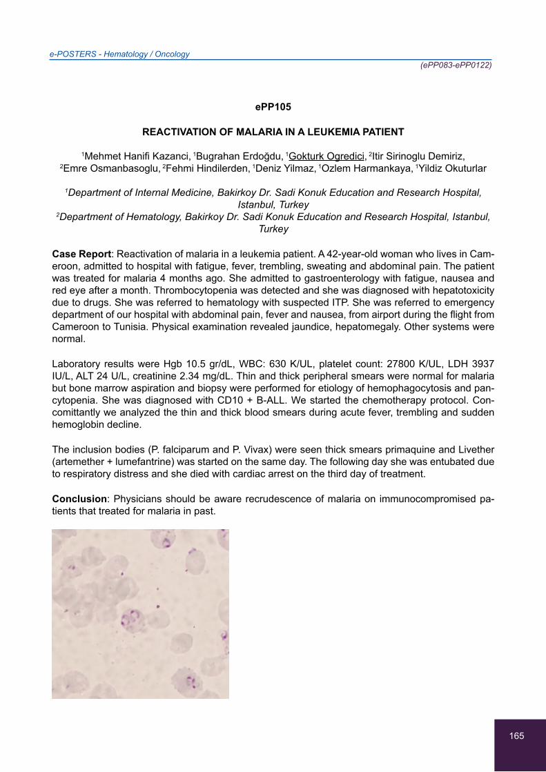
165
e-POSTERS - Hematology / Oncology(ePP083-ePP0122)
ePP105
REACTIVATION OF MALARIA IN A LEUKEMIA PATIENT
1Mehmet Hanifi Kazanci, 1Bugrahan Erdoğdu, 1Gokturk Ogredici, 2Itir Sirinoglu Demiriz,
2Emre Osmanbasoglu, 2Fehmi Hindilerden, 1Deniz Yilmaz, 1Ozlem Harmankaya, 1Yildiz Okuturlar
1Department of Internal Medicine, Bakirkoy Dr. Sadi Konuk Education and Research Hospital, Istanbul, Turkey
2Department of Hematology, Bakirkoy Dr. Sadi Konuk Education and Research Hospital, Istanbul, Turkey
Case Report: Reactivation of malaria in a leukemia patient. A 42-year-old woman who lives in Cam-eroon, admitted to hospital with fatigue, fever, trembling, sweating and abdominal pain. The patient was treated for malaria 4 months ago. She admitted to gastroenterology with fatigue, nausea and red eye after a month. Thrombocytopenia was detected and she was diagnosed with hepatotoxicity due to drugs. She was referred to hematology with suspected ITP. She was referred to emergency department of our hospital with abdominal pain, fever and nausea, from airport during the flight from Cameroon to Tunisia. Physical examination revealed jaundice, hepatomegaly. Other systems were normal.
Laboratory results were Hgb 10.5 gr/dL, WBC: 630 K/UL, platelet count: 27800 K/UL, LDH 3937 IU/L, ALT 24 U/L, creatinine 2.34 mg/dL. Thin and thick peripheral smears were normal for malaria but bone marrow aspiration and biopsy were performed for etiology of hemophagocytosis and pan-cytopenia. She was diagnosed with CD10 + B-ALL. We started the chemotherapy protocol. Con-comittantly we analyzed the thin and thick blood smears during acute fever, trembling and sudden hemoglobin decline.
The inclusion bodies (P. falciparum and P. Vivax) were seen thick smears primaquine and Livether (artemether + lumefantrine) was started on the same day. The following day she was entubated due to respiratory distress and she died with cardiac arrest on the third day of treatment.
Conclusion: Physicians should be aware recrudescence of malaria on immunocompromised pa-tients that treated for malaria in past.
166
e-POSTERS - Hematology / Oncology(ePP083-ePP0122)
ePP106
AETIOLOGY AND OUTCOME OF UPPER EXTREMITY THROMBOSIS
Chaozer Er, Navin Kuthiah
Department of General Medicine, Tan Tock Seng Hospital, Singapore
Background: Upper and lower extremities deep vein thrombosis (DVT) have different aetiologies.
Objectives: To study the epidemiology and outcome of upper extremity venous thrombosis in our institution.
Methods: We retrospectively examined the medical record of all patients with upper extremity ve-nous thrombosis that were managed by our department from 2014 October to 2015 October. We examined the aetiology, treatment duration and treatment outcome of those patients.
Results: Out of the 18 patients, 13 (72%) had DVT, 5 (28%) had superficial venous thrombosis. Out of the 13 patients with DVT, 7 (54%) were due to intravascular catheter, 4 (31%) had malignancy, 1 (7.5%) had Lemierre’s syndrome and 1 case (7.5%) was attributed to immobility from stroke and radial fracture. 8 patients had repeat imaging after DVT treatment, 5 (70%) of them had complete resolution of the DVT within 3 months. 1 patient had complete resolution of DVT shown on venogram performed for other purpose 2 years after DVT treatment. 1 patient had interval DVT improvement shown on repeat imaging 6 months later.
Conclusions: Secondary upper extremity DVTs are mainly contributed by intravascular catheter or malignancy. 3 months of anti-coagulation is generally adequate.

167
e-POSTERS - Hematology / Oncology(ePP083-ePP0122)
ePP107
MULTICENTRIC CASTELMAN’S DISEASE AND SARCOMA KAPOSI IN AN IMMUNOCOMPETENT, HIV (-) PATIENT WITH HERPESVIRUS-8 AND EBSTEIN-BARR VIRUS
CO-INFECTION
1Asimina Koulouridi, 1Emmanouil Tzagarakis, 1Afroditi Koufogianni, 1Sevasti-Alexandra Maragou,
1Despoina-Eleni Arna, 2Ioanna Manika, 1Kyriaki Petraki, 1Melina Kavousanaki, 2Dimitra Liapi,
1Charalampos Lydakis
1B’ Internal Medicine Department, Venizeleio General Hospital, Heraklion, Greece 2Hematology Department, Venizeleio General Hospital, Heraklion, Greece
Background/Aim: Sarcoma Kaposi (SK) and MulticentricCastelman’s Disease (MSD) have rarely been reported in immunocompetent, HIV(-) patients. Human herpes-8 (HHV-8) virus plays an im-portant role to the pathogenesis of these entities. Herein, we report a case of an immunocompetent patient, who was diagnosed with SK and MCD, HHV8(+).
Case Report: A 68-year’s old man, with a medical history of hypertension, with good performance status, presented in the emergency department complaining of fatigue, low-grade fever for two weeks, and loss of 15 kilos of body weight during the last three months. From the clinical examina-tion, subcutaneous nodules of different sizes were apparent on the lower extremities; small cervical, axillary and inguinal lymph nodes, splenomegaly and skin paleness were present. Laboratory tests revealed pancytopenia, elevated erythrocyte sedimentation rate and hypalbuminemia. Computed tomography of the chest and abdomen revealed multiple lymph nodes of >1cm diameter and hepat-osplenomegaly. Histologic specimens were obtained from two different locations. The biopsy results were: SK and HHV-8(+) MCD, from the inguinal node and SK and HHV-8(+) and EBV(+) MCD from the axillary lymph node. After these results, the patient was referred to the hematology and oncology department, where he received liposomal doxorubicin and rituximab with complete response of MCD and partial response of SK.
Conclusion: SK and MCD are common malignancies found in the HIV(+) population, but extremely rare but described in the immunocompetent.
168
e-POSTERS - Hematology / Oncology(ePP083-ePP0122)
ePP108
THINKING OUTSIDE THE BOX IN A BLEEDING PATIENT: A BIZARRE CASE OF ACQUIRED HEMOPHILIA- A CASE REPORT
1Dimitra Kozikopoulou, 1George Karlis, 2Emmanouil Spanou, 1Aikaterini Chasiou, 1Maria Stampori,
1Evagelos Papaoikonomou, 1Chrysostomos Psellas, 1Filippos Papakonstantinou, 1Nikitas Katsilis,
1Stylianos Karatapanis
11st Department of Internal Medicine, General Hospital of Rhodes, Rhodes, Greece 2Hematology Department, General Hospital of Rhodes, Rhodes, Greece
Background/Aim: Acquired hemophilia is a rare coagulation disorder, most frequently caused by autoantibodies against endogenous factor VIII. Most cases involve elderly people (60-70 years old), but there are also cases related to pregnancy. The cause of the disorder may be idiopathic (50% of cases), related to malignancy or autoimmune disorders. The disease presents with minor to severe bleeding, anemia and an isolated prolonged aPTT. We present a case of a 67-year old male with acquired hemophilia A.
Case presentation: Our patient was admitted to the surgical department due to extensive hemato-mas located to both lower limbs and left arm without previous trauma. At admission, he presented with severe anemia and isolated prolonged aPTT. The patient was receiving aspirin 100mg per day. Despite the appropriate daily blood transfusions, the hemoglobin level did not respond. Due to pro-longed aPTT, we conducted a mixing study of his blood and the diagnosis of acquired hemophilia, attributed to the presence of factor VIII inhibitor, was confirmed. He was treated with prednisone, an-ti-inhibitor coagulant complex (FEIBA), cyclophosphamide and rituximab with a very slow response. He was discharged two months later with normalized coagulation studies and adequate levels of hemoglobin.
Conclusion: The initial approach of a patient with hematomas and concurrent treatment with an-tiplatelet drugs is to exclude active bleeding. However, a single test (aPTT) was overlooked. High in-dex of clinical suspicion for acquired hemophilia should be considered in cases of isolated prolonged aPTT, bleeding and anemia.

169
e-POSTERS - Hematology / Oncology(ePP083-ePP0122)
ePP109
POEMS SYNDROME IN A PATIENT WITH PLASMACYTOMA
1Maria Alexandrou, 1Marios Christodoulou, 1Anthoulla Georgiou, 1Maria Nicolaou, 1Stella Fetta,
2Ourania Seimeni, 3Chrisa Shiakalli Tziakouri
1Department of Internal Medicine, Paphos General Hospital, Paphos, Cyprus 2Department of Hematology, Limassol General Hospital, Limassol, Cyprus
3Department of Radiology, Larnaca General Hospital, Larnaca, Cyprus
Background: POEMS (polyneuropathy, organomegaly, endocrinopathy, monoclonal gammopathy and skin changes) syndrome is a paraneoplasmatic syndrome due to underlying plasma cell neo-plasm. Physicians seem to be unfamiliar with the diagnosis of this rare, chronic and disabling condi-tion. We discuss a case of diagnosis of Poems syndrome presenting with polyneuropathy and lower limbs edema in a patient with underlying plasmacytoma.
Case report: A 56-year-old man presented to our hospital due to lower extremities paresthesia and weakness of six months duration. Clinical examination revealed areflexia of both lower and upper limps, as well as edema of the lower limbs and hypogonadism’s symptoms. Remarkable laboratory findings were thrombocytosis, erythrocytosis and lambda light chain restricted monoclonal gammop-athy, in serum immunofixation. Neurological assessment revealed the diagnosis of Chronic Inflam-matory Demyelinating Polyneuropathy (CIDP). Bone marrow biopsy showed normal cellularity which excluded the diagnosis of multiple myeloma. A full body CT scan showed splenomegaly, axillary and inguinal lymphadenopathy and a lytic bone lesion of the right ilium and ischium. Histology examina-tion of biopsy specimen from the lesion demonstrated a plasmacytoma.
Conclusion: Diagnosis of POEMS should be considered within the differential diagnosis in patients with polyneuropathy and biopsy of bone lesions can be useful for diagnosis of POEMS syndrome in difficult cases.
170
e-POSTERS - Hematology / Oncology(ePP083-ePP0122)
ePP110
VARIABLE IMMUNODEFICIENCY MANIFESTED AS AUTOIMMUNE HEMOLYTIC ANEMIA - AN INTERESTING CASE
1Emmanouil Spanou, 2Ourania Kariki, 2Despoina Diakonikola, 2Efstathia Anastasiou,
2Georgios Ntetskas, 2Nikitas Katsilis, 2Maria Stampori, 2Aikaterini Chasiou, 2Dimitra Kozikopoulou,
2Stylianos Karatapanis
1Hematology Department, General hospital of Rhodes, Rhodes, Greece 21st Department of Internal Medicine, General Hospital of Rhodes, Rhodes, Greece
Background/aim: Common variable immunodeficiency (CVI) is a primary immunodeficiency disor-der characterized by low levels of most or all of the immunoglobulin classes, a reduction of B lym-phocytes or plasma cells and clinically vulnerability to bacterial infections. VI is not a single disease but rather a collection of hypogammaglobulinemia syndromes resulting from many genetic defects. In the majority of cases, the diagnosis is not made until the third or fourth decade of life. The main complications of the disorder are considered to be the recurrent infections which can lead to perma-nent damage to organs involved, the autoimmune manifestations and the tendency to malignancies especially lymphomas. We present a patient diagnosed with autoimmune hemolytic anemia as a first manifestation CVI disorder.
Case Report: A 49-year-old male patient was admitted to our hospital because of anemia (Ht=15%). The laboratory tests showed findings compatible with the presence of autoimmune hemolysis (Coombs test+). Significant hepatosplenomegaly was noticed (20 cm and 17 cm respectively). The serum immunology tests were negative. A bone marrow biopsy and aspiration was performed which showed no infiltration from lymphocytes. The serum protein electrophoresis was abnormal (low gamma globulins levels). All serum immunoglobulins were low (IgG= 624, IgA=27.8, IgM= 33.6). The patient was initially treated with corticosteroids with no response. He consequently treated with rituximab with success.
Conclusion: The CVI is a rare hematologic disease which can manifest at first with autoimmune disorders such as autoimmune hemolytic anemia. The treatment with corticosteroids can be insuf-ficient. Therefore, the administration of rituximab may be necessary.
171
e-POSTERS - Hematology / Oncology(ePP083-ePP0122)
ePP111
AGRANULOCYTOSIS IN A PATIENT WITH UNDIAGNOSED T-LEUKEMIA, AGGREVATED BY LEFLUNOMIDE
1Despoina-Eleni Arna, 1Emmanouil Tzagarakis, 1Asimina Koulouridi, 1Sevasti-Alexandra Maragou,
1Aggeliki Kipraki, 1Ifigenia Kassotaki, 1Melina Kavousanaki, 1Kyriaki Petraki, 2Dimitra Liapi,
1Charalampos Lydakis
1B’ Internal Medicine Department, Venizeleio General Hospital, Heraklion, Greece 2Hematology Department, Venizeleio General Hospital, Heraklion, Greece
Background/Aim: Agranulocytosis is a rare condition with a reported incidence ranging from 1 to 5 cases per million yearly. Association with medications can be found in about 70% of cases, but only a very low percentage involves the use of leflunomide (ARAVA). Herein, we report a case of agranu-locytosis in a patient, who received leflunomide for a rheumatologic disease under investigation but which eventually proved to be T-leukemia.
Case report: A 59year old woman, with a medical history of hypothyroidism and hypertension, pre-sented in the emergency department, when agranulocytosis (WBC 174/μL) and severe neutropenia (Poly 1/μL) were detected in regularly ordered blood tests. The patient was recently investigated for a rheumatologic disease, and a low-grade leucopenia (WBC 4000/μL. During the past month, she had received leflunomide, for 10 days, ceased due to upper respiratory infection. 20 days later she presented with neutropenia. The clinical examination was normal throughout hospitalization. Extended laboratory and invasive exams have been performed, without significant findings. Bone marrow aspirate revealed no granular array cells. She received filgrastim (G-CSF) 48ui/day and broad-spectrum antibiotics, due to low grade fever. Since neutrophils were not increased, on the 7th day cholestyramine was added, to enhance the clearance of the metabolites of leflunomide. On day 8, neutrophils were 200/μL and on day 11 2.200/μL. Bone morrow aspirate finally revealed T-lymphocyte infiltration, compatible with LGL-T leukemia. She was referred to the hematology de-partment.
Conclusions: Agrunolocytosis is a rare, drug-induced complication, rarely described for lefluno-mide, where use of cholestyramine may be helpful.
172
e-POSTERS - Hematology / Oncology(ePP083-ePP0122)
ePP112
MEGALOBLASTIC ANEMIA WITH MASSIVE SPLENOMEGALY: COMPLETE RESPONSE AFTER B12 VITAMIN REPLACEMENT
Firuze Midi Kursat, Betül Erişmiş, Hakan Kursat, Gamze Gülçiçek, Yildiz Okuturlar
Department of Internal Medicine, Bakirkoy Dr. Sadi Konuk Education and Research Hospital, Istanbul, Turkey
Introduction: The most common explanations for poor vitamin B12 status are a low dietary intake of the vitamin (i.e., a low intake of animal-source foods) and malabsorption. We report an atypical clinical observation of megaloblastic anemia (MA), which is especially interesting in that it illustrates the difficulty in approaching a case of massive splenomegaly.
Case description: A 42-year-old male presented to the internal medicine outpatient clinic with one month history of fatigue. His blood pressure was 110/80 mmHg and all vital findings were normal. Findings on lymph node examinations were unremarkable. Abdominal examination revealed 5 cm splenomegaly. Laboratory findings reavealed severe macrocytic anaemia (haemoglobin, 4,58 g/dL; MCV 112 fL) in addition to leucopenia(2930/mm3) and thrombocytopaenia(44000/mm3), haemolysis (total bilirubine 2,33 mg/dL, indirect bilirubine 1,35 mg/dl), increased LDH (2134 U/L) and reticulo-cyte count, decreased haptoglobin levels, negative direct coombs test and severe vitamin B12 defi-ciency (<50 pg/ml). Abdominal ultrasonography showed massive splenomegaly (213x72 mm). Bone marrow aspiration and biopsy showed no lymphoproliferative but megaloblastic changings. Follow-ing cobalamin replacement therapy, the patient reported increased well-being, including appetite and weight gain. In the follow-up laboratory examinations, leucocyte and platelet counts in addition to serum bilirubin and LDH levels normalised. At the end of 2 months, laboratory findings, includ-ing haemoglobin level were all within the normal range and radiologic findings showed decrease in spleen size.
Discussion: Systemic disorders, chronic myeloproliferative/ lymphoproliferative disorders often can cause massive splenomegaly. Splenomegaly with pancytopenia is an unusual association of MA as most of the literature suggest only severe anemia as predominant symptom of MA.
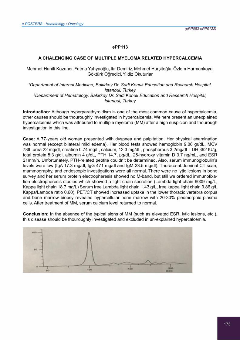
173
e-POSTERS - Hematology / Oncology(ePP083-ePP0122)
ePP113
A CHALENGING CASE OF MULTIPLE MYELOMA RELATED HYPERCALCEMIA
Mehmet Hanifi Kazancı, Fatma Yahyaoğlu, Itır Demiriz, Mehmet Hurşitoğlu, Özlem Harmankaya,
Göktürk Öğredici, Yildiz Okuturlar
1Department of Internal Medicine, Bakirkoy Dr. Sadi Konuk Education and Research Hospital, Istanbul, Turkey
2Department of Hematology, Bakirkoy Dr. Sadi Konuk Education and Research Hospital, Istanbul, Turkey
Introduction: Although hyperparathyroidism is one of the most common cause of hypercalcemia, other causes should be thouroughly investigated in hypercalcemia. We here present an unexplained hypercalcemia which was attributed to multiple myeloma (MM) after a high suspicion and thourough investigation in this line.
Case: A 77-years old woman presented with dyspnea and palpitation. Her physical examination was normal (except bilateral mild edema). Her blood tests showed hemoglobin 9.06 gr/dL, MCV 78fL.urea 22 mg/dl, creatine 0.74 mg/L, calcium, 12.3 mg/dL, phosphorous 3.2mg/dL LDH 392 IU/g, total protein 5.3 g/dl, albumin 4 g/dL, PTH 14.7, pg/dL, 25-hydroxy vitamin D 3.7 ng/mL, and ESR 21mm/h. Unfortunately, PTH-related peptite couldn’t be determined. Also, serum immunoglobulin’s levels were low (IgA 17.3 mg/dl, IgG 471 mg/dl and IgM 23.5 mg/dl). Thoraco-abdominal CT scan, mammography, and endoscopic investigations were all normal. There were no lytic lesions in bone survey and her serum protein electropheresis showed no M-band, but still we ordered immunofixa-tion electropheresis studies which showed a light chain secretion (Lambda light chain 6009 mg/L, Kappa light chain 18.7 mg/L) Serum free Lambda light chain 1.43 g/L, free kappa light chain 0.86 g/L Kappa/Lambda ratio 0.60). PET/CT showed increased uptake in the lower thoracic vertebra corpus and bone marrow biopsy revealed hypercellular bone marrow with 20-30% pleomorphic plasma cells. After treatment of MM, serum calcium level returned to normal.
Conclusion: In the absence of the typical signs of MM (such as elevated ESR, lytic lesions, etc.), this disease should be thouroughly investigated and excluded in un-explained hypercalcemia.
174
e-POSTERS - Hematology / Oncology(ePP083-ePP0122)
ePP114
2 CASES OF LEMIERRE’S SYNDROME: DO WE ANTI-COAGULATE?
Chaozer Er, Navin Kuthiah, Siow Yun Wong, Samuel Kang Lian Lee
Department of General Medicine, Tan Tock Seng Hospital, Singapore
Background: The role of anti-coagulation in Lemierre’s syndrome is controversial. We present two cases of Lemierre’s syndrome: one was anti-coagulated while the other was not.
Case1. 19-year-old gentleman presented with 1 week of fever and cough. Chest X-ray showed left sided pneumonia. Blood culture grew Fusobacterium necrophorium. He was intubated for respira-tory failure. Computed tomography (CT) of neck, chest, abdomen and pelvis showed left sided con-solidation with small developing lung abscess, large left pleural effusion, septic emboli in bilateral lungs, left internal jugular vein (IJV) thrombosis and bilateral mastoiditis. He was given 4 days of Enoxaparin. Repeat CT in 2 weeks showed interval recanalization of left IJV. With antibiotic and surgical decortication of lung, patient recovered well.
Case2. 70-year-old Indian lady was treated as asthma exacerbation. CT brain and cervical spine was done as she had a traumatic head injury following a fall. It showed marked swelling of the cervi-cal prevertebral soft tissues with narrowing of the airway. Patient developed stridor and worsened dyspnoea that required intubation. Magnetic resonance imaging (MRI) of neck showed multiple neck abscess and left IJV thrombosis. She completed 3 months of anti-coagulation. Repeat CT 3 months later showed resolution of IJV thrombosis. With antibiotic and repeated surgical drainage, patient recovered well. Intra-operative culture grew Klebsiella pneumoniae.
In conclusion, Lemierre’s syndrome have been treated successfully with or without anti-coagulation previously. Our cases shown the same finding. More prospective studies are required to provide definite answer.

175
e-POSTERS - Hematology / Oncology(ePP083-ePP0122)
ePP115
UNUSUAL INR PROLONGATION DUE TO CHILI PEPPER CONSUMPTION
Aikaterini Volonaki, Alexandros Kyriakos
Euromedica General Clinic, Rhodes, Greece
Background/Aim: To report an INR prolongation aetiology, not described in the literature before.
Case Report: A 55-year-old triathlon athlete underwent preoperative testing for scheduled shoulder surgery. Surprisingly his INR was 1.57, aPTT 34 sec and the rest of his lab tests were within normal limits. No history of smoking, alcohol consumption or NSAID intake. He reported mild bleeding upon minor lacerations but no mucosal bleeding or bruising. He underwent a thorough investigation with liver function tests and imaging that were normal. He confessed consuming at least 15 chili pep-pers (Capsicum annuum sp) a day for energy purposes. As no other cause was identified, he was instructed to refrain from chili peppers. A repeat INR that was performed 15 days later was 1.05. He was allowed to undergo surgery and a normal INR was found again. Quick time prolongation was witnessed a new 2 weeks after returning to his regular diet which included chili peppers, again with a normal aPTT. To our knowledge, capsaicin does not affect lab tests for clotting times, even though it has a dose-dependent action on platelet aggregation, factor VIII:C and factor IX concentration.
Conclusion: Further investigation is warranted for capsaicin medicinal actions and INR should be considered especially in areas where spice rich diet is seen.
176
e-POSTERS - Hematology / Oncology(ePP083-ePP0122)
ePP116
PATIENT WITH RECURRENT STROKE AND SPLENOMEGALY
1Andreas Kostis, 1Pinelopi Nicolaou, 1Maria Vrasida, 1Elena Georgiadou, 2Nikolaos Hadjisavvas,
1Emmelia Vounou
1 Unit- medical ward, Internal Medicine Department, Limassol General Hospital, Limassol, Cyprus 2Medical school, University of Nicosia, Nicosia, Cyprus, St George’s University of London, UK
Background: splenic marginal zone lymphomas are related to secondary antiphospholipid syn-drome. Case report: 57 years old patient, history of hypertension, hyperlipidaemia, gout. Presented to the A&E, February 2016, with right hemiparesis, dysarthria and diagnosed with stroke. Further management and control of risk factors were provided. Five months later (7/2016), he returned com-plaining of fatigue and weight loss. On investigation, anaemia, splenomegaly and mesenteric lymph nodes (smaller than 1cm) were found, no peripheral lymph nodes. Bone marrow and trephine biopsy were performed twice. The histology showed myelofibrosis. The patient presented again two months later (9/2016) with recurrent symptoms of stroke.
On investigation, his spleen was not enlarged and he had no anaemia. No treatment was adminis-tered since his last hospitalisation, apart from aspirin and clopidogrel. CT brain was with no findings but the MRI brain revealed stroke in the left pons. From investigation performed, high titres of anti-bodies against La, cardiolipin and β2- glycoprotein consistent with antiphospholipid syndrome were found. After repeated analysis at two months, he was started on anticoagulants, with the possible di-agnosis of antipospholipid syndrome. This could also explain the enlarged spleen and lymph nodes that disappeared with antiplatelets. On follow up, he developed splenomegaly, cervical lymph nodes, anaemia and pruritus. At this point, biopsy of the lymph nodes and splenectomy were performed. The histology showed splenic marginal zone B-cell lymphoma.
Conclusion: Rare causes of stroke, as secondary antiphospholipid syndrome, should be consid-ered in patients with recurrent strokes and other manifestations of a systemic disease.

177
e-POSTERS - Hematology / Oncology(ePP083-ePP0122)
ePP117
THROMBOTIC THROMBOPENIC PURPURA IN PREGNANCY: ONE RARE CASE OF FATAL INTRACEREBRAL HEMORRHAGE
1Athina Dimosiari, 1Christina Kydona, 1Konstantina Skarmoutsou-Tsimirika, 2Maria Sidiropoulou,
1Petros Doumtsis, 1Christos Vettas, 1Alexandros Garyfallos
14th Department of Internal Medicine, Hippokration Hospital, School of Medicine, Aristotle University of Thessaloniki, Thessaloniki, Greece
2Radiology Department, Hippokration Hospital, Thessaloniki, Greece
Thrombotic thrombopenic purpura (TTP) is a rare microangiopathitic hemolytic anemia, along with thrombocytopenia and neurological deficits, associated with several underlying conditions. It usually occurs in autoimmune diseases, with inhibition of ADAMTS13 by autoantibodies or during preg-nancy irrelevantly to autoimmunity, congenitally or acquired. Here we report a rare case of TTP in a 37-year old pregnant woman, with a TTP-free previous pregnancy and no identifiable underlying trigger. She presented with thrombopenia, microangioathitic hemolytic anemia, schistocytes in the peripheral blood smear, elevated LDH and signs of low fetal pulse.
She was treated with emergency caesarian section, daily plasmapheresis and intravenous pred-nisolone, resulting in remission within 3 days. All serological tests, viral and immunological profile were negative. Brain, chest and abdomen imaging did not uncover malignancy or other disorder. With remission established, on day 9, the patient presented with acute headache, diplopia, hyperten-sion and recurrence of thrombopenia, with brain imaging findings of Posterior Reversible Engepha-lopathy Syndrome, ending in intubation, due to rapid deterioration of conscious level (GCS:11). Si-multaneously the TTP relapsed aggressively, advancing in brain hemorrhage shortly after intubation. Although plasmapheresis was resumed intensively twice daily and various immunosuppressants were used -high dose prednisolone, intravenous immunoglobulin, rituximab- the patient deteriorated massively, with hemopericardium, lung alveolar hemorrhage, uncontrollable intracerebral bleeding with brain death.
Despite all efforts, our patient died on day 17, underlying that TTP is a rapidly deteriorating serious disease, leading to early death if not drastically treated. With a high relapse risk upon minor trigger, long intensive therapy and monitoring remain crucial.

178
e-POSTERS - Hematology / Oncology(ePP083-ePP0122)
ePP118
TINNITUS AS THE PRESENTING SYMPTOM OF ESSENTIAL THROMBOCYTHEMIA
Mahmoud Husni Ayesh (Haj Yousef)
Department of Medicine, King Abdullah University Hospital, Irbid -Jordan, Faculty of Medicine, Jordan University of Science and Technology, Irbid -Jordan
Background and purpose: Essential thrombocythemia (ET) is clonal myeloproliferative disorder (MPD), clinical picture is dominated by a predisposition to vascular occlusive events and hemor-rhages. Clinical manifestations of ET in adult Jordanian patients are lacking.
Aim: To describe and find the early symptoms of ET in adult Jordanian patients Methods: A retro-spective hospital-based study was conducted. All cases of ET diagnosed during the period January September 2002 October2017 at King Abdullah University Hospital were included in the study. The early symptom of the patient was extracted from the clinical note of the patients.
Results: 26 cases met the inclusion criteria for the diagnosis of ET, mean age 42 year (range 29-76), male/female ratio (43%/57%). Tinnitus as an early symptom of ET was found in 11 patients (42%) , asymptomatic in 6 patients (23 %), three patients with headache (12%), three patients with cerebrovascular accident (12%), two patients with portal vein thrombosis (8%), and one patient with hematuria (4%).
Conclusion: Tinnitus was the early symptom of ET in the majority of our patients. Thrombotic mani-festations were not common in our ET patients. We recommend that patients with tinnitus should check their platelet.
179
e-POSTERS - Hematology / Oncology(ePP083-ePP0122)
ePP119
FATAL HEMOPHAGOCYTIC LYMPHOHISTIOCYTOSIS, PROBABLY SECONDARY TO LEISHMANIASIS, IN AN IMMUNOCOMPETENT ADULT
1Asimina Koulouridi, 1Emmanouil Tzagarakis, 1Sevasti-Alexandra Maragou, 2Ioanna Manika,
1Despoina-Eleni Arna, 1Afroditi Koufogianni, 1Melina Kavousanaki, 2Dimitra Liapi, 1Kyriaki Petraki,
1Charalampos Lydakis
1B Internal Medicine Department, Venizeleio General Hospital, Heraklion, Greece 2Hematology Department, Venizeleio General Hospital, Heraklion, Greece
Background/ Aim: Hemophagocytic lymphohistiocytosis (HLH) is a rare syndrome, attributed to ge-netic factors, inflammatory/rheumatologic diseases, infections, or other, rare conditions. HLH is as-sociated with overactivation of macrophages and overproduction of inflammatory cytokines. Herein, we describe a rare case of an immunocompetent patient, diagnosed with HLH, who never responded to treatment.
Case Report: A 53 year’s old man, with a medical history of smoking and stroke, presented in the ER with fever of 10 days. The patient had been recently hospitalized for fever that responded to doxycycline. The patient underwent a full clinical and laboratory work-up, in the context of FUO, with no diagnostic findings, apart from positive IgG antibodies and PCR for Leishmania in the blood and bone marrow respectively. Though, no Leishmania amastigotes were found in the bone marrow aspirate. The patient received amphotericin B without remission of the fever, while his blood tests revealed thrombocytopenia, anemia, high ferritin levels and hypertriglyceridemia. Ultrasound and computed tomography showed mild splenomegaly. Diagnostic criteria of hemophagocytic syndrome were fulfilled, without a specific cause found, apart from probable leishmaniasis. Genetic testing for idiopathic HLH was negative. The patient received a second cycle of amphotericin, with no satisfac-tory response. Finally, he received etoposide, cyclosporine and dexamethasone, according to 2004 HLH protocol, with partial response to the treatment. After a long hospitalization, several complica-tions and hospital-acquired infections the patient passed away.
Conclusions: HLH is a rare, though difficult to diagnose and manage complication in several severe infections or inflammatory situations, needing prompt treatment.
180
e-POSTERS - Hematology / Oncology(ePP083-ePP0122)
ePP120
ΑDULT ONSET CYCLIC NEUTROPENIA ΙΝ 71 ΥΕARS OLD WOMAN
Anna Makina, Eirini Louka, Ioanna Skouri, Basileios Gogolas, Nikolaos Koutsoumpos,
Ioulia Kontolaimou, Efrosini Roulia, Zoi Alexiou
2nd General Department of Internal Medicine, General Hospital of Elefsis “Thriasio”, Elefsina, Greece
Background -Aim: Cyclic neutropenia is a rare blood disorder characterized by recurrent episodes of low levels of neutrophils. Neutropenia occur every 21 days and may last 4 to 6 days. These pa-tients are susceptible for recurrent infections, sometimes life threatening. In our case, we describe a case of cyclic neutopenia in an adult patient.
Case Report: A 71 years old woman was admitted to our Department with recurrent episodes of diarrhoea, apthous stomatitis, ulcers in perineum and fever for the past 6 months. She complained for sore throat and dysphagia. She had no family history of similar symptoms, she didn’t take any blameworthy drugs. Medical history with recurrent infections of upper respiratory system.
Physical examination: BP 90/60 mmHg, Τ: 36,.8°C, HR: 98/min, RR: 24/min. Apthous ulcers in oral mucosa and perineum. General abdominal tenderness, rest of the physical examination (-). Labora-tory tests Hct 28%, Hb 8,7, Neut 0,483 x10³/uL, BUN 128 mg/dl, creatinin 2.8 mg/dl, C3, C4 nor-mal. From her previous report referred three other same neutropenic episodes in last 6 months. All bloodstream cultures were without pathogens, stool and urine culture (-). All body Ct scan (-). Colon-oscopy: ulcers in ileocecal valve. Bone marrow study revealed increased number of large granular Lymphocytes and decreased neutrophil precursors. She was diagnosed with adult onset cyclic neu-tropenia and was treated with G-CSF and Pip/taz and linezolid iv, her condition improved.
Conclusions: Cyclic neutropenia usually presents in children but it must be considered in the dif-ferential diagnosis of neutropenia in adult patients where occurs spontaneously.
181
e-POSTERS - Hematology / Oncology(ePP083-ePP0122)
ePP121
PRIMARY CENTRAL NERVOUS SYSTEM LYMPHOMA IN A PATIENT WITH A CLINICAL MANIFESTATION OF FACIAL NERVE PALSY: CASE REPORT
1Andreas Tofarides, 1Eleni Papanicolaou, 1Marios Petrou, 1Aram Katsios, 1Christina Flourou, 1Andreas Ioannou, 2Constantinos Kaouranis, 2Michalis Michael, 1Chrysostomos Zintilis
1Internal Medicine Department, Nicosia General Hospital, Nicosia, Cyprus 2Hematology Department, Nicosia General Hospital, Nicosia, Cyprus
Background/ Aim: Primary Central Nervous System Lymphoma (PCNL) is a rare form of Non-Hodgkin Lymphoma, at a rate of 2-3% of all brain tumors, in immunocompetent patients. It is well known the immunocompromised patients are at greater risk of developing lymphoma. Facial nerve paralysis should be thoroughly investigated as it is observed from this case report, having a wide range of causal etiologies.
Case report: We report the case of 65 -year-old Greek-Cypriot patient, with past medical history of Parkinson’s disease, arterial hypertension, hyperlipidemia with first right facial nerve palsy and then bilateral, right ophtalmoplegia and right hearing reduction. Laboratory investigation and CT +MRI brain was without significant findings. Due to worsening clinical situation of the patient instead of giving acetylsalicylic acid and methylprednisolone we have made an MRI eye -socket and internal auditory meatus with of evidence of enhanced the nerves of bilateral internal auditory, trigeminal and occulomotor nerves. Cerebrospinal fluid examination (CSF) showed increased leukocytes with lym-phocytic type WBC 630/μl with 95% Lympho, increased protein 540 mg/dl and hypoglycaemia CSF 3 dl/dl, and cytologist positive for malignancy. The flow cytometry showed clonality of lymphocytes. Then the patient received a high dose methotrexate regimen according Memorial protocol with no clinical response, week later patient died.
Conclusion: Primary Central Nervous System Lymphoma (PCNL) may appear with clinical hetero-geneity and must be differentiated from vascular lesions, chronic granulomatous diseases and other neurological diseases.

182
e-POSTERS - Hematology / Oncology(ePP083-ePP0122)
ePP122
AN ATYPICAL CASE OF THROMBOTIC THROMBOPENIC PURPURA
1Konstantina Goula, 2Panagiotis Zikos, 1Dionisios Adrahtas, 1Ourania Drakoulogkona, 1Zoi Tegou
1Nephrology Department, “St. Andrews” General Hospital of Patras, Patras Greece 2Hematology Department, “St. Andrews” General Hospital of Patras, Patras, Greece
Background: Thrombotic thrombopenic purpura is a disorder of the blood-coagulation system char-acterized by clotting in small blood vessels resulting in thrombopenia and is caused by reduced activity of ADAMTS13. Cancer patients are at high risk for hematologic complications such as TTP. We report a rare case of prostate cancer which presented as TTP and acute kidney injury.
Case Report: A 68 year old man was admitted because of oliguria, abdominal pain, nausea, low grade fever since three days and lower urinary tract symptoms since one year. Past medical his-tory included hypertension, nephrolithiasis and gastrectomy. Clinical examination was unremarkable except for two small bruises on lower abdomen and on left leg. Laboratory examinations showed urea:189 mg/dl, creat:9,1 mg/dl, LDH:2437 U/L, platelet count 60.000/μL, Hb:12,7g/dl, fibrinogen 382 mg/dl , D-dimers 2,42 mg/l. Peripheral blood smear revealed schistocytosis and microangiopathic anemia consistent with a diagnosis of TTP. He was started on plasma exchange therapy, corticoids and dialysis. Renal function and hematologic parameters restored five days later. During investiga-tion for secondary causes of TTP, a high PSA was found (108 ng/μl) and a prostate biopsy revealed an invasive adenocarcinoma. The patient was treated with bicalutamide and leuprorelin. Two months after admission, he was in good clinical condition, PSA levels were falling and no plasma exchange therapy was required.
Conclusions: Cancer patients can be complicated by hemostasis disorders and TTP is in the dif-ferential diagnosis. Εarly initiation of plasma exchange is mandatory in some cases for recovery.

183
e-POSTERS - Cardiovascular Diseases(ePP123-ePP141)
ePP123
CLINICAL CHARACTERISTICS AND TREATMENT OF ATRIAL FIBRILLATION IN PATIENTS WITH CORONARY ARTERY DISEASE
George Dividis, George Fotos, Athanasios Samaras, Eleni Paschou, Dimitra Vasdeki,
Giannis Goulas, Eleni Kotsi, Tasos Kartas, Paraskevi Tsoukra, Evropi Forozidou, Apostolos Tzikas,
Haralampos Karvounis, George Giannakoulas
1st Department of Cardiology, AHEPA University Hospital, Thessaloniki, Greece
Background: The population of patients with coexisting coronary artery disease (CAD) and non-val-vular atrial fibrillation (NVAF)is increasing.In the era of novel oral anticoagulant (NOACs) there is a need for real world data on this subset of patients. Purpose To investigate the clinical characteristics and anticoagulation strategy in patients with CAD in routine clinical practice in a tertiary university hospital
Methods: We studied a population of 735 consecutive patients who were hospitalized to the cardiol-ogy department with any diagnosis and coexisting NVAF.History of CAD was defined as documented CAD, prior myocardial infarction,and/or history of coronary revascularization.
Results: A total of 336(45%) had CAD.These patients had higher CHA2DS2-VAScscore[CAD4.8(±1.6) vs noCAD3.8(±2.0),p<0.001],HAS-BLED score[CAD2.9(±1.0)vs noCAD2.4(±1.1),p<0.001].Comor-bidities such as diabetes mellitus [CAD n=151 vs noCAD n=108 p<0.001],heart failure [CAD n=206 vs noCAD n=126, p<0.001],chronic kidney disease [CAD n=75 vs noCAD n=40,p<0.001]and dyslipi-demia [CAD n=222 vs noCAD n=128, p<0.001]were found to be more frequent in patients with CAD.Patients with CAD had also more often a history of stroke/transient ischaemic attack[CAD n=69 vs noCAD n=59, p=0.033].Patients with CAD were less likely to be cardioverted pharmaceutically[CAD n=108 vs noCAD n=167, p=0.009].Single(SAPT) and dual antiplatelet treatment (DAPT) was more commonly used in patients with CAD[SAPT CAD n=57 vs noCAD n=19,p<0.001, DAPT CAD n=26 vsnoCAD n=16, p<0.001].Patients with CAD who received oral anticoagulation at discharge,were less often received NOACs [NOAC n=121 vs vitamin K antagonist n=115,p<0.001].
Conclusions: Comorbidities and cardiovascular risk factors are more frequent present in patients with CAD and NVAF.Pharmaceutical cardioversion is less often utilised in these patients in the real world.
184
e-POSTERS - Cardiovascular Diseases(ePP123-ePP141)
ePP124
GLUCAGON-LIKE PEPTIDE 1 RECEPTOR AGONISTS AND PROTECTION AGAINST STROKE: A SYSTEMATIC REVIEW AND META-ANALYSIS
Haralampos Milionis, Fotios Barkas, Moses Elisaf
Department of Internal Medicine, School of Medicine, University of Ioannina, Ioannina, Greece
Background: Glucagon-like peptide 1 receptor (GLP-1R) agonists, a novel class of antidiabetic agents, have been reported to exert neuroprotective effects in experimental stroke models, espe-cially if treatment starts before stroke.
Objective: To assess available data regarding the risk of stroke in diabetic individuals treated with GLP-1R agonists.
Methods: We conducted a meta-analysis of randomized, placebo-controlled trials (RCTs) involving GLP-1R agonists with cardiovascular outcomes and/or safety as primary endpoints. Results Four eligible multicenter (n=32,895) prospective double-blinded RCTs (ELIXA, LEADER, SUSTAIN, EX-CEL) were published up to November 2017. The analysis showed a non-significant trend towards a reduction of the risk of stroke in patients taking GLP-1R agonists compared with placebo (odds ratio, OR 0.88, 95%CI: 0.76-1.03, p=0.109; Q-value =3.57, p=0.31; I2= 15.95%). After excluding ELIXA trial, which included patients at high cardiovascular risk with a recent myocardial infarction, the net benefit was significant (OR 0.85, 95%CI:0.74-0.99, p=0.04; Q-value = 2.145, p=0.342; I2 = 6.76%).
Conclusions: Available data from RCTs dedicated to cardiovascular safety at least partially support the promising findings from previous experimental studies indicating protective effects of treatment with GLP-1R agonists against stroke. Nonetheless, the issue of neuroprotection ofthese agents needs to be investigated in future clinical studies focusing on stroke.

185
e-POSTERS - Cardiovascular Diseases(ePP123-ePP141)
ePP125
DESCRIPTION OF BRASH SYNDROME: BRADYCARDIA-RENAL FAILURE- AV NODE BLOCKADE-SHOCK-HYPERKALEMIA
1Nikolaos Vryzas, 2Alexandros G. Sachinidis, 3Ioannis Drikos, 2Agathoklis Efthimiadis,
1Efthalia Damvopoulou, 4Afroditi Anastasiadou, 1Georgios Katseas
1Department of Cardiology, General Hospital of Pella, Department of Giannitsa, Giannitsa, Greece 2Second Propedeutic Department of Medicine, Medical School, Aristotle University of Thessaloniki,
Hippokration Hospital, Thessaloniki, Greece 31st Pediatric Clinic, General Hospital of Elefsina, Thriassio, Greece
4Department of Internal Medicine, General Hospital of Pella, Department of Giannitsa, Giannitsa, Greece
Aim: BRASH syndrome refers to the constellation of bradycardia, renal impairment, av nodal block-ade, shock and hyperkalemia. Patients may be presented quite often with this clinical picture and laboratory findings and may seem difficult to treat, unless someone promptly recognizes the inter-connection between the mechanisms that make up this vicious cycle. We illustrate the case of a 68 year old man that was presented in the emergency department of our hospital with BRASH syn-drome and we analyze the pathophysiology behind it.
Case Report: Our patient was presented with excessive diaphoresis, dyspnea due to bronchospasm and hyperglycemia. He was treated with carvedilol, bisoprolol and telmisartan for arterial hyperten-sion, oxygen therapy and nebulizers for obstructive pulmonary disease, glargine insulin for diabetes type 2 and atorvastatin for dylsipidemia. His medications also included alfuzosin for benign prostatic hyperplasia and valproic acid, amisulpride and venlafaxine for bipolar disorder. We emphasize the synergistic actions of hyperkalemia and av nodal blockade to cause bradycardia that is dispropor-tionate for the levels of potassium. The acute renal impairment caused by bradycardia resulted in reduced clearance of valproic acid, amisulpride and venlafaxine which led to profound hyponatremia (Na 112 mEq/L). Telmisartan may further deteriorate kidney perfusion which in turn worsens hyper-kalemia and the vicious cycle continues.
To conlude, it should be noted that polypharmacy and comorbidities of patients that are presented with BRASH may further complicate the case and thus the early recognition of BRASH as a pattern may facilitate diagnosis and treatment.
186
e-POSTERS - Cardiovascular Diseases(ePP123-ePP141)
ePP126
DIMENSIONAL CHANGES IN THE LEFT VENTRICLE DUE TO THE EFFECT OF HYPERTENSION IN PATIENTS WITH STABLE ANGINA
1Dilbar Mohammed Saleem, 1Dhanarajan Vishnu, 1George Aneeta, 2Hrechko Svitlana
1Department of Internal Medicine, Bukovinian State Medical University, Chernivtsi, Ukraine 2Cardiology, Department of Hypertension, Regional Clinical Cardiology Dispensary,
Chernivtsi, Ukraine
Ischemic Heart Disease combined with Hypertension is known world wide to be the main cause of mortality.Disorders of Left ventricular geometry and it’s function are highly prevalent in this vulner-able population.Although routine screening by Echocardiography is critically volume dependent and prone to underestimation in LV ejection fraction.
To determine the effect of hypertension on the Functional state of the left ventricle in patients with stable angina as a predictor of mortality.The study involved 84 patients hospitalized in the Chernivtsi Regional Cardiology Clinic.The LV functional status of the patients were evaluated without using drugs and then underwent Bicycle Ergometry according to the modified Bruce protocol.The echocar-diographic examination was done and the results were analysed in groups; Group I-patients with stable angina, Class III hypertension, Group II-patients with stable angina, hypertension and heart failure, Group III-patients with stable angina without hypertension.Left ventricular mass index was significantly higher in patients with hypertension at hospital admission and during diagnostic stress test.LV internal dimension in systole was also higher in patients from group II-8.8% compared with group I.LV internal dimension in diastole was high in all groups and the maximum-12.0%(p<0.05) in patients from group I.A significant decrease in LV ejection fraction was noted in patients with hypertension at all stages of the study with a decline in group I of 20.3%(p<0.05) in group II of 28.4%(p<0.05).
In conclusion, patients with stable angina associated with hypertension were found to have a greater reduction in left ventricular systolic function which points to an increased risk of mortality.
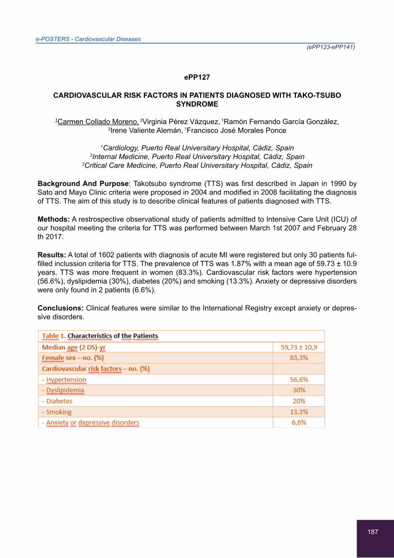
187
e-POSTERS - Cardiovascular Diseases(ePP123-ePP141)
ePP127
CARDIOVASCULAR RISK FACTORS IN PATIENTS DIAGNOSED WITH TAKO-TSUBO SYNDROME
1Carmen Collado Moreno, 2Virginia Pérez Vázquez, 1Ramón Fernando García González,
3Irene Valiente Alemán, 1Francisco José Morales Ponce
1Cardiology, Puerto Real Universitary Hospital, Cádiz, Spain 2Internal Medicine, Puerto Real Universitary Hospital, Cádiz, Spain
3Critical Care Medicine, Puerto Real Universitary Hospital, Cádiz, Spain
Background And Purpose: Takotsubo syndrome (TTS) was first described in Japan in 1990 by Sato and Mayo Clinic criteria were proposed in 2004 and modified in 2008 facilitating the diagnosis of TTS. The aim of this study is to describe clinical features of patients diagnosed with TTS.
Methods: A restrospective observational study of patients admitted to Intensive Care Unit (ICU) of our hospital meeting the criteria for TTS was performed between March 1st 2007 and February 28 th 2017.
Results: A total of 1602 patients with diagnosis of acute MI were registered but only 30 patients ful-filled inclussion criteria for TTS. The prevalence of TTS was 1.87% with a mean age of 59.73 ± 10.9 years. TTS was more frequent in women (83.3%). Cardiovascular risk factors were hypertension (56.6%), dyslipidemia (30%), diabetes (20%) and smoking (13.3%). Anxiety or depressive disorders were only found in 2 patients (6.6%).
Conclusions: Clinical features were similar to the International Registry except anxiety or depres-sive disorders.
188
e-POSTERS - Cardiovascular Diseases(ePP123-ePP141)
ePP128
THE INCIDENCE OF CARDIOVASCULAR RISK FACTORS AMONG ELECTIVE CORONARY ANGIOPLASTY PATIENTS
1Kateryna Bulavkina, 1Laima Caunīte, 1, 2Aija Mača
1Intensive Care Unit, Cardiology Department, Paul Stradins’s Clinical University Hospital, Riga, Latvia
2Internal Medicine Department, Riga Stradins’ University, Riga, Latvia
Background: Cardiovascular diseases are Nr.1 cause of death in Latvia. Since 1990, percutaneous coronary intervention (PCI) has been available in Latvia. ~200-400 stents are placed per million of population per year, which is equivalent to Finland, Spain and Portugal.
Aim: Determine most common cardiovascular risk factors among elective PCI patients in Latvia and compare to those of Europe.
Methods: A prospective research was held: questionnaires developed and 188 patients interviewed. Obtained data was analyzed with Microsoft excel. Results: 188 patients (aged 39 - 87) mean age 67,1. 63% of patients are males. 49,5% had previously had myocardial infarction; 57,9 % had under-gone PCI before and 6 patients (3,2%) - coronary artery bypass grafting. Most common cardiovas-cular risk factors were found to be patients’ age (males over 45 and females over 55 years) - 96%, elevated body mass index (BMI) - 80% (38% of had BMI >30), arterial hypertension - 71% (poorly controlled - 39%), abdominal obesity - 65%, insufficient physical activity (56%) and high stress levels (51%).
Conclusion: 63% already have two (age and gender), thus it is especially important to reduce the adjustable risk factors. In comparison with EUROASPIRE IV data, the incidence of BMI >30 is equal in both Latvia and Europe (38%), more patients smoke in Latvia - 20% (16% on average in Europe), blood pressure is better controlled in Latvia - 39% poorly controlled (52% in Europe). Three most common risk factors in Europe were abdominal obesity (58%), poorly controlled arterial blood pres-sure (42%) and diabetes (40%).
189
e-POSTERS - Cardiovascular Diseases(ePP123-ePP141)
ePP129
TELEMEDICAL APPLICATIONS IN HYPERTENSION MANAGEMENT
1Nikolaos Papakonstantinou, 1Rafaella Datseri, 3Stefanos Papakonstantinou, 2Christos Tsagkaris
1Comenius University, Faculty of Medicine, Bratislava, Slovakia 2Faculty of Medicine, School of Health Science, University of Crete, Heraklion, Greece
3IEK AKMI,Thessaloniki, Greece
Over the last years there have been a few attempts to utilize telemonitoring and e-health systems to achieve better surveillance of chronic patients. Some of these are considered quite successful, but the expensive specialized systems used limit their field of use.
For this study, we decided to focus on commercially available applications for smartphones and tablets. We reviewed 100 applications from the iOS app-store and google play-store. Of these we narrowed down to 18 applications through a primary reviewing process, to those which could work as diaries for daily blood pressure data collection. We conducted a systematic review of the functionality of each application and an ease-of-use trial with 20 participants to determine which of these applica-tions could find use in a clinical/ambulatory setting for long term self surveillance of patients primarily affected by hypertension. We identified 4 as being suitable for clinical/ambulatory use. SmartBP and Blood Pressure (BP) Watch proved most suitable for generalized record-keeping, Health2Sync showed great utility for hypertensive diabetics.
Finally Laborom, aside from having the core functions required, was able to keep records of history and past lab results and transmitting them to a physician on demand. We hope that our study can provide active physicians with an option for easier monitoring of the patients condition and keeping track of trends especially in their BP values. As we are aware, the applications we suggest may be-come obsolete, therefore we strive to at least establish a basic guideline for short term monitoring in the clinical setting.
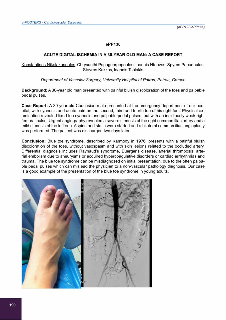
190
e-POSTERS - Cardiovascular Diseases(ePP123-ePP141)
ePP130
ACUTE DIGITAL ISCHEMIA IN A 30-YEAR OLD MAN: A CASE REPORT
Konstantinos Nikolakopoulos, Chrysanthi Papageorgopoulou, Ioannis Ntouvas, Spyros Papadoulas,
Stavros Kakkos, Ioannis Tsolakis
Department of Vascular Surgery, University Hospital of Patras, Patras, Greece
Background: A 30-year old man presented with painful bluish discoloration of the toes and palpable pedal pulses.
Case Report: A 30-year-old Caucasian male presented at the emergency department of our hos-pital, with cyanosis and acute pain on the second, third and fourth toe of his right foot. Physical ex-amination revealed fixed toe cyanosis and palpable pedal pulses, but with an insidiously weak right femoral pulse. Urgent angiography revealed a severe stenosis of the right common iliac artery and a mild stenosis of the left one. Aspirin and statin were started and a bilateral common iliac angioplasty was performed. The patient was discharged two days later.
Conclusion: Blue toe syndrome, described by Karmody in 1976, presents with a painful bluish discoloration of the toes, without vasospasm and with skin lesions related to the occluded artery. Differential diagnosis includes Raynaud’s syndrome, Buerger’s disease, arterial thrombosis, arte-rial embolism due to aneurysms or acquired hypercoagulative disorders or cardiac arrhythmias and trauma. The blue toe syndrome can be misdiagnosed on initial presentation, due to the often palpa-ble pedal pulses which can mislead the physician to a non-vascular pathology diagnosis. Our case is a good example of the presentation of the blue toe syndrome in young adults.

191
e-POSTERS - Cardiovascular Diseases(ePP123-ePP141)
ePP131
RARE ANATOMY OF CORONARY ARTERIES: COMBINATION OF TWO CORONARY ANOMALIES CASE REPORT
1,2Anastasia Nouli, 1Aikaterini Dimitra, 2Eleni Tsakiri, 3Dimosthenis Stavrianakis, 1,2,3Nikitas Moschos
1Cardiology Department, General Hospital of Rhodes, Rhodes, Greece 2Intensive Coronary Care Unit, General Hospital of Rhodes, Rhodes, Greece
3Cardiac Catheterization Laboratory, General Hospital of Rhodes, Rhodes, Greece
Background: Coronary artery anomalies (ACAs) are a diverse group of congenital disorders whose manifestations and pathophysiological mechanisms are highly variable. Most variations are benign; however, some may lead to angina, myocardial ischemia, syncope or sudden cardiac arrest.
Case Report: In a series of 1358 coronarographies we reported two cases of ACAs (0.15%). Our documented cases concern two different coronary anomalies. At first, we present a case of a 67yo patient with medical history of hypertension and dyslipidemia, who presented to the ER with epi-sodic syncope. He underwent a cardiac catheterization that showed an anomalous right coronary artery (RCA) arising from the left coronary sinus. A C/T coronary angiography was performed which revealed the RCA’s malignant coursing between the pulmonary artery and the aorta. Finally an inva-sive electrophysiologic study showed sick sinus syndrome. The patient with a permanent pacemaker implantation and b-blocker administration remains asymptomatic in the annual follow up. The sec-ond case is about a 75 yo patient with medical history of hypertension, diabetes and smoking, who proceeded to the ER with inferior STEMI. The coronarography incidentally revealed an anomalous left coronary artery (LCA) originating from the right sinus of Valsalva (separate ostium), an anatomi-cal variation of no clinical importance. The patient had a PCI in the culprit lesion of RCA. Due to contrast-induced acute kidney failure, we avoided performing a C/T coronarography.
192
e-POSTERS - Cardiovascular Diseases(ePP123-ePP141)
ePP132
AN UNEXPECTED CAUSE OF ACUTE CHEST PAIN IN AN ADULT MAN: A CASE OF EPIPERICARDIAL FAT NECROSIS (EPFN)
1,2Anastasia Nouli, 1Aikaterini Dimitra, 2Eleni Tsakiri, 3Dimosthenis Stavrianakis, 1,2,3Nikitas Moschos,
4Nikolaos Papapanagiotou, 4Eirini Delaporta
1Cardiology Department, General Hospital of Rhodes, Rhodes, Greece 2Intensive Coronary Care Unit, General Hospital of Rhodes, Rhodes, Greece
3Cardiac Catheterization Laboratory, General Hospital of Rhodes, Rhodes, Greece 4C/T and MRI Department, General Hospital of Rhodes, Rhodes, Greece
Background: EPFN is a rare self-limiting cause of an acute chest pain in an otherwise healthy in-dividual. It occurs within the mediastinum outside the pericardium. Typically manifesting as severe acute chest pain, EPFN can be mistaken for a serious disorder, such as pulmonary embolism, myo-cardial infarction or acute aortic syndrome.
Case Report: We report a case of a 52yo man who proceeded to the ER, due to acute chest pain. The ECG showed no ischemia and the troponine was negative. The D-dimers were slightly elevated 500 (reference concentration<250). Upon auscultation pericardial friction rub was audible. In the echocardiography there were no pathological findings and no pericardial fluid. The patient was ad-mitted to the Cardiology Department and was treated with ibuprofen. Due to the persistence of the pain two days after admission a chest C/T was performed, in order to exclude a possible acute aortic syndrome and pulmonary embolism. The C/T scan showed a well defined ovoid lesion of fat attenu-ation, surrounded by the increased attenuation of the anterior mediastinal paracardiac fat adjacent to the pericardium. The pericardium was minimally thickened. The symptom of the patient gradually recessed. He received anti-inflammatory treatment for 15 days. The inflammatory infiltrates revealed gradual resolution in the C/T scans we performed to follow-up the lesion (10 days and 1 month).
193
e-POSTERS - Cardiovascular Diseases(ePP123-ePP141)
ePP133
FREQUENCY AND CHARACTERISTICS OF ANEMIC SYNDROME IN PATIENTS WITH CHRONIC HEART FAILURE
1Budhbhushan Bhagat, 2Nataliia Pavliukovych, 3Oleksandr Pavliukovych, 1Guleria Pankaj
1Department of Internal Medicine,Cardiology,Higher State Educational Establishment of Ukraine “Bukovinian State Medical University”, Chernivtsi-city, Ukraine
2Department of Internal Medicine, Clinical Pharmacology and Occupational Diseases, Higher State Educational Establishment of Ukraine “Bukovinian State Medical University”, Chernivtsi-city,
Ukraine 3Department of Forensic Medicine and Medical Law, Higher State Educational Establishment of
Ukraine “Bukovinian State Medical University”, Chernivtsi-city, Ukraine
It is known that decreasing of hemoglobin (Hb) level below 12 g/dL is accompanied by progression of chronic heart failure (CHF) of ischemic origin.
Aim of our study was to find out frequency and origin of anemic syndrome (AS) in patients with CHF. We analyzed 2056 case records of hospitalized patients with CHF of ischemic origin. AS was diag-nosed in case of Hb below 130 g/dL in males and below 120 g/dL in females (WHO, 2003).
Among all examined patients AS was found in 69,21% (1423 cases), which corresponds to the lit-erature data. Among male patients AS was diagnosed in 1147 cases (76,22%), in females in 276 cases (49,19%). We found out that in patients after 45 years anemia is more frequent in males than in females. Only in rare cases anemia was documented as a separate diagnosis (2,81% in case of mild anemia, 50% in case of moderate anemia, 65,8% in case of severe AS). Hyperchromic anemia (MCH>33 pg) was diagnosed in 23 patients (1,62%), hypochromic anemia (MCH<27 pg) in 128 patients (8,99%); in most cases AS was of normochromic character (1272 patients, 89,39%). Mild macrocytosis (MCV 95-108) was found occasionally (19 cases, 1,34%), microcytosis (MCV<80) in 163 cases (11,45%), normocytosis in 1241 patients (87,21%).
Therefore, AS is comorbid to CHF in 69,21% of patients, predominantly in males. In most patients with CHF concomitant anemia is normochromic and normocytic, which requires further investigation of its etiology for an adequate correction of hemoglobin level.

194
e-POSTERS - Cardiovascular Diseases(ePP123-ePP141)
ePP134 - WITHDRAWN
195
e-POSTERS - Cardiovascular Diseases(ePP123-ePP141)
ePP135
EPIDEMIOLOGICAL STUDY OF MYOCARDIAL INFARCTION (MI) IN THE ISLAND COMPLEX OF DODECANESE IN SOUTH-EASTERN AEGEAN-GREECE
1,2Anastasia Nouli, 1Aikaterini Dimitra, 2Eleni Tsakiri, 3Dimosthenis Stavrianakis, 1,2,3Nikitas Moschos
1Cardiology Department, General Hospital of Rhodes, Rhodes, Greece 2Intensive Coronary Care Unit, General Hospital of Rhodes, Rhodes, Greece
3Cardiac Catheterization Laboratory, General Hospital of Rhodes, Rhodes, Greece
Background: Despite the decline in incidence and mortality of MI, only few studies have examined the apparent differences in its prevalence among geographical separate population groups, and whether known regional disparities in MI care have narrowed over time. Aim Determination of the incidence of MI in an isolated area of the Dodecanese islands, along with the contribution of the newly-established catheterization laboratory to the reduction in mortality rates.
Methods: A six-year retrospective study was conducted of all cases with MI. The prevalence of MI was compared to global statistics. The mortality rates during the period 03/2014-03/2017, with an active catheterization laboratory, were compared to those in 02/2011-02/2014. Information was ob-tained from the patients’ medical files.
Results: 781 patients with MI [387 STEMI (49.5%), 394 NSTEMI (50.5%)] undertook a coronarog-raphy in 03/2014-03/2017. The in-hospital mortality was reduced in comparison with the period 02/2011-02/2014 (6.1% vs 8.1%). The STEMIs were 52% anterior and 36% inferior. Men and women had a 3.4 years deviation in the median age of MI occurrence. The annual prevalence of MI was 1.2/1000 (1.5/1000 in Rhodes, 0.75/1000 in smaller islands).
Conclusion: Lifestyle in the islands of Dodecanese contributes to lower prevalence of MI. Women seem to experience their first MI on average only 3.4 years later than men. The establishment of the catheterization laboratory resulted to 20% relative reduction of in-hospital mortality. In half of the MIs LAD was the culprit vessel.
196
e-POSTERS - Cardiovascular Diseases(ePP123-ePP141)
ePP136
TELMISARTAN IN PATIENTS WITH CHRONIC HEART FAILURE AND DIABETES MELLITUS TYPE 2
1Nimisha Raut, 2Pavliukovych Nataliia, 3Pavliukovych Oleksandr, 1Mirang Patel, 1Jasmeet Kaur Saini
1Bukovinian State Medical University, Chernivtsi, Ukraine 2Department of Internal Medicine, Clinical Pharmacology and Occupational Diseases, Higher State
Educational Establishment of Ukraine “Bukovinian State Medical University”, Chernivtsi-city, Ukraine
3Department of Forensic Medicine and Medical Law, Higher State Educational Establishment of Ukraine “Bukovinian State Medical University”, Chernivtsi-city, Ukraine
Background:-Despite modern achievements in diagnosis and treatment of patients with chronic heart failure (CHF) and diabetes mellitus type 2 (DM), their incidence and unfavorable outcomes have been increased recently. The research aimed at the investigation of the possible clinical effec-tiveness of telmisartan in patients with CHF and comorbid DM.
Methods:-75 patients with CHF and DM were under investigation. They were randomized into 2 groups according to the prescribed treatment: I group - 40 patients who received statins, metformin and enalapril; II group - 35 patients for whom substitution of ACE inhibitor by telmisartan in daily dose of 40 mg was conducted. Clinical effectiveness of the prescribed treatment was estimated in 3 months by level of blood pressure (BP), fasting glucose (FG) and blood lipid spectrum (total choles-terol (TC), triacylglycerols (TG), HDL-cholesterol (HDL-C), LDL-cholesterol (LDL-C).
Results:-Comparison of BP in patients of both groups revealed no statistical difference. Valid de-creasing of FG level for 33% was detected in I group (5,8±0,11 comparing with 8,6±0,23 mmol/L, р<0,001). In II group lower level of FG (4,6±0,18 comparing with 8,5±0,82 mmol/L, р<0,001) was observed. Patients of II group were also characterized by more pronounced changes in blood lipid spectrum, such as decreasing of TC in 1,89 times (p<0,001), TG - by 12,5% (p<0,05), LDL-C - ap-proximately twice (p<0,001), increasing of HDL-C in 2,66 times (p<0,001).
Conclusion:-So, advisability of telmisartan prescription in treatment of patients with CHF and DM opens new perspectives for its application in this category of patients.
197
e-POSTERS - Cardiovascular Diseases(ePP123-ePP141)
ePP137
THE ROLE OF LNCRNAS TO THE GENESIS OF ATHEROSCLEROSIS
1Theodoros Angelopoulos, 1Emmanouela Laskaratou, 1Aggeliki Moustaka, 5Nikolaos Papakonstantinou, 3Eviana Petropoulou, 4Nikolaos Sevdalis, 1Christos Tsagkaris,
2Aggeliki Vakka
1Faculty of Medicine, School of Health Science, University of Crete, Heraklion, Crete, Greece2Faculty of Medicine, School of Health Science, University of Patras, Patras, Greece
3Department of Medical Laboratories, School of Health Science and Welfare, Athens University of Applied Sciences, Athens, Greece
4Faculty of Medicine, Medical University of Sofia, Sofia, Bulgaria5Faculty of Medicine, Commenius University of Bratislava, Bratislava, Slovakia
Introduction: The association of factors such as hypertension, hypercholesterolemia and illnesses such as diabetes mellitus with atherosclerosis is a common place in the literature. However, new studies demonstrate the contribution of long non-coding RNAs to atherosclerosis through the effect on epithelial, vascular smooth muscle cells and macrophages. Goal of the study: In this study we aim to collect and analyze results that correlate the effect of lncRNAs with atherosclerotic disease. Materials-
Method: A bibliographic retrospect was made on authoritative databases (Pubmed). Keywords (atherosclerosis, lncRNAs, ECs, VSMCs, vascular function) were put into search algorithms. Recent and earlier experimental studies that have been made on experimental animals and humans were selected for our study.
Results: It appears that lncRNAs are involved in vessel function, regulation of metabolism, inflam-mation and immunity. In particular, cytoplasmic lncRNAs can increase or decrease the transcription and translation of mRNAs, while nuclear lncRNAs can affect gene expression. Indeed, after bioinfor-matic analysis, it is suggested that lncRNAs increase cell proliferation and reduce apoptosis, factors that significantly lead to the appearance of atherosclerosis. Some examples of important lncRNAs are ANRIL and MALAT1
Discussion: It is necessary for further study to be done, in order to investigate the effect of lncRNAs on the occurrence of atherosclerosis at the level of basic and translational research. Particular men-tion should be made of the use of lncRNAs as biomarkers or therapeutic targets.

198
e-POSTERS - Cardiovascular Diseases(ePP123-ePP141)
ePP138
FREQUENCY AND CHARACTERISTICS OF ANEMIC SYNDROME IN PATIENTS WITH CHRONIC HEART FAILURE
Sarkar Arphika, Nataliia Pavliukovych, Oleksandr Pavliukovych, Nilang Chaudhari
Cardiology Department, Bukovinian State Medical University, Chernivsty, Ukraine
Anemia of any etiology aggravates clinical manifestation of coronary artery disease (CAD). Our investigation aimed at the analysis of the peculiarities of the clinical course of CAD in patients with comorbid anemia of different degree of severity. 72 patients were randomized into 2 groups: an experimental group 40 patients with CAD (stable angina of physical exertion) with comorbid ane-mia (hemoglobin less than 13 g/dl in males and less than 12 g/dl in females) and B control group 32 patients with CAD without anemia. Frequency of angina attacks was significantly higher and its duration was significantly longer in the patients of experimental group (p<0,05). All patients of experi-mental group (100%) used nitroglycerin to reduce pain during angina attack. In control group only 12 patients (37,5%) used short-acting nitrates for angina attack treatment, those who took them needed significantly less daily dose than patients of the experimental group (p<0,05). Patients of group A had complaints and objective findings conditioned by comorbid anemia such as tachycardia, skin dryness, dizziness, which aggravated the course of CAD itself. Comorbid anemia resulted in prolon-gation of patients hospital treatment due to CAD and increasing of frequency of their hospitalization (p<0,05). So, anemia of any origin aggravates the clinical course of CAD as it leads to the increased frequency of the angina attacks, increased need for nitrates during the attack, such patients are hospitalized more frequently due to exacerbation of the main disease and duration of their hospital treatment is longer.
199
e-POSTERS - Cardiovascular Diseases(ePP123-ePP141)
ePP139
CLINICAL AND EPIDEMIOLOGICAL CHARACTERISTICS OF PATIENTS WITH HYPERTENSIVE CRISIS TREATED AT THE EMERGENCY DEPARTMENT OF A HIGH
COMPLEXITY INSTITUTION, MEDELLIN, COLOMBIA, 2014-2015
Daniel Gallego-Gonzalez, Jorge Banol-Betancur, Lina Maria Martinez-Sanchez,
Maria de los Angeles Rodriguez-Gazquez, Juan Sebastian Marin-Cardenas,
Laura Isabel Jaramillo-Jaramillo, Luis Felipe Alvarez-Hernandez, Andres Eduardo Marin-Castro,
Dayana Andrea Quintero-Moreno
Escuela de Ciencias de la Salud, Facultad de Medicina, Universidad Pontificia Bolivariana, Medellin, Colombia
Objective: To determine the clinical and epidemiological characteristics of patients with hyperten-sive crisis treated in the emergency department of an institution of high complexity between 2014 and 2015.
Methods: A cross-sectional study involving patients older than 18 years who attended to the emer-gency deparment during the study period, diagnosed with any hypertensive disorder according to ICD-10; a sampling was not carried out. The analysis was performed using the vr.24 SPSS program, for quantitative variables with normal distribution, mean and standard deviation were calculated; for qualitative variables absolute and relative frequencies were estimated.
Results: A total of 156 patients were included, with a mean age of 63.2 ± 10.1 years, 73.7% were women and only 45.5% had a history of hypertension control. On admission, the most reported symptoms were headache 47.9%, dizziness 31.3%, paroxysmal dyspnea at night 18.8% and pre-cordial pain 13.5%. 22.4% had hipertensive emergency and 77.6% presented hypertensive urgency. 61.5% were admitted and managed mainly with ACE inhibitors in 25%. The ischemic stroke was the main target organ compromise, in 31.4% and 4 patients died in total 11.4%.
Conclusion: hypertensive urgency occurred more frequently in the study population, but emergen-cies had worse outcomes. In general, most patients received an appropriate management.

200
e-POSTERS - Cardiovascular Diseases(ePP123-ePP141)
ePP140
CAROTID ENDARTERECTOMY IN PATIENTS OLDER THAN 70 YEARS
Alexander Korotkikh, Dmitry Nekrasov
Regional clinical hospital №2, Tyumen, Russian Federation
Methods: 627 patients, who underwent CEA between 01 Sept 2015 and 30 March 2017, were in-cluded in prospective study. 187 (29,8%) were patients older than 70 years. Number of operations in study group was 227. The average age of all patients was 66,6 ± 10,8 years, the average age of the patients older than 70 years was 76,7 ± 3,5 years. Total amount was 356 males and among patients of study group was 82. Total symptomatic operations was 344 (54,9%), and in patients older than 70 years - 94 (50,3%). In acute phase of stroke performed 252 (30,2%) operations, and in study group - 72 (38,5%). In research assessed defeat of the contralateral CA and operational criteria as secondary endpoints. The primary endpoint are stroke, myocardial infarction, or death during the per procedural period or ipsilateral stroke thereafter.
Results: According to ultrasonography in 88 patients detected a hemodynamically significant lesion contralateral ICA, 40 of them were operated on after 1-2 months on the second stage. Four patients have occlusion of the contralateral ICA. Average time of ICA occlusion was 12,9 ±3,8 minutes. Av-erage time of occlusion of all operations was 12,5 ± 3,9 minutes (p>0,95). The primary endpoint in the group of patients older than 70 years was 0,5%. All patients showed improvement in neurologic status, regression of neurological symptoms, as well as improving the general condition of patients.
Conclusion: CEA in patients older than 70 years is an effective procedure since prevents the risk of recurrent and primary stroke.

201
e-POSTERS - Cardiovascular Diseases(ePP123-ePP141)
ePP141
INCREASED SALT INTAKE- A RISK FACTOR FOR HYPERTENSION
1Dilbar Mohammed Saleem, 1Dhanarajan Vishnu, 1George Aneeta, 2Hrechko Svitlana
1Department of Internal Medicine, Bukovinian State Medical University, Chernivtsi, Ukraine 2Department of Hypertension, Regional Cardiology Clinic, Chernivtsi, Ukraine
Its widely said “Give neither counsel nor salt till you are asked for it”.Salt is integral to most of food items but, more than a required level hampers the functional integrity of the organisms. With new criteria of hypertension, many more peoples are included under Hypertension Stage I.
Understanding importance of salt management in hypertension progression, is vital. According to FDA, normal intake should be less than 2300mg per day, to avoid hypertension and related compli-cations.The modern trend of processed foods makes average intake of sodium much more higher.This has increased the number of people in the hypertensive stage.To determine the effect of salt intake in increasing the blood pressure on normal healthy people. The study involved 75 healthy students from Chernivtsi University hostel between age group 20-26. The blood pressure before the study was evaluated.3 groups were made by common diet with different amount of salt intake. Group I-intake of diet with 2gm added salt, Group II-intake of diet with 4gm added salt, Group III-intake of diet with 8gm added salt.This particular diet was given to students for 2weeks.There was an aver-age rise of blood pressure by 2-3mmHg(p<0.05) in systolic for Group II compared to Group I and a 5-10mmHg(p<0.05) rise of systolic blood pressure for Group III. Diastolic blood pressure was also increased within expected range of systolic pressure.
In conclusion, students with increased salt intake had significant increase of blood pressure. 5% of the students had increased susceptibility to complications of hypertension.However, this effect is amplified in patients with heart failure.
202
e-POSTERS - Infectious Diseases(ePP142-ePP185)
ePP142
A PATIENT WITH WEIL’S DISEASE, HEMOPTYSIS AND ARDS: A CASE REPORT
Afroditi Roumpou, Elena Tsokola, Maria Koutroumpi, Ioanna Papaioannou, Christos Lampropoulos
Department of Internal Medicine, Argolidos General Hospital, Nafplion, Greece
Background/Aim: Weil’s disease is the severe form of leptospirosis whereas hemoptysis and/or Acute Respiratory Distress Syndrome (ARDS) are rare manifestations. A few case reports have been published mentioning the high mortality and the frequent necessity for mechanical ventilation. A case of a patient with Weil’s disease, massive hemoptysis and ARDS is described.
Case Report: A 35-year old farmer presented with fever and dyspnea the last five days. He mentioned massive hemoptysis (>200 ml) a few hours before admission. His medical history included malaria successfully treated two years ago. Clinical examination revealed jaundice, bilateral rales on lungs’ auscultation, respiratory rate >30 breaths/minute, cyanosis and high temperature (38.5oC). Tho-racic x-ray revealed bilateral pulmonary infiltrates while blood gas analysis showed pO2=41mmHg, pCO2=30mmHg, sO2=82%, pH=7.54, HCO3=25.7mmol/Lt, pO2/FiO2=195. Laboratory investigation showed WBC=20.200/μL, ESR=120/1h, Ht=31.1%, Hb=9.8gr/dL, PLTs=130.000, urea=55mg/dL, creatinine=1.6mg/dL, AST=47IU/L, ALT=58IU/L, bilirubin total/direct=7.8/3.9mg/dL, γ-GT=219IU/L and CRP=30mg/dL (<0.7). Abdominal ultrasound depicted increased liver’s size whereas cardiac ultrasound small pericardial effusion. A diagnosis of Weil’s disease with ARDS was considered and empirical treatment was started (ampicillin/sulbactam 3grx3, azithromycin 500mgx1, metronidazole 500mgx3) with addition of methylprednisolone 40mgx3. There was complete resolution of fever, x-ray’s findings and hemoptysis after 4 days of treatment. IgM antibodies against leptospira were positive. The patient was discharged one week later. Laboratory investigation showed WBC=10.100/μL, ESR=50/1h, Ht=33.9%, PLTs=430.000, bilirubin total/direct=2.4/1.05mg/dL and CRP=4.1mg/dL (<0.7). Renal function was normal.
203
e-POSTERS - Infectious Diseases(ePP142-ePP185)
ePP143
A RARE CASE OF LISTERIOSIS IN A YOUNG PATIENT PRESENTING WITH INCOMPLETE MISCARRIAGE
1Konstantinos Louis, 1Nikolaos Folas, 2Eleni Kourti, 2Anastasia Kopsaftopoulou, 2Maria Mylona
11st Department of Obstetrics-Gynecology, “Konstantopouleio” - “Patision” General Hospital, Nea Ionia, Athens, Greece
21st Department of Propaedeutic & Internal Medicine, “Laiko” General Hospital, Athens University Medical School, Athens, Greece
Introduction/Aim: Listeria monocytogenes (LM) is an important bacterial pathogen affecting espe-cially neonates, immunosuppressed patients, older adults and pregnant women. It usually causes mild maternal illness, but can be devastating to the fetus. Herein we report a case of a young patient with an incomplete miscarriage and listerial infection. Listeriosis- though a rare entity- should always be considered in the differential diagnosis of fever in pregnant women, who may present with symp-toms of miscarriage. Although women with comorbidities, such as diabetes, steroid use, or HIV are at higher risk of listerial infection, most cases occur in healthy pregnant women.
Case description: A 21-year old Caucasian woman, with a history of one previously successful pregnancy, presented firstly to the Emergency Department and was finally admitted to our Obstet-rics-Gynecology Department, due to fever up to 40° C since 2 days with concomitant acute upper abdomen pain for the last 2 hours. She reported she was 13 weeks pregnant and an occasional smoker. Blood chemistry revealed mild anemia (Hb=10,9 g/dl), leukocytosis (WBC=18300/μl) and elevated inflammation markers (CRP=232,57 mg/l, max 5mg/l). She was immediately taken into OR, as an incomplete miscarriage was suspected, where an abrasion was performed. The patient was initially started on cefoxitin IV and a vaginal tampon was placed for hemostasis combined with ergometrine maleate po. Listeria monocytogenes was isolated from the amniotic fluid cultures. The patient was started on combined antibiotic treatment with ampicillin/sulbactam and gentamycin IV, showed significant clinical response and exited the hospital three weeks after.
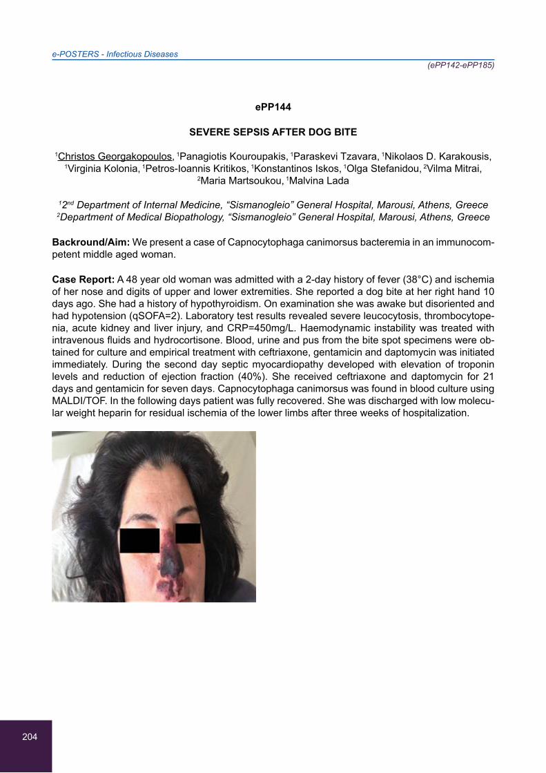
204
e-POSTERS - Infectious Diseases(ePP142-ePP185)
ePP144
SEVERE SEPSIS AFTER DOG BITE
1Christos Georgakopoulos, 1Panagiotis Kouroupakis, 1Paraskevi Tzavara, 1Nikolaos D. Karakousis,
1Virginia Kolonia, 1Petros-Ioannis Kritikos, 1Konstantinos Iskos, 1Olga Stefanidou, 2Vilma Mitrai,
2Maria Martsoukou, 1Malvina Lada
12nd Department of Internal Medicine, “Sismanogleio” General Hospital, Marousi, Athens, Greece 2Department of Medical Biopathology, “Sismanogleio” General Hospital, Marousi, Athens, Greece
Backround/Aim: We present a case of Capnocytophaga canimorsus bacteremia in an immunocom-petent middle aged woman.
Case Report: A 48 year old woman was admitted with a 2-day history of fever (38°C) and ischemia of her nose and digits of upper and lower extremities. She reported a dog bite at her right hand 10 days ago. She had a history of hypothyroidism. On examination she was awake but disoriented and had hypotension (qSOFA=2). Laboratory test results revealed severe leucocytosis, thrombocytope-nia, acute kidney and liver injury, and CRP=450mg/L. Haemodynamic instability was treated with intravenous fluids and hydrocortisone. Blood, urine and pus from the bite spot specimens were ob-tained for culture and empirical treatment with ceftriaxone, gentamicin and daptomycin was initiated immediately. During the second day septic myocardiopathy developed with elevation of troponin levels and reduction of ejection fraction (40%). She received ceftriaxone and daptomycin for 21 days and gentamicin for seven days. Capnocytophaga canimorsus was found in blood culture using MALDI/TOF. In the following days patient was fully recovered. She was discharged with low molecu-lar weight heparin for residual ischemia of the lower limbs after three weeks of hospitalization.
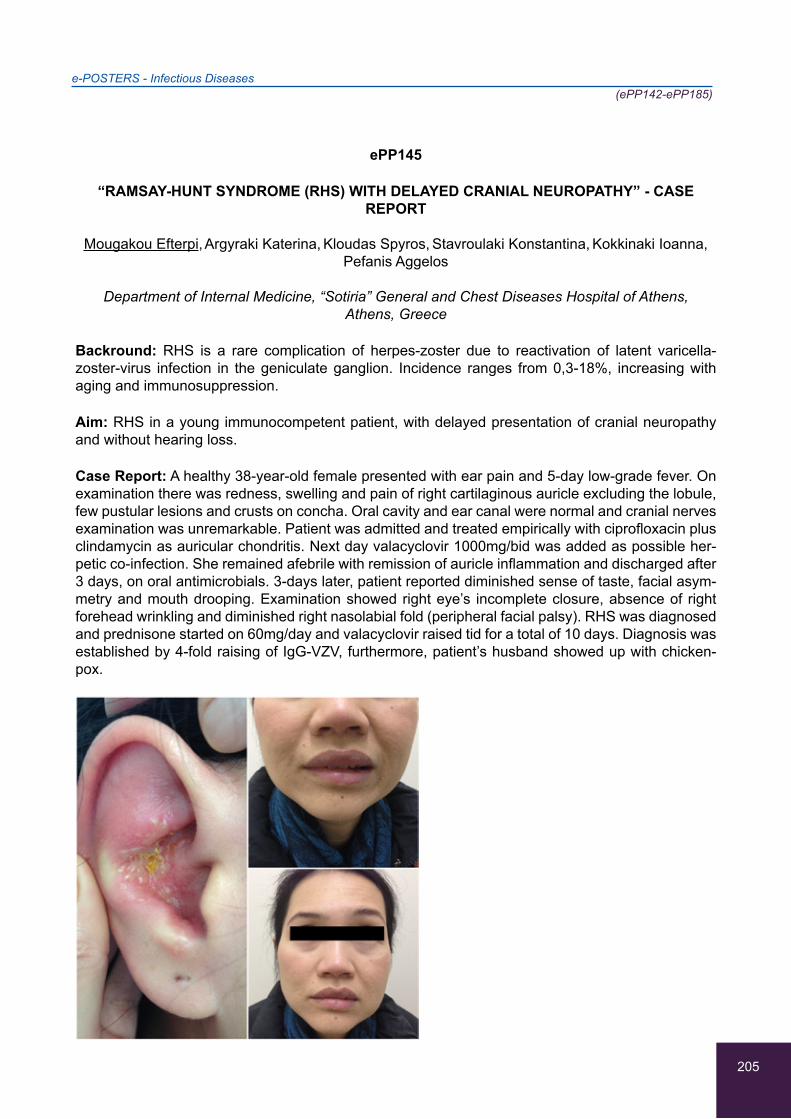
205
e-POSTERS - Infectious Diseases(ePP142-ePP185)
ePP145
“RAMSAY-HUNT SYNDROME (RHS) WITH DELAYED CRANIAL NEUROPATHY” - CASE REPORT
Mougakou Efterpi, Argyraki Katerina, Kloudas Spyros, Stavroulaki Konstantina, Kokkinaki Ioanna,
Pefanis Aggelos
Department of Internal Medicine, “Sotiria” General and Chest Diseases Hospital of Athens, Athens, Greece
Backround: RHS is a rare complication of herpes-zoster due to reactivation of latent varicella-zoster-virus infection in the geniculate ganglion. Incidence ranges from 0,3-18%, increasing with aging and immunosuppression.
Aim: RHS in a young immunocompetent patient, with delayed presentation of cranial neuropathy and without hearing loss.
Case Report: A healthy 38-year-old female presented with ear pain and 5-day low-grade fever. On examination there was redness, swelling and pain of right cartilaginous auricle excluding the lobule, few pustular lesions and crusts on concha. Oral cavity and ear canal were normal and cranial nerves examination was unremarkable. Patient was admitted and treated empirically with ciprofloxacin plus clindamycin as auricular chondritis. Next day valacyclovir 1000mg/bid was added as possible her-petic co-infection. She remained afebrile with remission of auricle inflammation and discharged after 3 days, on oral antimicrobials. 3-days later, patient reported diminished sense of taste, facial asym-metry and mouth drooping. Examination showed right eye’s incomplete closure, absence of right forehead wrinkling and diminished right nasolabial fold (peripheral facial palsy). RHS was diagnosed and prednisone started on 60mg/day and valacyclovir raised tid for a total of 10 days. Diagnosis was established by 4-fold raising of IgG-VZV, furthermore, patient’s husband showed up with chicken-pox.

206
e-POSTERS - Infectious Diseases(ePP142-ePP185)
ePP146
FEVER OF UNKNOWN ORIGIN (FUO): WITHOUT SPLEEN AND WITHOUT DIAGNOSIS
Magdalena Martín Pérez, Ylenia Avellaneda López, Jose María Perez de Navarro
Internal Medicine, Hospital Punta Europa, Algeciras, Spain
A 30-year-old patient with no personal history of interest except for a febrile episode of three months of evolution in the previous year with spontaneous resolution and without finding etiology. It enters again due to fever of two weeks of evolution of up to 39ºC with myalgia and associated headache. No other symptoms or epidemiological risk. Analytical performed including blood count shows only mild lymphopenia and thrombocytopenia, with smear of normal peripheral blood. Coagulation, iron study, LDH, renal function, thyroid profile, hepatic, proteinogram, autoimmunity, tumor markers, mul-tiple serologies, blood cultures (after more than 21 days) and urine cultures: negative. Chest x-ray, abdominal ultrasound, echocardiogram, gastroscopy: normal. Complete CT with nonspecific intra-abdominal adenopathies and mild hepato-homogeneous splenomegaly. Biopsies of esophagus, du-odenum and liver are taken with negative results and only doubtful positivity for Helicobacter Pylori in the stomach. Antibiotics are not administered due to good general condition and to avoid masking results. Given the multiple diagnostic doubts, PET-CT with splenic uptake and intra-abdominal lym-phadenopathies highly suggestive of lymphoproliferative process is requested. Bone marrow study negative. When the fever persisted and in the absence of other data, it was decided to perform a splenectomy and intra-abdominal adenopathy biopsy, which nevertheless turned out to be negative for malignancy.
Conclusions: After this result, we proposed whether surgery was the only alternative without be-ing able to forget the usefulness of PET, which, despite contributing significantly to the diagnosis of FUO, could give false positives like this, since the febrile symptoms subsided spontaneously and progressively.
207
e-POSTERS - Infectious Diseases(ePP142-ePP185)
ePP147
PERSISTENT BACILLUS CEREUS BACTEREMIA IN AN IMMUNOCOMPETENT PATIENT: A CHALLENGING ENEMY
Kourti Eleni, Kontrafouri Paraskevi, Kopsaftopoulou Anastasia, Reppas Konstantinos,
Efthymiou Konstantinos, Toskas Agelos, Mylona Maria
1st Department of Propaedeutic & Internal Medicine, “Laiko” General Hospital, Athens University Medical School, Athens, Greece
Introduction/Aim: Bacillus cereus is an aerobic to facultative anaerobic, Gram-variable bacilli found readily in soil and near fresh and marine water beds. Until identified as a clinically significant patho-gen, it was often thought to be a contaminant of blood cultures. Most clinically significant cases of bacteremia are usually associated with an immunocompromised host or the presence of foreign bod-ies. It is increasingly becoming a causative agent for both localized and systemic infections. Herein, we present an interesting case of persistent B. cereus bacteremia in an immunocompetent patient, in order to emphasize the importance of recognizing B. cereus as a potentially serious human patho-gen even in an immunocompetent host with need of early combination therapy.
Case description: An 85-year old Caucasian woman, with a history of chronic renal failure, was ad-mitted to our clinic due to fever, progressively worsening of generalized oedema, oliguria and hypo-tension. B. cereus was isolated from 3 blood cultures and empiric antibiotic treatment (ciprofloxacin and vancomycin) was initiated. Hemodialysis was performed once daily initially and then every other day. A week later, the central venous catheter was changed due to persistent bacteremia, and clin-damycin was added. Transthoracic and transesophageal echocardiogram were performed, exclud-ing endocarditis and a full body CT scan did not confirm an abcess. A 99mTc LeukoScan showed pericardial positive uptake. Due to persistent bacteremia 20 days later, the antibiotic treatment was changed to a gentamycin/meropenem/vancomycin combination IV-initiated. 3 days later, the blood cultures were negative and the patient showed significant clinical response.
208
e-POSTERS - Infectious Diseases(ePP142-ePP185)
ePP148
A RARE CASE OF SPONDYLODISCITIS CAUSED BY PROTEUS MIRABILIS
Ealing Loke, Veeraraghavan MA
Department of Internal Medicine, Khoo Teck Puat Hospital, Singapore
Infectious spondylodiscitis commonly results from local or haematogenous infections by micro or-ganism, with Staphylococcus being the most common cause of infective organism accounting for more than half of non-tuberculosis cases. Other bacterial pathogens include the Enterobacteriaceae group; of which the more common ones are Escherichia Coli and Proteus. This micro organisms are more commonly found in the urinary or gastrointestional track, and are usually associated with old age, the immunosupressed and diabetes. Patients present with back pain and fever, typically worsening at night. Our patient is an 84 year old male fom a nursing home, with poorpremorbids was recently admitted for 2 day history of fever associated with shortness of breath, chesty cough. He was febrile and tachpneic while in ED, and was drowzy. On examination, he was noted to have bilateral lower zone crepitations. The labs revealed raised inflammatory markers of total white cell 21, C-reactive protein 52. Chest x-ray showed bilateral consolidative changes.
Subsequently, blood cultures were positive for proteus mirabilis bacteremia and was started on IV Tazocin. He was recently hospitalized for proteus bacteremia secondary to a urinary track infec-tion. CT scan had features suggestive of spondylodiscitis, and pyelonephritis. His antibiotics were eventually de escalated to IV Cefatriaxone and oralised to PO Ciprofloxacin, completing 14 days of antibiotics.

209
e-POSTERS - Infectious Diseases(ePP142-ePP185)
ePP149
CASE REPORT OF SECONDARY HAEMOPHAGOCYTIC SYNDROME DUE TO MILIARY TUBERCULOSIS
1Charalampos Giannopoulos, 1Nikolaos Aleksiou, 2Eleni Christopoulou, 1Eirini Skoura,
1Alkiviadis Ioannou, 1Ioannis Mouzakitis, 1Tatiana Tataridou, 1Georgios Maikantis,
1Thomais Lamprou, 1Vasileios Apostolopoulos, 1Oksana Tsatov, 2Ioannis Gkinis, 1Marina Koupetori,
2Michail Komis, 1Stiliani Sybardi
1First Department of Internal Medicine, “Thriasio” General Hospital, Elefsina, Greece 2Department of Pneumology, “Thriasio” General Hospital, Elefsina, Greece
Introduction: Haemophagocytic syndrome (HS) is an uncommon hematologic disorder character-ized by severe hyperinflammation, caused by uncontrolled proliferation of T-lymphocytes and mac-rophages that secrete severe amounts of inflammatory cytokines. In rare cases, infectious agents, such as M.tuberculosis can trigger secondary HS.
Purpose: Prompt and targeted treatment of the underlying condition is effective for the resolution of secondary HS.
Case Report: A 42 years old male, with free medical history, was admitted because of prolonged fever, dyspnea, generalised lymphadenopathy. Labor: Severe neutropenia, anemia, thrombocyto-penia. Low fibrinogen levels, highly elevated ferritin levels. Testing for HIV, HBV, HCV, malaria, leishmaniasis:negative. Thorax radiography, HRCT: diffuse miliary opacities in lung parenchyma. Imaging and laboratory testing negative for extrapulmonary disease.
Results: Diagnosis of HS was made after fulfillment of five criteria including haemophagocytosis proved in bone marrow biopsy. After obtaining samples of sputum and gastric fluid, an initial empiric treatment with Isoniazid (H), Rifampicin(R), Pirazinamide (Z), Ethambutol (E) was administered, in combination with low-dose i.v steroids. Gradually clinical and laboratory improvement was noted with resolution of the HS after one week of treatment. M.tuberculosis was isolated only in gastric fluid. The molecular and compatible testing proved no resistance to antimycobacterial agents. The duration of treatment was set for 9 months (initially 2 months HRZE, following from 7 months HR).
Conclusions: Secondary HS is a rare clinical entity with irreversible tissue complications and poor prognosis. A targeted and aggressive therapy of the underlying condition may result in a gradual resolution of the syndrome without implementation of the internationally accepted therapy.

210
e-POSTERS - Infectious Diseases(ePP142-ePP185)
ePP150
BACTEREMIA CAUSED BY GRAM NEGATIVE BACTERIA IN PATIENTS IN AN INTENSIVE CARE UNIT
1Nikolaos Zotos, 2Eirini Tatsina, 3Nikolaos Varsamis, 4Aristea Zotou, 5Athanasios Margaritis,
1George Katagis, 1Marina Gerasimou, 1Foteini Adam, 1Anastasia Pournou
1Microbiology Department, General Hospital of Ioannina, Ioannina, Greece 2Microbiology Department, “Papageorgiou” General Hospital,Thessaloniki, Greece
3Pathology Department, General Hospital of Ioannina, Ioannina, Greece 4Cardiology Department, University Hospital of Ioannina, Ioannina, Greece
5I.C.U.,General Hospital of Ioannina, Ioannina, Greece
Aim: The aim of the study was to analyze bloodstream infections caused by Gram negative bacteria that were isolated in patients in the I.C.U during a period of one year.
Material and Method: All the epidemiologic and clinical characteristics of inpatients in the I.C.U were studied, as well as the antimicrobial susceptibility testing of all of the GNB isolated.
Results: 234 inpatients in the I.C.U were studied. 37 episodes of bacteremia caused by GNB in 23 patients (9.8%) were recorded.12 of them (55%) being Acinetobacter baumanii strains, 4 (18%) Klebsiella pneumoniae strains, 2 (9%) Pseudomonas aeruginosa strains and 4 (18%) other kinds of strains. Totally, 37 GNB were isolated and 13 of them were A.baumanii strains (92% of them were resistant to Carbapenems , while 8% were resistant to Colistin) , 8 were K.pneumoniae strains (50% were resistant to Carbapenems,13% of them were resistant to Colistin, and 38% of them were resist-ant to Tigecycline), 5 were P.Aeruginosa strains (40% of them were resistant to Carbapenems ,and all of them were susceptible to Colistin)
Conclusion: Bacteremia caused by Gram negative bacteria is an aggravating factor in patients in a critical condition. A significant proportion of the isolated strains were multi-drug resistant. A.baumanii was the most dominant of the pathogens isolated during the study. Even though Colistin is more ef-fective against the majority of them, strains resistant to it have started to emerge.
211
e-POSTERS - Infectious Diseases(ePP142-ePP185)
ePP151
SEVERE INFECTION CAUSED BY STREPTOCOCCI OF THE VIRIDANS GROUP IN A TERTIARY HOSPITAL
1Nikolaos Zotos, 2Eirini Tatsina, 3Nikolaos Varsamis, 4Aristea Zotou, 1Christos Mitsis,
1Christos Briasoulis, 1Lampros Papageorgiou, 1Evaggelia Chrisostomou, 1Anastasia Pournou
1Microbiology Department, General Hospital of Ioannina, Ioannina, Greece 2Microbiology Department, “Papageorgiou” General Hospital,Thessaloniki, Greece
3Pathology Department, General Hospital of Ioannina, Ioannina, Greece 4Cardiology Department, University Hospital of Ioannina, Ioannina, Greece
Aim: To record severe infection caused by streptococci viridans in a tertiary hospital.
Material and Method: During the last 8 years ,26017 samples of blood and various other biological fluids were cultured and estimated for the presence of streptococci. When gram positive, catalase negative cocci resembling the form of streptococci were isolated, a biochemical identification was conducted.
Results: The species of 328 isolated strains of streptococci were: 1) 102 Streptococci of the Mitis group (S.mitis, S.gordonii,S.oralis, S.sanguis, S.parasanguis) 2)87 strains of streptococci of the an-ginosus or S.milleri group (S. anginosus, S. contellatus, S. intermedius) 3) 63 strains of streptococci of the Salivarius group (S. Salivarius, S. thermophiles, S. vestibularis) 4) 42 strains of the Bovis group (S. bovis, S. equines, S. alactolyticus) 5)34 strains of the Mutans group (S.mutans, S.rattus, S. cricetus, S. downei, S. sobrinus, S. macacae). All of the isolated strains were susceptible to antimicrobial drugs and the MIC of penicillin was <=0.12μgr/ml (Sensitive), exept for one strain of S.sanguis and another strain of S.equinus .For these two strains the MIC of penicillin was 0.25 - 2 (Intermediate).
Conclusion: The most frequently isolated streptococci of the viridans group were those of the Mitis group , followed by those of the Anginosus group, the Salivarius group, the Bovis group and finally those of the Mutans group.In Greece they are still susceptible to antibiotics which should be admini-stared in accorrdance with the results of the antimicrobial susceptibility testing.
212
e-POSTERS - Infectious Diseases(ePP142-ePP185)
ePP152
A CASE REPORT OF BRUCELLOSIS IN A HEALTH CARE PROFESSIONAL
Dimitra Vasdeki, Christos Zisis, Dimitra Maritsa, Eleni Petroulaki, Fotini Malakoudi, Charitini Milona,
Stefanos Milonas
Second Department of Internal Medicine, General Hospital of Trikala, Trikala, Greece
Background/Aim: Brucellosis is a zoonotic disease that can be transmitted through contact with infected animals (sheep/goats/cattle/pigs), by eating undercooked meat or consuming unpasteur-ized/raw dairy products. Also, infection can occur by inhalation (breathing in the bacteria that causes brucellosis),but this risk is generally greater for people in microbiological laboratories.
Case Report: A 43-year-old woman assistant microbiologist presented to our Hospital with a 3-day history of fever, anorexia, malaise and headache. Her blood test revealed mild pancytopenia, ab-normal LFTs, increased CRP and LDH and the patient was initially given symptomatic treatment for a common flu infection. Within 5 days there was a remission of her clinical condition. The follow-up blood tests, after 15 days , were normal, except for a persistent mild leucocytopenia. Further blood tests were performed (HbsAg/ anti-HCV/ anti-HIV/ Monotest/ Widal/ anti-toxo/ ΑΝΑ/ anti-DNA/anti-ENA) and Μantοux but all were found normal. Chest X-rays and abdominal ultrasound were also normal. Serological tests for CMV and EBV revealed previous exposure to the viruses but no active infection. Due to the patient’s previous exposure to samples contaminated with B.melitensis in the laboratory, Brucellosis was suspected.
Consequently, the Rose-Bengal plate test (RBPT) was performed using antigen of B.melitensis, which revealed the presence of high level of antibodies (>100IU/ml). The diagnosis was further strengthened by a positive Wright test (1/320). Rifampicine p.o. once a day combined with doxycy-cline p.o. twice a day were eventually initiated for a duration of 6 weeks and the patient was finaly cured.

213
e-POSTERS - Infectious Diseases(ePP142-ePP185)
ePP153
MENINGOCOCCAL SEPTIC SHOCK WITH VARIOUS COMPLICATIONS IN A 70-YEAR-OLD IMMUNOCOMPETENT WOMAN
1Μakis Eleftheriotis, 1Marina Gkeka, 2Stelios Kouvardas, 1Sofia Kalantzi, 1Costas Christopoulos,
1Asimina Protogerou, 2Georgia Kouppari, 3Sofia Lafoyianni, 1Chariclia Loupa
12nd Department of Internal Medicine, “Amalia Fleming” General Hospital, Athens, Greece 2Laboratory of Microbiology, “Amalia Fleming” General Hospital, Athens, Greece
3Laboratory of Radiology, “Amalia Fleming” General Hospital, Athens, Greece
Background: N. meningitidis can exist as nasopharyngeal carriage or as pathogen that mainly causes meningitis and septicemia.However,it can also cause other infections like pneumonia and arthritis,which is frequently large-joint,oligoarticular and asymmetric.It’s found in approximately 7% of meningococcal infections and is either septic arthritis or immune-mediated,presenting 3 days to 2 weeks after disease onset,when the patient’s otherwise improving.A short NSAID course is mostly efficacious for immunoallergic type,but few may need long-term corticosteroid treatment.
Case report:The patient was presented with fever,chest pain,purpura and subscleral hemorrhage.She was upset but oriented.Vital signs:98 bpm,26 breaths/min,blood pressure:not measurable.She had no photophobia or neurologic-meningeal signs.Blood gas analysis revealed hypoxemia with metabolic acidosis and chest radiograph showed bilateral bronchopneumonic infiltrates and 6th left rib fracture,which was attributed to a fall at home due to hypotension.Aggressive hydration and vancomycin with ceftriaxone were initiated.After crystalloids’ failure to raise blood pressure nore-pinephrine was added and afterwards hydrocortisone(50mg x4).Blood tests showed acute kidney injury and DIC(ISTH score:5).Due to hemorrhagic lesions and the unresponsiveness to vasopres-sors brain and adrenal(for Waterhouse-Friderichsen syndrome exclusion)CT was performed,which revealed subarachnoid hemorrhage.FFP was administered,resulting to non-extension of purpura and hemorrhage absorption.After the 2ndday developed bilateral pleural effusions created from hy-dration and renal failure and ischemic hepatitis.Both resolved.Blood cultures revealed penicillin-sensitive N.meningitidis(MIC:0,0094).The 4thday she presented new fever along with polyarthritis.She was discharged with lornoxicam and fever resolved after 1 week.She continued treatment for 1 month,until CRP-ESR returned to normal.
Conclusions: Clinicians should be aware of these complications because fever and inflammatory markers’ rebound can lead to unnecessary initiation of nosocomial antibiotics.

214
e-POSTERS - Infectious Diseases(ePP142-ePP185)
ePP154
77 YEAR OLD MALE WITH FEVER, JAUNDICE, THROMBOCYTOPENIA AND BILATERAL PULMONARY INFILTRATES
Ioannis Andrianopoulos, Aggeliki Karahaliou, Xanthi Zikou, Athanasios Papathanasiou,
Maria Saranti, Vasilios Koulouras
Intensive Care Unit, University Hospital of Ioannina, Ioannina, Greece
Background/Aim: We present a rare cause of severe ARDS. Case report A 77 year old male pa-tient was admitted in our hospital with a five day history of fever(T38.3oC),jaundice(Tbil 9.3 mg/dl), acute kidney injury (Creatinine2.4 mg/dl),and thrombocytopenia(PLT26,000/μL). He was started on empirical antibiotic treatment while a thorough diagnostic work-up was undertaken (that included blood and urine cultures, blood smear examination and serological tests for autoimmune and in-fectious causes). Leptospira IgM antibodies were positive and a diagnosis of Leptospirosis was confirmed. A CT chest showed bilateral pulmonary infiltrates consistent with hemmorhagic alveolitis. Despite appropriate treatment patient continued to deteriorate as he developed respiratory failure he was intubated and was transferred to our ICU. On arrival in ICU he was tachycardic, hypotensive requiring vasopressors at a high dose(0.9mcg/kg/min), under mechanical oxygenation with very poor oxygegenation (po2 63mmHg,fi02 75%,Pa02/Fi02=84). He was treated with iv ceftriaxone, iv fluids and was put in prone position . Patient’s condition gradually improved in terms of oxygenation (reduction in FI02 %), hemodynamic status (reduction in the dose of vasopressors) and after 48h he was turned back in the supine position Patient continued to improve sedation was stopped and 5 days post his admission he was extubated. By that point his bilirubin, creatinine, WBC and platelet count had normalized.
The patient was discharged back to the general medicine ward being hemodynamically stable and with good oxygenation. Conclusion Leptospirosis is a rare cause of ARDS. Clinicians should be aware of this rare but life-threatening complication of Leptospirosis.
215
e-POSTERS - Infectious Diseases(ePP142-ePP185)
ePP155
RIFAMPICIN AS A CAUSE OF SEVERE HEMOLYTIC ANEMIA IN A PATIENT WITH LOCALIZED BRUCELLOSIS
Dafni Sveroni, Aggelos Stefos, Dimitrios Sagris, Asterios Polizos, Maria Boulbou,
George N. Dalekos, Eirini Rigopoulou
Department of Medicine and Research Laboratory of Internal Medicine, Medical School, Thessaly University, Larissa, Greece
Background: Since its introduction, rifampicin has been widely used in the treatment of tuberculo-sis, nasopharyngeal carriage of Neisseria meningitidis and Haemophilus influenzae, and to a lesser degree, in various antibiotic combinations for bacterial infections. Acute hemolysis as an adverse re-action to rifampicin is a rarely encountered complication. We present a patient with acute hemolysis due to rifampicin administration for localized brucellosis.
Case report: A 58-year-old woman with a history of localized brucellosis was admitted to our depart-ment due to high grade fever accompanied by abdominal and lumbar pain and laboratory evidence of acute hemolysis. She had been treated with doxycycline and ciprofloxacin for three months but she reported aggravated pain of the left hip despite treatment. Pelvic MRI revealed myositis of the left gluteus minimus muscle and six days before admission, rifampicin was added to her medication. Blood cultures were negative, and serology for malaria, mycoplasma, viral and other acute bacterial infections was unrevealed. ANA test was negative. Chest and abdominal CT scans, bone marrow biopsy and flow cytometry were performed and had no evidence of lymphoproliferative disorders. Direct antiglobulin test (Coombs) was strongly positive (++++) for IgG and C3d, and haptoglobins were undetectable. The diagnosis of hemolytic anemia due to rifampicin was established. After drug withdrawal, the patient was treated with intravenous immune globulin together with prednisolone that was gradually tapered and stopped. After 14 days of hospitalization the patient was discharged, with no evidence of relapse in a follow-up period of 1 year.
216
e-POSTERS - Infectious Diseases(ePP142-ePP185)
ePP156
PULMONARY TUBERCULOSIS REACTIVATION: WHAT ABOUT ALTITUDE?
1Filipa Bianchi-de-Aguiar, 2Rafaela Campanha, 2Cátia Guimarães, 2Margarida Simões-Raposo
1Serviço de Medicina - Unidade Funcional 4, Hospital de S. Francisco Xavier, Centro Hospitalar de Lisboa Ocidental, Lisboa, Portugal
2Serviço de Pneumologia, Hospital de Egas Moniz, Centro Hospitalar de Lisboa Ocidental, Lisboa, Portugal
Background: High altitudes are linked to decreased rates of pulmonary tuberculosis (PTB) infec-tion, disease and mortality. However, its relevance as a trigger for PTB reactivation in immunocom-petent patients is not documented.
Case Report: A 28-year-old healthy Nepalese female was referred to emergency with sudden left pleuritic back pain with shortness of breath, two weeks after arriving in Lisbon, having flown from Kathmandu and undergone a change in altitude of 1400 metres. She also had evening low-grade fever and fatigue since she arrived. Her mother-in-law had died of tuberculosis two years before. Chest radiography and CT scan showed a left upper lobe consolidation. Laboratory analysis were unremarkable, except for ESR of 79 mm/sec. HIV serology, blood cultures and urinary antigen test-ing were negative. Bronchial secretions’ cultures became positive for Mycobacterium tuberculosis complex. The patient was started on anti-tuberculous treatment and made a steady recovery.
Conclusions: The diagnosis of TB in the immunocompetent host requires high level of suspicion and persistence, mainly when it is manifested insidiously and with few laboratory and clinical signs. Epidemiologic history in these cases is very important. Altitude is a known influencing factor on pul-monary tuberculosis disease and it’s reasonable to think it may influence reactivation.
217
e-POSTERS - Infectious Diseases(ePP142-ePP185)
ePP157
SUCCESSFUL OUTCOME OF PERSISTENT BACTEREMIA DUE TO PSEUDOMONAS AERUGINOSA AFTER TREATMENT WITH CEFTOLOZANE-TAZOBACTAM. A CASE REPORT
Dimitrios Sagris, Aggelos Stefos, Maria Boulbou, Asterios Polizos, Eirini I. Rigopoulou,
George N. Dalekos
Department of Medicine and Research Laboratory of Internal Medicine, Medical School, University of Thessaly, Larissa, Greece
Background/Aim: Complicated urinary tract infections (cUTIs) due to Ps. aeruginosa, remain a ma-jor challenge in everyday clinical practice. Ceftolozane/tazobactam is a novel antibiotic with strong antipseudomonal activity. Hereby, we report a case of cUTI with persistent bacteremia due to Ps. aeruginosa with a favourable outcome after ceftolozane/tazobactam administration.
Case: A 65 year-old woman with a history of multiple sclerosis on methylprednisolone pulses was admitted to our department due to high fever accompanied by persistent flank pain in the right costovertebral angle, positive Giordano sign, leukocytosis and pyuria. After blood and urine cul-tures were obtained, ceftazidime/teicoplanin were initiated for cUTI, due to recent hospitalization, previous bladder catheterization and antibiotic use. CT urography revealed right ureteral dilation, though in subsequent ureteroscopy no stone or obstruction was found. After the procedure, fever up to 40oC with rigors occurred, and new blood cultures grew Ps. aeruginosa. Bacteremia persisted despite multiple courses of antibiotic regimens according to antibiogram, including ceftazidime, cip-rofloxacin, meropenem and amikacin. Multiple CT scans did not reveal localized infection sites and a TOE echocardiogram was negative for endocarditis. Ceftalozane/tazobactam was initiated and the patient became afebrile 2 days later, while multiple surveillance blood cultures had been sterile. After a 14-days course she was discharged and in the 3-months’ follow up period she remained afebrile without relapse.
Conclusions: Ceftolozane/Tazobactam has a potent antipseudomonal activity with excellent renal penetration and excretion, characteristics that make this novel antibiotic a promising weapon in the clinical practice.
218
e-POSTERS - Infectious Diseases(ePP142-ePP185)
ePP158
ACUTE HAV INFECTION INCIDENCE IN HIV POSITIVE MSM DURING A HAV OUTBREAK
1Andronikos Spyrou, 1Konstantinos Marousis, 1Maria Moutzouri-Sidiri, 1,2Nikolaos Paisios,
2Georgios Xylomenos, 1,2Panagiotis Gargalianos-Kakolyris
1First Department of Internal Medicine, Athens General Hospital “G. Gennimatas”, Athens, Greece 2Infectious Diseases Ward, Athens General Hospital “G. Gennimatas”, Athens, Greece
Backround: Hepatitis A virus (HAV) is considered a sexually transmitted infection (STI) through oral/anal sex. Acute hepatitis A outbreaks have been reported in men who have sex with men (MSM) in EU countries in 2017, including Greece.
Aim: A review of acute hepatitis A infection incidence to the HIV(+) patients treated by the G.GENNIMATAS Athens General Hospital Infectious Diseases Ward, analysis of demographics, clinical manifestations and laboratory findings of the sample.
Methods: Single-center, retrospective study of 11 HIV(+) patients diagnosed with acute HAV-infec-tion during January-October 2017. Diagnosis was made with presence of clinical symptoms, positive serum IgM anti-HAV and hepatic enzymes elevation.
Results: A total of 11 HIV(+) patients were diagnosed with acute HAV-infection, 7 of which were admitted for treatment. All were of male gender with a mean age of 39, with no intravenous drug use history (IDU) and 9 were MSM. The registered cases peak occurred from July until October. Apart from 1 patient, whose HIV-diagnosis was made following HAV-infection, all were on antiretroviral treatment. Jaundice was the most common presenting symptom, followed by fever and malaise. No B or C hepatitis co-infection was reported. Past syphilis infection was present in 4 cases. In all pa-tients sgot, sgpt, alp and γ-gt levels were elevated. In 3 cases INR prolongation was prominent.
Conclusions: The rise of HAV-infection cases among MSM was in accordance with the rest of the EU considering the HIV(+) population. Serology status should be monitored and HAV vaccination is of imperative importance to prevent transmission.
219
e-POSTERS - Infectious Diseases(ePP142-ePP185)
ePP159
INFECTIVE ENDOCARDITIS CASE AFTER CORONER ANGIOGRAPHY
Nadiye Sever, Mehmet Hanefi Kazancı, Ismet Bahtiyar, Faruk Karandere, Selda Tas,
Kubilay Karaboyun, Gokturk Ogredici, Ozlem Harmankaya, Yildiz Okuturlar
Department of Internal medicine, Bakirkoy Dr. Sadi Konuk Education and Research Hospital, Istanbul, Turkey
Purpose: In this case, we present a male infective endocarditis patient who came outpatient clinic with fever,weight loss and weakness.
Case: 58 years old male patient came over our outpatient clinic with fever and weight loss for 1 month. He had coronary angiography 6 months ago and he had 2 stents in his background. Physical examination; arterial tension:110/80 mmHg(brachial), pulse:112/m(ritmic), fever:38.4°C .There was bilaterale crepitance in basal lungs and tachycardia,4/6 S3 Gallop murmur in all centers of heart with auscultation.Spleen was palpable 2 cm.We palped mobile lymph nodes,the biggest one is 1,5 cm in neck and axiller area.In laboratory tests,creatinine:0.6 mg/dl , AST:16 U/L, ALT:22 U/L , LDH:130 leukocyte:5.94 x10³/uL Hemoglobin:8.85 gr/dl, PLT:184.000/uL, CRP:9.92 , sedimentation:74 mm/h, blood cultures were taken.Before he came our clinic,he had pet results which only had spleen in-volvement .We also couldn’t find any pathology with echocardiography.After eco, transesophageal echocardiography was tried.As a result, aortic valve had 3 cusps, there was one 1.2x0.6 cm veg-etation in NCC, and another 0.9x0.5 cm vegetation in LCC.Streptococcus gordonii was isolated in multiple blood culture.Antibiotheraphy with ceftriaxone 1x2 gr i.v. gentamicin 160 mg i.v. and rifampin 300 mg 1x2 oral were started.The patient was operated by cardiovascular surgery because of the vegatation was bigger than 1 cm.
Conclusion: Difficulties of diagnosis in this patient were; not to have artifical heart disease, to have angiography background interventionally, to have general symptoms like fever and weight loss.Infec-tive endocarditis is an important clinical problem which needs quick diagnosis and powerful antibiotic treatment.

220
e-POSTERS - Infectious Diseases(ePP142-ePP185)
ePP160
VISCERAL LEISHMANIASIS: A CASE REPORT
Zacharo Sachla, Panagiota Gklara, Christine-Chrysanthi Theocharidou, Eleni Dempela,
Georgios Papadimos, Konstantinos Gkogkos
Department of Internal Medicine, “Papanikolaou” Hospital, Thessaloniki, Greece
Aim/Background: Leishmaniasis is caused by a protozoa parasite from the leishmania species. In Greece, 94% of the reported cases consist of visceral leishmaniasis (otherwise called kala-azar), a potentially fatal form of the disease, if left untreated. We hereby report a case of visceral leishmania-sis, having occurred in the regional unit of Thessaloniki, in autumn 2017.
Case report: A 48 year old woman, with no past medical history, was admitted to our hospital with high fever (up to 40o C), night sweats, and rigors, having lasted for 15 days. Spleen and liver enlargement was noted during the physical examination. Splenomegaly (18,5cm) and hepatomegaly (19,1cm) were verified using ultrasound scan. Laboratory findings included pancytopenia (WBC=2890/mm3, Hb=8.9g/dl, Ht=27.2%, PLT=113.000/μL), elevated inflammation markers and increased IgG. The serologic tests for parasite antibodies were negative but visceral leishmaniasis was confirmed by the remarkably positive PCR test in blood sample. The patient was treated with liposomal amphotericin B, according to guidelines, resulting in complete recovery.
Conclusion: A thorough investigation for leishmaniasis should be performed in such patients, pre-senting with fever, sweats, hepatosplenomegaly, and pancytopenia, so that correct diagnosis and treatment are provided.
221
e-POSTERS - Infectious Diseases(ePP142-ePP185)
ePP161
A RARE CASE OF AN AFEBRILE PATIENT WITH PANCYTOPENIA AND HEPATOSPLENOMEGALY: A DIAGNOSTIC CHALLENGE
Anastasia Kopsaftopoulou, Paraskevi Kontrafouri, Eleni Kourti, Konstantinos Reppas, Maria Mylona,
Stamatia Driva, Aggelos Toskas, Petros P. Sfikakis
1st Department of Propaedeutic & Internal Medicine, “Laiko” General Hospital, Athens University Medical School, Athens, Greece
Introduction/Aim: Leishmaniasis is a vector-borne zoonosis with variable clinical presentations. Visceral, cutaneous and mucocutaneous types can emerge, depending on the Leishmania species and immune responses of the host. Atypical presentation of visceral leishmaniasis (VL) in immuno-compromised patients is always a diagnostic challenge. We present an interesting case of VL in an afebrile patient with rheumatoid arthritis presenting with hepatosplenomegaly and pancytopenia.
Case description: A 73-year old Caucasian man, with a history of rheumatoid arthritis under medica-tion with methotrexate (10mg per week) and adalimumab (once in 25 days) for the last 3 years,was admitted to our clinic due to progressive worsening of fatigue, weight loss and abdominal tension for the last 5 months. He was recently diagnosed as an outpatient patient with pancytopenia. He was afebrile and physical examination was unremarkable apart from hepatosplenomegaly. Blood chem-istry revealed pancytopenia, hypergammaglobulinaemia and elevated inflammation markers. During the first day of hospitalization the patient had two febrile waves up to 38,8ºC. The blood and urine cultures did not isolate any pathogen. The patient’s tests for HIV, HBV and HCV were negative. No signs of portal hypertension were found, while liver transient elastography revealed no signs of cir-rhosis. A bone marrow biopsy was performed with several intracellular and extracellular Leishmania parasites visualized directly in Giemsa-stained bone-marrow aspirate. Anti-K39 antibody was posi-tive and the title of anti-Leishmania antibodies was 1/3.200.The patient was started on liposomal amphotericin B for five days. One month after his discharge, he is asymptomatic with normal blood count.
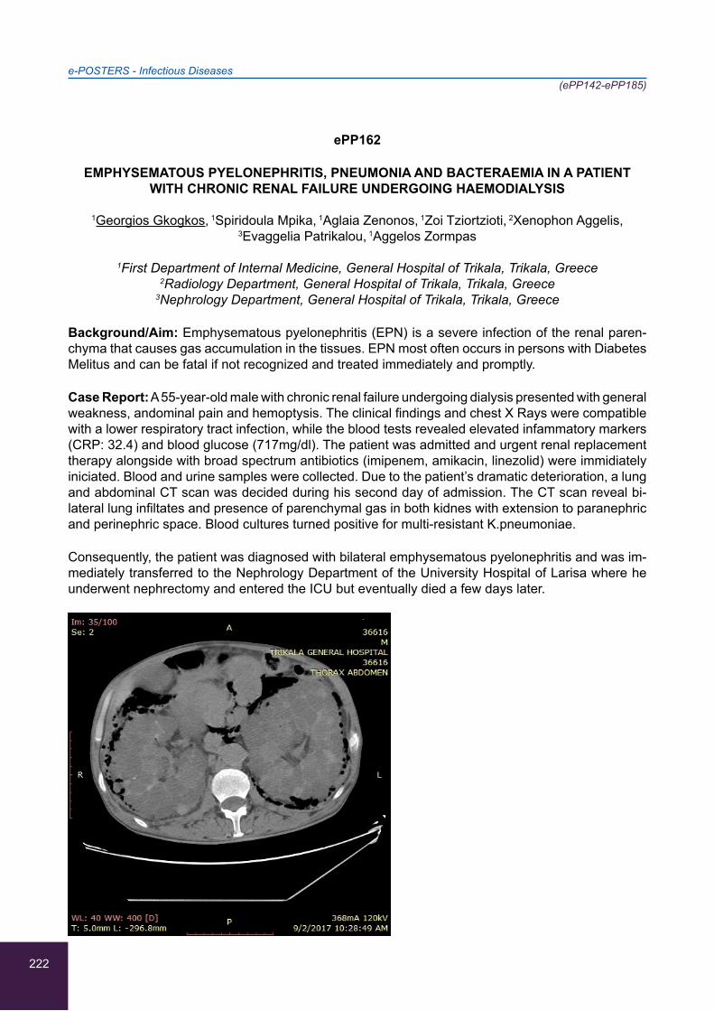
222
e-POSTERS - Infectious Diseases(ePP142-ePP185)
ePP162
EMPHYSEMATOUS PYELONEPHRITIS, PNEUMONIA AND BACTERAEMIA IN A PATIENT WITH CHRONIC RENAL FAILURE UNDERGOING HAEMODIALYSIS
1Georgios Gkogkos, 1Spiridoula Mpika, 1Aglaia Zenonos, 1Zoi Tziortzioti, 2Xenophon Aggelis,
3Evaggelia Patrikalou, 1Aggelos Zormpas
1First Department of Internal Medicine, General Hospital of Trikala, Trikala, Greece 2Radiology Department, General Hospital of Trikala, Trikala, Greece
3Nephrology Department, General Hospital of Trikala, Trikala, Greece
Background/Aim: Emphysematous pyelonephritis (EPN) is a severe infection of the renal paren-chyma that causes gas accumulation in the tissues. EPN most often occurs in persons with Diabetes Melitus and can be fatal if not recognized and treated immediately and promptly.
Case Report: A 55-year-old male with chronic renal failure undergoing dialysis presented with general weakness, andominal pain and hemoptysis. The clinical findings and chest X Rays were compatible with a lower respiratory tract infection, while the blood tests revealed elevated infammatory markers (CRP: 32.4) and blood glucose (717mg/dl). The patient was admitted and urgent renal replacement therapy alongside with broad spectrum antibiotics (imipenem, amikacin, linezolid) were immidiately iniciated. Blood and urine samples were collected. Due to the patient’s dramatic deterioration, a lung and abdominal CT scan was decided during his second day of admission. The CT scan reveal bi-lateral lung infiltates and presence of parenchymal gas in both kidnes with extension to paranephric and perinephric space. Blood cultures turned positive for multi-resistant K.pneumoniae.
Consequently, the patient was diagnosed with bilateral emphysematous pyelonephritis and was im-mediately transferred to the Nephrology Department of the University Hospital of Larisa where he underwent nephrectomy and entered the ICU but eventually died a few days later.
223
e-POSTERS - Infectious Diseases(ePP142-ePP185)
ePP163
NEUROLOGICAL COMPLICATIONS OF INFECTIVE ENDOCARDITIS THREE WEEKS AFTER ONSET OF TREATMENT
Stephanie Ioannou, Marina Gkeka, Gerasimos Eleftheriotis, Sofia Kalantzi, Asimina Protogerou,
Constantinos Christopoulos
Department of Internal Medicine, “Amalia Fleming” General Hospital, Melissia, Athens, Greece
Background: Neurological complications are common in patients with infective endocarditis and include acute encephalopathy, purulent or aseptic meningitis, meningoencephalitis, cardioembolic stroke, cerebral hemorrhage (due to stroke or ruptured mycotic aneurysm), cerebral abscess and epileptic seizures. These complications usually occur before or early after the onset of antimicrobial therapy.
Case Report: A 46-year-old male presented to the emergency department complaining of sudden onset of gait imbalance, dizziness and vomiting. These symptoms had been preceded by low-grade pyrexia which had lasted for less than 24 hours and had resolved spontaneously. Three weeks pre-viously, he had been diagnosed with S. viridans endocarditis involving the mitral valve and treated with intravenous penicillin for two weeks followed by intramuscular ceftriaxone which he was still receiving at the time of presentation. On clinical examination dysdiadochokinesia was found, sign of cerebellar dysfunction. Computed Tomography of the head revealed no pathological findings. An MRI of the brain showed appearances typical of septic infarct in the left cerebellar hemisphere. A repeated transesophageal echocardiogram showed no vegetation. The patient was admitted to the hospital and treated with a combination of intravenous penicillin and gentamycin. His neurological status improved rapidly and he was discharged after two weeks with instructions for continuous fol-low up of both cardiac and neurological status.
Conclusion: Neurological complications of infective endocarditis can occur late, even when ap-propriate antimicrobial treatment is well underway. As they can be potentially life threatening, they should always be suspected and thoroughly investigated. A careful neurological follow up of the patient is necessary.
224
e-POSTERS - Infectious Diseases(ePP142-ePP185)
ePP164
LEISHMANIASIS: AN INSIDIOUS DISEASE WITH MULTIPLE MANIFESTATIONS
1Spiridoula Mpika, 1Aglaia Zenonos, 1Georgios Gkogkos, 1Zoi Tziortzioti, 2Aggelos Zormpas,
2Eleni Gatsa
1First Department of Internal Medicine, General Hospital of Trikala, Trikala, Greece 2Hematology Department, General Hospital of Trikala, Trikala, Greece
Background/Aim: Leishmaniasis is endemic in the Mediterranean basin (L.Infantum). The parasite resides in macrophage and most infections remain asymptomatic. Herein we present two cases of visceral leishmaniasis with divergent clinical pictures, treated in our department.
Case report1: A 40-year-old male manifested anemia (Ht: 29.7%) in routine blood tests. The pa-tient presented afebrile, with palpable splenomegaly, diffuse gamma hyperglobulinemia and positive bone marrow smear for Leishmania. He was successfully treated with liposomal amphotericin at a dose of 21mg/kg, with successful recovery of the patient.
Case report2: An 80-year-old female was hospitalized due to high grade fever and weight loss for the past 15days. She had a 16-month history of anemia, a 4-month history of thrombocytopenia, diabetes and pulmonary TB infection two years ago.
Clinical examination: unremarkable.
Laboratory exams: Ht:24.7%, WBC: 6.800/μL, PLT: 65.000/μL, abdominal U/S: normal findings, diffuse gamma hyperglobulinemia, antibodies and PCR in blood specimen positive for Leishmania. She received a total dose of 45 mg/kg liposomal amphotericin, with no clinical improvement and the progression was fatal for the patient.
Conclusions: Leishmaniasis may present with a spectrum of clinical manifestations ranging from asymptomatic to lethal systemic illness. These cases are representative of that spectrum. Both had an insidious onset. Notably the second case had no splenomegaly until later stage. Recent TB infec-tion highlights the importance of cell-mediated immune response as determinant of progression from subclinical to overt disease for both infections.
225
e-POSTERS - Infectious Diseases(ePP142-ePP185)
ePP165
UTILITY OF NEUTROPHIL-LYMPHOCYTE RATIO, PLATELET-LYMPHOCYTE RATIO AND MEAN PLATELET VOLUME AS COST-EFFECTIVE DIAGNOSTIC AND PROGNOSTIC
MARKERS IN PATIENTS WITH DIABETES WHO DEVELOP TUBERCULOSIS
V.S. Srikanth, Praveen VP, Cinzia SK, J Ashwin Rajkumar
Amrita Institute of Medical Sciences and Research Centre (AIMS), Kochi, India
Objective: To study the potential role of NLR, PLR and MPV as diagnostic and prognostic markers in patients with diabetes who develop tuberculosis.
Secondary objectives: The sensitivity and prognostic utility of these 3 markers in comparison with the more established markers like CRP and ESR. Also as there is no clear marker available for pre-dicting diabetics who are predisposed and at risk of developing TB
Methodology of Investigation: A combined Retrospective + prospective study on NLR, PLR and MPV in 3 groups 1) Healthy controls,2) Type 2 diabetes patient with TB 3) Type 2 diabetes patient without TB 70 cases were taken in each group and 70 controls were taken and data obtained were analysed
Result and Discussion: In our study, we found that NLR and PLR were highly significant with a P value of 0.001 as compared to the CRP with P value 0.001. According to Stevenson et al. 15% of TB burden in India was attributed to diabetes. Diabetics is a major cause of conversion of latent to active TB, reactivation of old TB. MPV was not statistically significant to be used as a marker
Conclusion: Large number of Diabetic population who are at risk of developing TB can be easily identified and the prognosis can be monitored at Primary Health Care Centre with just a peripheral smear from which NLR and PLR can be calculated and need not depend on costly investigation like CRP. This will reduce the financial burden of the patient.
226
e-POSTERS - Infectious Diseases(ePP142-ePP185)
ePP166
CAUSES OF BLOOD STREAM INFECTION IN PATIENTS HOSPITALIZED IN THE FIRST DEPARTMENT OF INTERNAL MEDICINE OF TRIKALA HOSPITAL DURING A THREE YEAR
PERIOD
1Spiridoula Mpika, 1Georgios Gkogkos, 1Aglaia Zenonos, 1Zoi Tziortzioti, 2Vasiliki Ippa,
2Vasiliki Samara, 1Aggelos Zormpas
1First Department of Internal Medicine, General Hospital of Trikala, Trikala, Greece 2Microbiology Department, General Hospital of Trikala, Trikala, Greece
Background: A study of the causes of blood stream infections (BSIs) in patients hospitalized in the First Department of Internal Medicine in our hospital, during the period 2015-2017.
Aim: The determination of the microbal strain prevalence and antibiotic resistance.
Materials and Methods: We reviewed data of 583 blood cultures from patients hospitalized in our department during a 3-year period. Results: From a total of 583 blood cultures, 88 samples were positive (15%), 22 of them were contaminated and therefore ruled out. In the remaining 66 cases of true bacteraemia the following pathogens were isolated: E.coli 26% (16), Klebiella spp. 21%, Entero-coccus spp. 14% (9) ans S.aureus 13% (8).The remaining strains were mainly enterobacteriaceae. BSIs were distinguished in community onset-CO (80%) (blood cultures taken at the first two days of admission) and hospital BSIs (20%) (cultures taken after 2 days). CO BSIs were distinuished even further in CO-Community Acquired (CO-CA)(41% of total cases) and Health Care Associated (CO-HCA) (39%) (history of recent admission in hospital/rehabilitation centers or patient undergoing dialysis). The most common pathogen in CO-CA BSIs was E.coli (50%) (predominantly UTIs). CO-HCA BSIs were caused mainly by Klebsiella spp. (30%) -in more than half of the cases the pathogen was carbapenem resistant- and S.aureus (25%)-mostly in patients under dialysis. In hospital BSIs Enterococcus spp. was isolated in 33%.
Conclusion: The burden of HCA BSIs significant and often realted to multi-drug resistant (MDR) pathogens. Surveilance, prevention of spreading in-hospital MDR microbal strains and rational use of antibiotics are mandatory.
227
e-POSTERS - Infectious Diseases(ePP142-ePP185)
ePP167
A CASE REPORT OF PERITONEAL TUBERCULOSIS IN A PREVIOUSLY HEALTHY 27-YEAR-OLD-MAN
Eleni Marolachaki, Antonios-Rigas Papapanagiotou, Nikoletta Orphanou, Anna Ioannou, Maria Bachlitzanaki, Maria-Elena Zeniodi, Eleni Ioannidou
Internal Medicine Department, Rethymnon General Hospital, Rethymnon, Greece
Background/Aim: Tuberculosis of the peritoneum accounts less than 5% of all extrapulmonary tu-berculosis cases and about 1-2% of all forms of tuberculosis.The diagnosis of peritoneal tuberculosis can be extremely difficult because of its non-specific clinical manifestations that often mimic a variety of other abdominal diseases.
Case Report: A 27-year-old-man from Bangladesh without previous medical history presented at the emergency department with malaise, fever, shivering, nausea, vomiting and abdominal distension for about 15 days as well as weakness and weight loss. He has been living in Greece permanently for the last 7 years. The clinical examination revealed an underweighted febrile patient with hypoten-sion, tachycardia and abdominal tenderness while the blood exams showed elevated total cell count and CRP without any other abnormal findings. CT scan displayed ascites and mesenteric thicken-ing with lymphadenitis. The ascitic fluid examination showed a total cell count of 1600/μl, consisting predominantly of lymphocytes (>75%) and SAAG<1,1g/L. Staining for acid fast bacilli, fluid culture, PCR and cytologic examination where negative. Referring to the complete work up, blood cultures and blood tests for hepatitis/HIV/CMV/EBV,Brucella and Salmonella species where all negative. Gastroscopy and colonoscopy proved totally normal. Subsequentlly, a diagnostic laparoscopy was perfomed, the histology was compatible with tuberculosis and the culture was positive for mycobac-terium tuberculosis sensitive to antitubercular drugs. The patient underwent antitubercular treatment resulting in improvement.
Conclusions: Peritoneal tuberculosis is a rare extrapulmonary tuberculosis manifestation especially in patients without predisposing factors. It is often misdiagnosed while the early diagnosis and effec-tive therapy increases survival rates.
228
e-POSTERS - Infectious Diseases(ePP142-ePP185)
ePP168
XDR ACINETOBACTER BAUMANNII MENINGITIS IN A PATIENT WITH NO HISTORY OF HEAD TRAUMA OR NEUROSURGICAL INTERVENTION
Maria Meliou, Katerina Argyraki, Vaia Magaliou, Ioanna Kokkinaki, Spyridon Kloudas,
Frosso Konstantinou, Panagiotis Oikonomou, Angelos Pefanis
Department of Internal Medicine, “Sotiria” General and Chest Diseases Hospital of Athens, Athens, Greece
Background: Literature concerning multi-drug resistant Acinetobacter baumannii meningitis re-mains limited, all cases referring to neurosurgical patients.
Aim: Presentation of a unique case of health-care associated XDR Acinetobacter baumannii menin-gitis in a patient with cancer but no history of neurosurgical operation or head trauma.
Case Report: A 69 years old male patient, with SCLC stage IV, diagnosed 2 months earlier, presents with confusion and irritability of a few hours duration. Due to metastatic brain lesions he had previ-ously received whole-brain radiotherapy besides chemotherapy. Patient was afebrile, without focal neurological deficits or nuchal rigidity, GCS 10/15.
Significant laboratory findings: thrombocytopenia, leucocytosis and hyponatremia. Brain CT re-vealed 3 cerebellar metastases with oedema. Patient was treated with mannitol and high-dose dex-amethasone. Until lumbar puncture (LP) could be safely performed, ceftriaxone and ampicillin was empirically initiated. LP performed on day 3 revealed elevated neutrophils and protein. Microscopy: Gram-negative bacteria. Culture was positive for Acinetobacter baumannii, and treatment was modi-fied to meropenem, colistimethate sodium and gentamicin until antibiogram was available. However, antibiogram finally revealed an XDR isolate with MICs: meropenem >16, colistimethate >6, Tigecy-cline >265, amikacin=8. Patient died the same day, before targeted therapeutical action could be taken.
Conclusion: Extreme resistance (practically panresistance) of this isolate limited treatment options to intrathecal amikacin, with the danger of chemical meningitis due to the excipients of the regimen, or to iv amikacin in extremely high dosage. Generally, XDR Acinetobacter baumannii meningitis has limited therapeutic choices, including intrathecal administration of polymyxins, gentamicin or even tigecyclin (very few case reports).
229
e-POSTERS - Infectious Diseases(ePP142-ePP185)
ePP169
DISEASE BY LIGHT CHAIN DEPOSIT
1Jose Manuel Gil-Gascon, 1Rocio Rojas-Lorence, 1Gloria Perez-Vazquez, 2Esther Rubio-Martin,
1Eva Lozano-Loaiza, 1Carolina Luque-Amado
1Internal Medicine Unit, Hospital Punta Europa, Algeciras, Spain 2Nephrology Unit, Hospital Punta Europa, Algeciras, Spain
A 64-year-old woman was admitted in Internal Medicine Unit for a month of evolution of vomiting, weight loss, dark urine and transaminase elevation in laboratory tests. Physical examination was normal excepting subcutaneous jaundice. The blood test revealed INR 1.43, total protein 5.7 g/dl (albumin 2.1 g/dl), total bilirubin 2.4 mg/dl (direct bilirubin 1.3), GOT 221 UI/L, GPT 127 UI/L, GGT 2349 UI/L, alkaline phosphatase 1434 UI/L, total cholesterol 361 mg/dl and proteinogram with lamb-da light chains in serum 95.5 (without monoclonal peak). The hepatic autoimmunity, hepatotropic virus serology, and ceruloplasmin was normal. Urine with microalbumin/creatinine ratio 2472 mg/g. Abdominal ultrasound, abdominal MRI, and MR cholangiography with one uncomplicated right renal cyst. Gastroscopy with small hiatus hernia. With a possible diagnosis of amyloidosis was performed a biopsy of abdominal fat was performed with an ultrasound guided hepatic needle, and bone mar-row biopsy, compatible with monoclonal deposit of lambda light chains. The patient presented a renal and hepatic insufficiency with edema, ascites and increased jaundice and finally died 41 days after admission due to multiorgan failure.
This case is interesting because of the exceptionality of the deposition of lambda light chains in the liver, and probably also in the kidney, and cholestasis as part of the clinical presentation of the dis-ease.
230
e-POSTERS - Infectious Diseases(ePP142-ePP185)
ePP170
A STUDY OF THE ISOLATION INCIDENCE OF MICROORGANISMS IN CULTURES OF CENTRAL INTRAVASCULAR CATHETER TIPS AND THEIR ANTIMICROBIAL RESISTANCE
1Nikolaos Zotos, 2Eirini Tatsina, 1Christos Mitsis, 4Aristea Zotou, 1Christos Briasoulis, 1Foteini Adam,
1Panagiotis Christodoulou, 3Nikolaos Varsamis, 1Anastasia Pournou
1Microbiology Department, General Hospital of Ioannina, Ioannina, Greece 2Microbiology Department, “Papageorgiou” General Hospital,Thessaloniki, Greece
3Pathology Department, General Hospital of Ioannina, Ioannina, Greece 4Cardiology Department, University Hospital of Ioannina, Ioannina, Greece
Aim: The aim of the study was to investigate the incidence of isolation of pathogenic agents on cultures of central intravascular catheter tips in the General Hospital of Ioannina during a five year period (2012- 2016).
Material and Method: The material of the study was 712 catheter tips from inpatients from 2012 until 2016. The Microscansystem was employed for the identification and antimicrobial susceptibility testing of the pathogenic agents isolated.
Results: A microorganism was detected in 250 out of 712 samples (35.1%) . The most frequent microorganisms that were isolated were: Staphylococcus epidermidis (32.8% ) , Acinetobacter Baumanii (1.4%), Pseudomonas aeruginosa (9.6%),Klebsiella pneumoniae(8.8%),Enterococcus faecalis(4.8%),Proteus mirabilis(2.8%). The resistance to drugs can be seen on the table below:
Conclusion: The most common microorganism isolated was Staphylococcus epidermidis, a fact that raises the question of whether that was a pathogenic agent of infection or just a contamination of the sample with the microbial flora of the skin .In addition, strains of Acinetobacter.spp and Kleb-siella.spp resistant to antibiotics were isolated at a high rate, since most of them were isolated in patients of the I.C.U who are usually colonized by multi-drug resistant strains.
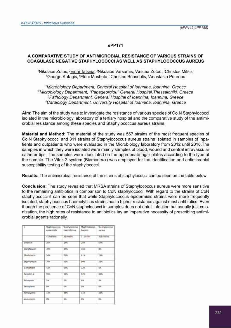
231
e-POSTERS - Infectious Diseases(ePP142-ePP185)
ePP171
A COMPARATIVE STUDY OF ANTIMICROBIAL RESISTANCE OF VARIOUS STRAINS OF COAGULASE NEGATIVE STAPHYLOCOCCI AS WELL AS STAPHYLOCOCCUS AUREUS
1Nikolaos Zotos, 2Eirini Tatsina, 3Nikolaos Varsamis, 4Aristea Zotou, 1Christos Mitsis,
1George Katagis, 1Eleni Mosheta, 1Christos Briasoulis, 1Anastasia Pournou
1Microbiology Department, General Hospital of Ioannina, Ioannina, Greece 2Microbiology Department, “Papageorgiou” General Hospital,Thessaloniki, Greece
3Pathology Department, General Hospital of Ioannina, Ioannina, Greece 4Cardiology Department, University Hospital of Ioannina, Ioannina, Greece
Aim: The aim of the study was to investigate the resistance of various species of Co.N Staphylococci isolated in the microbiology laboratory of a tertiary hospital and the comparative study of the antimi-crobial resistance among these species and Staphylococcus aureus strains.
Material and Method: The material of the study was 567 strains of the most frequent species of Co.N Staphylococci and 311 strains of Staphylococcus aureus strains isolated in samples of inpa-tients and outpatients who were evaluated in the Microbiology laboratory from 2012 until 2016.The samples in which they were isolated were mainly samples of blood, wound and central intravascular catheter tips. The samples were inoculated on the appropriate agar plates according to the type of the sample. The Vitek 2 system (Biomerieux) was employed for the identification and antimicrobial susceptibility testing of the staphylococci.
Results: The antimicrobial resistance of the strains of staphylococci can be seen on the table below:
Conclusion: The study revealed that MRSA strains of Staphylococcus aureus were more sensitive to the remaining antibiotics in comparison to CoN staphylococci. With regard to the strains of CoN staphylococci it can be seen that while Staphylococcus epidermidis strains were more frequently isolated, staphylococcus haemolyticus strains had a higher resistance against most antibiotics. Even though the presence of CoN staphylococci in samples does not entail infection but usually just colo-nization, the high rates of resistance to antibiotics lay an imperative necessity of prescribing antimi-crobial agents rationally.
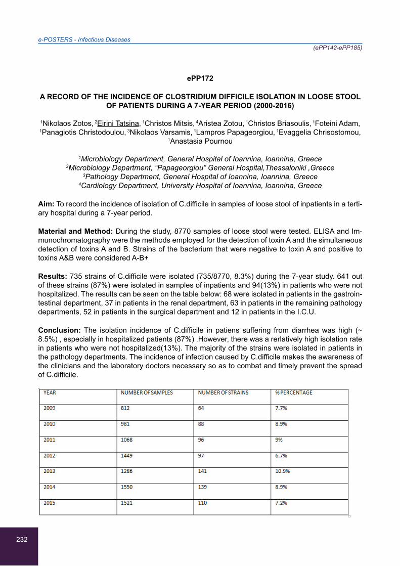
232
e-POSTERS - Infectious Diseases(ePP142-ePP185)
ePP172
A RECORD OF THE INCIDENCE OF CLOSTRIDIUM DIFFICILE ISOLATION IN LOOSE STOOL OF PATIENTS DURING A 7-YEAR PERIOD (2000-2016)
1Nikolaos Zotos, 2Eirini Tatsina, 1Christos Mitsis, 4Aristea Zotou, 1Christos Briasoulis, 1Foteini Adam,
1Panagiotis Christodoulou, 3Nikolaos Varsamis, 1Lampros Papageorgiou, 1Evaggelia Chrisostomou,
1Anastasia Pournou
1Microbiology Department, General Hospital of Ioannina, Ioannina, Greece 2Microbiology Department, “Papageorgiou” General Hospital,Thessaloniki ,Greece
3Pathology Department, General Hospital of Ioannina, Ioannina, Greece 4Cardiology Department, University Hospital of Ioannina, Ioannina, Greece
Aim: To record the incidence of isolation of C.difficile in samples of loose stool of inpatients in a terti-ary hospital during a 7-year period.
Material and Method: During the study, 8770 samples of loose stool were tested. ELISA and Im-munochromatography were the methods employed for the detection of toxin A and the simultaneous detection of toxins A and B. Strains of the bacterium that were negative to toxin A and positive to toxins A&B were considered A-B+
Results: 735 strains of C.difficile were isolated (735/8770, 8.3%) during the 7-year study. 641 out of these strains (87%) were isolated in samples of inpatients and 94(13%) in patients who were not hospitalized. The results can be seen on the table below: 68 were isolated in patients in the gastroin-testinal department, 37 in patients in the renal department, 63 in patients in the remaining pathology departments, 52 in patients in the surgical department and 12 in patients in the I.C.U.
Conclusion: The isolation incidence of C.difficile in patiens suffering from diarrhea was high (~ 8.5%) , especially in hospitalized patients (87%) .However, there was a rerlatively high isolation rate in patients who were not hospitalized(13%). The majority of the strains were isolated in patients in the pathology departments. The incidence of infection caused by C.difficile makes the awareness of the clinicians and the laboratory doctors necessary so as to combat and timely prevent the spread of C.difficile.
233
e-POSTERS - Infectious Diseases(ePP142-ePP185)
ePP173
PSEUDOMONAS PUTIDA: A CASE OF COMMUNITY ACQUIRED DRUG RESISTANT BACTEREMIA
1Dimitrios Pappas, 1Natalie El Gkotmi, 1Aikaterini Koutsi, 1Eleni Loutsidi, 1Aggeliki Papageorgiou, 1Georgios Nikolaidis, 2Alexandra Konstantinou, 2Alexandra Stamati
1Resident, 2nd department of Internal Medicine, General Hospital “Asklepieio”, Voula, Athens, Greece
2Internist, 2nd department of Internal Medicine, General Hospital “Asklepieio”, Voula, Athens, Greece
Background: Pseudomonas putida is a gram-negative rod belonging to rRNA group I of the genus Pseudomonas. Cases of Pseudomonas putida bacteremia, often associated with trauma hospitali-zation or immunosuppresion, have rarely been reported. Here we present a rare case of septic bac-teremia caused by a community acquired P. putida, resistant to carbapenemases.
Case Report: A 57 year old female Caucasian gardener, presented to the emergency department with a history of persistent fever up to 40°C, accompanied with shiver, nausea and vomiting, colic pain located in the right lumbar region. After clinical laboratory and imaging control an acute focal pyelonephrits on bilateral nephrolithiasis was diagnosed. Medical history and laboratory results re-vealed presence of hepatitis B under no antiviral therapy. The patient showed inadequate response to the initial empirical treatment with meropenem. Repeated blood cultures obtained upon hospital admission revealed the presence of P. putida carbapenemases resistant due to VIM mechanism susceptible only to amikacin, colistin and fosfomycin. P. putida is a common ingredient of soil fertiliz-ers also used in our patient’s workplace.
Conclusion: After being treated with proper antibiotic treatment, our patient remained afebrile, in a good clinical status with all inflammatory markers in remission and she was discharged.
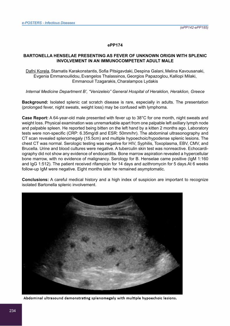
234
e-POSTERS - Infectious Diseases(ePP142-ePP185)
ePP174
BARTONELLA HENSELAE PRESENTING AS FEVER OF UNKNOWN ORIGIN WITH SPLENIC INVOLVEMENT IN AN IMMUNOCOMPETENT ADULT MALE
Dafni Korela, Stamatis Karakonstantis, Sofia Pitsigavdaki, Despina Galani, Melina Kavousanaki,
Evgenia Emmanouilidou, Evangelos Thalassinos, Georgios Papazoglou, Kalliopi Milaki,
Emmanouil Tzagarakis, Charalampos Lydakis
Internal Medicine Department B’, “Venizeleio” General Hospital of Heraklion, Heraklion, Greece
Background: Isolated splenic cat scratch disease is rare, especially in adults. The presentation (prolonged fever, night sweats, weight loss) may be confused with lymphoma.
Case Report: A 64-year-old male presented with fever up to 38°C for one month, night sweats and weight loss. Physical examination was unremarkable apart from one palpable left axillary lymph node and palpable spleen. He reported being bitten on the left hand by a kitten 2 months ago. Laboratory tests were non-specific (CRP: 6.35mg/dl and ESR: 50mm/hr). The abdominal ultrasonography and CT scan revealed splenomegaly (15.5cm) and multiple hypoechoic/hypodense splenic lesions. The chest CT was normal. Serologic testing was negative for HIV, Syphilis, Toxoplasma, EBV, CMV, and Brucella. Urine and blood cultures were negative. A tuberculin skin test was nonreactive. Echocardi-ography did not show any evidence of endocarditis. Bone marrow aspiration revealed a hypercellular bone marrow, with no evidence of malignancy. Serology for B. Henselae came positive (IgM 1:160 and IgG 1:512). The patient received rifampicin for 14 days and azithromycin for 5 days.At 6 weeks follow-up IgM were negative. Eight months later he remained asymptomatic.
Conclusions: A careful medical history and a high index of suspicion are important to recognize isolated Bartonella splenic involvement.
235
e-POSTERS - Infectious Diseases(ePP142-ePP185)
ePP175
PATIENT WITH FEBRILE SYNDROME AND REACTIVE ARTHRITIS AFTER INTRAVESICAL ADMINISTRATION OF BACILLUS CALMETTE GUERIN (BCG) AND ANTI-CYCLIC
CITRULLINATED PEPTIDE ANTIBODIES DEVELOPMENT
Konstantinos Kounouklas, Styliani Kaliampou, Andromachi Panagiotopoulou, Dimitra Stathi,
Georgia Farmaki, Melpomeni Tsepeli, Vasilis Fasias, Eleni Mostratou, Nikolaos Symeonidis
2nd Department of Internal Medicine of 251 General Airforce Hospital, Athens, Greece
Aim: Detection of anti-cyclic citrullinated peptide (anti-CCP) has recently been reported in patients with active pulmonary tuberculosis (TBC). Whether patients with intravesicular BCG immunotherapy develop anti-CCP, needs evaluation.
Case report: A 50-year-old, male, was admitted because of episodes of fever up to 39 C and urine discomfort . One week ago he had intravesicular administration of BCG for bladder cancer. On ex-amination patient had arthritis of left knee and at the second day of left ankle.
Differential diagnosis: Urinary-acute prostatitis infection, BCG infection after intravesical infusions. Laboratory evaluation, CT scans and knee aspiration did not confirm active TBC infection. Patient was treated with levofloxacin and because of persistence of fever, 300 mg isoniazide and 8 mg methylprednisone were added. Anti -CCP found positive, but patient had improvement and became asymptomatic, with resolution of arthritis.
Conclusions: anti-CCP antibodies have been considered very specific for rheumatoid arthritis (RA). However, studies have shown that these antibodies can be falsely positive in infectious diseases like TB, HIV, etc. Reactive arthritis following BCG immunotherapy is well described. Thus, a patient could be misdiagnosed as having early RA based on polyarthritis and anti-CCP positivity. The percentage of these patients develop anti-CCP needs further evaluation.
236
e-POSTERS - Infectious Diseases(ePP142-ePP185)
ePP176
POSTERIOR REVERSIBLE ENCEPHALOPATHY SYNDROME AS A COMPLICATION OF BACTERIAL MENINGITIS
Elena Krasnikova, Charalambos Ch. Christofidis, Alexandros Anastasiou, Orestes Papazisis,
Evdoxia Mitsiou, Charalambos Mandros,Evangelos Potolidis
Second Department of Internal Medicine, General Hospital of Volos, Volos, Greece 2General Hospital of Volos, Volos, Greece
Posterior reversible encephalopathy syndrome (PRES) is a clinico-radiological syndrome that mani-fests with headache, seizures, altered mental consciousness and visual disturbances, and is char-acterized by white matter vasogenic edema affecting predominantly the posterior occipital and pa-rietal lobes of the brain. This clinical syndrome is increasingly recognized due to improvement and availability of brain imaging, specifically magnetic resonance imaging (MRI). The syndrome should be promptly recognized, since it is reversible and treatable. Delayed diagnosis and treatment of PRES may lead to death or irreversible neurological deficit. The severity of the clinical symptoms varies widely, i.e. the visual disturbances can range from blurred vision, visual hallucinations, visual neglect, homonymous hemianopsia to cortical blindness, while altered consciousness may vary from mild confusion or agitation progressing even to coma. Other symptoms include nausea, vomiting and brainstem deficits. Seizures and status epilepticus are quite often the presenting manifestation. PRES is a syndrome of heterogeneous etiologies and is associated with clinical conditions such as preeclampsia, eclampsia, autoimmune disease, cancer chemotherapy, transplatation including bone marrow or stem cell transplatation, hypertension, and finally, infection/sepsis/shock.
We hereby report the case of an otherwise healthy adult female, presenting with fever, headache and generalized arthralgia, masking Neisseria meningitidis serotype B meningitis, later complicated by posterior reversible encephalopathy syndrome. Meningococcal meningitis is a well established po-tential fatal infection characterized by fever, headache, petechial rash, and vomiting in the majority of cases. Increased clinical suspicion and prompt diagnosis is crucial to ensure favorable outcomes.
237
e-POSTERS - Infectious Diseases(ePP142-ePP185)
ePP177
ATYPICAL INFECTIOUS MONONUCLEOSIS SYNDROME CAUSED BY PRIMARY CMV INFECTION ALONG WITH REACTIVATION OF LATENT EBV INFECTION
1Christos Zissis, 1Dimitra Maritsa, 1Eleni Petroulaki, 1Ioannis Liabas, 2Charitini Mylona, 3Eleni Gatsa,
1Stefanos Mylonas
12nd Department of Internal Medicine, General Hospital of Trikala, Trikala, Greece 2Medical School, Aristotle University of Thessaloniki, Thessaloniki, Greece
3Department of Hematology, General Hospital of Trikala, Trikala, Greece
Background/Aim: Cytomegalovirus(CMV) and Epstein-Barr-Virus(EBV) belong to the herpes-virus group. They cause infectious-mononucleosis-syndrome. After the infection, they remain latent in lymphocytes. They rarely show resurgence in conditions of cellular immunosuppression. Rare cases of co-infection with CMV and EBV - mainly in children - are reported in literature. Additionally, their antigens may show cross-reactions with all herpes-viruses’ antigens. The aim of this report is the demonstration of a rare case of atypical mononucleosis syndrome caused by primary-CMV-infection along with reactivation of latent-EBV-infection.
Case Report: A 56-year-old woman has been experiencing a feeling of tiredness and fatigue for months and was finally referred to the General Hospital of Trikala due to elevated transaminas-es (serum values three times higher than the upper-normal-limit) and splenomegaly(confirmed by ultrasound). Blood-tests, also, showed mild lymphocytocis-anemia-thrombocytopenia, hyper-gammaglobulinemia and high-LDH-levels. Hepatitis-serologic-testing was negative for HBV/HCV-infection(HBsAg/anti-HBs/anti-core/anti-HCV) and positive for old-HAV-infection. She underwent imaging testing (chest/abdomen computed-tomography) which confirmed splenomegaly, without revealing co-existing lymphadenopathy. Further testing (Mantoux/Monospot/HIV-test/Widal/Wright/anti-Brucella/anti-Leismania/anti-Toxoplasma/ANA/SLA/anti-LKM-1,2,3/copper-serum-levels/seru-loplasmin-levels/alpha-1-antitrypsin-serum-levels) was negative. The initial high anti-CMV-IgM and anti-CMV-IgG titers and following antibody-seroconversion were compatible with primary-CMV-in-fection. On the other hand, high EBV-IgM-VCA/EBV-IgG-VCA titers were found and seroconversion followed. Heterophile-Antibody-Test(Monospot) was negative. IgG-EBNA-titers were increased, as well. Since 90-95% of the population is infected by EBV until adulthood, the syndrome was-primarily-attributed to acute-CMV-infection along with reactivation of latent-EBV-infection, as a result of tran-sient cellular immunosuppression.
Conclusions: In medicine, a successful diagnostic approach requires both detailed clinical examina-tion and laboratory investigation - which sometimes includes special serological testing - but mainly, therapist’s ability to co-assess and interpret the collected data.
238
e-POSTERS - Infectious Diseases(ePP142-ePP185)
ePP178
MICROBIAL INFECTIONS AND ATHEROSCLEROSIS: INVESTIGATING A NOVEL RISK FACTOR?
1Rafaella Datseri, 2D. Desse, 2Eleni Kastritsi, 2Olga Kastritsi, 2Emmanouela Laskaratou,
3Georgios Matzakanis, 2Eleutheria Parasiri, 4Eviana Petropoulou, 5Nikolaos Sevdalis,
6Dimitrios Tremoulis, 7Sibel Kara Ali Achmet, 2Christos Tsagkaris
1Faculty of Medicine, Commenius University of Bratislava, Bratislava, Slovakia 2Faculty of Medicine, School of Health Science, University of Crete, Heraklion, Greece
3Faculty of Medicine, University of Patras, Patras, Greece 4Department of Medical Laboratories, University of Applied Science of Athens, Athens, Greece
5Faculty of Medicine, Medical University of Sofia, Bulgaria 6Biology Department, University of Patras, Patras, Greece
7Department of Nursery, University of Applied Science of Athens, Athens, Greece
Introduction: Atherosclerosis has been evinced epidemiologically as a major cause of morbidity and mortality. Even if current literature focuses on the metabolic background of the disease, the role of the pathogens and the immune background of endothelial damage appear interesting.
Purpose: The purpose of this study is to discuss the latest scientific results that evaluate the effect of some pathogens on the appearance of atherosclerotic plaque.
Materials And Methods: We reviewed literature through Pubmed and Cochrane. Key-words as atherosclerosis, infection, pathogens and endothelial instability were used. Retrospective and epide-miological studies hosted in high impact factor journals were the main sources of data.
Results: Pathogens contribute to the endothelial damage both in a direct and an indirect way. In the first case endothelial cells get infected, whereas in the second case secretion of inflammatory cytokines destabilizes the endothelium spreading away from the area of infection. Bacterial and viral strains are involved in atheromatosis and can be classified as common pathogens and pathogens of periodontal disease.
Discussion: The immunological background of the atherosclerotic disease is offered for further re-search, and it is essential that a high sensitivity technique of isolating pathogens from atherosclerotic plaques be defined. The conditions needed for the creation of atherosclerotic plaques in humans are not reflected accurately while using genetically modified mice, which makes it necessary that new studies based on human be conducted. The issue is raised through the anti-vaccination movement which has yet to reveal another aspect of the pathogenesis of infections.
239
e-POSTERS - Infectious Diseases(ePP142-ePP185)
ePP179
TREATMENT OF BLOOD CULTURE-NEGATIVE ENDOCARDITIS ON AN ARTIFICIAL METALLIC VALVE
Lamprini Skorda, Ioanna Gazouli, Georgios Kassianidis, Vladimiros Hatzis, Maria Tsekeri,
Antonios Alaveras
1st internal medicine department, Red Cross Hospital, Athens, Greece
Background: Treatment of blood culture negative endocarditis on a prosthetic metallic valve.
Case report: Female patient 49 yo was admitted in the internal medicine ward due to daily fever for the previous 20 days. She had been prescribed clarithromycin and cephuroxim. Personal medi-cal history: 1) familial hypercholesterolemia (homozygous) treated with loperamide 2) triple CABG (2014), 3) aortic valve replacement by a metallic valve treated with acenocoumarol. Blood tests on the day of admission: WBC 10.000, ESR 86, CRP 58, Hgb 8gr/dL, Probable diagnosis on admit-tance: endocarditis due to her medical history. The transthoracic ultrasound did not reveal any pa-thology, the oesophageal ultrasound revealed abscess on the artificial valve. The brain, thorax and abdomen CT revealed hepatosplenomegaly, pleural effusion.
Histopathology: negative.
Blood cultures: negative. After trying various antimicrobial combinations, she was administered imi-penem and daptomycin for 6 months. The patient’s fever dropped to normal, CRP and ESR returned to normal and there was a significant clinical improvement. She was tested regularly with oesopha-geal ultrasound she was diagnosed with a pseudoaneurysm of the mitral-aortic intervalvular fibrossa and was advised to be examined by a cardiac surgeon. She was examined by six cardiac surgeons and all of them consider the pseudoneurysm as inoperable.
Conclusion: Treatment of blood culture negative endocarditis is very difficult because of the lack of sufficient evidence in bibliography. Six months of antimicrobial treatment seem like a safe period in inoperable cases of cardio surgery.

240
e-POSTERS - Infectious Diseases(ePP142-ePP185)
ePP180
THE EFFECTIVENESS OF EBOLA CONTROL MEASURES IN WEST AFRICA
1Anastasia Papachristou, 1Anastasia Ntikoudi, 2Vasileios Saridakis
1Medical Department, National & Kapodistrian University of Athens, Athens, Greece 2Surgical Department, General Hospital of Nikaia, Athens, Greece
The Ebola virus outbreak in West Africa in 2013 reached an alarming diffusion rate starting from Guinea and in a few months, it was spread in several countries of the African continent. Ebola virus is transmitted to people as a result of direct contact with body fluids containing virus of an infected patient. Typical Ebola symptoms include fever, weakness, cramping, nausea, diarrhea, abdominal pain, cramping, nausea and vomiting for 5-7 days. Currently, several therapeutic agents and novel vaccines for Ebola virus have been developed and are now undergoing evaluation. There are no ap-proved vaccines or antiviral therapeutics for Ebola virus currently available for human use although many have already been tested. However, ring vaccination is predicted to offer a moderately benefi-cial supplement to ongoing non-pharmaceutical Ebola control efforts. Moreover, activated protein C, a recombinant inhibitor of factor VIIa/ tissue factor may be considered in future clinical experimental plans for severe Ebola infections in patients which are not responsive to standard care regimens.
Last but not least, additional treatment beds on the number of Ebola cases led to a substantial de-cline in EVD transmission during 2014-2015. Ebola poses a threat not only to West Africa but also to the international community. The most effective approach would be both limiting the international spread of Ebola and to control the disease at its source.
241
e-POSTERS - Infectious Diseases(ePP142-ePP185)
ePP181
IMPACT OF NEONATAL EARLY-ONSET SEPSIS CALCULATOR ON ANTIBIOTIC USE IN NEWBORNS WITH SUSPECTED INFECTION
1Lauma Vasiļevska, 1,2Ilze Meldere
1Riga Stradiņš University, Faculty of Medicine, Riga, Latvia 2Neonatology Intensive Care Unit, Children’s Clinical University Hospital, Riga, Latvia
Objective: A recently described neonatal early-onset sepsis (EOS) calculator has the potential to reduce newborn antibiotic exposure based on five risk factors. The objective was to compare actual antibiotic use to the stratification based on the sepsis calculator in newborns with suspected EOS and to investigate differences in EOS risk between newborns that received early (<12 h) versus late antibiotics (≥12 h of life).
Materials and methods: Retrospective review of infants born ≥ 34 weeks gestation who received antibiotics within 72 hours after birth. The EOS risk score per 1000 live births was calculated and each newborn was retrospectively assigned to the recommended category by the Kaiser Perma-nente neonatal EOS calculator. Statistical analysis was performed using IMB SPSS Statistics 22 software.
Results: The EOS calculator recommended not to start antibiotic therapy in 156 (77%) out of 205 infants. Antibiotic treatment was started early in 60 (29%) and late in 145 (71%) newborns. 13 (6%) positive blood cultures were identified. Newborns that received early antibiotics had higher maternal EOS score (median 1.39, IQR: 0.39-2.12) compared to newborns in the late treatment group (me-dian 0.03, IQR: 0.02-0.04). Clinical condition deteriorated in the late treatment group, including one newborn with low risk score, but afterwards proven purulent meningitis.
Conclusion: Antibiotic use in newborns could be significantly reduced by more than 77%, leading to less need for laboratory monitoring and more accurate antibiotic targeting. Newborns with initial low sepsis risk score clinically deteriorated beyond 12 h of life. Continuous good clinical observation is crucial.
242
e-POSTERS - Infectious Diseases(ePP142-ePP185)
ePP182
MALIGNANT EXTERNAL OTITIS IN PEDIATRIC AND ADULT PATIENTS
1Georgios Karabelias, 2Vassiliki Mamakou, 1,3Aikaterini Drylli
1 ENT Department, Penteli Children’s Hospital, Athens, Greece2Internal Medicine Department, General Psychiatric Hospital “Dromokaiteio”, Athens, Greece
3Audiology-Neurotology, Medical School, National and Kapodistrian University of Athens,Athens, Greece
Background: Malignant External Otitis (MEO) is a necrotizing infection of external auditory canal and temporal bone, leading to skull base osteomyelitis. Pseudomonas aeruginosa is the most com-mon causative organism, in more than 90% of patients. Despite the antibiotic treatment the survival rate is 50%.
Objectives / Aim: To describe similarities and differences of MEO in pediatric and adult patients.
Methods: A retrospective systematic review was conducted of articles, by using search terms such as [(malignant external otitis) AND children AND adult], on medical literature data base (PubMed).
Results: In adult population MEO tends to affect the elderly as well as patients with comorbidity such as diabetes mellitus, cardiovascular disease, chronic renal failure, autoimmune disease, hematologi-cal disease or cancer. Typically, includes otalgia persisting for longer than one months, chronic otor-rhea, headache and cranial involvement. HbA1c levels, inflammatory markers and imaging findings influence the prognosis of disease. MEO is reported in almost few cases of non-diabetic pediatric patients who are immunocompromised due to malignancy, malnutrition and hematological -severe anemia or granulocytopenia- and immunodeficiency disorders such as IgA deficiency and HIV. The clinical presentation contains fever, otalgia, otorrhea, fatigue and facial palsy nerve. Inflammatory indexes and imaging findings determine the prognosis of disease.
Conclusions: Pediatric patients are frequently immunodeficient and have a far better prediction, with practically no mortality. However, adult patients are usually elderly diabetics, with severe mortal-ity rate. Ciprofloxacin and other antipseudomonal agents are the imperative antibiotic treatment. The prognosis of disease has statistically significant impact of cranial nerve involvement.
243
e-POSTERS - Infectious Diseases(ePP142-ePP185)
ePP183
ASSOCIATION BETWEEN ROUTINE BIOCHEMICAL TESTS AND OUTCOME IN PATIENTS WITH SEPSIS
1,2Elina Drakou, 3Eleutherios Stratigakos, 4Spyridon Kottas, 5Dimitra Latsou, 1Alexandra Zata, 1Panagiotis Gargalianos-Kakolyris, 2,6Nikoleta Iacovidou, 2,6Athanasios Chalkias,
6,7Theodoros Xanthos
11st Department of Internal Medicine, General Hospital of Athens ‘Georgios Gennimatas’, Athens, Greece
2Postgraduate Study Program (MSc) “Cardiopulmonary Resuscitation”, Medical School, National and Kapodistrian University of Athens, Athens, Greece
3Department of Gastroenterology, General Hospital of Athens “Georgios Gennimatas”, Athens, Greece
4Hellenic Labour Inspectorate, Greek Ministry of Labour, Social Insurance and Social Solidarity, Athens, Greece
5Faculty of Social and Education Policy, University of Peloponnese, Korinthos, Greece6Hellenic Society of Cardiopulmonary Resuscitation, Athens, Greece7School of Medicine, European University of Cyprus, Nicosia, Cyprus
Background: Sepsis is a medical emergency associated with high rates of mortality and morbidity. In this study, we sought to investigate the association between biochemical markers and outcome of patients with sepsis.
Methods: We reviewed the files of all consecutive patients admitted to the 1st Department of Inter-nal Medicine, from January 2015 to December 2016. Demographics, duration of hospitalization, sur-vival, site of infection, type of the pathogen species, biochemical analysis, qSOFA and SOFA score at the ED and within 24h after hospital admission were recorded. The primary outcome was survival at discharge, while length of hospital stay was defined as the secondary outcome.
Results: The study population consisted of 90 patients (40 men and 50 women) with a mean age of 79±11.7 years and mean length of stay was 8.7±5.4 days. There was a statistically significant association between baseline and 24h SOFA score variation with survival (p=0.001). Although, no statistically significant association was observed between the biochemical data or SOFA score with length of stay.
Conclusion: In this study, biochemical markers were not associated with survival or length of stay. However, the number of survivors indicates the proper timing and effectiveness of targeted thera-peutic interventions in our Institution.
244
e-POSTERS - Infectious Diseases(ePP142-ePP185)
ePP184
ANGIOEDEMA AND INFECTIOUS MONONUCLEOSIS: A RARE CASE REPORT
1Christos Zissis, 1Dimitra Maritsa, 1Nikolaos Aggelis, 1Foteini Malakoudi, 2Charitini Mylona,
1Vaios Tasios, 1Stefanos Mylonas
12nd Department of Internal Medicine, General Hospital of Trikala, Trikala, Greece 2Medical School, Aristotle University of Thessaloniki, Thessaloniki, Greece
Background/Aim:”Angioedema” is the swelling of deep-dermis, subcutaneous or submucosal tissue due to vascular leakage. Acute episodes often involve the lips, eyes, face and-occasionally-other parts including the respiratory/gastrointestinal mucosa. Laryngeal-swelling can be life-threatening. -Hista-mine-mediated/Histaminergic angioedema (responsive to antihistamine/cortisone). 1.Angioedema-associated-with-urticaria (with/without urticarial rash) 2.Idiopathic -Bradykinin-mediated/Non-hista-minergic angioedema(resistant to antihistamine/cortisone). 1.Hereditary:Functional / Quantitative C1-esterase-inhibitor (C1-INH) deficiency. 2.Acquired: Production of anti-C1-INH / Overconsumption of C1-INH in autoimmune/lymphoproliferative diseases respectively. 3.ACE(angiotensin-converting-enzyme)-inhibitor-induced angioedema. 4.Idiopathic
The aim of this report is the demonstration of a rare case of angioedema in a teenager with infectious mononucleosis.
Case Report: A 14-year-old-female patient, without individual/family history of angioedema, referred to the emergency department because she was experiencing fever, vomiting and diarrhea for 24 hours, weakness and anorexia for 5 days. Clinical examination revealed cervical lymphadenopathy. Blood tests showed lymphocytosis, elevated LDH and transaminases. Μild splenomegaly was diag-nosed by abdominal-ultrasound. On-2nd-day-of-hospitalization, she acutely exhibited angioedema of the eyelids, face, lips and tongue, without urticaria, non-related-to-drugs and antihistamine-resist-ant. Further testing (immunological-control/thyroid-function/hepatitis-tests/HIV-test/anti-CMV/Widal/Wright/anti-Brucella/anti-Leismania/ASTO/Mantoux/cardiac-ultrasound/stool-urine-blood-cultures/serum&24-hour-urine protein) was normal. Chest/Abdomen-computed-tomography(CT) did not re-veal mesothoracic/abdominal lymphadenopathy. On-5th-day-of-hospitalization, cortisone was ad-ministered. On-6th-day the fever retreated. The edema subsided two days later. Heterophile-Anti-body-Test(Monospot) and EBV-IgM-VCA-titers were elevated, while C1-INH-levels were marginally low. A month later, EBV-antibody-seroconversion occurred, while C1-INH and C4-fragment levels were normal.
Conclusions: EBV is a lymphotropic virus, implicated in activation of hereditary-angioedema and appearance of lymphoproliferative/autoimmune diseases. However, there is no reference to its as-sociation with acquired-angioedema. In contrast, few cases implying correlation with histaminergic-angioedema and acute/chronic urticaria are described. In medicine, a rare diagnosis should, also, be based on bibliographical confirmation(evidence-based) rather than on spontaneous conclusions.
245
e-POSTERS - Infectious Diseases(ePP142-ePP185)
ePP185
OCULAR COMPLICATIONS OF AIDS
1Dimitrios Bakas, 2Angelos Anyfantis, 3Vasileios Kolokoutsas
1Ophthalmology Department, General Hospital Of Kastoria, Kastoria, Greece2Ophthalmology Clinic, General Hospital Of Drama, Drama, Greece
3PEDY-MY of Kalamata, Kalamatta, Greece
Introduction: Although in the asymptomatic vectors of the virus ocular disorders are rare, in patients with AIDS the incidence rates range from 75-95%.
Clinical Manifestations: The most common and major ocular manifestations of AIDS are: • Non-infectious AIDS retinopathy (HIV microangiopathy): The most common manifestation. It is charac-terized mainly by cotton wool spots and, less frequently, by intraretinal haemorrhages, microaneu-rysms, telangiectasia, vasculitis, retinal vascular occlusion. It is generally asymptomatic and does not require treatment. • CMV retinitis: The most common opportunistic eye infection. It is character-ized by a slowly progressive necrotic retinitis. It is treated with systemic or intravitreal injections of ganciclovir, foscarnet or sidofovir. • Progressive Outer Retinal Necrosis and Acute Retinal Necrosis: These are necrotizing retinitis, mainly due to HZV and more rarely HSV, with poor response to treatment and poor prognosis. Less frequent or minor manifestations include: • Adnexal complica-tions: Conjunctivitis, dry eye, blepharitis, molluscum contagiosum of eyelids, HZV or HSV infection, orbital cellulitis, chronic microsporidial keratitis. • Anterior uveitis (from CMV, syphilis, tuberculosis, toxoplasma). • Neoplasms: Kaposi’s sarcoma of eyelids, orbital or intraocular (mainly choroidal) lym-phoma. • Opportunistic infections: Bacterial or fungal keratitis, cryptococcal choroiditis, toxoplasmic chorioretinopathy, Pneumocystis carinii choroiditis, Candida endophthalmitis. • Neuroophthalmologi-cal manifestations: Cranial nerve and/or extraocular muscle palsies, retrobulbar optic neuritis (from syphilis, cryptococcus or HZV). • Toxic optic neuropathy: from pharmaceutical therapy, mainly dida-nosine and ethambutol.
Conclusion: Ophthalmic manifestations in people with AIDS are frequent and sometimes intense. Prophylactic examination by the ophthalmologist even with the most insignificant discomfort is nec-essary.
246
e-POSTERS - Miscellaneous(ePP186-ePP203)
ePP186
EFFICACY AND SAFETY OF RADIATION THERAPY IN PATIENTS WITH GRAVES’ OPHTHALMOPATHY: A CASE SERIES REPORT
1Eleni Fotiou, 1Maria Pittaka, 2Ioannis Kyprianou, 1Marilena Theodorou, 1Demetris Andreopoulos
1Department of Radiation Oncology, Bank of Cyprus Oncology Center, Nicosia, Cyprus 2Ophthalmology Department, Archbishop Makarios III Hospital, Nicosia, Cyprus
Background/Aim: Graves’ ophthalmopathy (GO) is the most common extrathyroidal manifestation of Graves’ disease. It is characterized by inflammation of orbital and periorbital tissue. Treatment of GO is very challenging, and the outcome is often unsatisfactory. Orbital radiotherapy (RT) has been used for over 60 years in the treatment of GO with variable efficacy. The purpose of this study was to review our practice of RT in GO and compare to published results.
Case report: We examined the clinical outcomes and tolerability of radiation therapy in a series of patients (subjects) with severe Graves’ ophthalmopathy between 2015 and 2017. Nine patients were included (66% female; median age 58 years), all euthyroid. Clinical evaluation of patients was performed using the modified NOSPECS score. Before treatment all patients had exophthalmos, whereas 7 (78%) had diplopia and 4 (44%) had decreased visual acuity. 7 (78%) patients were smokers and continued smoking during treatment against medical advice. A total dose of 20 Gy was delivered to each eye with Linear Accelerator in ten fractions, using 3D conformal technique and VMAT. All patients received IV methylprednisolone during RT. Of 9 patients, with a mean follow up of 7.5 months, all responded to RT. Exophthalmos and diplopia improved in 77% and 22%, respec-tively. No improvement of sight loss was noticed. Minor side effects were reported. Two patients scored grade 1 palpebral and periorbital erythema, two patients had mild conjunctivitis requiring care with eye drops and one had lacrimation. These findings suggest improvement in parameters proptosis mostly.
247
e-POSTERS - Miscellaneous(ePP186-ePP203)
ePP187
INTERVENTION OF THE INTERNAL MEDICINE- PALLIATIVE CARE SUPPORT TEAM IN THE ALS MULTIDISCIPLINARY OUTPATIENT SERVICE
1Virginia Perez-Vazquez, 2María Eugenia Hoz-Adame, 2Antonia Gonzalez-Quiñones,
3Gloria Perez-Vazquez, 4Luis Garcia-Aragon
1Internal Medicine Unit, Hospital Universitario Puerto Real, Puerto Real, Spain 2Palliative Care Unit, Hospital Universitario Puerto Real, Puerto Real, Spain
3Internal Medicine Unit, Hospital Punta Europa, Algeciras, Spain 4Primary Care, Area Gestion Sanitaria Campo de Gibraltar, Spain
Introduction: The Palliative Care Support Team has participated in the Amyotrophic Lateral Sclero-sis multidisciplinary outpatient service since it started in 2014. It is a service where in the course of a single consultation the patients and families are seen by the different specialist doctors involved in treatment of this illness. When the progress of the illness makes it difficult for the patients to be transferred to the Hospital, the service offers home care, thus maintaining the continuity of the treat-ment and the contact with all the doctors involved.
Objectives: Describe service provided in the treatment of this patients.
Equipment And Methods: It is a retrospective descriptive study based on the histories of the pa-tients diagnosed with Amyotrophic Lateral Sclerosis who have been treated from October 2014 to October 2016.
Results: 15 patients diagnosed have been treated, 8 needed home care, 23 home care visits have been made. The care plan was approached in a progressive way, and the team also organized the record of two Advanced Directive Documents. Five of the patients died in this period, with the unit intervention in patients´ last few days.
Conclusions: The participation in the multidisciplinary outpatient service allows the team to follow the progression of the illness in each patient, with trust relationship between doctors, patients and families, considering the patient´s wishes concerning the care planning and checking them through-out the process. It is thus easier to provide a high quality service when the patients require home care.

248
e-POSTERS - Miscellaneous(ePP186-ePP203)
ePP188
ACTIVITY OF A PALLIATIVE CARE SUPPORT TEAM IN ITS NINE YEARS OF WORK
2María Belen Domínguez-Fuentes, 1Virginia Perez-Vazquez, 2Pilar Balbuena Mora-Figueroa, 3Gloria Perez-Vazquez, 4Luis Garcia-Aragon
1Internal Medicine Unit, Hospital Universitario Puerto Real, Puerto Real, Spain 2Palliative Care Unit, Hospital Universitario Puerto Real, Puerto Real, Spain
3Internal Medicine Unit, Hospital Punta Europa, Algeciras, Spain 4Primary Care, Area Gestion Sanitaria Campo de Gibraltar, Spain
Introduction: The Palliative Care Support Team at the Puerto Real University Hospital was founded in January 2007 to provide home care to terminally ill oncology patients. We provide service in a geographically dispersed area with more than 300,000 inhabitants. Was formed initially by a part-time internal medicine doctor and a full-time nurse but due to its increasing demand since 2014 it comprises of two full-time internal medicine doctors and two full-time nurses.
Objectives: Compare the activity of the team in 2007 with 2015.
Equipment And Methods: Retrospective descriptive study is made based on the documents of the patients included in the Palliative Care Program in 2007 and in 2015. All patients have been reviewed.
Results: In 2007 a total of 132 patients were treated, while in 2005 they were 418. In 2015 639 home visits were made, far from the 74 visits made in 2007. In 2015 56% of the patients died in their homes compared with 47% of them died in their homes in 2007. In 2007 the average stay was 33.8 days and in 2015 is went up to 87.7 days.
Conclusions: The activity of the Team has increased since it started in 2007, this rise of the average stay in Palliative Care programs stands out due to an earlier referral which allows better care plan-ning. The higher home care visits, can explain the fact that in the first years the patients died mostly at the hospital, a tendency that has been recently inverted.
249
e-POSTERS - Miscellaneous(ePP186-ePP203)
ePP189
DYNAMICS OF PHYSICAL DEVELOPMENT OF CHILDREN ON BREASTFEEDING AND ON ARTIFICIAL FEEDING
Vladyslav Pysarenko
Medical Faculty, Zaporozhye State Medical University, Ukraine
Objective: To compare the level of physical development in children on free-feeding who are breast-fed and on artificial feeding, based on comparing of the main anthropometric parameters.
Materials and methods: The materials was 141 child development charts from outpatient clinics in Zaporozhye. The subjects were divided into groups - the boys group (70 persons) included 19 on artificial and 51 on breastfeeding; the girls group (71 persons) included 21 and 50 people respec-tively.
Results: The boys, who are on breastfeeding in the first half of the year outstrip the boys on artifi-cial feeding on body weight; the differences are gradually leveled to the year (statistically significant 2,3,4,7 months (p<0.05)), in the girls group this trend is not determined; the difference in favor of breastfeeding, which is also leveled by the year (statistically significant 1,2,3,4 months (p<0,05)) is determined for the body length in the male group, the difference in the female group isn’t significant, but repeats the tendency; breastfed boys show a tendency to a larger head circumference than boys on artificial feeding (4, 9 months are statistically significant (p<0,05)), in the girls group this trend is not determined.
Conclusions: 1. Boys who are breastfed ahead boys on artificial feeding by body weight; the differ-ences are gradually leveling by the year. Girls don’t repeat this trend. 2. In boys who are breastfed, the length of the body and the circumference of the head tend to increase more. Differences in girls depending on the type of feeding are not statistically reliable.

250
e-POSTERS - Miscellaneous(ePP186-ePP203)
ePP190
A CASE OF RHABDOMYOLYSIS DEVELOPING SECONDARY TO HYPOTHYROIDISM
1Zakir Guliyev, 1Gokturk Ogredici, 1Amil Rahimli, 1Bahadır Haydarov, 1Bahar Ozdemir,
2Pinar Karakaya, 1Mehmet Hursitoglu, 1Yildiz Okuturlar
1Department of Internal Medicine, Bakirkoy Dr. Sadi Konuk Education and Research Hospital, Instabul, Turkey
2Department of Endocrinology and Metabolism, Bakirkoy Dr. Sadi Konuk Education and Research Hospital, Instabul, Turkey
Introduction: The causes of rhabdomyolysis include trauma, exercise, muscle hypoxia, genetic defects, infections, changes in body temperature, metabolic and electrolyte disorders, drugs and tox-ins. Neuromuscular symptoms are present in 40% of patients with hypothyroidism at time of diagno-sis. Hypothyroid patients may exhibit myopathy and mildly elevated creatine phosphokinase (CPK) levels. Alarming acute kidney injury (AKI) develops in between 13% and 50% of rhabdomyolysis pa-tients. We describe a case of AKI diagnosed with primary hypothyroidism and with rhabdomyolysis determined with no additional precipitating factor.
Case: A 39-year-old male patient with no known history of chronic disease, drug use, exer-cise, injection or trauma presented to an external center due to muscle pain and nausea. Tests revealed CPK: 3372 U/L, lactate dehydrogenase(LDH): 572 U/L, creatinine 1.6 mg/dl and as-partate aminotransferase(AST): 65 U/L, and the patient was referred to our clinic. Abdominal ultrasonography(USG) at the external center was unremarkable. Tests performed by us revealed TSH: 334 mIU/L, free T3: 0.63 pg/ml. free T4:0.07 ng/dl, antithyroglobulin(TG): 224 IU/ml, and anti-thyroid peroxidase(TPO) 40 IU/ml. Thyroid USG was unremarkable. The patient was started on 100 mcg levothyroxine and hydration therapy. Follow-up after one month revealed TSH: 39.76 mIU/L, free T3:2.99 pg/ml, free T4:1.05 ng/dl, CPK: 294 U/L, LDH: 251 U/L, and AST:18 U/L.
Conclusion: Hypothyroid, a rare cause, should be considered in patients with rhabdomyolysis pre-senting to the emergency department and clinic with muscle weakness and muscle pain and with high muscle enzymes. Thyroid hormone replacement and conservative treatment are generally suf-ficient in such cases.
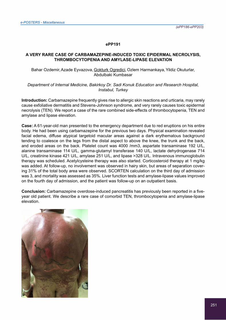
251
e-POSTERS - Miscellaneous(ePP186-ePP203)
ePP191
A VERY RARE CASE OF CARBAMAZEPINE-INDUCED TOXIC EPIDERMAL NECROLYSIS, THROMBOCYTOPENIA AND AMYLASE-LIPASE ELEVATION
Bahar Ozdemir, Azade Eyvazova, Gokturk Ogredici, Ozlem Harmankaya, Yildiz Okuturlar,
Abdulbaki Kumbasar
Department of Internal Medicine, Bakirkoy Dr. Sadi Konuk Education and Research Hospital, Instabul, Turkey
Introduction: Carbamazepine frequently gives rise to allergic skin reactions and urticaria, may rarely cause exfoliative dermatitis and Stevens-Johnson syndrome, and very rarely causes toxic epidermal necrolysis (TEN). We report a case of the rare combined side-effects of thrombocytopenia, TEN and amylase and lipase elevation.
Case: A 61-year-old man presented to the emergency department due to red eruptions on his entire body. He had been using carbamazepine for the previous two days. Physical examination revealed facial edema, diffuse atypical targetoid macular areas against a dark erythematous background tending to coalesce on the legs from the distal aspect to above the knee, the trunk and the back, and eroded areas on the back. Platelet count was 4000 /mm3, aspartate transaminase 192 U/L, alanine transaminase 114 U/L, gamma-glutamyl transferase 140 U/L, lactate dehydrogenase 714 U/L, creatinine kinase 421 U/L, amylase 251 U/L, and lipase >328 U/L. Intravenous immunoglobulin therapy was scheduled. Acetylcysteine therapy was also started. Corticosteroid therapy at 1 mg/kg was added. At follow-up, no involvement was observed in hairy skin, but areas of separation cover-ing 31% of the total body area were observed. SCORTEN calculation on the third day of admission was 3, and mortality was assessed as 35%. Liver function tests and amylase-lipase values improved on the fourth day of admission, and the patient was follow-up on an outpatient basis.
Conclusion: Carbamazepine overdose-induced pancreatitis has previously been reported in a five-year old patient. We describe a rare case of comorbid TEN, thrombocytopenia and amylase-lipase elevation.
252
e-POSTERS - Miscellaneous(ePP186-ePP203)
ePP192
MALIGNANT NEOPLASM DURING SYSTEMIC SCLEROSIS
Atig Amira, Guiga Ahmed, Ben Hadj Ali Emna, Bouker Ahmed, Bahri Fethi, Ghannouchi Neirouz
Department of internal medicine, Farhat Hached Hospital, Sousse, Tunisia
Background/aim : As in many autoimmune diseases, patients with scleroderma have an increased risk of malignancy compared to the general population. We report four new cases.
Case reports:
Case 1: One year after a diagnosis of SSc, physical examination of a 48-year-old female, noted a mass of 5 cm erythematous and vascularized in the right shoulder . Histologic evaluation revealed an inflammatory malignant histiocytofibroma.
Case 2: A 64-year-old female has a SSc diagnosed 8 years ago. she was admitted for cough and dyspnea. Thoracic CT showed an heterogeneous parenchymatous lesion. Transthoracic biopsy con-cluded to lung adenocarcinoma .
Case 3: A 57-year-old female presents a SSc. Initial Thoracic CT revealed a parietal mass with en-dothoracic development lysing the left fourth rib. A mass excision was performed and concluded to bone plasmacytoma. Ten years later, she consulted for thyroid goiter. Physical examination revealed a thyroid nodules. Total thyroidectomy was performed and the anatomopathological study was in favor of papillary carcinoma.
Case 4: A 78 years old man was diagnosed with systemic sclerosis since 2013. In February 2017, the patient was admitted for aggravation of the dyspnea with weight loss of 10kg. HRCT thorax showed a tissue mass in the anterior mediastinum of 52*81mm and parenchymatous nodule of 20mm. A biopsy of the nodule was performed and the microscopical examination showed a pulmo-nary adenocarcinoma
Conclusion: The implications of these observations is to encourage an active surveillance of scle-roderma patients for early detection of cancer.
253
e-POSTERS - Miscellaneous(ePP186-ePP203)
ePP193
AN UNUSUAL CASE REPORT OF RHABDOMYOLYSIS
Maria Bachlitzanaki, Maria Nisotaki, Kalliopi Diakaki, Eleni Valiraki-Valiri, Michaella Louka,
Emmanouil Vrentzos, Vasiliki Pitsouni
Internal Medicine Department, Rethymnon General Hospital, Rethymnon, Greece
Background/Aim: Rhabdomyolysis is a syndrome resulting from skeletal muscle injury that may cause the leakage of potentially toxic intracellular contents into plasma followed by multiple compli-cations such as electrolyte abnormalities, hyperuricemia, hypoalbuminemia, acute kidney injury and renal failure or even disseminated intravascular coagulation.
Case Report: A 23-year-old-man without previous medical history presented at the emergency de-partment with muscular pain and tenderness from about 48hours and dark urine during his last urination. The symptoms followed daily alcohol consumption (1 week of duration) and exertional activity(24hours before). The clinical examination revealed an afebrile, hemodynamically stable pa-tient and the blood sample that was obtained a few minutes after his arrival showed an extremely high level of creatine kinase(CK) that could not be measured (maximum level measured in our labo-ratory is about 250.000IU/L), as well as high transaminase(SGOT>>SGPT) and LDH levels. During his hospitalization, the patient underwent excessive fluid resuscitation and urinary alkalization fol-lowed by a rapid clinical and laboratory improvement without the need of renal replacement therapy. Furthermore, other causes of rhabdomyolysis (such as infection, trauma, use of drugs/medication, electrolyte abnormalities, hyperglycemia, thyroid gland dysfunction) where excluded. Results for genetic factors tests are still pending.
Conclusions: Although rhabdomyolysis is considered to be a common medical condition, this case report appears interesting as it concerns an extremely high level of CK (not previously mentioned bibliographically)in a healthy adult that was rapidly treated without causing renal failure.
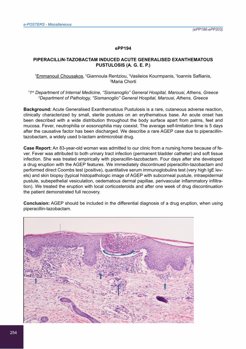
254
e-POSTERS - Miscellaneous(ePP186-ePP203)
ePP194
PIPERACILLIN-TAZOBACTAM INDUCED ACUTE GENERALISED EXANTHEMATOUS PUSTULOSIS (A. G. E. P.)
1Emmanouil Chousakos, 1Giannoula Rentziou, 1Vasileios Kourmpanis, 1Ioannis Saflianis,
2Maria Chorti
11st Department of Internal Medicine, “Sismanoglio” General Hospital, Marousi, Athens, Greece 2Department of Pathology, “Sismanoglio” General Hospital, Marousi, Athens, Greece
Background: Acute Generalised Exanthematous Pustulosis is a rare, cutaneous adverse reaction, clinically characterized by small, sterile pustules on an erythematous base. An acute onset has been described with a wide distribution throughout the body surface apart from palms, feet and mucosa. Fever, neutrophilia or eosonophilia may coexist. The average self-limitation time is 5 days after the causative factor has been discharged. We describe a rare AGEP case due to piperacillin-tazobactam, a widely used b-lactam antimicrobial drug.
Case Report: An 83-year-old woman was admitted to our clinic from a nursing home because of fe-ver. Fever was attributed to both urinary tract infection (permanent bladder catheter) and soft tissue infection. She was treated empirically with piperacillin-tazobactam. Four days after she developed a drug eruption with the AGEP features. We immediately discontinued piperacillin-tazobactam and performed direct Coombs test (positive), quantitative serum immunoglobulins test (very high IgE lev-els) and skin biopsy (typical histopathologic image of AGEP with subcorneal pustule, intraepidermal pustule, subepethelial vesiculation, oedematous dermal papillae, perivascular inflammatory infiltra-tion). We treated the eruption with local corticosteroids and after one week of drug discontinuation the patient demonstrated full recovery.
Conclusion: AGEP should be included in the differential diagnosis of a drug eruption, when using piperacillin-tazobactam.
255
e-POSTERS - Miscellaneous(ePP186-ePP203)
ePP195
RHABDOMYOLYSIS AND ACUTE KIDNEY INJURY INDUCED BY TREATMENT WITH LIPID-LOWERING DRUGS: A CASE REPORT
Georgia Samakidou, Dimitra Tsionara, Armonia Chatzigeorgiou, Ourania Triantafyllou,
Konstantinos Pateras, Konstantinos Krapis, Konstantinos Karakousis, Theodora Konstantoula,
Spiridon Karamagkiolis, Konstantinos Karamitsos
Department of Internal Medicine, General Hospital of Larissa, Larissa, Greece
Background: Rhabdomyolysis is a syndrome characterized by muscle necrosis and release of in-tracellular muscle constituents into the circulation. Its main manifestations are muscle weakness, myalgias and dark urine, while the laboratory tests reveal elevated serum muscle enzymes. There are many causes of rhabdomyolysis, that can be categorized into traumatic (including muscle trau-ma) and non-traumatic (including toxins and drugs). It is a severe condition which every clinician should be aware of in order to proceed to early diagnosis and management.
Case report: A 77-year old male presented to the ER complaining of myalgias and impaired mobility of the lower extremities for the past two days. His past medical history was significant for coronary heart disease (PTCA forty five days ago CABG four years ago) and diabetes mellitus. Clinical ex-amination revealed reduced muscle strength of the lower extremities with normal sensation. Labora-tory tests revealed an increase of serum CPK (31.350 IU/L ,with 190 IU/L as the upper limit), as well as increased levels of transaminases and impaired renal function. The syndrome was attributed to the initiation of treatment with high doses of simvastatin-ezetimibe due to recent PTCA ( 10mg of ezetimibe- 40mg of simvastatin twice daily) . The drug was discontinued and the patient underwent hemodialysis due to the constant deterioration of kidney function and acute pulmonary oedema.
Conclusion: Lipid-lowering drugs are very widely prescribed medications. It is important to remem-ber that close monitoring of these patients is essential in order to ensure safety and avoid serious side effects.
256
e-POSTERS - Miscellaneous(ePP186-ePP203)
ePP196
PAINFUL OPHTHALMOPLEGIA, A DIAGNOSTIC CHALLENGE
1Gloria Perez-Vazquez, 1Eva Lozano-Loaiza, 1Carolina Luque-Amado, 2Esther Rubio-Martin,
3Luis Garcia-Aragon, 3Juan-Antonio Jimenez-Casas
1Internal Medicine Unit, Hospital Punta Europa, Algeciras, Spain 2Nephrology Unit, Hospital Punta Europa, Algeciras, Spain
3Primary Care, Area Gestion Sanitaria Campo de Gibraltar, Spain
83-year-old male who attended with intense retrociliary pain in the right eye, blurred vision and diplo-pia, after capsulotomy the week before. Ischemic heart disease with 3-vessel disease, with double bypass in 2003 and subsequent coronary angiography in December 2016 with CD dilatation with a direct stent. Physical examination reactive mydriatic pupils, proptosis and bilateral conjunctival chemosis and right palpebral edema, limitation for ocular mobility in all planes, especially the exter-nal rectus of the OD. IOP was observed to be 20 mmHg with subsequent increase to 28 mmHg.
A study was performed with leukocytes of 9600 cels / dL, neutrophilia 92.4%, red series and normal platelets. Fibrinogen: 451 mg/dl. In the biochemical study creatinine 1.25 mg/dl., Total bilirubin 2.52 mg/dl (direct 0.83 mg / dl), normal rest. Negative onconeuronales. Normal CSF Mantoux: negative. The microbiological study was negative, negative serologies, and PCR for negative Mycobacte-ria. Negative fungal study. PCR for meningitis and negative encephalitis. Differential diagnosis was performed cranial MRI and MRI angiography of the Willis polygon that identifies cerebral atrophy and leukoaraiosis; Asymmetry due to thickening of the right cavernous sinus, with increased flow in the vascular sequence, NMR orbitals are extended with acute ischemic infarcts with signs of mild hemorrhagic transformation. CT scan of the paranasal sinuses showing bilateral exophthalmos and discrete thickening of the medial muscle in both orbits. Corticosteroid treatment with prednisone 90 mg daily for 7 days and subsequent dose reduction at a rate of 10 mg every 15 days, with improve-ment.
257
e-POSTERS - Miscellaneous(ePP186-ePP203)
ePP197
MULTIPLE CRANIAL NEUROPATHY CAUSED BY EPSTEIN-BARR VIRUS (EBV): AN UNCOMMON INVOLVEMENT
1Afroditi Spyrou, 1Anastasia Kalliotzoglou, 1Ioannis Liapis, 2Maria Stampori, 2Aikaterini Chasiou,
2Dimitra Kozikopoulou, 2Evangelos Papaoikonomou, 2George Karlis, 2Chrysostomos Psellas,
2Stylianos Karatapanis
1Neurology Department, General Hospital of Rhodes, Rhodes, Greece 21st Department of Internal Medicine, General Hospital of Rhodes, Rhodes, Greece
Background/Aim: Encountering a patient with cranial nerve paralysis could be not only very inter-esting but also very demanding concerning the differential diagnosis. Here, we present a case of polyneuritis cranialis caused by EBV.
Case presentation: A 80 year-old man admitted to the neurology department with complaints of low-pitched dysphonia, dysphagia and right sided facial asymmetry for 2 days. One week before the admission the patient experienced mild headache and drowsiness as well as fever up to 38C. The neurological examination revealed hoarse voice, right sided peripheral facial asymmetry, hypoesthe-sia on the right side of the face and decreased gag reflex. Laryngoscopy demonstrated right vocal cord paralysis. MRI of brain, neck computed tomography(CT) and thorax(CT) were normal. CSF analysis revealed lymphocytic pleocytosis. EBV-DNA was detected at the CSF by PCR (1.3 * 103 copies/ml). The patient was treated with intravenous acyclovir for 10 days as well as corticosteroids per-os for 15 days. One month after discharge the patient was significantly improved regarding not only the facial asymmetry but also the hoarseness and the swallowing. The laryngoscopic image was also improved.
Conclusion : Although multiple cranial neuropathy is a frequent problem encountered in neurologi-cal practice EBV it is very rarely involved. Here, we described a case of cranial polyneuropathy due to EBV virus. The diagnosis was verified by EBV DNA in the CSF. Therefore, it’s valuable to take into consideration the EBV infection in the differential diagnosis of cranial nerve paralysis.
258
e-POSTERS - Miscellaneous(ePP186-ePP203)
ePP198
WHEN NEUROLOGY COMES ACROSS NEPHROLOGY: A CASE REPORT
1Afroditi Spyrou, 1Anastasia Kalliotzoglou, 1Ioannis Liapis, 2Maria Stampori, 2Dimitra Kozikopoulou,
2Aikaterini Chasiou, 2Georgios Ntetskas, 2Nikitas Katsilis, 2Evangelos Papaoikonomou,
2Stylianos Karatapanis
1Neurology Department, General Hospital of Rhodes, Rhodes, Greece 21st Department of Internal Medicine, General Hospital of Rhodes, Rhodes, Greece
Background/Aim: It is generally accepted that several medical cases might be considerably com-plicated and multidimensional, requiring a spherical approach .The aim of this abstract is to present a case of Guillain-Barre syndrome complicated with renal involvement.
Case presentation : A 60 year-old woman, with no past history of renal or neurological disease admitted to our hospital due to seven days of progressive weakness of her extremities as well as paresthesia at her hands. Laboratory tests revealed proteinuria (2g/24h) and normal renal function. Although both the electrophysiological and the spinal fluid investigation were normal on admission, her weakness progressed rapidly (48h) to the point where she became bed-bound. The diagnosis of Guillain-Barre was verified with the repetition of electrophysiological and CSF studies. Immu-noglobulin and intravenous corticosteroids were given for five days but with no improvement at all. Plasmapheresis was instituted and oral prednisolone was commenced tapered over six months. Immunological investigation, including antineutrophil cytoplasmic antibodies, and virological screen-ings were negative. After 3 months the patient was able to walk with normal renal function and no residual proteinuria.
Conclusion: Inflammatory neuropathies have been reported to occur in association with proteinuria. Their underlying immuno-pathogenic mechanisms remain unknown as well as a definite treatment regarding this type of cases. Here we pointed out the necessity for further research concerning not only the pathogenesis but also the treatment and the prognosis of acute inflammatory neuropathy complicated by renal disease, with many issues remaining unresolved.
259
e-POSTERS - Miscellaneous(ePP186-ePP203)
ePP199
OUT OF POCKET PAYMENTS IN GREECE & CHRONIC DISEASES: DIABETES MELLITUS CASE
1,2,3,4Paraskevi Koufopoulou, 1Ioannis Kyriazis, 1,5Grigoria Betsi, 2Athanassios Vozikis, 6Kyriakos Souliotis, 2Dionysios Ithakisios,1,7,8Sara-Eleni Amprahim
1KAT General Hospital of Attica, Kifisia, Greece2Department of Economics, Laboratory of Health Economics & Management, Faculty of
Economics, Business & International Studies, University of Piraeus, Piraeus, Greece3Hellenic Association of Political Scientists, Athens, Greece
4Cretan Studies Medical Association, Chania, Crete, Greece5Health Care Management, Faculty of Social Sciences, Hellenic Open University, Patra, Greece
6Department of Social & Educational Policy, Faculty of Social & Political Studies, University of Peloponnese, Korinthos, Greece
7Cardiopulmonary Rejuvenation, Department of Medicine, Faculty of Health Sciences, National & Kapodistrian University of Sciences, Athens, Greece
8Salud y Calidad de Vida, Universidad de Huelva, Huelva, Spain
Background: Diabetes is the fourth leading death cause worldwide, with adult diabetic patients reaching 366 million in 2030. There are 60 million diabetic patients in the European Union and 1 mil-lion in Greece as a consequence of overweight, obesity, unhealthy nutrition etc. Instant result is the financing of health systems, from patients (out of pocket payments).
Aim: The evolution of private health expenditure in Greece, through diabetes mellitus.
Method: In the framework of the PhD Dissertation titled about Shadow Economy in health care sector in Greece (Department of Economics, Laboratory of Health Economics and Management, University of Piraeus), Greek and foreign literature was collected through the electronic databases PubMed, Google Scholar, Cochrane Library, Elsevier, CINAHL and Science Direct for the period 2010 - 2016, regarding private health expenditure through diabetes.
Results: Diabetes mellitus cost, ranges from 3% to 6% of total health expenditure (average annual per capita expenditure is 2.834 €). In Greece, with a 35.4% private health cost share, the average cost per patient is estimated at € 1,300 per year (31.1% annual medication, 44.8% required exams and 24.1% wage staff). Furthermore, the average annual cost would be over 3,000 € per year and triple the total financial burden of the disease.
Conclusions: Chronic diseases, such as diabetes mellitus, react on morbidity and health costs. Pre-vention is the only solution, through early diagnosis primarily from the health system and secondarily from the patients. Reconstruction of services through the contribution of Primary Health Care are the main point.

260
e-POSTERS - Miscellaneous(ePP186-ePP203)
ePP200
A DIFFICULT TO TREAT PNEUMONIA IN A GERIATRIC PATIENT REVEALED PULMONARY TUBERCULOSIS ON HIV INFECTION: SUSPECT THE INNOCENT
1Ilias Lyroudis, 1Assimina Nikolakopoulou, 2Georgios Noutsos, 1Emily G. Tsaroucha,
1Despina Thiriou, 1Ekaterini Ralli, 3Malvina Lada, 1Aggeliki Rapti
12nd Pulmonology Department, Hospital of Chest Diseases of Athens, Athens, Greece 2Physiotherapy Department, Technological Educational Institution of Athens, Athens, Greece
32nd Department of Internal Medicine, “Sismanoglion” Hospital, Athens, Greece
Tuberculosis and HIV remain two of the most serious threats on public health worldwide while their combination makes them even more dangerous. TB is the most common infection associated with HIV, but also the leading cause of death among HIV-positive people. Immunodeficiency caused by HIV infection makes the diagnosis of TB even harder.
Case Report: 71yo non-smoker woman with a history of diabetes, arterial hypertension and a colon cancer resection 2 years ago. She admitted with dry cough, fever, fatigue and night sweats during the last 4 months. A month ago a CXR revealed fluid on the right costophrenic angle. she was treated with antibiotics without any improvement. A new CXR showed consolidation of the right lung and fluid accumulation. At first it was treated as a pneumonia with parapneumonic effusion with piperacillin/tazobactam and azithromycin. Because of the persisting symptoms a CT scan was performed: Con-solidation of the right middle lobe with atelectasis and pleural effusion, small perilymphatic nodules in both lungs, enlarged mediastinal and hilar lymph nodes. Lab results: Positive sputum smear, negative Mantoux, HIV confirmed with ELISA, CD4: 333cells/ml, FNB negative for cancer, positive sputum cultures for M. Tuberculosis. A 4-drug(HREZ) anti-TB treatment was given for 6 months with total recovery.
Conclusion: The coexistence of HIV/TB may present with atypical clinical and radiological signs. There should always be a suspicion of these 2 diseases regardless of age, sex and social status. It is also essential that prior to the initiation of anti-TB treatment virological testing is performed.

261
e-POSTERS - Miscellaneous(ePP186-ePP203)
ePP201
PULMONARY AMYLOIDOSIS
2Virginia Perez-Vazquez, 1Gloria Perez-Vazquez, 2Beatriz Sainz-Vera, 3Luis Garcia-Aragon,
1Carolina Luque-Amado, 4Esther Rubio-Martin, 1Eva Lozano-Loaiza
1Internal Medicine Unit, Hospital Punta Europa, Algeciras, Spain 2Internal Medicine Unit, Hospital Universitario Puerto Real, Puerto Real, Spain
3Primary Care, Area Gestion Sanitaria Campo de Gibraltar, Gibraltar, Spain 4Nephrology Unit, Hospital Punta Europa, Algeciras, Spain
74-year-old patiente with COPD is admitted to Neumology Unit for hemoptysis, he was diagnosticated of lung amyloidosis (amiloide AA) in December of 2016, without evidence of systemic amyloidosis, biopsy bone marrow was negative. In 2017 the patient was admitted in Internal Medicine Department with a constitutional syndrome, 20 kg weight loss in three months. Chest- TC Scan was realized find-ing a big mass in right upper lobe of right lung, with another cavitations of new appearance. A biopsy of this mass was red congo stain positive. Bone biopsy and cardiac-RMI were also negative. During this admission the patient had syncopes with brain imaging studies and 24-hour heart rate monitor-ing were negatives. In sputum microbiological study was isolated Haemophilus influenzae and the Pneumococus Antigen in orine was positive. All mycobacterial cultives were negative. The patient received antibiotic treatment, endovenous corticoids and fluids for a week. The final diagnosis was pulmonary amyloidosis with lung infection of Pneumococus and Haemophilus influenzae.

262
e-POSTERS - Miscellaneous(ePP186-ePP203)
ePP202
PECULIARITIES OF PSYCHO-SOMATIC STATUS AND QUALITY OF LIFE IN PATIENTS WITH COPD
Alpha James, Natalia Pavliukovych, Oleksandr Pavliukovych
Pulmonology Department, Bukovinian State Medical University, Chernivitsi, Ukraine
Chronic Obstructive Pulmonary Disease (COPD) is a topical problem of pulmonology due to its high morbidity, mortality and impact on the patients’ quality of life.
Aim: to analyze psycho-somatic status and quality of life (QL) of patients with COPD. 65 male patients with COPD were randomized into two groups according to the degree of disease sever-ity: group A - 30 patients with COPD II (FEV1=58,8±2,11%), group B - 35 patients with COPD III (FEV1=41,3±3,62%). Control group comprised 24 healthy individuals of the same age and gender. Assessment of psycho-somatic status was based on the Test of Patient’s Differential Self-assess-ment, which includes analysis of well-being (WB), activity (A) and mood (M). QL was determined by Mezzich J. E., Cohen M., Ruiperez N. questionnaire. In group A psycho-somatic status was lower compared to healthy individuals, WB decreased significantly (4,3±0,18 against 3,5±0,16, p<0,05). In group B all components of psycho-somatic status were lower than in patients of group A (WB - 2,1±0,09 against 4,3±0,18, A - 2,5±0,11 against 3,9±0,12, M - 2,0±0,22 against 3,8±0,07, p<0,05 in all cases) and compared to healthy individuals. Overall perception of QL in patients of group A was lower by 23%, and in patients of group B - by 42% correspondingly compared with control group (p<0,05 in both cases). Indices of WB, M and QL correlated directly with FEV1 (p<0,05). So, pro-longed course of COPD is accompanied by changing of the psycho-somatic status and worsening of patients’ QL, which directly correlates with COPD degree of severity.
263
e-POSTERS - Miscellaneous(ePP186-ePP203)
ePP203
AN AUDIT TO EXAMINE THE COMPLIANCE OF DOCUMENTING THE PROCESS OF OBTAINING AN INFORMED CONSENT
Siow Yun Wong, Samuel Kang Lian Lee, Chaozer Er
Department of General Medicine, Tan Tock Seng Hospital, Singapore
Background: A consent form is a proof that a consent has been obtained. It does not obviate the need of documentation of the discussion about the procedure or treatment in the medical record. We felt that the documentation of the process of obtaining an informed consent is poor in our depart-ment.
Aim: To examine if the process of obtaining an informed consent is properly documented in the medical record
Method: We retrospectively examined the case notes of all patients admitted under the care of con-sultant general physician A and B in the Department of General Medicine, Tan Tock Seng Hospital, Singapore from July 2017-August 2017 to identify those who have had procedures performed on them during their admissions. Among those procedures, we examined the documentation of consent taking to see if procedure nature, indication, complication and alternative treatment were explained and documented in the case notes.
Result: Out of the 35 procedure we audited, only 23% had the procedure nature explanation, 37 % had the procedure indication explanation, 31% had the procedure complication explanation and 9% had the alternative treatment explanation documented.
Conclusion: The documentation of the process of obtaining an informed consent was poor. We relied mainly on a piece of consent form as a proof of obtaining an informed consent.

264

265
10th International Congress of Internal MedicineMarch 22-24, 2018, Athens, Greece
Index
INDEX
266
AAchmet Sibel Kara Ali 238
Adam Foteini 210, 230, 232
Adamantou Angelos 80
Adamantou Magdalini 45, 155, 159
Adrahtas Dionisios 182
Aggelis Nikolaos 244
Aggelis Xenophon 222
Aggelou Evangelos 27
Agiasotelli Danai 29
Agrogianni Maria 70, 82
Ahmed Bouker 252
Ahmed Guiga 252
Akarsu Cevher 138
Akdeniz Yasemin Sefika 43, 46, 124, 137, 138, 139
Akinosoglou Karolina 56
Aktaş Hanife Şerife 38
Alaveras Antonios 239
Aleksiou Nikolaos 209
Alemán Irene Valiente 9, 187
Alexandris Konstantinos 85
Alexandrou Maria 169
Alexi Angelos 45, 80, 155
Alexiou Nikolaos 89
Alexiou Zoi 180
Alexopoulos Theodoros 20, 21, 29
Alexopoulou Alexandra 19, 20, 21, 29
Aloizos George 81, 156, 158
Alpha James 262
Alvarez-Hernandez Luis Felipe 199
Amira Atig 252
Amprahim Sara-Eleni 259
Anagnostopoulou Euaggelia 71, 72
Anastasiadis Georgios 48
Anastasiadis Stilianos 89
Anastasiadou Afroditi 185
Anastasiadou Christiana 85
Anastasiou Alexandros 236
Anastasiou Efstathia 17, 23, 65, 66, 170
Anastassios Philippou 37
Andreopoulos Demetris 246
Andrianopoulos Ioannis 64, 79, 214
Aneeta George 140, 186, 201
Angelakis Athanasios 33, 34, 60
Angelopoulos Theodoros 197
Angelopoulou Stella-Maria 55
Antonopoulos Stavros 83
Anyfantis Angelos 245
Apostolidou Eirini 39
Apostolopoulos Vasileios 209
Apsemidou Athanasia 75
Arelaki Stella 39
Argiana Vasiliki 81, 156, 158
Argyraki Katerina 86, 205, 228
Arna Despoina-Eleni 95, 167, 171, 179
Arphika Sarkar 198
Atalay Hande 47, 120, 125, 126, 142
Athanasopoulou Athanasia 163
Avellaneda-López Ylenia 76
Avgoustinou Andonia 114
Aydemir Birsen 126
Aygoyleas George 83
Azariadi Kalliopi 35
BBachlitzanaki Maria 227, 253
Bahtiyar Ismet 219
Bakarozi M. 18
Bakas Dimitrios 245
Baltayiannis Gerasimos 64, 79
Bankov Tsvetan 114
Banol-Betancur Jorge 199
Bao-Bao Hsu 77
Barbullushi Myftar 112
Barkas Fotios 15, 184
Basat Okcan 42
INDEX
267
Battezzati Pier M. 30
Baygül Arzu 42
Bayrak Savas 136
Beka Angeliki Anna 22
Betsi Grigoria 259
Bhagat Budhbhushan 193
Bianchi-de-Aguiar Filipa 216
Bitsani Aikaterini 117
Boulbou Maria 215, 217
Boyuk Banu 47, 120, 122, 123, 125, 126, 142
Bozer Denis Sabriye 43, 137
Briasoulis Christos 211, 230, 231, 232
Bruns Tony 30
Bulavkina Kateryna 188
Buuren Henk R. van 30
CCabuk Fatmagul Kusku 138, 139
Cafka Majlinda 112
Campanha Rafaela 216
Caunīte Laima 188
Celebi Aslan 120, 125
Cetin Irem 120
Cetin Seher Irem 47, 125, 126, 142
Chalkiadaki Anthi 59, 74
Chalkias Athanasios 243
Charalambidis Dimos 64
Charalambous George 127
Charitos Dimitrios 67, 70
Chasiou Aikaterini 17, 23, 27, 65, 66, 168, 170, 257, 258
Chatzidavid Sevastianos 90, 102
Chatzigeorgiou Armonia 255
Chatzikonstantinou Thomas 121
Chaudhari Nilang 198
Chiotou-Sato Anastasia 161
Chorti Maria 254
Chousakos Emmanouil 254
Chrisostomou Evaggelia 211, 232
Christodoulou Marios 169
Christodoulou Panagiotis 230, 232
Christodoulou Sofia 80, 159
Christofidis Charalambos Ch. 236
Christopoulos Constantinos 213, 223
Christopoulou Eleni 209
Christou Konstantinos 55
Chrysanthos Nikolaos 59, 71, 72, 74
Cirak Musa 136
Constantinou Haris 45, 80, 159
Corpechot Christophe 30
DDafoulas Georgios 49
Dalekos George N. 18, 30, 35, 52, 215, 217
Daminov Botir 107, 109
Damvopoulou Efthalia 185
Datseri Rafaella 133, 145, 189, 238
Deftereos Savas 39
Delaporta Eirini 192
Demir Esra 47
Demiriz Itir 100, 165, 173
Dempela Eleni 220
Desse D. 238
Deutsch Melanie 63
Dhanarajan Vishnu 140
Diakaki Kalliopi 253
Diakonikola Despoina 65, 170
Diamantea Filia 20, 21
Dimitra Aikaterini 13, 191, 192, 195
Dimitriou Panagiotis 40
Dimosiari Athina 177
Dividis Georgios 8, 50, 183
Dogansen Sema Ciftci 46, 122, 123, 124, 137, 138, 139
Domínguez-Fuentes María Belen 248
Domvri Kalliopi 161
Doumtsis Petros 177
Dourakis Spyros P. 19, 20, 21, 29, 32
INDEX
268
Drakou Elina 243
Drakoulis Christos 62, 67, 70, 82
Drakoulis Nikolaos 53
Drakoulogkona Ourania 182
Drikos Ioannis 185
Driva Stamatia 221
Drosos Georgios 154
Drylli Aikaterini 97, 98, 242
Dural Ahmet Cem 139
EEfraimi Charis 134, 135
Efthimiadis Agathoklis 185
Efthymiou Konstantinos 207
Eken Kamile Gulcin 120
Ekizoglu Ismail 120
Eleftheriotis Gerasimos 213, 223
Elisaf Moses 184
Ellina Christina 62, 67
Ellina Olga 111
Emmanouilidou Evgenia 92, 234
Emna Ben Hadj Ali 252
Eracleous Eraclis 84
Er Chaozer 166, 174, 263
Erdoğdu Bugrahan 165
Erişmiş Betül 78, 172
Esen Ayse 124
Eyvazova Azade 251
FFamilias Ioannis 66
Farmaki Athanasia 70
Farmaki Georgia 235
Fasias Vasilis 235
Fedorivna Marchuk Yuliya 140
Fethi Bahri 252
Fetta Stella 169
Filiotis Nikolaos 84
Floreani Annarosa 30
Flouda Sofia 81
Flourou Christina 87, 181
Folas Nikolaos 203
Forozidou Evropi 8, 50, 183
Fotiou Eleni 155, 246
Fotos Georgios 8, 50, 183
Fragioudaki Sofia 154
Fytrolakis Georgios 59, 74
GGabeta S. 18
Galani Despina 92, 234
Galanis K. 18
Galanis Petros 11
Gallego-Gonzalez Daniel 199
Ganga Pedro Luis Quirós 103
Garcia-Aragon Luis 41, 73, 91, 93, 94, 99, 106, 116, 157, 160, 162, 247, 248, 256, 261
Gargalianos-Kakolyris Panagiotis 218, 243
Garyfallos Alexandros 177
Gatos Ilias 32, 33, 34, 60
Gatsa Eleni 224, 237
Gatselis Nikolaos K. 18, 30, 35, 52
Gavalas Ioannis 117
Gazouli Ioanna 239
Georgakopoulos Christos 204
Georgiadi Evgenia 53
Georgiadou Elena 176
Georgiopoulos George 19
Georgiou Anthoulla 169
Georgopoulos Ilias 128, 129, 130, 131, 132
Georgousi Kleoniki 54
Gerasimou Marina 210
German Vasilios 81, 156, 158
Germenis Anastasios E. 51
Geronatsios Konstantinos 96
Giamarelou Aggeliki 79
Giannakas Kontantinos 70
Giannakoulas Georgios 8, 50, 183
INDEX
269
Giannopoulos Charalampos 89, 209
Gigi Eleni 26
Gika Meropi 111
Gil-Gascon Jose-Manuel 94, 157, 160, 229
Gioka Maria 20, 21
Gioulekas Fotios 11
Gkeka Marina 213, 223
Gkinis Ioannis 209
Gkizeli Konstantina 89
Gklara Panagiota 121, 220
Gkogkos Georgios 222, 224, 226
Gkogkos Konstantinos 121, 220
Gkotmi Natalie El 233
Gkousios Themistoklis 39
Goet Jorn C. 30
Gogolas Basileios 180
Gogos Charalambos 56
Gonzalez-Quiñones Antonia 247
González Ramón Fernando García 9, 103, 187
Goula Konstantina 182
Goulas Ioannis 8, 50. 183
Gouva Mary 127
Guimarães Cátia 216
Gülçiçek Gamze 172
Guliyev Zakir 250
Guzel Savas 125, 126
HHadjisavvas Nikolaos 176
Hadziyannis Emilia 29
Hahalis Georgios 143, 144, 146, 147, 149, 150, 151, 152, 153
Hansen Bettina E. 30
Haris Constantinou 155
Harmankaya Ozlem 165, 173, 219, 251
Harms Maren H. 30
Hatzis Vladimiros 239
Hatzitolios Apostolos I. 55
Haydarov Bahadır 250
Hera Maroulio 128, 129, 130, 131, 132
Heras Panagiotis 58, 128, 129, 130, 131, 132, 148
Heras Vasilios 58, 148
Hindilerden Fehmi 100, 165
Hirschfield Gideon 30
Hoz-Adame María Eugenia 247
Hrechko Svitlana 186, 201
Hsu Bao-Bao 77
Huang Kuang-Yung 77
Hursitoglu Mehmet 173, 250
IIacovidou Nikoleta 243
Idrizi Alma 112
Internos Iosif 82
Invernizzi Pietro 30
Ioannidis Dimitrios 67
Ioannidou Eleni 227
Ioannou Alkiviadis 89, 209
Ioannou Anna 227
Ioannou Antreas 40, 181
Ioannou Stephanie 223
Ippa Vasiliki 226
Iskos Konstantinos 204
Ithakisios Dionysios 259
JJanssen Harry L.A. 30
Jaramillo-Jaramillo Laura Isabel 199
Jimenez-Casas Juan-Antonio 41, 73, 106, 116, 162, 256
Jursevica Liga 105
KKachrimanidis Ioannis 54
Kagiafa Dimitra 61
Kakkos Stavros 190
Kalafatis Emmanouil 54
Kalagiakou Evangelia 62
INDEX
270
Kalantzi Sofia 213, 223
Kalessis Alkistis 83
Kaliampou Styliani 235
Kaliouli Christiana 62, 67, 70
Kalliotzoglou Anastasia 257, 258
Kalmoukos Panagiotis 26
Kalogeraki Loukia 118
Kalogiannis Konstantinos 75, 96
Kalogridis Thodoris 53
Kalopitas Georgios 75, 96
Kaltsa Anastasia 161
Kanavaki Aikaterini 33, 60
Kani Chara 49
Kaouranis Constantinos 181
Kapatais Andreas 28, 69
Kapelios Christos 88
Kapouralos Andreas 58, 128, 129, 130, 131, 132, 148
Karabelias Georgios 97, 98, 242
Karaboyun Kubilay 101, 219
Karagiannaki Anastasia 14
Karagianni Maria 62, 67, 70
Karahaliou Aggeliki 214
Karaivazi Sofia 161
Karakasiliotis Ioannis 22
Karakaya Pinar 46, 122, 123, 124, 137, 138, 139, 250
Karakonstantis Stamatis 92, 234
Karakousis Konstantinos 255
Karakousis Nikolaos D. 204
Karamagkiolis Spiridon 255
Karamanli Κonstantina 28, 69
Karamitsos Konstantinos 255
Karandere Faruk 219
Kara Oğuzhan 42
Karaoulani Theofani 28, 69
Karasavidou Larisa 67
Karatapanis Stylianos 17, 23, 27, 65, 66, 168, 170, 257, 258
Karatarakis Konstantinos 59
Kardas Przemyslaw 36
Kariki Ourania 27, 65, 66, 170
Karlis George 23, 66, 168, 257
Kartas Anastasios 8, 50, 183
Karvounis Haralambos 8, 50, 183
Kassianidis Georgios 239
Kassotaki Ifigenia 95, 171
Kastritsi Eleni 145, 238
Kastritsi Olga 145, 238
Katagis George 210, 231
Katsarou Irini 114
Katsarou Martha-Spyridoula 53
Katseas Georgios 185
Katsili Anna 17, 23
Katsilis Nikitas 17, 27, 65, 168, 170, 258
Katsios Aram 87, 181
Kavallieratos Fotios 81, 156, 158
Kavousanaki Melina 92, 95, 167, 171, 179, 234
Kayalar Arzu Ozdemir 136
Kazanci Mehmet Hanifi 165, 173, 219
Kechagias Georgios 82
K.Filippatos 164
Khan Numan 10
Kipraki Aggeliki 171
Kirby Joshua 10
Kiziler Ali Rıza 126
Kloudas Spyridon 86, 228
Kocaoglu Ali 120
Kokkinaki Ioanna 86, 205, 228
Kokores Ioanna 111
Kolokoutsas Vasileios 245
Kolonia Virginia 204
Komis Michail 209
Konstantinou Alexandra 233
Konstantinou Frosso 86, 228
Konstantoula Theodora 255
Kontaxakis Ioannis 59, 74
INDEX
271
Kontolaimou Ioulia 180
Kontrafouri Paraskevi 117, 134, 135, 207, 221
Kopsaftopoulou Anastasia 134, 135, 203, 207, 221
Kordali Christina 115
Korela Dafni 92, 234
Korkolopoulou Pinelopi 71
Koroboki Eleni 11, 15
Korodima Maria 97
Korotkikh Alexander 200
Kostaki Stavroula 55
Kostanti Eleonora 79
Kostis Andreas 176
Kostourou Danai-Thomais 55
Kotrotsiou Evangelia 127
Kotsanti Sotiria 117
Kotsi Eleni 8, 183
Kottas Spyridon 243
Koudouna Aspasia 117
Koufogianni Afroditi 92, 95, 167, 179
Koufopoulou Paraskevi 259
Koukoulis George K. 35
Koulouras Vasilios 64, 79, 214
Koulouridi Asimina 95, 167, 171, 179
Koumanis Theologos 161
Koumoutsea Dafni 84
Kounouklas Konstantinos 235
Koupetori Marina 89, 209
Kouppari Georgia 213
Kourmpanis Vasileios 254
Kouroupakis Panagiotis 204
Kourti Eleni 134, 135, 154, 203, 207, 221
Koussis Panos 32
Koutroumpi Maria 68, 202
Koutsi Aikaterini 233
Koutsilieris Michael 37
Koutsoumpos Nikolaos 180
Koutsouri Christina-Panagiota 56
Koutsouris Panagiotis 54
Kouvardas Stelios 213
Kowdley Kris V. 30
Kozikopoulou Dimitra 27, 65, 66, 168, 170, 257, 258
Krapis Konstantinos 255
Krasnikova Elena 236
Krasovskagia Irina 85, 119
Krentiris Dimitrios 84
Kritikos Petros-Ioannis 204
Kucane Linda 105
Kumbasar Abdulbaki 251
Kurczewska-Michalak Marta 36
Kursat Firuze Midi 78, 172
Kursat Hakan 78, 100, 172
Kuthiah Navin 166, 174
Kydona Christina 177
Kypraki Aggeliki 95
Kyprianou Ioannis 246
Kyriakos Alexandros 175
Kyriazis Ioannis 54, 259
LLabropoulos Panagiotis 28, 69
Lada Malvina 204, 260
Lafoyianni Sofia 213
Lagiou Maria 53
Lagoudaki Irini 118
Laina Angeliki 19
Lai Ning-Sheng 77
Lammers Willem J. 30
Lampropoulos Christos 68, 115, 202
Lamprou Thomais 209
Lappa Theodora 54
Laskaratou Emmanouela 145, 197, 238
Latsis Becator 28, 69
Latsou Dimitra 54, 243
Lee Samuel Kang Lian 174, 263
Lelekis Moyssis 54
INDEX
272
Leontaki Maria 59, 74
Leveta Georgia 141
Liabas Ioannis 237
Lialiaris Theodoros 161
Liapi Dimitra 167, 171, 179
Liapis Ioannis 257, 258
Liatis Stavros 48, 49
Likaj Erjola 112
Likouresi Anna 114
Lindor Keith D. 30
Loke Ealing 208
Lontou Stavroula-Panagiota 90, 102
Lopez-Saez Juan-Bosco 93
López Ylenia Avellaneda 206
Louis Konstantinos 203
Louka Chrysostomos 45, 155, 159
Louka Eirini 180
Louka Michaella 253
Loupa Chariclia 213
Loupas Thanasis 34
Loutsidi Eleni 233
Lozano-Loaiza Eva Maria 41, 73, 93, 99, 106, 116, 162, 229, 256, 261
Lu Ming-Chi 77
Luque-Amado Carolina 41, 73, 93, 99, 106, 116, 162, 229, 256, 261
Lydakis Charalampos 92, 95, 167, 171, 179, 234
Lygoura Vasiliki 30
Lyroudis Ilias 163, 260
MMača Aija 188
Machaira Aggeliki 118
Magaliou Vaia 228
Maikantis Georgios 89, 209
Makaritsis Konstantinos P. 11, 12, 14, 52
Makina Anna 180
Makrilakis Konstantinos 49
Malakoudi Fotini 212, 244
Malliarou Evgenia 154
Mamakou Vassiliki 242
Mandros Charalambos 236
Manesis Emanuel 32, 33, 34, 60
Mani Ilianna 19, 20, 21, 29
Manika Ioanna 167
Manios Efstathios 11
Manolakopoulos Spilios 63
Mantzourani Marina 102
Maragou Sevasti-Alexandra 95, 167, 171, 179
Margaritis Athanasios 210
Marin-Cardenas Juan Sebastian 199
Marin-Castro Andres Eduardo 199
Maritsa Dimitra 212, 237, 244
Marketos Andreas 54
Marmara Maria 97
Marolachaki Eleni 227
Marousis Konstantinos 218
Martinez-Sanchez Lina Maria 199
Martín-Pérez Magdalena 76
Martsoukou Maria 204
Mason Andrew L. 30
Mastrogianni Elpida 90
Matzakanis Georgios 145, 238
Mavromara Penelope 22
Mayo Marlyn J. 30
Mechmet Akif Chοusko 39
Megalopoulos Aggelos 85
Meldere Ilze 241
Melios Achilleas 71
Meliou Maria 86, 228
Memtsa T.P. 164
Meritsi Aggeliki 32
Mert Meral 43, 46, 122, 123, 124, 137, 138, 139
Michael Michalis 181
Michel Patrik 12
Milaki Kalliopi 92, 234
Milionis Haralambos 11, 15, 184
INDEX
273
Milona Charitini 212
Milonas Dimitris 55
Milonas Stefanos 212
Mimidis Konstantinos 22
Minns Marissa 16
Miranda-Sancho Esperanza 94
Mitrai Vilma 204
Mitsikostas Dimos-Dimitrios 11
Mitsiou Evdoxia 236
Mitsis Christos 211, 230, 231, 232
Mora-Figueroa Pilar Balbuena 248
Moreno Carmen Collado 9, 103, 187
Mosawi Aamir Al 113
Moschos Nikitas 13, 191, 192, 195
Moschovis Dimitrios 82
Mosheta Eleni 231
Mostratou Eleni 235
Mougakou Efterpi 205
Mouskeftara Ioanna-Maria 75
Moustaka Aggeliki 197
Moustogiannis Athanasios 37
Moutzouri-Sidiri Maria 218
Mouzakitis Ioannis 209
Mpika Spiridoula 222, 224, 226
Mpitsakis Antonios 59, 74
Mylona Charitini 237, 244
Mylona Maria 48, 49, 88, 117, 134, 135, 154, 203, 207, 221
Mylonas Stefanos 237, 244
Mylopoulou Theodora 22
NNakos Andreas 84
Nakou Alexandra 20, 21
Naoumis Nikolaos 67
Neirouz Ghannouchi 252
Nekrasov Dmitry 200
Nevens Frederik 30
Ng Khai Jing 77
Ng Xin Ning 77
Nicolaou Maria 169
Nicolaou Pinelopi 176
Nikolaidis Georgios 233
Nikolaidou Christina 143, 144, 146, 147, 149, 150, 151, 152, 153
Nikolakopoulos Konstantinos 190
Nikolakopoulou Assimina 260
Nikolaou Vassilios E 82
Nisotaki Maria 253
Nouli Anastasia 13, 191, 192, 195
Noutsos Georgios 163, 260
Ntaios George 11, 12, 14, 15
Ntetskas Georgios 17, 23, 27, 65, 66, 170, 258
Ntikoudi Anastasia 240
Ntouvas Ioannis 190
OOade Stephanie 10, 16
Ogredici Gokturk 46, 101, 122, 123, 124, 137, 138, 165, 173, 219, 250, 251
Oikonomou Panagiotis 86, 228
Okuturlar Yildiz 43, 46, 47, 78, 100, 101, 122, 123, 124, 136, 137, 138, 139, 142, 165, 172, 173,
219, 250, 251
Olympios Georgos 45, 80, 159
Orphanou Nikoletta 227
Ortaboz Damla 42
Osmanbasoglu Emre 165
Ouranou Evaggelia 28, 69
Ozbas Isil 124
Ozdemir Bahar 101, 250, 251
PPaisios Nikolaos 218
Palatianou Maria 62, 67, 70
Palomar-Muñoz Carmen 157, 160
Panagiotopoulou Andromachi 235
Pankaj Guleria 193
Panos George 40
INDEX
274
Panoutsakou Maria 56
Panteleakou Eleni 33, 60
Papachristou Anastasia 240
Papadeli Despina 89
Papadimos Georgios 121, 220
Papadopoulos Evgenia 62
Papadopoulos Nikolaos 63, 81, 156, 158
Papadopoulos Vasilis 22
Papadopoulou A. 164
Papadopoulou Stavroula 143, 144, 146, 147, 149, 150, 151, 152, 153
Papadoulas Spyros 190
Papageorgiou Aggeliki 233
Papageorgiou Giorgos 127
Papageorgiou Lampros 211, 232
Papageorgopoulou Chrysanthi 190
Papagianni Marianthi 55
Papagiouvanni Ioanna 26
Papaioannou Ioanna 68, 115, 202
Papaioannou Nikolaos 75
Papaioannou Paraskevi 117
Papakonstantinou Filippos 17, 23, 27, 65, 66, 168
Papakonstantinou Nikolaos 133, 189, 197
Papakonstantinou Stefanos 189
Papanastasiou Labrini 141
Papanicolaou Eleni 40, 181
Papanikola Despoina 17
Papaoikonomou Evangelos 27, 65, 66, 168, 257, 258
Papapanagiotou Antonios-Rigas 227
Papapanagiotou Nikolaos 192
Papasavva Maria 53
Papastergiou Vasilios 17, 23
Papathanasiou Athanasios 64, 214
Papavasileiou Vasileios 10, 11, 12, 15, 16
Papavdi Maria 63
Papazisis Orestes 236
Papazoglou Georgios 92, 234
Pappas Dimitrios 233
Parasiri Eleutheria 145, 238
Paraskevopoulou Penelope 111
Pardali Christianna 11
Pares Albert 30
Parisis Christoforos 111
Paschou Eleni 8, 50, 98, 183
Pashou Stiliani 119
Patel Mirang 196
Pateras Konstantinos 255
Patrikalou Evaggelia 222
Patsavela Stamatia 71
Pavlidou Anna 81, 156, 158
Pavliukovych Natalia 193, 196, 198, 262
Pavliukovych Oleksandr 193, 196, 198, 262
Pavlou Mary 28, 69
Pefanis Aggelos 86, 205, 228
Peraki Antigoni 158
Perez de Navarro Jose María 76, 206
Pérez Magdalena Martín 206
Perez-Navarro Jose-Maria 160, 162
Perez-Vazquez Gloria 41, 73, 91, 93, 94, 99, 106, 116, 157, 160, 162, 229, 247, 248, 256, 261
Perez-Vazquez Virginia 91, 93, 99, 247, 248, 261
Peristeri Athanasia-Marina 51
Perlepe Kalliopi 14
Peschos Dimitrios 127
Petersons Aivars 105
Petraina Alexandra 53
Petraki Kyriaki 95, 167, 171, 179
Petridis Nikolaos 75, 96
Petropoulou Eviana 145, 197, 238
Petropoulou Foteini 141
Petrou Christos 161
Petroulaki Eleni 212, 237
Petrou Marios 45, 181
Phaedra Eleftheriou 161
Pilianidis Georgios 75, 96
INDEX
275
Pillai M. Gopala Krishna 24, 25
Piskinpasa Hamide 43, 46, 122, 123, 124, 137, 138, 139
Pitsigavdaki Sofia 92, 234
Pitsouni Vasiliki 253
Pittaka Maria 246
Pleros Christos 118
Plytzanopoulou Petrini 111
Polat Ozlem 43, 136, 137
Politis Panayiotis 111
Polizos Asterios 215, 217
Ponce Francisco José Morales 9, 103, 187
Ponsioen Cyriel Y. 30
Potaris Konstantinos 163
Potolidis Evangelos 236
Poulas Andreas 115
Pouliou Eftychia 54
Pouriki Sophia 19, 20, 21
Pournou Anastasia 210, 211, 230, 231, 232
Prastiti Michaela 155
Praveen VP 225
Preniasi Emanouela 161
Protogerou Asimina 213, 223
Psarelis Savvas 87
Psellas Chrysostomos 17, 23, 168, 257
Pysarenko Vladyslav 249
QQuintero-Moreno Dayana Andrea 199
RRahimli Amil 122, 123, 250
Rajkumar J Ashwin 225
Ralli Ekaterini 260
Ramirez-Yesa Daniel 91
Ranjitha 44
Rapti Aggeliki 163, 260
Rasulova Khurshidakhon 107, 109
Raut Nimisha 196
Rentziou Giannoula 254
Reppas Konstantinos 134, 135, 207, 221
Rigopoulou Eirini I. 18, 52, 215, 217
Ritis Konstantinos 39, 52
Rodotheou Dania 45, 80, 155, 159
Rodriguez-Gazquez Maria de los Angeles 199
Rodriguez-Mora Maria 91
Rojas-Lorence Rocio 229
Rontogianni Dimitra 70
Roulia Efrosini 180
Roumpakis Christoforos 141
Roumpou Afroditi 68, 115, 202
Rovithi Maria 59, 74
Rroji Merita 112
Rubio-Martin Esther 99, 106, 162, 229, 256, 261
Rubio-Muñoz Esther 41, 73, 116
SSabanis Nikolaos 98
Sachinidis Alexandros G. 185
Sachla Zacharo 220
Sadri Sogol 43, 137
Saflianis Ioannis 254
Sagris Dimitrios 11, 12, 52, 215, 217
Sahin Tunahan 100
Saini Jasmeet Kaur 196
Sainz-Vera Beatriz 261
Saitis Asterios 35, 52
Saleem Dilbar Mohammed 140, 186, 201
Samakidou Georgia 255
Samaras Athanasios 8, 50, 183
Samara Vasiliki 226
Samarkos Michael 102
Saranti Maria 64, 79, 214
Saridakis Vasileios 240
Sarigianni Maria 26
Savopoulos Christos 55
Saydam Nurbanu 100
INDEX
276
Scordilis Dorothea 133, 145
Seferi Saimir 112
Seimeni Ourania 169
Setsidou Eleni 96
Sevdali Eirini 51
Sevdalis Nikolaos 197, 238
Sever Nadiye 101, 219
Sevim Ecem 42
Sfikakis Petros P. 49, 221
Sidiropoulou Maria 177
Simões-Raposo Margarida 216
Sionti Vasilki 127
Sirbilantze Tamta 115
Sivritepe Rıdvan 42
Skarmoutsou-Tsimirika Konstantina 177
Skarpari Maria 159
SK Cinzia 225
Skendros Panagiotis 39, 52
Skopelitis Elias 62
Skorda Lamprini 239
Skorpou Vasiliki 156, 158
Skouloudi Marina 90, 102
Skoura Eirini 89, 209
Skouri Ioanna 180
Smala Liga 105
Solomou Solon 28, 69
Souliotis Kyriakos 259
Soultatos Alexandros 33, 60
Soumpourou Eyridiki 83
Soylu Aliye 78
Spahia Nereida 112
Spanou Emmanouil 168, 170
Speletas Matthaios 51
Spencer-Lane Bethany 10, 16
Spengos Konstantinos 11
Spyros Kloudas 205
Spyrou Afroditi 257, 258
Spyrou Andronikos 218
Srikanth V.S. 24, 25, 44, 225
Stamatelopoulos Kimon 19
Stamati Alexandra 233
Stampori Maria 23, 168, 170, 257, 258
Stathi Dimitra 235
Stavrianakis Dimosthenis 13, 191, 192, 195
Stavroulaki Konstantina 205
Stefani Dimitra 54
Stefanidou Olga 204
Stefanopoulos Anastasios 58, 128, 129, 130, 131, 132, 148
Stefos Aggelos 215, 217
Steiner Thorsten 12
Stratigakos Eleutherios 243
Stylianidou S. 164
Stylianou Andreas 45, 80, 155, 159
Sveroni Dafni 215
Sveroni Eirini 81, 156, 158
Sybardi Stiliani 89, 209
Symeonidis David 83
Symeonidis Nikolaos 235
TTabakopoulos Dimitrios 84
Tabakopoulos Marios 84
Tampaki Maria 102
Tasiopoulou Karmen 85, 119
Tasios Vaios 244
Taso Orjona 37
Tas Selda 101, 219
Tassopoulos Georgios 58, 128, 129, 130, 131, 132, 148
Tastemir Sofia 161
Tatakis Fotios 54
Tataridou Maria 81
Tataridou Tatiana 89, 209
Tatsina Eirini 210, 211, 230, 231, 232
Tegou Zoi 182
Tekmen Yeliz 136
INDEX
277
Teodossiu Giovanni Apostolo 84
Tevetoglu Isil Ozbas 43, 46, 100, 136, 137, 139
Thalassinos Evangelos 92, 234
Thalassinou Panagiota 84
Thanopoulou Anastasia 32
Theocharidou Christine-Chrysanthi 220
Theodorou Marilena 246
Theodorou Μargarita 28, 69
Theofanous Konstantina 74
Theotokas Ioannis 32, 33, 60
Thiriou Despina 163, 260
Thomaidou Eleni 8
Thomaidou Elina 50
Thomas Akash 44
Thorburn Douglas 30
Tinoco-Bernal Maria Jose 91
Tofarides Antreas 40, 87, 181
Toliopoulos Αpostolos 28, 69
Toskas Aggelos 207, 221
Toskas Pantelis 88
Tremoulis Dimitrios 238
Triantafillidou A. 164
Triantafyllou Maria 90, 102
Triantafyllou Ourania 255
Tsagkaris Christos 133, 145, 189, 197, 238
Tsakalis Konstantinos 48
Tsakiri Eleni 13, 191, 192, 195
Tsalouchidou Viktoria 161
Tsamis Ioannis 54
Tsantekidou Ellada 119
Tsaroucha Emily G. 163, 260
Tsatov Oksana 89, 209
Tsekeri Maria 239
Tsentidis Charalambos 67
Tsepeli Melpomeni 235
Tserkezis Georgios 111
Tsifi Angeliki 90, 102
Tsinti Gerasimina 51
Tsionara Dimitra 255
Tsokola Elena 68, 115, 202
Tsolakis Ioannis 190
Tsoukra Paraskevi 8, 50, 183
Tsoutsouli Vasiliki 121
Tung Chien-Hsueh 77
Tzagarakis Emmanouil 95, 167, 171, 179, 234
Tzavara Paraskevi 204
Tziakouri Chrisa Shiakalli 169
Tzikas Apostolos 8, 50, 183
Tzimopoulos Konstantinos 163
Tziomalos Konstantinos 55
Tziortzioti Zoi 222, 224, 226
Tzitzikas I. 164
Tzouvala Maria 82
UUcak Sema 38, 42
Utku Irem Kırac 47
VVafiadis Irene 33, 34, 60
Vakka Aggeliki 197
Vakrou Styliani 88
Valiraki-Valiri Eleni 253
Vallianatou Kalliopi 62
Vamvaka Eumorfia 27
Vamvakousis Vasilios 82
Varsamis Nikolaos 210, 211, 230, 231, 232
Vasdeki Dimitra 8, 50, 183, 212
Vasiļevska Lauma 241
Vasilieva Larisa 19, 20, 21, 29
Vassilopoulou Sofia 11
Vasudevan Senthilvel 25, 44
Vazgiourakis Vasilios 48
Vázquez Virginia Pérez 9, 103, 187
Veeraraghavan MA 208
Veimou Chrysoula 82
Veletza Stavroula 22
INDEX
278
Vemmos Konstantinos 11, 12, 15
Vemmou Anastasia 11
Venizelos Ioannis 143, 144, 146, 147, 149, 150, 151, 152, 153
Ventiadi Kyriaki 141
Venu Rama P 24
Verhelst Xavier 30
Vettas Christos 177
Vishnu Dhanarajan 186, 201
Vithoulkas Andonis 114
Volonaki Aikaterini 175
Vordoni Aikaterini 70
Vounou Emmelia 176
Vozikis Athanassios 259
Vrasida Maria 176
Vrentzos Emmanouil 253
Vryzas Nikolaos 185
WWong Siow Yun 174, 263
XXanthos Theodoros 243
Xylomenos Georgios 218
YYahyaoğlu Fatma 43, 173
Yazici Sevde 100
Yilmaz Deniz 165
(Yousef Haj) Mahmoud Husni Ayesh 178
Yovos Ioannis 161
Yürüyen Mehmet 136
ZZacharioglou Evangelia 114
Zacharopoulou Eirini 82
Zachou Kalliopi 18, 30, 35, 52
Zakopoulou Victoria 127
Zampetas Dimitrios 19, 20, 21, 29
Zarogoulidis Konstantinos 161
Zata Alexandra 243
Zeniodi Maria-Elena 227
Zenonos Aglaia 222, 224, 226
Zerva Kanelina 48
Zevolis Evangelos 37
Zikos Panagiotis 182
Zikou Xanthi 64, 79, 214
Zintilis Chrysostomos 40, 87, 181
Zissis Christos 212, 237, 244
Zlatkou Christina 161
Zokaris Dimitrios 84
Zormpas Aggelos 222, 224, 226
Zotos Nikolaos 210, 211, 230, 231, 232
Zotou Aristea 210, 211, 230, 231, 232
Zoumpoulis Pavlos 32, 33, 34, 60
Ztriva Eleftheria 55


