Embed Size (px)
Citation preview
doi:10.1152/ajpregu.00483.2011 302:R283-R291, 2012. First published 2 November 2011;Am J Physiol Regul Integr Comp Physiol
Jiun-Lin Horng and Pung-Pung HwangChia-Cheng Lin, Li-Yih Lin, Hao-Hsuan Hsu, Violette Thermes, Patrick Prunet,
) acclimated to acidic freshwaterOryzias latipesAcid secretion by mitochondrion-rich cells of medaka (
You might find this additional info useful...
48 articles, 29 of which can be accessed free at:This article cites http://ajpregu.physiology.org/content/302/2/R283.full.html#ref-list-1
including high resolution figures, can be found at:Updated information and services http://ajpregu.physiology.org/content/302/2/R283.full.html
can be found at:and Comparative PhysiologyAmerican Journal of Physiology - Regulatory, Integrativeabout Additional material and information
http://www.the-aps.org/publications/ajpregu
This infomation is current as of January 16, 2012.
ISSN: 0363-6119, ESSN: 1522-1490. Visit our website at http://www.the-aps.org/.Physiological Society, 9650 Rockville Pike, Bethesda MD 20814-3991. Copyright © 2012 by the American Physiological Society. ranging from molecules to humans, including clinical investigations. It is published 12 times a year (monthly) by the Americanilluminate normal or abnormal regulation and integration of physiological mechanisms at all levels of biological organization,
publishes original investigations thatAmerican Journal of Physiology - Regulatory, Integrative and Comparative Physiology
on January 16, 2012ajpregu.physiology.org
Dow
nloaded from
Acid secretion by mitochondrion-rich cells of medaka (Oryzias latipes)acclimated to acidic freshwater
Chia-Cheng Lin,1 Li-Yih Lin,2 Hao-Hsuan Hsu,1 Violette Thermes,3 Patrick Prunet,4 Jiun-Lin Horng,5,*and Pung-Pung Hwang1,*1Institute of Cellular and Organismic Biology, Academia Sinica, Taipei, Taiwan, Republic of China; 2Department of LifeScience, National Taiwan Normal University, Taipei, Taiwan, Republic of China; and 3Institut National de la RechercheAgronomique, Station Commune de Recherches en Ichtyophysiologie, Biodiversité et Environnement, Campus de Beaulieu,Rennes, France; 4Institut National de la Recherche Agronomique, UR1037, SCRIBE, IFR140, Biogenouest, Rennes, France;and 5Department of Anatomy, Taipei Medical University, Taipei, Taiwan, Republic of China
Submitted 29 August 2011; accepted in final form 30 October 2011
Lin CC, Lin LY, Hsu HH, Thermes V, Prunet P, Horng JL,Hwang PP. Acid secretion by mitochondrion-rich cells of medaka(Oryzias latipes) acclimated to acidic freshwater. Am J Physiol RegulIntegr Comp Physiol 302: R283–R291, 2012. First published Novem-ber 2, 2011; doi:10.1152/ajpregu.00483.2011.—In the present study,medaka embryos were exposed to acidified freshwater (pH 5) toinvestigate the mechanism of acid secretion by mitochondrion-rich(MR) cells in embryonic skin. With double or triple in situ hybrid-ization/immunocytochemistry, the Na�/H� exchanger 3 (NHE3) andH�-ATPase were localized in two distinct subtypes of MR cells.NHE3 was expressed in apical membranes of a major proportion ofMR cells, whereas H�-ATPase was expressed in basolateral mem-branes of a much smaller proportion of MR cells. Gill mRNA levelsof NHE3 and H�-ATPase and the two subtypes of MR cells in yolksac skin were increased by acid acclimation; however, the mRNAlevel of NHE3 was remarkably higher than that of H�-ATPase. Ascanning ion-selective electrode technique was used to measure H�,Na�, and NH4
� transport by individual MR cells in larval skin.Results showed that Na� uptake and NH4
� excretion by MR cellsincreased after acid acclimation. These findings suggested that theNHE3/Rh glycoprotein-mediated Na� uptake/NH4
� excretion mech-anism plays a critical role in acidic equivalent (H�/NH4
�) excretionby MR cells of the freshwater medaka.
ionocytes; Na�/H� exchanger; Rh glycoprotein
IN MAMMALS, METABOLIC ACIDS are mainly excreted by proximaltubules of the kidneys, and about 60% of H� secretion ismediated by the Na�/H� exchanger (NHE). NHE3 mediates�50% of the overall apical NHE activity of proximal tubulesfor H� secretion, which is also the first step in bicarbonatereabsorption (5, 45). Mice with targeted disruption of NHE3exhibited metabolic acidosis and decreased renal absorption ofNa�, fluids, and HCO3
�. Furthermore, animals given a chronicacid load showed increased NHE3 activity and protein in bothproximal tubules and thick ascending limbs of the loop ofHenle (5). On the other hand, in the epididymis, acidification ofluminal fluid is essential for sperm maturation, sperm storage,and fertility. Previous studies mainly focused on the role ofH�-ATPase in acid secretion; however, experiments on anNHE3 inhibitor and NHE3-deficient mice recently showed theimportant role of NHE3 in luminal acidification (32). All of
these studies indicate the essential function of NHE3 in acidexcretion in mammals.
In freshwater fish, gills account for more than 90% of theacid-base regulation function. In fish embryos, the acid-excreting function is performed by the embryonic skin.Using zebrafish embryonic skin for acid-base regulationstudies, researchers have proven that metabolic acid excre-tion is mainly conducted by H�-ATPase-rich (HR) cells, asubtype of mitochondrion-rich (MR) cells, with an increasedcell number and H�-secreting function after acid acclima-tion (18, 19, 29). In rainbow trout (Oncorhynchus mykiss),H�-ATPase-expressing peanut agglutinin (PNA)� MR cellsresponded to a hypercapnic stimulus and were believed tofunction in acid excretion (15).
NHE3 was also found to be expressed in zebrafish HR cellsand medaka MR cells, and treatment with an NHE inhibitordecreased Na� uptake by those cells (48, 49). Upregulation ofboth the Na� uptake function and NHE expression was alsoreported in tilapia under low-Na� freshwater (FW) experimen-tal conditions (24). Recent studies on medaka demonstratedthat the NH4
�-dependent Na� uptake by MR cells relies on thecoupling of the Rh glycoprotein (Rhcg1) and NHE3 (48). Inaddition, other studies have proposed the important role ofNHE3 in acid excretion by MR cells in FW teleosts. InOsorezan dace (Tribolodon hakonensis) and tilapia (Oreochro-mis mossambicus), NHE3 was expressed in the apical mem-brane of gill MR cells, and its expression and/or cell numberincreased during acclimation to acidic FW (14, 16). In rainbowtrout, both NHE2 and NHE3 are expressed in PNA� MR cells,and only NHE2 mRNA increased after hypercapnic acidosis(25); however, PNA� MR cells were previously proposed to bebase-secreting MR cells (15, 25, 34). Taken together, ourcurrent understanding of the functional role of NHEs in acid-secreting mechanisms of teleosts in FW is still fragmentary andneeds further exploration.
In this study, we used FW-acclimated euryhaline medaka asa model to test our hypothesis that NHE plays a major role inacid-secretion function during acclimation to acidic FW. Ex-periments were designed to answer several specific questions:1) Are NHE3 or NHE2 (or both) and H�-ATPase expressed inspecific groups of MR cells?; 2) are the expressions ofNHE2/3, H�-ATPase, and Rhcg1 regulated during acclimationto acidic FW? Does an acidic environment affect cell densitiesof acid-secreting MR cells?; and 3) are ionic (H�, NH4
�, andNa�) transports by MR cells regulated during acclimation toacidic FW?
* J.-L. Horng and P.-P. Hwang contributed equally to this study.Address for reprint requests and other correspondence: P.-P. Hwang, Insti-
tute of Cellular and Organismic Biology, Academia Sinica, Taipei, Taiwan,ROC. (e-mail: [email protected]).
Am J Physiol Regul Integr Comp Physiol 302: R283–R291, 2012.First published November 2, 2011; doi:10.1152/ajpregu.00483.2011.
0363-6119/12 Copyright © 2012 the American Physiological Societyhttp://www.ajpregu.org R283
on January 16, 2012ajpregu.physiology.org
Dow
nloaded from
MATERIALS AND METHODS
Experimental animals. Mature Japanese medaka (Oryzias latipes)were reared in tanks with circulating tap water at 27°C with a 14:10-hlight-dark photoperiod. Females spawned every day, and fertilizedegg clusters were collected from the belly of females and rinsed withrunning tap water to remove any sludge and separate the clusters intosingle eggs. The eggs were incubated in different artificial FWs forspecific experiments. Embryos usually hatched at 7–8 days postfer-tilization (dpf), and newly hatched larvae were used for the followingexperiments. During the experiments, the embryos were not fed, andthe freshwater was changed daily to ensure optimal water quality. Theexperimental protocols were approved by the Academia Sinica Insti-tutional Animal Care and Utilization Committee (approval no.:RFiZOOHP2009060).
Acclimation experiments. All of the incubating solutions wereprepared by adding various salts (Sigma-Aldrich, St. Louis, MO) todouble-distilled water. The FW contained (in mM) 0.5 NaCl, 0.2CaSO4, 0.2 MgSO4, 0.16 KH2PO4, and 0.16 K2HPO4 (pH 7.0).Acidified FW was produced by adjusting FW to pH 5 with H2SO4 forembryo acidic acclimation. For the acidic acclimation of adultmedaka, acidic FW was adjusted to pH 4. Embryonic and adultmedaka were acclimated for 7 and 14 days, respectively, to acidic FWand FW, and all showed normal behaviors with no mortality duringthe acclimation period. During the experiments, acidic FW wascontinuously pumped into the experimental tank bottom with anelectrical pump to maintain a stable pH. pH values of the experimentalmedia were checked with a pH meter (Mettler Toledo MP225,Schwerzenbach, Switzerland).
Preparation of total RNA. Gills from 6 individuals were collectedand homogenized in TRIzol reagent (Ambion, Woodward, TX). TotalRNA was purified following the manufacturer’s protocol. The totalamount of RNA was determined at absorbances of 260 and 280 nm byspectrophotometry (ND-1000, NanoDrop Technology, Wilmington,DE). All RNA pellets were stored at �20°C.
RT-PCR. For cDNA synthesis, 5 �g of total RNA was reverse-transcribed in a final volume of 20 �l containing 0.5 mM dNTPs, 2.5�M oligo(dT)20, 5 mM dithiothreitol, 40 units of an RNase inhibitor,and 200 units of SuperScript III RT (Invitrogen, Carlsbad, CA) for 1.5h at 55°C, followed by incubation at 70°C for 15 min. Then 20 unitsof Escherichia coli RNase H (Invitrogen, Carlsbad, CA) were addedto remove the remnant RNA with a 20-min incubation at 37°C. ForPCR amplification, 1 �l of cDNA was used as a template in a 25-�lfinal reaction volume containing 0.25 �M dNTP, 1.25 units ofGen-Tag polymerase (Genemark, Taipei, Taiwan), and 0.2 �M ofeach primer. The primer sets are shown in Table 1.
Molecular cloning and sequencing analysis. Partial open readingframes of medaka slc9a, slc9a3, and atp6v1a homologs obtained fromthe genome were carefully confirmed with the expressed sequence tagdatabase. Specific primers were designed for cloning and the RT-PCRanalysis. Thus, PCR products obtained were subcloned into a
pGEM-T Easy vector (Promega, Madison, WI), and the nucleotidesequences were determined with an ABI 377 sequencer (AppliedBiosystems, Warrington, UK). Sequence analysis was conducted withthe BLASTx program (NCBI).
Quantitative qRT-PCR. mRNA expression levels of forkhead box I3 (foxi3; Ref. 39), NHE2 (slc9a2, ENSORLG00000012399; recentlyreannotated as NHE4 by Ensembl), NHE3 (slc9a3, ENSORLG00000009128), V-type H�-ATPase V1 subunit A (atp6v1a, ENSORLG00000006642), and Rhcg1 (48) were measured by a qRT-PCRwith a Roche Lightcycler 480 (Roche, Penzberg, Germany). Thesample in each well had a final volume of 10 �l and contained 5 �lof 2 � SYBR Green master mix (Roche), 3.2 ng of cDNA, and 50 nMof primers pairs. The standard curve of each gene was checked in alinear range with ribosomal protein (RP)L7 as an internal control.Primer sets for the qRT-PCR are given in Table 1. The specificity ofthe primer sets that we used was confirmed by the presence of a singleband of correct size on gel electrophoresis. In addition, the presenceof a single peak in the dissociation curve analysis represented thespecific product expected from the primer pair.
Whole-mount in situ hybridization. The slc9a2, slc9a3, and atp6v1afragments were obtained by a PCR and inserted into the pGEM-TEasy vector (Promega, Madison, WI). The inserted fragments wereamplified with the T7 and SP6 primers by PCR, and the products wereused as templates for in vitro transcription with T7 and SP6 RNApolymerase (Roche) in the presence of digoxigenin (DIG)-UTP(Roche) to synthesize sense and anti-sense probes, respectively. DIG-labeled RNA probes were examined using RNA gels, and a dot blotassay was used to confirm their quality and concentrations. Medakaembryos were anesthetized on ice and fixed with 4% paraformalde-hyde in a PBS (1.4 mM NaCl, 0.2 mM KCl, 0.1 mM Na2HPO4, and0.002 mM KH2PO4; pH 7.4) solution at 4°C overnight. Afterward,samples were washed with diethylpyrocarbonate-PBST (PBS with0.1% Tween-20) several times (for 10 min each). After PBST wash-ing, samples were incubated with hybridization buffer (HyB, 50%formamide, 5� SSC, and 0.1% Tween 20) at 65°C for 5 min and withHyB containing 500 �g/ml yeast tRNA at 65°C for 4 h beforehybridization. After overnight hybridization with 100 ng/ml DIG-labeled antisense or sense RNA probes, embryos were serially washedwith 2 � SSC (at 65°C for 20 min), 0.2 � SSC (at 65°C for 30 min,twice), and PBST at room temperature (RT) for 10 min. Afterward,embryos were immunoreacted with an alkaline phosphatase (AP)-coupled anti-DIG antibody (1:8,000) and then stained with nitro bluetetrazolium (NBT) (Roche) and 5-bromo-4-chloro-3-indolyl phos-phate (BCIP) (Roche) for the AP reaction in the AP system. Fluores-cence staining was conducted with a commercial kit, TSA PlusFluorescence Systems (Perkin-Elmer, Boston, MA, USA). Fluores-cence signals detected by DIG-labeled RNA probes were amplifiedthrough fluorescein-tyramide signal amplification. Images were ob-tained with a microscope (Leica, Heidelberg, Germany).
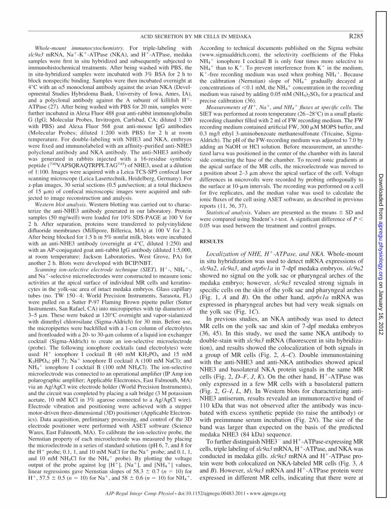
Table 1. Specific primer sequences of the quantitative real-time RT-PCR and in situ hybridization
Gene Name Forward Primer Sequence (5=-3=) Reverse Primer Sequence (5=-3=)
qRT-PCRrpl7 GAGATCCGCCTGGCTCGTA GGGCTGACTCCGTTGATACCTfoxi3 GAACATAGAGCAAGGGCAAACAT CGTCGTGGGCGTCTTAACGTslc9a2 ATCGTCTGTTGTGCCCTC CAGTTCCACTCGTGCTCTslc9a3 ATGCCTGATGTCACTGCT GTGTCGGTGCTGCTTCCTatp6v1a ACAAGTGCGCCCAGTAAC CCCGAAGGCTCCAGGTATrhcg1 TGGGAGATGATGGGAAGATAAGC GCAATGCTCCATAGGCAATCAA
In situ hybridizationslc9a2 CTGCCATCTTTCTCAGGTTTTCAT GGAGATGGCAAAGAGCTCAGCTACslc9a3 GCGCTGTGGATCCTGGTCTGTTGG GTAGGACAGGTATCCCACCACAAAatp6v1a TTTGTGCATGGAGTTTCAGGACCA GATCACAGTTTTTCCACACCCGAA
qRT-PCR, quantitative RT-PCR.
R284 ACID SECRETION BY MR CELLS IN MEDAKA
AJP-Regul Integr Comp Physiol • doi:10.1152/ajpregu.00483.2011 • www.ajpregu.org
on January 16, 2012ajpregu.physiology.org
Dow
nloaded from
Whole-mount immunocytochemistry. For triple-labeling withslc9a3 mRNA, Na�-K�-ATPase (NKA), and H�-ATPase, medakasamples were first in situ hybridized and subsequently subjected toimmunohistochemical treatments. After being washed with PBS, thein situ-hybridized samples were incubated with 3% BSA for 2 h toblock nonspecific binding. Samples were then incubated overnight at4°C with an �5 monoclonal antibody against the avian NKA (Devel-opmental Studies Hybridoma Bank, University of Iowa, Ames, IA),and a polyclonal antibody against the A subunit of killifish H�-ATPase (27). After being washed with PBS for 20 min, samples werefurther incubated in Alexa Fluor 488 goat anti-rabbit immunoglobulinG (IgG; Molecular Probes, Invitrogen, Carlsbad, CA; diluted 1:200with PBS) and Alexa Fluor 568 goat anti-mouse IgG antibodies(Molecular Probes; diluted 1:200 with PBS) for 2 h at roomtemperature. For double-labeling with NHE3 and NKA, embryoswere fixed and immunolabeled with an affinity-purified anti-NHE3polyclonal antibody and NKA antibody. The anti-NHE3 antibodywas generated in rabbits injected with a 16-residue syntheticpeptide (730VAPSQRAQTRPPLTAG745) of NHE3, used at a dilutionof 1:100. Images were acquired with a Leica TCS-SP5 confocal laserscanning microscope (Leica Lasertechnik, Heidelberg, Germany). Forz-plan images, 30 serial sections (0.5 �m/section; at a total thicknessof 15 �m) of confocal microscopic images were acquired and sub-jected to image reconstruction and analysis.
Western blot analysis. Western blotting was carried out to charac-terize the anti-NHE3 antibody generated in our laboratory. Proteinsamples (50 mg/well) were loaded for 10% SDS-PAGE at 100 V for2 h. After separation, proteins were transferred to polyvinylidenedifluoride membranes (Millipore, Billerica, MA) at 100 V for 2 h.After being blocked for 1.5 h in 5% nonfat milk, blots were incubatedwith an anti-NHE3 antibody (overnight at 4°C, diluted 1:250) andwith an AP-conjugated goat anti-rabbit IgG antibody (diluted 1:5,000,at room temperature; Jackson Laboratories, West Grove, PA) foranother 2 h. Blots were developed with BCIP/NBT.
Scanning ion-selective electrode technique (SIET). H�-, NH4�-,
and Na�-selective microelectrodes were constructed to measure ionicactivities at the apical surface of individual MR cells and keratino-cytes in the yolk-sac area of intact medaka embryos. Glass capillarytubes (no. TW 150–4; World Precision Instruments, Sarasota, FL)were pulled on a Sutter P-97 Flaming Brown pipette puller (SutterInstruments, San Rafael, CA) into micropipettes with tip diameters of3–5 �m. These were baked at 120°C overnight and vapor-sialanizedwith dimethyl chlorosilane (Sigma-Aldrich) for 30 min. Before use,the micropipettes were backfilled with a 1-cm column of electrolytesand frontloaded with a 20- to 30-�m column of a liquid ion exchangercocktail (Sigma-Aldrich) to create an ion-selective microelectrode(probe). The following ionophore cocktails (and electrolytes) wereused: H� ionophore I cocktail B (40 mM KH2PO4 and 15 mMK2HPO4; pH 7); Na� ionophore II cocktail A (100 mM NaCl); andNH4
� ionophore I cocktail B (100 mM NH4Cl). The ion-selectivemicroelectrode was connected to an operational amplifier (IP Amp ionpolarographic amplifier; Applicable Electronics, East Falmouth, MA)via an Ag/AgCl wire electrode holder (World Precision Instruments),and the circuit was completed by placing a salt bridge (3 M potassiumacetate, 10 mM KCl in 3% agarose connected to a Ag/AgCl wire).Electrode vibration and positioning were achieved with a steppermotor-driven three-dimensional (3D) positioner (Applicable Electron-ics). Data acquisition, preliminary processing, and control of the 3Delectrode positioner were performed with ASET software (ScienceWares, East Falmouth, MA). To calibrate the ion-selective probe, theNernstian property of each microelectrode was measured by placingthe microelectrode in a series of standard solutions (pH 6, 7, and 8 forthe H� probe; 0.1, 1, and 10 mM NaCl for the Na� probe; and 0.1, 1,and 10 mM NH4Cl for the NH4
� probe). By plotting the voltageoutput of the probe against log [H�], [Na�], and [NH4
�] values,linear regressions gave Nernstian slopes of 58.3 � 0.7 (n � 10) forH�, 57.5 � 0.5 (n � 10) for Na�, and 58 � 0.6 (n � 10) for NH4
�.
According to technical documents published on the Sigma website(www.sigmaaldrich.com), the selectivity coefficients of the FlukaNH4
� ionophore I cocktail B is only four times more selective toNH4
� than to K�. To prevent interference from K� in the medium,K�-free recording medium was used when probing NH4
�. Becausethe calibration (Nernstian) slope of NH4
� gradually decayed atconcentrations of 0.1 mM, the NH4
� concentration in the recordingmedium was raised by adding 0.05 mM (NH4)2SO4 for a practical andprecise calibration (36).
Measurements of H�, Na�, and NH4� fluxes at specific cells. The
SIET was performed at room temperature (26–28°C) in a small plasticrecording chamber filled with 2 ml of FW recording medium. The FWrecording medium contained artificial FW, 300 �M MOPS buffer, and0.3 mg/l ethyl 3-aminobenzoate methanesulfonate (Tricaine, Sigma-Aldrich). The pH of the FW-recording medium was adjusted to 7.0 byadding an NaOH or HCl solution. Before measurement, an anesthe-tized larva was positioned in the center of the chamber with its lateralside contacting the base of the chamber. To record ionic gradients atthe apical surface of the MR cells, the microelectrode was moved toa position about 2–3 �m above the apical surface of the cell. Voltagedifferences in microvolts were recorded by probing orthogonally tothe surface at 10-�m intervals. The recording was performed on a cellfor five replicates, and the median value was used to calculate theionic fluxes of the cell using ASET software, as described in previousreports (11, 36, 37).
Statistical analysis. Values are presented as the means � SD andwere compared using Student’s t-test. A significant difference of P 0.05 was used between the treatment and control groups.
RESULTS
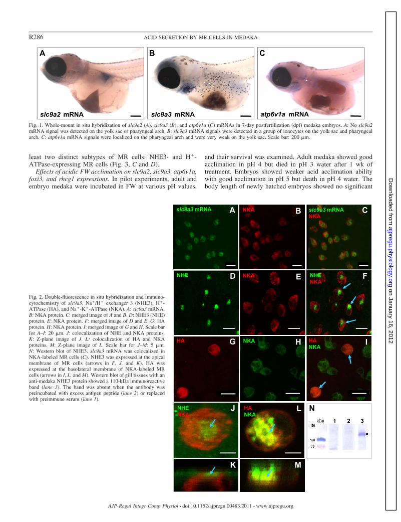
Localization of NHE, H�-ATPase, and NKA. Whole-mountin situ hybridization was used to detect mRNA expressions ofslc9a2, slc9a3, and atp6v1a in 7-dpf medaka embryos. slc9a2showed no signal on the yolk sac or pharyngeal arches of themedaka embryo; however, slc9a3 revealed strong signals inspecific cells on the skin of the yolk sac and pharyngeal arches(Fig. 1, A and B). On the other hand, atp6v1a mRNA wasexpressed in pharyngeal arches but had very weak signals onthe yolk sac (Fig. 1C).
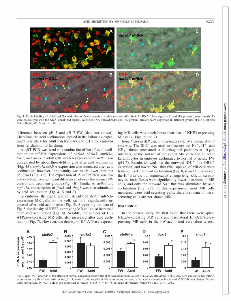
In previous studies, an NKA antibody was used to detectMR cells on the yolk sac and skin of 7-dpf medaka embryos(36, 45). In this study, we used the same NKA antibody todouble-stain with slc9a3 mRNA (fluorescent in situ hybridiza-tion), and results showed the colocalization of both signals ina group of MR cells (Fig. 2, A–C). Double immunostainingwith the anti-NHE3 and anti-NKA antibodies showed apicalNHE3 and basolateral NKA protein signals in the same MRcells (Fig. 2, D–F, J, K). On the other hand, H�-ATPase wasonly expressed in a few MR cells with a basolateral pattern(Fig. 2, G–I, L, M). In Western blots for characterizing anti-NHE3 antiserum, results revealed an immunoreactive band of110 kDa that was not observed after the antibody was incu-bated with excess synthetic peptide (to raise the antibody) orwith preimmune serum incubation (Fig. 2N). The size of theband was larger than expected on the basis of the predictedmedaka NHE3 (84 kDa) sequence.
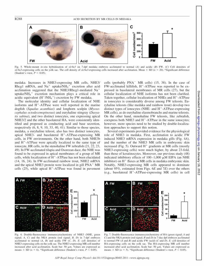
To further distinguish NHE3� and H�-ATPase-expressing MRcells, triple labeling of slc9a3 mRNA, H�-ATPase, and NKA wasconducted in medaka gills. slc9a3 mRNA and H�-ATPase pro-tein were both colocalized on NKA-labeled MR cells (Fig. 3, Aand B). However, slc9a3 mRNA and H�-ATPase protein wereexpressed in different MR cells, indicating that there were at
R285ACID SECRETION BY MR CELLS IN MEDAKA
AJP-Regul Integr Comp Physiol • doi:10.1152/ajpregu.00483.2011 • www.ajpregu.org
on January 16, 2012ajpregu.physiology.org
Dow
nloaded from
least two distinct subtypes of MR cells: NHE3- and H�-ATPase-expressing MR cells (Fig. 3, C and D).
Effects of acidic FW acclimation on slc9a2, slc9a3, atp6v1a,foxi3, and rhcg1 expressions. In pilot experiments, adult andembryo medaka were incubated in FW at various pH values,
and their survival was examined. Adult medaka showed goodacclimation in pH 4 but died in pH 3 water after 1 wk oftreatment. Embryos showed weaker acid acclimation abilitywith good acclimation in pH 5 but death in pH 4 water. Thebody length of newly hatched embryos showed no significant
Fig. 1. Whole-mount in situ hybridization of slc9a2 (A), slc9a3 (B), and atp6v1a (C) mRNAs in 7-day postfertilization (dpf) medaka embryos. A: No slc9a2mRNA signal was detected on the yolk sac or pharyngeal arch. B: slc9a3 mRNA signals were detected in a group of ionocytes on the yolk sac and pharyngealarch. C: atp6v1a mRNA signals were localized on the pharyngeal arch and were very weak on the yolk sac. Scale bar: 200 �m.
Fig. 2. Double-fluorescence in situ hybridization and immuno-cytochemistry of slc9a3, Na�/H� exchanger 3 (NHE3), H�-ATPase (HA), and Na�-K�-ATPase (NKA). A: slc9a3 mRNA.B: NKA protein. C: merged image of A and B. D: NHE3 (NHE)protein. E: NKA protein. F: merged image of D and E. G: HAprotein. H: NKA protein. I: merged image of G and H. Scale barfor A–I: 20 �m. J: colocalization of NHE and NKA proteins.K: Z-plane image of J. L: colocalization of HA and NKAproteins. M: Z-plane image of L. Scale bar for J–M: 5 �m.N: Western blot of NHE3. slc9a3 mRNA was colocalized inNKA-labeled MR cells (C). NHE3 was expressed at the apicalmembrane of MR cells (arrows in F, J, and K). HA wasexpressed at the basolateral membrane of NKA-labeled MRcells (arrows in I, L, and M). Western blot of gill tissues with ananti-medaka NHE3 protein showed a 110-kDa immunoreactiveband (lane 3). The band was absent when the antibody waspreincubated with excess antigen peptide (lane 2) or replacedwith preimmune serum (lane 1).
R286 ACID SECRETION BY MR CELLS IN MEDAKA
AJP-Regul Integr Comp Physiol • doi:10.1152/ajpregu.00483.2011 • www.ajpregu.org
on January 16, 2012ajpregu.physiology.org
Dow
nloaded from
difference between pH 5 and pH 7 FW (data not shown).Therefore, the acid acclimation applied in the following exper-iment was pH 4 for adult fish for 2 wk and pH 5 for embryosfrom fertilization to hatching.
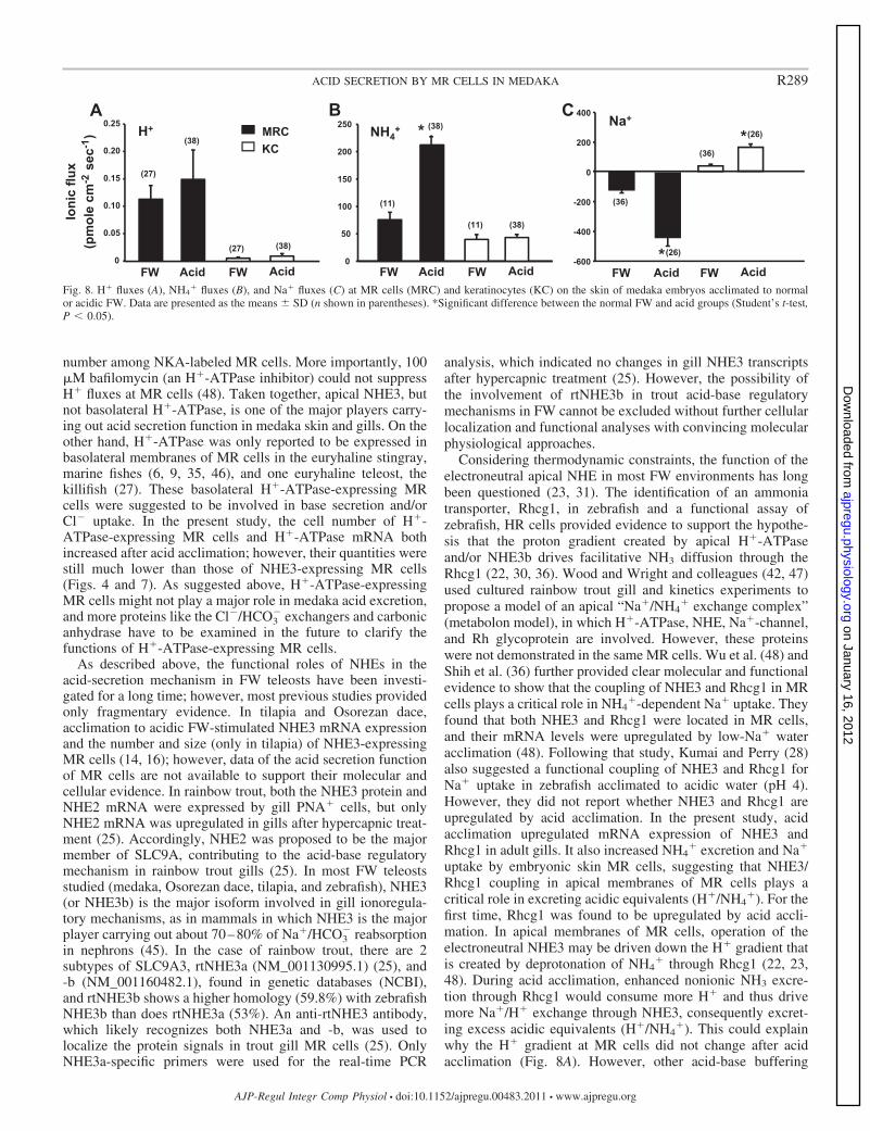
A qRT-PCR was used to examine the effect of acid accli-mation on mRNA expressions of slc9a2, slc9a3, atp6v1a,foxi3, and rhcg1 in adult gills. mRNA expression of slc9a3 wasupregulated by about three-fold in gills after acid acclimation(Fig. 4A). atp6v1a mRNA expression also increased after acidacclimation; however, the quantity was much lower than thatof slc9a3 (Fig. 4C). The expression of slc9a2 mRNA was lowand exhibited no significant difference between the normal FWcontrol and treatment groups (Fig. 4B). Similar to slc9a3 andatp6v1a, transcription of foxi3 and rhcg1 was also stimulatedby acid acclimation (Fig. 4, D and E).
In embryos, the signal and cell density of slc9a3 mRNA-expressing MR cells on the yolk sac both significantly in-creased after acid acclimation (Fig. 5). Supporting the data ofFig. 5, the density of NHE3-expressing MR cells also increasedafter acid acclimation (Fig. 6). Notably, the number of H�-ATPase-expressing MR cells also increased after acid accli-mation (Fig. 7). However, the density of H�-ATPase-express-
ing MR cells was much lower than that of NHE3-expressingMR cells (Figs. 6 and 7).
Ionic fluxes at MR cells and keratinocytes of yolk-sac skin ofembryos. The SIET was used to measure net Na�, H�, andNH4
� fluxes (measured at 2 orthogonal positions at 10-�mintervals) at the surface of individual MR cells and adjacentkeratinocytes in embryos acclimated to normal or acidic FW(pH 5). Results showed that the outward NH4
� flux (NH4�
excretion) and inward Na� flux (Na� uptake) of MR cells wereboth induced after acid acclimation (Fig. 8, B and C); however,the H� flux did not significantly change (Fig. 8A). In keratin-ocytes, ionic fluxes were significantly lower than those in MRcells, and only the outward Na� flux was stimulated by acidacclimation (Fig. 8C). In this experiment, most MR cellsmeasured were acid-secreting cells; therefore, data of base-secreting cells are not shown (48).
DISCUSSION
In the present study, we first found that there were apicalNHE3-expressing MR cells and basolateral H�-ATPase-ex-pressing MR cells in the FW-acclimated euryhaline teleost,
Fig. 3. Triple labeling of slc9a3 mRNA with HA and NKA proteins in adult medaka gills. Slc9a3 mRNA (black signal) (A) and HA protein (green signal) (B)were colocalized with the NKA signal (red signal). slc9a3 mRNA (arrowheads) and HA protein (arrows) were expressed in different groups of NKA-labeledMR cells (C, D). Scale bar: 50 �m.
Fig. 4. qRT-PCR analysis of the effects of normal and acidic freshwater (FW) acclimation on slc9a3 (A), slc9a2 (B), atp6v1a (C), foxi3 (D), and rhcg1 (E) mRNAexpressions in gills of adult fish. slc9a3, foxi3, atp6v1a, and rhcg1 mRNA expressions increased after acid acclimation, but that of slc9a2 did not change. Valueswere normalized by rpl7. Values are expressed as means � SD (n � 6). *Significant difference (Student’s t-test, P 0.05).
R287ACID SECRETION BY MR CELLS IN MEDAKA
AJP-Regul Integr Comp Physiol • doi:10.1152/ajpregu.00483.2011 • www.ajpregu.org
on January 16, 2012ajpregu.physiology.org
Dow
nloaded from
medaka. Increases in NHE3-expressing MR cells, NHE3/Rhcg1 mRNA, and Na� uptake/NH4
� excretion after acidacclimation suggested that the NHE3/Rhcg1-mediated Na�
uptake/NH4� excretion mechanism plays a critical role in
acidic equivalent (H�/NH4�) excretion by FW medaka.
The molecular identity and cellular localization of NHEisoforms and H�-ATPase were well reported in the marinedogfish (Squalus acanthias) and longhorn sculpin (Myoxo-cephalus octodecemspinosus) and euryhaline stingray (Dasya-tis sabina), and two distinct ionocytes, one expressing apicalNHE3/2 and the other basolateral HA, were consistently iden-tified and proposed as conducting acid and base secretion,respectively (6, 8, 9, 10, 33, 40, 41). Similar to those species,medaka, a euryhaline teleost, also has two distinct ionocytes,apical NHE3- and basolateral H�-ATPase-expressing MRcells, in FW environments. On the other hand, both NHE3band H�-ATPase were apically localized to the same type ofionocyte, HR cells, in the stenohaline FW zebrafish (21, 22, 23,49). In FW-acclimated tilapia and Osorezan dace, the NHE wasfound to be expressed in apical membranes of a group of MRcells, while localization of H�-ATPase has not been elucidated(14, 16, 24). In FW-acclimated rainbow trout, NHE2 mRNAand the apical NHE3 protein were localized in gill PNA� MRcells (25), while apical H�-ATPase was found in pavement
cells (probably PNA� MR cells) (15, 38). In the case ofFW-acclimated killifish, H�-ATPase was reported to be ex-pressed in basolateral membranes of MR cells (27), but thecellular localization of NHE isoforms has not been clarified.Taken together, cellular localization of NHEs and H�-ATPasein ionocytes is considerably diverse among FW teleosts. Eu-ryhaline teleosts (like medaka and rainbow trout) develop twodistinct types of ionocytes (NHE- and H�-ATPase-expressingMR cells), as do euryhaline elasmobranchs and marine teleosts.On the other hand, stenohaline FW teleosts, like zebrafish,coexpress both NHE3 and H�-ATPase in the same ionocytes;however, more species need to be studied by double-localiza-tion approaches to support this notion.
Several experiments provided evidence for the physiologicalrole of NHE3 in medaka. First, acclimation to acidic FWinduced NHE3 mRNA expression in medaka gills (Fig. 4A),and the number of the NHE3 MR cells in embryonic skinincreased (Fig. 5). Outward H� gradients at MR cells (mostlyNHE3-expressing cells) were much higher, by about 23-fold,than those of keratinocytes. Moreover, our previous study (48)indicated inhibitory effects of 100–1,000 �M EIPA (an NHEinhibitor) on H� fluxes at MR cells in medaka embryonic skin.Notably, NHE3-expressing MR cells appeared to dominate(about 95%, estimated from Figs. 6E and 7E) over the others(e.g., basolateral H�-ATPase-expressing MR cells) in cell
Fig. 7. Double-fluorescence immunocytochemistry of HA (green signal; A andC) and the NKA protein (red signal; B and D) in 7-day dpf embryos acclimatedto normal FW (A and B) and acidic FW (acid) (C and D). E: cell densities ofHA-expressing cells on the yolk sac. The HA-expressing MR cell numberincreased after acid acclimation. Scale bar: 50 �m. Values are expressed asmeans � SD (n � 5). *Significant difference (Student’s t-test, P 0.05).
Fig. 5. Whole-mount in-situ hybridization of slc9a3 on 7-dpf medaka embryos acclimated to normal (A) and acidic (B) FW. (C) Cell densities ofslc9a3-expressing cells on the yolk sac. The cell density of slc9a3-expressing cells increased after acclimation. Mean � SD (n � 20). *Significant difference(Student’s t-test, P 0.05).
Fig. 6. Double-fluorescence immunocytochemistry of NHE3 (NHE, greensignal; A, C) and the NKA protein (red signal; B, D) in 7-dpf embryosacclimated to normal (A, B) and acidic FW (C, D). E: cell densities ofNHE3-expressing cells on the yolk sac. The NHE3-expressing MR cell numberincreased after acid acclimation. Scale bar: 50 �m. Values are expressed asmeans � SD (n � 6). *Significant difference (Student’s t-test, P 0.05).
R288 ACID SECRETION BY MR CELLS IN MEDAKA
AJP-Regul Integr Comp Physiol • doi:10.1152/ajpregu.00483.2011 • www.ajpregu.org
on January 16, 2012ajpregu.physiology.org
Dow
nloaded from
number among NKA-labeled MR cells. More importantly, 100�M bafilomycin (an H�-ATPase inhibitor) could not suppressH� fluxes at MR cells (48). Taken together, apical NHE3, butnot basolateral H�-ATPase, is one of the major players carry-ing out acid secretion function in medaka skin and gills. On theother hand, H�-ATPase was only reported to be expressed inbasolateral membranes of MR cells in the euryhaline stingray,marine fishes (6, 9, 35, 46), and one euryhaline teleost, thekillifish (27). These basolateral H�-ATPase-expressing MRcells were suggested to be involved in base secretion and/orCl� uptake. In the present study, the cell number of H�-ATPase-expressing MR cells and H�-ATPase mRNA bothincreased after acid acclimation; however, their quantities werestill much lower than those of NHE3-expressing MR cells(Figs. 4 and 7). As suggested above, H�-ATPase-expressingMR cells might not play a major role in medaka acid excretion,and more proteins like the Cl�/HCO3
� exchangers and carbonicanhydrase have to be examined in the future to clarify thefunctions of H�-ATPase-expressing MR cells.
As described above, the functional roles of NHEs in theacid-secretion mechanism in FW teleosts have been investi-gated for a long time; however, most previous studies providedonly fragmentary evidence. In tilapia and Osorezan dace,acclimation to acidic FW-stimulated NHE3 mRNA expressionand the number and size (only in tilapia) of NHE3-expressingMR cells (14, 16); however, data of the acid secretion functionof MR cells are not available to support their molecular andcellular evidence. In rainbow trout, both the NHE3 protein andNHE2 mRNA were expressed by gill PNA� cells, but onlyNHE2 mRNA was upregulated in gills after hypercapnic treat-ment (25). Accordingly, NHE2 was proposed to be the majormember of SLC9A, contributing to the acid-base regulatorymechanism in rainbow trout gills (25). In most FW teleostsstudied (medaka, Osorezan dace, tilapia, and zebrafish), NHE3(or NHE3b) is the major isoform involved in gill ionoregula-tory mechanisms, as in mammals in which NHE3 is the majorplayer carrying out about 70–80% of Na�/HCO3
� reabsorptionin nephrons (45). In the case of rainbow trout, there are 2subtypes of SLC9A3, rtNHE3a (NM_001130995.1) (25), and-b (NM_001160482.1), found in genetic databases (NCBI),and rtNHE3b shows a higher homology (59.8%) with zebrafishNHE3b than does rtNHE3a (53%). An anti-rtNHE3 antibody,which likely recognizes both NHE3a and -b, was used tolocalize the protein signals in trout gill MR cells (25). OnlyNHE3a-specific primers were used for the real-time PCR
analysis, which indicated no changes in gill NHE3 transcriptsafter hypercapnic treatment (25). However, the possibility ofthe involvement of rtNHE3b in trout acid-base regulatorymechanisms in FW cannot be excluded without further cellularlocalization and functional analyses with convincing molecularphysiological approaches.
Considering thermodynamic constraints, the function of theelectroneutral apical NHE in most FW environments has longbeen questioned (23, 31). The identification of an ammoniatransporter, Rhcg1, in zebrafish and a functional assay ofzebrafish, HR cells provided evidence to support the hypothe-sis that the proton gradient created by apical H�-ATPaseand/or NHE3b drives facilitative NH3 diffusion through theRhcg1 (22, 30, 36). Wood and Wright and colleagues (42, 47)used cultured rainbow trout gill and kinetics experiments topropose a model of an apical “Na�/NH4
� exchange complex”(metabolon model), in which H�-ATPase, NHE, Na�-channel,and Rh glycoprotein are involved. However, these proteinswere not demonstrated in the same MR cells. Wu et al. (48) andShih et al. (36) further provided clear molecular and functionalevidence to show that the coupling of NHE3 and Rhcg1 in MRcells plays a critical role in NH4
�-dependent Na� uptake. Theyfound that both NHE3 and Rhcg1 were located in MR cells,and their mRNA levels were upregulated by low-Na� wateracclimation (48). Following that study, Kumai and Perry (28)also suggested a functional coupling of NHE3 and Rhcg1 forNa� uptake in zebrafish acclimated to acidic water (pH 4).However, they did not report whether NHE3 and Rhcg1 areupregulated by acid acclimation. In the present study, acidacclimation upregulated mRNA expression of NHE3 andRhcg1 in adult gills. It also increased NH4
� excretion and Na�
uptake by embryonic skin MR cells, suggesting that NHE3/Rhcg1 coupling in apical membranes of MR cells plays acritical role in excreting acidic equivalents (H�/NH4
�). For thefirst time, Rhcg1 was found to be upregulated by acid accli-mation. In apical membranes of MR cells, operation of theelectroneutral NHE3 may be driven down the H� gradient thatis created by deprotonation of NH4
� through Rhcg1 (22, 23,48). During acid acclimation, enhanced nonionic NH3 excre-tion through Rhcg1 would consume more H� and thus drivemore Na�/H� exchange through NHE3, consequently excret-ing excess acidic equivalents (H�/NH4
�). This could explainwhy the H� gradient at MR cells did not change after acidacclimation (Fig. 8A). However, other acid-base buffering
Fig. 8. H� fluxes (A), NH4� fluxes (B), and Na� fluxes (C) at MR cells (MRC) and keratinocytes (KC) on the skin of medaka embryos acclimated to normal
or acidic FW. Data are presented as the means � SD (n shown in parentheses). *Significant difference between the normal FW and acid groups (Student’s t-test,P 0.05).
R289ACID SECRETION BY MR CELLS IN MEDAKA
AJP-Regul Integr Comp Physiol • doi:10.1152/ajpregu.00483.2011 • www.ajpregu.org
on January 16, 2012ajpregu.physiology.org
Dow
nloaded from
systems, like the HCO3� reabsorption mechanism, may also
consume part of the H�.Similar to zebrafish (18), medaka enhance their acid secre-
tion function by not only increasing the number of acid-secreting ionocytes (Fig. 6) but also stimulating the functionalcapacity at each ionocyte (Fig. 8). In medaka, the functionalregulation at each NHE3 ionocyte may be achieved by regu-lating activity and/or mRNA expression of the relavant trans-porters if we compare the data of ionocyte number and trans-porter mRNA expressions. The number of NHE3-expressingcells was increased 1.3-fold (estimated from Fig. 6) after acidacclimation, while mRNA expressions of NHE3 and Rhcg1were stimulated about 2.9–3.4 times (estimated from Fig. 4),suggesting an upregulation of the transporter mRNA expressedat each ionocyte.
One remarkable function of mammalian kidneys is to ex-crete net acid in the urine when experiencing a sustainedincrement in acid loading (4). Recent studies also found thatapical H�-ATPase and Rhcg play critical roles in excretingacid equivalents (H�/NH4
�) by collecting duct-intercalatedcells (43). Deletion of an Rhcg strongly reduced renalammonium secretion, caused metabolic acidosis in acid-challenged mice, and impaired restoration of a normalacid-base status (2, 43).
In mammals, chronic acid loads (hours to days) induce theappearance of cells positive for the proliferation markers BrdU,PCNA, or Ki67 in the collecting duct, suggesting the prolifer-ation of acid-secreting, �-intercalated cells (44). Chronic acidadaptation can increase the NHE3 protein and activity inproximal tubules through the Pyk2/c-Src pathway (4). How-ever, no study has shown the effect of acid loading on the cellnumber of proximal tubules. In the collecting duct, the tran-scription factor, foxi1, was identified to have a critical role indetermining cell differentiation (1). Foxi1�/� mice lose theproper gene expression pattern needed to maintain an adequateacid-base homeostasis and thus develop distal renal tubularacidosis (3). In our previous study, the HR cell number in-creased after 4 days of acid acclimation through epithelial stemcell proliferation and ionocyte differentiation (19). An acidicenvironment simultaneously stimulated zgcm2 mRNA expres-sion, a specific transcription factor for HR cell differentiationin zebrafish, illustrating that this cell fate-related transcriptionfactor may be involved in the chronic acid adaptation mecha-nism (7, 13, 20). foxi3a and foxi3b were demonstrated to act asmain regulators for specification and differentiation of iono-cytes in zebrafish skin/gills; knockdown of zfoxi3a/3b wasfound to block differentiation of all ionocytes (12, 20, 26). Inmedaka, foxi3 is expressed in progenitor and differentiated MRcells (39). mRNA expression of foxi3 also increased after acidacclimation (Fig. 4D), suggesting the involvement of foxi3 inpromoting differentiation of NHE3-expressing MR cells (andthus their function) to cope with an acidic environment.
Perspectives and Significance
Teleosts appear to have evolved two distinct pathwaysmediated by the apical NHE and apical H�-ATPase, respec-tively, to carry out acid-secretion functions in FW. Medaka,dace, and tilapia are all euryhaline species and develop apicalNHE3 in MR cells to excrete H�, as do euryhaline elasmo-branchs and marine teleosts. Stenohaline FW teleosts, like
zebrafish, coexpress both NHE3 and H�-ATPase in the sameionocytes but mainly employ apical H�-ATPase for the acid-secretion function. In contrast to these general patterns, rain-bow trout express apical NHE2 in PNA� MR cells and apicalH�-ATPase in PNA� MR cells, and both transporters wereproposed to be involved in the acid-secretion mechanism.Further studies with convincing molecular physiological ap-proaches, and on more species, are needed to explore theevolutional physiological significance of the two distinct acid-secretion mechanisms in FW teleosts.
ACKNOWLEDGMENTS
This study was financially supported by the grants to P. P. Hwang fromAcademia Sinica and the National Science Council, Taiwan, Republic ofChina. We thank Y. C. Tung for her assistance during the experiments.
DISCLOSURES
No conflicts of interest, financial or otherwise, are declared by the authors.
AUTHOR CONTRIBUTIONS
Author contributions: C.-C.L., L.-Y.L., H.-H.H., and J.-L.H. performedexperiments; C.-C.L., L.-Y.L., H.-H.H., and J.-L.H. analyzed data; C.-C.L.,L.-Y.L., V.T., P.P., J.-L.H., and P.-P.H. interpreted results of experiments;C.-C.L., H.-H.H., J.-L.H., and P.-P.H. prepared figures; C.-C.L. and J.-L.H.drafted manuscript; L.-Y.L., J.-L.H., and P.-P.H. edited and revised manu-script; V.T., P.P., J.-L.H., and P.-P.H. conception and design of research;P.-P.H. approved final version of manuscript.
REFERENCES
1. Al-Awqati Q, Schwartz GJ. A fork in the road of cell differentiation inthe kidney tubule. J Clin Invest 113: 1528–1530, 2004.
2. Biver S, Belge H, Bourgeois S, Van Vooren P, Nowik M, Scohy S,Houillier P, Szpirer J, Szpirer C, Wagner CA, Devuyst O, Marini AM.A role for Rhesus factor Rhcg in renal ammonium excretion and malefertility. Nature 456: 339–343, 2008.
3. Blomqvist SR, Vidarsson H, Fitzgerald S, Johansson BR, Ollerstam A,Brown R, Persson AE, Bergström GG, Enerbäck S. Distal renal tubularacidosis in mice that lack the forkhead transcription factor Foxi1. J ClinInvest 113: 1560–1570, 2004.
4. Bobulescu IA, Moe OW. Na�/H� exchangers in renal regulation ofacid-base balance. Semin Nephrol 26: 334–344, 2006.
5. Bobulescu IA, Moe OW. Luminal Na�/H� exchange in the proximaltubule. Pflügers Arch 458: 5–21, 2009.
6. Catches JS, Burns JM, Edwards SL, Claiborne JB. Na�/H� antiporter,V-H�-ATPase and Na�/K�-ATPase immunolocalization in a marineteleost (Myoxocephalus octodecemspinosus). J Exp Biol 209: 3440–3447,2006.
7. Chang WJ, Horng JL, Yan JJ, Hsiao CD, Hwang PP. The transcriptionfactor, glial cell missing 2, is involved in differentiation and functionalregulation of H�-ATPase-rich cells in zebrafish (Danio rerio). Am JPhysiol Regul Integr Comp Physiol 296: R1192–R1201, 2009.
8. Choe KP, Edwards SL, Claiborne JB, Evans DH. The putative mech-anism of Na� absorption in euryhaline elasmobranchs exists in the gills ofa stenohaline marine elasmobranch, Squalus acanthias. Comp BiochemPhysiol A Mol Integr Physiol 146: 155–162, 2007.
9. Choe KP, Kato A, Hirose S, Plata C, Sindic A, Romero MF, ClaiborneJB, Evans DH. NHE3 in an ancestral vertebrate: primary sequence,distribution, localization, and function in gills. Am J Physiol Regul IntegrComp Physiol 289: R1520–R1534, 2005.
10. Claiborne JB, Choe KP, Morrison-Shetlar AI, Weakley JC, Havird J,Freiji A, Evans DH, Edwards SL. Molecular detection and immunolog-ical localization of gill Na�/H� exchanger in the dogfish (Squalus acan-thias). Am J Physiol Regul Integr Comp Physiol 294: R1092–R1102,2008.
11. Donini A, O’Donnell MJ. Analysis of Na�, Cl�, K�, H� and NH4�
concentration gradients adjacent to the surface of anal papillae of themosquito Aedes aegypti: application of self-referencing ion-selective mi-croelectrodes. J Exp Biol 208: 603–610, 2005.
12. Esaki M, Hoshijima K, Kobayashi S, Fukuda H, Kawakami K, HiroseS. Visualization in zebrafish larvae of Na� uptake in mitochondria-rich
R290 ACID SECRETION BY MR CELLS IN MEDAKA
AJP-Regul Integr Comp Physiol • doi:10.1152/ajpregu.00483.2011 • www.ajpregu.org
on January 16, 2012ajpregu.physiology.org
Dow
nloaded from
cells whose differentiation is dependent on foxi3a. Am J Physiol RegulIntegr Comp Physiol 292: R470–R480, 2007.
13. Esaki M, Hoshijima K, Nakamura N, Munakata K, Tanaka M,Ookata K, Asakawa K, Kawakami K, Wang W, Weinberg ES, HiroseS. Mechanism of development of ionocytes rich in vacuolar-type H�-ATPase in the skin of zebrafish larvae. Dev Biol 329: 116–129, 2009.
14. Furukawa F, Watanabe S, Inokuchi M, Kaneko T. Responses of gillmitochondria-rich cells in Mozambique tilapia exposed to acidic environ-ments (pH 4.0) in combination with different salinities. Comp BiochemPhysiol A Mol Integr Physiol 158: 468–476, 2011.
15. Galvez F, Reid SD, Hawkings G, Goss GG. Isolation and characteriza-tion of mitochondria-rich cell types from the gill of freshwater rainbowtrout. Am J Physiol Regul Integr Comp Physiol 282: R658–R668, 2002.
16. Hirata T, Kaneko T, Ono T, Nakazato T, Furukawa N, Hasegawa S,Wakabayashi S, Shigekawa M, Chang MH, Romero MF, Hirose S.Mechanism of acid adaptation of a fish living in a pH 3.5 lake. Am JPhysiol Regul Integr Comp Physiol 284: R1199–R1212, 2003.
18. Horng JL, Lin LY, Huang CJ, Katoh F, Kaneko T, Hwang PP.Knockdown of V-ATPase subunit A (atp6v1a) impairs acid secretion andion balance in zebrafish (Danio rerio). Am J Physiol Regul Integr CompPhysiol 292: R2068–R2076, 2007.
19. Horng JL, Lin LY, Hwang PP. Functional regulation of H�-ATPase-richcells in zebrafish embryos acclimated to an acidic environment. Am JPhysiol Cell Physiol 296: C682–C692, 2009.
20. Hsiao CD, You MS, Guh YJ, Ma M, Jiang YJ, Hwang PP. A positiveregulatory loop between foxi3a and foxi3b is essential for specification anddifferentiation of zebrafish epidermal ionocytes. PLoS One 2: e302, 2007.
21. Hwang PP, Lee TH. New insights into fish ion regulation and mitochon-drion-rich cells. Comp Biochem Physiol A Mol Integr Physiol 148:479–497, 2007.
22. Hwang PP, Lee TH, Lin LY. Ion regulation in fish gills: recent progressin the cellular and molecular mechanisms. Am J Physiol Regul IntegrComp Physiol 301: R28–R47, 2011.
23. Hwang PP, Perry S. Ionic and acid-base regulation. In: Zebrafish, FishPhysiology Series, edited by Perry S, Ekker, M, Farrell, AP, Brauner, CJ.San Diego, CA: Academic, p. 311–314, 2010.
24. Inokuchi M, Hiroi J, Watanabe S, Hwang PP, Kaneko T. Morpholog-ical and functional classification of ion-absorbing mitochondria-rich cellsin the gills of Mozambique tilapia. J Exp Biol 212: 1003–1010, 2009.
25. Ivanis G, Esbaugh AJ, Perry SF. Branchial expression and localizationof SLC9A2 and SLC9A3 sodium/hydrogen exchangers and their possiblerole in acid-base regulation in freshwater rainbow trout (Oncorhynchusmykiss). J Exp Biol 211: 2467–2477, 2008.
26. Jänicke M, Carney TJ, Hammerschmidt M. Foxi3 transcription factorsand Notch signaling control the formation of skin ionocytes from epider-mal precursors of the zebrafish embryo. Dev Biol 307: 258–271, 2007.
27. Katoh F, Hyodo S, Kaneko T. Vacuolar-type proton pump in thebasolateral plasma membrane energizes ion uptake in branchial mitochon-dria-rich cells of killifish Fundulus heteroclitus, adapted to a low ionenvironment. J Exp Biol 206: 793–803, 2003.
28. Kumai Y, Perry SF. Ammonia excretion via Rhcg1 facilitates Na�
uptake in larval zebrafish, Danio rerio, in acidic water. Am J Physiol RegulIntegr Comp Physiol 301: 1517–1528, 2011.
29. Lin LY, Horng JL, Kunkel JG, Hwang PP. Proton pump-rich cellsecretes acid in skin of zebrafish larvae. Am J Physiol Cell Physiol 290:C371–C378, 2006.
30. Nakada T, Hoshijima K, Esaki M, Nagayoshi S, Kawakami K, HiroseS. Localization of ammonia transporter Rhcg1 in mitochondrion-rich cellsof yolk sac, gill, and kidney of zebrafish and its ionic strength-dependentexpression. Am J Physiol Regul Integr Comp Physiol 293: R1743–R1753,2007.
31. Parks SK, Tresguerres M, Goss GG. Theoretical considerations under-lying Na� uptake mechanisms in freshwater fishes. Comp BiochemPhysiol C Toxicol Pharmacol 148: 411–418, 2008.
32. Pholpramool C, Borwornpinyo S, Dinudom A. Role of Na�/H� ex-changer 3 in the acidification of the male reproductive tract and malefertility. Clin Exp Pharmacol Physiol 38: 353–359, 2011.
33. Piermarini PM, Evans DH. Immunochemical analysis of the vacuolarproton-ATPase B-subunit in the gills of a euryhaline stingray (Dasyatissabina): effects of salinity and relation to Na�/K�-ATPase. J Exp Biol204: 3251–3259, 2001.
34. Reid SD, Hawkings GS, Galvez F, Goss GG. Localization and charac-terization of phenamil-sensitive Na� influx in isolated rainbow trout gillepithelial cells. J Exp Biol 206: 551–559, 2003.
35. Reis-Santos P, McCormick SD, Wilson JM. Ionoregulatory changesduring metamorphosis and salinity exposure of juvenile sea lamprey(Petromyzon marinus L.). J Exp Biol 211: 978–988, 2008.
36. Shih TH, Horng JL, Hwang PP, Lin LY. Ammonia secretion in skin ofzebrafish larvae (Danio rerio). Am J Physiol Cell Physiol 295: C1625–C1632, 2008.
36a.Shih TH, Horng JL, Liu ST, Hwang PP, Lin LY. Rhcg1 and NHE3b areinvolved in ammonium-dependent sodium uptake by zebrafish larvaeacclimated to low-sodium water. Am J Physiol Regul Integr Comp Physiol302: 84–93, 2012.
37. Smith PJ, Hammar K, Porterfield DM, Sanger RH, Trimarchi JR.Self-referencing, non-invasive, ion selective electrode for single celldetection of trans-plasma membrane calcium flux. Microsc Res Tech 46:398–417, 1999.
38. Sullivan GV, Fryer JN, Perry SF. Immunolocalization of proton pumps(H�-ATPase) in pavement cells of rainbow trout gill. J Exp Biol 198:2619–2629, 1995.
39. Thermes V, Lin CC, Hwang PP. Expression of Ol-foxi3 and Na�/K�-ATPase in ionocytes during the development of euryhaline medaka (Ory-zias latipes) embryos. Gene Expr Patterns 10: 185–192, 2010.
40. Tresguerres M, Katoh F, Fenton H, Jasinska E, Goss GG. Regulationof branchial V-H�-ATPase, Na�/K�-ATPase and NHE2 in response toacid and base infusions in the Pacific spiny dogfish (Squalus acanthias). JExp Biol 208: 345–354, 2005.
41. Tresguerres M, Parks SK, Katoh F, Goss GG. Microtubule-dependentrelocation of branchial V-H�-ATPase to the basolateral membrane in thePacific spiny dogfish (Squalus acanthias): a role in base secretion. J ExpBiol 209: 599–609, 2006.
42. Tsui TKN, Hung CYC, Nawata CM, Wilson JM, Wright PA, WoodCM. Ammonia transport in cultured gill epithelium of freshwater rainbowtrout: The importance of Rhesus glycoproteins and the presence of anapical Na�/NH4
� exchange complex. J Exp Biol 212: 878–892, 2009.43. Wagner CA, Devuyst O, Belge H, Bourgeois S, Houillier P. The rhesus
protein RhCG: a new perspective in ammonium transport and distalurinary acidification. Kidney Int 79: 154–161, 2011.
44. Wagner CA, Devuyst O, Bourgeois S, Mohebbi N. Regulated acid-basetransport in the collecting duct. Pflügers Arch 458: 137–156, 2009.
45. Wagner CA, Finberg KE, Breton S, Marshansky V, Brown D, GeibelJP. Renal vacuolar-ATPase. Physiol Rev 84: 1263–1314, 2004.
46. Wilson JM, Laurent P, Tufts BL, Benos DJ, Donowitz M, Vogl AW,Randall DJ. NaCl uptake by the branchial epithelium in freshwater teleostfish: an immunological approach to ion-transport protein localization. JExp Biol 203: 2279–2296, 2000.
47. Wright PA, Wood CM. A new paradigm for ammonia excretion inaquatic animals: role of Rhesus (Rh) glycoproteins. J Exp Biol 212:2303–2312, 2009.
48. Wu SC, Horng JL, Hwang PP, Wen ZH, Lin CS, Lin LY. Ammonium-dependent sodium uptake in mitochondrion-rich cells of medaka (Oryziaslatipes) larvae. Am J Physiol Cell Physiol 298: C237–C250, 2010.
49. Yan JJ, Chou MY, Kaneko T, Hwang PP. Gene expression of Na�/H�
exchanger in zebrafish H�-ATPase-rich cells during acclimation to low-Na� and acidic environments. Am J Physiol Cell Physiol 293: C1814–C1823, 2007.
R291ACID SECRETION BY MR CELLS IN MEDAKA
AJP-Regul Integr Comp Physiol • doi:10.1152/ajpregu.00483.2011 • www.ajpregu.org
on January 16, 2012ajpregu.physiology.org
Dow
nloaded from


