Embed Size (px)
Citation preview

Activation of ERK/MAP Kinase in the Amygdala Is Required forMemory Consolidation of Pavlovian Fear Conditioning
Glenn E. Schafe,1 Coleen M. Atkins,2 Michael W. Swank,3 Elizabeth P. Bauer,1 J. David Sweatt,2 andJoseph E. LeDoux1
1W. M. Keck Foundation Laboratory of Neurobiology, Center for Neural Science, New York University, New York, New York10003, 2Division of Neuroscience, Baylor College of Medicine, Houston, Texas 77030, and 3Department of Psychiatry,Cornell University Medical Center, White Plains, New York 10605
Although much has been learned about the neurobiologicalmechanisms underlying Pavlovian fear conditioning at the sys-tems and cellular levels, relatively little is known about the mo-lecular mechanisms underlying fear memory consolidation. Thepresent experiments evaluated the role of the extracellularsignal-regulated kinase/mitogen-activated protein kinase (ERK/MAPK) signaling cascade in the amygdala during Pavlovian fearconditioning. We first show that ERK/MAPK is transiently acti-vated–phosphorylated in the amygdala, specifically the lateralnucleus (LA), at 60 min, but not 15, 30, or 180 min, after condi-tioning, and that this activation is attributable to paired presen-tations of tone and shock rather than to nonassociative auditory
stimulation, foot shock sensitization, or unpaired tone–shockpresentations. We next show that infusions of U0126, an inhibitorof ERK/MAPK activation, aimed at the LA, dose-dependentlyimpair long-term memory of Pavlovian fear conditioning butleaves short-term memory intact. Finally, we show that bathapplication of U0126 impairs long-term potentiation in the LA invitro. Collectively, these results demonstrate that ERK/MAPKactivation is necessary for both memory consolidation of Pav-lovian fear conditioning and synaptic plasticity in the amygdala.
Key words: amygdala; fear conditioning; ERK; MAPK; learning; LTP
Considerable evidence has implicated the lateral and basal nucleiof the amygdala (LBA) in the plastic changes underlying acquisi-tion and retention of Pavlovian fear conditioning. Lesion, tracttracing, and electrophysiological studies suggest that fear condi-tioning involves transmission of sensory information to the lateralnucleus of the amygdala (LA) where alterations in synaptic trans-mission are thought to encode key aspects of the learning (Fendtand Fanselow, 1999; Maren, 1999; LeDoux, 2000). However, al-though fear conditioning has received much attention at the systemsand cellular levels, relatively little is known about the molecularmechanisms that underlie consolidation of fear memory in the LA.
One relatively recent discovery is the role of the mitogen-activated protein (MAP) family of kinases in synaptic plasticityand memory. These include the p38 MAP kinase (MAPK) and Jun(or stress-activated protein) kinase members, which have beenimplicated in stress-related cellular responses to injury or inflam-mation, and also the extracellular signal-regulated kinase (ERK),which has been implicated in cellular growth and differentiation(Kornhauser and Greenberg, 1997; Impey et al., 1999; Oruban etal., 1999). In neurons, ERK/MAPK has been shown to be potentlyactivated by phosphorylation after synaptically driven increases inintracellular Ca21 (Rosen et al., 1994; Impey et al., 1999). Fur-thermore, ERK/MAPK has been shown to be activated–phospho-rylated in the hippocampus after long-term potentiation (LTP)induction in the Schaffer collateral pathway, an effect that isblocked, along with LTP, by pretreatment with inhibitors of ERK/MAPK activation (English and Sweatt, 1996, 1997; Impey et al.,1999). Finally, LTP induction in the hippocampus has been shown
to induce the transcription of cAMP response element (CRE)-mediated genes, an effect that is prevented by inhibitors of ERKactivation (Impey et al., 1998b).
In support of the hypothesis that MAPK regulation is necessaryfor memory consolidation, several recent studies have demon-strated learning and memory impairments after manipulation ofthe ERK/MAPK pathway. These include impaired memory con-solidation of taste aversion and spatial learning after administra-tion of inhibitors of MAPK activation into the gustatory cortex(Berman et al., 1998) or hippocampus (Blum et al., 1999), respec-tively. Furthermore, several recent studies have implicated theMAPK signaling pathway in auditory fear conditioning, one usingtransgenic mice deficient in Ras, an upstream regulator of ERK/MAPK (Brambilla et al., 1997), and the others using either sys-temic (Atkins et al., 1998; Selcher et al., 1999) or intracerebroven-tricular (Schafe et al., 1999) administration of ERK/MAPKinhibitors. However, these studies were limited in that they did notspecifically evaluate the role of ERK/MAPK in fear memoryconsolidation in the amygdala, the presumed locus of memorystorage in Pavlovian fear conditioning.
The following series of experiments was aimed at further defin-ing the role of the ERK/MAP kinase signaling pathway in memoryconsolidation of auditory fear conditioning. We first show thatERK/MAPK is transiently activated in the amygdala after fearconditioning. We next show that pharmacological blockade ofERK/MAPK activation in the amygdala dose-dependently impairsfear memory consolidation. Finally, we show that bath applicationof an inhibitor of ERK/MAPK activation impairs LTP in the LAin vitro.
MATERIALS AND METHODSSubjects. Adult male Sprague Dawley rats (Hilltop Labs, Scottdale, PA)were housed individually in plastic Nalgene cages and maintained on a 12hr light /dark cycle. Food and water were provided ad libitum throughoutthe experiment.
Western blotting. For Western blotting studies, rats were habituated tohandling and to the conditioning chamber for 3 d before training. On thetraining day, rats received five conditioning trials consisting of a 20 sec, 5kHz, 75 dB tone that coterminated with a 0.5 sec, 1.0 mA foot shock. Theintertrial interval (ITI) was, on average, 120 sec, and the total training time
Received June 23, 2000; revised Aug. 14, 2000; accepted Aug. 15, 2000.This research was supported in part by National Institute of Mental Health Grants
MH 46516, MH 00956, MH 39774, MH 11902, and MH 570161. This work was alsosupported by grants from the National Alliance for Research on Schizophrenia andDepression and the W. M. Keck Foundation to New York University. We thankAnnemieke Schoute for assistance with the histology.
G.E.S., C.M.A., and M.W.S. contributed equally to this work.Correspondence should be addressed to Dr. Glenn E. Schafe, Center for Neural
Science, New York University, 4 Washington Place, Room 809, New York, NY 10003.E-mail: [email protected] © 2000 Society for Neuroscience 0270-6474/00/208177-11$15.00/0
The Journal of Neuroscience, November 1, 2000, 20(21):8177–8187

lasted 14 min. Control (“Box”) rats were handled and exposed to theconditioning box for an equivalent amount of time but were not exposed totones or shocks. At the appropriate time interval after training, rats weredeeply anesthetized with pentobarbital (100 mg/kg, i.p.) and decapitated.Brains were frozen and stored at 280°C until processed. Amygdalapunches were obtained with a 1 mm punch tool (Fine Science Tools, FosterCity, CA) from 400-mm-thick sections taken on a sliding freezing mic-rotome. The punches included the LA and the basal nucleus and possiblyportions of the lateral central nucleus and cortical tissue directly lateral tothe external capsule. Punches were briefly sonicated in 100–200 ml ofice-cold buffer (20 mM Tris-HCl, pH 7.5, 1 mM EGTA, 1 mM EDTA, 25mg/ml aprotinin, 25 mg/ml leupeptin, 1 mM sodium pyrophosphate, 500 mMphenylmethylsulfonyl fluoride, 4 mM para-nitrophenyl-phosphate, and 1mM sodium orthovanadate). Sample buffer was immediately added to thehomogenates, and the samples were boiled at 95°C for 10 min. Homoge-nates were electrophoresed on 10% SDS-polyacrylamide gels and blottedto Immobilon-P (Millipore, Bedford, MA). Western blots were blocked inTTBS buffer (50 mM Tris-HCl, pH 7.5, 150 mM NaCl, and 0.05% Tween20) with 3% bovine serum albumin and then incubated with an anti-phospho-MAPK (1:1000; New England Biolabs, Beverly, MA) or ananti-total MAPK antibody (1:1000; Upstate Biotechnology, Lake Placid,NY). Blots were then incubated with anti-rabbit conjugated to horseradishperoxidase (Cappel, West Chester, PA) and developed using enhancedchemiluminescence (Amersham Pharmacia Biotech, Arlington Heights,IL). Western blots were developed in the linear range used for densitom-etry. Total protein amounts were determined by a BCA assay for eachhomogenate. Densitometry was conducted using NIH Image software. Toassess for changes in the activation of ERK/MAPK, total kinase levelswere first normalized to total protein levels for each sample. Then, acti-vated kinase levels were normalized to total kinase levels. Finally, acti-vated kinase levels in paired rats were expressed as a percentage of thosein controls.
Immunohistochemistry. For immunohistochemical studies, rats were ha-bituated to handling and to the conditioning chamber for 3 d beforetraining. On the training day, rats received five conditioning trials consist-ing of a 20 sec, 5 kHz, 75 dB tone that coterminated with a 0.5 sec, 1.0 mAfoot shock (as in the Western blot experiments). At the appropriate timeinterval, rats were rapidly and deeply anesthetized with pentobarbital (100mg/kg, i.p.) and perfused through the heart with ice-cold PBS, followed byice-cold 4% paraformaldehyde in 0.1 M phosphate buffer (PB). Brains wereremoved and post-fixed in 4% paraformaldehyde–PB for 24 hr and thencryoprotected in 20% glycerol–0.1 M PB for 48 hr. Immunohistochemistrywas performed as described previously (Swank, 2000a,b). Briefly, 30 mmfree-floating sections through amygdala were obtained using a slidingmicrotome. Every third section was processed for activated–phosphory-lated ERK/MAPK (pMAPK) immunoreactivity. After blocking in Tris-buffered saline (TBS) containing 4% normal goat serum–0.2% TritonX-100, slices were incubated at 4°C for 72 hr in anti-phospho-MAPK(rabbit polyclonal antibody, 1:4000; New England Biolabs) in TBS–2%goat serum–0.1% Triton X-100. After extensive washes in TBS, tissuesections were incubated in biotinylated goat anti-rabbit [prediluted;Kirkegaard & Perry Laboratories (KPL), Gaithersburg, MD] for 2 hr atroom temperature. Tissue was again rinsed in TBS followed by incubationin streptavidin–HRP (KPL) at room temperature for 1 hr, rinsed in TBS,and developed in cobalt-enhanced DAB (Enhance Black; KPL) for 10 min.Sections were mounted on subbed slides and coverslipped.
Quantification of pMAPK-labeled cells. Sections from comparable an-teroposterior levels were selected for scoring, ;3.2–3.3 mm posterior tobregma. At this level, the LA, central nucleus of amygdala (CE), and basalnuclei are all well represented (Fig. 1 E). Cell counts were taken from atleast three sections per rat and scored using a defined boundary approxi-mately equivalent to the size of the LA or the CE using either ImagePro(Media Cybernetics, Silver Spring, MD) or NIH Image. Because everythird section through the amygdala was processed for immunohistochem-istry, it was not necessary to correct for double-counting.
Drugs. In behavioral studies, U0126 (Promega, Madison, WI) was dis-solved in 100% DMSO to a final stock concentration of 4 mg/ml. Forconditioning, the drug was diluted 1:1 in ACSF. U0126 is a specificinhibitor of MAP kinase kinase (MEK), an upstream regulator of ERK/MAPK activation (Favata et al., 1998). In a recent study using hippocam-pal homogenates, the effects of U0126 have been shown to be specific toERK/MAPK and to have no effect at a range of concentrations on otherkinases, such as PKA, calcium-calmodulin kinase II, or PKC (Roberson etal., 1999).
Behavioral procedures. Behavioral procedures were conducted as de-scribed previously (Schafe et al., 1999; Schafe and LeDoux, 2000). UnderNembutal anesthesia (45 mg/kg, i.p.), rats were first implanted bilaterallywith 26 gauge stainless steel guide cannulas aimed at the LA (for details,see Schafe and LeDoux, 2000). The guide cannulas were fixed to screws inthe skull using a mixture of acrylic and dental cement, and a 33 gaugedummy cannula was inserted into each guide to prevent clogging. Allsurgical procedures were conducted in accordance to the National Insti-tutes of Health Guide for the Care and Use of Experimental Animals andwere approved by the New York University Animal Care and Use Com-mittee. Rats were given at least 5 d to recover before experimentalprocedures.
On the day before conditioning, rats were habituated to the conditioning
chamber and to dummy cannula removal for a minimum of 10–15 min. Thefollowing day, rats were given an intra-LBA infusion of either 0.5 ml 50%DMSO (vehicle) or one of two doses of U0126 in 50% DMSO (1.0 or 0.1mg/side in 0.5 ml; 0.25 ml /min). Injectors remained in the cannulas for 1min after drug infusion to allow diffusion of the drug from the tip.
Thirty minutes after drug infusions, rats were trained with either asingle conditioning trial consisting of a 30 sec, 5 kHz, 75 dB tone thatcoterminated with a 1.0 sec, 1.5 mA foot shock, or with five conditioningtrials consisting of a 20 sec, 5 kHz, 75 dB tone that coterminated with a 0.5sec, 1.0 mA foot shock (ITI of 120 sec).
Testing for conditioned fear responses (freezing) in rats conditionedwith a single trial were conducted at 1 or 24 hr after conditioning. For eachtest, rats were placed in a distinctive environment (for details, see Schafeet al., 1999) and exposed to either five or eight conditioned stimulus (CS)tones (5 kHz, 75 dB, 30 sec), respectively. For rats conditioned with fivetrials, testing occurred at 1, 3, 6 (three tones each test), and 24 (10 tones)hr later (5 kHz, 75 dB, 20 sec). Total seconds freezing during the CSpresentations were scored for each rat, and this number was expressed asa percentage of the total CS presentation time. All data were analyzedwith ANOVA and Newman–Keuls post hoc t tests. Differences wereconsidered significant if p , 0.05.
At the end of each behavioral experiment, rats were anesthetized by anoverdose of chloral hydrate (600 mg/kg) and perfused with 10% bufferedformalin. Nissl staining and light microscopy were used to verify thelocation of the cannula tips within the amygdala.
Slice electrophysiology. Electrophysiological experiments in amygdalaslices were conducted as documented previously (for a detailed descrip-tion, see Weisskopf et al., 1999). Briefly, male Sprague Dawley rats (3–5weeks old) were deeply anesthetized with halothane, and the brain wasrapidly removed and transferred to ice-cold ACSF containing (in mM): 115NaCl, 3.3 KCl, 1 MgSO4, 2 CaCl2, 25.5 NaHCO3, 1.2 NaH2PO4, 5 lacticacid, and 25 glucose, equilibrated with 95% O2–5% CO2. Coronal slices(400-mm-thick) containing the amygdala were cut and recovered in aholding chamber at 32–34°C for 30 min and were then allowed to return toroom temperature for at least another 30 min before recording. An uprightmicroscope equipped with infrared differential interference contrast optics(Olympus Optical, Tokyo, Japan) was used to perform whole-cell patchrecordings under visual guidance. Glass recording electrodes were filledwith (in mM): 130 K-gluconate, 0.6 EGTA, 2 MgCl2, 5 KCl, 10 HEPES, 2Mg-ATP, and 0.3 Na3-GTP, pH 7.3 (290–300 mOsm). The electrodestypically had resistances of 4–8 MV. All cells were allowed to remain attheir resting potentials.
Stimuli (150 msec duration) were delivered through bipolar stainlesssteel electrodes placed in the ventral striatum, just medial to the dorsallateral amygdala (LAd). This stimulating protocol activates fibers thatoriginate, at least in part, in the auditory thalamus (LeDoux et al., 1990;Weisskopf et al., 1999). The stimulation intensity was kept at a minimumand adjusted for each cell (between 80 and 140 mA) to produce a reliableEPSP without also recruiting polysynaptic responses or spiking. Baselineresponses were monitored at 0.1 Hz. After stabilization of baseline re-sponses, LTP was induced by pairing trains of 10 stimuli at 30 Hz with 1nA, 5 msec depolarizations given 5–10 msec after the onset of each EPSPin the train. This pattern of stimulation yields an action potential at thepeak of each EPSP of the train. This pairing was given 15 times at 10 secintervals. For each cell, the stimulation intensity for LTP induction was thesame as that used to elicit baseline EPSPs.
Picrotoxin (75 mM) was included in the bath in all experiments to blockfast GABAergic transmission but was not observed to produce epilepti-form bursting in the amygdala. Drugs were made up in DMSO stocksolution and diluted 1000 times into the superfusing ACSF, yielding a finalconcentration of U0126 of 10 mM. In control experiments, slices weresuperfused with 0.1% DMSO vehicle. The addition of U0126 to the bathwas not observed to affect membrane potential (Vm vehicle, 271.38 6 1.75;Vm U0126, 270.39 6 0.86).
In all experiments, the slope of the EPSP was measured, and LTP foreach time point was expressed as a percentage of the preinduction base-line. Data were analyzed with ANOVA and Newman–Keuls post hoc ttests.
RESULTSERK/MAPK is transiently activated in the amygdalaafter Pavlovian fear conditioningPrevious studies have shown that ERK/MAPK is activated in thehippocampus after Pavlovian fear conditioning (Atkins et al.,1998). Other recent studies have shown activation of ERK/MAPKin insular cortex and hippocampus after either taste aversion (Ber-man et al., 1998; Swank, 2000a) or spatial learning (Blum et al.,1999), respectively. Given the well established role of the LBA infear conditioning, our first objective was to determine whetherERK/MAPK is similarly activated in the LBA and whether thisactivation is characterized by a specific time course. For this seriesof experiments, we first used Western immunoblotting techniquesto quantify total amounts of activated ERK/MAPK from amygdala
8178 J. Neurosci., November 1, 2000, 20(21):8177–8187 Schafe et al. • ERK/MAPK and Fear Memory Consolidation

tissue punches. Next, we used immunohistochemistry to anatomi-cally localize expression of activated ERK/MAPK to particularamygdala nuclei. For each experiment, we used an antibody thatrecognizes activated–phosphorylated ERK/MAPK (pMAPK). Tocontrol for total amount of ERK protein, Western blot experimentsalso used an antibody that recognizes total (both phosphorylatedand unphosphorylated) ERK/MAPK (tMAPK). For these exper-iments, we attempted to strike a balance between giving enoughconditioning trials to observe significant regulation of ERK/MAPK with our biochemical methods (Atkins et al., 1998) but notso many that we would be unable to chart a time course ofERK/MAPK activation. Thus, we settled on five tone–shock pair-ings given over the course of 14 min.
Western blottingResults of the Western blotting can be viewed in Figure 1A–D inwhich both histograms for both pMAPK and tMAPK immunore-activity can be found (Fig. 1A,C), as well as representative blotsfrom both trained (Paired) and sham-trained (Box) controls (Fig.1B,D). For this latter group, rats were handled and exposed to theconditioning box for an equivalent amount of time but were notexposed to tones or shocks. Fear conditioning resulted in signifi-cant increases in pMAPK immunoreactivity for both p42 and p44
ERK/MAPK at 60 min after training but not at other time points.The ANOVA (kinase by time point) for pMAPK scores revealed asignificant effect for time point (F(3,80) 5 5.66; p , 0.01). The effectfor kinase (F(1,80) 5 0.33) and the interaction (F(3,80) 5 0.79) werenot found to differ. Newman–Keuls post hoc t tests revealed thatdifferences existed between 15 and 60 min time points for the p42kinase ( p , 0.05). No significant differences were detected be-tween other time points. Furthermore, this increase was not ac-counted for by changes in total ERK/MAPK (Fig. 1C,D). Here, theANOVA showed a nonsignificant effect of kinase (F(1,80) 5 0.14),time point (F(3,80) 5 0.53), and interaction (F(3,80) 5 0.83). Thus,ERK/MAPK is transiently activated in the amygdala after fearconditioning, with a peak at 60 min.
ImmunohistochemistryThe transient increase in pMAPK as assessed by Western blottingwas confirmed by immunohistochemistry (Fig. 1E–J). Representa-tive photomicrographs of pMAPK labeling are presented in Figure1G–J, and cell counts of pMAPK-labeled cells in the LA arepresented in Figure 1F. pMAPK-labeled cells were found scatteredthroughout the LA, particularly in the ventral portions of LAd andventrolateral lateral amygdala (LAvl). There were also scatteredlabeled cells in ventromedial lateral amygdala (LAvm). pMAPK
Figure 1. Time course of ERK/MAPK activation in the amygdala. A, Mean 6 SE percent pMAPK immunoreactivity from amygdala punches taken fromrats decapitated at 15 (n 5 10), 30 (n 5 10), 60 (n 5 14), or 180 (n 5 10) min after conditioning. Rats were presented with five tone–foot shock pairings.Paired samples in each group were normalized relative to sham-trained (Box) controls (for details, see Materials and Methods). p42 and p44 correspondto the molecular weights (in kilodaltons) of the two isoforms of mammalian ERK (ERK1 and ERK2) that are recognized by the pMAPK antibody. *p ,0.05 relative to the 15 min time point. B, Representative pMAPK blots from Box and Paired conditions at different time points after conditioning. C,Mean 6 SE percent tMAPK immunoreactivity at different time points after conditioning (15, 30, 60, or 180 min) from the samples in A. Paired samplesin each group were normalized relative to sham-trained (Box) controls. D, Representative tMAPK blots from Box and Paired conditions at different timepoints after conditioning. E, Schematic of the amygdala at approximately bregma 23.3 (according to Paxinos and Watson, 1997). F, Mean 6 SEpMAPK-immunoreactive cells in the LA at 15 (n 5 5), 30 (n 5 5), 60 (n 5 5), or 180 (n 5 5) min after conditioning. Rats were presented with fivetone–foot shock pairings. *p , 0.05 relative to the 15 min time point. G, Representative photomicrograph of pMAPK labeling in the amygdala at 15 minafter conditioning. CPu, Caudate/putamen; EC, external capsule. H, Representative photomicrograph of pMAPK labeling in the amygdala at 30 min afterconditioning. I, Representative photomicrograph of pMAPK labeling in the amygdala at 60 min after conditioning. J, Representative photomicrograph ofpMAPK labeling in the amygdala at 180 min after conditioning.
Schafe et al. • ERK/MAPK and Fear Memory Consolidation J. Neurosci., November 1, 2000, 20(21):8177–8187 8179
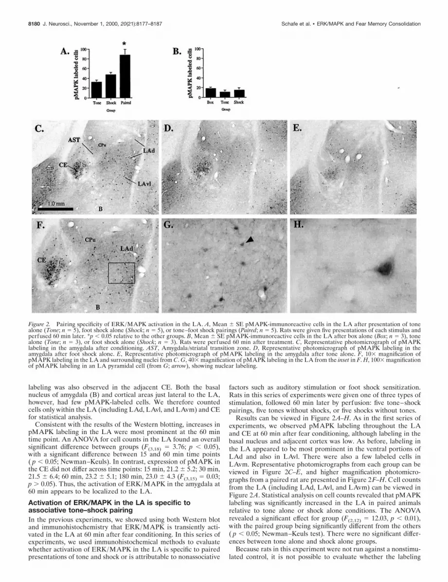
labeling was also observed in the adjacent CE. Both the basalnucleus of amygdala (B) and cortical areas just lateral to the LA,however, had few pMAPK-labeled cells. We therefore countedcells only within the LA (including LAd, LAvl, and LAvm) and CEfor statistical analysis.
Consistent with the results of the Western blotting, increases inpMAPK labeling in the LA were most prominent at the 60 mintime point. An ANOVA for cell counts in the LA found an overallsignificant difference between groups (F(3,18) 5 3.76; p , 0.05),with a significant difference between 15 and 60 min time points( p , 0.05; Newman–Keuls). In contrast, expression of pMAPK inthe CE did not differ across time points: 15 min, 21.2 6 5.2; 30 min,21.5 6 6.4; 60 min, 23.2 6 5.1; 180 min, 23.0 6 4.3 (F(3,15) 5 0.03;p . 0.05). Thus, the activation of ERK/MAPK in the amygdala at60 min appears to be localized to the LA.
Activation of ERK/MAPK in the LA is specific toassociative tone–shock pairingIn the previous experiments, we showed using both Western blotand immunohistochemistry that ERK/MAPK is transiently acti-vated in the LA at 60 min after fear conditioning. In this series ofexperiments, we used immunohistochemical methods to evaluatewhether activation of ERK/MAPK in the LA is specific to pairedpresentations of tone and shock or is attributable to nonassociative
factors such as auditory stimulation or foot shock sensitization.Rats in this series of experiments were given one of three types ofstimulation, followed 60 min later by perfusion: five tone–shockpairings, five tones without shocks, or five shocks without tones.
Results can be viewed in Figure 2A–H. As in the first series ofexperiments, we observed pMAPK labeling throughout the LAand CE at 60 min after fear conditioning, although labeling in thebasal nucleus and adjacent cortex was low. As before, labeling inthe LA appeared to be most prominent in the ventral portions ofLAd and also in LAvl. There were also a few labeled cells inLAvm. Representative photomicrographs from each group can beviewed in Figure 2C–E, and higher magnification photomicro-graphs from a paired rat are presented in Figure 2F–H. Cell countsfrom the LA (including LAd, LAvl, and LAvm) can be viewed inFigure 2A. Statistical analysis on cell counts revealed that pMAPKlabeling was significantly increased in the LA in paired animalsrelative to tone alone or shock alone conditions. The ANOVArevealed a significant effect for group (F(2,12) 5 12.03, p , 0.01),with the paired group being significantly different from the others( p , 0.05; Newman–Keuls test). There were no significant differ-ences between tone alone and shock alone groups.
Because rats in this experiment were not run against a nonstimu-lated control, it is not possible to evaluate whether the labeling
Figure 2. Pairing specificity of ERK/MAPK activation in the LA. A, Mean 6 SE pMAPK-immunoreactive cells in the LA after presentation of tonealone (Tone; n 5 5), foot shock alone (Shock; n 5 5), or tone–foot shock pairings (Paired; n 5 5). Rats were given five presentations of each stimulus andperfused 60 min later. *p , 0.05 relative to the other groups. B, Mean 6 SE pMAPK-immunoreactive cells in the LA after box alone (Box; n 5 3), tonealone (Tone; n 5 3), or foot shock alone (Shock; n 5 3). Rats were perfused 60 min after treatment. C, Representative photomicrograph of pMAPKlabeling in the amygdala after conditioning. AST, Amygdala/striatal transition zone. D, Representative photomicrograph of pMAPK labeling in theamygdala after foot shock alone. E, Representative photomicrograph of pMAPK labeling in the amygdala after tone alone. F, 103 magnification ofpMAPK labeling in the LA and surrounding nuclei from C. G, 403 magnification of pMAPK labeling in the LA from the inset in F. H, 1003 magnificationof pMAPK labeling in an LA pyramidal cell (from G; arrow), showing nuclear labeling.
8180 J. Neurosci., November 1, 2000, 20(21):8177–8187 Schafe et al. • ERK/MAPK and Fear Memory Consolidation

observed in tone alone and shock alone groups reflects basal levelsof ERK/MAPK activation in the LA or increases relative to thisbaseline. Thus, it may be argued that the increase in pMAPKlabeling observed in the LA after tone–shock pairings is simplyattributable to an additive effect of separate cells responsive to toneand shock alone. To evaluate this possibility, we ran an additionalassay, comparing pMAPK labeling in the LA in rats receivingsham training (box only), tone alone, or foot shock alone. Resultscan be viewed in Figure 2B. It is evident from the figure that nodifferences existed between groups. The ANOVA showed no sig-nificant effects (F(2,6) 5 2.75). Thus, the levels of pMAPK labelingobserved after presentations of tones or shocks alone appear toreflect basal levels of ERK/MAPK activation, which suggests thatthe increase in pMAPK labeling observed in the LA is not likely tobe attributable to an additive effect of tone- and shock-responsivecells. Rather, the increase in pMAPK observed in the LA appearsto be specific to pairing of tone and shock.
To specifically evaluate the associative specificity of pMAPKlabeling in the LA, we next examined activation of ERK/MAPK inthe LA after unpaired presentations of tone and shock (Schafe etal., 1999). Rats in this experiment were handled and habituated tothe conditioning context for 3 d as in previous experiments. On thetraining day, rats received either five paired presentations of toneand shock (as in the previous experiments) or five unpaired tone–shock presentations. For this latter group, the unconditioned stim-ulus (US) shock preceded the tone CS by 60 sec, and at least 120sec were allowed to pass between a tone CS presentation and thenext trial. Rats in each group were killed by perfusion 60 min afterstimulation, and brains were processed using immunohistochemis-try. Results can be viewed in Figure 3. Relative to unpairedcontrols, rats in the paired condition were observed to have signif-icantly more labeled cells in the LA (including LAd, LAvl, andLAvm) (t(8) 5 2.48, p , 0.05). The percent increase in labeling inpaired rats (;30%) appeared to be somewhat less than that in theprevious experiment in which paired rats were compared with ratsreceiving shock alone, which could reflect the fact that unpairedpresentations of tone and shock produce less, but not necessarilyno, learning about the tone (Schafe et al., 1999). Nonetheless,activation of ERK/MAPK in the LA appears to be specific toassociative pairing of tone and shock rather than reflecting nonas-sociative processes.
Interestingly, rats receiving shock alone stimulation were notobserved to have increases in pMAPK in the LA, which might, forexample, be expected to accompany contextual fear conditioning.This result stands in contrast to the findings of other recent studiesin the fear conditioning literature that have shown regulation oftranscription factors such as early growth response gene 1 in theamygdala, and particularly the LA, after contextual fear condition-ing (Rosen et al., 1998; Malkani and Rosen, 2000). However, therelative lack of pMAPK labeling after shock presentation in thepresent study may be a result of at least two factors. First, it may bepossible that the time course of ERK/MAPK activation aftercontextual learning is different from that after auditory fear con-ditioning. Second, because rats in our labeling experiments re-ceived extensive preexposure to the conditioning apparatus (3 d)before stimulation, it is possible that latent inhibition may haveobscured any potential context learning and accompanying regula-tion of ERK/MAPK. Future experiments using different trainingprotocols will be necessary to determine whether contextual fearconditioning is also characterized by increases in ERK/MAPKactivation in the amygdala and whether the pattern of expression issimilar to that after auditory fear conditioning.
Pharmacological blockade of ERK/MAPK activation inthe amygdala impairs fear memory consolidationThe previous experiments showed that ERK/MAPK is transientlyactivated in the amygdala and that this activation is specific toassociative pairing of tone and shock. In the next series of exper-iments, we asked whether ERK/MAPK activation in the LBA isobligatory for fear memory consolidation. Several recent studies
using either systemic (Atkins et al., 1998; Selcher et al., 1999) orintracerebroventricular (Schafe et al., 1999) pharmacological ma-nipulations have implicated the ERK/MAPK signaling pathway infear conditioning in rodents. To date, however, no study has tar-geted these manipulations to the amygdala. Thus, in the presentexperiments, rats received bilateral intra-amygdala infusions ofdifferent doses of U0126, a specific inhibitor of MEK, an upstreamregulator of ERK/MAPK activation (Favata et al., 1998). In theseexperiments, rats were infused with U0126 or vehicle before fearconditioning and assessed for retention of fear memory at varioustime points thereafter. Although the LA was the main target, ourinfusions also likely affected the adjacent basal nucleus. We there-fore refer to the affected area as the LBA.
In the first series of experiments, rats were given single-trialPavlovian fear conditioning, a protocol that matches that of previ-ous reports from our laboratory (Schafe et al., 1999; Schafe andLeDoux, 2000). For this series of experiments, rats were tested forfear retention at either 1 or 24 hr after conditioning (Fig. 4A). Inthe second series of experiments, rats were given five-trial Pavlov-ian fear conditioning, a protocol that was identical to that used inthe Western blot and immunohistochemical experiments reported
Figure 3. Associative specificity of ERK/MAPK activation in the LA. A,Mean 6 SE pMAPK-immunoreactive cells in the LA after paired (Paired;n 5 5) or unpaired (Unpaired; n 5 5) presentations of tone and shock. Ratswere perfused 60 min later. *p , 0.05 relative to the unpaired group. B,Representative photomicrograph of pMAPK labeling in the amygdala afterpaired stimulation. C, Representative photomicrograph of pMAPK label-ing in the amygdala after unpaired stimulation.
Schafe et al. • ERK/MAPK and Fear Memory Consolidation J. Neurosci., November 1, 2000, 20(21):8177–8187 8181

earlier in this paper. For this series of experiments, rats were testedfor fear retention at 1, 3, 6, and 24 hr after training (Fig. 5A).Finally, we evaluated the effectiveness of U0126 at blocking ERK/MAPK activation in the LBA. For this experiment, rats were giveninfusions of different doses of U0126 (1.0 or 0.l mg) or vehicle 30min before five-trial fear conditioning and killed 1 hr later. Punchesfrom the LBA surrounding the cannula tips were subjected toERK/MAPK immunoblotting.
Single-trial learningResults of the one-trial conditioning can be viewed in Figure 4B–F.Infusions of U0126 had no effect on post-shock freezing (Fig. 4B),suggesting that foot shock sensitivity was not disrupted by the drug.The ANOVA (drug dose 3 trial) showed an effect only for trials(F(1,13) 5 336.6; p , 0.01). The effects for dose (F(2,13) 5 0.89) andinteraction (F(2,13) 5 0.89) were not significant. Twenty four hourslater, however, rats treated with U0126 showed a dose-dependentimpairment of long-term memory (LTM) to the tone (Fig. 4C).The ANOVA (dose 3 trial) for LTM scores revealed a significanteffect for group (drug dose; F(2,13) 5 5.54; p , 0.02). The effects fortrials (F(7,91) 5 1.72) and interaction (F(14,91) 5 1.27) were notsignificant. Newman–Keuls post hoc t tests revealed that significantdifferences existed between vehicle controls and the high dose ofU0126 on trials three through eight ( p , 0.05), although nodifferences existed between vehicle controls and the low dose onany trial. Thus, long-term retention of Pavlovian fear memory isdose-dependently disrupted by U0126, which suggests that activa-tion of ERK/MAPK is necessary for fear memory consolidation.
To control for possible nonspecific effects of U0126 on sensory orperformance factors related to fear memory, three additional ex-periments were conducted. First, rats in the previous experimentwere reconditioned drug-free ;1 week later and were able toreacquire normal levels of fear (Fig. 4D). The ANOVA (dose 3trials) revealed only an effect for trials (F(4,52) 5 2.89; p , 0.05).The effects for dose (F(2,13) 5 0.19) and interaction (F(8,52) 5 0.93)were not found to be significant. Thus, infusions of U0126 did notappear to result in damage to the amygdala that might account forthe inability of rats to retain fear memories over the course of 24 hr.Second, separate groups of rats injected with the highest dose of
U0126 (1.0 mg) or vehicle before training were shown to have intactshort-term memory (STM) for the tone 1 hr after training (Fig.4E). The ANOVA (dose 3 trials) showed a nonsignificant effect ofdose (F(1,14) 5 0.13), trials (F(4,56) 5 1.58), or interaction (F(4,56) 51.19). Thus, U0126 did not appear to produce deficits in tonesensitivity during training or to affect the formation of LTM byimpairing shorter forms of synaptic plasticity in the LBA. Third,infusion of the highest dose of U0126 24 hr before conditioning hadno effect on the expression of STM 1 hr after training, which wasassessed at approximately the same time as LTM in the previousexperiment (Fig. 4F). The ANOVA (dose 3 trials) showed anonsignificant effect of dose (F(1,10) 5 0.03), trials (F(4,40) 5 1.36),or interaction (F(4,40) 5 1.12). Thus, it is unlikely that the freezingdeficits observed in the LTM test (Fig. 4C) are attributable to somenonspecific effect of U0126 on general activity levels (i.e., hyper-activity) that might compete with normal behavioral expression 24hr after the infusion. Collectively, these findings strongly favor theconclusion that U0126 impairs fear memory retention by blockingmemory consolidation processes.
Multiple-trial learningIn the previous behavioral experiments, the effects of U0126 wereevaluated using single-trial fear conditioning methods. Althoughmatching that of previous behavioral protocols used in our labora-tory (Schafe et al., 1999; Schafe and LeDoux, 2000), it failed todirectly match the five-pairing training protocol used in the West-ern blot and immunohistochemical experiments reported earlier inthe paper. Furthermore, STM and LTM were assayed in differentrats. Thus, in the next series of experiments, rats were treated withmultiple doses of U0126 before multiple-trial Pavlovian fear con-ditioning (five CS–US pairings). This training protocol matchedexactly that of the one used in the Western blot and immunohis-tochemical experiments (Figs. 1–3). Additionally, we extended ourtesting protocol to include multiple memory tests within the samerats both on the day of training and 24 hr later to ascertain the timecourse of the amnesic effects of U0126 (Fig. 5A).
Figure 5B depicts suppression of ERK/MAPK activation bymultiple doses of U0126. The ANOVA (dose 3 kinase) revealed asignificant effect for dose (F(2,40) 5 54.74; p , 0.01) and a nonsig-
Figure 4. Effects of intra-LBA administration of U0126 on single-trial fear conditioning. A, Schematic of behavioral protocol. B, Mean 6 SE post-shockfreezing immediately after the conditioning trial in rats given intra-LBA infusions of 50% DMSO (vehicle; n 5 4), 0.1 mg of U0126 (n 5 4), or 1.0 mgof U0126 (n 5 8). Rats were given a single tone–foot shock pairing. C, Mean 6 SE auditory LTM in the rats from B. Rats were assessed for LTM at 24hr after conditioning. D, Mean 6 SE auditory fear memory after reconditioning in the rats from C. Rats were reconditioned drug-free ;1 week after theinitial drug infusions, training, and testing. E, Mean 6 SE auditory STM in rats given intra-LBA infusions of 50% DMSO vehicle (n 5 8) or 1.0 mg ofU0126 (n 5 8). Rats were assessed for STM at 1 hr after conditioning. F, Mean 6 SE auditory STM in rats given intra-LBA infusions of 50% DMSOvehicle (n 5 6) or 1.0 mg of U0126 (n 5 6) 24 hr before conditioning and STM testing (see adjacent schematic of behavioral procedures).
8182 J. Neurosci., November 1, 2000, 20(21):8177–8187 Schafe et al. • ERK/MAPK and Fear Memory Consolidation

nificant effect for trials (F(1,40) 5 0.05) or interaction (F(2,40) 50.01). Newman–Keuls post hoc t tests revealed that both drug dosessignificantly reduced pMAPK immunoreactivity relative to vehiclecontrols and that no significant differences existed between thedrug doses.
Consistent with the findings of the one-trial conditioning exper-iment, post-shock freezing was not affected by U0126 (Fig. 5C).The ANOVA showed only a significant effect of trials (F(4,64) 532.57; p , 0.01). Thus, as before, the drug did not appear to affectfoot-shock sensitivity. The effects for group (F(2,16) 5 0.50) andinteraction (F(8,64) 5 0.56) were not significant. Also consistentwith the previous behavioral experiments, STM assessed either 1or 3 hr later was found to be intact (Fig. 5D,E). The ANOVA(dose 3 trials) for fear memory at 1 hr showed a nonsignificanteffect of dose (F(2,16) 5 0.15), trials (F(2,32) 5 0.64), or interaction(F(4,32) 5 1.98) and the ANOVA for fear memory at 3 hr showed asimilar nonsignificant effect of dose (F(2,16) 5 1.38), trials (F(2,32) 51.67), or interaction (F(4,32) 5 0.68). However, at 6 hr after condi-tioning, differences began to emerge in the group treated with thehighest dose of U0126 (Fig. 5F). The ANOVA for fear memory at6 hr revealed a significant effect of dose (F(2,16) 5 5.40; p , 0.05)and a nonsignificant effect for trials (F(2,32) 5 0.02) or interaction(F(4,32) 5 0.19). This difference became more pronounced thefollowing day (Fig. 5G). The ANOVA for LTM scores revealed asignificant effect for group (F(2,16) 5 9.22; p , 0.01) and trials(F(9,144) 5 3.21; p , 0.01) but not interaction (F(18,144) 5 0.63).Furthermore, Newman–Keuls post hoc t tests revealed that signif-icant differences existed between vehicle controls and rats infusedwith the highest dose of U0126 on every trial but the second ( p ,0.05). No differences were detected between vehicle controls or thelow dose of U0126 on any trial. Thus, intra-LBA administration ofU0126 dose-dependently impairs both ERK/MAPK activation andfear memory consolidation.
HistologyHistological verification of cannula placements can be viewed inFigure 6A–D (see figure legend for details). Cannula tips were
observed to lie throughout the LBA at various rostrocaudal levels.Only rats with cannula tips at or within the boundaries of the LBAwere included in the data analysis.
In vitro application of inhibitors of ERK/MAPK activationto the amygdala impairs long-term potentiation in the LAPrevious studies using both in vivo and in vitro recording methodshave demonstrated LTP in the LA after stimulation of auditoryafferent pathways (Chapman et al., 1990; Clugnet and LeDoux,1990; Rogan and LeDoux, 1995; Huang and Kandel, 1998; Weiss-kopf et al., 1999). Furthermore, neural activity in the LA has beenshown to be modified during auditory fear conditioning in a man-ner similar to that observed after artificial LTP induction (Mc-Kernan and Shinnick-Gallagher, 1997; Rogan et al., 1997). Thispattern of findings suggests that an LTP-like process in the LA mayunderlie fear conditioning. If true, then amygdala LTP should beimpaired, like fear memory consolidation, by inhibitors of ERK/MAPK activation. To evaluate this hypothesis, we next used an invitro slice preparation to induce LTP in the LA with or withoutbath application of U0126. In these experiments, we measured LTPat “thalamic” input synapses to the LA by placing stimulatingelectrodes in the ventral striatum, which contains fibers originatingin the auditory thalamus traveling en route to the LA (LeDoux etal., 1990) (Fig. 7A). Consistent with other studies in our lab, weinduced “associative” LTP by pairing trains of presynaptic stimu-lation with depolarizations of the postsynaptic cell (Weisskopf etal., 1999). This method has been shown to be effective at enhancingEPSPs between pairs of neocortical neurons (Markram et al.,1997). Furthermore, because fear conditioning is thought to involveconvergence of auditory and nociceptive inputs onto single neuronsin the LA, this induction protocol may have distinct advantagesover traditional tetanic stimulation because pairing is more consis-tent with the cellular mechanisms thought to underlie naturalassociative learning (Weisskopf et al., 1999).
Results can be viewed in Figure 7B in which the mean 6 SEslope of the EPSP (relative to baseline) for each group is presented,as well as representative traces for each group from an individual
Figure 5. Effects of intra-LBA administration of U0126 on multiple-trial fear conditioning. A, Schematic of behavioral protocol. B, Representative blotsand mean 6 SE percent pMAPK immunoreactivity from amygdala punches taken from rats given intra-LBA infusions of 50% DMSO (vehicle; n 5 6),0.1 mg of U0126 (n 5 6), or 1.0 mg of U0126 (n 5 6). *p , 0.05 relative to vehicle controls. C, Mean 6 SE post-shock freezing between conditioning trialsin rats given intra-LBA infusions of 50% DMSO (vehicle; n 5 8), 0.1 mg of U0126 (n 5 4), or 1.0 mg of U0126 (n 5 7). Rats were given five tone–footshock pairings. D, Mean 6 SE auditory fear memory assessed at 1 hr after conditioning in the rats from C. E, Mean 6 SE auditory fear memory assessedat 3 hr after conditioning in the rats from C. F, Mean 6 SE auditory fear memory assessed at 6 hr after conditioning in the rats from C. G, Mean 6 SEauditory fear memory assessed at 24 hr after conditioning in the rats from C.
Schafe et al. • ERK/MAPK and Fear Memory Consolidation J. Neurosci., November 1, 2000, 20(21):8177–8187 8183

experiment before and 40 min after LTP induction. It is evident inthe figure that U0126-treated cells showed impaired LTP shortlyafter the pairing protocol and remained impaired throughout thetesting session. An ANOVA (group 3 time) showed a significanteffect for group (F(1,7) 5 7.06; p , 0.05), a significant effect of time(F(44,308) 5 2.05; p , 0.01), and a nonsignificant group 3 timeinteraction (F(44,308) 5 0.68). Newman–Keuls post hoc t testsshowed that there was a significant difference between vehicle andU0126-treated cells at every time point ( p , 0.05). Furthermore,baseline synaptic transmission was not affected by U0126 (Fig. 7C).The ANOVA (across time) for synaptic transmission scores re-vealed no significant effects (F(50,100) 5 1.17). Thus, treatment withU0126 impairs both fear memory consolidation and synaptic plas-ticity in the LA.
DISCUSSIONSeveral recent studies have implicated the ERK/MAPK signalingpathway in fear memory consolidation. Ras-deficient mice havebeen shown to have impaired fear memory consolidation, as well as
LTP in the amygdala (Brambilla et al., 1997). Furthermore, recentbehavioral studies have shown that either systemic (Atkins et al.,1998; Selcher et al., 1999) or intracerebroventricular (Schafe et al.,1999) administration of drugs that block ERK/MAPK activationimpairs fear memory consolidation. In contrast to these previousstudies that used more global manipulations, the present series ofexperiments evaluated the role of ERK/MAPK in fear memoryconsolidation specifically in the LA. Additionally, we askedwhether ERK/MAPK activation is required for LTP in the LA.The findings indicated that ERK/MAPK was transiently activatedin the amygdala, particularly the LA, after fear conditioning, andthat this effect was specific to associative presentations of tone andshocks. Furthermore, pharmacological inhibition of ERK/MAPKactivation in the LBA impaired memory consolidation of auditoryfear conditioning after either single- or multiple-trial fear condi-tioning. Finally, bath application of U0126 to amygdala slicesimpaired LTP in the LA without affecting routine synaptic trans-mission. Collectively, the findings of the present study stronglyfavor the view that an ERK/MAPK-dependent process underliesmemory consolidation and synaptic plasticity in the amygdala andbuild nicely on the findings of recent papers that have demon-strated the involvement of other intracellular processes in theamygdala in fear memory consolidation, including protein andRNA synthesis, and PKA (Bailey et al., 1999; Schafe and LeDoux,2000).
The involvement of ERK/MAPK in both LTP and fear memoryconsolidation parallels that required for simpler forms of synapticplasticity in invertebrates. In Aplysia cocultured sensory and motorneurons, inhibition of MAP kinase activity by anti-MAPK anti-bodies or the MEK inhibitor PD098059 selectively interferes withlong-term facilitation (LTF) but has no effect on short-term facil-itation (Martin et al., 1997). Furthermore, stimulation that leads toLTF has been shown to be accompanied by translocation of MAPkinase to the sensory neuron nucleus in which it is thought toengage activators of transcription (Martin et al., 1997). Thesefindings are in parallel to those of the LTP literature in whichtreatment with the cAMP activator forskolin has been shown tolead to activation and nuclear translocation of ERK/MAPK inhippocampus (Martin et al., 1997). Furthermore, pharmacologicalinhibition of the ERK/MAPK signaling pathway in the hippocam-pus impairs LTP in area CA1 (English and Sweatt, 1996, 1997;Atkins et al., 1998; Coogan et al., 1999; Kanterewicz et al., 2000),and LTP-inducing stimulation of hippocampal cells leads to in-creases in CRE-mediated transcription, an effect that is blocked,along with LTP, by inhibitors of ERK/MAPK activation (Impey etal., 1998b). Collectively, these findings are consistent with the viewthat synaptic plasticity in a wide range of species involves activationand nuclear translocation of ERK/MAPK where it may interactwith nuclear transcription factors to promote the long-lasting pro-tein synthesis-dependent changes thought to underlie memoryformation.
Recently, it has become clear that the cAMP-response element-binding protein (CREB) is a nuclear target of MAPKs (Frank andGreenberg, 1994; Impey et al., 1998b; Roberson et al., 1999). Anumber of studies have implicated CREB in a variety of forms oflearning and memory in both invertebrates and vertebrates (Yin etal., 1994, 1995; Guzowski and McGaugh, 1997; Kogan et al., 1997;Lamprecht et al., 1997). Importantly, transgenic mice lacking the aand d isoforms of CREB have been shown to have impaired LTM,but not STM, for auditory and contextual fear conditioning(Bourtchuladze et al., 1994). Together with the findings of thepresent studies, these observations suggest that fear memory con-solidation in the LBA may involve activation of nuclear transcrip-tion factors such as CREB. In support of this hypothesis, CRE-mediated gene transcription has been shown recently to increase inthe amygdala after contextual fear conditioning (Impey et al.,1998a), and overexpression of CREB in the LBA using viralvectors has been shown to facilitate LTM of fear-potentiated startle(Josselyn et al., unpublished observations). The extent to which theinvolvement of CREB in the LBA in memory consolidation of fear
Figure 6. Histological verification of cannula placements. A, Cannula tipplacements from rats trained with a single pairing and tested for LTM 24hr later (see Fig. 4B–D). Rats were infused with ACSF (black squares), 0.1mg of U0126 (white triangles), or 1.0 mg of U0126 (dark gray triangles). B,Cannula tip placements from rats trained with a single pairing and testedfor STM 1 hr later (see Fig. 4E). Rats were infused with ACSF (blacksquares) or 1.0 mg of U0126 (dark gray triangles). C, Cannula tip placementsfrom rats trained with a single pairing and tested for STM 1 hr later (seeFig. 4F). Rats were infused with ACSF (black squares) or 1.0 mg of U0126(dark gray triangles) 24 hr before conditioning and STM testing. D, Cannulatip placements from rats trained with multiple pairings and tested for fearmemory at 1, 3, 6, and 24 hr after conditioning (see Fig. 5C–G). Rats wereinfused with ACSF (black squares), 0.1 mg of U0126 (white triangles), or 1.0mg of U0126 (dark gray triangles). Panels were adapted from Paxinos andWatson (1997).
8184 J. Neurosci., November 1, 2000, 20(21):8177–8187 Schafe et al. • ERK/MAPK and Fear Memory Consolidation

is dependent on activation by the ERK/MAPK signaling pathwayremains to be determined.
Interestingly, we have shown recently that intra-LBA adminis-tration of inhibitors of PKA activity dose-dependently impairsmemory consolidation of auditory fear conditioning (Schafe andLeDoux, 2000). Like the results obtained using U0126 in thepresent study, immediate post-training infusions of Rp-cAMPSimpaired LTM of auditory fear conditioning but left STM intact.This pattern of results is consistent with a recent report showingimpaired amygdala LTP after bath application of Rp-cAMPS(Huang and Kandel, 1998), and, along with the present findings,suggests that both PKA and MAPK are involved in synapticplasticity and fear memory consolidation in the LBA. Consistentwith this hypothesis, recent reports have shown that nuclear trans-location of activated ERK/MAPK and Ca 21 stimulation of CRE-mediated gene transcription depends on PKA (Impey et al.,1998b). Furthermore, it has been shown recently that both PKAand PKC are upstream regulators of ERK/MAPK in area CA1 ofthe hippocampus and that PKA-mediated CREB phosphorylationdepends on ERK/MAPK activation (Roberson et al., 1999). Col-lectively, these findings suggest a complex interaction betweenprotein kinase signaling cascades in gene expression and synapticplasticity. The extent to which PKA and ERK/MAPK interact inthe LBA to promote gene transcription and fear memory is aquestion that awaits further study.
The impairment of fear memory consolidation as well as amyg-dala LTP by U0126 provides further evidence that an LTP-likeprocess in the LA may underlie fear memory consolidation. Pre-vious studies have demonstrated the involvement of ERK/MAPKin multiple forms of hippocampal LTP (English and Sweatt, 1997;Atkins et al., 1998; Impey et al., 1998b; Coogan et al., 1999;Kanterewicz et al., 2000). In the present experiments, we show thatbath application of U0126 impairs associative “thalamic” LTP inthe LA induced by pairing trains of presynaptic stimulation withpostsynaptic depolarization. This induction protocol, which isknown to produce backpropagating action potentials and to openvoltage-gated calcium channels (VGCCs) (Stuart et al., 1997), has
been shown to produce an NMDA-independent form of LTP thatis blocked by the L-type VGCC blocker nifedipine (Weisskopf etal., 1999). Collectively, these findings suggest that Ca21 influx viaVGCCs and resultant ERK/MAPK activation may be an impor-tant series of initial events whereby long-term fear memories areestablished in the LA (Fig. 8). We are currently addressing thisimportant question in our laboratory.
In the present study, STM of auditory fear was intact to 3 hr afterinfusion of U0126 and fear conditioning but was impaired at 6 and24 hr. This time course of memory decay is consistent with a recentreport from our laboratory showing intact STM at 4 hr afterconditioning and treatment with inhibitors of protein synthesis orPKA activity (Schafe and LeDoux, 2000). It is also consistent withreports that have shown that fear memory is insensitive to disrup-tion by inhibitors of protein synthesis and PKA at 6 hr aftertraining (Bourtchuladze et al., 1998; Schafe and LeDoux, 2000).Collectively, these findings suggest a fairly long time course ofmemory decay after disruption of intracellular processes necessaryfor LTM. However, it is obvious that amygdala LTP under theinfluence of U0126 was impaired almost immediately after induc-tion. This pattern of results is quite common in the literature,particularly in those studies using slice physiology in which STM isalmost invariably observed to last longer than LTP after the samemanipulation. For example, Brambilla et al. (1997) observed intactcontextual and auditory fear STM in Ras-deficient mice at 30 minafter training (longer periods were not evaluated), but LTP inamygdala slices from Ras-deficient mice was impaired almost im-mediately after induction and decayed to baseline levels by 30 min.Similarly, both a/d CREB knock-outs and mice overexpressingR(AB), an inhibitory form of PKA, were shown to have intactcontextual fear memory from 30 to 60 min, respectively, after fearconditioning, whereas LTP, measured in hippocampus, was im-paired almost immediately after induction (Bourtchuladze et al.,1994; Abel et al., 1997). Thus, there appears to be a discrepancybetween the time course of memory decay and LTP decay, whichclearly suggests that LTP induction in the laboratory is unlikely torepresent an accurate model of the establishment of a short-term
Figure 7. Impaired amygdala LTP by U0126. A, Schematic of the amygdala slice preparation, showing placement of stimulating and recording electrodes.Afferent fibers from the auditory thalamus enter the LA medially, coursing through the ventralmost part of the striatum just above the central nucleus.Recordings were made just below the site of termination of auditory thalamic fibers terminating in the LAd. IC, Internal capsule; OT, optic tract; EC,external capsule. B, Mean 6 SE percent EPSP slope (relative to baseline) in cells treated with 0.1% DMSO vehicle (n 5 5; black squares) or 10 mM U0126(n 5 4; gray triangles) before and after LTP induction. U0126 was applied at the time indicated by solid bar, plus variable time before breaking into thecell indicated by dashed bar. Traces from an individual experiment before and 40 min after induction are shown in the inset. Traces are averages of fiveresponses. C, Mean 6 SE percent EPSP slope (relative to baseline) in cells (n 5 3) before and after treatment with U0126 (10 mM; solid bar). Traces froman individual experiment before and 30 min after application of U0126 are shown in the inset. Traces are averages of five responses.
Schafe et al. • ERK/MAPK and Fear Memory Consolidation J. Neurosci., November 1, 2000, 20(21):8177–8187 8185
memory trace per se. However, it may be possible that STM andLTM are independent cellular processes that are characterized bydistinct molecular mechanisms and that a process akin to LTPinduction engages the long-term process exclusive of the short-termprocess in the behaving animal. If true, this may account for why somany manipulations that disrupt LTP also impair LTM but notnecessarily STM. Additional experiments, particularly in awakerats in which both electrophysiology and behavior can be evaluatedsimultaneously, will be necessary to evaluate this question.
The results of the present study clearly suggest that an ERK/MAPK-dependent process underlies synaptic plasticity and fearmemory consolidation in the LBA. These findings expand nicelyon those of previous studies showing the involvement of ERK/MAPK in other types of learning and memory in tissue-specificareas, such as insular cortex or hippocampus (Berman et al., 1998;Blum et al., 1999), and make an important first step toward under-standing the cellular and molecular processes underlying emotionalmemory formation in the amygdala.
REFERENCESAbel T, Nguyen PV, Barad M, Deuel TAS, Kandel ER, Bourchuladze R
(1997) Genetic demonstration of a role for PKA in the late phase of LTPand in hippocampus-based long-term memory. Cell 88:615–626.
Atkins CM, Selcher JC, Petraitis JJ, Trzaskos JM, Sweatt JD (1998) TheMAPK cascade is required for mammalian associative learning. NatNeurosci 1:602–610.
Bailey DJ, Kim JJ, Sun W, Thompson RF, Helmstetter FJ (1999) Acqui-
sition of fear conditioning in rats requires the synthesis of mRNA in theamygdala. Behav Neurosci 113:276–282.
Berman DE, Hazvi S, Rosenblum K, Seger R, Dudai Y (1998) Specificand differential activation of mitogen-activated protein kinase cascadesby unfamiliar taste in the insular cortex of the behaving rat. J Neurosci18:10037–10044.
Blum S, Moore AN, Adams F, Dash PK (1999) A mitogen-activated pro-tein kinase cascade in the CA1/CA2 subfield of the dorsal hippocampusis essential for long-term spatial memory. J Neurosci 19:3535–3544.
Bourtchuladze R, Frenguelli B, Blendy J, Cioff D, Schutz G, Silva AJ(1994) Deficient long-term memory in mice with a targeted mutation ofthe cAMP-responsive element-binding protein. Cell 79:59–68.
Bourtchuladze R, Abel T, Berman N, Gordon R, Lapidus K, Kandel ER(1998) Different training procedures recruit either one or two criticalperiods for contextual memory consolidation, each of which requiresprotein synthesis and PKA. Learn Mem 5:365–374.
Brambilla R, Gnesutta N, Minichiello L, White G, Roylance AJ, HerronCE, Ramsey M, Wolfer VC, Cestari V, Rossi-Arnaud C, Grant SE,Chapman PF, Lipp H-P, Sturani E, Klein R (1997) A role for the Rassignaling pathway in synaptic transmission and long-term memory. Na-ture 390:281–286.
Chapman PF, Kairiss EW, Keenan CL, Brown TH (1990) Long-termsynaptic potentiation in the amygdala. Synapse 6:271–278.
Clugnet M, LeDoux JE (1990) Synaptic plasticity in fear conditioningcircuits: induction of LTP in the lateral nucleus of the amygdala bystimulation of the medial geniculate body. J Neurosci 10:2818–2824.
Coogan AN, O’Leary DM, O’Conner JJ (1999) p42/44 MAP kinase in-hibitor PD098059 attenuates multiple forms of synaptic plasticity in ratdentate gyrus in vitro. J Neurophysiol 81:103–110.
English JD, Sweatt JD (1996) Activation of p42 mitogen-activated proteinkinase in hippocampal long-term potentiation. J Biol Chem 271:24329–24332.
English JD, Sweatt JD (1997) A requirement for the mitogen-activatedprotein kinase cascade in hippocampal long-term potentiation. J BiolChem 272:19103–19106.
Favata MF, Horiuchi KY, Manos EJ, Daulerio AJ, Stradley DA, FeeserWS, Van Dyk DE, Pitts WJ, Earl RA, Hobbs F, Copeland RA, MagoldaRL, Scherle PA, Trzaskos JM (1998) Identification of a novel inhibitor ofmitogen-activated protein kinase kinase. J Biol Chem 273:18623–18632.
Fendt M, Fanselow MS (1999) The neuroanatomical and neurochemicalbasis of conditioned fear. Neurosci Biobehav Rev 23:743–760.
Frank DA, Greenberg ME (1994) CREB: a mediator of long-term mem-ory from mollusks to mammals. Cell 79:5–8.
Guzowski JF, McGaugh JL (1997) Antisense oligodeoxynucleotide-mediated disruption of hippocampal cAMP response element bindingprotein levels impairs consolidation of memory for water maze training.Proc Natl Acad Sci USA 94:2693–2698.
Huang YY, Kandel ER (1998) Postsynaptic induction and PKA-dependentexpression of LTP in the lateral amygdala. Neuron 21:169–178.
Impey S, Smith DM, Obrietan K, Donahue R, Wade C, Storm DR (1998a)Stimulation of cAMP response element (CRE)-mediated transcriptionduring contextual learning. Nat Neurosci 1:595–601.
Impey S, Obrietan K, Wong S, Poser S, Yano S, Wayman G, Deloulme JC,Chan G, Storm DR (1998b) Cross talk between ERK and PKA isrequired for Ca 21 stimulation of CREB-dependent transcription andERK nuclear translocation. Neuron 21:869–883.
Impey S, Obrietan K, Storm DR (1999) Making new connections: role ofERK/MAP kinase signaling in neuronal plasticity. Neuron 23:11–14.
Kanterewicz BI, Urban NN, McMahon DB, Norman ED, Giffen LJ, FavataMF, Scherle PA, Trzskos JM, Barrionuevo G, Klann E (2000) Theextracellular signal-regulated kinase cascade is required for NMDAreceptor-independent LTP in area CA1 but not area CA3 of the hip-pocampus. J Neurosci 20:3057–3066.
Kogan JH, Frankland PW, Blendy JA, Coblentz J, Marowitz Z, Schutz G,Silva AJ (1997) Spaced training induces normal long-term memory inCREB mutant mice. Curr Biol 7:1–11.
Kornhauser JM, Greenberg ME (1997) A kinase to remember: dual rolesfor MAP kinase in long-term memory. Neuron 18:839–842.
Lamprecht R, Hazvi S, Dudai Y (1997) cAMP response element-bindingprotein in the amygdala is required for long- but not short-term condi-tioned taste aversion memory. J Neurosci 17:8443–8450.
LeDoux JE (2000) Emotion circuits in the brain. Annu Rev Neurosci23:155–184.
LeDoux JE, Farb C, Ruggiero DA (1990) Topographic organization ofneurons in acoustic thalamus that project to the amygdala. J Neurosci10:1043–1054.
Malkani S, Rosen JB (2000) Specific induction of early growth responsegene 1 in the lateral nucleus of the amygdala following contextual fearconditioning in rats. Neuroscience 97:693–702.
Maren S (1999) Long-term potentiation in the amygdala: a mechanism foremotional learning and memory. Trends Neurosci 22:561–567.
Markram H, Lubke J, Frotscher M, Sakmann B (1997) Regulation ofsynaptic efficacy by coincidence of postsynaptic APs and EPSPs. Science275:213–215.
Martin KC, Michael D, Rose JC, Barad M, Casadio A, Zhu H, Kandel ER(1997) MAP kinase translocates into the nucleus of the presynaptic celland is required for long-term facilitation in Aplysia. Neuron 18:899–912.
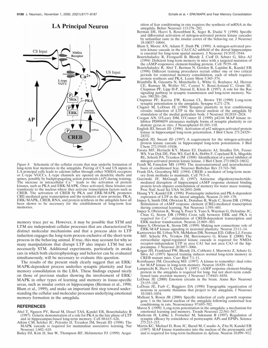
Figure 8. Schematic of the cellular events that may underlie formation oflong-term fear memories in the amygdala. Pairing of CS and US inputs inLA principal cells leads to calcium influx through either NMDA receptorsor L-type VGCCs. L-type channels are opened on dendritic shafts andspines, possibly by backpropagating action potentials (AP) during training.The increase in intracellular Ca 21 leads to the activation of proteinkinases, such as PKA and ERK/MAPK. Once activated, these kinases cantranslocate to the nucleus where they activate transcription factors such asCREB. The activation of CREB by PKA and ERK/MAPK promotesCRE-mediated gene transcription and the synthesis of new proteins. PKA,ERK/MAPK, CREB, RNA, and protein synthesis in the amygdala have allbeen shown to be necessary for the establishment of long-term fearmemories.
8186 J. Neurosci., November 1, 2000, 20(21):8177–8187 Schafe et al. • ERK/MAPK and Fear Memory Consolidation

McKernan MG, Shinnick-Gallagher P (1997) Fear conditioning induces alasting potentiation of synaptic currents in vitro. Nature 390:607–611.
Oruban PC, Chapman PF, Brambilla R (1999) Is the Ras-MAPK signal-ling pathway necessary for long-term memory formation? Trends Neu-rosci 22:38–44.
Paxinos G, Watson C (1997) The rat brain in stereotaxic coordinates:computer graphics files, Ed 3. San Diego: Academic.
Roberson ED, English JD, Adams JP, Selcher JC, Kondratick C, Sweatt JD(1999) The mitogen-activated protein kinase cascade couples PKA andPKC to cAMP response element binding protein phosphorylation in areaCA1 of hippocampus. J Neurosci 19:4337–4348.
Rogan MT, LeDoux JE (1995) LTP is accompanied by commensurateenhancement of auditory-evoked responses in a fear conditioning circuit.Neuron 15:127–136.
Rogan MT, Staubli U, LeDoux JE (1997) Fear conditioning induces as-sociative long-term potentiation in the amygdala. Nature 390:604–607.
Rosen JB, Fanselow MS, Young SL, Sitcoske M, Maren S (1998)Immediate-early gene expression in the amygdala following footshockstress and contextual fear conditioning. Brain Res 796:132–142.
Rosen LB, Ginty DD, Weber MJ, Greenberg ME (1994) Membrane de-polarization and calcium influx stimulate MEK and MAP kinase viaactivation of ras. Neuron 12:1207–1221.
Schafe GE, LeDoux JE (2000) Memory consolidation of auditory Pavlov-ian fear conditioning requires protein synthesis and PKA in the amyg-dala. J Neurosci RC96:1–5.
Schafe GE, Nadel NV, Sullivan GM, Harris A, LeDoux JE (1999) Mem-ory consolidation for contextual and auditory fear memory is dependenton protein synthesis, PKA, and MAP kinase. Learn Mem 6:97–110.
Selcher JC, Atkins CM, Trzaskos JM, Paylor R, Sweatt JD (1999) Anecessity for MAP kinase activation in mammalian spatial learning.Learn Mem 6:478–490.
Stuart G, Spruston N, Sakmann B, Hauser M (1997) Action potentialinitiation and backpropagation in neurons of the mammalian CNS.Trends Neurosci 20:125–131.
Swank MW (2000a) Phosphorylation of MAP kinase and CREB in mousecortex and amygdala during taste aversion learning. NeuroReport11:1625–1630.
Swank MW (2000b) Pharmacological antagonism of tyrosine kinases andMAP kinase in brainstem blocks taste aversion learning in mice. PhysiolBehav 69:499–503.
Weisskopf MG, Bauer EP, LeDoux JE (1999) L-type voltage-gated cal-cium channels mediate NMDA-independent associative long-term po-tentiation at thalamic input synapses to the amygdala. J Neurosci19:10512–10519.
Yin JCP, Wallach JS, Del Vecchio M, Wilder EL, Zhuo H, Quinn WG,Tully T (1994) Induction of a dominant negative CREB transgene spe-cifically blocks long-term memory in Drosophila. Cell 79:49–58.
Yin JCP, Del Vecchio M, Zhuo H, Tully T (1995) CREB as a memorymodulator: induced expression of a dCREB2 activator isoform enhanceslong-term memory in Drosophila. Cell 81:107–115.
Schafe et al. • ERK/MAPK and Fear Memory Consolidation J. Neurosci., November 1, 2000, 20(21):8177–8187 8187


