Embed Size (px)
Citation preview

ACTIN MICROFILAMENTS, CELL SHAPE, AND SECRETORY
PROCESSES IN ISOLATED RAT HEPATOCYTES
Effect of Phalloidin and Cytochalasin D
MARC PRENTKI, CHRISTINE CHAPONNIER, BERNARD JEANRENAUD, and
GIULIO GABBIANI
From the Laboratoires de Recherches M6dicales and the D6partement de Pathologie, Geneva University Medical School, Geneva, Switzerland
ABSTRACI"
The effects of phalloidin and cytochalasin D, drugs which, respectively, stabilize and destabilize actin microfilaments, have been tested on isolated rat hepatocytes. Both drugs produced a modification of cell shape, characterized by protrusions bulging from the cytoplasm. In phalloidin-treated hepatocytes, an accumulation of actin microfilamentous network was detectable at the base of each protrusion by electron microscopy, immunofluoreseence, and HMM decoration. This accu- mulation of microfilaments was absent in cytochalasin D-treated cells. The release of triglycerides, an index of very low density lipoprotein secretion, was inhibited by phalloidin or cytochalasin D, and accompanied by an increase in cellular triglycerides. At the electron microscope examination, triglyceride accumulation was represented by fat droplets and vesicle-enclosed, very low density lipoprotein- like particles. Total protein and albumin secretion was only very slightly modified by either one of these drugs. With the use of various phalloidin analogs, a correlation was observed between their respective ability to stabilize F-actin in vitro, and their effects on cell shape and triglyceride secretion. In conclusion, phaUoidin, and cytochalasin D: (a) modify the shape of isolated hepatocytes; (b) inhibit lipoprotein secretion. These effects possibly result from a modification of actin microfilament function.
KEY WORDS isolated hepatocytes phalloidin �9 cytochalasin D microfilaments antiactin antibodies cell shape secretory processes
In eucariotic cells, microtubular and microfila- mentous proteins appear to be of importance in various physiological phenomena such as mainte- nance of cell shape (9, 10, 39) and secretory processes (4, 28, 35, 40, 43). In the liver, micro- tubules have been proposed to be implicated in
the release of proteins (31, 48) and lipoproteins (30, 47, 48), possibly via a polymerization-depo- lymerization cycle of tubulin (41,42). By analogy with the microtubular system, the existence of a polymerization-depolymerization cycle of actin could represent an additional system for control- ling both shape and secretion of liver cells.
Two families of drugs appear to interfere with actin microfilaments. The first is the cytochalasins which destabilize the actin filament structure, al- though their precise mode of action is still disputed
592 J. CELL BIOLOGY �9 The Rockefeller University Press �9 0021-9525/79/06/0592/16 $1.00 Volume 81 June 1979 592-607
on January 27, 2016jcb.rupress.org
Dow
nloaded from
Published June 1, 1979

(49). While cytochalasin B inhibits transmem- brane glucose transport in various cell types (2, 33), cytochalasin D does not (2). Fur thermore, cytochalasin D is 8 - 1 0 times more potent in altering cell shape of cultured fibroblasts than the B analog (2). The second is the cyclic peptide phalloidin from the genus Amanita (53), which has been shown in vitro to react stoichiometrically with muscle actin (for a review, see reference 54), to enhance the rate of actin polymerization, and to stabilize the F-actin structure (13, 32, 34). Liver membranes (1) and hepatocytes (16) ob- tained from rats treated in vivo with phalloidin, or membranes exposed to phalloidin in vitro (23, 32), show an increased amount of actin microfila- ments. Fur thermore , microinjection of this drug into cultured fibroblasts leads to a redistribution of the immunofluorescence pattern of actin (50).
In the present study, the effects of phalloidin and cytochalasin D on the cell shape and secretory processes of isolated rat hepatocytes were investi- gated.
M A T E R I A L S A N D M E T H O D S
Animals
6-wk-old male albino rats derived from a Wistar strain bred in these laboratories were used. They weighed between 165 and 185 g, and were fed ad libitum with standard laboratory chow.
Preparation and Incubation o f Rat
Hepatocytes
Liver cells were isolated by a method described previously for mouse hepatocytes (38), with a few modifications. Livers were first perfused in situ at 37~ with a nonrecirculating calcium-free phosphate buffer (130 ml, pH 7.7) gassed with oxygen, then for 6 min with identical recirculating medium. Peffusion was con- tinued for 20-25 min with a recirculating calcium-free phosphate buffer containing collagenase (50 rag/100 ml), bovine serum albumin (2 g/100 ml), glucose (150 rag/100 ml) and soybean trypsin inhibitor (14 rag/100 mt). Livers were removed, and, after gentle stirring in a Petri dish containing a calcium-free phosphate buffer with bovine serum albumin (0.5 g/100 ml, pH 7.4), isolated cells thus obtained were further incubated for 3 min at 37~ in a shaking incubator. The cells were filtered through a nylon gauze, centrifuged, and washed three times with this buffer. They were then preincu- bated under an atmosphere of O~: CO~ (95:5 vol/vol) for 45 min in a Krebs-Ringer bicarbonate buffer (pH 7.4, 37~ containing glucose (15 raM), bovine charcoal- treated serum (25%) and bovine defatted albumin (1.5 g/100 ml) prepared as described elsewhere (30). After
preincubation, cells were centrifuged, resuspended in the same medium, and distributed as 1.5-rrd aliquots into 10-ml Eflenmeyer flasks. Drugs were added at time zero of incubation. Phalloidin was stored at 4~ as a 100-fold concentrated solution in isotonic NaC1. Cyto- chalasin D was stored at 4~ as a 200-fold concentrated solution in dimethylsutfoxide. Dimethylsulfoxide (final concentration 0.5% in incubation vials) did not interfere with any of the parameters measured. Incubations were stopped by transferring cell suspensions into conical tubes placed in ice-water that were centrifuged at 4~ (3,000 g, 5 rain). Pellets and supernates thus obtained were used for measurements of various metabolic indi- ces. All results are expressed per g of wet weight of hepatocytes - SEM, measured at time zero of incuba- tion. Although figures and tables often show results of one particular series of experiments, all experiments reported have been repeated 2-4 times, and experimen- tal vials were run in triplicate or quadruplicate,
Electron Microscopy
Isolated hepatocytes were fixed at room temperature for 30 min in 3% glutaraldehyde in cacodylate buffer (0.1 M) with 1% saccharose (55), pH 7.2-7.4. After rinsing in buffer, they were postfixed at 4~ with 2% OsO4 in Millonig's phosphate buffer at pH 7.3-7,4 (36). Sections - 1 /zm thick were cut with glass knives on an ultramicrotome (C. Reichert A. G., Vienna, Austria) and stained with methylene blue and Azur II. Thin sections were cut with diamond knives, mounted on noncoated grids, stained with uranyl acetate and lead- citrate, coated with a thin layer of carbon, and examined with a Philips EM 300 electron microscope. Heavy meromyosin (HMM) was prepared by tryptic digestion (56) from rabbit skeletal muscle myosin (46) and stored at -25~ in 25% glycerol. For the electron microscope study, isolated hepatocytes were placed in 50% glycerol standard salt solution (0.1 M KCI, 0.005 M MgCI~, and 0.006 M phosphate buffer, pH 7.0) for 12-24 h, then for 12 h in 25% glycerol and 4 h in 5% glycerol. Hepatocytes were incubated for 40 h with HMM (5-6 mg/ml in 0.015 M KCI). As control, phosphate buffer (0.01 M, pH 7.0) containing KC1 (0.015 M) was used instead of HMM (27). Hepatocytes were then fixed and processed for electron microscope examination as de- scribed above.
lmmuno fluorescence
Anti-actin antibodies (AAA) were obtained from the serum of two patients with chronic aggressive hepatitis (8, 15) having a titer of 1/1,280 and 1/640, respectively, when tested on rat intestinal smooth muscle. These sera were passed on columns of sepharose covalently linked (12) with rabbit skeletal muscle actin (8), followed by elution of the antibody at pH 2.7. The specificity of these antibodies was tested by immunodiffusion, immu- noelectrophoresis and immunofluorescence (8, 18).
PRE~'r~a E~r AL. Effect of Phalloidin and Cytochala~in D on Hepatocytes 593
on January 27, 2016jcb.rupress.org
Dow
nloaded from
Published June 1, 1979

Anti-smooth muscle myosin antibodies (AMA) were obtained and their specificity was demonstrated as pre- viously described (7, 14). Isolated hepatocytes were incubated with AAA or AMA as follows: 100 /zl of hepatocytes in suspension (8. l& cells/ml) were diluted in 10 ml of Hanks-FCS (5%) solution. One drop of the suspension was dried on a glass slide for 30 min, fixed with absolute ethanol for 30 s, rinsed three times in phosphate-buffered saline (PBS), and incubated for 15 min with AAA or AMA. After incubation, sections were washed in PBS and then stained for 15 rain with either fluorescein-conjugated IgG fraction of goat anti- serum to human IgG (Code No. 64.170, Miles Seravac, Lausanne, Switzerland) for AAA, or with fluorescein- conjugated IgG fraction of sheep antiserum to rabbit IgG (Behring Werke AG, Marburg Lahn, West Ger- many) for AMA. After rewashing in PBS and mounting in 90% glycerol in PBS, the level of fluorescence was compared with that found in control preparations treated with human ),-globulins or rabbit serum instead of AAA or AMA.
SDS Polyacrvlamide Gel Electrophoresis 24 • 10 ~ cells of control, phalloidin- or cytochalasin
D-treated (1 h) hepatocytes were collected in Hanks- BBS (Gibco, Bio AG, Basel, Switzerland) and washed twice to eliminate albumin. After centrifugation at 300 g, pellets were suspended at 4~ in Tris-HCI-NP-40 buffer containing 0.05% Nonidet (Nonidet P-40, British Drug House, Poole, England), 3 mM MgCI2, 10 mM NaCI, and 10 mM Tris-HCl, pH 7.4 (6). After sonica- tion (twice 5 s) with a Branson sonifier (Branson Sonic Power Co., Danbury, Conn.), hepatocytes were centri- fuged (20,000 g, 20 min). The resulting supernates (considered as total extract and containing 9.5 mg of proteins per ml) were centrifuged at 120,000 g for 60 min in a Beckman L5-50 ultracentrifuge (Beckman Co., Zurich, Switzerland), using a rotor type 50. Supernates obtained from ultracentrifugation contained 4.6 mg/ml protein (controls) and 4.0 mg/ml (phalloidin-treated cells); pellets contained 6.8 mg/ml protein (controls) and 9.6 mg/ml (phalloidin-treated cells). Total extracts, as well as supernates and pellets obtained after ultracentrif- ugation, were dissolved in 500 /~1 of Tris-HCl buffer boiled in sample buffer (29) and run on SDS-10% acrylamide tube gels (29), charging 50 /~g of proteins per gel. Protein content of samples was measured with the biuret technique (22). The gels were stained with Coomassie blue, then planed by means of a Chromoscan (Joyce-Loebl Ltd., Goteshead, England).
Cell Shape Study Cells were incubated in triplicate vials for I h, without
addition (controls) or in the presence of various phallo- peptides. Samples of each vial were then taken, placed on a slide under a light microscope (• 100), and pictures of each sample were taken. On each photograph, 120- 300 cells could be counted and classified as follows: (a)
control-like (i.e., round shape) cells; (b) cells with abnormal shape (i.e., occurrence of several protrusions). Results were expressed as percent of total cells counted.
Biochemical Determinations Intracellular water volume and total potassium con-
tent were determined as follows: cells were incubated for 2 h in a medium containing 3H20 (1.4 tzCi/ml) as an extracellular plus intracellular marker and [hydroxy14C methyl]-inulin (0.14/~Ci/ml) as extracellular marker, At the end of incubation, cell suspensions were centrifuged (500 g, 2 min). Supernates were removed and the dpm/ ml of ~H and 14C determined from supernates (0.5 ml), using quenching correction curves, and after substraction of the contamination of laC in the 3H channel. 2 ml of distilled water were then added to the cell pellets, After sonication, cell debris were centrifuged (23,000 g, 5 min), and supernates (0.1 ml) were used for potassium measurement using flame photometry, and counted (0.5 ml) in a liquid scintillation spectrometer (Packard, Tri- Carb model 3880, Instrument Inc., Downers Grove, II1.) so that the dpm of aH and of ~4C in the first centrifugation pellets could be known. Intracellular wa- ter volume was the difference between the SH space and the ~4C space, i.e.,
. dpm ~-I dpm 14C dpm aH/ml dpm 14C/ml.
Aliquots of supernates were used for the following measurements: 0.1 ml for urea (5), 0.2 ml for triglycer- ides (30), and 0.05 ml for lactate dehydrogenase (LDH) (5). ATP was determined with the luciferin-luciferase technique as described elsewhere (25). Labeled amino acid incorporation into cellular and released proteins was carded out according to a technique described previously (31).
Prelabeling Experiments In experiments to investigate the secretion of prela-
beled proteins and triglycerides, the following experi- mental design was adopted. Cells were preincubated (45 min) in a medium containing L-[U-~4C]amino acid mix- ture (0.21 mg/ml; 1.9 /.~Ci/ml) and albumin-J9, 10- 3H]oleate (1.2 raM; 4.5 /zCi/ml). After centrifugation, supernates were discarded and cells washed twice with 15 ml of Krebs-Ringer bicarbonate buffer (4~ This temperature was used to minimize, due to our own previous observations, the exit of prelabeled compounds during cell washing and distribution. Cells were resus- pended in a medium (4~ containing unlabeled albu- min-oleate (0.9 raM), unlabeled amino acid mixture (0.21 mg/ml), and cycloheximide (10 -4 M), a concentra- tion which totally blocks protein synthesis. Cells were then distributed and incubated at 37~ in different flasks containing either saline or drugs. At different time intervals, aliquots were taken to measure the appearance of prelabeled proteins and triglycerides in the medium.
594 THE JOURNAL OF CELL BIOLOGY �9 VOLUME 81, 1979
on January 27, 2016jcb.rupress.org
Dow
nloaded from
Published June 1, 1979

Chemicals
All organic and inorganic chemicals were purchased from E. Merck (Darmstadt, Germany), Fluka AG (Buchs, Switzerland), Sigma Chemical Co. (St. Louis, Mo.), and were of analytical grade. Labeled compounds were secured from the Radiochemical Centre (Amer- sham, Buckinghamshire, England). Amino acid mixture (TC amino acids Hela • 100) was obtained from the Difco Laboratories (Detroit, Mich.), and coilagenase (CLS IV) and soybean trypsin inhibitor from Worthing- ton Biochemical Corporation (Freehold, N. Y.). Phal- loidin, secophalloidin, phalloidin sulfoxid A and phallo- idin sulfoxid B were a generous gift of Professor Th. Wieland (Max-Pianek Institute, Heidelberg, Germany). Phalloidin was also obtained from Boehringer (Ingel- heim/Rh., Germany). Virosin was a generous gift of Dr. T. Staron (I. M. B. A., Luc6, France).
RESULTS
Effects o f Phalloidin and Cytochalasin D on
Hepatocyte Shape
As shown by Fig. 1, control hepatocytes, incu- bated with or without fatty acids, had a round appearance. At high magnification a microfila- mentous network could always be seen at the periphery of the cytoplasm and in the microvillous projections. The ultrastructure of hepatocytes in- cubated with phalloidin or cytochalasin D was similar to that of controls, with the remarkable exception of the cell contour. As shown by Fig. 2, phalloidin-treated cells were characterized by the presence of several protrusions bulging from the cytoplasm, at the base of which a clear band was observed. In cytochalasin D-treated cells (Fig. 3), numerous protrusions were also present but no clear band was seen at their base. In contrast to the lasting effect of phalloidin (from 5 min to >2 h), that of cytochalasin D on cell shape was transient, being maximal within 5 min and disap- pearing after ~25 rain in presence of the drug. When observed at higher magnification (Figs. 4 and 5), the protrusions of both phalloidin- or cytochalasin D-treated cells contained the usual cytoplasmic organelles, and few microvilli when compared to control cells. In cytochalasin D- treated cells, protrusions and the inner part of the hepatocyte were in direct continuity (Fig. 4). In contrast, at the base of each protrusion of the phalloidin-treated cells, a characteristic filamen- tous network separated the extruded from the internal cytoplasm (Fig. 5). Single microfilaments were difficult to resolve even at high magnifica- tion. When visible (Fig. 5, inset) their diameter
measured 4-7 nm. After incubation in glycerol, the shape and the cytoplasmic topography of phaUoidin-treated hepatocytes were no longer similar to that of phalloidin-treated, glutaralde- hyde-fixed hepatocytes. In particular, microfila- ment bundles could not be precisely located at the base of the protrusions. However, the intracyto- plasmic bundles of microfilaments were character- istically decorated by HMM (Fig. 6).
After immunofluorescent staining with anti-ac- tin antibodies, control hepatocytes were diffusely positive (Fig. 7b). In hepatocytes treated with phalloidin, anti-actin antibody staining was local- ized principally as bright bands at the cell periph- ery, corresponding to the base of cell protrusions (Fig. 7 c). In hepatocytes treated with cytochalasin D, the intensity and distribution of staining with A A A were analogous to that of controls, and no immunofluorescent positive bands were seen in relation to protrusions (Fig. 7d). Staining with anti-myosin was not modified in phalloidin-treated hepatocytes when compared to controls (data not shown). Control sections treated with normal hu- man y globulins (Fig. 7a) or rabbit serum were negative.
The amount of actin present in total homoge- hates, as well as in ultracentrifugation supernates and pellets of control and phaUoidin-treated hep- atocytes, was semiquantified using SDS polyacryl- amide gel electrophoresis. Planimetric evaluation of gel scannings (Fig. 8) showed that the total extract of control and phalloidin-treated cells con- tained similar amounts of a protein having the same electrophoretic mobility as actin. The pellet obtained after ultracentrifugation of extracts from phalloidin-treated cells was richer in such protein (co-migrating with actin) than pellets from control hepatocytes (Fig. 8). Concurrently, in phalloidin- treated cells the supernate obtained after ultracen- trifugation was poorer in this protein than the control supernates (data not shown). The amounts of actin-like protein present in total homogenates, in supernates or ultracentrifugation pellets of con- trol or cytochalasin D-treated cells were similar.
To control the action of microtubules on cell shape, hepatocytes were incubated with colchicine (10 -5 M) from 5 rain to 2 h. No changes in cell shape were observed at any time.
Effects o f Phalloidin and Cytochalasin D on Hepatic Secretory Processes
Despite the morphological changes, it should be noted that intracellular water volume, potassium
I~ENTgl ET AI~. Effect of Phalloidin and Cytochalasin D on Hepatocytes 595
on January 27, 2016jcb.rupress.org
Dow
nloaded from
Published June 1, 1979

lq6URE 1 Control isolated hepatocyte. The cell shape is round, and the most conspicuous cytoplasmic organelles are rough endoplasmic reticulum, mitochondria, liposomes, and glycogen granules. Microvilli are present at the cell periphery. Bar, 1 /zm. • 6,500.
and ATP content, and urea output of phalloidin- treated cells remained unmodified, while lactic acid dehydrogenase output was kept at a minimal value (Table I), Similarly, ATP content remained normal in cytochalasin D-treated cells (controls: 3.30 + 0.08; cytochalasin, 3 /xg/ml: 3.38 + 0.01 /xmol/g wet weight). This, together with the data summarized below, indicates that phalloidin- or
cytochalasin D-treated hepatocytes were function- ally well preserved.
As illustrated by Fig. 9, the triglyceride output (an index of very low density lipoprotein [VLDL} secretion by isolated hepatocytes [30]) was linear for 2 h. In the presence of phalloidin (10 -~' M) or cytocbalasin D (3 ~g/ml), the output of triglycer- ides into the medium was markedly curtailed from
596 THE JOURNAL OF CELL BIOLOGY" VOLUME 8l , 1979
on January 27, 2016jcb.rupress.org
Dow
nloaded from
Published June 1, 1979

30 min of incubation onwards. The effects of phalloidin and cytochalasin D on triglyceride se- cretion were dose-dependent. When reaching maximal inhibitory concentrations, phalloidin used at 10 -4 M and cytochalasin D at 20 /zg/ml decreased triglyceride output to the same extent as phalloidin 10 -5 M and cytochalasin D 3 /.~g/ml (data not shown). As a result of this, all subse-
quent experiments were carried out with 10 -5 M phalloidin or 3 /zg/ml cytochalasin D. Of further interest was the observation, summarized in Table II, that the decrease in triglyceride output brought about by phalloidin was accompanied by an accu- mulation of triglycerides within the hepatocytes, such that total triglycerides (i.e., cellular plus medium triglycerides) were identical in control
I~URE 2 Isolated hepatoeyte incubated with oleate (1 mM) and phaltoidin (10 -~ M, 1 h). The cell periphery shows several bubble-like protrusions. The cytoplasmic content of protrusions is similar to that of the perinuclear cytoplasm. Very few microvilli are visible at the cell periphery. Bar, 1 /xm. • 6,200.
PRENTKI ET AL. Effect o f Phalloidin and Cytochalasin D on Hepatocytes 597
on January 27, 2016jcb.rupress.org
Dow
nloaded from
Published June 1, 1979
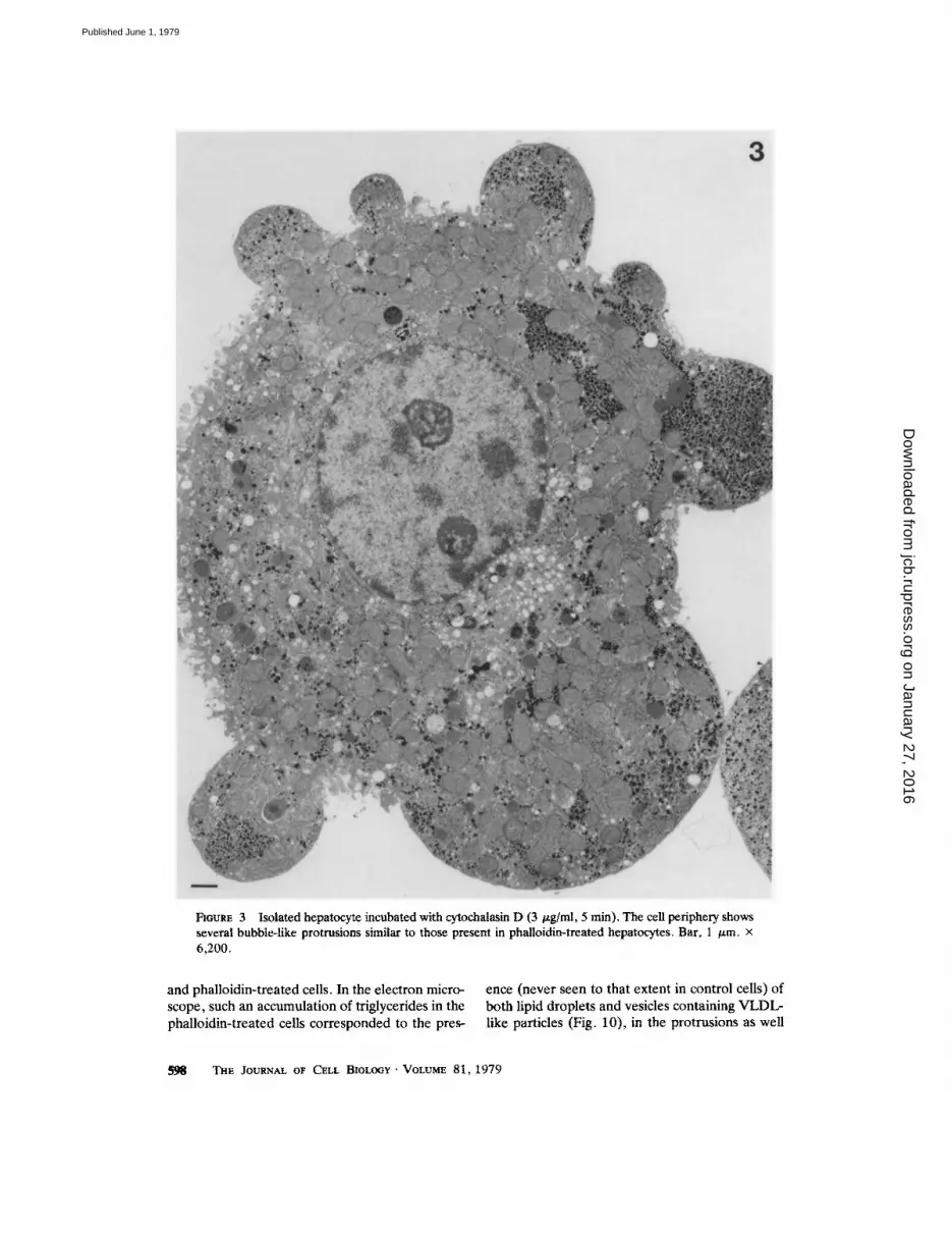
FIGURE 3 Isolated hepatocyte incubated with cytochalasin D (3 v.g/ml, 5 rain). The cell periphery shows several bubble-like protrusions similar to those present in phalloidin-treated hepatocytes. Bar, 1 /~m. x 6,200.
and phalloidin-treated cells. In the electron micro- scope, such an accumulation of triglycerides in the phalloidin-treated cells corresponded to the pres-
ence (never seen to that extent in control cells) of both lipid droplets and vesicles containing VLDL- like particles (Fig. 10), in the protrusions as well
SgB ThE JOURNAL OF CELL BIOLOGY �9 VOLUME 81, 1979
on January 27, 2016jcb.rupress.org
Dow
nloaded from
Published June 1, 1979

as in the remaining cytoplasm. Lipid droplets were conspicuously more numerous than vesicles con- taining VLDL.
Since VLDL secretion requires protein synthe- sis (3), it was important to assess the integrity of this process by measuring the incorporation of labeled amino acids into proteins. As shown in Table III, phalloidin decreased the labeled protein output but also total protein synthesis (~25%), a value in accord with previous results (24), while cytochalasin D barely changed protein secretion or synthesis. Similar data were obtained when albumin output was measured (data not shown). To decide whether the inhibitory effect of phallo- idin upon protein and triglyceride secretion was secondary to decreased protein synthesis, it was necessary to dissociate protein synthesis from pro- tein and triglyceride secretion. To do this, the release, in the presence of cycloheximide, of tri- glycerides and proteins previously labeled during a preincubation of hepatocytes with labeled pre- cursors (see Materials and Methods) was investi-
gated. As shown by Fig. 11, phalloidin reduced only slightly the release of prelabeled proteins into the medium, while that of prelabeled triglycerides was markedly curtailed.
Specificity o f Phalloidin for Microfilaments
As phalloidin has been well documented to interact with and bind to muscle actin in vitro (for a review, see reference 54), the following experi- ments were carried out to suggest similar specific- ity in the liver. It was observed (Table IV) that the monocyclic secophalloidin that does not react with F-actin (34, 54) and, therefore, does not protect it from heat denaturation, had no effect either on the shape of the hepatocyte or on triglyceride secretion. For the other phallopep- tides including phalloidin, a reasonable fit be- tween protective effect from heat denaturation of F-actin, change in cell shape, and inhibitory effect upon triglyceride secretion was observed (Table IV). It should be pointed out that the in vitro protection of F-actin from heat denaturation was
FIrORE 4 Cytoplasmic protrusion of an isolated hepatocyte incubated with cytochalasin D (3 /xg/ml, 5 min). No microfilaments are present between the inner cytoplasm and the protrusion. Note a VLDL-Iike containing vesicle (arrow). Bar, 1 ~m. x 15,700.
PREN'rKI ET AL. Effect of Phalloidin and Cytochalasin D on Hepatocytes 599
on January 27, 2016jcb.rupress.org
Dow
nloaded from
Published June 1, 1979

FIGURE 5 Cytoplasmic protrusion from an isolated hepatocyte incubated with oleate (1 mM) and phalloidin (10 -5 M, 1 h). The protrusion is separated from the inner cytoplasm by a clear band. Cytoplasmic organelles in the protrusion and in the inner cytoplasm appear normal. Note VLDL-like containing vesicles (arrows) and lipid droplets (1 d). The inset shows a detail of the band separating a protrusion from the inner cytoplasm. This is composed of a network where single microfilaments (4-7 nm in diameter) are difficult to resolve (arrows). Bar, 1 /xm. x 20,800; (inset) Bar, 0.1 /xm. x 108,000.
600
on January 27, 2016jcb.rupress.org
Dow
nloaded from
Published June 1, 1979

FIGURE 6 Decoration with HMM of actin microfilaments in phalloidin-treated (10 -5 M, 1 h) isolated rat hepatocytes. (a) Periphery of an isolated hepatocyte incubated with 0.01 M phosphate buffer (pH 7.0), containing 0.015 M KC1. (b) Periphery of an isolated hepatocyte decorated after incubation with HMM. Bar, 0.1 ~m. • 55,000.
achieved in the presence of both actin and phal- loidin at equimolar concentration (10 -5 M), a concentration of the drug used throughout the present study.
Effects of Virosin
A few experiments were carried out to charac- terize the effects of virosin, a toxin isolated from Amanita virosa (11) which also produces a marked accumulation of actin filaments in hepatocytes following in vivo administration for several days (G. Gabbiani, unpublished observations). The morphological observations and the immunofluo- rescence studies done in the presence of either virosin (16 p.g/ml) or phalloidin (8 /.~g/ml = 10 -'~ M) were identical. Various metabolic indices, such as ATP content, triglyceride secretion, labeled protein synthesis and secretion, gave results simi- lar to those recorded with phalloidin.
DISCUSSION
Morphological Changes Produced by
Phalloidin and Cytochalasin D
Several factors have been implicated in the regulation of cell shape, for instance: (a) the cytoskeletal protein tubulin (44) which, under our experimental conditions, does not seem to play a structural role in hepatocytes, since no cell shape change in the presence of colchicine was observed; (b) interaction of actin and myosin via a system analogous to that described for smooth and striated muscles (21 ); (c) microfilament formation and disappearance through polymerization and depolymerization of actin (19, 20, 26). Other factors, which have not been taken into consider- ation here, such as 10-nm filaments, are probably important as well.
In the present experiments, both phaUoidin and cytochalasin D were found to produce a dramatic
PRENTKI ET AL. Effect of Phalloidin and Cytochalasin D on Hepatocytes 601
on January 27, 2016jcb.rupress.org
Dow
nloaded from
Published June 1, 1979

previously reported, although microfilament or- ganization in phalloidin-treated cell was not inves- tigated (52). The main differences found during the present study between the effects of phaUoidin and cytochalasin D were that the effect of cytocha- lasin D upon cell shape was reversible and that few microfilaments could be observed in hepatc~ cytes treated with cytochalasin D, the protrusions and the inner cell body being in direct continuity. In contrast, the protrusions produced by phal- loidin were separated from the rest of the hepato-
FIGURE 7 Indirect immunofluoreseent staining with AAA of control, phalloidin-treated and cytochalasin D- treated isolated rat hepatocytes. On the left, immunoflu- orescent staining; on the right, interference contrast optics of the same hepatocytes. (a) Control hepatocyte stained with human ~/-globulins instead of AAA; no staining. (b) Control hepatocyte stained with AAA; the staining is diffusely cytoplasmic. (c) Hepatocyte incu- bated with phalloidin for 1 h, then stained with AAA. In addition to a diffuse cytoplasmic staining, immunoflu- orescent bands are present at the base of each protru- sion. (d) Hepatocyte incubated with cytochalasin D for 5 min, then stained with AAA. The intensity and distribution of the staining is similar to that of control. x 400.
alteration of hepatocyte shape, characterized by the appearance of numerous protrusions, while cellular organelles remained unaltered. Changes in hepatocyte shape due to phalloidin have been
9 I I I I I I
r
| O FmUSE 8 Profile of SDS polyacrylamide gels from: (a) total extract of control hepatocytes; (b) total extract of phalloidin-treated (10 -s M, 1 h) hepatocytes; (c) ultracentrifugation pellet of control hepatocytes; (d) ultracentrifugation pellet of phalloidin-treated hepato- cytes. The dotted line corresponds to the actin peaks. The surfaces of the material co-migrating with actin in a and b are similar; but the surface of the material co- migrating with actin in d is larger than that in c.
~02 THE JOURNAL OF CELL BIOLO(;Y ' VOLUME 81, 1979
on January 27, 2016jcb.rupress.org
Dow
nloaded from
Published June 1, 1979

TABLE I
Assessment of Cellular Integrity of Isolated Rat Hepatocytes in the Presence of Phalloidin
A. Intracellular water B. Intraeellular pores- D. Urea output into the E. LDH output into Addition volume 5ium content C. ATP content medium the medium
mllg wet wt meq/I izmol/g wet wt ~mollg wet wt UIg wet wt
None 0.48 - 0.03 145.8 --- 3.9 2.77 --- 0.25 14.94 --- 1.15 6.11 --- 2.46 Phalloidin 0.49 • 0.03 147.1 • 4.0 2.75 • 0.38 15.92 --- 2.34 6.03 • 1.53
Hepatocytes from fed rats were incubated for 2 h (A-D) or 1 h (E) with and without phaUoidin (10 -5 M), in 1.5 ml of Krebs-Ringer bicarbonate buffer (pH 7.4) containing bovine charcoal-treated serum (25%), bovine serum albumin (1.5 g/100 ml) and glucose (15 mM). Each figure is the mean • SEM of three different experiments carried out in triplicate. LDH = lactate dehydrogenase.
~ Cont ro l
o--.--o Cy tocha [as in D
o - - - o Pha l lo id in
2 - i o
o A 8'0 ,~o Time(rain)
FIOURE 9 Effect of phalloidin and cytochalasin D on triglyceride (TG) secretion by isolated rat hepatocytes. Experimental conditions were those described in Table I, with albumin-oleate (0.9 mM). Phailoidin: 10 -5 M; cytochalasin D: 3/~g/ml. Each point is the mean -- SEM of four values.
crease of actin recovered in the pellets obtained from cytochalasin-treated cells, compared to con- trols, was anticipated. This, however, was not the case, due either to the limited sensitivity of the gel scanning technique or to the fact that cytochalasin D may destabilize F-actin by a mechanism other than simple depolymerization. It is also possible that cytochalasin D destabilizes bundles but does not depolymerize individual filaments. Unorga- nized bundles are probably difficult to see in the electron microscope but could nevertheless sedi- ment during ultracentrifugation.
Taken together, these observations suggest that, in isolated hepatocytes, a normal equilibrium between G- and F-actin is necessary for the main- tenance of cell shape. When this equilibrium is altered by either phalloidin (stabilizing F-actin) or cytochalasin D (destabilizing F-actin), a change in hepatocyte shape results. The changes in cell shape produced by either one of these drugs suggest that actin is connected to the plasma- lemma of the hepatocyte, as previously proposed for other cell types (17, 37, 51).
cyte by an accumulation of actin microfilaments detectable by electron microscopy, immunofluo- rescence and HMM binding. Furthermore, SDS polyacrylamide gel electrophoresis revealed that the total content of actin from phalloidin-treated hepatocytes was unchanged when compared to controls, whereas polymerized actin was re- covered in greater amount in the ultracentrifuga- tion pellets of phalloidin-treated hepatocyte ex- tracts than in controls. This is in accordance with previous in vivo and in vitro data showing that phalloidin promotes actin polymerization and sta- bilizes polymerized actin (13, 23, 32, 50). In the presence of cytochalasin D, the total content of actin in hepatocytes was unchanged. Since cyto- chalasin D appears to destabilize F-actin, a de-
Effects o f Phalloidin and Cytochalasin D
upon Secretory Processes
Although the effects of phalloidin upon secre- tory processes were studied in more detail than those of cytochalasin D, the most prominent find- ing was that either one of these drugs caused a marked, rapid decrease in triglyceride (VLDL) secretion. At maximal inhibitory concentration, the effect of phalloidin was more pronounced than that of cytochalasin D. In phalloidin-treated cells, the diminution of triglyceride secretion was ac- companied by intracellular triglyceride accumula- tion, resulting in unimpairment of total triglycer- ides (cell plus medium). This accumulation of lipids was represented in the electron microscope by an accumulation (never seen to such an extent
PRENT~a ET AL. Effect of Phalloidin and Cytochalasin D on Hepatocytes 603
on January 27, 2016jcb.rupress.org
Dow
nloaded from
Published June 1, 1979

TABLE II
Effect of PhaUoidin on Triglyceride Secretion by lsolated Rat Hepatocytes
Triglycerides secreted into the me- Addition dium (A) Cellular triglycerides (B) Total triglycerides (A + B)
~mol /g Wel wt
Exp. 1 None 2.51 -- 0.13 8.05 -+ 0.42 10.52 -+ 0.29 Phalloidin 1.16 - 0.07* 10.26 -+ 0.38~ 11.41 -+ 0.34w Exp. 2 None 3.14 -+ 0.24 11.62 • 0.46 14.76 -+ 0.58 Phalloidin 1.27 -+ 0.07* 13.27 - 0.22 l[ 14.65 -+ 0.26w
Hepatocytes from fed rats were incubated with 1.0 m M oleate bound to albumin for 2 h under experimental conditions described in Table I. Phalloidin" 10 -5 M. Each figure is the mean -+ SEM of 3-4 values.
* p < 0.0025. * p < 0.01. w NS. II p < 0 .025.
FIGURE 10 Cytoplasm of an isolated hepatocyte incubated with oleate (1 mM) and phalloidin (10 -5 M, 1 h). The cytoplasm contains many VLDL-like-containing vesicles (arrows) and is otherwise normal. Bar, 1 /xm. x 29,500.
TIlE JOURNAL OF CELL BIOLOGY" VOLUME 81, 1979
on January 27, 2016jcb.rupress.org
Dow
nloaded from
Published June 1, 1979

TABLE l l I
Effect of Phalloidin and Cytochalasin D on the Incorporation of Labeled Amino Acids into Proteins by Isolated Rat Hepatocytes
Proteins secreted into the me- dium (A) Cellular Proteins (B) Total Proteins (A + B)
dpm x 10-S/g wet wt
Control 6.29 - 0.07 12.75 - 0.34 19.03 - 0.42 Phalloidin 3.69 - 0.08 10.52 + 0.60 14.20 +- 0.72 Cytochalasin D 5.89 -+ 0.06 13.73 -+ 0.26 19.59 -+ 0.30
Hepatocytes were incubated for 2 h, as described in Table I, with amino acid mixture (21.2 mg/100 ml) and randomly labelled 14C-amino acids (0.5 /xCi/ml). Phalloidin: 10 4 M. Cytochalasin D: 3 /~g/ml. Each figure is the mean -+ SEM of three values.
in control cells) of vesicles containing VLDL-like particles as well as free lipid droplets, the latter being more conspicuous than the former. Lipid droplets, and vesicles containing VLDL-like par- tides, were present in both protrusions and the remaining cytoplasm of phalloidin-treated cells. This indicates that the intracellular migration of VLDL had been, at some ill-defined step(s), interfered with by these drugs.
Cytochalasin D (Table III) or phalloidin (Fig. 11) had only a very slight inhibitory effect upon protein secretion. This is in marked contrast with previous results obtained with colchicine (an in- hibitor of the microtubular system), in which inhibition of triglyceride as well as total protein secretion was observed in the presence of the drug (30, 31, 45, 48). The observation that phalloidin or cytochalasin D inhibits triglyceride secretion but only very slightly protein secretion suggests that the intracellutar routes are, at least in part, different for the two types of secretory products, and that in the case of proteins a step or a series of steps of the intracellular migration are unaf- fected by drugs that appear to interfere with actin microfilaments.
Specificity o f Phalloidin Ac t ion
The specificity of phalloidin for actin was sug- gested by the use of phallopeptide analogs. Thus, secophalloidin which has been shown not to react with actin, and therefore does not protect F-actin from heat denaturation or from depolymerization by potassium iodide (34, 54), did not change the shape of hepatocytes or affect triglyceride secre- tion. Moreover, a satisfactory relationship was obtained between the ability of phallopeptides to react with actin in vitro and their effect on hepa-
~ - -= Cont ro l ....... Phal [o id in
3 "
Time Iminl 0 15 30 eO 120
Timr
FIGURE 11 EffeCt of phalloidin on the secretion of prelabeled proteins (left panel) and prelabeled triglycer- ides, TG, (right panel), in the presence of cyclohexi- mide, by isolated rat hepatocytes. For experimental design, see Materials and Methods. Phalloidin: 10 -5 M. Each point is the mean +- SEM of three values.
tocyte shape and triglyceride secretion. These observations do not exclude the possibility that phallopeptides interact, in addition, with struc- tures other than microfilaments. However, the fact that similar cell shape and secretory changes were observed with phalloidin or cytochalasin D treatment suggests that the effects of these drugs are mediated, at least in part, through perturba- tion of microfilaments.
In conclusion, phalloidin and cytochalasin D change the cell shape and inhibit lipoprotein secre- tion in isolated hepatocytes. Both drugs have been shown to interfere with the organization of actin microfilaments. Hence, it is conceivable that, in isolated rat hepatocytes, microfilaments play a role in the control of cell shape and lipoprotein secretion.
PRENTKI ET AL, Effect of Phalloidin and Cytochalasin D on Hepatocytes 605
on January 27, 2016jcb.rupress.org
Dow
nloaded from
Published June 1, 1979

TABLE I V
Relationships between Stabilization o f F-Act in by Various Phallopeptides and their Respective Ef fect on Cell
Shape and Triglyceride Secretion o f lsolated Ra t Hepatocytes
A. Relative turbidity of Addition F-actin solution B. Cell with protrusions C. Trigiycetide secretion
% from control % of total i~mol/g wet wt
N o n e 100 0 3 .24 -+ 0.11
Secophalloidin 96 0 2 .97 +-- 0 .14
Phalloidin Sulfoxid A 67 8.1 - 0 .7 2 .80 +- 0.17
Phalloidin Sulfoxid B 24 90.0 - 2 .0 1.54 _ 0.22
Phalloidin 12 98.3 -+ 0.5 1.34 - 0 .02
Exp. A: Protect ive effect of phal lopept ides (10 -s M) on F-actin (10 -5 M) f rom dena tu ra t ion by heat ing to 70~ for
3 man in 0.1 M KCI, 1 m M Tris , p H 7 .4 . D a t a f rom J. X . de Vries , A . J. Schiifer, H . Faulstich, and Th . Wieland,
Hoppe-Seyler 's Z . Physiol . Chem. 3 5 7 : 1 1 3 9 - 1 1 4 3 , 1976, used for compar i son with exps. B and C, with au thors ' author izat ion.
Exps. B and C: He pa toc y t e s f rom fed rats were incubated as descr ibed in Tab le I. All drugs (5 .10 -5 M) , except
phalloidin (10-s) . Exp . B: exper imenta l conditions as descr ibed in Mater ia ls and Methods . Incubat ion t ime: 1 h.
Exp. C: 1.0 m M oleate bound to a lbumin . Incubat ion t ime: 2 h. E a c h figure is the m e a n -+ S E M of three values.
W e are great ly indeb ted to Miss M. B r u n s m a n n , Miss C.
Gri l let , and Miss M. -C. Clottu for their excellent
technical assistance. W e thank Mr . J. C. Ru mb e l i and
Mr. E . D e n k i n g e r for photographic work , and Mrs. C.
McVeigh for typing this manuscr ip t . This s tudy has
great ly benef i ted f r o m the initial c o m m e n t s and advices
of D r . C. Patzel t , which are great ly acknowledged .
This work has b e e n suppor ted by grants 3 . 2 1 8 0 . 7 6 ,
3 .1540 .77 , and 3 . 6 9 2 0 . 7 6 of the Swiss Nat ional Science
Founda t ion , Be rne , Switzerland, and a grant- in-aid of
Z y m a , Nyon , Switzer land.
Received for publicat ion 16 N o v e m b e r 1977, and in
revised f o r m 15 January 1979.
R E F E R E N C E S
1. AOOSTIm, B., V. M. GOVI~DAN, and W. Hor't~o~r~. 1975. Morpho- logical changes induced by phalloidin in the rat liver. In Pathogenesis and Mechanimls of Liver Cell Necrosis. D. Keppler, editor. MTP Press Ltd., Lancaster, England. 175-192.
2. A'rl~s, S. J., and S, Lm. 1978. Dihydrocytochalasin B. Biological effects and binding to 3T3 cells. J. Cell Biol. ?6:360-370.
3. Bm~t-oN, H., A. I. Koog, O. S'r~N, and Y. STEm. 1973. Assembly and secretion of very low density lipoproteins by rat liver following inhibition of protein synthesis with cycloheximide. Biochim. Biophys. Acta. 306:106-114.
4. BAUDtrr~, H., C. STOCK, D. V:NCENT, and J. F. GaEmEa. 1975. Microfilamentous system and secretion of enzyme in the exocrine pancreas.J. Cd/Biol. 66:165-181.
5. BEaO~mVEa, H. U., EDrroa. 1974. In Methods of Enzymatic Analysis. Second English Edition. Academic Press, Inc., New York and London.
6. BRAY, D., and C. "I'nou/d. 1976. Unpolymerized actin in fibroblasts and brain. J. Mol. Biol. 105:527-544.
7. Btrmmx:m, K., and D. BItAv, 1975. Purification and structural analysis of myosin from brain and other non-musele tissues. J. MoL Biol. 99:1- 14.
8, CnmoNmEa, C., L. KO:-ILEa, and G. G.~BIA~. 1977. Fixation of human anti-actin autoantibodies on skeletal muscle fibers. Clan. Exp. Immunol. 2"]:278-284.
9. CLUE, M., and J. A. SrumCH. 1977. Nonmusele contractile pro- teins: the role of actin and myosin in cell motility and shape determi- nation. Ann. Rev. Biochem. 46:797-822.
10. COl-tEN, C. 1977. Protein assemblies and cell form. Trends Biochem. Sci. 2:51-55.
11. COUITILLOT, M., and T. ST~ON. 1970. Amanita Virosa Fr. Pr6cislon
sur I'esp~e, mise en 6vidence de sa tontine principale (virosine). Ann. Phytopathol. Soc. Jpn. 2:561-584.
12. CUAlamc~a, P., M. WtLCI~K, and C. B. At~FtNSgN. 1968. Selective enzyme purification by affinity chromatography. Proc. Natl. Aead. Sci. U. S. A. 61:636-643.
13. DANCKEa, P., I. L6W, W. HASSELaACat, and T. WmL.,~D. 1975. Interaction of actin with phalloidin: polymerization and stabilization of F-actin. Biochim. Biophys. Acta. 400:.407--414.
14. Fuawmm, K., and T. D. POLLAgO. 1976. Fluorescent antibody localization of myosin in the cytoplasm, cleavage furrow, and mitotic spindle of human cells. J. Cell BIOl. '71:848-875.
15. G ~ m m , G., G. B. RYe, P. LAMEHN, P. VA~ALLI, (J. MAJNO, C. A. BotmEt, A. CSUCltAUD, and E. F. L0SCHER. 1973. Human smooth muscle autoantibody: its identification as anti-actin antibody and a study of its binding to "non-muscular" cells. Am. J. PathoL 72: 473--488.
16. Gnmamm, G., R. Mo~rl~sAuo, B. TucnwEa~.a, M. SALAS, and L, Oltct. 1975. Phalloidin-induced hyperphisia of actin filaments in rat hepatocytes. Lab, Invest. 33:562-569.
17. GMmL~NI, G., M. DA PXADA, G. RtCHm, DS, and A. PLF'rscHEa. 1976. Actin associated with membranes of monoamine storage organ- elles. Proe. Soc. Exp. Biol. Med. 1$2:135-138.
18. G~amm, G., C. CHAI'ONmEI, A. ZUm~E, and P. VASSmSa. 1977. Actin and tubulin cocap with surface immnnogiobulins in mouse B- lymphocytes. Nature (Lond. ). 269:697-698.
19. GOLmO~N, R. D., and D. M. KmPs. 1973. Functions of cytoplasmic fibers in non-muscle motility. Cold Spring Harbor Syrup. Quant. Biol. 3-]:523-534.
20. GOLDMAN, R. D. 1975. The use of heavy meromyosin binding as an ultrastructural cytochemical method for locali~ng and determining the possible functions of actin-like microfilaments in non-muscle cells. J. Hiaochem. Cytoehem. 23:529-542.
21. GOLD~O,I~, R. D., J. A, SOtLO~, and J. M. STAaOEa. 1976. Organi- zational changes of actin-like microfilaments during animal cell move- ment. In Cell Motility, Book A: Motility, Muscle and Non-Muscle Cells. R. Goldman, T. Pollard, and J. Rosenbaum, editors. Cold Spring Harbor Laboratory, New York. 217-245.
22. GoRNALL, A. G., C. J. BAkDAVaLL, and M. M. DAWD. 1949. Determination of serum proteins by means of the biuret reaction. J. Biol. Chem. 1-]'/:751-766.
23. GoWND.~, V. M., H. FAULaTIclt, TH. WmLAND, B. Aoosnm, and W. I-lnSSELaACrl. 1972. In vitro effect of phalloidin on a plasma membrane preparation from rat liver. Naturwissensehaflen. 59:521- 522.
24. GlmWLA, E., and G. Pot./. 1977. Phalloidin poisoning of isolated hepatocytes: inhibition of protein synthesis. Experienria (Basel). 33: 603-604.
25. HEINDEL, J. J., S. W. CUSH~O~N, and B. JEANa~^uD. 1974. Cell- associated fatty acid levels and energy-requiring processes in mouse adipocytes. Am..L Physiol. 226:16-24.
26. HrrcncooK, S. E. 1977. Regulation of motility in nonmuscle cells. J. Cell Biol. 74:1-15.
27. IsmgAwA, H., R. BlSCnoFF, and H. HOLrZEa. 1969. Formation of
THE JOURNAL OF CELL BIOLOGY �9 VOLUME 81, 1979
on January 27, 2016jcb.rupress.org
Dow
nloaded from
Published June 1, 1979

arrowhead complexes with heavy meromyosin in a variety of cell types. J, Cell Biol. 43:312-328.
28. LacY, P. E., S. L. HOWELL, D. A. YOUNU, and C. J. FINK. 1968. New hypothesis of insulin secretion. Nature (Lond,). 219:.1177-1179.
29. L.~MLI, U. K, 1970. Cleavage of structural proteins during the assembly of the head of bacteriophage T 4. Nature (Lond.). 227:680- 685.
30. LE MAttCHA~D, Y., A. SmoH, F. ASSlMACOPOULO~JEANNET. L. OacL C. RotnLLrat, and B. Jr.~r,'tENAUD. 1973. A role for the microtubular system in the release of very low density lipoproteius by perfused mouse liver.J. Biol. Chem. 248.'6862-6870.
31. LE Ik, CtAIICHAND, Y+, C. PATZELT, F../L$SIMAOOPOULO~JEANNET, E. G. LOTEN, add B. JEANI~HAUD. 1974. Evidence for the role of the microtubular system in the secretion of newly syathes~zed albumin and other proteins by the liver.,/. Clio. Invest. 53:1512-1517.
32. LENC, SFrXV, A. M., I. L6w, T. W m ~ , P, DANCK~Jt, and W. HASS~t~SACtL 1974. Interaction of phalloidin with actin. Proc, Natl. Acad. Sci. U. S. A. "/1:2803-2807.
33. Lo~s , E. G., and B. JEAr~RENAffD. 1974. Effects of cytoehalasin B, colchicine and vincristine on the metabolism of isolated fat-cells. Biochem, J. 140:.185-192.
34. L6w, I,, and T. WmLAND. 1974. The interaction of phalloidin, some of its derivatives, and of other cyclic peptides with muscle actin as studied by viseosimetry. FEBS (Fed. Eur. Biochem, Soc,) Lett. 44: 340-343.
35. MaL~SSE, W. J., F. MAL~dSSE-LAoAE, M. O, W~t,~r~lt, and P. E. LACY. 1971. The stimuhm-secretion coupling of glucose-induced insulin release. V. The partidpation of a micrombular-microfilamentous sys- tem. Diabetes. 20:.257-265.
36. MaLLO~3, G. 1961~ Advantage of a phosphate buffer for OsO~ solution in fixation. J. AppL Physiol. 32:1637.
37. M o ~ r ~ , M. S. 1976. Actin filament-raembrane attachment in the microvini of intestinal epithelial cells. In Cell Motflity, Book B: Actin, Myosin and Associated Proteins. R. Goldman, T. Pollard, and J. Rosenbaum, editors. Cold Sprin 8 Harbor Laboratory, New York. 631- 650.
38, MOLLmt, P., A. Snqo~, L. Oaca, and B. J~xmt~tn~. 1976. Secre- tory proce~es, carbohydrate and lipid metaboliun in isolated mouse bepatocytes. Aspects of regulation by glucagun and insulin. Biochira. Biophys. Acta. 428:480-494.
39. OL~Sr~D, J. B., and G. G. Bo]tJSY. 1973. Microtubules. Ann. Roy. Biochem. 42:507-540.
40. OSXLUSD, R. E. 1977, Contractile proteins and pancreatic beta-cell secretion. Diabetes. 26:245-254.
41. PAI'Z~LT, C,, A. SINOH, Y. I.s MA~CrL*~D, L. Oitc:, and B. J~I~at~N- AUD. 1975. Colchicine-binding protein of the liver. J. Call Biol. 66: 609--620.
42, PATZELT, C., A. S~aH, Y, LE MAICHAND, and B. JKANTtENAUD.
1975. Characterization of hepatic microtubules: possible functional models. In Microtobules and Microtubule Inhibitors. M. Borgers and M. De Brnhander, editors. North-HoBand Publishing Company, Am- sterdam. 165-176.
43. PATZ~LT, C., D. BROWN, and B. Jr, A~tENAUD. 1977. Inhibitory effect of colchicine on amylase secretion by rat parotid glands. J. Cell Biol. 73:578-593.
44. Pou'mt, K. R. 1976. Introduction: motility in cells. In Cell Motility, Book A: Motility, Muscle and Non-Muscle Cells. R. Goldman, T. Pollard, and J. Rosenbaum, editors~ Cold Spring Harbor Laboratory, New York. 1-28.
45. REDMAN, C, M., D. BANEIUEE, K. HOWELL, and G. E. PALADE. 1975. Colehicine inhibition of p l ~ a protein release from rat hepato- eytes.J. Cell Biol. 66:42-59.
46. RICHAtos, E. G., C. -S. (2~NG, D. B. M~Nzet, and H. S, OLcorr. 1967. C~romatograpby of Myosin on Diethylaminoethyl-Sephadex A- 50. Biochemistry 6:528-M0.
47. S:NO~, A,, Y. L~ M.t, ICHa~D, L, Oitcl, and B. JEANItENAUO. 1975. Colchicine administration to mice: a metabolic and ulirastructurul study. Eur. J. Clio. Invest, $:495-505.
48. STmrL O , L. SAt~omL and Y. STF~N. 1974. Colchicine-induced inhibition of lipoprotein and protein secretion into the serum and lack of hiterferenee with secretion of bilin~ phospholipids and cholesterol by rat liver in vivo. J. Cell BIOL 62:90-103.
49, TANNENaAUM, J., S. W. Ts and G. C. GoDMAr~. 1977. The binding sites of cytochalasin D. Evidence that they may be peripheral membrane proteins. J, Cell, Physiol. 91:225-238.
50. W~II.~ND, J., M. OsnoL'~, and K~ W ~ . 1977, Phalloidin-induced actin polymerization in the cytopla.~a of cultured cells interferes with cell locomotion and growth. Proc. Natl. Acad. Sci. U. S. A. 74:5613- 5617,
51. WmmNo, R. R. 1976. Membrane association and polymerization of actin. In Cell Motility, Book B: Actio, Myosin and Associated Proteins. R, Goldman, T. Pollard, and J. Rosenl~mm, editors. Cold Spring Harbor Laboratory, New York. 671-684.
52. Wmss, E., I. Srmtz, M. Flatcar , and R. Kto]~.t. 1973. Electron micre~copy of isolated rat hepatoeytes before and after treatment with phalloidin. Beitr. Path. Bd. 150:345-356,
53. Wrst.Ja~D, T. 1968. Poisonous principles of mushrooms of the genus amanita. Science (Wash. D, C.). 159:.946-952.
54. WmLO~. T. 1977. Interaction of phallotoxim with actin. Adv. Enzyme Regul. 15:285-300.
55. WtSSE, E. 1970. An electron microscopic study of the fenem'ated endothelial lining of rat liver shiusoids. J. Ullrastruct. Res. 31:125-150.
56. WooDmUM, D. T., S. A. l~ctt, and T. D. POLL, tiP. 1975. Evidence for biased bidirectional polymerization of actin filaments using heavy meromyosin prepared by an improved method. J. Cell Biol. 67:231- 237.
PRENTKI ET AL. E f f e c t o f P ha l l o id in a n d Cy tocha las in D o n H e p a t o c y t e s 607
on January 27, 2016jcb.rupress.org
Dow
nloaded from
Published June 1, 1979


