Embed Size (px)
Citation preview

Virology 443 (2013) 363–374
Contents lists available at SciVerse ScienceDirect
Virology
0042-68http://d
n CorrE-m
journal homepage: www.elsevier.com/locate/yviro
The P6 protein of Cauliflower mosaic virus interacts with CHUP1, a plantprotein which moves chloroplasts on actin microfilaments
Carlos A. Angel a, Lindy Lutz c, Xiaohua Yang b, Andres Rodriguez a, Adam Adair a,Yu Zhang a, Scott M. Leisner c, Richard S. Nelson b, James E. Schoelz a,n
a Division of Plant Sciences, University of Missouri, Columbia, MO 65211, USAb The Division of Plant Biology, The Samuel Roberts Noble Foundation, Ardmore OK 73401, USAc Department of Biological Sciences, University of Toledo, Toledo OH 43606, USA
a r t i c l e i n f o
Article history:Received 11 February 2013Returned to author for revisions11 March 2013Accepted 18 May 2013Available online 12 June 2013
Keywords:Intracellular movementSubcellular localizationInclusion bodiesCaMV
22/$ - see front matter & 2013 Elsevier Inc. Ax.doi.org/10.1016/j.virol.2013.05.028
esponding author. Fax: +1 573 882 0588.ail address: [email protected] (J.E. Schoe
a b s t r a c t
The gene VI product, protein 6 (P6), of Cauliflower mosaic virus (CaMV) assembles into large, amorphousinclusion bodies (IBs) that are considered sites for viral protein synthesis and viral genome replicationand encapsidation. P6 IBs align with microfilaments and require them for intracellular trafficking, a resultimplying that P6 IBs function to move virus complexes or virions within the cell to support virusphysiology. Through a yeast two-hybrid screen we determined that CHUP1, a plant proteinallowing chloroplast transport through an interaction with chloroplast and microfilament, interactswith P6. The interaction between CHUP1 and P6 was confirmed through colocalization in vivo andco-immunoprecipitation assays. A truncated CHUP1 fused with enhanced cyan fluorescent protein,unable to transport chloroplasts, inhibited intracellular movement of P6–Venus inclusions. Silencing ofCHUP1 in N. edwardsonii impaired the ability of CaMV to infect plants. The findings suggest that CHUP1supports CaMV infection through an interaction with P6.
& 2013 Elsevier Inc. All rights reserved.
Introduction
Plant viruses have at least three distinct activities necessary tocomplete their disease cycle in a host: those required to replicate,encapsidate, and move the virus throughout the plant. Some plantviruses encode additional proteins with functions dedicated to vectortransmission or defeating plant defenses, but it is generally acceptedthat the genomic capacity of plant viruses is small, with an upperlimit of perhaps 15–20 proteins. To overcome their limited codingcapacity, it is likely that each viral protein has multiple functions andcontacts with host factors. Collectively these functions and interac-tions determine the outcome of the infection.
The P6 protein of Cauliflower mosaic virus (CaMV) is an exampleof a plant virus protein with multiple functions and interactions. P6functions to support the synthesis of viral proteins from the 35S viralRNA, suppresses gene silencing, and inhibits SA-mediated plantdefenses (Hohn and Fütterer, 1997; Love et al., 2007, 2012; Parket al., 2001). In addition, P6 functions as an avirulence determinantin some solanaceous and cruciferous species, and is a chlorosissymptom determinant in susceptible hosts (Baughman et al., 1988;Daubert et al., 1984; Hapiak et al., 2008; Schoelz et al., 1986). P6 is
ll rights reserved.
lz).
the most abundant protein component of the amorphous, electrondense inclusion bodies (IBs) present during virus infection (Odelland Howell, 1980; Shockey et al., 1980). P6-containing IBs inducedduring virus infection are likely “virion factories”, as they are theprimary site for CaMV protein synthesis, genome replication, andassembly of virions (Hohn and Fütterer, 1997). P6 physically interactswith the CaMV capsid (P4) and movement (P1) proteins, as well asthe two viral proteins necessary for insect transmission (P2 and P3)(Hapiak et al., 2008; Himmelbach et al., 1996; Lutz et al., 2012;Ryabova et al., 2002).
Recently, a new function for P6 was suggested when it wasshown that P6 IBs induced by ectopic expression of P6 associatewith actin microfilaments, microtubules and the endoplasmicreticulum, and were capable of intracellular movement alongmicrofilaments (Harries et al., 2009a). Furthermore, latrunculinB, a pharmacological agent that disrupts microfilaments by pre-venting polymerization of actin monomers, abolished CaMV locallesion formation in N. edwardsonii, an indication that intactmicrofilaments are essential for CaMV infections (Harries et al.,2009a). Collectively, these experiments suggested that P6 IBsmight be responsible for intracellular trafficking of virions toplasmodesmata, in addition to the role of the P6 protein intranslation of the 35S RNA and gene silencing suppression.
Prior to the report by Harries et al. (2009a), the P6 protein ofCaMV had not been considered to have a role in CaMV movement,

4D1D D3 D2
Host Range, Symptoms & P6 Self Association Mini TAV
Zinc Finger
P6 Self Association
Chloroplast Interaction
Leucine zipper
Proline-rich, interacts with Profilin/Actin Leucine zipper
Coiled-coil Actin binding
CaMV P6 binding 1004 aa
CHUP1 structure
P6 structure:
C.A. Angel et al. / Virology 443 (2013) 363–374364
but there was evidence suggesting its function in this activity.CaMV virions move from cell to cell through plasmodesmatamodified into tubules through the function of its movementprotein, P1 (Kasteel et al., 1996; Perbal et al., 1993). However, itis unlikely that the CaMV P1 protein transports the virions to theplasmodesmata since P1 does not appear to directly interact withthe virion. The CaMV P3 protein does interact with virions throughthe formation of a tetrameric structure anchored into the virions(Leclerc et al., 1998, 2001). Electron microscopy studies haveindicated that P1 and P3 colocalize with virions only within theplasmodesmata, and it has been suggested that the P3/virioncomplex travels to the plasmodesmata independently from P1(Stavolone et al., 2005). Consequently, there is a need for a secondCaMV protein to fulfill the role of intracellular transport. Since P6-containing IBs are the site for virion accumulation and they arecapable of movement, they may be responsible for deliveringvirions to CaMV P1 located at the plasmodesmata (Harries et al.,2009a; Schoelz et al., 2011). At the very least, there must be amechanism that would account for the transfer of CaMV virionsfrom the P6 IBs to the plasmodesmata.
In this study we utilized a yeast two-hybrid assay to identifyhost proteins that interact with CaMV P6. We show here that P6physically interacts with CHUP1 (Chloroplast Unusual Positioning1), a protein which is encoded by a single gene in Arabidopsis andis localized to the outer membrane of chloroplasts (Oikawa et al.,2003, 2008). A CHUP1-ECFP (Enhanced Cyan Fluorescent Protein)fusion protein was observed to relocate a P6-Venus fusion proteinto chloroplasts in vivo. An interaction between CHUP1 and P6 alsowas demonstrated through co-immunoprecipitation studies fromplant extracts. Overexpression of a truncated CHUP1 in a transientexpression assay in N. benthamiana blocked the movement of P6IBs. This observation was correlated with a delayed rate of CaMVlocal lesion development when CHUP1 was silenced in N. edward-sonii. Our data provide an explanation for the subcellular localiza-tion of P6 IBs to, and movement along, microfilaments. In additionour data provide further evidence that a complex composed of P6and virions may contribute to intracellular movement of CaMVparticles necessary to infect the host.
436 aa
ECFP, GFP
436 aa CHUP1 Start
EYFP
GFP, RFP or Venus
CaMV P6
520 aa
RNA Binding
P6 fusions
CHUP1 fusions
eIF3g-Venus Venus
324 aa
eIF3g
GFP
CHUP1 FL
1004 aa
154 aa
GFP
Fig. 1. CHUP1, P6 and eIF3g constructs used for confocal microscopy orco-immunoprecipitation. (A) Structure of CHUP1 and CaMV P6 proteins. Thefunctions of P6 domains D1–D4 tested for self interaction (Li and Leisner, 2002)and interaction with a portion of CHUP1. The mini TAV is the minimal region for thetranslational transactivation function. The functions of CHUP1 domains are shownbelow. (B) Structure of P6, CHUP1, eIF3g fusions developed for confocal microscopy.
Results
A yeast two-hybrid screen reveals that the CaMV P6 protein interactswith CHUP1
Previous studies showed that CaMV P6 interacts with theribosomal proteins L13, L18, and L24, along with eukaryotictranslation factor 3 subunit g (eIF3g) (Bureau et al., 2004; Lehet al., 2000; Park et al., 2001), consistent with the role of P6 in re-initiation of translation of the polycistronic 35S mRNA. P6 alsointeracts with the RNA silencing protein DRB4 (Haas et al., 2008),which is consistent with the role of P6 as a silencing suppressor.To identify additional proteins that interact with CaMV P6, a yeasttwo-hybrid screen of an A. thaliana cDNA library composed oftranscripts representing one-week old seedlings was performed byHybrigenics Services (Paris, France). The bait consisted of the full-length sequence of P6 of CaMV strain W260 (Wintermantel et al.,1993). Of the 85 Arabidopsis clones found in this screen, 17 wereidentified as eIF3g. None of the other proteins previously shown tointeract with P6 (e.g. L13, L18, L24 or DRB4) appeared in this Y2Hscreen. Nonetheless, the result with eIF3g demonstrates thecapacity of our screen to identify host proteins previously shownto interact with P6 through two-hybrid screens (Park et al., 2001).
One of the additional clones selected in the Hybrigenics screenwas identified as CHUP1 (At3G25690), a unique Arabidopsis geneencoding a protein that localizes to the outer membrane of
chloroplasts (Oikawa et al., 2003). Previously it was shown thatCHUP1 has four important functional domains (Fig. 1A). TheN-terminus contains a hydrophobic domain that targets CHUP1to the chloroplast outer envelope. A second domain is a coiled-coilmotif that interacts with the plasma membrane, permittingCHUP1 to serve as a bridge to anchor chloroplasts to plasmamembranes along the cell wall, and also is important for homo-dimerization of the protein. A third domain binds to F-actin bothin vitro and in vivo, and a fourth proline-rich domain interacts withprofilin and actin. In addition, there are two embedded leucine-zipper motifs, one in the coiled-coil region and the other down-stream from the proline-rich region, each of which may beimportant for intramolecular interactions. (Oikawa et al., 2003,2008; Lehmann et al., 2011; Schmidt von Braun and Schleiff,2008). In the Hybrigenics yeast two hybrid assay, CaMV P6 wasshown to interact with the coiled-coil region of CHUP1 (Fig. 1A).
To identify the specific region(s) of P6 that interact with CHUP1,a second yeast two-hybrid assay was performed. Four domains ofP6, previously investigated for their role in self-association(D1–D4; Fig. 1A) (Li and Leisner, 2002), were used as bait forCHUP1. Yeast cells co-transformed with the full length P6 fused tothe LexA DNA-binding domain in the pEG202 plasmid and aportion of the coiled-coil domain of CHUP1 fused to the B42

D1
D4D3
D2
6 7
1 2 3 4
5
P6P6
CHUP1
CHUP1
CHUP1CHUP1
CHUP1
CHUP1
Fig. 2. Interaction of CaMV P6 and CHUP1 proteins in yeast two-hybrid screens.(A) Full-length CaMV P6 and P6 domains were screened for interaction with the363 nt sequence of CHUP1 protein originally identified in the Hybrigenics screen.The black boxes indicate the LexA DNA-binding domain of the yeast vector pEG202and the hatched boxes indicate B24 activation domain present in the yeast plasmidpJG4. (B) β-galactosidase activity of yeast transformants expressing the constructsillustrated in panel A. (C) Growth of yeast transformants on media with (left) andwithout (right) leucine.
C.A. Angel et al. / Virology 443 (2013) 363–374 365
activation domain in the pJG4-5 plasmid showed leucine-independentgrowth and β-galactosidase activity (Fig. 2). Similarly, the four P6domains necessary for self-association were each independently fusedto the LexA DNA-binding domain and co-transformed with the B42–CHUP1 fusion. Yeast co-transformed with either domains D2 or D4and CHUP1 grew on leucine-deficient media and the highest level ofβ-galactosidase activity, indicating a strong interaction, was observedwith the D2–CHUP1 combination (Fig. 2). Interestingly, no interactionwas detected between domains D3 or D1 with CHUP1. Since domainD3 was shown previously to interact with eIF3g and L24 (Park et al.,2001), our results show that host proteins interact with differentdomains within P6.
Validation of co-localization studies with CaMV P6 and eIF3g
Although P6 has been shown to associate with plant proteinsand with itself, these interactions have not been visualizedthrough co-localization studies in live cells. As a prerequisite forevaluating co-localization of CHUP1 with P6 in vivo, we firstsought to establish, as a standard, the co-localization of P6 with
itself and with eIF3g. By fusing the green fluorescent protein (GFP)to the C-terminus of P6, it was possible to image amorphous IBs inlive, infected cells (Harries et al., 2009a). To visualize P6-P6associations in vivo, we co-agroinfiltrated P6–GFP with P6–RFP(Campbell et al., 2004; Heim and Tsien, 1996). Of the 38 P6–GFPIBs and 40 P6–RFP IBs that we counted, we found that GFP and RFPsignals co-localized in 79% of the IBs (Fig. 3A–C). To visualize thelocalization of eIF3g in cells individually and in the presence of P6,a full-length eIF3g cDNA from A. thaliana was fused to a sequenceencoding the yellow fluorescent protein Venus (Venus) (Nagaiet al., 2002). N. benthamiana leaf sections agroinfiltrated with onlyeIF3g–Venus contained numerous small, aggregates distributedwithin the cytoplasm (Fig. 3D and E). Upon co-agroinfiltration ofeIF3g–Venus with P6–RFP, the eIF3g–Venus signal was relocated toand concentrated within the P6–RFP inclusion bodies (Fig. 3F–H).This result and those from earlier studies showing that ribosomesappeared to aggregate around IBs in CaMV-infected ChineseCabbage leaves (Conti et al., 1972) together support the conclusionthat translation occurs in the vicinity of the IBs.
CHUP1 and CaMV P6 co-localize in vivo
To examine the influence of CHUP1 on the subcellular localizationof P6, we generated a cDNA clone consisting of the first 436 codons forA. thaliana CHUP1 fused to the 5′ end of ECFP (enhanced cyanfluorescent protein) coding sequence (Fig. 1B) (Heim and Tsien, 1996).This CHUP1 fusion, CHUP1436–ECFP, was similar in composition to aclone (CHUP11–500–GFP) previously shown to interact with actin, to betargeted to the chloroplast outer envelope, and to associate with theplasmamembrane (Oikawa et al., 2003, 2008). Upon agroinfiltration ofCHUP1436–ECFP into leaf sections of N. benthamiana, the ECFP signalwas observed in a band that surrounded chloroplasts (Fig. 4A–C), inagreement with findings from previous studies (Oikawa et al., 2003,2008). In addition, the ectopic expression of CHUP1436–ECFP causedchloroplasts to aggregate (Fig. 4B). To visualize P6 in live cells in thisstudy, we fused Venus to the C-terminus of the CaMV strain W260 P6coding sequence and the construct was agroinfiltrated intoN. benthamiana leaf panels. In agreement with a previous study(Harries et al., 2009a), CaMV P6–Venus IBs in the absence of additionalectopically expressed CHUP1, were well-defined, but they were notspecifically associated with chloroplasts (Fig. 4D–F).
Co-agroinfiltration of CHUP1436–ECFP with P6–Venus revealedthat these proteins consistently co-localized in cells. Uponco-agroinfiltration, the CHUP1436–ECFP fusion was associated withand surrounded chloroplasts, as observed previously whenexpressed in the absence of P6–Venus (compare Fig. 4A with G).Two patterns were associated with the P6–Venus signal duringco-agroinfiltration. In one pattern, signal representing P6–Venuswas observed surrounding chloroplasts, reminiscent of theCHUP1436–ECFP pattern (Fig. 4G–J), in addition to being morediffuse and distributed over a larger area than when infiltrated byitself. In the second pattern, the P6–Venus formed compact IBsembedded in a CHUP1 matrix (Suppl. Fig. 1). To examine whetherthe co-localization of CHUP1 with P6 was dependent on thecoiled-coil domain of CHUP1, as indicated in the yeast two-hybrid screen, we eliminated the portion of the coiled-coil domainthat contained the P6 binding region (Suppl. Fig. 4) to createCHUP154–GFP. Agroinfiltation of CHUP1154–GFP into N. benthami-ana leaf tissue revealed that it retained the capacity for localizationto the chloroplast outer membrane (Suppl. Fig. 2A–C). Furthermore,expression of CHUP154–GFP caused the chloroplasts to aggregate,similar to the CHUP1436–ECFP construct. Co-agroinfiltration ofCHUP1154–GFP with P6–RFP showed that the capacity for co-localization of the two proteins was abolished (Suppl. Fig. 2D–F).This experiment confirmed that the coiled-coil domain of CHUP1was necessary for its association with P6.

Fig. 3. Colocalization of CaMV P6–GFP, P6–RFP, and eIF3g–Venus in N. benthamiana leaves by confocal microscopy. (A) Expression at 4 dpinf of P6–GFP. (B) Expression at 4 dpiof P6-RFP in the same cells as A. (C) Overlay of panels A and B. (D) Expression of eIF3g–Venus alone, at 3 dpi. (E) Bright field overlay of D. (F) Expression of eIF3g–Venus at4 dpinf. (G) Expression of P6–RFP in the same cells as F. (H) Overlay of panels E and F with brightfield image. Magnification bar is 10 mm.
C.A. Angel et al. / Virology 443 (2013) 363–374366
To examine whether the full length CHUP1 would co-localizewith CaMV P6 in vivo, a full length CHUP1 gene with GFP fused tothe 3′ end (CHUP1FL–GFP) (Fig. 1) was obtained from Dr. Sam-GeunKong (Kyushu University). Upon co-bombardment of N. benthami-ana leaves with CHUPFL–GFP and P6–RFP, the CHUP1FL–GFP wasassociated with chloroplasts (Fig. 5A and C), as reported by Oikawaet al. (2003, 2008). In this image, a compact P6-RFP IB co-localizedwith the CHUP1FL–GFP protein and was adjacent to a chloroplast,and P6-RFP signal was also present in a more diffuse pattern thatoverlapped with the CHUP1FL–GFP signal surrounding each of thechloroplasts (Fig. 5B and D). Therefore, CHUP1FL–GFP co-localizedwith P6–RFP, similar to the results obtained with CHUP1436–ECFP.
P6–RFP Is co-immunoprecipitated with P6-GFP and CHUP1436-GFP
To develop a standard for co-immunoprecipitation of proteins thatinteract with P6, we co-agroinfiltrated P6–RFP with P6–GFP, as theself-aggregating properties of this protein have been well established,both in a yeast two-hybrid assay (Li and Leisner, 2002) and in vivo(Fig. 3A–C). Both P6–GFP and P6–RFP were readily detected by westernblot in agroinfiltrated tissues when expressed individually (Fig. 6A andB, lanes 3 and 4) or when co-expressed (Fig. 6A and B, lane 6).Furthermore, antibodies to RFP and GFP did not cross react with eachother at levels that would influence IP results (Fig. 6A and B). In aprevious study we found that P6 is partially processed into smallerprotein products (Yu et al., 2003). The processing of P6–GFP andP6–RFP into smaller protein products was largely blocked through theaddition of protease and phosphatase inhibitors, including PMSF,
Na3VO4 and NaF, although even in the presence of these inhibitors asmaller band was visible in the western blot for P6–RFP (Fig. 6B).
To investigate the interaction of P6 with itself and with CHUP1during immunoprecipitation analyses, plant tissue extracts co-agroinfiltrated with either P6–GFP/P6–RFP or CHUP1436–GFP/P6–RFPwere incubated with GFP antibodies immobilized onto sepharosebeads. Following extensive washes, the bound proteins were elutedfrom the beads, separated by gel electrophoresis, blotted onto anitrocellulose membrane, and probed with antibodies to RFP. Thefull-length P6–RFP protein and a smaller processed product weredetected upon co-immunoprecipitation with P6–GFP (Fig. 6C, lane 6)or CHUP1436-GFP (Fig. 6C, lane 5), but were not detected when P6–GFPwas omitted from the co-immunoprecipitation assay (Fig. 6C, lane 4).As an additional negative control for the co-immunoprecipitation ofP6, plant tissue extracts containing P6–GFP and P6–RFP were incu-bated with FLAG-M2 antibodies immobilized onto sepharose beads.In this assay, P6–RFP was not co-immunoprecipitated, as revealed in awestern blot with RFP antibodies (data not shown), confirming thatdetection of P6–RFP depends on the immunoprecipitation of CHUP436–GFP or P6–GFP. We concluded that the co-immunoprecipitation assaydetected a P6–CHUP1436 interaction with equivalent sensitivity as aP6–P6 interaction.
P6 and chloroplast targeting signals in CHUP1 do not overlap
A previous study had shown that fusion of GFP to theN-terminus of CHUP1 blocked its interaction with chloroplasts(Oikawa et al., 2008). The presence of GFP was proposed to mask
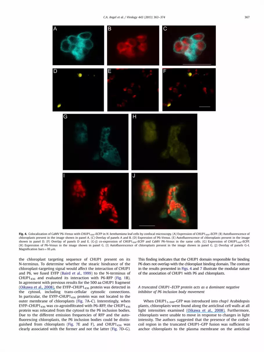
Fig. 4. Colocalization of CaMV P6–Venus with CHUP1436–ECFP in N. benthamiana leaf cells by confocal microscopy. (A) Expression of CHUP1436–ECFP. (B) Autofluorescence ofchloroplasts present in the image shown in panel A. (C) Overlay of panels A and B. (D) Expression of P6–Venus. (E) Autofluorescence of chloroplasts present in the imageshown in panel D. (F) Overlay of panels D and E. (G–J) co-expression of CHUP1436–ECFP and CaMV P6–Venus in the same cells. (G) Expression of CHUP1436–ECFP.(H) Expression of P6–Venus in the image shown in panel G. (I) Autofluorescence of chloroplasts present in the image shown in panel G. (J) Overlay of panels G–I.Magnification bars¼10 mm.
C.A. Angel et al. / Virology 443 (2013) 363–374 367
the chloroplast targeting sequence of CHUP1 present on itsN-terminus. To determine whether the stearic hindrance of thechloroplast-targeting signal would affect the interaction of CHUP1and P6, we fused EYFP (Baird et al., 1999) to the N-terminus ofCHUP1436 and evaluated its interaction with P6-RFP (Fig. 1B).In agreement with previous results for the 500 aa CHUP1 fragment(Oikawa et al., 2008), the EYFP–CHUP1436 protein was detected inthe cytosol, including trans-cellular cytosolic connections.In particular, the EYFP–CHUP1436 protein was not located to theouter membrane of chloroplasts (Fig. 7A–C). Interestingly, whenEYFP–CHUP1436 was co-agroinfiltrated with P6–RFP, the CHUP1436protein was relocated from the cytosol to the P6 inclusion bodies.Due to the different emission frequencies of RFP and the auto-fluorescing chloroplasts, the P6 inclusion bodies could be distin-guished from chloroplasts (Fig. 7E and F), and CHUP1436 wasclearly associated with the former and not the latter (Fig. 7D–G).
This finding indicates that the CHUP1 domain responsible for bindingP6 does not overlap with the chloroplast binding domain. The contrastin the results presented in Figs. 4 and 7 illustrate the modular natureof the association of CHUP1 with P6 and chloroplasts.
A truncated CHUP1–ECFP protein acts as a dominant negativeinhibitor of P6 inclusion body movement
When CHUP11–500–GFP was introduced into chup1 Arabidopsisplants, chloroplasts were found along the anticlinal cell walls at alllight intensities examined (Oikawa et al., 2008). Furthermore,chloroplasts were unable to move in response to changes in lightintensity. The authors suggested that the presence of the coiled-coil region in the truncated CHUP1–GFP fusion was sufficient toanchor chloroplasts to the plasma membrane on the anticlinal

Fig. 5. Colocalization of CaMV P6–RFP with CHUP1FL–GFP after particle bombardment of N. benthamiana leaves. (A) Expression of CHUP1FL–GFP. (B) Expression of P6–RFP.(C) Autoflourescence of chloroplasts. (D) Overlay of panels A–C. Confocal images were taken three days post particle bombardment. Magnification bar in panel (D) is 10 mm.
Sample
CHUP1-GFP
P6-GFP
P6-RFP
-
-
-
+
-
-
-
+
-
-
-
+
+
-
+
-
+
+
72kDa
95kDa
72kDa
95kDa
Total Blot: GFP
Total Blot: RFP
72kDa
95kDa
Co-IP: GFP Blot: RFP
2 3 4 5 61
Fig. 6. Co-immunoprecipitation of CaMV P6–RFP by CaMV P6–GFP, and CHUP1436–GFPafter agroinfiltration of N. benthamiana leaves. Lane 1, mock leaf control; Lane 2,Expression of CHUP1436–GFP alone; Lane 3, Expression of P6–GFP only; Lane 4,Expression of P6–RFP only; Lane 5, Co-expression of CHUP1436–GFP and P6–RFP;Lane 6, Co-expression of P6–GFP and P6–RFP. (A) Western blot for total proteinsinput probed against GFP antibodies. (B) Western blot for total protein input probedagainst RFP antibodies. (C) Western blot for immunoprecipitation using GFPantibodies and probed against RFP antibodies. All samples taken were taken at2–3 dpinf, and co-agroinfiltrated with a construct expressing the TBSV P19 silen-cing suppressor.
C.A. Angel et al. / Virology 443 (2013) 363–374368
side, but the deletion of the C-terminus abolished chloroplastmovement in response to light (Oikawa et al., 2008).
Since our CHUP1436–ECFP construct did not contain the domainfor chloroplast realignment, but did localize to chloroplasts simi-larly to the CHUP11–500–GFP construct, we investigated whether itstransient overexpression from the 35S promoter might block themovement of P6 IBs due to the absence of the movement domain.Three days after co-agroinfiltration of leaves with P6–Venus andCHUP1436–ECFP, P6 IBs were essentially immobilized within cellsthrough our observation period of 295 s (Suppl. Movie 1).By contrast, P6 IBs resulting from agroinfiltration of P6–Venusalone exhibited rapid movement over a distance of 10 mm inN. benthamiana cells (Suppl. Movie 2), similar to the results ofHarries et al. (2009a). Furthermore, P6–Venus IBs also exhibitedcomparable movement when co-agroinfiltrated with a binaryvector that expressed free ECFP (Suppl. Movie 3). Thus, our workindicated that not only does CHUP1 interact with CaMV P6, but atruncated CHUP1 deficient in chloroplast movement can block thetransport of P6 IB-like complexes.
Supplementary material related to this article can be foundonline at http://dx.doi.org/10.1016/j.virol.2013.05.028.
Silencing CHUP1 slows the rate of CaMV local lesion formation inN. edwardsonii
To investigate whether CHUP1 has a role in CaMV infections, wesought to silence CHUP1 through a virus-induced gene silencing(VIGS) assay in N. edwardsonii. As a prelude to investigating theCHUP1 influence on CaMV infections, we first cloned and sequencedthe entire CHUP1 gene from N. benthamiana and a portion of the genefrom N. edwardsonii, both hosts of CaMV. The N. benthamiana CHUP1gene (NbCHUP1) is 4914 bp in length and encodes nine exons (Suppl.Fig. 3). The encoded amino acid sequences of the NbCHUP1 and
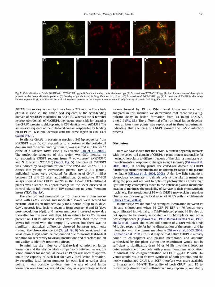
Fig. 7. Colocalization of CaMV P6-RFP with EYFP-CHUP1436 in N. benthamiana by confocal microscopy. (A) Expression of EYFP-CHUP1436. (B) Autofluorescence of chloroplastspresent in the image shown in panel A. (C) Overlay of panels A and B. Magnification bar, 10 mm. (D) Expression of EYFP–CHUP1436. (E) Expression of P6–RFP in the imageshown in panel D. (F) Autofluorescence of chloroplasts present in the image shown in panel D. (G) Overlay of panels D–F. Magnification bar is 10 mm.
C.A. Angel et al. / Virology 443 (2013) 363–374 369
AtCHUP1 exons vary in identity from a low of 22% in exon II to a highof 93% in exon VI. The amino acid sequence of the actin-bindingdomain of NbCHUP1 is identical to AtCHUP1, whereas the N-terminalhydrophobic domain of NbCHUP1, the region responsible for targetingthe CHUP1 protein to chloroplasts, is 72% identical with AtCHUP1. Theamino acid sequence of the coiled-coil domain responsible for bindingAtCHUP1 to P6 is 70% identical with the same region in NbCHUP1(Suppl. Fig. 4).
To silence CHUP1 in Nicotiana species a 345 bp sequence fromNbCHUP1 exon IV, corresponding to a portion of the coiled-coildomain and the actin binding domain, was inserted into the RNA2clone of a Tobacco rattle virus (TRV) vector (Liu et al., 2002).The nucleotide sequence of this region was 98% identical tocorresponding CHUP1 regions from N. edwardsonii (NeCHUP1)and N. tabacum (NtCHUP1) (Suppl. Fig. 5). Silencing of NeCHUP1was induced by co-agroinfiltration of the RNA1 and RNA2–CHUP1clones into young N. edwardsonii plants (TRV–CHUP1 plants).Individual leaves were evaluated for silencing of CHUP1 mRNAbetween 21 and 26 after agroinfiltration. Quantitative RT-PCRassays showed that CHUP1 mRNA in the TRV–CHUP1-infiltratedplants was silenced to approximately 5% the level observed incontrol plants infiltrated with TRV containing no gene fragmentinsert (TRV; Fig. 8A).
The silenced and associated control plants were then inocu-lated with CaMV virions and inoculated leaves were scored fornecrotic local lesion numbers daily for a period of up to 19 days.CaMV necrotic local lesions began to form between 9 and 12 (dayspost-inoculation (dpi), and lesion numbers increased every daythereafter for the next 7–8 days. Mean values for CaMV lesionspresent on CHUP1-silenced leaves were lower than those fromleaves infiltrated with the empty TRV vector, but there was nosignificant statistical difference observed between treatmentsthrough the observation period (Suppl. Fig. 6). We considered thatlocal lesion assays could be confounded due to normal leaf-to-leafvariation for receptivity to virion inoculations and therefore inhibitour ability to identify treatment effects.
To minimize the influence of leaf-to-leaf variation on lesionformation and thereby facilitate comparisons between leaves, thelesion number for individual leaves at 19 dpi was used to approx-imate the capacity of each leaf for CaMV local lesion formation.By recording local lesion numbers for each leaf at earlier timepoints, it was possible to determine the rate of local lesionformation over time, expressed each day as a percentage of total
lesions formed by 19 dpi. When local lesion numbers wereanalyzed in this manner, we determined that there was a sig-nificant delay in lesion formation from 14–18 dpi (ANOVA,p¼0.01) (Fig. 8B). The differential effect on local lesion develop-ment at later time points was reproduced in three experiments,indicating that silencing of CHUP1 slowed the CaMV infectionprocess.
Discussion
Here we have shown that the CaMV P6 protein physically interactswith the coiled-coil domain of CHUP1, a plant protein responsible formoving chloroplasts to different regions of the plasma membrane onmicrofilaments in response to changes in light intensity (Oikawa et al.,2003, 2008). In healthy plants, the coiled-coil domain of CHUP1functions to anchor the protein and its chloroplast cargo to the plasmamembrane (Oikawa et al., 2003, 2008). Under low light conditions,chloroplasts accumulate in palisade cells at the plasma membranealong the periclinal cell wall to optimize photosynthesis. Under highlight intensity, chloroplasts move to the anticlinal plasma membranelocation to minimize the possibility of damage to their photosyntheticmachinery. The association of P6 with CHUP1 may explain a previousobservation concerning the localization of P6 IB's with microfilaments(Harries et al., 2009a).
In our assays we did not find strong co-localization between P6IBs and chloroplasts when P6–GFP, P6–RFP or P6–Venus wereagroinfiltrated individually. In CaMV-infected plants, P6 IBs also donot appear to be closely associated with chloroplasts and otherhost components (Fujisawa et al., 1967; Rubio-Huertos et al., 1968;Shalla et al., 1980). The coiled-coil domain of CHUP1 which bindsP6 is also responsible for homo-dimerization of the protein and itsinteraction with the plasma membrane (Oikawa et al., 2003, 2008;Lehmann et al., 2011). Thus, it may be that native CHUP1 is alreadybound to chloroplasts and plasma membrane, and amountssynthesized by the plant during the experiment would not besufficient to significantly draw P6 or P6 IBs into the chloroplastouter membrane or compete with plasma membrane association.In contrast, the co-agroinfiltration of CHUP1436–ECFP with P6–Venus would result in de novo synthesis of both proteins, and thenewly synthesized CHUP1436–ECFP therefore was more availableto interact with P6–YFP. The ability of both CHUP1 and P6 to,respectively, dimerize and self-interact, may explain (a) our ability

0.0
0.2
0.4
0.6
0.8
1.0
1.2
Mock TRV TRV-CHUP1
Rel
ativ
e Ex
pres
sion
of C
HU
P1
0
10
20
30
40
50
60
70
80
90
100
9 10 11 12 13 14 15 16 17 18
Lesi
ons
(% o
f tot
al fo
rmed
)
dpi
Empty vector Silenced
Fig. 8. Silencing CHUP1 in N. edwardsonii results in a delay in CaMV lesionformation. (A) Analysis of CHUP1 mRNA silencing in leaves of N. edwardsoniiinduced through virus-induced gene silencing using a TRV vector. QuantitativeRT-PCR analysis was used to determine the relative expression ratio of CHUP1 inleaves of N. edwardsonii inoculated with buffer (Mock), TRV without insert (TRV)and TRV containing a fragment of the CHUP1 gene (TRV–CHUP1). Error barsillustrate the standard deviation about the mean for three biological replicatesfor TRV and TRV–CHUP1 treatments. (B) The percentage of total local lesions overtime in plants expressing or silenced for CHUP1 mRNA expression through virus-induced gene silencing. Each value represents the number of local lesions inducedby CaMV at that time point divided by the number of local lesions present at theend of the experiment (19 dpi) and reported as a percentage. Each value representsthe mean percent of lesions present for single leaves from 10 individual plantsinoculated with TRV or single leaves from 11 individual plants inoculated withTRV–CHUP1 +/− standard error. Lesion numbers were obtained from the sameplants through the time period. The percentage of total lesions was less in CHUP1silenced plants using the aggregate value from 14 through 18 dpi for each replicateand comparing between treatments (ANOVA, po0.01).
C.A. Angel et al. / Virology 443 (2013) 363–374370
to observe co-localization of CHUP1 with two different cargoes,the P6 and chloroplasts and (b) the formation of large aggregatesof CHUP1, P6 and chloroplasts. The ability of P6 to compete withplasma membrane for binding to the coiled-coil domain of CHUP1and the ability of P6 to bind to the coiled-coil domain and stillallow CHUP1 dimerization all require further investigation topredict strengths of interactions between these componentsin vivo.
We found that CHUP1 interacted with domain D2 of CaMV P6,and to a lesser extent domain D4 (Figs. 1 and 2). Domain D2corresponds to the mini-TAV function of P6 (De Tapia et al., 1993;Kobayashi and Hohn, 2004), a function necessary for translation ofviral proteins on the polycistronic 35 S RNA. P6 is thought tointeract with eIF3g to facilitate translation reinitiation. It may bethat domains D2 and D4 facilitate interactions with a variety ofhost proteins to carry out essential but disparate functions such asviral gene expression and intracellular movement. Kobayashi andHohn (2004) probed the structure of P6 by making a series of
small deletions within P6 and then testing their affect on replica-tion of the virus and infectivity in whole plants. Small deletionswithin domains D2, D3 or D4 (Fig. 1) abolished replication, withthe exception of a pair of small deletions at the C-terminus of P6.Interestingly, small deletions with domain D1 had a minimal effecton replication, but were necessary for efficient CaMV spread inboth cruciferous and solanaceous hosts (Kobayashi and Hohn,2004), providing further evidence that P6 has a role in virusmovement. It is tempting to speculate that deletions within D2might abolish CaMV infection due to lack of TAV function, but thatdeletions with D1 might affect the structure of P6 to slow theintra- or intercellular movement of the virus.
Although P6 associates with and traffics along microfilamentsduring its ectopic expression as a GFP fusion (Harries et al., 2009a),the mechanism of the interaction was not determined in thatstudy. CHUP1 is necessary for movement of chloroplasts onmicrofilaments, and transient expression of a truncated CHUP1acts as a dominant negative inhibitor of chloroplast movement(Oikawa et al., 2008). Essentially the same truncated CHUP1construct reported to block chloroplast movement also immobi-lized P6 IBs when co-agroinfiltrated with the P6 gene (Suppl.Movie 1). This finding may explain the mechanism by which CaMVP6 localizes to microfilaments for movement of IBs within the cell.Specifically, the association of P6, through its D2 domain, with thecoiled-coil domain of CHUP1 would allow for self-association of P6through domain D3 (Li and Leisner, 2002) and/or D1 (Haas et al.,2005) while simultaneously linking IBs to microfilaments orchloroplasts through the actin or chloroplast binding domains ofCHUP1 (Fig. 1).
Likewise, our finding that silencing expression of CHUP1 inN. edwardsonii inhibited the normal induction of local lesions byCaMV may help to explain a previous observation where treat-ment of N. edwardsonii leaves with latrunculin B, a pharmacolo-gical agent that disrupts microfilaments, abolished the develop-ment of local lesions by CaMV (Harries et al., 2009a). Here, we nowsuggest that the interaction of P6 with CHUP1 not only supportstransport of ectopically-expressed P6 in the cell, but local virusaccumulation and spread, likely including the intracellular trans-port of P6 IBs to the plasmodesmata.
Several recent studies have shown that plant viruses or hostproteins necessary for sustained virus spread utilize the actomyo-sin system to support virus or virus component movement withinthe cell (Amari et al., 2011; Avisar et al., 2008; Harries et al., 2009b;Harries et al., 2010; Wei et al., 2010). Plant virus proteins shown torequire microfilaments for intracellular movement include the126-kDa protein of TMV, Hsp70h of BYV, TGBp2 and TGBp3 ofpotexviruses, and the 6K2 protein of Tobacco etch virus (reviewedin Harries et al., 2010). Two types of experimental approaches havebeen utilized to implicate myosins in virus or ectopically-expressed virus protein movement; silencing myosins throughthe use of VIGS (Harries et al., 2009b) and expressing truncatedmyosins that function as dominant negative inhibitors (Amariet al., 2011; Avisar et al., 2008; Wei et al., 2010). However, nostudy to date has shown a physical association between a plantvirus protein and a myosin or any other motor-like protein.
Here we have shown an interaction between a virus protein, P6,and a plant protein, CHUP1, known to interact with microfila-ments. Interestingly, however, our attempt to fully block CaMVsystemic accumulation by VIGS of CHUP1 delayed, but did notprevent accumulation (Fig. 8). Although remaining CHUP1 in theplant during VIGS may have been sufficient to allow CaMVaccumulation, it also may be that redundancies exist in the plantwhich support CaMV spread in the absence of CHUP1. In studies ofmyosin involvement in virus movement, attempts to abolish virusinfections by blocking interactions with myosins have only beenpartially successful. For example, silencing of myosin class XI-2 in

C.A. Angel et al. / Virology 443 (2013) 363–374 371
N. benthamiana reduced the size of lesions induced by GFP-taggedTMV, but infection and spread still occurred (Harries et al., 2009b).Similarly, dominant negative inhibition of class XI-K and XI-2myosins in N. benthamiana reduced the size of foci induced byGFP-tagged GFLV but infections were not abolished (Amari et al.,2011). These studies underscore the difficulties in trying tocharacterize the role of individual plant proteins in intracellularmovement of viruses. They demonstrate the involvement of hostproteins in movement, but in each case an alternate protein,possibly a second family member or an unrelated protein withfunctional redundancy, must exist to complement the activity ofthe plant protein that was silenced. Because CHUP1 is not amember of a multigene family the functional redundancy wouldnecessarily be supplied by a protein with no sequence identity.Myosins could fulfill these requirements and require further studyfor their involvement in CaMV accumulation and movement.
Methods
Plants and viruses
Seeds of N. benthamiana (PI 555478) and N. edwardsonii(PI 555704) were obtained from the U.S. Tobacco GermplasmCollection at North Carolina State University (Lewis andNicholson, 2007). All plants were propagated under greenhouseconditions at the University of Missouri (Columbia, MO). Virions ofCaMV W260 strain were purified from turnip infected leaves(Brassica rapa subsp. rapa cv. Just Right), according to Schoelzet al. (1986), and mechanically inoculated onto leaves of Arabi-dopsis or N. edwardsonii plants. CaMV experiments withN. edwardsonii were conducted in the greenhouse during themonths of November through March or in growth chambers setwith light intensity of 150 mmol/m2/s, 10 h day length, and1972 1C (Qiu et al., 1997).
Yeast two-hybrid analysis
Yeast two-hybrid (Y2H) screening was performed by HybrigenicsServices, S.A.S (Paris, France). The coding sequence for the full lengthCaMV P6 protein was PCR-amplified from plasmid pW260 (Schoelzand Shepherd, 1988) and cloned into pB29 as an N-terminal fusion toLexA (N-P6–LexA-C). The sequence of the entire construct was verifiedand used as a bait to screen a random-primed A. thaliana cDNA libraryconstructed from 1-week old seedlings into pP6 prey vector. pB29 andpP6 are derived from the original pBTM116 (Vojtek and Hollenberg,1995)and pGADGH (Bartel et al., 1993) plasmids, respectively. Eightyonemillion clones (8-fold the complexity of the library) were screenedusing a mating approach with Y187 (mata) and L40ΔGal4 (mata) yeaststrains as previously described (Fromont-Racine et al., 1997). Eightyfive His+ colonies were selected on a medium lacking tryptophan,leucine and histidine. The prey fragments of the positive clones wereamplified by PCR, sequenced at their 5′ and 3′ junctions, and theresulting sequences were used to identify the corresponding interact-ing proteins in the GenBank database (NCBI).
To identify the domains of P6 that interact with CHUP1, asecond Y2H assay was performed using only the 363 nt region ofA. thaliana CHUP1 identified in the Hybrigenics Y2H. The regioncorresponding to positions 489–851 of the CHUP1 codingsequence (Oikawa et al., 2003, AT3G25690) was amplified fromA. thaliana Col-0 genomic DNA by PCR, with forward and reverseprimers containing EcoRI and XhoI sites, respectively. The PCRproduct was cloned into pGEM-T easy vector (Promega, MadisonWI) for nucleotide sequence confirmation, and subsequentlycloned into the yeast plasmid pJG4-5 (Gyuris et al., 1993), aplasmid that contained the activation domain (Li and Leisner,
2002). The four P6 self-association domains were previouslycloned into the yeast plasmid pEG202 and the Y2H analysis wasperformed as described in Li and Leisner (2002). All PCR primerswere synthesized by Integrated DNA Technologies (Coralville, IA),and all sequencing reactions were performed at the DNA CoreFacility of the University of Missouri (Columbia, MO).
Cloning of A. thaliana CHUP1, eIF3g, and CaMV P6 fused to fluorescentproteins
Total RNA from A. thaliana Col-0 was extracted using an RNeasyplant mini kit (Qiagen, Valencia CA), and DNAse treated withTurbo DNA-free (Ambion, Austin TX). cDNA synthesis was doneusing an Improm-II TM reverse transcription system kit (Promega,Madison WI), with 15 nt oligo (dt) primers following the manu-facturer's instructions. Using this cDNA template, a DNA fragmentcorresponding to the first 1308 nt of the CHUP1 coding sequencewas amplified by PCR and cloned into a pGEM-T Easy vector(Promega, Madison WI). The nucleotide sequence of the CHUP1insert was determined at the DNA Core Facility at the University ofMissouri to confirm that no mutations had been introduced duringPCR. The 1308 nt CHUP1 fragment was cloned into pDONR-201 andthen cloned into selected pSITE expression vectors (Chakrabartyet al., 2007, Martin et al., 2009), resulting in both N- andC-terminal fusions of a truncated CHUP1 to fluorescent proteinsusing Gateway Technology s (Invitrogen, Carlsbad CA), followingthe manufacturer's instructions. The entire 1560 nt of CaMV geneVI, minus its stop codon, was amplified from the plasmid pW260(Schoelz and Shepherd, 1988) by PCR and cloned into pDONR-201vector using Gateway Technology s. After the gene VI nucleotidesequence was verified, it was cloned into selected pSITE vectors,resulting in the fusion of fluorescent proteins to the C-terminus ofP6. For the eIF3g gene (AT3G11400), a full-length cDNA clone wasobtained from the ABRC, and the coding sequence without its stopcodon was cloned into pDONR-201, and then into a pSITE vector,creating a fusion at the C-terminus of eIF3g with Venus. pSITE vectorscontaining the CHUP1, eIF3g, and P6 sequences were electroporatedinto A. tumefaciens strain AGL-1 (Lazo et al., 1991). Candidate colonieswere selected on appropriate antibiotics, and screened for the pSITEclones by colony PCR. To create CHUP154–GFP, the PCR primers CHUPF(5′GGGGGAAGCTTCACCATGTTTGTCCGGATAGGGTTT3′) and CHUPR(5′GAAGGTGAATTACTCGAGTATTACGG3′) was used to amplify the first154 codons of CHUP1 and the PCR product was subsequently clonedinto the HindIII–XhoI sites of pKYLX7–GFP (Angel et al., 2011). Thisplaced the CHUP154 coding sequence under the control of the CaMV35 S promoter and fused it in-frame to the mGFP5 coding sequence(Siemering et al., 1996) (Fig. 1).
Agroinfiltration and biolistic transient expression assays and confocalmicroscopy
Agrobacterium cultures containing pSITE vectors were agroin-filtrated into leaves of 8–12 weeks old N. benthamiana plants asdescribed by Angel et al. (2011). To extend and enhance thetransient expression of target proteins, we co-agroinfiltrated anAgrobacterium culture that expressed the Tomato bushy stunt virusP19 protein. The p19 gene had been cloned previously in theA. tumefaciens binary vector pKYLX7 (Angel et al., 2011). The finaloptical density at 600 nm for individual constructs was 0.6–1.0.
For biolistic particle delivery involving CHUP1FL–GFP and P6–RFP, we used a PDS-1000/He system (Bio-Rad, Hercules CA),present in the Plant Transformation Core Facility at the Universityof Missouri. The CHUP1FL–GFP and P6–RFP plasmid DNAs werecoated onto 0.6 mm gold microcarriers (Bio-Rad, Hercules CA) andshot into detached N. benthamiana leaves according to the

C.A. Angel et al. / Virology 443 (2013) 363–374372
manufacturer's protocol. Expression of CHUP1FL–GFP and P6–RFPproteins was assessed three days post-bombardment.
Confocal laser scanning microscopy was performed at theUniversity of Missouri Molecular Cytology Core (Columbia, MO),using a Zeiss LSM 510 META microscope, under multitrack modeset with the following parameters for excitation/emission filterswavelengths: 458 nm/480–520 nm for ECFP, 488 nm/501–530 forGFP, 514 nm/535–590 mn for Venus and EYFP, 543 nm/565–615 nm for RFP, and 488 nm or 543 nm/ 650–710 nm for chlor-oplast auto-fluorescence. N. benthamiana leaves were observedbetween 2 and 4 days postinfiltration (dpinf)for transient expres-sion. Time-lapse images to show movement of P6–Venus inclusionbodies with and without CHUP1436–ECFP, were obtained in a ZeissLSM 5 LIVE line-scanning confocal microscope every 5 s from asingle optical plane during 5 minutes. Excitation/emission filterswere 488 nm/520–555 nm for Venus (YFP), and 405 nm/445–505 nm for ECFP, respectively. Confocal images were processedusing LSM software (Carl Zeiss, Peabody MA), and movies wereassembled using ImageJ software (http://imagej.nih.gov/ij).
Co-immunoprecipitation of CaMV P6–RFP with AtCHUP1–EGFP andCaMV P6 EGFP
The co-immunoprecipitations were done according to Lee et al.,(2003), with a few modifications. Briefly, N. benthamiana plants6–10 weeks old were agroinfiltrated with the GFP and RFPconstructs simultaneously or individually, including the TBSVP19 silencing suppressor. Infiltrated leaf panels were collected at2–3 dpinf, and ground at 1:2 ratio (wt./vol.) tissue: extractionbuffer (25 mM Tris pH 7.5, 150 mM NaCL, 1 mM DTT, 1 mM PMSF,25 mM Na3VO4, 5 mM NaF, 1X plant proteases inhibitor cocktail(Sigma, St. Louis MO), 1 mM CaCL2, 0.1% Triton X-100, 5% Glycerol,and 0.5% NP40). The extract was filtered through Miracloth andclarified at 2000 g for 10 min at 4 1C. Then, 200–300 ml of extractwere pre-cleared with 20–30 ml of packed Protein G-Sepharosebeads previously washed (Invitrogen, Frederick, MD). After cen-trifugation at 2000 g, the pellet containing the beads was dis-carded, and the cleared extract was incubated overnight with2.0 mg of the polyclonal GFP antibody (Santa Cruz Biotechnology,Santa Cruz, CA), and fresh 20–30 ml of washed-packed Protein Gbeads. After a brief centrifugation to collect the beads, the pelletwas washed 4–5 times with extraction buffer, and then resus-pended in 30 ml of 1X loading sample buffer. After boiling for10 min, the sample was centrifuged to pellet the beads, and thesupernatant was collected and run in an 8% SDS-PAGE. Gelproteins from total extracts, pull down assays, and the Co-IPswere transferred to a 0.45 mm nitrocellulose membrane. Westernblot analyses were performed incubating the blocked membranewith rabbit-anti-RFP (Invitrogen, Eugene, OR) or goat-anti-GFP(Santa Cruz Biotechnology, Santa Cruz, CA) antibodies at 1: 5000or 1: 1000 dilutions in 2.5% dry skim milk in TBS–Tween 0.2%.Following several washes, horseradish peroxidase conjugates ofgoat-anti-rabbit or rabbit-anti-goat antibodies (Sigma, Saint Louis,MO), were used for RFP and GFP blots respectively, at a 1:5000(vol./vol.) dilution. Finally, the blots were exposed to a chemilu-minescent substrate and developed by X-ray autoradiography.
Cloning of N. benthamiana CHUP1 gene, VIGS, and quantitativeRT-PCR
To clone the full length N. benthamiana CHUP1 gene, genomicDNA was extracted as described by Dellaporta et al., 1983, and PCRprimers were designed based on mRNA sequences annotated aspartial CHUP1 unigenes from N. tabacum (SGN-U447326) andN. benthamiana (SGN-U513917), contained at the Sol GenomicsNetwork database (SGN; http://solgenomics.net/, Bombarely et al.,
2011). Amplified PCR fragments were purified by agarose gelelution using a QIAquick Gel Extraction kit (Qiagen, Valencia CA),then cloned into the pGEM-T Easy vector (Promega, Madison WI),and candidate clones were submitted for sequencing in bothorientations at the DNA Sequencing Core Facility at the Universityof Missouri (Columbia MO). Sequences were analyzed by BLASTN(Altschul et al., 1997) and Clustal W2 (Larkin et al., 2007), andcontigs were assembled manually and using CAP3 (Huang andMadan, 1999).
To target the CHUP1 mRNA for VIGS in N. edwardsonii, a 345 bpgenomic DNA sequence from exon IV of the N. benthamiana and N.edwardsonii CHUP1 genes (See Supplemental Fig. 5), was amplified byPCR with the forward primer 5′-GAATTCAATTTGAAACATACAAATGAG-3′ and the reverse primer 5′-CTCGAGACTAAATCTGCTTGTGGAACT-3′.The amplified DNA fragment was cloned into the pGEM-T Easy vectorand the nucleotide sequence confirmed. Forward and reverse primerscontained EcoRI and XhoI sites respectively to facilitate cloning of the345 bp nbCHUP1 sequence into a modified pTRV2 vector (Liu et al.,2002). After the sequences of the clones were verified, the pTRV2–CHUP1 plasmid was electroporated into A. tumefaciens AGL-1 cells,and selected colonies were screened for the 345 bp insert by colonyPCR. TRV infections were initiated by co-agroinfiltration of the pTRV1and pTRV2–CHUP1 vectors into leaves of 5–6 weeks old N. edwardsoniiplants. As a negative control, the pTRV2 empty plasmid vector wasco-agroinfiltated with the pTRV1 vector. To further assess the envir-onmental conditions for induction of VIGS, N. edwardsonii andN. benthamiana plants were agroinoculated with a TRV vector carryinga phytoene desaturase (PDS) gene sequence (Liu et al., 2002) to inducebleaching of leaves. CaMV was inoculated to CHUP1-silenced and TRVempty vector plants when the entire leaves of PDS-silenced plantsexhibited photobleaching.
Partially purified CaMV virions (Schoelz et al., 1986) weremechanically inoculated to the four youngest expanded leavesper plant, 21–24 days after the agroinfiltration of TRV vectors(approximately four leaves above the leaves agroinfiltrated withTRV). The number of necrotic local lesions elicited by CaMV wasevaluated every day from their initial formation at approximately9 dpi until 19 dpi. The progression of local lesion development,expressed as a percentage of daily lesions based on the totalnumber of lesions at 19 dpi per each leaf, was analyzed bythe ANOVA.
To confirm silencing of the CHUP1 transcript in N. edwardsonii,quantitative real time PCR (qRT-PCR) was performed on RNAextracted from upper, non-inoculated leaves, collected 24 daysafter agroinfiltration of the TRV vectors. In addition, samples ofcomparable leaves from a healthy N. edwardsonii plant wereincluded as a negative control. The forward primer NbChup1aF5′-TGGAACTACTGCTCGGAAAGA-3′ and the reverse primer NbChu-p1aR 5′-TTGGTTAGCTTCAAGCAGCAT-3′ amplified a 84 nt fragmentof the N. benthamiana CHUP1 exon III. EF-1A was the internalloading control. General procedures for qRT-PCR assays andanalysis are described in Harries et al. (2009b).
Acknowledgments
The authors thank Dr. Michael Goodin (University of Kentucky)for the gift of Agrobacterium pSITE expression vectors, andDr. Sam-Geun Kong (Kyushu University) for the gift of CHUP1FL–GFP.We thank Dr. Howard Berg (The Donald W. Danforth Center, St. LouisMO), Dr. Aleksandr Jurkevic and Dr. Zhanyuan Zhang (University ofMissouri) for their assistance with confocal microscopy and biolositics.We thank Dr. Malay Saha, Dr. David Pintel, Dr. Shrikesh Sachdev, Dr.Boovaraghan Balaji, Bethany A. Bishop, and Sandra Valdes for technicalassistance. This project was supported by the Agriculture and FoodResearch Initiative Competitive Grants Program no. 2010-65108-20525

C.A. Angel et al. / Virology 443 (2013) 363–374 373
from the USDA National Institute of Food and Agriculture and TheSamuel Roberts Noble Foundation, Inc.
Appendix A. Supporting information
Supplementary data associated with this article can be found inthe online version at http://dx.doi.org/10.1016/j.virol.2013.05.028.
References
Altschul, S.F., Madden, T.L., Schaffer, A.A., Zhang, J., Zhang, Z., Miller, W.,Lipman, D.J., 1997. Gapped BLAST and PSI-BLAST: a new generation of proteindatabase search programs. Nucleic Acids Res. 25, 3389–3402.
Amari, K., Lerich, A., Schmitt-Keichinger, C., Dolja, V.V., Ritzenthaler, C., 2011.Tubule-guided cell-to-cell movement of a plant virus requires class XI myosinmotors. PLoS Pathog. 7, e1002327.
Angel, C.A., Hsieh, Y-C.H., Schoelz, J.E., 2011. Comparative analysis of the capacity oftombusvirus P22 and P19 proteins to function as avirulence determinants innicotiana species. Mol. Plant Microbe Interact. 24, 91–99.
Avisar, D., Prokhnevsky, A.I., Dolja, V.V., 2008. Class VIII myosins are required forplasmodesmatal localization of a closterovirus Hsp70 homolog. J. Virol. 82,2836–2843.
Baird, P.L., Chien, CT., Sternglanz, R., Fields, S., 1999. Circular permutation andreceptor insertion within green fluorescent proteins. Proc. Natl. Acad. Sci. U. S.A. 96, 11241–11246.
Bartel, P.L., Chien, C-T., Sternglanz, R., Fields, S., 1993. Using the two-hybrid systemto detect protein–protein interactions. In: Hartley, D.A. (Ed.), Cellular Interac-tions in Development: A Practical Approach. Oxford University Press, Oxford,pp. 153–179.
Baughman, G.A., Jacobs, J.D., Howell, S.H., 1988. Cauliflower mosaic virus gene VIproduces a symptomatic phenotype in transgenic tobacco plants. Proc. Natl.Acad. Sci. USA 85, 733–737.
Bombarely, A., Menda, N., Tecle, I.Y., Buels, R.M., Strickler, S., Fischer-York, T., et al.,2011. The sol genomics network (solgenomics.net): growing tomatoes usingPerl. Nucleic Acids Res. 39, D1149–D1155.
Bureau, M., Leh, V., Haas, M., Geldreich, A., Ryabova, L., Yot, P., Keller, M., 2004. P6protein of Cauliflower mosaic virus, a translational reinitiator, interactswith ribosomal protein L13 from Arabidopsis thaliana. J. Gen. Virol. 85,3765–3775.
Campbell, R.E., Tour, O., Palmer, A.E., Steinbach, P.A., Baird, G.S., Zacharias, D.A.,Tsien, R.Y., 2004. A monomeric red fluorescent protein. Proc. Natl. Acad. Sci.USA 99, 7877–7882.
Chakrabarty, R., Banerjee, R., Chung, S.M., Farman, M., Citovsky, V., Hogenhout, S.A.,Tzfira, T, Goodin, M., 2007. pSITE vectors for stable integration or transientexpression of autofluorescent protein fusions in plants: probing Nicotianabenthamiana-virus interactions. Mol. Plant-Microbe Interact. 20, 740–750.
Conti, G.G., Vegetti, G., Bassi, M., Favali, M.A., 1972. Some ultrastructural andcytochemical observations on chinese cabbage leaves infected with Cauliflowermosaic virus. Virology 47, 694–700.
Daubert, S.D., Schoelz, J., Debao, L., Shepherd, R.J., 1984. Expression of diseasesymptoms in Cauliflower mosaic virus genomic hybrids. J. Mol. Appl. Genet. 2,537–547.
Dellaporta, S.L., Wood, J.W., Hicks, J.B., 1983. A plant DNA minipreparation: VersionII. Plant Mol. Biol. Rep. 1, 19–21.
De Tapia, M., Himmelbach, A., Hohn, T., 1993. Molecular dissection of theCauliflower mosaic virus translation transactivator. EMBO J. 12, 3305–3314.
Fromont-Racine, M., Rain, J.C., Legrain, P., 1997. Toward a functional analysisof the yeast genome through exhaustive two-hybrid screens. Nat. Genet. 16,277–282.
Fujisawa, I., Rubio-Huertos, M., Matsui, C., Yamaguchi, A., 1967. Intracellularappearance of Cauliflower mosaic virus particles. Phytopathology 57, 1130–1132.
Gyuris, J., Golemis, E., Chertkov, H., Brent, R., 1993. Cdi1, a human G1 and S phaseprotein phosphatase that associates with Cdk2. Cell 75, 791–803.
Haas, M., Azevedo, J., Moissiard, G., Geldreich, A., Himber, C., Bureau, M., Fukuhara,T., Keller, M., Voinnet, O., 2008. Nuclear import of CaMV P6 is required forinfection and suppression of the RNA silencing factor DRB4. EMBO J. 6,2102–2112.
Haas, M., Geldreich, A., Bureau, M., Dupuis, L., Leh, V., Vetter, G., Kobayashi, K.,Hohn, T., Ryabova, L., Yot, P., Keller, M., 2005. The open reading frame VIproduct of Cauliflower mosaic virus is a nucleocytoplasmic protein: its Nterminus mediates its nuclear export and formation of electron-dense viro-plasms. Plant Cell 17, 927–943.
Hapiak, M., Li, Y.Z., Agama, K., Swade, S., Okenka, G., Falk, J., Khandekar, S., Raikhy,G., Anderson, A., Pollock, J., Zellner, W., Schoelz, J., Leisner, S.M., 2008. Cauli-flower mosaic virus gene VI product N-terminus contains regions involved inresistance-breakage, self-association and interactions with movement protein.Virus Res. 138, 119–129.
Harries, P., Palanichelvam, K., Yu, W., Schoelz, J.E., Nelson, R.S., 2009a. TheCauliflower mosaic virus protein P6 forms motile inclusions that traffic alongactin microfilaments and stabilize microtubules. Plant Physiol. 149, 1005–1016.
Harries, P.A., Park, J-W., Sasaki, N., Ballard, K.D., Maule, A.J., Nelson, R.S., 2009b.Differing requirements for actin and myosin by plant viruses for sustainedintercellular movement. Proc. Natl. Acad. Sci. USA 106, 17594–17599.
Harries, P.A., Schoelz, J.E., Nelson, R.S., 2010. Intracellular transport of viruses andtheir components: Utilizing the cytoskeleton and membrane highways. Mol.Plant-Microbe Interact. 23, 1381–1393.
Heim, R., Tsien, R.Y., 1996. Engineering green fluorescent protein for improved bright-ness, longer wavelengths and fluorescence energy transfer. Curr. Biol. 6, 178–182.
Himmelbach, A., Chapdelaine, Y., Hohn, T., 1996. Interaction between Cauliflowermosaic virus inclusion body protein and capsid protein: implications for viralassembly. Virology 217, 147–157.
Hohn, T., Fütterer, J., 1997. The proteins and functions of plant pararetroviruses:knowns and unknowns. Crit. Rev. Plant Sci. 16, 133–167.
Huang, X., Madan, A., 1999. CAP3: a DNA sequence assembly program. Genome Res.9, 868–877.
Kasteel, D.T.J., Perbal, M.C., Boyer, J.C., Wellink, J., Goldbach, R.W., Maule, A.J., vanLent, J.W.M., 1996. The movement proteins of cowpea mosaic virus andCauliflower mosaic virus induce tubular structures in plant and insect cells. J.Gen. Virol. 77, 2857–2864.
Kobayashi, K., Hohn, T., 2004. The avirulence domain of Cauliflower mosaic virustranslactivator/viroplasmin is a determinant of viral virulence in susceptiblehosts. Mol. Plant Microbe Interact. 17, 475–483.
Larkin, M.A., Blackshields, G., Brown, N.P., Chenna, R., McGettigan, P.A., McWilliam,H., Valentin, F., Wallace, I.M., Wilm, A., Lopez, R., Thompson, J.D., Gibson, T.J.,Higgins, D.G., 2007. Clustal W and Clustal X version 2.0. Bioinformatics 23,2947–2948.
Lazo, G.R., Stein, P.A., Ludwig, R.A., 1991. A DNA transformation-competentArabidopsis genomic library in Agrobacterium. BioTechnology 9, 963–967.
Leclerc, D., Burri, L., Kajava, A.V., Mougeot, J.L., Hess, D., Lustig, A., Kleemann, G.,Hohn, T., 1998. The open reading frame III product of Cauliflower mosaic virusforms a tetramer through a N-terminal coiled-coil. J. Biol. Chem. 273,29015–29021.
Leclerc, D., Stavolone, L., Meier, E., Guerra-Peraza, O., Herzog, E., Hohn, T., 2001.The product of ORF III in Cauliflower mosaic virus interacts with the viral coatprotein through its C-terminal proline rich domain. Virus Genes 22, 159–165.
Lee, S.S., Cho, H.S., Yoon, G.M., Ahn, J.W., Kim, H.H., Pai, H.S., 2003. Interaction ofNtCDPK1 calcium-dependent protein kinase with NtRpn3 regulatory subunit ofthe 26S proteasome in Nicotiana tabacum. Plant J. 33, 825–840.
Leh, V., Yot, P., Keller, M, 2000. The Cauliflower mosaic virus translational transacti-vator interacts with the 60 S ribosomal subunit protein L18 of Arabidopsisthaliana. Virology 266, 1–7.
Lehmann, P., Bohnsack, M.T., Schleiff, E., 2011. The functional domains of thechloroplast unusual positioning protein 1. Plant Sci. 180, 650–654.
Lewis, R.S., Nicholson, J.S., 2007. Aspects of the evolution of Nicotiana tabacum L.and the status of the United States Nicotiana germplasm collection. Genet.Resour. Crop Evol. 54, 727–740.
Li, Y., Leisner, S.M., 2002. Multiple domains within the Cauliflower mosaic virus gene VIproduct interact with the full-length protein. Mol. Plant Microbe Interact. 15,1050–1057.
Liu, Y., Schiff, M., Dinesh-Kumar, S.P., 2002. Virus-induced gene silencing in tomato.Plant J. 31, 777–786.
Love, A.J., Laird, J., Holt, J., Hamilton, A.J., Sadanandom, A., Milner, J.J., 2007.Cauliflower mosaic virus protein P6 is a suppressor of RNA silencing. J. Gen.Virol. 88, 3439–3444.
Love, A.J., Geri, C., Laird, J., Carr, C., Yun, B-W., Loake, G.J., Tada, Y., Sasanandom, A.,Milner, J.J., 2012. Cauliflower mosaic virus protein P6 inhibits signalingresponses to salicylic acid and regulates innate immunity. PLoS ONE 7 (10),e47535, doi: 10. 1371/journal.pone.0047535.
Lutz, L., Raikhy, G., Leisner, S.M., 2012. Cauliflower mosaic virusmajor inclusion bodyprotein interacts with the aphid transmission factor, the virion-associatedprotein, and gene VII product. Virus Res. 170, 150–153.
Martin, K., Kopperud, K., Chakrabarty, R., Banerjee, R., Brooks, R., Goodin, M.M.,2009. Transient expression in Nicotiana benthamiana fluorescent marker linesprovides enhanced definition of protein localization, movement and interac-tions in planta. Plant J. 59, 150–162.
Nagai, T., Ibata, K., Park, E.S., Kubota, M., Mikoshiba, K., Miyawaki, A, 2002. A variantof yellow fluorescent protein with fast and efficient maturation for cell-biological applications. Nat. Biotechnol. 20, 87–90.
Odell, J.T., Howell, S.H., 1980. The identification, mapping, and characterization ofmRNA for P66, a Cauliflower mosaic virus-coded protein. Virology 102, 349–359.
Oikawa, K., Kasahara, M., Kiyosue, T., Kagawa, T., Suetsugu, N., Takahashi, F., Kanegae, T.,Niwa, Y., Kadota, A., Wada, M., 2003. Chloroplast unusual positioning 1 is essentialfor proper chloroplast positioning. Plant Cell 15, 2805–2815.
Oikawa, K., Yamasato, A., Kong, S-G., Kasahara, M., Nakai, M., Takahashi, F., Ogura, Y.,Kagawa, T., Wada, M., 2008. Chloroplast outer envelope protein CHUP1 isessential for chloroplast anchorage to the plasma membrane and chloroplastmovement. Plant Phys. 148, 829–842.
Park, H-S., Himmelbach, A., Browning, K.S., Hohn, T., Ryabova, L.A., 2001. A plantviral “reinitiation” factor interacts with the host translational machinery. Cell106, 723–733.
Perbal, M.C., Thomas, C.L., Maule, A.J., 1993. Cauliflower mosaic virus gene-I product(P1) forms tubular structures which extend from the surface of infectedprotoplasts. Virology 195, 281–285.
Qiu, S.G., Wintermantel, W.M., Sha, Y., Schoelz, J.E., 1997. Light-dependent systemicinfection of solanaceous species by Cauliflower mosaic virus can be conditionedby a viral gene encoding an aphid transmission factor. Virology 227, 180–188.

C.A. Angel et al. / Virology 443 (2013) 363–374374
Rubio-Huertos, M., Matsui, C., Yamaguchi, A., Kamei, T., 1968. Electron microscopyof X-body formation in cells of cabbage infected with Brassica virus 3.Phytopathology 58, 548–549.
Ryabova, L.A., Pooggin, M.M., Hohn, T., 2002. Viral strategies of translationinitiation: ribosomal shunt and reinitiation. Prog. Nucleic Acid Res. Mol. Biol.72, 1–39.
Schmidt von Braun, S., Schleiff, E., 2008. The chloroplast outer membrane proteinCHUP1 interacts with actin and profilin. Planta 227, 1151–1159.
Schoelz, J.E., Harries, P.A., Nelson, R.S., 2011. Intracellular transport of plant viruses:finding the door out of the cell. Mol. Plant 4, 813–831.
Schoelz, J.E., Shepherd, R.J., 1988. Host range control of Cauliflower mosaic virus.Virology 162, 30–37.
Schoelz, J.E., Shepherd, R.J., Daubert, S.D., 1986. Gene VI of CaMV encodes a hostrange determinant. Mol. Cell. Biol. 6, 2632–2637.
Shalla, T.A., Shephered, R.J., Peterson, L.J., 1980. Comparative cytology of nineisolates of Cauliflower mosaic virus. Virology 102, 381–388.
Shockey, M.W., Gardner Jr., C.O., Melcher, U., Essenberg, R.C., 1980. Polypeptidesassociated with inclusion bodies from leaves of turnip infected with Cauliflowermosaic virus. Virology 105, 575–581.
Siemering, K.R., Golbik, R., Sever, R., Haseloff, J., 1996. Mutations that suppress thethermosensitivity of green fluorescent protein. Curr. Biol. 6, 1653–1663.
Stavolone, L., Villani, M.E., Leclerc, D., Hohn, T., 2005. A coiled-coil interactionmediates Cauliflower mosaic virus cell-to-cell movement. Proc. Natl. Acad. Sci.USA 102, 6219–6224.
Vojtek, A., Hollenberg, S.M., 1995. Ras-Raf interaction: two-hybrid analysis. Meth-ods Enzymol. 255, 331–342.
Wei, T., Huang, T-S., McNeil, J., Laliberté, J-F., Hong, J., Nelson, R.S., Wang, A., 2010.Sequential recruitment of the endoplasmic reticulum and chloroplasts for plantpotyvirus replication. J. Virol. 84, 799–809.
Wintermantel, W.M., Anderson, E.J., Schoelz, J.E., 1993. Identification of domainswithin gene VI of Cauliflower mosaic virus that influence systemic infection ofNicotiana bigelovii in a light-dependent manner. Virology 196, 789–798.
Yu, W., Murfett, J., Schoelz, J.E., 2003. Differential induction of symptoms inArabidopsis by P6 of Cauliflower mosaic virus. Mol. Plant Microbe Interact. 16,35–42.


