Embed Size (px)
Citation preview
Afadin Regulates Puncta Adherentia Junction Formationand Presynaptic Differentiation in Hippocampal NeuronsDaisaku Toyoshima1,3¤, Kenji Mandai1,3*, Tomohiko Maruo1,3, Irwan Supriyanto2,3, Hideru Togashi1,3,
Takahito Inoue1,3, Masahiro Mori2,3, Yoshimi Takai1,3*
1 Department of Biochemistry and Molecular Biology, Kobe University Graduate School of Medicine, Kobe, Hyogo, Japan, 2 Faculty of Health Sciences, Kobe University
Graduate School of Health Sciences, Kobe, Hyogo, Japan, 3 CREST, Japan Science and Technology Agency, Kobe, Hyogo, Japan
Abstract
The formation and remodeling of mossy fiber-CA3 pyramidal cell synapses in the stratum lucidum of the hippocampus areimplicated in the cellular basis of learning and memory. Afadin and its binding cell adhesion molecules, nectin-1 and nectin-3, together with N-cadherin, are concentrated at puncta adherentia junctions (PAJs) in these synapses. Here, weinvestigated the roles of afadin in PAJ formation and presynaptic differentiation in mossy fiber-CA3 pyramidal cell synapses.At these synapses in the mice in which the afadin gene was conditionally inactivated before synaptogenesis by usingnestin-Cre mice, the immunofluorescence signals for the PAJ components, nectin-1, nectin-3 and N-cadherin, disappearedalmost completely, while those for the presynaptic components, VGLUT1 and bassoon, were markedly decreased. Inaddition, these signals were significantly decreased in cultured afadin-deficient hippocampal neurons. Furthermore, theinterevent interval of miniature excitatory postsynaptic currents was prolonged in the cultured afadin-deficienthippocampal neurons compared with control neurons, indicating that presynaptic functions were suppressed or a numberof synapse was reduced in the afadin-deficient neurons. Analyses of presynaptic vesicle recycling and paired recordingsrevealed that the cultured afadin-deficient neurons showed impaired presynaptic functions. These results indicate thatafadin regulates both PAJ formation and presynaptic differentiation in most mossy fiber-CA3 pyramidal cell synapses, whilein a considerable population of these neurons, afadin regulates only PAJ formation but not presynaptic differentiation.
Citation: Toyoshima D, Mandai K, Maruo T, Supriyanto I, Togashi H, et al. (2014) Afadin Regulates Puncta Adherentia Junction Formation and PresynapticDifferentiation in Hippocampal Neurons. PLoS ONE 9(2): e89763. doi:10.1371/journal.pone.0089763
Editor: Michael A. Fox, Virginia Tech Carilion Research Institute, United States of America
Received June 5, 2013; Accepted January 25, 2014; Published February 27, 2014
Copyright: � 2014 Toyoshima et al. This is an open-access article distributed under the terms of the Creative Commons Attribution License, which permitsunrestricted use, distribution, and reproduction in any medium, provided the original author and source are credited.
Funding: This work was supported by the Global COE Program ‘‘Global Center for Education and Research in Integrative Membrane Biology’’ (http://www.jsps.go.jp/english/eglobalcoe/) and the Targeted Proteins Research Program (http://www.tanpaku.org/e_index.php) from the Ministry of Education, Culture, Sports,Science and Technology (MEXT), Japan, Grants-in-Aid for Scientific Research (S) (21227005 to Y.T.) and (C) (24590354 to K.M. and 22500293 to H.T.) and for YoungScientists (B) (25860214 to T.M.) from the Japan Society for the Promotion of Science (https://www.jsps.go.jp/english/e-grants/), and the grants (to Y.T.) from theNaito Foundation (https://www.naito-f.or.jp/en/), the Sagawa Foundation for Promotion of Cancer Research (http://www.sagawa-gan.or.jp/), and the YasudaMedical Foundation (http://www.yasuda-mf.or.jp/). The funders had no role in study design, data collection and analysis, decision to publish, or preparation of themanuscript.
Competing Interests: The authors have declared that no competing interests exist.
* E-mail: [email protected] (YT); [email protected] (KM)
¤ Current address: Department of Pediatrics, Kobe University Graduate School of Medicine, Kobe, Hyogo, Japan
Introduction
Synapses are specialized intercellular junctions that are indis-
pensable for neuronal transmission. Most excitatory synapses are
formed on the heads of dendritic spines and have asymmetric
structures. Synapses contain at least two types of junctional
structures: synaptic junctions (SJs) and puncta adherentia junctions
(PAJs) [1]. PAJs resemble adherens junctions (AJs) of epithelial cells
in their molecular architecture and are regarded as mechanical
adhesion sites between axons and their target dendrites, while SJs
function as sites of neurotransmission. SJs are associated both with
synaptic vesicles docked at the presynaptic active zones where
Ca2+ channels are localized, and with postsynaptic densities (PSDs)
where specific neurotransmitter receptors are concentrated. PAJs,
in contrast, contain symmetrical paramembranous dense materials
and are not associated with synaptic vesicles. Both SJs and PAJs
are highly developed as separate clusters consisting of distinctive
macromolecular complexes in mossy fiber-CA3 pyramidal cell
synapses in the stratum lucidum of the hippocampus. The
synapses in this area undergo activity-dependent remodeling and
reorganization, which is implicated in the cellular basis of learning
and memory [2]. However, the molecular mechanisms underlying
activity-dependent remodeling and reorganization are poorly
understood. Moreover, it is not fully understood how contacts
between axons and dendrites are initiated, or how presynaptic and
postsynaptic components are recruited to the contact sites to
establish synapses.
Many cell-cell adhesion molecules (CAMs) are localized at
synapses and are implicated in synaptogenesis [3,4]. Among them,
N-cadherin was shown to be localized at the presynaptic and
postsynaptic sides of PAJs, but not at SJs, in an adult cerebellar
nucleus [5]. Moreover, neuroligin 1 and neuroligin 2 were shown
to be localized at the PSDs of excitatory synapses in the neocortex
and GABAA-receptor-containing inhibitory postsynapses in the
hippocampus, respectively [6–8]. Neurexins were shown to be
concentrated in presynaptic terminals in the pons and hippocam-
pus [9]. Furthermore, nectins were found to be co-localized with
N-cadherin at PAJs, but not at SJs, at mossy fiber-CA3 pyramidal
PLOS ONE | www.plosone.org 1 February 2014 | Volume 9 | Issue 2 | e89763
cell synapses in the hippocampus [10]. Nectins comprise a family
of four members (nectin-1 nectin-2, nectin-3 and nectin-4) [11,12].
All members have three immunoglobulin-like loops in their
extracellular regions flanked by a single transmembrane region.
The cytoplasmic tail of nectins binds the filamentous actin (F-
actin)-binding protein, l-afadin and its shorter variant, s-afadin.
Although N-cadherin and l-afadin are symmetrically localized at
the presynaptic and postsynaptic sides of PAJs at mossy fiber-CA3
pyramidal cell synapses, nectin-1 and nectin-3 are asymmetrically
localized at the presynaptic and postsynaptic sides of PAJs,
respectively [10].
Afadin is homologous to a human AF-6 gene product [13,14].
AF-6 was originally identified as the fusion partner of the ALL-1
gene involved in acute myeloid leukemia with chromosome
translocation [15]. In mice, rats and humans, afadin is encoded
by an Mllt4 gene that produces several translational products,
presumably by alternative splicing. The largest afadin protein,
which is called l-afadin, binds F-actin through its F-actin-binding
domain in the C-terminus, but other short variants lack this
domain. In the brain, l-afadin and s-afadin, the latter of which is
one of the short variants, are mainly expressed [13]. Here, l-afadin
is referred to simply as afadin. Afadin binds many proteins through
multiple domains, including two Ras-associated domains, a
forkhead-associated domain, a dilute domain, a PDZ domain,
three proline-rich domains, and an F-actin-binding domain from
the N-terminus to the C-terminus. The afadin-binding proteins
thus far identified include a-catenin, p120ctn, ponsin, ADIP,
LMO7, PLEKHA7, ZO-1, Rap1, Rit, Rin, Eph receptors,
neurexins, Jagged-1, JAM, SPA-1, Bcr, c-Src, LMO2, profilin,
and nArgBp2 [14,16].
We and another group generated the afadin straight knockout
mouse lines [17,18]. Because systemic ablation of afadin caused
early embryonic lethality, an afadin-floxed mouse line was
generated and camk2a-Cre conditional ablation was utilized to
inactivate the afadin gene in the hippocampus after postnatal day 9
(P9) [19]. In the mutant mice, the active zone protein, bassoon,
and the postsynaptic density protein, PSD-95, accumulate at
mossy fiber-CA3 pyramidal cell synapses, but perforated PSDs
tend to be more frequently observed than in control mice. Because
perforated PSDs are observed in synapses that undergo remod-
eling [2], these results suggest that afadin is likely to regulate the
remodeling of synapses. However, the role of afadin in synapto-
genesis remains to be determined.
In the present study, we analyzed the role of afadin in PAJ
formation and presynaptic differentiation using a nestin-Cre
mouse line, in which the nestin promoter starts to operate around
E10.5 [20]. These experiments revealed that afadin regulates PAJ
formation and presynaptic differentiation at mossy fiber-CA3
pyramidal cell synapses in the stratum lucidum of the hippocam-
pus.
Materials and Methods
MiceThe afadin-floxed mice [19] and nestin-Cre mice [21] were
described previously. The heterozygous mice carrying the afadin
conditional allele are referred to as afadin+/f. Genotyping was
performed with a REDExtract-N-Amp Tissue PCR kit (Sigma).
The mutant and control samples were prepared from the same
litter. The morning after coitus and the day of birth were defined
as E0.5 and P0, respectively. All animal experiments were
performed in strict accordance with the guidelines of the
institution and approved by the administrative panel on laboratory
animal care of Kobe University. The protocol was approved by
the Committee on the Ethics of Animal Experiments of Kobe
University Graduate School of Medicine (Permit Number:
P130205). All efforts were made to minimize suffering.
Western blottingMouse forebrains were dissected, placed in tubes, frozen in
liquid nitrogen, and stored at 280uC until use. Tissues were
homogenized with a Teflon-glass homogenizer in 20 mM Tris-
HCl, pH 7.5, 1 mM EDTA, 1 mM Na3VO4, 10 mM NaF, 1 mM
phenylmethylsulfonyl fluoride, 10 mg/ml leupeptin, and 1.5 mg/ml
aprotinin. Then, 150 mM NaCl and 10% (wt/vol) glycerol were
added to the homogenates. The homogenates were centrifuged at
8006g at 4uC for 10 min and the supernatants were collected.
Protein concentrations were determined using the Bio-Rad protein
assay (Bio-Rad). Protein lysates (20 mg each) were separated by
SDS-PAGE, transferred to PVDF membranes, and incubated with
antibodies (Abs). Immunodetection was performed with Immobi-
lon Western (Millipore) and a LAS-4000 luminescent image
analyzer (Fujifilm).
Immunohistochemistry and immunocytochemistryFor immunohistochemistry, deeply-anesthetized mice were
perfused with an ice-cold fixative composed of 2% paraformalde-
hyde, 4% sucrose, 1 mM sodium pyruvate, Hanks’ balanced salt
solution (HBSS) containing 1 mM CaCl2 and 1 mM MgCl2 (Life
Technologies), 3 units/ml heparin sodium, and 10 mM HEPES
(pH 7.3). The brains were dissected and incubated in the same
fixative at 4uC for 4 h, and then they were dehydrated overnight
in 30% sucrose, 1 mM sodium pyruvate, HBSS containing 1 mM
CaCl2 and 1 mM MgCl2, and 10 mM HEPES (pH 7.3). The
brains were placed in OCT compound (Tissue Tek) and frozen on
dry ice. Sections of 14-mm thickness were mounted on glass slides
and incubated at 62uC for 20 min in HistoVT One antigen
retrieval solution (Nacalai Tesque), and then blocked at room
temperature for 20 min in 100 mM phosphate buffer (PB)
(pH 7.4) containing 10% goat serum, 1% bovine serum albumin,
and 0.25% Triton X-100. The specimens were incubated at 4uCfor 48 h with primary Abs in CanGetSignal immunoreaction
enhancer solution B (Toyobo). After washing for 10 min three
times in 100 mM PB containing 0.05% saponin, the samples were
incubated at 4uC for 24 h with secondary Abs and 1 mg/ml DAPI
(Nacalai Tesque) in the immunoreaction enhancer solution. After
washing three times for 10 min in 100 mM PB containing 0.05%
saponin, the samples were mounted in FluorSave reagent (Merck)
and observed with an LSM510 META confocal laser scanning
microscope (Carl Zeiss). For immunostaining of cultured hippo-
campal neurons, cells were fixed with the above-mentioned
fixative without heparin sodium at 37uC for 15 min. The fixed
cells were permeabilized at room temperature for 5 min with
0.25% Triton-X and 0.005% Tween-20 in Tris-buffered saline
(TBS) containing 1 mM CaCl2, and then blocked with 10% goat
serum in TBS containing 0.005% Tween-20 and 1 mM CaCl2 at
37uC for 20 min. Then, the cells were incubated with primary Abs
in the solution used for blocking at 4uC overnight. After washing 3
times for 5 min in 0.005% Tween-20 in TBS containing 1 mM
CaCl2 at room temperature, the cells were incubated with Alexa
Fluor-conjugated secondary Abs (Life Technologies) at room
temperature for 45 min. Maximum intensity projection images
were created from around 10 confocal images collected at a 0.4-
mm step along the z-axis with an LSM700 or LSM510 META
confocal laser scanning microscope (Carl Zeiss) under exactly the
same conditions for both control and afadin-deficient neurons.
The immunofluorescence signals for nectin-1, nectin-3, N-
cadherin, b-catenin, VGLUT1 and bassoon in cultured neurons
Afadin and Presynaptic Differentiation
PLOS ONE | www.plosone.org 2 February 2014 | Volume 9 | Issue 2 | e89763
were measured in synaptotagmin I –positive punctae that located
between 5 mm and 45 mm away from cell bodies along dendrites
for each genotype (20 punctae per neuron, totally 100 punctae for
each genotype) and subjected to statistical analysis.
AbsRabbit anti-l-afadin and rabbit anti-l/s-afadin were prepared as
described [13]. The Abs listed below were purchased from
commercial sources: rat anti-nectin-1, clone 48–12 (MBL); rat
anti-nectin-3, clone 103-A1 (MBL); mouse anti-N-cadherin, clone
32 (BD Biosciences); rabbit anti-N-cadherin (Takara); rabbit anti-
b-catenin (Sigma); rabbit anti-synapsin I (Millipore); guinea pig
anti-VGLUT1 (Millipore); mouse anti-bassoon (Enzo Life Scienc-
es); mouse anti-PSD-95, clone 7E3-1B8 (Enzo Life Sciences) and
clone K28/43 (NeuroMab); mouse anti-actin (clone C4) (Milli-
pore); and chicken anti-MAP2 (Abcam). Alexa Fluor-conjugated
secondary Abs (Life Technologies) were used for immunohisto-
chemistry and immunocytochemistry.
Cell cultureHippocampal neuron cultures were prepared from E18.5
embryos, which were generated by the breeding of afadinf/f and
afadin+/f;nestin-Cre mice. To identify the mutants and littermate
controls, the embryos were set aside on ice in 1 mM sodium
pyruvate, HBSS without CaCl2 and MgCl2 (Life Technologies),
and 10 mM HEPES (pH 7.3) for approximately 3 h while
genotyping. Dissociated hippocampal neurons were prepared as
described [22] with slight modifications. In brief, hippocampal
neurons dissociated with trypsin were plated at a density of 5–
76103 cells/cm2 on poly-L-lysine-coated coverslips in Neurobasal
medium (Life Technologies) containing B27 supplement (Life
Technologies) and GlutaMAX (Life Technologies), and cultured
in a 5% CO2 incubator. Neurons for electrophysiology experi-
ments were initially cultured in MEM (Life Technologies)
containing 10% fetal bovine serum for 18 h. To label recycling
synaptic vesicles, cultured neurons were incubated with the rabbit
polyclonal synaptotagmin I luminal domain Ab (Synaptic Systems,
#105 102, 1:50) in culture medium at 37uC for 30 min in 5%
CO2. To quantify the immunofluorescence signal for synaptotag-
min I uptake, the intensities of sixty punctae along dendrites for
each genotype were measured and subjected to statistical analysis.
ElectrophysiologyThe hippocampal cultures at 14–21 days in vitro (DIV) were
transferred to a recording chamber and superfused with an
external solution (pH 7.4) containing: 148.8 mM Na+, 2.7 mM
K+, 149.2 mM Cl2, 2.8 mM Ca2+, 2.0 mM Mg2+, 11.6 mM
Figure 1. Conditional ablation of afadin in the brain. (A) Expression levels of various synaptic components in P14 forebrains. The indicatedsynaptic proteins were analyzed by Western blotting. Twenty mg of protein lysates were loaded in each lane. (B) Expression pattern of afadin in theCA3 stratum lucidum at P14. Hippocampal sections were stained with the l-afadin Ab (red) and 49,6-diamidino-2-phenylindole dihydrochloride (DAPI)(blue). The signal for afadin in nuclei was likely non-specific because it was not abolished by genetic ablation of afadin. The results shown arerepresentative of three independent experiments. SR, stratum radiatum; SL, stratum lucidum; SP, stratum pyramidale. Control, afadin+/f; cKO, afadinf/
f;nestin-Cre. Scale bars, 25 mm.doi:10.1371/journal.pone.0089763.g001
Figure 2. Decreased immunofluorescence signals for the CAMsat the mossy fiber-CA3 pyramidal cell synapses in the afadincKO brain. Coronal hippocampal sections at P14 were stained with theindicated Ab against nectin-1, nectin-3 or N-cadherin (red) and DAPI(blue). The results shown are representative of three independentexperiments. SR, stratum radiatum; SL, stratum lucidum; SP, stratumpyramidale. Control, afadin+/f; cKO, afadinf/f;nestin-Cre. Scale bars,25 mm.doi:10.1371/journal.pone.0089763.g002
Afadin and Presynaptic Differentiation
PLOS ONE | www.plosone.org 3 February 2014 | Volume 9 | Issue 2 | e89763
HCO32, 0.4 mM H2PO4
2, 5.6 mM D-glucose, 0.01 mM
gabazine, and 10 mg/l phenol red. The cultures were kept at
room temperature. Recordings were obtained from the cells held
at –70 mV with patch pipettes (2–5 MV) using an EPC 10
amplifier (HEKA Elektronik). Pipettes were filled with a solution
(pH 7.2) containing 135 mM K-gluconate, 5 mM KCl, 10 mM
HEPES, 1 mM EGTA, 2 mM Mg-ATP, 5 mM creatine phos-
phate, 0.4 mM GTP, and 0.07 mM CaCl2. The actual membrane
potentials were corrected for the liquid junction potential.
Miniature excitatory postsynaptic currents (mEPSCs) were
recorded in the presence of tetrodotoxin (1 mM) added to the
above-mentioned external solution. mEPSCs were analyzed off-
line using Synaptosoft mini analysis software. For recording from
synaptically-coupled pairs of neurons, presynaptic action poten-
tials were evoked by injecting depolarizing current (1 ms, 1.5 to
2 nA) at 0.1 Hz. Series resistance (typically between 5 and 15 MV)
was regularly monitored and cells were excluded if a change of
more than 20% occurred. Gabazine, ATP, CrP, EGTA and GTP
were purchased from Sigma/Fluka. For data acquisition, signals
were filtered at 10 kHz and digitally recorded using PATCH-
MASTER software (HEKA Elektronik).
Statistical analysisStatistical analysis of the difference between mean values was
performed with two-tailed Student’s t test. The criterion for
statistical significance was set at P,0.05. All values are reported as
the mean 6 s.e.m.
Results
Conditional ablation of afadin in the brainIn the present study, we used afadinf/f;nestin-Cre mice and their
afadin+/f littermates as controls. We first confirmed that afadin is
indeed ablated in the afadin conditional knockout (cKO) brain.
Western blotting of P14 brain extracts demonstrated that both the
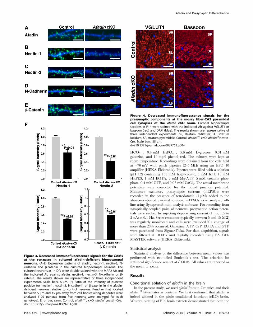
Figure 3. Decreased immunofluorescence signals for the CAMsat the synapses in cultured afadin-deficient hippocampalneurons. (A–E) Expression patterns of afadin, nectin-1, nectin-3, N-cadherin and b-catenin in the cultured hippocampal neurons. Thecultured neurons at 14 DIV were double-stained with the MAP2 Ab andthe indicated Ab against afadin, nectin-1, nectin-3, N-cadherin or b-catenin. The results shown are representative of three independentexperiments. Scale bars, 5 mm. (F) Ratio of the intensity of punctaepositive for nectin-1, nectin-3, N-cadherin or b-catenin in the afadin-deficient neurons relative to control neurons. Punctae that locatedbetween 5 mm and 45 mm away from cell bodies along dendrites wereanalyzed (100 punctae from five neurons were analyzed for eachgenotype). Error bar, s.e.m. Control, afadin+/f; cKO, afadinf/f;nestin-Cre.doi:10.1371/journal.pone.0089763.g003
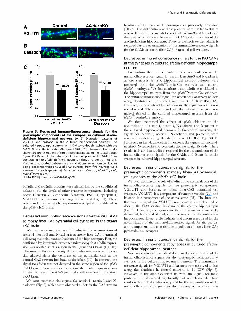
Figure 4. Decreased immunofluorescence signals for thepresynaptic components at the mossy fiber-CA3 pyramidalcell synapses of the afadin cKO brain. Coronal hippocampalsections at P14 were stained with the indicated Ab against VGLUT1 orbassoon (red) and DAPI (blue). The results shown are representative ofthree independent experiments. SR, stratum radiatum; SL, stratumlucidum; SP, stratum pyramidale. Control, afadin+/f; cKO, afadinf/f;nestin-Cre. Scale bars, 25 mm.doi:10.1371/journal.pone.0089763.g004
Afadin and Presynaptic Differentiation
PLOS ONE | www.plosone.org 4 February 2014 | Volume 9 | Issue 2 | e89763
l-afadin and s-afadin proteins were almost lost by the conditional
ablation, but the levels of other synaptic components, including
nectin-1, nectin-3, N-cadherin, b-catenin, PSD-95, synapsin I,
VGLUT1 and bassoon, were largely unaltered (Fig. 1A). These
results indicate that afadin expression was specifically ablated in
the afadin cKO brain.
Decreased immunofluorescence signals for the PAJ CAMsat mossy fiber-CA3 pyramidal cell synapses in the afadincKO brain
We next examined the role of afadin in the accumulation of
nectin-1, nectin-3 and N-cadherin at mossy fiber-CA3 pyramidal
cell synapses in the stratum lucidum of the hippocampus. First, we
confirmed by immunofluorescence microscopy that afadin expres-
sion was ablated in this region in the afadin cKO brain (Fig. 1B).
The immunofluorescence signal for afadin was observed as dots
that aligned along the dendrites of the pyramidal cells at the
control CA3 stratum lucidum, as described [10]. In contrast, the
signal for afadin was not detected in the same region of the afadin
cKO brain. These results indicate that the afadin expression was
ablated at mossy fiber-CA3 pyramidal cell synapses in the afadin
cKO brain.
We next examined the signals for nectin-1, nectin-3 and N-
cadherin (Fig. 2), which were observed as dots in the CA3 stratum
lucidum of the control hippocampus as previously described
[10,23]. The distributions of these proteins were similar to that of
afadin. However, the signals for nectin-1, nectin-3 and N-cadherin
disappeared almost completely in the CA3 stratum lucidum of the
afadin-deficient hippocampus. These results indicate that afadin is
required for the accumulation of the immunofluorescence signals
for the CAMs at mossy fiber-CA3 pyramidal cell synapses.
Decreased immunofluorescence signals for the PAJ CAMsat the synapses in cultured afadin-deficient hippocampalneurons
To confirm the role of afadin in the accumulation of the
immunofluorescence signals for nectin-1, nectin-3 and N-cadherin
at the synapses in vitro, hippocampal neuron cultures were
prepared from the afadinf/f;nestin-Cre embryos and control
afadin+/f embryos. We first confirmed that afadin was ablated in
the hippocampal neurons from the afadinf/f;nestin-Cre embryos.
The immunofluorescence signal for afadin was observed as dots
along dendrites in the control neurons at 14 DIV (Fig. 3A).
However, in the afadin-deficient neurons, the signal for afadin was
not observed. These results indicate that afadin expression was
indeed ablated in the cultured hippocampal neurons from the
afadinf/f;nestin-Cre embryos.
We then examined the effects of afadin ablation on the
accumulation of nectin-1, nectin-3, N-cadherin and b-catenin in
the cultured hippocampal neurons. In the control neurons, the
signals for nectin-1, nectin-3, N-cadherin and b-catenin were
observed as dots along the dendrites at 14 DIV (Fig. 3B–F).
However, in the afadin-deficient neurons, the signals for nectin-1,
nectin-3, N-cadherin and b-catenin decreased significantly. These
results indicate that afadin is required for the accumulation of the
immunofluorescence signals for the CAMs and b-catenin at the
synapses in cultured hippocampal neurons.
Decreased immunofluorescence signals for thepresynaptic components at mossy fiber-CA3 pyramidalcell synapses of the afadin cKO brain
We next examined the role of afadin in the accumulation of the
immunofluorescence signals for the presynaptic components,
VGLUT1 and bassoon, at mossy fiber-CA3 pyramidal cell
synapses. VGLUT1 is a component of synaptic vesicles [24] and
bassoon is a component of the active zone [25]. The immuno-
fluorescence signals for VGLUT1 and bassoon were observed as
dots in the CA3 stratum lucidum of the control hippocampus
(Fig. 4). However, the signals for these proteins were markedly
decreased, but not abolished, in this region of the afadin-deficient
hippocampus. These results indicate that afadin is required for the
accumulation of the immunofluorescence signals for the presyn-
aptic components at a considerable population of mossy fiber-CA3
pyramidal cell synapses.
Decreased immunofluorescence signals for thepresynaptic components at synapses in cultured afadin-deficient hippocampal neurons
Next, we confirmed the role of afadin in the accumulation of the
immunofluorescence signals for the presynaptic components at
synapses in the cultured hippocampal neurons. The immunoflu-
orescence signals for VGLUT1 and bassoon were observed as dots
along the dendrites in control neurons at 14 DIV (Fig. 5).
However, in the afadin-deficient neurons, the signals for these
proteins were decreased significantly but not abolished. These
results indicate that afadin is required for the accumulation of the
immunofluorescence signals for the presynaptic components at
Figure 5. Decreased immunofluorescence signals for thepresynaptic components at the synapses in cultured afadin-deficient hippocampal neurons. (A, B) Expression patterns ofVGLUT1 and bassoon in the cultured hippocampal neurons. Thecultured hippocampal neurons at 14 DIV were double-stained with theMAP2 Ab and the indicated Ab against VGLUT1 or bassoon. The resultsshown are representative of three independent experiments. Scale bars,5 mm. (C) Ratio of the intensity of punctae positive for VGLUT1 orbassoon in the afadin-deficient neurons relative to control neurons.Punctae that located between 5 mm and 45 mm away from cell bodiesalong dendrites were analyzed (100 punctae from five neurons wereanalyzed for each genotype). Error bar, s.e.m. Control, afadin+/f; cKO,afadinf/f;nestin-Cre.doi:10.1371/journal.pone.0089763.g005
Afadin and Presynaptic Differentiation
PLOS ONE | www.plosone.org 5 February 2014 | Volume 9 | Issue 2 | e89763
synapses in a considerable population of cultured hippocampal
neurons.
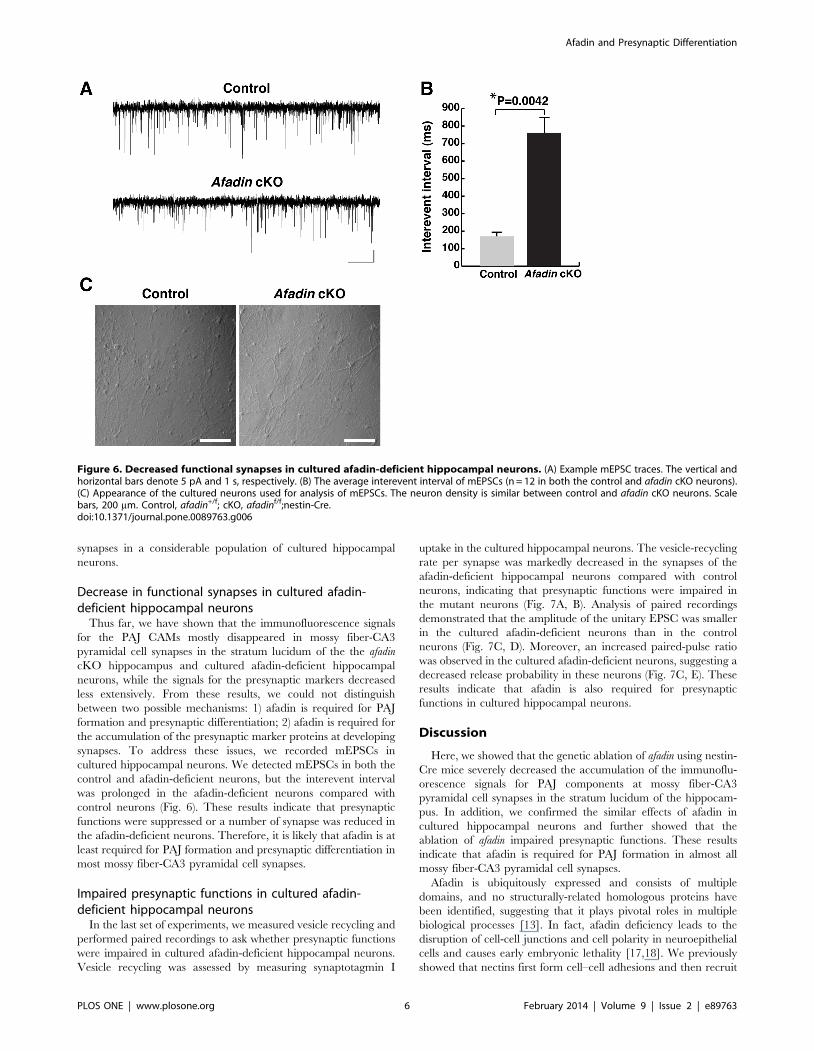
Decrease in functional synapses in cultured afadin-deficient hippocampal neurons
Thus far, we have shown that the immunofluorescence signals
for the PAJ CAMs mostly disappeared in mossy fiber-CA3
pyramidal cell synapses in the stratum lucidum of the the afadin
cKO hippocampus and cultured afadin-deficient hippocampal
neurons, while the signals for the presynaptic markers decreased
less extensively. From these results, we could not distinguish
between two possible mechanisms: 1) afadin is required for PAJ
formation and presynaptic differentiation; 2) afadin is required for
the accumulation of the presynaptic marker proteins at developing
synapses. To address these issues, we recorded mEPSCs in
cultured hippocampal neurons. We detected mEPSCs in both the
control and afadin-deficient neurons, but the interevent interval
was prolonged in the afadin-deficient neurons compared with
control neurons (Fig. 6). These results indicate that presynaptic
functions were suppressed or a number of synapse was reduced in
the afadin-deficient neurons. Therefore, it is likely that afadin is at
least required for PAJ formation and presynaptic differentiation in
most mossy fiber-CA3 pyramidal cell synapses.
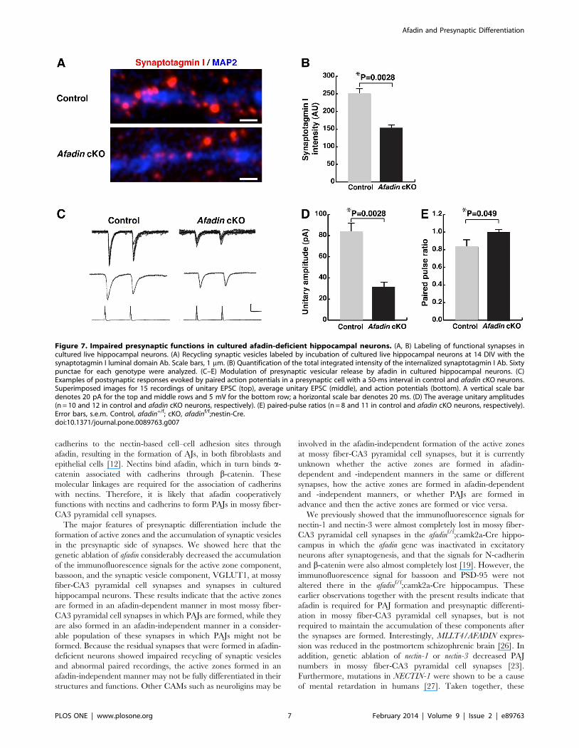
Impaired presynaptic functions in cultured afadin-deficient hippocampal neurons
In the last set of experiments, we measured vesicle recycling and
performed paired recordings to ask whether presynaptic functions
were impaired in cultured afadin-deficient hippocampal neurons.
Vesicle recycling was assessed by measuring synaptotagmin I
uptake in the cultured hippocampal neurons. The vesicle-recycling
rate per synapse was markedly decreased in the synapses of the
afadin-deficient hippocampal neurons compared with control
neurons, indicating that presynaptic functions were impaired in
the mutant neurons (Fig. 7A, B). Analysis of paired recordings
demonstrated that the amplitude of the unitary EPSC was smaller
in the cultured afadin-deficient neurons than in the control
neurons (Fig. 7C, D). Moreover, an increased paired-pulse ratio
was observed in the cultured afadin-deficient neurons, suggesting a
decreased release probability in these neurons (Fig. 7C, E). These
results indicate that afadin is also required for presynaptic
functions in cultured hippocampal neurons.
Discussion
Here, we showed that the genetic ablation of afadin using nestin-
Cre mice severely decreased the accumulation of the immunoflu-
orescence signals for PAJ components at mossy fiber-CA3
pyramidal cell synapses in the stratum lucidum of the hippocam-
pus. In addition, we confirmed the similar effects of afadin in
cultured hippocampal neurons and further showed that the
ablation of afadin impaired presynaptic functions. These results
indicate that afadin is required for PAJ formation in almost all
mossy fiber-CA3 pyramidal cell synapses.
Afadin is ubiquitously expressed and consists of multiple
domains, and no structurally-related homologous proteins have
been identified, suggesting that it plays pivotal roles in multiple
biological processes [13]. In fact, afadin deficiency leads to the
disruption of cell-cell junctions and cell polarity in neuroepithelial
cells and causes early embryonic lethality [17,18]. We previously
showed that nectins first form cell–cell adhesions and then recruit
Figure 6. Decreased functional synapses in cultured afadin-deficient hippocampal neurons. (A) Example mEPSC traces. The vertical andhorizontal bars denote 5 pA and 1 s, respectively. (B) The average interevent interval of mEPSCs (n = 12 in both the control and afadin cKO neurons).(C) Appearance of the cultured neurons used for analysis of mEPSCs. The neuron density is similar between control and afadin cKO neurons. Scalebars, 200 mm. Control, afadin+/f; cKO, afadinf/f;nestin-Cre.doi:10.1371/journal.pone.0089763.g006
Afadin and Presynaptic Differentiation
PLOS ONE | www.plosone.org 6 February 2014 | Volume 9 | Issue 2 | e89763
cadherins to the nectin-based cell–cell adhesion sites through
afadin, resulting in the formation of AJs, in both fibroblasts and
epithelial cells [12]. Nectins bind afadin, which in turn binds a-
catenin associated with cadherins through b-catenin. These
molecular linkages are required for the association of cadherins
with nectins. Therefore, it is likely that afadin cooperatively
functions with nectins and cadherins to form PAJs in mossy fiber-
CA3 pyramidal cell synapses.
The major features of presynaptic differentiation include the
formation of active zones and the accumulation of synaptic vesicles
in the presynaptic side of synapses. We showed here that the
genetic ablation of afadin considerably decreased the accumulation
of the immunofluorescence signals for the active zone component,
bassoon, and the synaptic vesicle component, VGLUT1, at mossy
fiber-CA3 pyramidal cell synapses and synapses in cultured
hippocampal neurons. These results indicate that the active zones
are formed in an afadin-dependent manner in most mossy fiber-
CA3 pyramidal cell synapses in which PAJs are formed, while they
are also formed in an afadin-independent manner in a consider-
able population of these synapses in which PAJs might not be
formed. Because the residual synapses that were formed in afadin-
deficient neurons showed impaired recycling of synaptic vesicles
and abnormal paired recordings, the active zones formed in an
afadin-independent manner may not be fully differentiated in their
structures and functions. Other CAMs such as neuroligins may be
involved in the afadin-independent formation of the active zones
at mossy fiber-CA3 pyramidal cell synapses, but it is currently
unknown whether the active zones are formed in afadin-
dependent and -independent manners in the same or different
synapses, how the active zones are formed in afadin-dependent
and -independent manners, or whether PAJs are formed in
advance and then the active zones are formed or vice versa.
We previously showed that the immunofluorescence signals for
nectin-1 and nectin-3 were almost completely lost in mossy fiber-
CA3 pyramidal cell synapses in the afadinf/f;camk2a-Cre hippo-
campus in which the afadin gene was inactivated in excitatory
neurons after synaptogenesis, and that the signals for N-cadherin
and b-catenin were also almost completely lost [19]. However, the
immunofluorescence signal for bassoon and PSD-95 were not
altered there in the afadinf/f;camk2a-Cre hippocampus. These
earlier observations together with the present results indicate that
afadin is required for PAJ formation and presynaptic differenti-
ation in mossy fiber-CA3 pyramidal cell synapses, but is not
required to maintain the accumulation of these components after
the synapses are formed. Interestingly, MLLT4/AFADIN expres-
sion was reduced in the postmortem schizophrenic brain [26]. In
addition, genetic ablation of nectin-1 or nectin-3 decreased PAJ
numbers in mossy fiber-CA3 pyramidal cell synapses [23].
Furthermore, mutations in NECTIN-1 were shown to be a cause
of mental retardation in humans [27]. Taken together, these
Figure 7. Impaired presynaptic functions in cultured afadin-deficient hippocampal neurons. (A, B) Labeling of functional synapses incultured live hippocampal neurons. (A) Recycling synaptic vesicles labeled by incubation of cultured live hippocampal neurons at 14 DIV with thesynaptotagmin I luminal domain Ab. Scale bars, 1 mm. (B) Quantification of the total integrated intensity of the internalized synaptotagmin I Ab. Sixtypunctae for each genotype were analyzed. (C–E) Modulation of presynaptic vesicular release by afadin in cultured hippocampal neurons. (C)Examples of postsynaptic responses evoked by paired action potentials in a presynaptic cell with a 50-ms interval in control and afadin cKO neurons.Superimposed images for 15 recordings of unitary EPSC (top), average unitary EPSC (middle), and action potentials (bottom). A vertical scale bardenotes 20 pA for the top and middle rows and 5 mV for the bottom row; a horizontal scale bar denotes 20 ms. (D) The average unitary amplitudes(n = 10 and 12 in control and afadin cKO neurons, respectively). (E) paired-pulse ratios (n = 8 and 11 in control and afadin cKO neurons, respectively).Error bars, s.e.m. Control, afadin+/f; cKO, afadinf/f;nestin-Cre.doi:10.1371/journal.pone.0089763.g007
Afadin and Presynaptic Differentiation
PLOS ONE | www.plosone.org 7 February 2014 | Volume 9 | Issue 2 | e89763
results indicate the importance of afadin and its binding proteins,
nectin-1 and nectin-3, in both physiology and pathology.
It was shown that AF-6/afadin regulates the morphology of
dendritic spines in cultured hippocampal neurons [28]. Addition-
ally, it was shown that afadin is required for the maintenance of
dendritic arborization and synapse number in cultured hippo-
campal neurons [29]. These two studies employed afadin
knockdown or overexpression of mutant forms of afadin. These
earlier observations indicate that afadin regulates synaptogenesis
through postsynaptic differentiation. Taken together with the
present results, it is likely that afadin is involved in synaptogenesis
through both presynaptic and postsynaptic differentiation. In
contrast, it was recently shown by use of conditional afadin-
deficient mice with a Nex-Cre mouse line that the genetic ablation
of afadin neither changed the localizations of nectin-1 and nectin-3
in the CA1 region nor perturbed the presynaptic functions in the
CA3 region, although it was shown that synapse number was
decreased in the CA1 region of the afadin-deficient hippocampus
[30]. Some of these results are apparently inconsistent with our
present results. The exact reason for this inconsistency is not
known, but it may be due to the different regions analyzed in the
hippocampus, the different promoters used for the Cre mice, the
different nectin Abs used, or the different samples used for
electrophysiology experiments.
Author Contributions
Conceived and designed the experiments: KM HT YT. Performed the
experiments: DT KM TM IS HT TI MM. Analyzed the data: DT KM
TM IS HT TI MM. Wrote the paper: KM YT.
References
1. Spacek J, Lieberman AR (1974) Ultrastructure and three-dimensionalorganization of synaptic glomeruli in rat somatosensory thalamus. J Anat 117:
487–516.
2. Yuste R, Bonhoeffer T (2001) Morphological changes in dendritic spinesassociated with long-term synaptic plasticity. Annu Rev Neurosci 24: 1071–
1089.3. Takeichi M (2007) The cadherin superfamily in neuronal connections and
interactions. Nat Rev Neurosci 8: 11–20.4. Sudhof TC (2008) Neuroligins and neurexins link synaptic function to cognitive
disease. Nature 455: 903–911.
5. Uchida N, Honjo Y, Johnson KR, Wheelock MJ, Takeichi M (1996) Thecatenin/cadherin adhesion system is localized in synaptic junctions bordering
transmitter release zones. J Cell Biol 135: 767–779.6. Kasugai Y, Swinny JD, Roberts JD, Dalezios Y, Fukazawa Y, et al. (2010)
Quantitative localisation of synaptic and extrasynaptic GABAA receptor
subunits on hippocampal pyramidal cells by freeze-fracture replica immunola-belling. Eur J Neurosci 32: 1868–1888.
7. Song JY, Ichtchenko K, Sudhof TC, Brose N (1999) Neuroligin 1 is apostsynaptic cell-adhesion molecule of excitatory synapses. Proc Natl Acad
Sci U S A 96: 1100–1105.
8. Varoqueaux F, Jamain S, Brose N (2004) Neuroligin 2 is exclusively localized toinhibitory synapses. Eur J Cell Biol 83: 449–456.
9. Dean C, Scholl FG, Choih J, DeMaria S, Berger J, et al. (2003) Neurexinmediates the assembly of presynaptic terminals. Nat Neurosci 6: 708–716.
10. Mizoguchi A, Nakanishi H, Kimura K, Matsubara K, Ozaki-Kuroda K, et al.(2002) Nectin: an adhesion molecule involved in formation of synapses. J Cell
Biol 156: 555–565.
11. Takai Y, Nakanishi H (2003) Nectin and afadin: novel organizers of intercellularjunctions. J Cell Sci 116: 17–27.
12. Takai Y, Ikeda W, Ogita H, Rikitake Y (2008) The immunoglobulin-like celladhesion molecule nectin and its associated protein afadin. Annu Rev Cell Dev
Biol 24: 309–342.
13. Mandai K, Nakanishi H, Satoh A, Obaishi H, Wada M, et al. (1997) Afadin: Anovel actin filament-binding protein with one PDZ domain localized at
cadherin-based cell-to-cell adherens junction. J Cell Biol 139: 517–528.14. Mandai K, Rikitake Y, Shimono Y, Takai Y (2013) Afadin/AF-6 and Canoe:
Roles in Cell Adhesion and Beyond. Prog Mol Biol Transl Sci 116: 433–454.15. Prasad R, Gu Y, Alder H, Nakamura T, Canaani O, et al. (1993) Cloning of the
ALL-1 fusion partner, the AF-6 gene, involved in acute myeloid leukemias with
the t(6;11) chromosome translocation. Cancer Res 53: 5624–5628.16. Kurita S, Yamada T, Rikitsu E, Ikeda W, Takai Y (2013) Binding between the
Junctional Proteins Afadin and PLEKHA7 and Implication in the Formation ofAdherens Junction in Epithelial Cells. J Biol Chem in press.
17. Ikeda W, Nakanishi H, Miyoshi J, Mandai K, Ishizaki H, et al. (1999) Afadin: A
key molecule essential for structural organization of cell-cell junctions of
polarized epithelia during embryogenesis. J Cell Biol 146: 1117–1132.
18. Zhadanov AB, Provance DW Jr, Speer CA, Coffin JD, Goss D, et al. (1999)
Absence of the tight junctional protein AF-6 disrupts epithelial cell-cell junctions
and cell polarity during mouse development. Curr Biol 9: 880–888.
19. Majima T, Ogita H, Yamada T, Amano H, Togashi H, et al. (2009)
Involvement of afadin in the formation and remodeling of synapses in the
hippocampus. Biochem Biophys Res Commun 385: 539–544.
20. Betz UA, Voßhenrich CA, Rajewsky K, Muller W (1996) Bypass of lethality with
mosaic mice generated by Cre-loxP-mediated recombination. Curr Biol 6:
1307–1316.
21. Tronche F, Kellendonk C, Kretz O, Gass P, Anlag K, et al. (1999) Disruption of
the glucocorticoid receptor gene in the nervous system results in reduced
anxiety. Nat Genet 23: 99–103.
22. Togashi H, Abe K, Mizoguchi A, Takaoka K, Chisaka O, et al. (2002) Cadherin
regulates dendritic spine morphogenesis. Neuron 35: 77–89.
23. Honda T, Sakisaka T, Yamada T, Kumazawa N, Hoshino T, et al. (2006)
Involvement of nectins in the formation of puncta adherentia junctions and the
mossy fiber trajectory in the mouse hippocampus. Mol Cell Neurosci 31: 315–
325.
24. Bellocchio EE, Reimer RJ, Fremeau RT Jr, Edwards RH (2000) Uptake of
glutamate into synaptic vesicles by an inorganic phosphate transporter. Science
289: 957–960.
25. tom Dieck S, Sanmartı-Vila L, Langnaese K, Richter K, Kindler S, et al. (1998)
Bassoon, a novel zinc-finger CAG/glutamine-repeat protein selectively localized
at the active zone of presynaptic nerve terminals. J Cell Biol 142: 499–509.
26. Katsel P, Davis KL, Haroutunian V (2005) Variations in myelin and
oligodendrocyte-related gene expression across multiple brain regions in
schizophrenia: a gene ontology study. Schizophr Res 79: 157–173.
27. Suzuki K, Hu D, Bustos T, Zlotogora J, Richieri-Costa A, et al. (2000)
Mutations of PVRL1, encoding a cell-cell adhesion molecule/herpesvirus
receptor, in cleft lip/palate-ectodermal dysplasia. Nat Genet 25: 427–430.
28. Xie Z, Huganir RL, Penzes P (2005) Activity-dependent dendritic spine
structural plasticity is regulated by small GTPase Rap1 and its target AF-6.
Neuron 48: 605–618.
29. Srivastava DP, Copits BA, Xie Z, Huda R, Jones KA, et al. (2012) Afadin is
required for maintenance of dendritic structure and excitatory tone. J Biol Chem
287: 35964–35974.
30. Beaudoin GM III, Schofield CM, Nuwal T, Zang K, Ullian EM, et al. (2012)
Afadin, a ras/rap effector that controls cadherin function, promotes spine and
excitatory synapse density in the hippocampus. J Neurosci 32: 99–110.
Afadin and Presynaptic Differentiation
PLOS ONE | www.plosone.org 8 February 2014 | Volume 9 | Issue 2 | e89763