Embed Size (px)
Citation preview
A
TP2
Toataorp
MtfadepsvtCCpbSrweid1rdcntfib
GASTROENTEROLOGY 2003;125:1508–1530
GA Technical Review on Perianal Crohn’s Disease
his literature review and the recommendations therein were prepared for the American Gastroenterological Association Clinicalractice Committee. The paper was approved by the Committee on May 18, 2003, and by the AGA Governing Board on July 25,
tugtpt
his report is a technical review of the normal anat-omy, definitions, etiology, classification, epidemiol-
gy, diagnosis, disease activity assessment, and medicalnd surgical treatment of perianal Crohn’s disease. Al-hough descriptions of surgical therapy are providedlong with the indications, outcomes, and complicationsf surgical intervention, the reader is cautioned that thiseview is not meant to provide the details of how toerform the specific individual surgical procedures.
Strategy for LiteratureIdentificationA search of the online bibliographic databases
EDLINE (1966 to August 2002) and Current Con-ents/Science Edition (1996 to August 2002) was per-ormed to identify potentially relevant English-languagerticles. The Medical Subject Heading terms “Crohn’sisease” or “inflammatory bowel disease” or “regionalnteritis” AND “fistulas” or “perianal” were used toerform keyword searches of the database. Manualearches of the reference lists from the potentially rele-ant papers and the proceedings from annual meetings ofhe American Gastroenterological Association, Americanollege of Gastroenterology, and American Society ofolon and Rectal Surgeons from 1990 to 2003 wereerformed to identify additional studies that may haveeen missed using the computer-assisted search strategy.tudies selected were retrospective or prospective studieseporting on the classification, epidemiology, diagnosisith magnetic resonance imaging (MRI) or anorectal
ndoscopic ultrasonography (EUS), treatment with med-cal therapy, and surgical treatment of perianal Crohn’sisease. This technical review is not based solely on levelstudies (population-based natural history studies and
andomized, double-blind, placebo-controlled trials ofiagnostic modalities and therapeutic interventions), be-ause only 5 level 1 studies were identified and they doot provide a sufficient basis for an “evidence-based”echnical review and medical position statement. There-ore, this technical review represents the consensus opin-on of the authors based on a comprehensive review of theest available evidence.
Normal Anatomy, Definitions,Etiology, and ClassificationNormal Anatomy
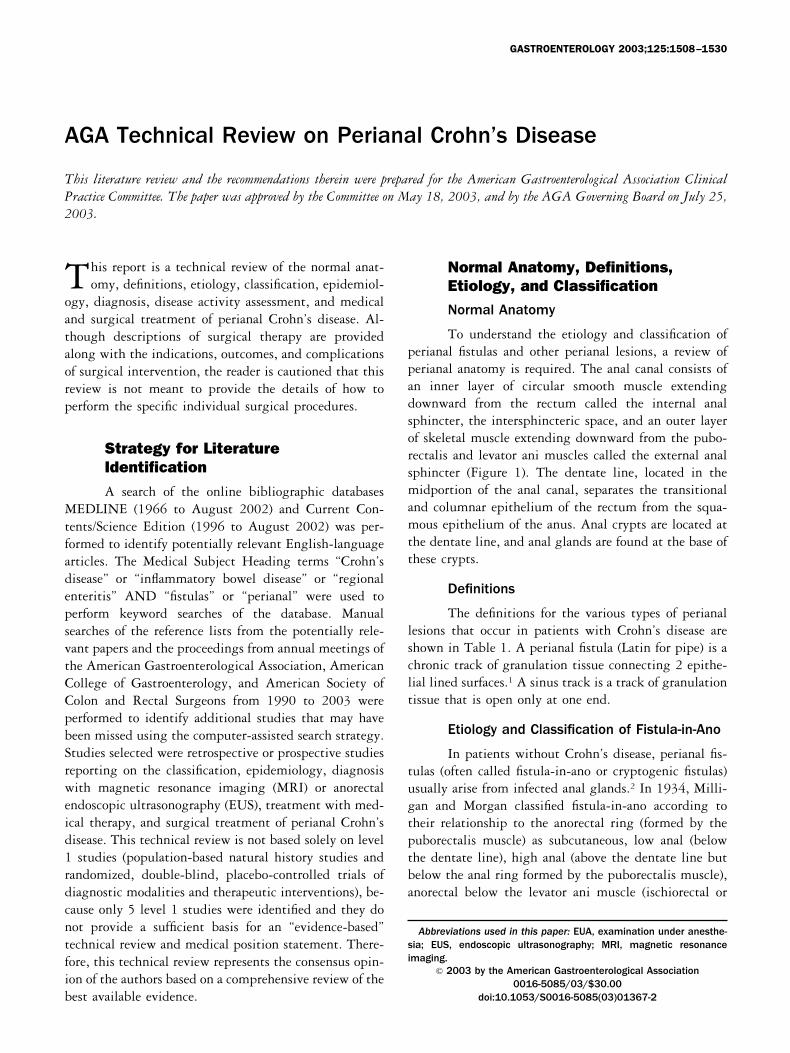
To understand the etiology and classification oferianal fistulas and other perianal lesions, a review oferianal anatomy is required. The anal canal consists ofn inner layer of circular smooth muscle extendingownward from the rectum called the internal analphincter, the intersphincteric space, and an outer layerf skeletal muscle extending downward from the pubo-ectalis and levator ani muscles called the external analphincter (Figure 1). The dentate line, located in theidportion of the anal canal, separates the transitional
nd columnar epithelium of the rectum from the squa-ous epithelium of the anus. Anal crypts are located at
he dentate line, and anal glands are found at the base ofhese crypts.
Definitions
The definitions for the various types of perianalesions that occur in patients with Crohn’s disease arehown in Table 1. A perianal fistula (Latin for pipe) is ahronic track of granulation tissue connecting 2 epithe-ial lined surfaces.1 A sinus track is a track of granulationissue that is open only at one end.
Etiology and Classification of Fistula-in-Ano
In patients without Crohn’s disease, perianal fis-ulas (often called fistula-in-ano or cryptogenic fistulas)sually arise from infected anal glands.2 In 1934, Milli-an and Morgan classified fistula-in-ano according toheir relationship to the anorectal ring (formed by theuborectalis muscle) as subcutaneous, low anal (belowhe dentate line), high anal (above the dentate line butelow the anal ring formed by the puborectalis muscle),norectal below the levator ani muscle (ischiorectal or
Abbreviations used in this paper: EUA, examination under anesthe-ia; EUS, endoscopic ultrasonography; MRI, magnetic resonancemaging.
© 2003 by the American Gastroenterological Association0016-5085/03/$30.00
doi:10.1053/S0016-5085(03)01367-2
003.
ppadsorsm
ba
si
amtt
lsclt
infralevator), anorectal above the levator ani muscle (pel-virectal or supralevator), and submucous (high intermus-cular between the internal and external anal sphincters).3
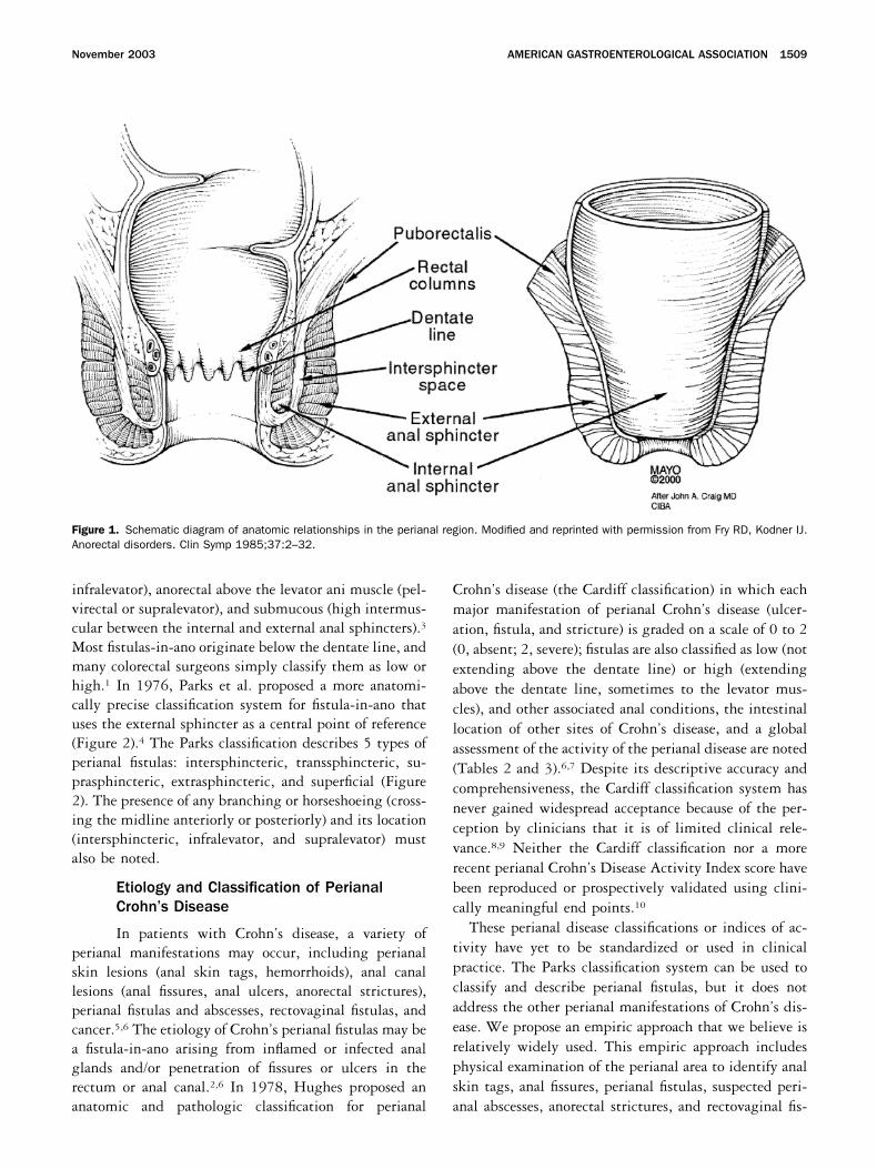
Most fistulas-in-ano originate below the dentate line, andmany colorectal surgeons simply classify them as low orhigh.1 In 1976, Parks et al. proposed a more anatomi-cally precise classification system for fistula-in-ano thatuses the external sphincter as a central point of reference(Figure 2).4 The Parks classification describes 5 types ofperianal fistulas: intersphincteric, transsphincteric, su-prasphincteric, extrasphincteric, and superficial (Figure2). The presence of any branching or horseshoeing (cross-ing the midline anteriorly or posteriorly) and its location(intersphincteric, infralevator, and supralevator) mustalso be noted.
Etiology and Classification of PerianalCrohn’s Disease
In patients with Crohn’s disease, a variety ofperianal manifestations may occur, including perianalskin lesions (anal skin tags, hemorrhoids), anal canallesions (anal fissures, anal ulcers, anorectal strictures),perianal fistulas and abscesses, rectovaginal fistulas, andcancer.5,6 The etiology of Crohn’s perianal fistulas may bea fistula-in-ano arising from inflamed or infected analglands and/or penetration of fissures or ulcers in therectum or anal canal.2,6 In 1978, Hughes proposed ananatomic and pathologic classification for perianal
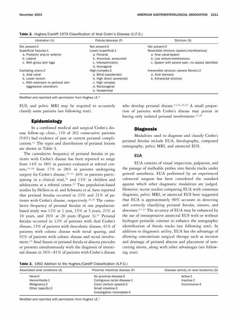
Crohn’s disease (the Cardiff classification) in which eachmajor manifestation of perianal Crohn’s disease (ulcer-ation, fistula, and stricture) is graded on a scale of 0 to 2(0, absent; 2, severe); fistulas are also classified as low (notextending above the dentate line) or high (extendingabove the dentate line, sometimes to the levator mus-cles), and other associated anal conditions, the intestinallocation of other sites of Crohn’s disease, and a globalassessment of the activity of the perianal disease are noted(Tables 2 and 3).6,7 Despite its descriptive accuracy andcomprehensiveness, the Cardiff classification system hasnever gained widespread acceptance because of the per-ception by clinicians that it is of limited clinical rele-vance.8,9 Neither the Cardiff classification nor a morerecent perianal Crohn’s Disease Activity Index score havebeen reproduced or prospectively validated using clini-cally meaningful end points.10
These perianal disease classifications or indices of ac-tivity have yet to be standardized or used in clinicalpractice. The Parks classification system can be used toclassify and describe perianal fistulas, but it does notaddress the other perianal manifestations of Crohn’s dis-ease. We propose an empiric approach that we believe isrelatively widely used. This empiric approach includesphysical examination of the perianal area to identify analskin tags, anal fissures, perianal fistulas, suspected peri-anal abscesses, anorectal strictures, and rectovaginal fis-
Figure 1. Schematic diagram of anatomic relationships in the perianal region. Modified and reprinted with permission from Fry RD, Kodner IJ.Anorectal disorders. Clin Symp 1985;37:2–32.
November 2003 AMERICAN GASTROENTEROLOGICAL ASSOCIATION 1509
tulas as well as an endoscopic evaluation to determinewhether or not there is macroscopically evident inflam-mation of the rectum. Fistulas are then classified as either“simple” or “complex.” A simple fistula is low (superfi-
cial or low intersphincteric or low transsphincteric originof the fistula tract), has a single external opening, has nopain or fluctuation to suggest perianal abscess, has noevidence of a rectovaginal fistula, and has no evidence ofanorectal stricture. The presence of active rectal Crohn’sdisease may make a simple fistula more complicated tomanage. Patients with simple fistulas may have higherrates of healing.11
A complex fistula is high (high intersphincteric orhigh transsphincteric or extrasphincteric or suprasphinc-teric origin of the fistula tract), may have multipleexternal openings, may be associated with the presence ofpain or fluctuation to suggest a perianal abscess, may beassociated with the presence of a rectovaginal fistula, maybe associated with the presence of an anorectal stricture,and may be associated with the presence of active rectaldisease at endoscopy. Additional diagnostic evaluationwith examination under anesthesia (EUA), anorectal
Figure 2. The Parks classification. (A) A superficial fistula tracksbelow both the internal anal sphincter and external anal sphinctercomplexes. (B) An intersphincteric fistula tracks between the internalanal sphincter and the external anal sphincter in the intersphinctericspace. (C) A transsphincteric fistula tracks from the intersphinctericspace through the external anal sphincter. (D) A suprasphinctericfistula leaves the intersphincteric space over the top of the puborec-talis and penetrates the levator muscle before tracking down to theskin. (E) An extrasphincteric fistula tracks outside of the external analsphincter and penetrates the levator muscle into the rectum. Modifiedand reprinted with permission from Parks et al.4 © British Journal ofSurgery Society Ltd. Reproduced with permission. Permission isgranted by John Wiley & Sons Ltd on behalf of the BJSS Ltd.
Table 1. Definition of Various Types of Perianal Lesions inPatients With Crohn’s Disease
Type of lesion Description
Skin tag Two types:1. Large, edematous, hard, cyanotic skin
tags. Typically arising from a healed analfissure or ulcer. Excision contraindicateddue to problems with wound healing.
2. “Elephant ear” tags that are flat and broador narrow, soft painless skin tags. Maycause perianal hygiene problems and canbe safely excised.
Hemorrhoids Prolapsing internal hemorrhoids. Uncommon inCrohn’s disease. Often present as largeexternal skin tags.
Fissure Anal fissures are broad based and deep withundermining of the edges. There may beassociated large skin tags and a cyanotic hueto the surrounding skin. They tend to bemultiple and may be placed eithereccentrically around the anal canal or in themidline in contrast to idiopathic fissure-in-ano,which tend to lie in the midline. Typicallypainless (pain should raise suspicion forperianal abscess or acute/chronicconventional anal fissure). Conventional analfissures occasionally are treated byconventional fissure treatment includinglateral sphincterotomy.
Anal ulcer Anal ulcers are usually associated with rectalinflammation and may lead to destruction ofthe anorectum, anorectal strictures, complexanorectal fistulas, and perianal abscess.
Low fistula Superficial, low intersphincteric, or low trans-sphincteric fistulas. May arise from either theanal glands (cryptogenic) or from penetratingulceration of the anal canal or rectum.
High fistula High intersphincteric, high transsphincteric,suprasphincteric, extrasphincteric fistulas.Arise from penetrating ulceration of the analcanal or rectum.
Rectovaginalfistula
Superficial, intersphincteric, transsphincteric,suprasphincteric, extrasphincteric fistulas.Arise from penetrating ulceration of the analcanal or rectum into the vagina.
Perianalabscess
Potential anorectal spaces may becomeinfected with an abscess, including perianal,ishiorectal, deep postnatal, intersphincteric,and supralevator.
Anorectalstricture
May be short annular diaphragm-like strictures�2 cm in length or longer tubular stricturesarising from rectal inflammation. May arisefrom either the anal glands (cryptogenic) orfrom penetrating ulceration of the anal canalor rectum.
Cancer Squamous cell carcinoma, basal cell carcinoma,or adenocarcinoma arising from malignantdegeneration of nonhealing perianal fistulasor sinus tracts.
1510 AMERICAN GASTROENTEROLOGICAL ASSOCIATION GASTROENTEROLOGY Vol. 125, No. 5
EUS, and pelvic MRI may be required to accuratelyclassify some patients (see following text).
EpidemiologyIn a combined medical and surgical Crohn’s dis-
ease follow-up clinic, 110 of 202 consecutive patients(54%) had evidence of past or current perianal compli-cations.12 The types and distribution of perianal lesionsare shown in Table 4.
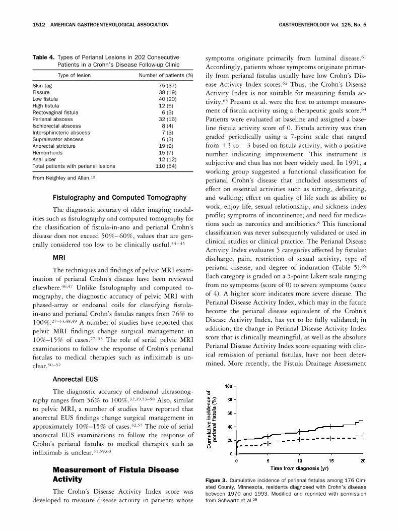
The cumulative frequency of perianal fistulas in pa-tients with Crohn’s disease has been reported to rangefrom 14% to 38% in patients evaluated at referral cen-ters,13–18 from 17% to 28% in patients undergoingsurgery for Crohn’s disease,19–21 36% in patients partic-ipating in a clinical trial,22 and 13% in children andadolescents at a referral center.23 Two population-basedstudies by Hellers et al. and Schwartz et al. have reportedthat perianal fistulas occurred in 23% and 21% of pa-tients with Crohn’s disease, respectively.24,25 The cumu-lative frequency of perianal fistulas in one population-based study was 12% at 1 year, 15% at 5 years, 21% at10 years, and 26% at 20 years (Figure 3).25 Perianalfistulas occurred in 12% of patients with ileal Crohn’sdisease, 15% of patients with ileocolonic disease, 41% ofpatients with colonic disease with rectal sparing, and92% of patients with colonic disease and rectal involve-ment.24 Anal fissure or perianal fistula or abscess precedesor presents simultaneously with the diagnosis of intesti-nal disease in 36%–81% of patients with Crohn’s disease
who develop perianal disease.14,21,24,25 A small propor-tion of patients with Crohn’s disease may persist inhaving only isolated perianal involvement.13,26
DiagnosisModalities used to diagnose and classify Crohn’s
perianal fistulas include EUA, fistulography, computedtomography, pelvic MRI, and anorectal EUS.
EUA
EUA consists of visual inspection, palpation, andthe passage of malleable probes into fistula tracks undergeneral anesthesia. EUA performed by an experiencedcolorectal surgeon has been considered the standardagainst which other diagnostic modalities are judged.However, recent studies comparing EUA with consensusdiagnosis, pelvic MRI, or anorectal EUS have suggestedthat EUA is approximately 90% accurate in detectingand correctly classifying perianal fistulas, sinuses, andabscesses.27–33 The accuracy of EUA may be enhanced bythe use of intraoperative anorectal EUS with or withouthydrogen peroxide contrast to enhance the sonographicidentification of fistula tracks (see following text). Inaddition to diagnostic utility, EUA has the advantage ofallowing concomitant surgical therapy such as incisionand drainage of perianal abscess and placement of non-cutting setons, along with other advantages (see follow-ing text).
Table 2. Hughes/Cardiff 1979 Classification of Anal Crohn’s Disease (U.F.S.)
Ulceration (U) Fistula/abscess (F) Stricture (S)
Not present-0 Not present-0 Not present-0Superficial fissures-1 Lower/superficial-1 Reversible stricture (spasm/membranous)
a. Posterior and/or anterior a. Perianal a. Anal canal-spasmb. Lateral b. Anovulval, anoscrotal b. Low rectum-membranousc. With gross skin tags c. Intersphincteric c. Spasm with severe pain, no sepsis identified
d. AnovaginalCavitating ulcers-2 High/complex-2 Irreversible stricture (severe fibrotic)-2
a. Anal canal a. Blind supralevator a. Anal stenosisb. Lower rectum b. High direct (anorectal) b. Extrarectal stricturec. With extension to perineal skin c. High complex
(aggressive ulceration) d. Rectovaginale. Ileoperineal
Modified and reprinted with permission from Hughes LE.6
Table 3. 1992 Addition to the Hughes/Cardiff Classification (A.P.D.)
Associated anal conditions (A) Proximal intestinal disease (P) Disease activity (in anal locations) (D)
None-0 No proximal disease-0 Active-1Hemorrhoids-1 Contiguous rectal disease-1 Inactive-2Malignancy-2 Colon (rectum spared)-2 Inconclusive-3Other (specify)-3 Small intestine-3
Investigation incomplete-4
Modified and reprinted with permission from Hughes LE.7
November 2003 AMERICAN GASTROENTEROLOGICAL ASSOCIATION 1511
Fistulography and Computed Tomography
The diagnostic accuracy of older imaging modal-ities such as fistulography and computed tomography forthe classification of fistula-in-ano and perianal Crohn’sdisease does not exceed 50%–60%, values that are gen-erally considered too low to be clinically useful.34–45
MRI
The techniques and findings of pelvic MRI exam-ination of perianal Crohn’s disease have been reviewedelsewhere.46,47 Unlike fistulography and computed to-mography, the diagnostic accuracy of pelvic MRI withphased-array or endoanal coils for classifying fistula-in-ano and perianal Crohn’s fistulas ranges from 76% to100%.27–33,48,49 A number of studies have reported thatpelvic MRI findings change surgical management in10%–15% of cases.27–33 The role of serial pelvic MRIexaminations to follow the response of Crohn’s perianalfistulas to medical therapies such as infliximab is un-clear.50–52
Anorectal EUS
The diagnostic accuracy of endoanal ultrasonog-raphy ranges from 56% to 100%.32,39,53–58 Also, similarto pelvic MRI, a number of studies have reported thatanorectal EUS findings change surgical management inapproximately 10%–15% of cases.32,57 The role of serialanorectal EUS examinations to follow the response ofCrohn’s perianal fistulas to medical therapies such asinfliximab is unclear.51,59,60
Measurement of Fistula DiseaseActivityThe Crohn’s Disease Activity Index score was
developed to measure disease activity in patients whose
symptoms originate primarily from luminal disease.61
Accordingly, patients whose symptoms originate primar-ily from perianal fistulas usually have low Crohn’s Dis-ease Activity Index scores.62 Thus, the Crohn’s DiseaseActivity Index is not suitable for measuring fistula ac-tivity.63 Present et al. were the first to attempt measure-ment of fistula activity using a therapeutic goals score.64
Patients were evaluated at baseline and assigned a base-line fistula activity score of 0. Fistula activity was thengraded periodically using a 7-point scale that rangedfrom �3 to �3 based on fistula activity, with a positivenumber indicating improvement. This instrument issubjective and thus has not been widely used. In 1991, aworking group suggested a functional classification forperianal Crohn’s disease that included assessments ofeffect on essential activities such as sitting, defecating,and walking; effect on quality of life such as ability towork, enjoy life, sexual relationship, and sickness indexprofile; symptoms of incontinence; and need for medica-tions such as narcotics and antibiotics.8 This functionalclassification was never subsequently validated or used inclinical studies or clinical practice. The Perianal DiseaseActivity Index evaluates 5 categories affected by fistulas:discharge, pain, restriction of sexual activity, type ofperianal disease, and degree of induration (Table 5).65
Each category is graded on a 5-point Likert scale rangingfrom no symptoms (score of 0) to severe symptoms (scoreof 4). A higher score indicates more severe disease. ThePerianal Disease Activity Index, which may in the futurebecome the perianal disease equivalent of the Crohn’sDisease Activity Index, has yet to be fully validated; inaddition, the change in Perianal Disease Activity Indexscore that is clinically meaningful, as well as the absolutePerianal Disease Activity Index score equating with clin-ical remission of perianal fistulas, have not been deter-mined. More recently, the Fistula Drainage Assessment
Figure 3. Cumulative incidence of perianal fistulas among 176 Olm-sted County, Minnesota, residents diagnosed with Crohn’s diseasebetween 1970 and 1993. Modified and reprinted with permissionfrom Schwartz et al.25
Table 4. Types of Perianal Lesions in 202 ConsecutivePatients in a Crohn’s Disease Follow-up Clinic
Type of lesion Number of patients (%)
Skin tag 75 (37)Fissure 38 (19)Low fistula 40 (20)High fistula 12 (6)Rectovaginal fistula 6 (3)Perianal abscess 32 (16)Ischiorectal abscess 8 (4)Intersphincteric abscess 7 (3)Supralevator abscess 6 (3)Anorectal stricture 19 (9)Hemorrhoids 15 (7)Anal ulcer 12 (12)Total patients with perianal lesions 110 (54)
From Keighley and Allan.12
1512 AMERICAN GASTROENTEROLOGICAL ASSOCIATION GASTROENTEROLOGY Vol. 125, No. 5
has been used to classify fistulas as either open andactively draining or closed (Table 6).62 A fistula isopen and actively draining if the investigator canexpress purulent material from the fistula with theapplication of gentle pressure. The acute effects oftherapy are assessed by determining if a patient has afistula response (defined as closure of at least 50% offistulas present at baseline maintained for at least 4weeks) or has complete fistula closure (defined asclosure of all fistulas present at baseline maintained forat least 4 weeks). The maintenance effects of therapyare assessed by determining the time to loss of fistularesponse or the time to loss of complete fistula closure.The terminology “closure of fistulas” probably doesnot accurately reflect the findings of EUA, pelvic MRI,or anorectal EUS, which often show persistent fistulatracts even when fistula drainage has ceased; thus,alternative terminology such as “cessation of drainage”should be considered. Although not addressed by theFistula Drainage Assessment, it should be noted thatit is important to reduce or eliminate anal pain inaddition to fistula drainage.
Medical TreatmentThe pharmaceutical agents with definite or po-
tential efficacy for treating patients with perianal Crohn’sdisease include antibiotics, azathioprine and 6-mercap-topurine, infliximab, cyclosporine, and tacrolimus.
Antibiotics
There are no controlled trials showing that anti-biotics are effective in the treatment of Crohn’s perianalfistulas. The current clinical practice of using metroni-dazole or ciprofloxacin is based on uncontrolled caseseries and the absence of other therapeutic alternativesperceived to be safe for use in all patients.66–75 A repre-sentative experience with metronidazole is that of Bern-stein et al., who reported a series of 21 patients withperianal Crohn’s disease treated with metronidazole 20mg � kg�1 � day�1.67 All patients had a clinical responsewith a decrease in pain and tenderness. Ten of the 18patients (56%) had complete healing of their perianaldisease. Clinical improvement typically occurred after6–8 weeks of therapy. A follow-up study of 17 of thesepatients, along with 9 additional patients, showed exac-erbation of disease with dosage reduction as well as theoccurrence of paresthesias in 13 of 26 patients (50%) (theparesthesias disappeared with dose reduction or drugdiscontinuation in 7 patients, paresthesias persisted un-der ongoing treatment in 5 patients, and hot/cold sen-sitivity persisted after drug discontinuation in 1 pa-tient).68 Clinicians prescribing antibiotics for fistulastypically use metronidazole at doses ranging from 750 to1500 mg/day or ciprofloxacin 1000 mg/day for up to3–4 months. Adverse events associated with metronida-zole include metallic taste, glossitis, nausea, and a distalperipheral sensory neuropathy.76 Adverse events associ-ated with ciprofloxacin are uncommon but include head-ache, nausea, diarrhea, and rash.77 A recent cost-utilitystudy suggested that metronidazole combined with6-mercaptopurine was a more cost-effective treatment
Table 5. Perianal Crohn’s Disease Activity Index
Categories affected by fistulas Score
DischargeNo discharge 0Minimal mucous discharge 1Moderate mucous or purulent discharge 2Substantial discharge 3Gross fecal soiling 4
Pain/restriction of activitiesNo activity restriction 0Mild discomfort, no restriction 1Moderate discomfort, some limitation activities 2Marked discomfort, marked limitation 3Severe pain, severe limitation 4
Restriction of sexual activityNo restriction in sexual activity 0Slight restriction in sexual activity 1Moderate limitation in sexual activity 2Marked limitation in sexual activity 3Unable to engage in sexual activity 4
Type of perianal diseaseNo perianal disease/skin tags 0Anal fissure or mucosal tear 1�3 Perianal fistulae 2�3 Perianal fistulae 3Anal sphincter ulceration or fistulae with
significant undermining of skin 4Degree of induration
No induration 0Minimal induration 1Moderate induration 2Substantial induration 3Gross fluctuance/abscess 4
From Irvine.65
Table 6. Fistula Drainage Assessment
End point Definition
Improvement Closure of individual fistulas defined as no fistuladrainage despite gentle finger compression.Improvement defined as a decrease frombaseline in the number of open drainingfistulas of �50% for at least 2 consecutivevisits (i.e., at least 4 weeks).
Remission Closure of individual fistulas defined as no fistuladrainage despite gentle finger compression.Remission defined as closure of all fistulasthat were draining at baseline for at least 2consecutive visits (i.e., at least 4 weeks).
Modified and reprinted with permission from Present et al.62
November 2003 AMERICAN GASTROENTEROLOGICAL ASSOCIATION 1513
strategy than infliximab-based treatment strategies (seefollowing text).78 However, this study may have under-estimated the cost of patients with fistulas79; in addition,given that no controlled data support the efficacy of theseagents, this conclusion is highly questionable.
Azathioprine and 6-Mercaptopurine
There are no controlled trials with fistula closureas the primary end point showing that the antimetabo-lites azathioprine and 6-mercaptopurine are effective inthe treatment of Crohn’s perianal fistulas. The currentclinical practice of using these medications is based on ameta-analysis of 5 controlled trials in which fistula clo-sure was examined as a secondary end point80 and un-controlled case series in adults81 and children.82 Themeta-analysis included 70 patients from 5 controlledtrials of azathioprine or 6-mercaptopurine for Crohn’sdisease in which the details of fistula closure were de-scribed and could be used as a secondary end point(fistula closure was identified as a secondary end pointpost-hoc in 4 of the 5 studies specifically for the purposesof performing the meta-analysis).64,83–86 The results ofthe meta-analysis showed that 22 of 41 patients (54%)treated with azathioprine or 6-mercaptopurine had fis-tula healing versus 6 of 29 patients (21%) treated withplacebo, with a pooled odds ratio of 4.44 favoring heal-ing of fistulas.80 Clinical practice varies widely withrespect to dosing and administration of azathioprine and6-mercaptopurine. Controlled trials indicate that aza-thioprine at doses of 2.0–3.0 mg � kg�1 � day�1 and6-mercaptopurine at a dose of 1.5 mg � kg�1 � day�1 iseffective for the treatment of Crohn’s disease. Some cli-nicians prefer to start either drug at a dose of 50 mg/dayand gradually titrate to effect or to a “therapeutic” bloodmetabolite concentration to minimize the occurrence ofadverse events, although the efficacy of this strategy isunproven. Adverse events associated with azathioprineand 6-mercaptopurine include leukopenia, allergic reac-tions, infection, pancreatitis, drug-induced hepatitis,and possibly non-Hodgkin’s lymphoma.80,87,88 Patientstreated with these medications should have regular mon-itoring of leukocyte counts and liver transaminase levels.
Infliximab
There are 2 controlled trials in which fistula clo-sure was the primary end point showing that infliximab(a mouse/human chimeric monoclonal antibody to tumornecrosis factor) is effective in the treatment of Crohn’sperianal fistulas.62,89 Uncontrolled case series have alsosuggested that infliximab is of benefit for this indica-tion.90–92 In one placebo-controlled trial, 94 patientswith draining Crohn’s fistulas (85 patients had perianal
fistulas) were randomized to treatment with 3 infusionsof placebo or infliximab at doses of 5 or 10 mg/kg at 0,2, and 6 weeks.62 Closure of at least 50% of fistulas wasmaintained for at least 4 weeks (primary study end point)in 26% of placebo-treated patients when compared with68% of patients treated with infliximab 5 mg/kg and56% of patients treated with infliximab 10 mg/kg (P �0.002 and P � 0.02, respectively). Closure of all fistulaswas maintained for at least 4 weeks (secondary study endpoint) in 13% for placebo, 55% for infliximab 5 mg/kg,and 38% for infliximab 10 mg/kg (P � 0.001 and P �0.04, respectively). The median duration of closure was 3months. Eleven percent of the patients treated withinfliximab developed a perianal abscess, possibly due toclosure of the cutaneous end of the fistula tract before therest of the fistula tract closed. In a second controlled trial,306 patients with actively draining fistulas received 3doses of infliximab 5 mg/kg at 0, 2, and 6 weeks.89
Patients who responded to therapy (closure of at least50% of fistulas maintained for at least 4 weeks) were thenrandomized into 2 groups at week 14; group I receivedmaintenance doses of placebo every 8 weeks beginning atweek 14, and group II received maintenance doses ofinfliximab 5 mg/kg every 8 weeks beginning at week 14.The primary end point was “the time to loss of response”through week 54. A total of 195 of 306 patients (69%)had a fistula response at week 14. The median time toloss of response through week 54 was 14 weeks forplacebo-treated patients and �40 weeks for patientstreated with infliximab 5 mg/kg (P � 0.001). At week54, 39% of patients who received infliximab mainte-nance therapy had complete closure of all draining fis-tulas when compared with 19% of those who receivedplacebo (P � 0.009). Nevertheless, a recently reportedobservational study suggested that many patients treatedwith infliximab for fistulas will still require surgicalintervention.93 Another open study suggested that thecombination of EUA and infliximab therapy for perianalfistulas led to more frequent and durable rates of heal-ing.94 Adverse events observed in patients treated withinfliximab include infusion reactions, delayed hyper-sensitivity reactions, formation of human antichimericantibodies, formation of antinuclear antibodies and anti–double-stranded DNA antibodies, and drug-induced lu-pus.95–97 Concomitant immunosuppressive therapy withazathioprine, 6-mercaptopurine, or methotrexate is rec-ommended to reduce the frequency of these reactions,which are largely due to an immunogenic response to themurine component of the chimeric antibody.98 There isalso an increased overall rate of infections and, rarely,serious infections including pneumonia, sepsis, tubercu-
1514 AMERICAN GASTROENTEROLOGICAL ASSOCIATION GASTROENTEROLOGY Vol. 125, No. 5
losis, histoplasmosis, coccidioidomycosis, listeriosis,Pneumocystis carinii pneumonia, and aspergillosis oc-cur.95–97,99–103 It is recommend that patients undergopurified protein derivative skin testing before treatmentwith infliximab.97
Cyclosporine
There are no controlled trials showing that cyclo-sporine is an effective therapy for Crohn’s perianal fistu-las. The current clinical practice of using intravenouscyclosporine is based on 10 uncontrolled case series thatinclude a total of 64 patients.104–113 The overall initialresponse rate in these studies was 83%. Cyclosporine wasadministered as a continuous intravenous infusion (be-cause of poor oral bioavailability with older formulations)at a dose of 4 mg � kg�1 � day�1. Clinical improvementoccurred rapidly, typically within 7 days. Respondingpatients were converted to oral cyclosporine. Relapserates were high when oral cyclosporine was discontin-ued.106,111,112,114 Adverse events observed in patientstreated with cyclosporine include renal insufficiency, hir-sutism, hypertension, paresthesias, headache, seizure,tremor, gingival hyperplasia, hepatotoxicity, and an in-creased incidence of infection (including P. carinii pneu-monia).114
Tacrolimus
Uncontrolled case series have suggested that ta-crolimus may be beneficial in the treatment of Crohn’sperianal fistulas.115–118 A single small placebo-controlledtrial randomized 46 patients with actively drainingCrohn’s fistulas (43 patients had perianal fistulas) totreatment with placebo or oral tacrolimus at an initialdose of 0.20 mg � kg�1 � day�1.119 Closure of at least 50%of fistulas maintained for at least 4 weeks (primary studyend point) occurred in 8% of placebo-treated patientswhen compared with 43% of patients treated with ta-crolimus (P � 0.004). Closure of all fistulas maintainedfor at least 4 weeks (secondary study end point) was 8%for placebo and 10% for tacrolimus (P � 0.86). Adverseevents observed in patients treated with tacrolimus in-clude headache, increased serum creatinine level, insom-nia, leg cramps, paresthesias, and tremor, typically re-solved with dose reduction. The major toxicity observedin patients treated with tacrolimus was an increase inserum creatinine level from baseline to a value �1.5mg/dL (designated before the study as nephrotoxicityrequiring tacrolimus dose reduction), which occurred in8 of 21 patients (38%) treated with tacrolimus comparedwith 0 of 25 (0%) placebo-treated patients (P � 0.008).
Other Miscellaneous Medical Therapies
There are no other controlled trials of other med-ical therapies for the treatment of Crohn’s perianal fis-tulas. Uncontrolled case reports and case series havereported that a number of other medical therapies mightbe of benefit, including elemental diet, bowel rest withtotal parenteral nutrition, mycophenolate mofetil, meth-otrexate, thalidomide, granulocyte colony-stimulatingfactor, and hyperbaric oxygen.120–143 Controlled trials areneeded before these therapies can be recommended forroutine use.
Surgical TreatmentEarly reports from referral centers have suggested
that the clinical course of perianal Crohn’s disease isrelatively benign and that a low percentage of patients(approximately 10%–18%) require proctectomy, thusleading to the recommendation of a conservative ap-proach with respect to surgery.144–146 However, popula-tion-based natural history studies and more recent re-ports have suggested that the rates of recurrent diseaseand proctectomy are relatively high, suggesting a greaterrole for judicious surgical intervention.24,25,147,148
Crohn’s perianal disease lesions that are potential indi-cations for surgical therapy include skin tags, hemor-rhoids, anal fissures, anorectal strictures, perianal ab-scesses, perianal fistulas, rectovaginal fistulas, and cancer.Surgical therapies for Crohn’s perianal disease includelateral sphincterotomy in selected patients with analfissures, dilation of anorectal strictures, incision anddrainage of perianal abscess, fistulotomy, placement ofnoncutting setons, transanal endorectal advancementflap, intestinal resection, temporary diverting colostomyor ileostomy, and proctectomy or proctocolectomy.149
Obstetric surgical procedures involving the perineal areasuch as vaginal delivery and episiotomy must also beconsidered.
Skin Tags
As noted in Table 1, 2 types of skin tags mayoccur: (1) typical Crohn’s disease skin tags, which arelarge, edematous, hard, cyanotic, and often tender orpainful, and (2) a variety of other skin tags includinglong (up to 2 cm) narrow polypoid lesions (fibroepithelialpolyp) and large flat “elephant ear” tags, which are softand painless. Although some investigators have advo-cated excisional biopsy of typical Crohn’s disease skintags to aid in the diagnosis,150 the typical appearance andrisk of postoperative complications, poor wound healing,and rare requisite for proctectomy preclude routine bi-opsy or excision.12 On the other hand, the fibroepithelial
November 2003 AMERICAN GASTROENTEROLOGICAL ASSOCIATION 1515
polypoid tags and elephant ear tags can be locally excisedif clinically indicated in patients experiencing difficultywith perianal hygiene/toilet without concerns aboutwound healing (however, excision of these innocuoustags is rarely required).
Hemorrhoids
Simple hemorrhoidectomy, the newer procedurefor prolapsing hemorrhoids, and banding of hemorrhoidsin patients with Crohn’s disease are usually contraindi-cated due to the frequent occurrence of postoperativecomplications, including poor wound healing, anorectalstenosis, and a high rate of proctectomy,12,151,152 not-withstanding one recent report suggesting that simplehemorrhoidectomy can be safely performed in selectedpatients.153 When symptomatic prolapsing or bleedinghemorrhoids fail to respond to conservative measures, inthe absence of active anorectal Crohn’s disease, elasticband ligation may be used with great effect (VictorFazio, unpublished experience, May 2003). It should alsobe noted that in patients without a preceding history/diagnosis of Crohn’s disease who have a nonhealedhemorrhoidectomy wound 2–3 months postoperatively,investigation including colonoscopy is warranted to ruleout occult Crohn’s disease.
Anal Fissures
Anal fissures in patients with Crohn’s diseaseare usually painless6,15,144 and spontaneously heal inmore than 80% of patients.144,154 Operative interven-tion in unselected patients does not improve the out-come and should generally be avoided.12 However, inpatients who have pain due to the fissure itself (not tolocal sepsis) and who do not have macroscopic evi-dence of rectal inflammation, lateral sphincterotomymay achieve healing without subsequent incontinencein most patients.153,155–157 Fissurectomy is contraindi-cated. The role of topical glyceryl trinitrate, isosorbidedinitrate, diltiazem, botulinum toxin, and other phar-macologic agents that relax the anal sphincter to treatanal fissures in patients with Crohn’s disease is unknown.
Anorectal Strictures
Anal or rectal strictures may arise as complica-tions of ulceration of the anal canal or rectum, perianalabscesses, and perianal fistulas and are often associatedwith ongoing rectal inflammation, complex perianal fis-tulas, and rectovaginal fistulas. Anal strictures may beshort (�2 cm in length) and annular, resulting in adiaphragm-like deformity, or long and tubular. Symp-toms are typically those of urgency, incontinence, tenes-mus, frequency, and difficulty with defecation. Many
patients are asymptomatic and do not require treatment.For symptomatic patients, treatment consists of dilationwith one-finger, Hegar’s dilators not to exceed one fin-ger’s width, or coaxial balloons.12,158–161 Repeat dilationsare often required. Home dilators can be tried. Care mustbe taken to avoid overdilation with injury to the analsphincter and incontinence. Some patients will requireproctectomy.158,162 There are no clinical trials or prospec-tive outcome data to evaluate the efficacy or durability ofdilation of anorectal strictures.
Perianal Abscess
The presence of perianal pain, tenderness, or fluc-tuation in a patient with Crohn’s disease suggests thepossibility that a perianal abscess may have formed as aresult of a cryptoglandular infection4 or obstruction of aperianal fistula tract.163 Perianal abscesses must bedrained surgically.12,147,152,155,160,161,164–172 A superficialperianal abscess can be defined as a subcutaneous perianalabscess near the skin surface, a perianal abscess close tothe anal verge, an intersphincteric abscess with no ceph-alad extent above the dentate line, or an ischiorectalabscess inferior to the postanal space (superficial to theanococcygeal ligament). Superficial perianal abscessesmay be associated with a low perianal fistula and can betreated with incision and drainage. Fistulotomy of anassociated low perianal fistula should not be performed inthis setting because of the risk of poor wound healing(see following text). A deep perianal abscess can bedefined as a perianal abscess at some distance from theskin surface, a deep postanal space abscess (i.e., ischio-rectal abscess bounded superiorly by the levator ani,inferiorly by the anococcygeal ligament and medial as-pect of the external sphincter), or a supralevator abscess(which may arise from an intersphincteric abscess or fromextension of a transsphincteric abscess, or from an extra-sphincteric origin such as a perforated rectum or ilealCrohn’s disease perforation extending into the supraleva-tor space). Deep perianal abscesses may be associatedwith a high perianal fistula and should be treated withincision and drainage, followed by placement of a non-cutting seton if an associated high perianal fistula can beidentified or placement of a mushroom catheter if anassociated fistula cannot be identified. The placement ofsetons, while allowing prolonged drainage of the abscess,in essence perpetuates a perianal fistula.
Perianal Fistulas
The surgical treatment of perianal fistulas inpatients with Crohn’s disease is determined by thepresence or absence of macroscopic evidence of inflam-mation in the rectum and the type and location of the
1516 AMERICAN GASTROENTEROLOGICAL ASSOCIATION GASTROENTEROLOGY Vol. 125, No. 5
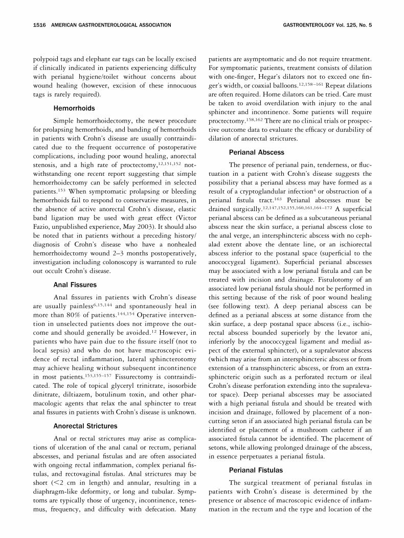
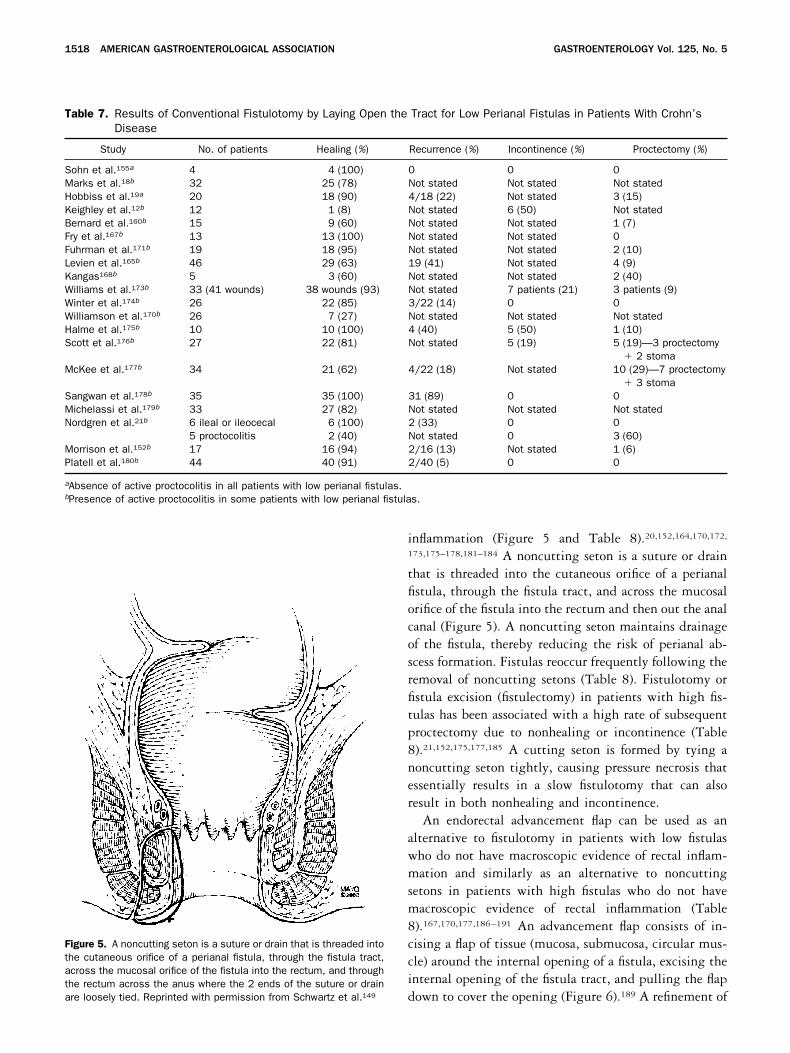
fistula. Patients who have low fistulas (superficial, lowintersphincteric, low transsphincteric) may be treatedby laying open the fistula tract through 1- or 2-stagefistulotomy (Figure 4 and Table 7).12,18,19,21,152,
155,160,165,167,168,170,171,173–180 The initial healing ratesin 21 studies of fistulotomy in patients with low fistulasranged from 8% to 100%, with healing rates of 80%–100% in 13 of 21 studies, 60%–79% in 5 of 13 studies,and �59% in 3 of 21 studies (Table 7). The recurrencerates in 10 studies of patients with fistulotomy whoinitially healed ranged from 5% to 89%, with recurrencerates of 0%–20% in 7 of 10 studies, 21%–40% in 1 of10 studies, and �41% in 2 of 10 studies (Table 7). Therates of incontinence following fistulotomy range from0% to 50%, and the rates of proctectomy or long-termdiverting stoma following fistulotomy range from 6% to60% (Table 7). The wide range of rates of recurrence,incontinence, and proctectomy may reflect differences inpatient selection and referral center bias, along withother differences. In general, there is a trend toward
greater healing rates following fistulotomy for low fistu-las in patients without macroscopic evidence of inflam-mation of the rectum when compared with patients withactive inflammation of the rectosigmoid colon; however,even in patients without macroscopic evidence of rectalinflammation, a nonhealing wound following fistulot-omy for a low fistula will lead to proctectomy in somepatients (Table 7). For this reason, many surgical expertsadvocate the use of a noncutting seton (see followingtext) rather than fistulotomy in patients with low fistulasand active inflammation of the rectosigmoid colon.
High fistulas involving a significant portion of theexternal anal sphincter (such as high intersphincteric,high transsphincteric, suprasphincteric, or extrasphinc-teric fistulas) require a more conservative surgical ap-proach to reduce the risk of incontinence. Noncuttingsetons are the treatment of choice in patients with highfistulas who have active inflammation of the rectosig-moid colon and may also be used in patients with highfistulas who do not have macroscopic evidence of rectal
Figure 4. In the absence of active proctocolitis, simple low transsphincteric, intersphincteric, and superficial fistulas can be treated with afistulotomy. Reprinted with permission from Schwartz et al.149
November 2003 AMERICAN GASTROENTEROLOGICAL ASSOCIATION 1517
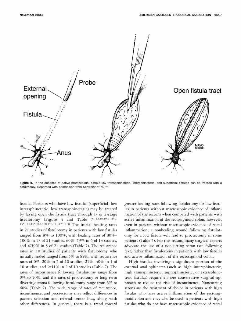
inflammation (Figure 5 and Table 8).20,152,164,170,172,
173,175–178,181–184 A noncutting seton is a suture or drainthat is threaded into the cutaneous orifice of a perianalfistula, through the fistula tract, and across the mucosalorifice of the fistula into the rectum and then out the analcanal (Figure 5). A noncutting seton maintains drainageof the fistula, thereby reducing the risk of perianal ab-scess formation. Fistulas reoccur frequently following theremoval of noncutting setons (Table 8). Fistulotomy orfistula excision (fistulectomy) in patients with high fis-tulas has been associated with a high rate of subsequentproctectomy due to nonhealing or incontinence (Table8).21,152,175,177,185 A cutting seton is formed by tying anoncutting seton tightly, causing pressure necrosis thatessentially results in a slow fistulotomy that can alsoresult in both nonhealing and incontinence.
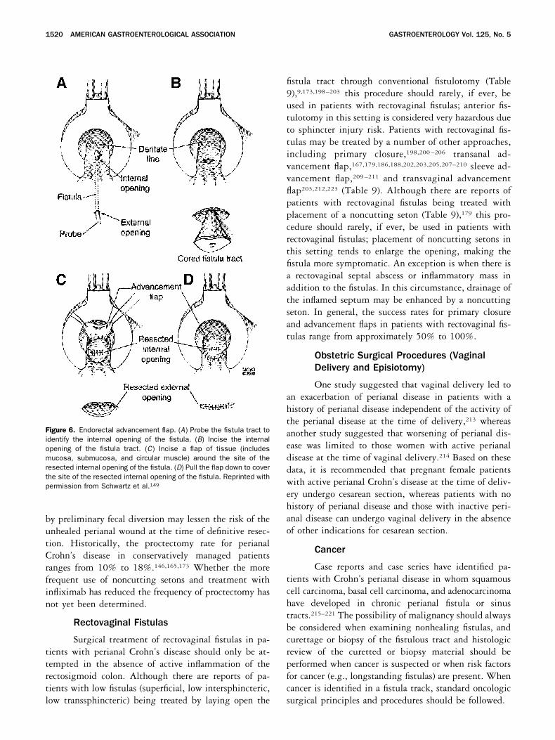
An endorectal advancement flap can be used as analternative to fistulotomy in patients with low fistulaswho do not have macroscopic evidence of rectal inflam-mation and similarly as an alternative to noncuttingsetons in patients with high fistulas who do not havemacroscopic evidence of rectal inflammation (Table8).167,170,177,186–191 An advancement flap consists of in-cising a flap of tissue (mucosa, submucosa, circular mus-cle) around the internal opening of a fistula, excising theinternal opening of the fistula tract, and pulling the flapdown to cover the opening (Figure 6).189 A refinement of
Figure 5. A noncutting seton is a suture or drain that is threaded intothe cutaneous orifice of a perianal fistula, through the fistula tract,across the mucosal orifice of the fistula into the rectum, and throughthe rectum across the anus where the 2 ends of the suture or drainare loosely tied. Reprinted with permission from Schwartz et al.149
Table 7. Results of Conventional Fistulotomy by Laying Open the Tract for Low Perianal Fistulas in Patients With Crohn’sDisease
Study No. of patients Healing (%) Recurrence (%) Incontinence (%) Proctectomy (%)
Sohn et al.155a 4 4 (100) 0 0 0Marks et al.18b 32 25 (78) Not stated Not stated Not statedHobbiss et al.19a 20 18 (90) 4/18 (22) Not stated 3 (15)Keighley et al.12b 12 1 (8) Not stated 6 (50) Not statedBernard et al.160b 15 9 (60) Not stated Not stated 1 (7)Fry et al.167b 13 13 (100) Not stated Not stated 0Fuhrman et al.171b 19 18 (95) Not stated Not stated 2 (10)Levien et al.165b 46 29 (63) 19 (41) Not stated 4 (9)Kangas168b 5 3 (60) Not stated Not stated 2 (40)Williams et al.173b 33 (41 wounds) 38 wounds (93) Not stated 7 patients (21) 3 patients (9)Winter et al.174b 26 22 (85) 3/22 (14) 0 0Williamson et al.170b 26 7 (27) Not stated Not stated Not statedHalme et al.175b 10 10 (100) 4 (40) 5 (50) 1 (10)Scott et al.176b 27 22 (81) Not stated 5 (19) 5 (19)—3 proctectomy
� 2 stomaMcKee et al.177b 34 21 (62) 4/22 (18) Not stated 10 (29)—7 proctectomy
� 3 stomaSangwan et al.178b 35 35 (100) 31 (89) 0 0Michelassi et al.179b 33 27 (82) Not stated Not stated Not statedNordgren et al.21b 6 ileal or ileocecal 6 (100) 2 (33) 0 0
5 proctocolitis 2 (40) Not stated 0 3 (60)Morrison et al.152b 17 16 (94) 2/16 (13) Not stated 1 (6)Platell et al.180b 44 40 (91) 2/40 (5) 0 0
aAbsence of active proctocolitis in all patients with low perianal fistulas.bPresence of active proctocolitis in some patients with low perianal fistulas.
1518 AMERICAN GASTROENTEROLOGICAL ASSOCIATION GASTROENTEROLOGY Vol. 125, No. 5
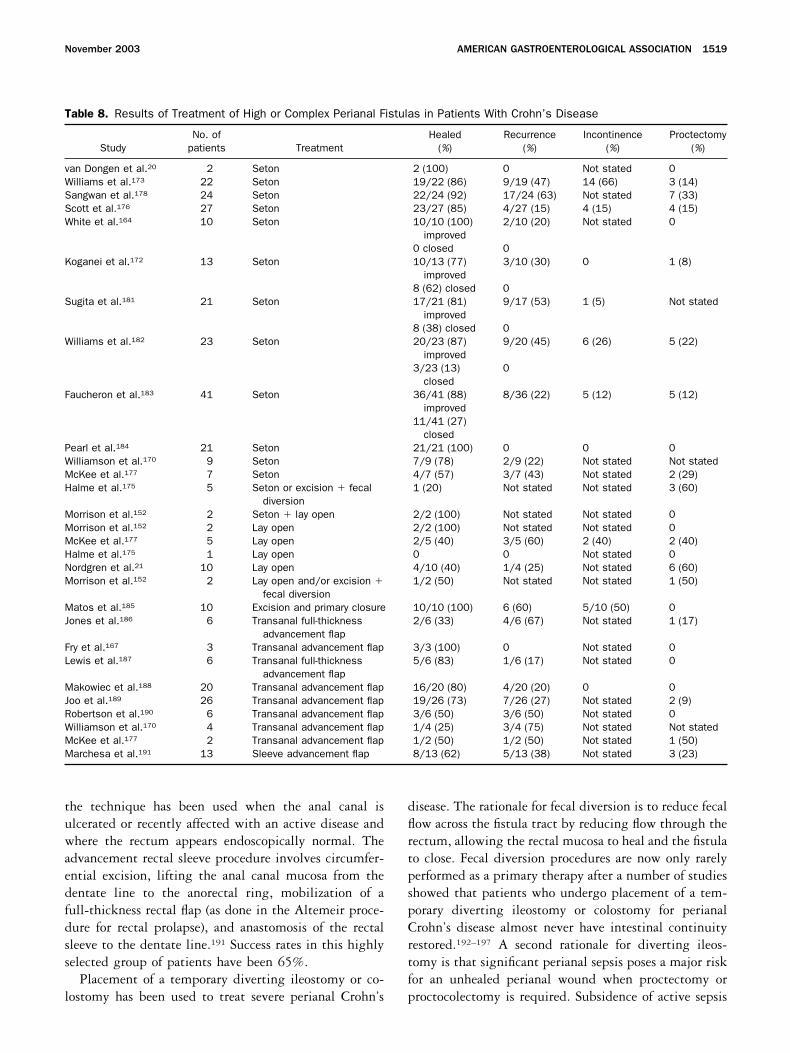
the technique has been used when the anal canal isulcerated or recently affected with an active disease andwhere the rectum appears endoscopically normal. Theadvancement rectal sleeve procedure involves circumfer-ential excision, lifting the anal canal mucosa from thedentate line to the anorectal ring, mobilization of afull-thickness rectal flap (as done in the Altemeir proce-dure for rectal prolapse), and anastomosis of the rectalsleeve to the dentate line.191 Success rates in this highlyselected group of patients have been 65%.
Placement of a temporary diverting ileostomy or co-lostomy has been used to treat severe perianal Crohn’s
disease. The rationale for fecal diversion is to reduce fecalflow across the fistula tract by reducing flow through therectum, allowing the rectal mucosa to heal and the fistulato close. Fecal diversion procedures are now only rarelyperformed as a primary therapy after a number of studiesshowed that patients who undergo placement of a tem-porary diverting ileostomy or colostomy for perianalCrohn’s disease almost never have intestinal continuityrestored.192–197 A second rationale for diverting ileos-tomy is that significant perianal sepsis poses a major riskfor an unhealed perianal wound when proctectomy orproctocolectomy is required. Subsidence of active sepsis
Table 8. Results of Treatment of High or Complex Perianal Fistulas in Patients With Crohn’s Disease
StudyNo. of
patients TreatmentHealed
(%)Recurrence
(%)Incontinence
(%)Proctectomy
(%)
van Dongen et al.20 2 Seton 2 (100) 0 Not stated 0Williams et al.173 22 Seton 19/22 (86) 9/19 (47) 14 (66) 3 (14)Sangwan et al.178 24 Seton 22/24 (92) 17/24 (63) Not stated 7 (33)Scott et al.176 27 Seton 23/27 (85) 4/27 (15) 4 (15) 4 (15)White et al.164 10 Seton 10/10 (100)
improved2/10 (20) Not stated 0
0 closed 0Koganei et al.172 13 Seton 10/13 (77)
improved3/10 (30) 0 1 (8)
8 (62) closed 0Sugita et al.181 21 Seton 17/21 (81)
improved9/17 (53) 1 (5) Not stated
8 (38) closed 0Williams et al.182 23 Seton 20/23 (87)
improved9/20 (45) 6 (26) 5 (22)
3/23 (13)closed
0
Faucheron et al.183 41 Seton 36/41 (88)improved
8/36 (22) 5 (12) 5 (12)
11/41 (27)closed
Pearl et al.184 21 Seton 21/21 (100) 0 0 0Williamson et al.170 9 Seton 7/9 (78) 2/9 (22) Not stated Not statedMcKee et al.177 7 Seton 4/7 (57) 3/7 (43) Not stated 2 (29)Halme et al.175 5 Seton or excision � fecal
diversion1 (20) Not stated Not stated 3 (60)
Morrison et al.152 2 Seton � lay open 2/2 (100) Not stated Not stated 0Morrison et al.152 2 Lay open 2/2 (100) Not stated Not stated 0McKee et al.177 5 Lay open 2/5 (40) 3/5 (60) 2 (40) 2 (40)Halme et al.175 1 Lay open 0 0 Not stated 0Nordgren et al.21 10 Lay open 4/10 (40) 1/4 (25) Not stated 6 (60)Morrison et al.152 2 Lay open and/or excision �
fecal diversion1/2 (50) Not stated Not stated 1 (50)
Matos et al.185 10 Excision and primary closure 10/10 (100) 6 (60) 5/10 (50) 0Jones et al.186 6 Transanal full-thickness
advancement flap2/6 (33) 4/6 (67) Not stated 1 (17)
Fry et al.167 3 Transanal advancement flap 3/3 (100) 0 Not stated 0Lewis et al.187 6 Transanal full-thickness
advancement flap5/6 (83) 1/6 (17) Not stated 0
Makowiec et al.188 20 Transanal advancement flap 16/20 (80) 4/20 (20) 0 0Joo et al.189 26 Transanal advancement flap 19/26 (73) 7/26 (27) Not stated 2 (9)Robertson et al.190 6 Transanal advancement flap 3/6 (50) 3/6 (50) Not stated 0Williamson et al.170 4 Transanal advancement flap 1/4 (25) 3/4 (75) Not stated Not statedMcKee et al.177 2 Transanal advancement flap 1/2 (50) 1/2 (50) Not stated 1 (50)Marchesa et al.191 13 Sleeve advancement flap 8/13 (62) 5/13 (38) Not stated 3 (23)
November 2003 AMERICAN GASTROENTEROLOGICAL ASSOCIATION 1519
by preliminary fecal diversion may lessen the risk of theunhealed perianal wound at the time of definitive resec-tion. Historically, the proctectomy rate for perianalCrohn’s disease in conservatively managed patientsranges from 10% to 18%.146,165,173 Whether the morefrequent use of noncutting setons and treatment withinfliximab has reduced the frequency of proctectomy hasnot yet been determined.
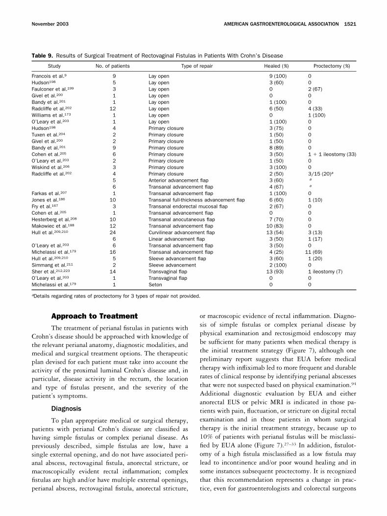
Rectovaginal Fistulas
Surgical treatment of rectovaginal fistulas in pa-tients with perianal Crohn’s disease should only be at-tempted in the absence of active inflammation of therectosigmoid colon. Although there are reports of pa-tients with low fistulas (superficial, low intersphincteric,low transsphincteric) being treated by laying open the
fistula tract through conventional fistulotomy (Table9),9,173,198–203 this procedure should rarely, if ever, beused in patients with rectovaginal fistulas; anterior fis-tulotomy in this setting is considered very hazardous dueto sphincter injury risk. Patients with rectovaginal fis-tulas may be treated by a number of other approaches,including primary closure,198,200 –206 transanal ad-vancement flap,167,179,186,188,202,203,205,207–210 sleeve ad-vancement flap,209 –211 and transvaginal advancementflap203,212,223 (Table 9). Although there are reports ofpatients with rectovaginal fistulas being treated withplacement of a noncutting seton (Table 9),179 this pro-cedure should rarely, if ever, be used in patients withrectovaginal fistulas; placement of noncutting setons inthis setting tends to enlarge the opening, making thefistula more symptomatic. An exception is when there isa rectovaginal septal abscess or inflammatory mass inaddition to the fistulas. In this circumstance, drainage ofthe inflamed septum may be enhanced by a noncuttingseton. In general, the success rates for primary closureand advancement flaps in patients with rectovaginal fis-tulas range from approximately 50% to 100%.
Obstetric Surgical Procedures (VaginalDelivery and Episiotomy)
One study suggested that vaginal delivery led toan exacerbation of perianal disease in patients with ahistory of perianal disease independent of the activity ofthe perianal disease at the time of delivery,213 whereasanother study suggested that worsening of perianal dis-ease was limited to those women with active perianaldisease at the time of vaginal delivery.214 Based on thesedata, it is recommended that pregnant female patientswith active perianal Crohn’s disease at the time of deliv-ery undergo cesarean section, whereas patients with nohistory of perianal disease and those with inactive peri-anal disease can undergo vaginal delivery in the absenceof other indications for cesarean section.
Cancer
Case reports and case series have identified pa-tients with Crohn’s perianal disease in whom squamouscell carcinoma, basal cell carcinoma, and adenocarcinomahave developed in chronic perianal fistula or sinustracts.215–221 The possibility of malignancy should alwaysbe considered when examining nonhealing fistulas, andcurettage or biopsy of the fistulous tract and histologicreview of the curetted or biopsy material should beperformed when cancer is suspected or when risk factorsfor cancer (e.g., longstanding fistulas) are present. Whencancer is identified in a fistula track, standard oncologicsurgical principles and procedures should be followed.
Figure 6. Endorectal advancement flap. (A) Probe the fistula tract toidentify the internal opening of the fistula. (B) Incise the internalopening of the fistula tract. (C) Incise a flap of tissue (includesmucosa, submucosa, and circular muscle) around the site of theresected internal opening of the fistula. (D) Pull the flap down to coverthe site of the resected internal opening of the fistula. Reprinted withpermission from Schwartz et al.149
1520 AMERICAN GASTROENTEROLOGICAL ASSOCIATION GASTROENTEROLOGY Vol. 125, No. 5
Approach to TreatmentThe treatment of perianal fistulas in patients with
Crohn’s disease should be approached with knowledge ofthe relevant perianal anatomy, diagnostic modalities, andmedical and surgical treatment options. The therapeuticplan devised for each patient must take into account theactivity of the proximal luminal Crohn’s disease and, inparticular, disease activity in the rectum, the locationand type of fistulas present, and the severity of thepatient’s symptoms.
Diagnosis
To plan appropriate medical or surgical therapy,patients with perianal Crohn’s disease are classified ashaving simple fistulas or complex perianal disease. Aspreviously described, simple fistulas are low, have asingle external opening, and do not have associated peri-anal abscess, rectovaginal fistula, anorectal stricture, ormacroscopically evident rectal inflammation; complexfistulas are high and/or have multiple external openings,perianal abscess, rectovaginal fistula, anorectal stricture,
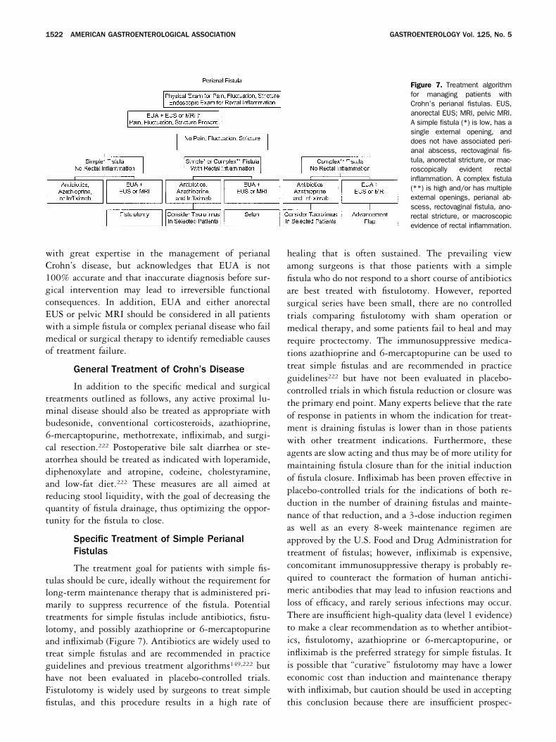
or macroscopic evidence of rectal inflammation. Diagno-sis of simple fistulas or complex perianal disease byphysical examination and rectosigmoid endoscopy maybe sufficient for many patients when medical therapy isthe initial treatment strategy (Figure 7), although onepreliminary report suggests that EUA before medicaltherapy with infliximab led to more frequent and durablerates of clinical response by identifying perianal abscessesthat were not suspected based on physical examination.94
Additional diagnostic evaluation by EUA and eitheranorectal EUS or pelvic MRI is indicated in those pa-tients with pain, fluctuation, or stricture on digital rectalexamination and in those patients in whom surgicaltherapy is the initial treatment strategy, because up to10% of patients with perianal fistulas will be misclassi-fied by EUA alone (Figure 7).27–33 In addition, fistulot-omy of a high fistula misclassified as a low fistula maylead to incontinence and/or poor wound healing and insome instances subsequent proctectomy. It is recognizedthat this recommendation represents a change in prac-tice, even for gastroenterologists and colorectal surgeons
Table 9. Results of Surgical Treatment of Rectovaginal Fistulas in Patients With Crohn’s Disease
Study No. of patients Type of repair Healed (%) Proctectomy (%)
Francois et al.9 9 Lay open 9 (100) 0Hudson198 5 Lay open 3 (60) 0Faulconer et al.199 3 Lay open 0 2 (67)Givel et al.200 1 Lay open 0 0Bandy et al.201 1 Lay open 1 (100) 0Radcliffe et al.202 12 Lay open 6 (50) 4 (33)Williams et al.173 1 Lay open 0 1 (100)O’Leary et al.203 1 Lay open 1 (100) 0Hudson198 4 Primary closure 3 (75) 0Tuxen et al.204 2 Primary closure 1 (50) 0Givel et al.200 2 Primary closure 1 (50) 0Bandy et al.201 9 Primary closure 8 (89) 0Cohen et al.205 6 Primary closure 3 (50) 1 � 1 ileostomy (33)O’Leary et al.203 2 Primary closure 1 (50) 0Wiskind et al.206 3 Primary closure 3 (100) 0Radcliffe et al.202 4 Primary closure 2 (50) 3/15 (20)a
5 Anterior advancement flap 3 (60) a
6 Transanal advancement flap 4 (67) a
Farkas et al.207 1 Transanal advancement flap 1 (100) 0Jones et al.186 10 Transanal full-thickness advancement flap 6 (60) 1 (10)Fry et al.167 3 Transanal endorectal mucosal flap 2 (67) 0Cohen et al.205 1 Transanal advancement flap 0 0Hesterberg et al.208 10 Transanal anocutaneous flap 7 (70) 0Makowiec et al.188 12 Transanal advancement flap 10 (83) 0Hull et al.209,210 24 Curvilinear advancement flap 13 (54) 3 (13)
6 Linear advancement flap 3 (50) 1 (17)O’Leary et al.203 6 Transanal advancement flap 3 (50) 0Michelassi et al.179 16 Transanal advancement flap 4 (25) 11 (69)Hull et al.209,210 5 Sleeve advancement flap 3 (60) 1 (20)Simmang et al.211 2 Sleeve advancement 2 (100) 0Sher et al.212,223 14 Transvaginal flap 13 (93) 1 ileostomy (7)O’Leary et al.203 1 Transvaginal flap 0 0Michelassi et al.179 1 Seton 0 0
aDetails regarding rates of proctectomy for 3 types of repair not provided.
November 2003 AMERICAN GASTROENTEROLOGICAL ASSOCIATION 1521
with great expertise in the management of perianalCrohn’s disease, but acknowledges that EUA is not100% accurate and that inaccurate diagnosis before sur-gical intervention may lead to irreversible functionalconsequences. In addition, EUA and either anorectalEUS or pelvic MRI should be considered in all patientswith a simple fistula or complex perianal disease who failmedical or surgical therapy to identify remediable causesof treatment failure.
General Treatment of Crohn’s Disease
In addition to the specific medical and surgicaltreatments outlined as follows, any active proximal lu-minal disease should also be treated as appropriate withbudesonide, conventional corticosteroids, azathioprine,6-mercaptopurine, methotrexate, infliximab, and surgi-cal resection.222 Postoperative bile salt diarrhea or ste-atorrhea should be treated as indicated with loperamide,diphenoxylate and atropine, codeine, cholestyramine,and low-fat diet.222 These measures are all aimed atreducing stool liquidity, with the goal of decreasing thequantity of fistula drainage, thus optimizing the oppor-tunity for the fistula to close.
Specific Treatment of Simple PerianalFistulas
The treatment goal for patients with simple fis-tulas should be cure, ideally without the requirement forlong-term maintenance therapy that is administered pri-marily to suppress recurrence of the fistula. Potentialtreatments for simple fistulas include antibiotics, fistu-lotomy, and possibly azathioprine or 6-mercaptopurineand infliximab (Figure 7). Antibiotics are widely used totreat simple fistulas and are recommended in practiceguidelines and previous treatment algorithms149,222 buthave not been evaluated in placebo-controlled trials.Fistulotomy is widely used by surgeons to treat simplefistulas, and this procedure results in a high rate of
healing that is often sustained. The prevailing viewamong surgeons is that those patients with a simplefistula who do not respond to a short course of antibioticsare best treated with fistulotomy. However, reportedsurgical series have been small, there are no controlledtrials comparing fistulotomy with sham operation ormedical therapy, and some patients fail to heal and mayrequire proctectomy. The immunosuppressive medica-tions azathioprine and 6-mercaptopurine can be used totreat simple fistulas and are recommended in practiceguidelines222 but have not been evaluated in placebo-controlled trials in which fistula reduction or closure wasthe primary end point. Many experts believe that the rateof response in patients in whom the indication for treat-ment is draining fistulas is lower than in those patientswith other treatment indications. Furthermore, theseagents are slow acting and thus may be of more utility formaintaining fistula closure than for the initial inductionof fistula closure. Infliximab has been proven effective inplacebo-controlled trials for the indications of both re-duction in the number of draining fistulas and mainte-nance of that reduction, and a 3-dose induction regimenas well as an every 8-week maintenance regimen areapproved by the U.S. Food and Drug Administration fortreatment of fistulas; however, infliximab is expensive,concomitant immunosuppressive therapy is probably re-quired to counteract the formation of human antichi-meric antibodies that may lead to infusion reactions andloss of efficacy, and rarely serious infections may occur.There are insufficient high-quality data (level 1 evidence)to make a clear recommendation as to whether antibiot-ics, fistulotomy, azathioprine or 6-mercaptopurine, orinfliximab is the preferred strategy for simple fistulas. Itis possible that “curative” fistulotomy may have a lowereconomic cost than induction and maintenance therapywith infliximab, but caution should be used in acceptingthis conclusion because there are insufficient prospec-
Figure 7. Treatment algorithmfor managing patients withCrohn’s perianal fistulas. EUS,anorectal EUS; MRI, pelvic MRI.A simple fistula (*) is low, has asingle external opening, anddoes not have associated peri-anal abscess, rectovaginal fis-tula, anorectal stricture, or mac-roscopically evident rectalinflammation. A complex fistula(**) is high and/or has multipleexternal openings, perianal ab-scess, rectovaginal fistula, ano-rectal stricture, or macroscopicevidence of rectal inflammation.
1522 AMERICAN GASTROENTEROLOGICAL ASSOCIATION GASTROENTEROLOGY Vol. 125, No. 5
tively collected data detailing the morbidity (rates ofincontinence and proctectomy) following fistulotomy.Additionally, there are insufficient data to determinewhether long-term maintenance therapy with infliximabis required in patients with simple fistulas who respondto induction therapy. Tacrolimus and cyclosporine arenot appropriate treatment for simple fistulas because oftoxicity.
Specific Treatment of Complex PerianalFistulas
The treatment goal for patients with complexfistulas is typically fistula closure and then suppression ofrecurrence. Potential treatments for complex fistulas in-clude antibiotics, azathioprine and 6-mercaptopurine,infliximab, and surgery (dilation of anal strictures, place-ment of noncutting setons, endorectal advancement flap,repair of rectovaginal fistulas, fecal diversion, and proc-tectomy) (Figure 7). Tacrolimus and cyclosporine mayrarely be used in selected patients. Antibiotics are widelyused to treat complex fistulas and are recommended inpractice guidelines and treatment algorithms149,222 buthave not been evaluated in placebo-controlled trials.Relapse rates for complex fistulas are high after antibiotictherapy is discontinued, and their use should probably beadjunctive in combination with other medical agents orsurgery in this setting. Similarly, the immunosuppres-sive medications azathioprine and 6-mercaptopurinehave been used to treat complex fistulas and are recom-mended in practice guidelines222 but have not beenevaluated in placebo-controlled trials for fistula closure.These agents are slow acting and thus are of more utilityfor maintaining fistula closure than for the initial induc-tion of fistula closure. In contrast to antibiotics andimmunosuppressive medications, infliximab has beenproven to be effective in placebo-controlled trials forreduction in the number of draining fistulas and main-tenance of that reduction, and treatment of fistulas witha 3-dose induction regimen and an every 8-week main-tenance regimen of infliximab is approved by the Foodand Drug Administration; although therapy with inflix-imab is expensive and concomitant immunosuppressivetherapy is probably required, these factors are of lessconsequence in patients with complex perianal fistulaswho have an otherwise poor prognosis. Surgical therapyfor complex perianal disease is largely palliative. Perianalabscesses should be drained and anal strictures dilated.Noncutting setons can be placed in fistula tracts inpatients with macroscopic rectal inflammation, and en-dorectal advancement flap procedures for high perianalfistulas and rectovaginal fistulas can be performed inpatients without rectal inflammation. However, the re-
currence rates following removal of noncutting setonsand following endorectal advancement flap proceduresare both relatively high. Setons can be left in placeindefinitely; however, given the alternative of suppressivemedical therapy, patients may not prefer this option.Fistulotomy and cutting setons are relatively contraindi-cated due to risk of nonhealing and incontinence. Be-cause infliximab therapy can completely close all fistulatracks in many patients with complex fistulas, mostgastroenterologists now believe that infliximab is theinitial treatment of choice in this setting (it is debated bysurgeons whether infliximab or noncutting setons is theinitial treatment of choice for the subgroup of patientswith complex perianal fistulas who do not have activerectal disease, because there are no data on patient ac-ceptance of long-term noncutting setons versus treat-ment with infliximab). In patients treated with inflix-imab, azathioprine, 6-mercaptopurine, or methotrexateshould be initiated before or coadministered routinelyboth to counteract an immunogenic reaction to inflix-imab and as maintenance of remission therapy. Manypatients will require combination maintenance therapywith azathioprine, 6-mercaptopurine, or methotrexateand infliximab. Temporary adjunctive therapy with an-tibiotics may be considered. It is clear from clinical trialswith infliximab that routine EUA and seton placementbefore initiating infliximab therapy is not mandatory.Whether or not the outcome with infliximab therapy forcomplex fistulas is improved through a multimodalityapproach that includes both temporary placement ofsetons and therapy with infliximab, azathioprine or6-mercaptopurine, and an antibiotic is unknown. Therationale for such an approach is that the temporaryplacement of setons guarantees resolution of sepsis(which can be a reason for infliximab failure); the setonsare removed after 1 or 2 doses of infliximab to allow thefistulas to close. Patients with complex fistulas whoinitially fail treatment with infliximab should undergoanorectal EUS or pelvic MRI as well as EUA withplacement of setons as indicated while continuing treat-ment with infliximab, azathioprine or 6-mercaptopurine,and antibiotics. Tacrolimus or cyclosporine can rarely beconsidered in selected patients who fail multimodalitytreatment with other medical and surgical therapies,including infliximab.222 This practice is based on uncon-trolled case series with cyclosporine and a single short-term placebo-controlled trial with tacrolimus thatshowed a reduction in the number of draining fistulas;however, nephrotoxicity and other side effects occur fre-quently and should be used with caution. In addition,the trials examining tacrolimus and cyclosporine have
November 2003 AMERICAN GASTROENTEROLOGICAL ASSOCIATION 1523
been of short duration, without determining whethermaintenance therapy after initial fistula closure is safeand effective. As a last resort, fecal diversion or proctec-tomy may be undertaken.
Specific Treatment of Rectovaginal Fistulas
The treatment goals for patients with recto-vaginal fistulas are initially to decrease fistula drainageto a minimal level that is “acceptable” or ideally tocompletely close the fistula, then suppression of recur-rence, and then cure if possible. Potential treatmentsfor rectovaginal fistulas include both medical therapyand surgery. 6-Mercaptopurine, infliximab, cyclospor-ine, and tacrolimus have all been used to treat recto-vaginal fistulas in uncontrolled series.81,90,106,107,112,117
A recent controlled trial of maintenance infliximabinfusions in patients with fistulas who responded toinfliximab included a subgroup of patients with rec-tovaginal fistulas.89 In general, the rates of closure forrectovaginal fistulas appear to be lower than for peri-anal fistulas. Surgical treatment of rectovaginal fistulascan only be performed when there is endoscopic heal-ing of the rectosigmoid mucosa. Thus, standard med-ical therapy with conventional corticosteroids, aza-thioprine, 6-mercaptopurine, methotrexate, andinfliximab should be administered as indicated tocontrol active luminal inflammatory disease in therectosigmoid colon.222 Other perianal disease shouldbe treated as previously outlined. If the rectovaginalfistula persists after the patient has received medicaltherapy to treat both the fistula itself and the rectosig-moid mucosa and there is no evidence of an anorectalstricture or active rectal disease, then surgical repairwith transanal or transvaginal advancement flaps, orlaparotomy with primary closure or sleeve advance-ment flap, can be performed. Temporary divertingileostomy or colostomy may be performed simulta-neously in selected cases. If the advancement flapprocedures (which involve coring out the fistula) fail,the rectovaginal fistula may actually increase in sizeand result in a worsening of symptoms. Thus, ad-vancement flap surgery should be reserved for patientswith disabling symptoms (by their definition), that is,stool per vagina, recurrent vaginitis, and vulvar exco-riation. As a last resort, fecal diversion or proctectomymay be undertaken. Some women in whom rectovagi-nal fistula drainage has diminished but not stoppedfollowing medical and/or surgical therapy may chooseto accept residual fistula drainage over proctectomywith an ostomy to optimize their overall quality oflife.
ConclusionsPerianal disease occurs frequently in patients with
Crohn’s disease. Diagnostic evaluation with physical ex-amination and rectosigmoid endoscopy, supplemented insome cases with EUA and anorectal ultrasonography orpelvic MRI, is required to determine the location andtype of fistulas and the presence or absence of macro-scopic rectal inflammation. Skin tags and hemorrhoidsshould not be operated on. Most anal fissures should notbe operated on. Lateral sphincterotomy can be consideredin selected cases. Anorectal strictures should be dilatedand perianal abscesses drained. Simple fistulas can betreated with antibiotics, infliximab, or fistulotomy. Thetreatment goal is cure without suppressive maintenancetherapy. Complex perianal disease should be treated ini-tially with infliximab and azathioprine or 6-mercapto-purine, followed by maintenance therapy with azathio-prine or 6-mercaptopurine, in some cases combined withinfliximab. Antibiotics may be used as adjunctive ther-apy during the induction phase of treatment. EUA andplacement of noncutting setons or performing endorectaladvancement flap procedures is reserved for patients whofail a trial of medical therapy. Tacrolimus or cyclosporinecan rarely be considered in selected patients who failmultimodality treatment with other medical and surgi-cal therapies, including infliximab, before proceeding tofecal diversion or proctectomy.
WILLIAM J. SANDBORNMayo ClinicRochester, Minnesota
VICTOR W. FAZIOCleveland ClinicCleveland, Ohio
BRIAN G. FEAGANUniversity of Western OntarioLondon, Ontario, Canada
STEPHEN B. HANAUERUniversity of ChicagoChicago, Illinois
References1. Goligher J. Fistula-in-ano. In: Goligher J, ed. Surgery of the anus,
rectum, and colon. 5th ed. Bailliere Tindall, 1984;178–220.2. Parks A. The pathogenesis and treatment of fistula-in-ano. BMJ
1961;1:463–1469.3. Milligan ET, Morgan CN. Surgical anatomy of the anal canal with
special reference to anorectal fistulae. Lancet 1934;2:1213.4. Parks AG, Gordon PH, Hardcastle JD. A classification of fistula-
in-ano. Br J Surg 1976;63:1–12.5. Buchmann P, Alexander-Williams J. Classification of perianal
Crohn’s disease. Clin Gastroenterol 1980;9:323–330.
1524 AMERICAN GASTROENTEROLOGICAL ASSOCIATION GASTROENTEROLOGY Vol. 125, No. 5
6. Hughes LE. Surgical pathology and management of anorectalCrohn’s disease. J R Soc Med 1978;71:644–651.
7. Hughes LE. Clinical classification of perianal Crohn’s disease.Dis Colon Rectum 1992;35:928–932.
8. Alexander-Williams J, Hellers G, Hughes LE, Minervini S, Spe-ranza V. Classification of perianal Crohn’s disease. Gastroen-terol Int 1992;5:216–220.
9. Francois Y, Vignal J, Descos L. Outcome of perianal fistulae inCrohn’s disease—value of Hughes’ pathogenic classification.Int J Colorectal Dis 1993;8:39–41.
10. Pikarsky AJ, Gervaz P, Wexner SD. Perianal Crohn disease: anew scoring system to evaluate and predict outcome of surgicalintervention. Arch Surg 2002;137:774–777; discussion 778.
11. Bell SJ, Williams AB, Wiesel P, Wilkinson K, Cohen RCG, KammMA. The clinical course of fistulating Crohn’s disease. AlimentPharmacol Ther 2003;17:1145–1151.
12. Keighley MR, Allan RN. Current status and influence of opera-tion on perianal Crohn’s disease. Int J Colorectal Dis 1986;1:104–107.
13. Farmer RG, Hawk WA, Turnbull RB Jr. Clinical patterns in Crohn’sdisease: a statistical study of 615 cases. Gastroenterology1975;68:627–635.
14. Williams DR, Coller JA, Corman ML, Nugent FW, VeidenheimerMC. Anal complications in Crohn’s disease. Dis Colon Rectum1981;24:22–24.
15. Fielding JF. Perianal lesions in Crohn’s disease. J R Coll SurgEdinb 1972;17:32–37.
16. Goebell H. Perianal complications in Crohn’s disease. NethJ Med 1990;37(suppl 1):S47–S51.
17. Greenstein AJ, Kark AE, Dreiling DA. Crohn’s disease of thecolon. I. Fistula in Crohn’s disease of the colon, classificationpresenting features and management in 63 patients. Am JGastroenterol 1974;62:419–429.
18. Marks CG, Ritchie JK, Lockhart-Mummery HE. Anal fistulas inCrohn’s disease. Br J Surg 1981;68:525–527.
19. Hobbiss JH, Schofield PF. Management of perianal Crohn’sdisease. J R Soc Med 1982;75:414–417.
20. van Dongen LM, Lubbers EJ. Perianal fistulas in patients withCrohn’s disease. Arch Surg 1986;121:1187–1190.
21. Nordgren S, Fasth S, Hulten L. Anal fistulas in Crohn’s disease:incidence and outcome of surgical treatment. Int J ColorectalDis 1992;7:214–218.
22. Rankin GB, Watts HD, Melnyk CS, Kelley ML Jr. National Coop-erative Crohn’s Disease Study: extraintestinal manifestationsand perianal complications. Gastroenterology 1979;77:914–920.
23. Tolia V. Perianal Crohn’s disease in children and adolescents.Am J Gastroenterol 1996;91:922–926.
24. Hellers G, Bergstrand O, Ewerth S, Holmstrom B. Occurrenceand outcome after primary treatment of anal fistulae in Crohn’sdisease. Gut 1980;21:525–527.
25. Schwartz DA, Loftus EV Jr, Tremaine WJ, Panaccione R, Harm-sen WS, Zinsmeister AR, Sandborn WJ. The natural history offistulizing Crohn’s disease in Olmsted County, Minnesota. Gas-troenterology 2002;122:875–880.
26. Lockhart-Mummery HE. Symposium. Crohn’s disease: anal le-sions. Dis Colon Rectum 1975;18:200–202.
27. Lunniss PJ, Barker PG, Sultan AH, Armstrong P, Reznek RH,Bartram CI, Cottam KS, Phillips RK. Magnetic resonance imag-ing of fistula-in-ano. Dis Colon Rectum 1994;37:708–718.
28. Barker PG, Lunniss PJ, Armstrong P, Reznek RH, Cottam K,Phillips RK. Magnetic resonance imaging of fistula-in-ano: tech-nique, interpretation and accuracy. Clin Radiol 1994;49:7–13.
29. deSouza NM, Hall AS, Puni R, Gilderdale DJ, Young IR, KmiotWA. High resolution magnetic resonance imaging of the analsphincter using a dedicated endoanal coil. Comparison of mag-
netic resonance imaging with surgical findings. Dis Colon Rec-tum 1996;39:926–934.
30. Haggett PJ, Moore NR, Shearman JD, Travis SP, Jewell DP,Mortensen NJ. Pelvic and perineal complications of Crohn’sdisease: assessment using magnetic resonance imaging. Gut1995;36:407–410.
31. Spencer JA, Chapple K, Wilson D, Ward J, Windsor AC, AmbroseNS. Outcome after surgery for perianal fistula: predictive valueof MR imaging. AJR Am J Roentgenol 1998;171:403–406.
32. Schwartz DA, Wiersema MJ, Dudiak KM, Fletcher JG, Clain JE,Tremaine WJ, Zinsmeister AR, Norton ID, Boardman LA, DevineRM, Wolff BG, Young-Fadok TM, Diehl NN, Pemberton JH, Sand-born WJ. A comparison of endoscopic ultrasound, magneticresonance imaging, and exam under anesthesia for evaluationof Crohn’s perianal fistulas. Gastroenterology 2001;121:1064–1072.
33. Beets-Tan RG, Beets GL, van der Hoop AG, Kessels AG, VliegenRF, Baeten CG, van Engelshoven JM. Preoperative MR imagingof anal fistulas: does it really help the surgeon? Radiology2001;218:75–84.
34. Glass RE, Ritchie JK, Lennard-Jones JE, Hawley PR, Todd IP.Internal fistulas in Crohn’s disease. Dis Colon Rectum 1985;28:557–561.
35. Fazio VW, Wilk P, Turnbull RB Jr, Jagelman DG. The dilemma ofCrohn’s disease: ileosigmoidal fistula complicating Crohn’s dis-ease. Dis Colon Rectum 1977;20:381–386.
36. Kuijpers HC, Schulpen T. Fistulography for fistula-in-ano. Is ituseful? Dis Colon Rectum 1985;28:103–104.
37. Pomerri F, Pittarello F, Dodi G, Pianon P, Muzzio PC. Radiologicdiagnosis of anal fistulae with radio-opaque markers. RadiolMed (Torino) 1988;75:632–637.
38. Weisman RI, Orsay CP, Pearl RK, Abcarian H. The role of fistu-lography in fistula-in-ano. Report of five cases. Dis Colon Rec-tum 1991;34:181–184.
39. Schratter-Sehn AU, Lochs H, Vogelsang H, Schurawitzki H,Herold C, Schratter M. Endoscopic ultrasonography versus com-puted tomography in the differential diagnosis of perianorectalcomplications in Crohn’s disease. Endoscopy 1993;25:582–586.
40. Van Outryve MJ, Pelckmans PA, Michielsen PP, Van MaerckeYM. Value of transrectal ultrasonography in Crohn’s disease.Gastroenterology 1991;101:1171–1177.
41. Fishman EK, Wolf EJ, Jones B, Bayless TM, Siegelman SS. CTevaluation of Crohn’s disease: effect on patient management.AJR Am J Roentgenol 1987;148:537–540.
42. Berliner L, Redmond P, Purow E, Megna D, Sottile V. Computedtomography in Crohn’s disease. Am J Gastroenterol 1982;77:548–553.
43. Yousem DM, Fishman EK, Jones B. Crohn disease: perirectaland perianal findings at CT. Radiology 1988;167:331–334.
44. Goldberg HI, Gore RM, Margulis AR, Moss AA, Baker EL. Com-puted tomography in the evaluation of Crohn disease. AJR Am JRoentgenol 1983;140:277–282.
45. Kerber GW, Greenberg M, Rubin JM. Computed tomographyevaluation of local and extraintestinal complications of Crohn’sdisease. Gastrointest Radiol 1984;9:143–148.
46. Laniado M, Makowiec F, Dammann F, Jehle EC, Claussen CD,Starlinger M. Perianal complications of Crohn disease: MR im-aging findings. Eur Radiol 1997;7:1035–1042.
47. Hussain SM, Outwater EK, Joekes EC, Ulrich F, Delemarre HB,Bemelman WA, Li X, Mitchell DG. Clinical and MR imagingfeatures of cryptoglandular and Crohn’s fistulas and abscesses.Abdom Imaging 2000;25:67–74.
48. Skalej M, Makowiec F, Weinlich M, Jenss H, Laniado M, Star-linger M [Magnetic resonance imaging in perianal Crohn’s dis-ease]. Dtsch Med Wochenschr 1993;118:1791–1796.
49. Koelbel G, Schmiedl U, Majer MC, Weber P, Jenss H, Kueper K,
November 2003 AMERICAN GASTROENTEROLOGICAL ASSOCIATION 1525
Hess CF. Diagnosis of fistulae and sinus tracts in patients withCrohn disease: value of MR imaging. AJR Am J Roentgenol1989;152:999–1003.
50. Van Assche G, Vanbeckevoort D, Bielen D, Coremans G, AerdenI, Noman M, D’Hoore A, Penninckx F, Marchal G, Cornillie F,Rutgeerts P. Magnetic resonance imaging of the effects ofinfliximab on perianal fistulizing Crohn’s disease. Am J Gastro-enterol 2003;98:332–339.
51. Agnholt J, Dahlerup J, Lyhne-Nielsen S, Buntzen S, Tottrup A,Lundorf E. Treatment of fistulizing Crohn’s disease with inflix-imab—one year follow by MRI-scan, ultrasound, and clinicalexamination (abstr). Gastroenterology 2002;122:A613.
52. Bell SJ, Halligan S, Windsor AC, Williams AB, Wiesel P, KammMA. Response of fistulating Crohn’s disease to infliximab treat-ment assessed by magnetic resonance imaging. Aliment Phar-macol Ther 2003;17:387–393.
53. Tio TL, Mulder CJ, Wijers OB, Sars PR, Tytgat GN. Endosonog-raphy of peri-anal and peri-colorectal fistula and/or abscess inCrohn’s disease. Gastrointest Endosc 1990;36:331–336.
54. Wijers O, Tio T, Tytgat G. Endosonography (transrectal andtransvaginal) in the assessment of perianorectal fistulas andabscesses: experience with 127 cases. In: Demling L, Fruhmor-gan P, eds. Non-neoplastic diseases of the anorectum. Kluwer,London, 1992;65–78.
55. Orsoni P, Barthet M, Portier F, Panuel M, Desjeux A, Grimaud JC.Prospective comparison of endosonography, magnetic reso-nance imaging and surgical findings in anorectal fistula andabscess complicating Crohn’s disease. Br J Surg 1999;86:360–364.
56. Stewart LK, McGee J, Wilson SR. Transperineal and transvaginalsonography of perianal inflammatory disease. AJR Am J Roent-genol 2001;177:627–632.
57. Sloots CE, Felt-Bersma RJ, Poen AC, Cuesta MA, MeuwissenSG. Assessment and classification of fistula-in-ano in patientswith Crohn’s disease by hydrogen peroxide enhanced transanalultrasound. Int J Colorectal Dis 2001;16:292–297.
58. Solomon MJ. Fistulae and abscesses in symptomatic perianalCrohn’s disease. Int J Colorectal Dis 1996;11:222–226.
59. van Bodegraven AA, Sloots CE, Felt-Bersma RJ, Meuwissen SG.Endosonographic evidence of persistence of Crohn’s disease-associated fistulas after infliximab treatment, irrespective ofclinical response. Dis Colon Rectum 2002;45:39–45; discus-sion 45–46.
60. Rasul I, Wilson SR, Cohen Z, Greenberg GR. Infliximab therapyfor Crohn’s disease fistuale: discordance between perianal ul-trasound findings and clinical response (abstr). Gastroenterol-ogy 2001;120:A619.
61. Best WR, Becktel JM, Singleton JW, Kern F Jr. Development of aCrohn’s disease activity index. National Cooperative Crohn’sDisease Study. Gastroenterology 1976;70:439–444.
62. Present DH, Rutgeerts P, Targan S, Hanauer SB, Mayer L, vanHogezand RA, Podolsky DK, Sands BE, Braakman T, DeWoodyKL, Schaible TF, van Deventer SJ. Infliximab for the treatment offistulas in patients with Crohn’s disease. N Engl J Med 1999;340:1398–1405.
63. Sandborn WJ, Feagan BG, Hanauer SB, Lochs H, Lofberg R,Modigliani R, Present DH, Rutgeerts P, Scholmerich J, StangeEF, Sutherland LR. A review of activity indices and efficacyendpoints for clinical trials of medical therapy in adults withCrohn’s disease. Gastroenterology 2002;122:512–530.
64. Present DH, Korelitz BI, Wisch N, Glass JL, Sachar DB, Paster-nack BS. Treatment of Crohn’s disease with 6-mercaptopurine.A long-term, randomized, double-blind study. N Engl J Med1980;302:981–987.
65. Irvine EJ. Usual therapy improves perianal Crohn’s disease asmeasured by a new disease activity index. McMaster IBD StudyGroup. J Clin Gastroenterol 1995;20:27–32.
66. Ursing B, Kamme C. Metronidazole for Crohn’s disease. Lancet1975;1:775–777.
67. Bernstein LH, Frank MS, Brandt LJ, Boley SJ. Healing of perinealCrohn’s disease with metronidazole. Gastroenterology 1980;79:357–365.
68. Brandt LJ, Bernstein LH, Boley SJ, Frank MS. Metronidazoletherapy for perineal Crohn’s disease: a follow-up study. Gastro-enterology 1982;83:383–387.
69. Schneider MU, Strobel S, Riemann JF, Demling L. [Treatment ofCrohn’s disease with metronidazole]. Dtsch Med Wochenschr1981;106:1126–1129.
70. Schneider MU, Laudage G, Guggenmoos-Holzmann I, RiemannJF. [Metronidazole in the treatment of Crohn disease. Results ofa controlled randomized prospective study]. Dtsch Med Wochen-schr 1985;110:1724–1730.
71. Jakobovits J, Schuster MM. Metronidazole therapy for Crohn’sdisease and associated fistulae. Am J Gastroenterol 1984;79:533–540.
72. Solomon MJ, McLeod RS, O’Connor BI, Steinhart AH, GreenbergGR, Cohen Z. Combination ciprofloxacin and metronidazole insevere perianal Crohn’s disease. Can J Gastroenterol 1993;7:571–573.
73. Turunen U, Farkkila M, Seppala K. Long-term treatment of peri-anal or fistulous Crohn’s disease with ciprofloxacin. Scand JGastroenterol Suppl 1989;24:144.
74. Turunen U, Farkkila M, Valtonen V. Long-term outcome of cipro-floxacin treatment in severe perianal or fistulous Crohn’s dis-ease (abstr). Gastroenterology 1993;104:A793.
75. Wolf JL. Ciprofloxacin may be useful in Crohn’s disease (abstr).Gastroenterology 1990;98:A212.
76. Freeman CD, Klutman NE, Lamp KC. Metronidazole. A therapeu-tic review and update. Drugs 1997;54:679–708.
77. Davis R, Markham A, Balfour JA. Ciprofloxacin. An updatedreview of its pharmacology, therapeutic efficacy and tolerability.Drugs 1996;51:1019–1074.
78. Arseneau KO, Cohn SM, Cominelli F, Connors AF Jr. Cost-utilityof initial medical management for Crohn’s disease perianalfistulae. Gastroenterology 2001;120:1640–1656.
79. Feagan BG, Vandervoort MK, Wong CJ, Bernstein CN, FedorakRN, Steinhart AH, Williams CN, Wong JB, Sandborn WJ. Thehealth related quality of life (HRQL) of patients with fistulizingCrohn’s disease (CD). Gastroenterology 2001;120:2601.
80. Pearson DC, May GR, Fick GH, Sutherland LR. Azathioprine and6-mercaptopurine in Crohn disease. A meta-analysis. Ann InternMed 1995;123:132–142.
81. Korelitz BI, Present DH. Favorable effect of 6-mercaptopurine onfistulae of Crohn’s disease. Dig Dis Sci 1985;30:58–64.
82. Jeshion WC, Larsen KL, Jawad AF, Piccoli DA, Verma R, MallerES, Baldassano RN. Azathioprine and 6-mercaptopurine for thetreatment of perianal Crohn’s disease in children. J Clin Gastro-enterol 2000;30:294–298.
83. Klein M, Binder HJ, Mitchell M, Aaronson R, Spiro H. Treatmentof Crohn’s disease with azathioprine: a controlled evaluation.Gastroenterology 1974;66:916–922.
84. Rosenberg JL, Levin B, Wall AJ, Kirsner JB. A controlled trial ofazathioprine in Crohn’s disease. Am J Dig Dis 1975;20:721–726.
85. Rhodes J, Bainton D, Beck P, Campbell H. Controlled trial ofazathioprine in Crohn’s disease. Lancet 1971;2:1273–1276.
86. Willoughby JM, Beckett J, Kumar PJ, Dawson AM. Controlled trialof azathioprine in Crohn’s disease. Lancet 1971;2:944–947.
87. Present DH, Meltzer SJ, Krumholz MP, Wolke A, Korelitz BI.6-Mercaptopurine in the management of inflammatory boweldisease: short- and long-term toxicity. Ann Intern Med 1989;111:641–649.
88. Dayharsh GA, Loftus EV Jr, Sandborn WJ, Tremaine WJ, Zins-meister AR, Witzig TE, Macon WR, Burgart LJ. Epstein-Barr virus-
1526 AMERICAN GASTROENTEROLOGICAL ASSOCIATION GASTROENTEROLOGY Vol. 125, No. 5
positive lymphoma in patients with inflammatory bowel diseasetreated with azathioprine or 6-mercaptopurine. Gastroenterology2002;122:72–77.
89. Sands B, Van Deventer S, Bernstein C, Kamm M, RachmilewiczD, Chey W, Lashner B, Wolf D, Blank M, Wild G, Fedorak R,Feagan B, Anderson F, Marsters P, Rutgeerts P. Long-termtreatment of fistulizing Crohn’s disease: response to infliximabin the ACCENT II trial through 54 weeks (abstr). Gastroenterol-ogy 2002;122:A81.
90. Ricart E, Panaccione R, Loftus EV, Tremaine WJ, Sandborn WJ.Infliximab for Crohn’s disease in clinical practice at the MayoClinic: the first 100 patients. Am J Gastroenterol 2001;96:722–729.
91. Cohen RD, Tsang JF, Hanauer SB. Infliximab in Crohn’s disease:first anniversary clinical experience. Am J Gastroenterol 2000;95:3469–3477.
92. Farrell RJ, Shah SA, Lodhavia PJ, Alsahli M, Falchuk KR, MichettiP, Peppercorn MA. Clinical experience with infliximab therapy in100 patients with Crohn’s disease. Am J Gastroenterol 2000;95:3490–3497.
93. Poritz LS, Rowe WA, Koltun WA. Remicade does not abolish theneed for surgery in fistulizing Crohn’s disease. Dis Colon Rec-tum 2002;45:771–775.
94. Regueiro M, Mardini H. Treatment of perianal fistulizing Crohn’sdisease with infliximab alone or as an adjunct for exam underanesthesia with seton placement. Inflamm Bowel Dis 2003;9:98–103.
95. Sandborn WJ, Hanauer SB. Antitumor necrosis factor therapy forinflammatory bowel disease: a review of agents, pharmacology,clinical results, and safety. Inflamm Bowel Dis 1999;5:119–133.
96. Schaible TF. Long term safety of infliximab. Can J Gastroenterol2000;14(suppl C):29C–32C.
97. Remicade (infliximab) for IV injection. Package Insert, 2002.98. Sandborn WJ, Hanauer SB. Infliximab in the treatment of
Crohn’s disease: a user’s guide for clinicians. Am J Gastroen-terol 2002;97:2962–2972.
99. Keane J, Gershon S, Wise RP, Mirabile-Levens E, Kasznica J,Schwieterman WD, Siegel JN, Braun MM. Tuberculosis associ-ated with infliximab, a tumor necrosis factor alpha-neutralizingagent. N Engl J Med 2001;345:1098–1104.
100. Warris A, Bjorneklett A, Gaustad P. Invasive pulmonary aspergil-losis associated with infliximab therapy. N Engl J Med 2001;344:1099–1100.
101. Nakelchik M, Mangino JE. Reactivation of histoplasmosis aftertreatment with infliximab. Am J Med 2002;112:78.
102. Kamath BM, Mamula P, Baldassano RN, Markowitz JE. Listeriameningitis after treatment with infliximab. J Pediatr Gastroen-terol Nutr 2002;34:410–412.
103. Morelli J, Wilson FA. Does administration of infliximab increasesusceptibility to listeriosis? Am J Gastroenterol 2000;95:841–842.
104. Fukushima T, Sugita A, Masuzawa S, Yamazaki Y, Tsuchiya S.Effects of cyclosporin A on active Crohn’s disease. Gastroen-terol Jpn 1989;24:12–15.
105. Lichtiger S. Cyclosporine therapy in inflammatory bowel dis-ease: open-label experience. Mt Sinai J Med 1990;57:315–319.
106. Hanauer SB, Smith MB. Rapid closure of Crohn’s disease fis-tulas with continuous intravenous cyclosporin A. Am J Gastro-enterol 1993;88:646–649.
107. Present DH, Lichtiger S. Efficacy of cyclosporine in treatment offistula of Crohn’s disease. Dig Dis Sci 1994;39:374–380.
108. Markowitz J, Rosa J, Grancher K, Aiges H, Daum F. Long-term6-mercaptopurine treatment in adolescents with Crohn’s dis-ease. Gastroenterology 1990;99:1347–1351.
109. Abreu-Martin J, Vasiliauskas E, Gaiennie J, Voigt B, Targan SR.
Continuous infusion cyclosporine is effective for acute severeCrohn’s disease...but for how long (abstr)? Gastroenterology1996;110:A851.
110. O’Neill J, Pathmakanthan S, Goh J, Costello S, MacMathuna P,O’Connell R, Crowe J, Lennon J. Cyclosporine A induces remis-sion in fistulous Crohn’s disease but relapse occurs upon ces-sation of treatment (abstr). Gastroenterology 1997;112:A1056.
111. Hinterleitner TA, Petritsch W, Aichbichler B, Fickert P, Ranner G,Krejs GJ. Combination of cyclosporine, azathioprine and pred-nisolone for perianal fistulas in Crohn’s disease. Gastroenter-ology 1997;35:603–608.
112. Egan LJ, Sandborn WJ, Tremaine WJ. Clinical outcome followingtreatment of refractory inflammatory and fistulizing Crohn’s dis-ease with intravenous cyclosporine. Am J Gastroenterol 1998;93:442–448.
113. Gurudu SR, Griffel LH, Gialanella RJ, Das KM. Cyclosporinetherapy in inflammatory bowel disease: short-term and long-term results. J Clin Gastroenterol 1999;29:151–154.
114. Sandborn WJ. A critical review of cyclosporine therapy in inflam-matory bowel disease. Inflamm Bowel Dis 1995;1:48–63.
115. Sandborn WJ. Preliminary report on the use of oral tacrolimus(FK506) in the treatment of complicated proximal small boweland fistulizing Crohn’s disease. Am J Gastroenterol 1997;92:876–879.
116. Fellermann K, Ludwig D, Stahl M, David-Walek T, Stange EF.Steroid-unresponsive acute attacks of inflammatory bowel dis-ease: immunomodulation by tacrolimus (FK506). Am J Gastro-enterol 1998;93:1860–1866.
117. Lowry PW, Weaver AL, Tremaine WJ, Sandborn WJ. Combinationtherapy with oral tacrolimus (FK506) and azathioprine or 6-mer-captopurine for treatment-refractory Crohn’s disease perianalfistulae. Inflamm Bowel Dis 1999;5:239–245.
118. Ierardi E, Principi M, Rendina M, Francavilla R, Ingrosso M,Pisani A, Amoruso A, Panella C, Francavilla A. Oral tacrolimus(FK 506) in Crohn’s disease complicated by fistulae of theperineum. J Clin Gastroenterol 2000;30:200–202.
119. Sandborn WJ, Present DH, Isaacs KL, Wolf DC, Greenberg E,Hanauer SB, Feagan BG, Mayer L, Johnson T, Galanko J, MartinC, Sandler RS. Tacrolimus for the treatment of fistulas in pa-tients with Crohn’s disease: a randomized, placebo-controlledtrial. Gastroenterology 2003;125:380–388.
120. Voitk AJ, Echave V, Brown RA, Gurd FN. Use of elemental dietduring the adaptive stage of short gut syndrome. Gastroenter-ology 1973;65:419–426.
121. Segal AW, Levi AJ, Loewi G. Levamisole in the treatment ofCrohn’s disease. Lancet 1977;2:382–385.
122. Axelsson C, Jarnum S. Assessment of the therapeutic value ofan elemental diet in chronic inflammatory bowel disease. ScandJ Gastroenterol 1977;12:89–95.
123. Russell RI, Hall MJ. Elemental diet therapy in the managementof complicated Crohn’s disease. Scott Med J 1979;24:291–295.
124. Teahon K, Bjarnason I, Pearson M, Levi AJ. Ten years’ experi-ence with an elemental diet in the management of Crohn’sdisease. Gut 1990;31:1133–1137.
125. Jones VA. Comparison of total parenteral nutrition and elemen-tal diet in induction of remission of Crohn’s disease. Long-termmaintenance of remission by personalized food exclusion diets.Dig Dis Sci 1987;32:100S–107S.
126. Fukuda Y, Kosaka T, Okui M, Hirakawa H, Shimoyama T. Effi-cacy of nutritional therapy for active Crohn’s disease. J Gastro-enterol 1995;30(suppl 8):83–87.
127. Milewski PJ, Irving MH. Parenteral nutrition in Crohn’s disease.Dis Colon Rectum 1980;23:395–400.
128. Harford FJ Jr, Fazio VW. Total parenteral nutrition as primarytherapy for inflammatory disease of the bowel. Dis Colon Rec-tum 1978;21:555–557.
November 2003 AMERICAN GASTROENTEROLOGICAL ASSOCIATION 1527
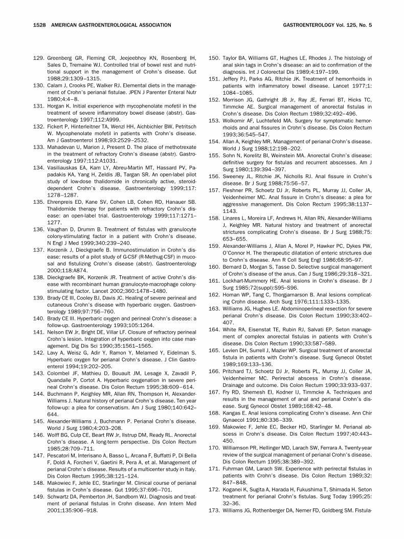
129. Greenberg GR, Fleming CR, Jeejeebhoy KN, Rosenberg IH,Sales D, Tremaine WJ. Controlled trial of bowel rest and nutri-tional support in the management of Crohn’s disease. Gut1988;29:1309–1315.
130. Calam J, Crooks PE, Walker RJ. Elemental diets in the manage-ment of Crohn’s perianal fistulae. JPEN J Parenter Enteral Nutr1980;4:4–8.
131. Horgan K. Initial experience with mycophenolate mofetil in thetreatment of severe inflammatory bowel disease (abstr). Gas-troenterology 1997;112:A999.
132. Fickert P, Hinterleitner TA, Wenzl HH, Aichbichler BW, PetritschW. Mycophenolate mofetil in patients with Crohn’s disease.Am J Gastroenterol 1998;93:2529–2532.
133. Mahadevan U, Marion J, Present D. The place of methotrexatein the treatment of refractory Crohn’s disease (abstr). Gastro-enterology 1997;112:A1031.
134. Vasiliauskas EA, Kam LY, Abreu-Martin MT, Hassard PV, Pa-padakis KA, Yang H, Zeldis JB, Targan SR. An open-label pilotstudy of low-dose thalidomide in chronically active, steroid-dependent Crohn’s disease. Gastroenterology 1999;117:1278–1287.
135. Ehrenpreis ED, Kane SV, Cohen LB, Cohen RD, Hanauer SB.Thalidomide therapy for patients with refractory Crohn’s dis-ease: an open-label trial. Gastroenterology 1999;117:1271–1277.
136. Vaughan D, Drumm B. Treatment of fistulas with granulocytecolony-stimulating factor in a patient with Crohn’s disease.N Engl J Med 1999;340:239–240.
137. Korzenik J, Dieckgraefe B. Immunostimulation in Crohn’s dis-ease: results of a pilot study of G-CSF (R-Methug-CSF) in muco-sal and fistulizing Crohn’s disease (abstr). Gastroenterology2000;118:A874.
138. Dieckgraefe BK, Korzenik JR. Treatment of active Crohn’s dis-ease with recombinant human granulocyte-macrophage colony-stimulating factor. Lancet 2002;360:1478–1480.
139. Brady CE III, Cooley BJ, Davis JC. Healing of severe perineal andcutaneous Crohn’s disease with hyperbaric oxygen. Gastroen-terology 1989;97:756–760.
140. Brady CE III. Hyperbaric oxygen and perineal Crohn’s disease: afollow-up. Gastroenterology 1993;105:1264.
141. Nelson EW Jr, Bright DE, Villar LF. Closure of refractory perinealCrohn’s lesion. Integration of hyperbaric oxygen into case man-agement. Dig Dis Sci 1990;35:1561–1565.
142. Lavy A, Weisz G, Adir Y, Ramon Y, Melamed Y, Eidelman S.Hyperbaric oxygen for perianal Crohn’s disease. J Clin Gastro-enterol 1994;19:202–205.
143. Colombel JF, Mathieu D, Bouault JM, Lesage X, Zavadil P,Quandalle P, Cortot A. Hyperbaric oxygenation in severe peri-neal Crohn’s disease. Dis Colon Rectum 1995;38:609–614.
144. Buchmann P, Keighley MR, Allan RN, Thompson H, Alexander-Williams J. Natural history of perianal Crohn’s disease. Ten yearfollow-up: a plea for conservatism. Am J Surg 1980;140:642–644.
145. Alexander-Williams J, Buchmann P. Perianal Crohn’s disease.World J Surg 1980;4:203–208.
146. Wolff BG, Culp CE, Beart RW Jr, Ilstrup DM, Ready RL. AnorectalCrohn’s disease. A long-term perspective. Dis Colon Rectum1985;28:709–711.
147. Pescatori M, Interisano A, Basso L, Arcana F, Buffatti P, Di BellaF, Doldi A, Forcheri V, Gaetini R, Pera A, et al. Management ofperianal Crohn’s disease. Results of a multicenter study in Italy.Dis Colon Rectum 1995;38:121–124.
148. Makowiec F, Jehle EC, Starlinger M. Clinical course of perianalfistulas in Crohn’s disease. Gut 1995;37:696–701.
149. Schwartz DA, Pemberton JH, Sandborn WJ. Diagnosis and treat-ment of perianal fistulas in Crohn disease. Ann Intern Med2001;135:906–918.
150. Taylor BA, Williams GT, Hughes LE, Rhodes J. The histology ofanal skin tags in Crohn’s disease: an aid to confirmation of thediagnosis. Int J Colorectal Dis 1989;4:197–199.
151. Jeffery PJ, Parks AG, Ritchie JK. Treatment of hemorrhoids inpatients with inflammatory bowel disease. Lancet 1977;1:1084–1085.
152. Morrison JG, Gathright JB Jr, Ray JE, Ferrari BT, Hicks TC,Timmcke AE. Surgical management of anorectal fistulas inCrohn’s disease. Dis Colon Rectum 1989;32:492–496.
153. Wolkomir AF, Luchtefeld MA. Surgery for symptomatic hemor-rhoids and anal fissures in Crohn’s disease. Dis Colon Rectum1993;36:545–547.
154. Allan A, Keighley MR. Management of perianal Crohn’s disease.World J Surg 1988;12:198–202.
155. Sohn N, Korelitz BI, Weinstein MA. Anorectal Crohn’s disease:definitive surgery for fistulas and recurrent abscesses. Am JSurg 1980;139:394–397.
156. Sweeney JL, Ritchie JK, Nicholls RJ. Anal fissure in Crohn’sdisease. Br J Surg 1988;75:56–57.
157. Fleshner PR, Schoetz DJ Jr, Roberts PL, Murray JJ, Coller JA,Veidenheimer MC. Anal fissure in Crohn’s disease: a plea foraggressive management. Dis Colon Rectum 1995;38:1137–1143.
158. Linares L, Moreira LF, Andrews H, Allan RN, Alexander-WilliamsJ, Keighley MR. Natural history and treatment of anorectalstrictures complicating Crohn’s disease. Br J Surg 1988;75:653–655.
159. Alexander-Williams J, Allan A, Morel P, Hawker PC, Dykes PW,O’Connor H. The therapeutic dilatation of enteric strictures dueto Crohn’s disease. Ann R Coll Surg Engl 1986;68:95–97.
160. Bernard D, Morgan S, Tasse D. Selective surgical managementof Crohn’s disease of the anus. Can J Surg 1986;29:318–321.
161. Lockhart-Mummery HE. Anal lesions in Crohn’s disease. Br JSurg 1985;72(suppl):S95–S96.
162. Homan WP, Tang C, Thorgjarnarson B. Anal lesions complicat-ing Crohn disease. Arch Surg 1976;111:1333–1335.
163. Williams JG, Hughes LE. Abdominoperineal resection for severeperianal Crohn’s disease. Dis Colon Rectum 1990;33:402–407.
164. White RA, Eisenstat TE, Rubin RJ, Salvati EP. Seton manage-ment of complex anorectal fistulas in patients with Crohn’sdisease. Dis Colon Rectum 1990;33:587–589.
165. Levien DH, Surrell J, Mazier WP. Surgical treatment of anorectalfistula in patients with Crohn’s disease. Surg Gynecol Obstet1989;169:133–136.
166. Pritchard TJ, Schoetz DJ Jr, Roberts PL, Murray JJ, Coller JA,Veidenheimer MC. Perirectal abscess in Crohn’s disease.Drainage and outcome. Dis Colon Rectum 1990;33:933–937.
167. Fry RD, Shemesh EI, Kodner IJ, Timmcke A. Techniques andresults in the management of anal and perianal Crohn’s dis-ease. Surg Gynecol Obstet 1989;168:42–48.
168. Kangas E. Anal lesions complicating Crohn’s disease. Ann ChirGynaecol 1991;80:336–339.
169. Makowiec F, Jehle EC, Becker HD, Starlinger M. Perianal ab-scess in Crohn’s disease. Dis Colon Rectum 1997;40:443–450.
170. Williamson PR, Hellinger MD, Larach SW, Ferrara A. Twenty-yearreview of the surgical management of perianal Crohn’s disease.Dis Colon Rectum 1995;38:389–392.
171. Fuhrman GM, Larach SW. Experience with perirectal fistulas inpatients with Crohn’s disease. Dis Colon Rectum 1989;32:847–848.
172. Koganei K, Sugita A, Harada H, Fukushima T, Shimada H. Setontreatment for perianal Crohn’s fistulas. Surg Today 1995;25:32–36.
173. Williams JG, Rothenberger DA, Nemer FD, Goldberg SM. Fistula-
1528 AMERICAN GASTROENTEROLOGICAL ASSOCIATION GASTROENTEROLOGY Vol. 125, No. 5
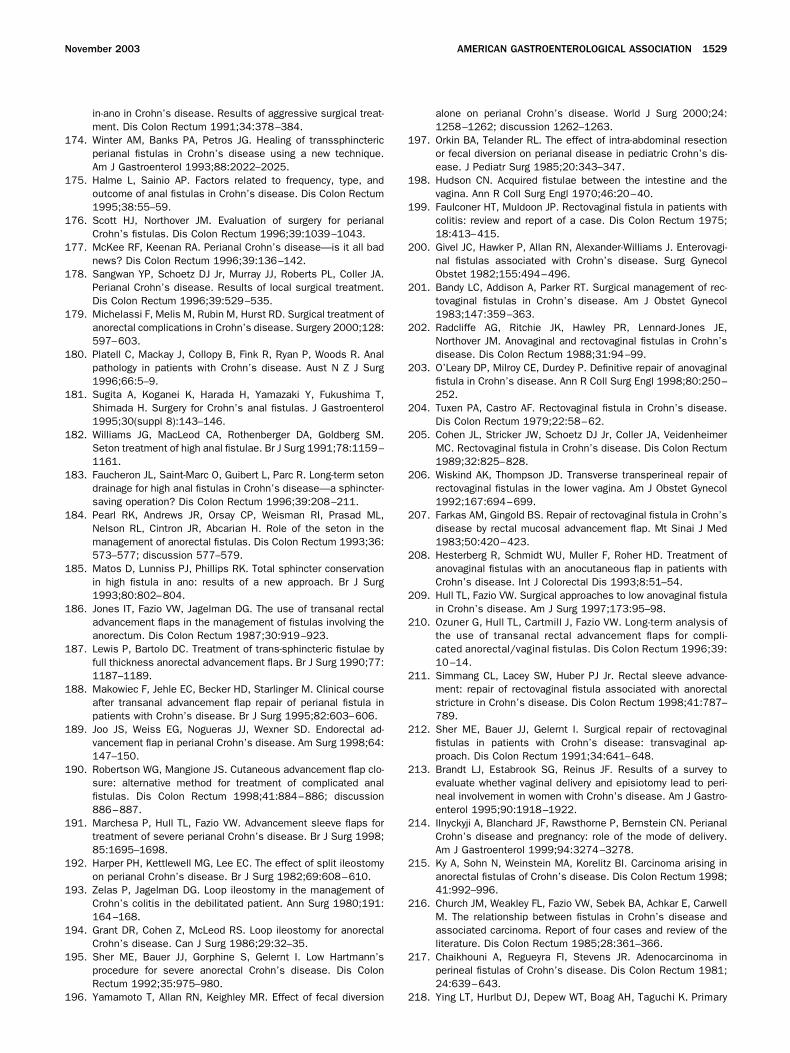
in-ano in Crohn’s disease. Results of aggressive surgical treat-ment. Dis Colon Rectum 1991;34:378–384.
174. Winter AM, Banks PA, Petros JG. Healing of transsphinctericperianal fistulas in Crohn’s disease using a new technique.Am J Gastroenterol 1993;88:2022–2025.
175. Halme L, Sainio AP. Factors related to frequency, type, andoutcome of anal fistulas in Crohn’s disease. Dis Colon Rectum1995;38:55–59.
176. Scott HJ, Northover JM. Evaluation of surgery for perianalCrohn’s fistulas. Dis Colon Rectum 1996;39:1039–1043.
177. McKee RF, Keenan RA. Perianal Crohn’s disease—is it all badnews? Dis Colon Rectum 1996;39:136–142.
178. Sangwan YP, Schoetz DJ Jr, Murray JJ, Roberts PL, Coller JA.Perianal Crohn’s disease. Results of local surgical treatment.Dis Colon Rectum 1996;39:529–535.
179. Michelassi F, Melis M, Rubin M, Hurst RD. Surgical treatment ofanorectal complications in Crohn’s disease. Surgery 2000;128:597–603.
180. Platell C, Mackay J, Collopy B, Fink R, Ryan P, Woods R. Analpathology in patients with Crohn’s disease. Aust N Z J Surg1996;66:5–9.
181. Sugita A, Koganei K, Harada H, Yamazaki Y, Fukushima T,Shimada H. Surgery for Crohn’s anal fistulas. J Gastroenterol1995;30(suppl 8):143–146.
182. Williams JG, MacLeod CA, Rothenberger DA, Goldberg SM.Seton treatment of high anal fistulae. Br J Surg 1991;78:1159–1161.
183. Faucheron JL, Saint-Marc O, Guibert L, Parc R. Long-term setondrainage for high anal fistulas in Crohn’s disease—a sphincter-saving operation? Dis Colon Rectum 1996;39:208–211.
184. Pearl RK, Andrews JR, Orsay CP, Weisman RI, Prasad ML,Nelson RL, Cintron JR, Abcarian H. Role of the seton in themanagement of anorectal fistulas. Dis Colon Rectum 1993;36:573–577; discussion 577–579.
185. Matos D, Lunniss PJ, Phillips RK. Total sphincter conservationin high fistula in ano: results of a new approach. Br J Surg1993;80:802–804.
186. Jones IT, Fazio VW, Jagelman DG. The use of transanal rectaladvancement flaps in the management of fistulas involving theanorectum. Dis Colon Rectum 1987;30:919–923.
187. Lewis P, Bartolo DC. Treatment of trans-sphincteric fistulae byfull thickness anorectal advancement flaps. Br J Surg 1990;77:1187–1189.
188. Makowiec F, Jehle EC, Becker HD, Starlinger M. Clinical courseafter transanal advancement flap repair of perianal fistula inpatients with Crohn’s disease. Br J Surg 1995;82:603–606.
189. Joo JS, Weiss EG, Nogueras JJ, Wexner SD. Endorectal ad-vancement flap in perianal Crohn’s disease. Am Surg 1998;64:147–150.
190. Robertson WG, Mangione JS. Cutaneous advancement flap clo-sure: alternative method for treatment of complicated analfistulas. Dis Colon Rectum 1998;41:884–886; discussion886–887.
191. Marchesa P, Hull TL, Fazio VW. Advancement sleeve flaps fortreatment of severe perianal Crohn’s disease. Br J Surg 1998;85:1695–1698.
192. Harper PH, Kettlewell MG, Lee EC. The effect of split ileostomyon perianal Crohn’s disease. Br J Surg 1982;69:608–610.
193. Zelas P, Jagelman DG. Loop ileostomy in the management ofCrohn’s colitis in the debilitated patient. Ann Surg 1980;191:164–168.
194. Grant DR, Cohen Z, McLeod RS. Loop ileostomy for anorectalCrohn’s disease. Can J Surg 1986;29:32–35.
195. Sher ME, Bauer JJ, Gorphine S, Gelernt I. Low Hartmann’sprocedure for severe anorectal Crohn’s disease. Dis ColonRectum 1992;35:975–980.
196. Yamamoto T, Allan RN, Keighley MR. Effect of fecal diversion
alone on perianal Crohn’s disease. World J Surg 2000;24:1258–1262; discussion 1262–1263.
197. Orkin BA, Telander RL. The effect of intra-abdominal resectionor fecal diversion on perianal disease in pediatric Crohn’s dis-ease. J Pediatr Surg 1985;20:343–347.
198. Hudson CN. Acquired fistulae between the intestine and thevagina. Ann R Coll Surg Engl 1970;46:20–40.
199. Faulconer HT, Muldoon JP. Rectovaginal fistula in patients withcolitis: review and report of a case. Dis Colon Rectum 1975;18:413–415.
200. Givel JC, Hawker P, Allan RN, Alexander-Williams J. Enterovagi-nal fistulas associated with Crohn’s disease. Surg GynecolObstet 1982;155:494–496.
201. Bandy LC, Addison A, Parker RT. Surgical management of rec-tovaginal fistulas in Crohn’s disease. Am J Obstet Gynecol1983;147:359–363.
202. Radcliffe AG, Ritchie JK, Hawley PR, Lennard-Jones JE,Northover JM. Anovaginal and rectovaginal fistulas in Crohn’sdisease. Dis Colon Rectum 1988;31:94–99.
203. O’Leary DP, Milroy CE, Durdey P. Definitive repair of anovaginalfistula in Crohn’s disease. Ann R Coll Surg Engl 1998;80:250–252.
204. Tuxen PA, Castro AF. Rectovaginal fistula in Crohn’s disease.Dis Colon Rectum 1979;22:58–62.
205. Cohen JL, Stricker JW, Schoetz DJ Jr, Coller JA, VeidenheimerMC. Rectovaginal fistula in Crohn’s disease. Dis Colon Rectum1989;32:825–828.
206. Wiskind AK, Thompson JD. Transverse transperineal repair ofrectovaginal fistulas in the lower vagina. Am J Obstet Gynecol1992;167:694–699.
207. Farkas AM, Gingold BS. Repair of rectovaginal fistula in Crohn’sdisease by rectal mucosal advancement flap. Mt Sinai J Med1983;50:420–423.
208. Hesterberg R, Schmidt WU, Muller F, Roher HD. Treatment ofanovaginal fistulas with an anocutaneous flap in patients withCrohn’s disease. Int J Colorectal Dis 1993;8:51–54.
209. Hull TL, Fazio VW. Surgical approaches to low anovaginal fistulain Crohn’s disease. Am J Surg 1997;173:95–98.
210. Ozuner G, Hull TL, Cartmill J, Fazio VW. Long-term analysis ofthe use of transanal rectal advancement flaps for compli-cated anorectal/vaginal fistulas. Dis Colon Rectum 1996;39:10–14.
211. Simmang CL, Lacey SW, Huber PJ Jr. Rectal sleeve advance-ment: repair of rectovaginal fistula associated with anorectalstricture in Crohn’s disease. Dis Colon Rectum 1998;41:787–789.
212. Sher ME, Bauer JJ, Gelernt I. Surgical repair of rectovaginalfistulas in patients with Crohn’s disease: transvaginal ap-proach. Dis Colon Rectum 1991;34:641–648.
213. Brandt LJ, Estabrook SG, Reinus JF. Results of a survey toevaluate whether vaginal delivery and episiotomy lead to peri-neal involvement in women with Crohn’s disease. Am J Gastro-enterol 1995;90:1918–1922.
214. Ilnyckyji A, Blanchard JF, Rawsthorne P, Bernstein CN. PerianalCrohn’s disease and pregnancy: role of the mode of delivery.Am J Gastroenterol 1999;94:3274–3278.
215. Ky A, Sohn N, Weinstein MA, Korelitz BI. Carcinoma arising inanorectal fistulas of Crohn’s disease. Dis Colon Rectum 1998;41:992–996.
216. Church JM, Weakley FL, Fazio VW, Sebek BA, Achkar E, CarwellM. The relationship between fistulas in Crohn’s disease andassociated carcinoma. Report of four cases and review of theliterature. Dis Colon Rectum 1985;28:361–366.
217. Chaikhouni A, Regueyra FI, Stevens JR. Adenocarcinoma inperineal fistulas of Crohn’s disease. Dis Colon Rectum 1981;24:639–643.
218. Ying LT, Hurlbut DJ, Depew WT, Boag AH, Taguchi K. Primary
November 2003 AMERICAN GASTROENTEROLOGICAL ASSOCIATION 1529
adenocarcinoma in an enterocutaneous fistula associatedwith Crohn’s disease. Can J Gastroenterol 1998;12:265–269.
219. Korelitz BI. Carcinoma arising in Crohn’s disease fistulae: an-other concern warranting another type of surveillance. Am JGastroenterol 1999;94:2337–2339.
220. Wong NA, Shirazi T, Hamer-Hodges DW, Corfield AP, LessellsAM. Adenocarcinoma arising within a Crohn’s-related anorectalfistula: a form of anal gland carcinoma? Histopathology 2002;40:302–304.
221. Kulaylat MN, Gallina G, Bem J, Zeid M. Carcinoma arising inanorectal fistulas of Crohn’s disease. Dis Colon Rectum 1999;42:826–828.
222. Hanauer SB, Sandborn W. The Practice Parameters Committeeof the American College of Gastroenterology. Management ofCrohn’s disease in adults. Am J Gastroenterol 2001;96:635–643.
223. Bauer JJ, Sher ME, Jaffin H, Present D, Gelerent I. Transvaginalapproach for repair of rectovaginal fistulae complicating Crohn’sdisease. Ann Surg 1991;213:151–158.
Address requests for reprints to: Chair, Clinical Practice Committee,AGA National Office, c/o Membership Department, 4930 Del RayAvenue, Bethesda, Maryland 20814. Fax: (301) 654-5920.
W. J. S. has consulted for Prometheus Laboratories, GlaxoSmithKline,Centocor, Novartis, and Fujisawa. He has received research support fromPrometheus Laboratories, Schering-Plough, Centocor, and Fujisawa. Hehas participated in continuing medical education events indirectly spon-sored by Prometheus Laboratories, Schering-Plough, and Centocor.B. G. F. has consulted for Centocor and Novartis. He has received researchsupport from Schering-Plough and Centocor. He has participated in con-tinuing medical education events indirectly sponsored by Schering-Ploughand Centocor. S. B. H. has consulted for and received research supportfrom Centocor. He has participated in continuing medical educationevents indirectly sponsored by Schering-Plough and Centocor.
The Clinical Practice Committee thanks the following individuals whosecritiques of this review paper provided valuable guidance to the authors:Jeffrey L. Barnett, M.D., Geert D’Haens, M.D., Ph.D., Francis A. Farraye,M.D., M.Sc., Jeffrey A. Katz, M.D., and Gary R. Lichtenstein, M.D.
1530 AMERICAN GASTROENTEROLOGICAL ASSOCIATION GASTROENTEROLOGY Vol. 125, No. 5