Embed Size (px)
Citation preview

Aging-Related Changes in the Nigrostriatal Dopamine System andthe Response to MPTP in Nonhuman Primates: DiminishedCompensatory Mechanisms as a Prelude to Parkinsonism
Timothy J. Collier1, Jack Lipton2, Brian F. Daley1, Stephane Palfi3, Yaping Chu4, CarylSortwell1, Roy A.E. Bakay5, John R. Sladek Jr6, and Jeffrey H. Kordower41 Department of Neurology, University of Cincinnati, Cincinnati, OH
2 Department of Psychiatry, University of Cincinnati, Cincinnati, OH
3 CEA-CNRS 2210 and Mircen program, Service Hospitalier Frederic Joliot, Orsay, France andNeurosurgical Department CHU Henri Mondor AP/HP, Universite Paris XII, Creteil, France
4 Department of Neurological Sciences, Rush University Medical Center, Chicago, IL
5 Department of Neurological Surgery, Rush University Medical Center, Chicago, IL
6 Department of Psychiatry, University of Colorado Health Sciences Center, Denver, CO
AbstractAging is the most prominent risk factor for Parkinson’s disease. Yet, consensus of how advancingage may predispose the dopamine (DA) system to parkinsonism is lacking. Three age-ranges offemale rhesus monkeys, 8–9, 15–17 and 21–31 years, received unilateral DA depletion withintracarotid 1-methyl-4-phenyl-1,2,3,6-tetrahydropyridine (MPTP). Morphological and biochemicalanalyses of DA-depleted and intact hemispheres revealed three primary findings: 1) The intactstriatum exhibited age-related declines in dopamine (DA) and homovanillic acid (HVA) that werepresent by middle-age; 2) In the MPTP-treated striatum, the compensatory increase in DA activitywas absent in old monkeys; and, 3) Age-associated morphological changes included declines in thedensity of tyrosine hydroxylase (TH) positive fibers in striatum, decreased nigral soma size andoptical density of TH, but no significant loss of neurons. These findings suggest that aging produceschanges in the nigrostriatal DA system that approach the threshold for expression of parkinsonianfeatures, and that progressive impairment of plasticity may be central to the role of aging indevelopment of parkinsonism.
Keywordsaging; dopamine; substantia nigra; parkinsonism; MPTP; monkey
The mesostriatal dopamine (DA) system exhibits dramatic degenerative changes inParkinson’s disease (PD). The morphological and biochemical patterns of these changes arewell characterized and consistent (Fahn, 2003). Aging is a well-established risk factor for PD(Granieri et al., 1991; Mayeux et al., 1995; Morens et al., 1996; Tanner and Goldman,
Corresponding Author: Timothy J. Collier, Ph.D. Department of Neurology, University of Cincinnati, P.O. Box 670525, 265, AlbertSabin Way, Cincinnati, OH 45267, Email: [email protected], Telephone: 513-558-1168, FAX: 513-558-2965Publisher's Disclaimer: This is a PDF file of an unedited manuscript that has been accepted for publication. As a service to our customerswe are providing this early version of the manuscript. The manuscript will undergo copyediting, typesetting, and review of the resultingproof before it is published in its final citable form. Please note that during the production process errors may be discovered which couldaffect the content, and all legal disclaimers that apply to the journal pertain.
NIH Public AccessAuthor ManuscriptNeurobiol Dis. Author manuscript; available in PMC 2008 April 1.
Published in final edited form as:Neurobiol Dis. 2007 April ; 26(1): 56–65.
NIH
-PA Author Manuscript
NIH
-PA Author Manuscript
NIH
-PA Author Manuscript

1996;Totaro et al., 2005). In contrast to PD, the pattern of aging-related changes in the DAsystem vary considerably among species (e.g. Morgan and Finch, 1988). Even within a speciesvariability exists in the exact pattern of changes reported and whether age-related changescould be a precursor to the degeneration seen in PD. For nonhuman primates, there tends to beagreement that declines in striatal DA content accompany advancing age, but associatedchanges in midbrain DA neuron numbers and function are a source of disagreement (Goldman-Rakic and Brown, 1981;Irwin et al., 1994;Pakkenberg et al., 1995; Emborg et al., 1998;Gerhardt et al., 2002;McCormack et al., 2004). In addition, while inclusion of young adult,middle-aged, and elderly individuals is considered proper experimental design for studies ofthe biology of aging, in reality this can be difficult to accomplish and many reports in theliterature compare only two of these age groups. As a consequence significant gaps exist inour understanding of the development of changes in the DA system over the lifespan inprimates. In the present study we were fortunate to have available for study three age rangesof female rhesus monkeys: 8–9 years (N=3), 15–17 years (N=4), and 21–31 years (N=6). It isdocumented that rhesus monkeys exhibit aging-related changes consistent with a rate of aging3:1 as compared to humans (Andersen et al., 1999). Accordingly, our groups of animalscorrespond to humans aged 24–27 years, 45–51 years, and 63–93 years. We contend thesecorrespond to generally held views of young adulthood, middle-age, and advanced age. In thepresent study we treated monkeys with unilateral intracarotid infusion of the DA neurotoxin1-methyl-4-phenyl-1,2,3,6-tetrahydropyridine (MPTP) to create unilateral degeneration of themesostriatal DA system. Our goal was not to revisit the previously established aging-relatedincreased vulnerability to MPTP toxicity (Ovadia et al., 1995;McCormack et al., 2004), butrather to study aging-related differences and similarities in the response to equivalent insult tothe DA system. Therefore, the MPTP dose was adjusted for monkeys of differing age to yielda constant behavioral endpoint: complete disuse of the contralateral arm and hand. Cell countsconfirm that this MPTP dosing regimen produced the expected severe depletion of tyrosinehydroxylase (TH) positive neurons in substantia nigra ipsilateral to treatment while maintainingcell counts in the contralateral hemisphere that did not differ from those in untreated monkeys.Thus, after establishing our behavioral endpoint, we compared the morphology andbiochemistry of the DA system ipsilateral to MPTP treatment to document aging-relatedchanges in the response to lesion and the hemisphere contralateral to MPTP treatment todocument changes due to aging itself.
Materials and MethodsExperimental Subjects and MPTP Treatment
Subjects were female rhesus monkeys (macaca mulatta) weighing 5.3–10.1kg. Three agegroups were studied: young adult (8–9yr) N=3, middle-aged (15–17yr) N=4, and aged (21–31yr) N=6. All animals were born in captivity with chronological age documented by birthrecords. Animals were housed in individual primate cages and cared for in the AALACapproved Biological Resources Laboratory at the University of Illinois-Chicago. All monkeyswere treated with unilateral intracarotid administration of 1-methyl-4-phenyl-1,2,3,6-tetrahydropyridine as previously described (Emborg et al., 2001) (MPTP-HCl, SigmaChemical, St. Louis, MO, first dose = 3.5 mg for monkeys ≥ 7kg body weight, 3.0 mg formonkeys <7kg body weight, 2.3 mg for monkeys >17 years of age, re-treatment, whennecessary to achieve behavioral endpoint, = 2.0 mg) (Table 1). Treatment resulted in equivalentbehavioral signs in all subjects, principally characterized by complete disuse of the forelimbcontralateral to infusion. No obvious impairment of contralateral leg function was noted, andnone of the treated monkeys exhibited spontaneous rotational behavior. Care and use of theseanimals was in compliance with all applicable laws and regulations as well as principlesexpressed in the National Institutes of Health, United States Public Health Service Guide forthe Care and Use of Laboratory Animals. This study was approved by the Animal Care and
Collier et al. Page 2
Neurobiol Dis. Author manuscript; available in PMC 2008 April 1.
NIH
-PA Author Manuscript
NIH
-PA Author Manuscript
NIH
-PA Author Manuscript

Use Committees of the University of Illinois-Chicago and Rush University Medical Centerwhere the live animal aspects of the study were conducted.
Measurement of Spontaneous ActivityPrior to MPTP treatment and at 3 months after treatment, monkeys were transferred from theirhome cage to a cage equipped with a Plexiglas front panel for video tape recording ofspontaneous activity. Activity was recorded for 30 minutes with no observers in the room.Videotapes were analyzed off-line by an examiner blinded to the experimental condition usingmotion tracking and analysis software (Ethovision 2.3, Noldus, Wageningen, TheNetherlands). The analysis provides an objective measurement of kinematic parameters. In thepresent study we analyzed total distance moved (cm) as an index of general motor activity.
Tissue CollectionThree months following induction of behavioral symptoms, animals were killed bypentobarbital overdose (50 mg/kg with effect confirmed by absence of corneal reflex) andperfused with ice-cold physiological saline. Perfusion both serves to reduce the temperatureof the tissue, aiding in stabilization of tissue catecholamines, and removes potentialcontamination of samples by catecholamines in blood. Brains were removed, and eachforebrain was divided into coronal slabs of 4 mm thickness on ice. Tissue punches, 1.3 mm indiameter, were taken from standard locations (Sladek et al., 1995) in the dorsal-lateral caudatenucleus and putamen at precommissural and commissural levels of the striatum (Figure 1).Punches were frozen on dry ice and stored at −80 degrees C until processing with highperformance liquid chromatography (HPLC) for detection of dopamine (DA) and homovanillicacid (HVA). Measures of 3,4-dihydroxyphenylacetic acid (DOPAC) also were obtained duringHPLC analysis. When present, DOPAC amounts were <5% of HVA levels and in manysamples were below the level of detection. Since DOPAC levels were not consistently obtainedand HVA is the metabolite of greater relevance in nonhuman primate studies, we chose not topresent DOPAC levels. After collection of fresh tissue punches for HPLC, remaining tissueblocks through the striatum and the brain caudal to the striatum was immersion fixed in 4%paraformaldehyde solution and sunk in 30% sucrose solution in preparation for sectioning andimmunocytochemical staining for TH.
Dopamine BiochemistryLevels of DA and HVA were analyzed using HPLC. Punches of caudate nucleus and putamenwere dissected out, placed in vials with 200 μl of an anti-oxidant solution (perchlorate andsodium metabisulfite) and immediately frozen on powdered dry ice and stored at –80°C. Atthe time of analysis samples were thawed and homogenized using an ultrasonic tissuehomogenizer. Supernatants were separated and detected via HPLC and pellets were reservedfor protein determination (Pierce BCA Protein Reagent Kit). Samples were simultaneouslyexamined for DA and HVA using a 12 channel coulometric array detector (CoulArray 5200,ESA) attached to a Waters 2695 Solvent Delivery System. All procedures within the laboratorywere carried out at GLP quality control levels utilizing appropriate 6-point standard curves andinterspersed quality control samples to ensure HPLC calibration.
Immunocytochemical AnalysisFixed tissue was sectioned on a freezing microtome in the coronal plane at 50μm sectionthickness. Staining for TH (mouse antiTH monoclonal, 1:4,000, Chemicon International,Temecula, CA) as a marker for DA neurons and their processes was visualized by using theVectastain ABC protocol (Vector Laboratories, Burlingame, CA). Stereological estimates ofTH stained substantia nigra (SN) neurons were performed using a BX52 Olympus microscope(Olympus America Inc.) equipped with Microbrightfield stereological software and a Microfire
Collier et al. Page 3
Neurobiol Dis. Author manuscript; available in PMC 2008 April 1.
NIH
-PA Author Manuscript
NIH
-PA Author Manuscript
NIH
-PA Author Manuscript

CCD camera (Optronics, Goleta, CA). The camera settings were maintained throughout theentirety of the experiment. Using the optical fractionator procedure, neurons were sampled ina uniform, systematic, but random design. The region of interest was outlined under lowmagnification (1.25X), demarcating SN by the caudal edge of the mammillary bodies rostrally,the cerebral peduncle ventrally and laterally, and the lateral edge of the third cranial nerverootlets medially. Approximately, 5% of the outlined region was sampled with a series ofcounting frames that were randomly and systematically distributed through the region ofinterest. Using a 60X magnification lens with a 1.4 N/A the section thickness was empiricallydetermined by first bringing the top of the section into focus and then using the Microbrightfieldprogram to step through the z-axis in 1μm increments until the very bottom of the section wasin focus. Once the top of the section was in focus, the z-plane was lowered at 1–2 μm intervalsand cell counts were made according to stereological principles while focusing down throughthe z-axis. Care was taken to ensure that the bottom and top guard zones were never includedin the analysis. Eight to ten equally spaced sections (1 in 6 series) were sampled from eachsubject. After obtaining the cell counts the stereological software generates accurate estimatesof the total number of cells in the whole cell population based on stereological principles andequations. Additional image analysis was performed on TH stained neurons sampled fromthree cytoarchitectural subdivisions of the SN: dorsal tier, ventral tier, and pars lateralis. Asfew as 10 cells from the lesioned hemisphere and up to 80 cells from the intact hemispherewere sampled from a single 50X magnification field positioned in the center of these regionsfrom 3 tissue sections at the level of the third cranial nerve rootlets. Sampling at this levelprovides the clearest demarcation of these subregions. Sampling of a single microscopic fieldplaced in the center of these regions insures that the cells measured are within the region, butprecludes more complete sampling. Both intensity of TH staining and soma area werequantified using computer-assisted imaging (Sigma Scan/Sigma Scan Pro software, JandelScientific, San Rafael, CA). The TH intensity measure is semi-quantitative. Sections from allmonkeys were processed simultaneously for TH immunoreactivity, ensuring constant reactionconditions. As such, variations in expression of TH immunoreactivity by individual cells whilenot appropriate as a linear correlate of protein expression can be used in assessment of relativedifferences among cell regions and age groups. The staining intensity value for each cellrepresented the average of TH immunoreactivity over the cross-sectional area of each cellexpressed relative to the staining intensity of white matter in the cerebral peduncle in the sametissue section. Measures of staining intensity and soma area for each SN subregion wereaveraged for the 3 tissue sections to provide a single mean value for each cell region for eachmonkey. These mean values were used for statistical analysis across age groups. Finally, theaverage intensity of TH fiber staining in the striatum was determined using the same softwarepackage. A single 50X magnification microscopic field in the center of the caudate nucleusand putamen of each subject (Figure 1) was assessed in four coronal tissue sections (at300μm intervals) immediately rostral to the decussation of the anterior commisssure andexpressed relative to staining intensity of white matter in the internal capsule in the same tissuesection. Measures of staining intensity of fibers were averaged across tissue sections to providea single mean value for each structure for each monkey. These mean values were used forstatistical analysis across age groups.
StatisticsComparisons of motor activity scores, regional levels of DA and HVA in tissue punches,stereological cell number estimates, intensity of TH staining in nigral neurons and striatalneuropil, and TH neuron soma area were analyzed using analysis of variance (ANOVA)followed by Fisher’s protected least significant difference (PLSD) test (Statview software).Motor activity was analyzed with a repeated measures design (Super ANOVA software).
Collier et al. Page 4
Neurobiol Dis. Author manuscript; available in PMC 2008 April 1.
NIH
-PA Author Manuscript
NIH
-PA Author Manuscript
NIH
-PA Author Manuscript

ResultsSpontaneous locomotion in a cage used for videotaping was assessed over a 30 minute durationrecording session prior to MPTP treatment and at 3 months after treatment. Total distancetraveled was computed using the Ethovision software program. At baseline, young adult,middle-aged, and aged monkeys were progressively less active (Figure 2). All monkeys wereunilaterally depleted of striatal DA by MPTP treatment to achieve the behavioral endpoint ofcomplete disuse of the contralateral arm and hand. All monkeys held the affected arm in aflexed posture and would not retrieve food with this hand. Overt impairment of the contralaterallower limb was not detected, but not explicitly tested, nor did subjects exhibit spontaneousrotational behavior. Statistical analysis revealed significant differences attributable to age (F(2,5)=6.91, p<0.04), treatment (pre- and post-MPTP, F(1,5)=41.09, p<0.002) and the age ×treatment interaction (F(2,5)=9.89, p<0.02). Post-hoc analysis indicated that significantdifferences were attributable to the large decrease in baseline spontaneous activity in agedmonkeys compared to young monkeys (-79%, p=0.005). After MPTP treatment, furtherreductions in activity were observed for all age groups, but none of these changes reachedstatistical significance.
Measurement of DA and the metabolite HVA from both intact and DA-depleted caudatenucleus and putamen revealed aging-related changes. For the intact hemisphere, both caudateand putamen showed statistically significant declines in DA that began in middle-age (caudate-55%, putamen –54%) and were sustained in old age (caudate -43%, putamen –30%) (Figure3) (Caudate: F(5,42)=41.98, p<0.0001; p<0.0001 for comparison of young intact to middle-aged and aged intact; Putamen: F(5,42)=32.90, p<0.0001; p<0.0001 for comparison of youngintact to middle-aged and aged intact). HVA also was significantly reduced from young adultlevels in the caudate nucleus and putamen of middle-aged (caudate -46%, putamen –50%) andaged monkeys (caudate –20%, putamen -30%)(Figure 3)(Caudate: F(5,42)=29.83, p<0.0001;p<0.05 for comparison of young intact to middle-aged and aged intact; Putamen: F(5,42)=24.19, p<0.0001; p≤0.0001 for comparison of young intact to middle-aged and aged intact).No differences among age groups were detected for the HVA/DA ratio in either striatalstructure.
In the DA-depleted striatum, there were the expected dramatic decreases in DA (-92-99%) andHVA (-85-94%) in all age groups as compared to their intact hemisphere (Figure 3)(Caudateand Putamen DA and HVA p<0.0001 for comparison of lesioned hemisphere to intacthemisphere). No statistically significant differences in the extent of depletion were foundbetween the age groups. This confirms that equivalent damage to the DA system was achievedin animals of all age groups. The most dramatic aging-related change in DA biochemistry wasfound for the HVA/DA ratio. In both caudate nucleus and putamen, the anticipated significantincrease in DA activity in response to MPTP-induced degeneration was present in young andmiddle-aged monkeys, but completely absent in the oldest age group (Figure 3)(Caudate: F(5,41)=7.42, p<0.0001; p<0.005 for comparison of intact to lesioned hemispheres, p<0.009 forcomparison of middle-age lesion to old lesion, p<0.04 for comparison of young lesion to bothmiddle-age and old lesion; Putamen: F(5,41)=5.85, p<0.0005; p<0.005 for comparison of intactto lesioned hemispheres, p<0.003 for comparison of young and middle-age lesion to old lesion).
Light microscopy for TH immunoreactive (THir) neurons of the substantia nigra pars compacta(SNC) in the intact hemisphere viewed at low magnification suggested the presence of adramatic aging-related decline in neuron number and dendritic neuropil (Figure 4). However,when SNC THir neuron numbers were quantified using unbiased stereology, no differences inneuron counts were found across age groups (Figure 6). Marked decreases in cell numbersaccompanied MPTP treatment (Figures 4 & 5) and these were not different with increasing
Collier et al. Page 5
Neurobiol Dis. Author manuscript; available in PMC 2008 April 1.
NIH
-PA Author Manuscript
NIH
-PA Author Manuscript
NIH
-PA Author Manuscript

chronological age (Figure 6) (F(5,16)=40.86, p<0.0001; p<0.0001 for comparison of intact tolesioned hemisphere for all ages).
Despite the absence of overt cell loss in SNC with advancing age, analysis of THir neuronstaining intensity and soma area revealed some aging-related changes (Figures 5 & 7). Thesechanges accounted for the dramatic difference in appearance of SNC in the intact hemisphereat low magnification. For this analysis, cells were separately sampled in the maincytoarchitectural subdivisions of SNC: the dorsal tier, ventral tier, and pars lateralis (Figure7). For dorsal tier and ventral tier neurons, an aging-related decrease in THir intensity wasdetected in the intact hemisphere of the oldest monkeys as compared to young adult monkeys(dorsal tier: F(5,16)=4.26, p<0.02; ventral tier: F(5,16)=3.84, p<0.02; p<0.02 for comparisonof young to old). Neurons in pars lateralis exhibited no statistically significant decline in THirintensity (p=0.053), but showed significant declines in soma area in both middle-aged and agedmonkeys (F(5,16)=7.40, p<0.001; p<0.03 for comparison of young to middle-age and old). Ingeneral, both THir intensity and soma area decreased in surviving neurons following MPTPtreatment, but only some of these changes achieved statistical significance. In pars lateralis,MPTP treatment produced significant declines in soma area in young adult and aged monkeys,with this decrease exaggerated in the oldest group (F(5,16)=7.40, p<0.001; p<0.03 forcomparison of intact to MPTP-treated, p<0.04 for comparison of young MPTP treated to oldMPTP-treated). No significant changes in THir intensity accompanied the declines in somaarea in pars lateralis. In addition, aged monkeys exhibited a significant decrease in THir somaarea in dorsal tier neurons after MPTP treatment (F(5,16)=3.43, p<0.03; p<0.01 for comparisonof aged intact to aged MPTP-treated).
Analysis of THir fiber staining in striatum also revealed aging-related changes. With advancingage the intensity of fiber staining decreased in both caudate nucleus and putamen, with thesedeclines being more pronounced and expressed earlier in the putamen (Figure 8)(F(5,20)=6.37,p<0.002; Caudate: p<0.01 for comparison of young to old; Putamen: p<0.04 for comparisonof young to middle-aged and old). THir fibers did not measure above levels of background incentral caudate and putamen in the MPTP-treated hemisphere.
DiscussionThe incidence of Parkinson’s disease increases dramatically with advancing chronological age(Mayeux et al., 1995;Tanner and Goldman, 1996;Totaro et al., 2005). Yet, surprisingly littleis known about whether the aging primate DA system responds differently to insults and ifsuch changes might contribute to the expression of parkinsonian syndromes. Indeed, somebelieve that the pattern of changes in the DA system in PD and aging are distinct and may bearno relationship one to the other (Fearnley and Lees, 1991;Kish et al., 1992;Kubis et al.,2000). It was the goal of the present study to revisit this comparison.
Choices were made in the design of our study, and the findings presented need to be interpretedwithin that context. First, intracarotid administration of MPTP was used to provide theopportunity to analyze biochemical and morphological correlates of age-related responses totoxic insult (brain hemisphere ipsilateral to MPTP infusion) and DA system changesattributable to aging itself (brain hemisphere contralateral to MPTP infusion) in the same cohortof monkeys. Disagreement exists in the literature as to whether this model produces a purelyunilateral DA depletion, or produces variable damage to the contralateral DA system (e.g. Pateet al., 1993). Consensus suggests that contralateral effects are associated with increasing dosesof MPTP and need not be an inevitable consequence of intracarotid administration (Bankiewiczet al., 1986;Guttman et al., 1990;Palomobo et al., 1990;Oiwa et al., 2003). Our MPTP doseswere on the low end of the spectrum for rhesus monkeys and comparisons of SNC THir cellcounts and THir intensity from the intact hemisphere of our treated animals do not differ from
Collier et al. Page 6
Neurobiol Dis. Author manuscript; available in PMC 2008 April 1.
NIH
-PA Author Manuscript
NIH
-PA Author Manuscript
NIH
-PA Author Manuscript

the same measures in untreated young, middle-aged, and aged monkeys analyzed in our otherongoing studies (Kanaan et al., 2006). Second, MPTP treatment was used to achieve the samebehavioral endpoint in monkeys of all ages: complete disuse of the contralateral arm and hand.This approach precludes analysis of aging-related changes in sensitivity to MPTP toxicity,assessed by comparing the response of subjects of different age to the same MPTP dose, andshifts the focus to aging-related changes associated with a comparable insult produced by age-adjusted MPTP doses as defined by a behavioral endpoint. Third, brain samples were collectedat a single time point, three months after establishing behavioral dysfunction. As such, theshort-term dynamics of the response to MPTP were lost in favor of evaluation of a relativelystable response to damage that potentially models the biology of the DA system in individualsof different ages expressing parkinsonism and embarking upon a course of therapy.
Our analysis of striatal DA and HVA confirmed previous studies documenting aging-relateddeclines (Goldman-Rakic and Brown, 1981;Irwin et al., 1994;Gerhardt et al.,2002;McCormack et al., 2004.) and these were associated with a progressive decrease inspontaneous motor activity. In addition, we found that significant decreases in DA content incaudate nucleus and putamen already were expressed in middle-age and sustained in advancedage (McCormack et al., 2004). Following MPTP treatment, the expected significant depletionsof DA in caudate and putamen were observed in the ipsilateral striatum, and the extent ofdepletion did not vary among age groups. This supports our contention that MPTP dosingadjusted to attain a common behavioral endpoint resulted in an equivalent toxic insult forcomparison across age groups. In response to equivalent insult, the most dramatic aging-relatedchange was in DA activity in response to MPTP-induced degeneration. While the well-documented compensatory increase in DA activity reflected in the HVA/DA ratio (Zigmondet al., 1990) was present in young adult and middle-aged monkeys after MPTP treatment, thisbiochemical compensation was completely absent in monkeys of advanced age. Increased DAactivity was a highly variable response in middle-aged animals yet probably accounts for therelative preservation of motor performance in this age group despite the presence of significantdecreases in striatal DA content. In contrast, the absence of compensatory changes in DAactivity in the oldest monkeys was a consistent finding and in combination with sustaineddecreases in DA content likely contributes to the significant deficits in motor activity.
Disagreement exists among previous studies regarding morphological correlates in SNC DAneurons that accompany aging-related declines in striatal DA (Pakkenberg et al., 1995; Emborget al., 1998; Gerhardt et al., 2002;McCormack et al., 2004). In our hands, estimates of SNCTHir cell numbers in the intact hemisphere using unbiased stereology found no loss of neuronsacross the three age groups. The difference between the present study and some previous onesmay reflect differences in detectability of TH. The loss of THir perikarya seen in aged monkeysin some studies likely reflects a phenotypic down-regulation of TH to undetectable levels.Environmental and genetic differences among colonies of aged monkeys may result in someanimals displaying TH levels below the level of detection in some studies but not others. Thedecrease in TH optical density seen in our study supports that view. As expected, dramatic lossof these neurons accompanied MPTP treatment, but the magnitude of cell loss did not differwith aging.
Despite the absence of evidence for overt DA neuron loss in the intact hemisphere withadvancing age, some significant decreases in soma area and THir intensity were detected. Forthis analysis, neuron cell bodies were sampled in the main cytoarchitectural subdivisions ofSNC: dorsal tier, ventral tier, and pars lateralis. These regions are documented to exhibitdifferent degrees of cell loss in PD, with neurons of ventral tier and pars lateralis being moreprone to degeneration and dorsal tier neurons being more resistant (German et al.,1989;Fearnley and Lees, 1991;Damier et al., 1999). This pattern holds true for susceptibilityto MPTP-induced degeneration. While such regional distinctions have not been extensively
Collier et al. Page 7
Neurobiol Dis. Author manuscript; available in PMC 2008 April 1.
NIH
-PA Author Manuscript
NIH
-PA Author Manuscript
NIH
-PA Author Manuscript

studied with regard to aging, one often cited report indicates that SNC cell loss in human agingfollows an opposite pattern, with most degeneration occurring in the dorsal tier (Fearnley andLees, 1991). We detected aging-related changes in all three SNC subdivisions. Statisticallysignificant changes in dorsal and ventral tier neurons consisted of decreased THimmunoreactivity, while changes in pars lateralis consisted of cell shrinkage. In allsubdivisions, further declines in soma area and THir were associated with MPTP treatment.Thus, despite the absence of detectable loss of SNC neurons with advancing age, progressivedeclines in TH expression or soma size likely represent a neuronal response to accumulatingdamage since the same changes are exaggerated by exposure to MPTP. No major differencesin intensity of THir and soma area were found for vulnerable ventral tier and resistant dorsaltier neurons. Both with aging and in response to MPTP exposure, the previously characterizedvulnerable pars lateralis neurons exhibited the most pronounced changes.
Aging-related changes in TH expression in SNC cell bodies was accompanied by decreasedTHir fiber staining in caudate and putamen. Decreased fiber staining was more pronounced inthe putamen and was detectable in middle-age. Differences in THir fiber staining in caudatenucleus only was significant for the comparison of young to old animals. This pattern ofchanges is consistent with declines in THir fibers in putamen preceding decreased THexpression at the level of SNC cell bodies. Decreased THir in fibers was reduced significantlyin middle-age, but decreased THir in neuronal soma only reached statistical significance in oldsubjects.
The etiology of idiopathic PD remains obscure and a focus of considerable debate. Indeed, itis likely that PD is a family of syndromes rather than a unitary entity. Individuals may arriveat a symptomatic state via any number of combinations of genetic risks and accumulated insultsto the DA system. Conceptual models of PD have evolved from expression of unsuccessfulaging, to the Calne-Langston hypothesis of an insult superimposed upon aging-related changes(Calne and Langston, 1983), to the multiple-hit hypothesis that combines more than oneenvironmental or genetic event with aging-related changes (Thiruchelvam et al., 2003,2004;Carvey et al., 2006;Ling et al., 2006). In these models, the aging-related change identifiedis progressive depletion of striatal DA, approaching the threshold for PD symptoms overdecades of the lifespan. This remains a likely event and in our study of nonhuman primatesstriatal DA depletion is significant even in middle-age. However in addition, the current studyidentifies a dramatic loss of nigrostriatal biochemical compensatory mechanisms in agedindividuals. We previously have documented that these same aged animals exhibit saturationof striatal trophic compensation, rendering old animals incapable of increasing striatal trophicactivity in response to MPTP-induced DA depletion (Collier et al., 2005). In our view, this lossof compensatory mechanisms may be more central to the role of aging in the development ofPD. With advancing age this loss of plasticity renders the organism less capable of recoveringfrom the lifelong series of insults to the DA system that lead with increasing probability toexpression of parkinsonian signs.
Acknowledgements
The authors gratefully acknowledge the talents and contributions of Barbara Blanchard, Nicholas Campbell, MichelleGartland, Nicholas Kanaan, Jeff Moriano, James Stansell, Drs. Marina Emborg, Liza Leventhal, and Ben Roitberg.This work was supported by NIH award AG17092 (TJC).
ReferencesAndersen AH, Zhang Z, Zhang M, Gash DM, Avison MJ. Age-associated changes in rhesus CNS
composition identified by MRI. Brain Res 1999;829:90–98. [PubMed: 10350533]Bankiewicz KS, Oldfield EH, Chiueh CC, Doppman JL, Jacobowitz DM, Kopin IJ. Hemiparkinsonism
in monkeys after unilateral internal carotid artery infusion of 1-methyl-4-phenyl-1,2,3,6-tetrahydropyridine (MPTP). Life Sci 1986;39:7–16. [PubMed: 3487691]
Collier et al. Page 8
Neurobiol Dis. Author manuscript; available in PMC 2008 April 1.
NIH
-PA Author Manuscript
NIH
-PA Author Manuscript
NIH
-PA Author Manuscript

Calne DB, Langston JW. Aetiology of Parkinson’s disease. Lancet 1983;2:1457–1459. [PubMed:6140548]
Carvey, PM.; Punati, A.; Newman, MB. Cell Transplant. 15. 2006. Progressive dopamine neuron loss inParkinson’s disease: The multiple hit hypothesis; p. 239-250.
Collier TJ, Ling ZD, Carvey PM, Fletcher-Turner A, Yurek DM, Sladek JR Jr, Kordower JH. Striataltrophic factor activity in aging monkeys with unilateral MPTP-induced parkinsonism. Exper Neurol2005;191:S60–S67. [PubMed: 15629762]
Damier P, Hirsch EC, Agid Y, Graybiel AM. The substantia nigra of the human brain. II. Patterns of lossof dopamine-containing neurons in Parkinson’s disease. Brain 1999;122 (Pt 8):1437–1448. [PubMed:10430830]
Emborg ME, Shin P, Roitberg B, Sramek JG, Chuy Y, Stebbins GT, Hamilton JS, Suzdak PD, SteinerJP, Kordower JH. Systemic administration of the immunophilin ligand GPI 1046 in MPTP-treatedmonkeys. Exp Neurol 2001;168:171–182. [PubMed: 11170732]
Fahn, S. Description of Parkinson’s disease as a clinical syndrome. In: Federoff, HJ.; Burke, RE.; Fahn,S.; Fiskum, G., editors. Parkinson’s disease. The life cycle of the dopamine neuron. 991. Annals ofthe New York Academy of Sciences; New York: 2003. p. 1-14.
Fearnley JM, Lees AJ. Aging and Parkinson’s disease substantia nigra regional selectivity. Brain1991;114:2283–2301. [PubMed: 1933245]
Gerhardt GA, Cass WA, Yi A, Zhang Z, Gash DM. Changes in somatodendritic but not terminal dopamineregulation in aged rhesus monkeys. J Neurochem 2002;80:168–177. [PubMed: 11796755]
German DC, Manaye K, Smith WK, Woodward DJ, Saper CB. Midbrain dopaminergic cell loss inParkinson’s disease: computer visualization. Ann Neurol 1989;26:507–514. [PubMed: 2817827]
Goldman-Rakic PS, Brown RM. Regional changes of monoamines in cerebral cortex and subcorticalstructures of aging rhesus monkeys. Neuroscience 1981;6:177–187. [PubMed: 6111765]
Guttman M, Fibiger HC, Jakubovic A, Calne DB. Intracarotid 1-methyl-4-phenyl-1,2,3,6-tetrahydropyridine administration: biochemical and behavioral observations in a primate model ofhemiparkinsonism. J Neurochem 1990;54:1329–1334. [PubMed: 1690267]
Irwin I, DeLanney LE, McNeill T, Chan P, Forno LS, Murphy GM Jr, Di Monte DA, Sandy MS, LangstonJW. Aging and the nigrostriatal dopamine system: a non-human primate study. Neurodegen1994;3:251–265.
Kanaan NM, Kordower JH, Collier TJ. Accumulation of intracellular inclusions in dopaminergicsubregions of the midbrain during normal aging in the rhesus monkey: relevance in differentialsusceptibility to degeneration. Soc Neurosci. 2006Abstract 75.1
Kish SJ, Shannak K, Rajput A, Deck JHN, Hornykiewicz O. Aging produces a specific pattern of striataldopamine loss: implications for the etiology of idiopathic Parkinson’s disease. J Neurochem1992;58:642–648. [PubMed: 1729408]
Kubis N, Faucheux BA, Ransmayr G, et al. Preservation of midbrain catecholaminergic neurons in veryold human subjects. Brain 2000;123:366–373. [PubMed: 10648443]
Ling, Z.; Zhu, Y.; Tong, CW.; Snyder, JA.; Lipton, JW.; Carvey, PM. Exp Neurol. 2006 Feb 24.Progressive dopamine neuron loss following supra-nigral lipopolysaccharide (LPS) infusion into ratsexposed to LPS prenatally. [Epub ahead of print]
Mayeux R, Marder K, Cote LJ, Denaro J, Hemenegildo N, Mejia H, Tang MX, Lantigua R, Wilder D,Gurland B, et al. The frequency of idiopathic Parkinson’s disease by age, ethnic group, and sex innorthern Manhattan, 1988–1993. Am J Epidemiol 1995;142:820–827. [PubMed: 7572958]
McCormack AL, Di Monte DA, Delfani K, Irwin I, DeLanney LE, Langston WJ, Janson AM. Aging ofthe nigrostriatal system in the squirrel monkey. J Comp Neurol 2004;471:387–395. [PubMed:15022260]
Morgan, DG.; Finch, CE. Dopaminergic changes in the basal ganglia. A generalized phenomenon ofaging in mammals. In: Joseph, JA., editor. Central determinants of age-related declines in motorfunction. 515. Annals of the New York Academy of Sciences; New York: 1988. p. 145-160.
Oiwa Y, Eberling JL, Nagy D, Pivirotto P, Emborg ME, Bankiewicz KS. Overlesioned hemiparkinsoniannon human primate model: correlation between clinical, neurochemical and histochemical changes.Front Biosci 2003;8:a155–166. [PubMed: 12957824]
Collier et al. Page 9
Neurobiol Dis. Author manuscript; available in PMC 2008 April 1.
NIH
-PA Author Manuscript
NIH
-PA Author Manuscript
NIH
-PA Author Manuscript

Ovadia A, Zhang Z, Gash DM. Increased susceptibility to MPTP toxicity in middle-aged rhesus monkeys.Neurobiol Aging 1995;16:931–937. [PubMed: 8622784]
Pakkenberg H, Andersen BB, Burns RS, Pakkenberg B. A stereological study of substantia nigra in youngand old rhesus monkeys. Brain Res 1995;693:201–206. [PubMed: 8653409]
Palomobo E, Porrino LF, Bankiewicz KS, Crane AM, Sokoloff L, Kopin IJ. Local cerebral glucoseutilization in monkeys with hemiparkinsonism induced by intracarotid infusion of the neurotoxinMPTP. J Neurosci 1990;10:860–869. [PubMed: 2319306]
Pate BD, Kawamata T, Yamada T, McGeer EG, Hewitt KA, Snow BJ, Ruth TJ, Calne DB. Correlationof striatal fluorodopa uptake in the MPTP monkey with dopaminergic indices. Ann Neurol1993;34:331–338. [PubMed: 8363350]
Sladek, JR., Jr; Elsworth, J.; Taylor, JR.; Roth, RH.; Redmond, DE, Jr. Methods in Cell Transplantation.RB Landes; Austin TX: 1995. Techniques for neural transplantation in non-human primates; p.391-408.
Tanner CM, Goldman SM. Epidemiology of Parkinson’s disease. Neurol Clin 1996;14:317–335.[PubMed: 8827174]
Totaro R, Marini C, Pistoia F, Sacco S, Russo T, Carolei A. Prevalence of Parkinson’s disease in theL’Aquila district, central Italy. Acta Neurol Scand 2005;112:24–28. [PubMed: 15932352]
Thiruchelvam M, McCormack A, Richfield EK, Baggs RB, Cory-Slechta DA. Age-related irreversibleprogressive nigrostriatal dopaminergic neurotoxicity in the paraquat and maneb model of Parkinson’sdisease. Eur J Neurosci 2003;18:589–600. [PubMed: 12911755]
Thiruchelvam M, Powers JM, Cory-Slechta DA, Richfield EK. Risk factors for dopaminergic neuronloss in human alpha-synuclein transgenic mice. Eur J Neurosci 2004;19:845–854. [PubMed:15009131]
Zigmond MJ, Abercrombie ED, Berger TW, Grace AA, Stricker EM. Compensations after lesions ofcentral dopaminergic neurons: some clinical and basic implications. Trends Neurosci 1990;13:290–296. [PubMed: 1695406]
Collier et al. Page 10
Neurobiol Dis. Author manuscript; available in PMC 2008 April 1.
NIH
-PA Author Manuscript
NIH
-PA Author Manuscript
NIH
-PA Author Manuscript

Figure 1.Locations of tissue punches for HPLC and regions analyzed for THir fiber staining. Tissuepunches, 1.3mm in diameter (*), were taken from the dorsal lateral caudate nucleus andputamen at precommissural and commissural levels of the striatum for analysis of DA andHVA. A single 50X magnification microscopic field (rectangle) in caudate and putamen wasanalyzed for intensity of THir fiber staining in four coronal tissue sections from each subject.Tissue punches in ventral medial caudate and putamen were analyzed for striatal trophic factoractivity reported in Collier et. al., 2005. Abbreviations: Cd = Caudate nucleus, IC = internalcapsule, Pt = putamen.
Collier et al. Page 11
Neurobiol Dis. Author manuscript; available in PMC 2008 April 1.
NIH
-PA Author Manuscript
NIH
-PA Author Manuscript
NIH
-PA Author Manuscript

Figure 2.Spontaneous locomotor activity progressively decreases with advancing age. Data representstotal distance traveled (cm) in a video recording cage over a thirty minute interval prior toMPTP treatment (Pre) and 3 months after induction of unilateral parkinsonism (Post). Prior toMPTP treatment, spontaneous activity was noted to decrease progressively with advancingage, reaching statistical significance for the comparison of young adult animals to aged animals(* p=0.005). Further decreases in motor activity were noted after MPTP treatment, but thesewere not statistically different between age groups.
Collier et al. Page 12
Neurobiol Dis. Author manuscript; available in PMC 2008 April 1.
NIH
-PA Author Manuscript
NIH
-PA Author Manuscript
NIH
-PA Author Manuscript

Figure 3.With advancing age, striatal DA content declines and the capacity to increase DA activity inresponse to insult is lost in the oldest monkeys. Significant aging-related decreases in DAcontent of the caudate nucleus and putamen of the intact hemisphere were detected in middle-age and sustained in old age. As expected, dramatic loss of striatal DA accompanied MPTPexposure in the opposite hemisphere (caudate and putamen: + p<0.0001 for comparison ofyoung intact to middle-aged and aged intact; * p<0.0001 for comparison of lesioned hemisphereto intact hemisphere). Decreases in striatal HVA also accompanied aging and exposure toMPTP (caudate and putamen: + p<0.05 for comparison of young intact to middle-aged andaged intact; * p<0.0001 for comparison of lesioned hemisphere to intact hemisphere). Large
Collier et al. Page 13
Neurobiol Dis. Author manuscript; available in PMC 2008 April 1.
NIH
-PA Author Manuscript
NIH
-PA Author Manuscript
NIH
-PA Author Manuscript

increases in striatal DA activity followed exposure to MPTP in young and middle-agedmonkeys, but this compensatory response was completely absent in aged monkeys (Caudate:* p<0.005 for comparison of intact to lesioned hemispheres, + p<0.009 for comparison ofmiddle-age lesion to old lesion, ++ p<0.04 for comparison of young lesion to both middle-ageand old lesion. Putamen: * p<0.005 for comparison of intact to lesioned hemispheres, + p<0.003for comparison of young and middle-age lesion to old lesion).
Collier et al. Page 14
Neurobiol Dis. Author manuscript; available in PMC 2008 April 1.
NIH
-PA Author Manuscript
NIH
-PA Author Manuscript
NIH
-PA Author Manuscript

Figure 4.Low magnification views of TH immunoreactivity in SNC of the intact hemisphere suggest adecrease in cell number and neuropil density accompany advancing age (calibrationbar=600μm). However, quantitation of THir cell numbers with stereology indicates no aging-related loss of neurons. The difference in appearance at low magnification was accounted forby a progressive increase in the frequency of THir neurons that stained lightly and decreasedin cross-sectional area with advancing age (Figure 5). The expected dramatic loss of THir SNCneurons accompanied MPTP exposure in the ipsilateral hemisphere. * denote regionsassociated with high magnification views presented in Figure 5.
Collier et al. Page 15
Neurobiol Dis. Author manuscript; available in PMC 2008 April 1.
NIH
-PA Author Manuscript
NIH
-PA Author Manuscript
NIH
-PA Author Manuscript

Figure 5.With advancing age, the number of THir SNC neurons that stain lightly and exhibit lowercross-sectional area increase in the intact hemisphere. While THir intensity and soma area arepreserved through middle-age, old subjects exhibited increased variability in these measures.SNC neurons surviving the MPTP insult in the ipsilateral hemisphere exhibited variable furtherdeclines in THir intensity and soma area. * in Figure 4 depict locations of higher magnificationviews presented here. Calibration bar = 50μm.
Collier et al. Page 16
Neurobiol Dis. Author manuscript; available in PMC 2008 April 1.
NIH
-PA Author Manuscript
NIH
-PA Author Manuscript
NIH
-PA Author Manuscript

Figure 6.Counts of THir neurons using stereology reveal no aging-related change in the intacthemisphere of monkeys. The expected significant loss of THir neurons accompanied exposureto MPTP. The magnitude of this loss was not different among age groups (* p<0.0001 forcomparison of intact to lesioned hemisphere).
Collier et al. Page 17
Neurobiol Dis. Author manuscript; available in PMC 2008 April 1.
NIH
-PA Author Manuscript
NIH
-PA Author Manuscript
NIH
-PA Author Manuscript
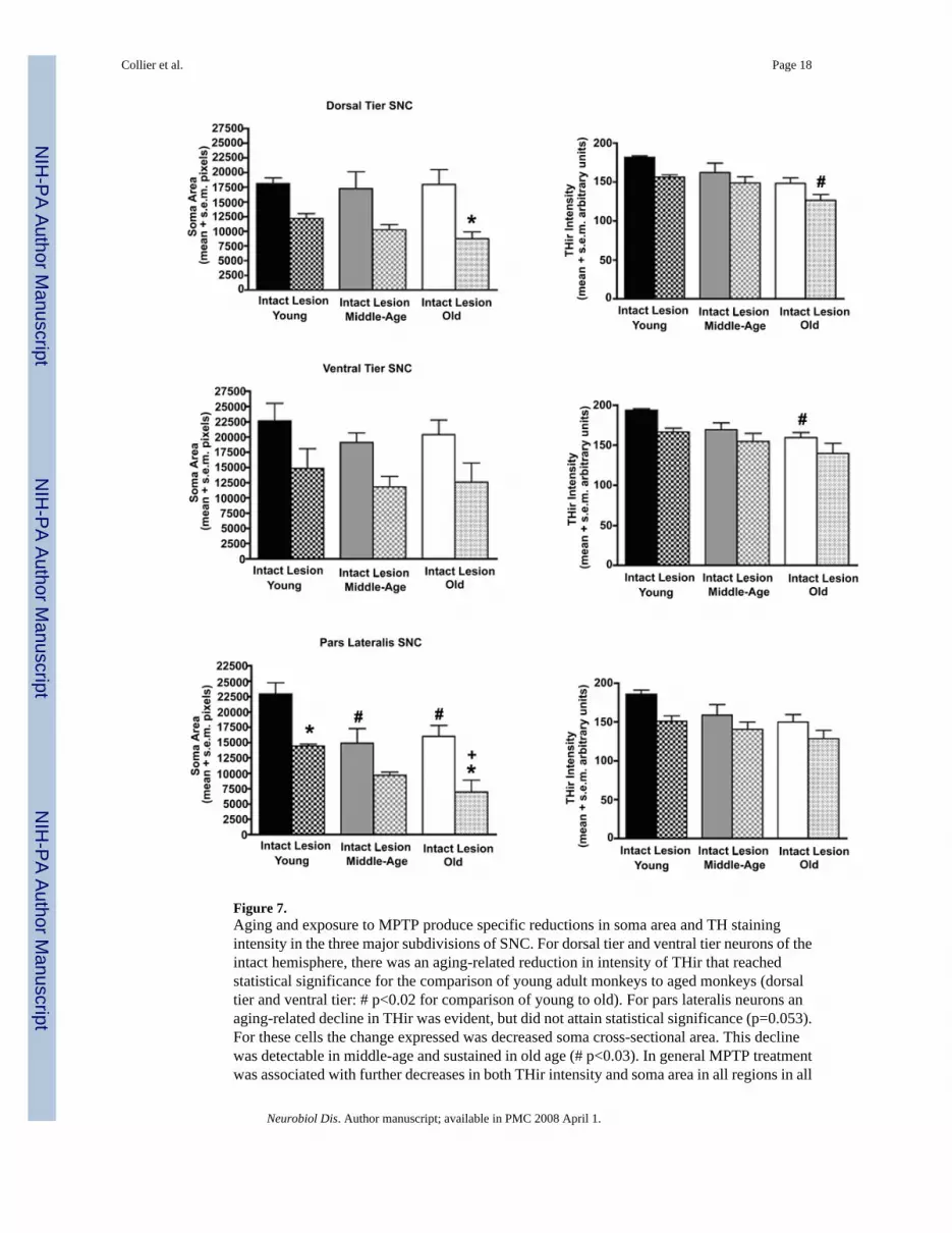
Figure 7.Aging and exposure to MPTP produce specific reductions in soma area and TH stainingintensity in the three major subdivisions of SNC. For dorsal tier and ventral tier neurons of theintact hemisphere, there was an aging-related reduction in intensity of THir that reachedstatistical significance for the comparison of young adult monkeys to aged monkeys (dorsaltier and ventral tier: # p<0.02 for comparison of young to old). For pars lateralis neurons anaging-related decline in THir was evident, but did not attain statistical significance (p=0.053).For these cells the change expressed was decreased soma cross-sectional area. This declinewas detectable in middle-age and sustained in old age (# p<0.03). In general MPTP treatmentwas associated with further decreases in both THir intensity and soma area in all regions in all
Collier et al. Page 18
Neurobiol Dis. Author manuscript; available in PMC 2008 April 1.
NIH
-PA Author Manuscript
NIH
-PA Author Manuscript
NIH
-PA Author Manuscript

age groups. Statistically significant decreases in soma area were detected for dorsal tier neuronsof aged monkeys (* p<0.01) and pars lateralis neurons of young and old monkeys (* p<0.03).This cell shrinkage after MPTP exposure was exaggerated in the oldest animals when comparedto young adult animals (+ p<0.04).
Collier et al. Page 19
Neurobiol Dis. Author manuscript; available in PMC 2008 April 1.
NIH
-PA Author Manuscript
NIH
-PA Author Manuscript
NIH
-PA Author Manuscript

Figure 8.The intensity of THir fiber staining decreased in caudate nucleus and putamen with advancingage in the intact hemisphere. Decreased fiber staining was more pronounced and expressedearlier in the putamen (+ Caudate: p<0.01 for comparison of young to old; Putamen: p<0.04for comparison of young to middle-aged and old).
Collier et al. Page 20
Neurobiol Dis. Author manuscript; available in PMC 2008 April 1.
NIH
-PA Author Manuscript
NIH
-PA Author Manuscript
NIH
-PA Author Manuscript

NIH
-PA Author Manuscript
NIH
-PA Author Manuscript
NIH
-PA Author Manuscript
Collier et al. Page 21
Table 1Experimental Subjects and MPTP Dose.
Animal I.D. Age (yr) Weight (kg) MPTP (mg)Young6965 8 6.0 3.0; 2.06966 8 5.3 3.07009 9 6.4 3.0; 2.0Middle-aged6337 17 10.0 3.56327 17 6.4 3.06339 14 9.5 3.5; 2.07014 17 7.3 3.5Old6724 31 7.2 2.36725 30 7.3 2.3; 2.06254 26.5 6.9 2,3; 2.06334 20.5 10.1 2.36826 24.5 8.9 2.36823 25 7.4 2.3
Neurobiol Dis. Author manuscript; available in PMC 2008 April 1.