Embed Size (px)
Citation preview
AUTHOR COPY ONLYR
EPRODUCTIONRESEARCHAKT (protein kinase B) is implicated in meiotic maturation ofporcine oocytes
Jaroslav Kalous, Michal Kubelka, Petr Solc, Andrej Susor and Jan Motlık
Institute of Animal Physiology and Genetics, Academy of Sciences of the Czech Republic, Rumburska 89, 277 21Libechov, Czech Republic
Correspondence should be addressed to J Kalous; Email: [email protected]
Abstract
The aim of this study was to investigate the involvement of the serine/threonine protein kinase AKT (also called protein kinase B) in
the control of meiosis of porcine denuded oocytes (DOs) matured in vitro. Western blot analysis revealed that the two principal AKT
phosphorylation sites, Ser473 and Thr308, are phosphorylated at different stages of meiosis. In freshly isolated germinal vesicle
(GV)-stage DOs, Ser473 was already phosphorylated. After the onset of oocyte maturation, the intensity of the Ser473 phosphorylation
increased, however, which declined sharply when DOs underwent GV breakdown (GVBD) and remained at low levels in metaphase
I- and II-stage (MI- and MII-stage). In contrast, phosphorylation of Thr308 was increased by the time of GVBD and reached maximum at
MI-stage. A peak of AKT activity was noticed around GVBD and activity of AKT declined at MI-stage. To assess the role of AKT during
meiosis, porcine DOs were cultured in 50 mM SH-6, a specific inhibitor of AKT. In SH-6-treated DOs, GVBD was not inhibited; on the
contrary, a significant acceleration of meiosis resumption was observed. The dynamics of the Ser473 phosphorylation was not affected;
however, phosphorylation of Thr308 was reduced, AKT activity was diminished at the time of GVBD, and meiotic progression was
arrested in early MI-stage. Moreover, the activity of the cyclin-dependent kinase 1 (CDK1) and MAP kinase declined when SH-6-treated
DOs underwent GVBD, indicating that AKT activity is involved in the regulation of CDK1 and MAP kinase. These results suggest that
activity of AKT is not essential for induction of GVBD in porcine oocytes but plays a substantial role during progression of meiosis to
MI/MII-stage.
Reproduction (2009) 138 645–654
Introduction
In mammalian ovaries, the oocytes are naturallyarrested in prophase of the first meiotic division, atthe so-called germinal vesicle (GV) stage. The fullygrown oocytes resume meiosis in vivo after LH surge orin vitro when released from antral follicles and furthercultured under suitable conditions.
cAMP is one of the main regulators of the oocyte entryinto meiosis. In mammalian oocytes, the concentration ofcAMP is regulated by gonadotropins such as FSH andLH acting through the follicular cells (Bornslaeger &Schultz 1985). In mouse oocytes, elevated levels of cAMPand activated protein kinase A (PKA) are involved inmaintaining oocytes in GV stage (Tsafriri et al. 1996, Contiet al. 1998). PKA activity is maintained at high levels bythe increased cAMP, thus preventing maturation in vitroand in vivo (Masciarelli et al. 2004). Degradation of cAMPby a phosphodiesterase type IIIA (PDE3A) leads toa decrease of PKA activity and initiates resumption ofmeiosis in rodent oocytes (Conti 2002, Kovo et al. 2006).Similarly, in porcine oocytes, resumption of meiosis isalso associated with a decrease in cAMP concentrationand subsequent inactivation of PKA (Kim & Menino 1995).
q 2009 Society for Reproduction and Fertility
ISSN 1470–1626 (paper) 1741–7899 (online)
The activity of maturation promoting factor (MPF) iscrucial for meiotic maturation of oocytes. MPFcomplex consists of the catalytic subunit p34cdc2
kinase (also termed cyclin-dependent kinase 1(CDK1)) and regulatory subunit cyclin B (Gautier &Maller 1991, Jessus & Beach 1992). The activity ofCDK1 is controlled by phosphorylation at threeresidues; phosphorylation on Thr161 is necessary forCDK1 kinase activation, whereas phosphorylation ofCDK1 by WEE1 and MYT1 kinases on either Thr14 orTyr15 residues dominantly inhibits activation of CDK1(Coleman & Dunphy 1994). This inhibition is reversedwhen CDK1 is dephosphorylated by activatedCDC25C phosphatase (Nilsson & Hoffmann 2000).It has been reported that PKA phosphorylates andinactivates CDC25C in frog oocytes (Duckworth et al.2002, Shibuya 2003). Also in mouse oocytes, theWEE1 kinase is a substrate for PKA in vitro (Han et al.2005). These findings support the concept thatPKA maintains meiotic arrest via dual regulation ofthe kinase that phosphorylates CDK1 and the phos-phatase that dephosphorylates the same residues ofCDK1 (Han & Conti 2006).
DOI: 10.1530/REP-08-0461
Online version via www.reproduction-online.org
AUTHOR COPY ONLY
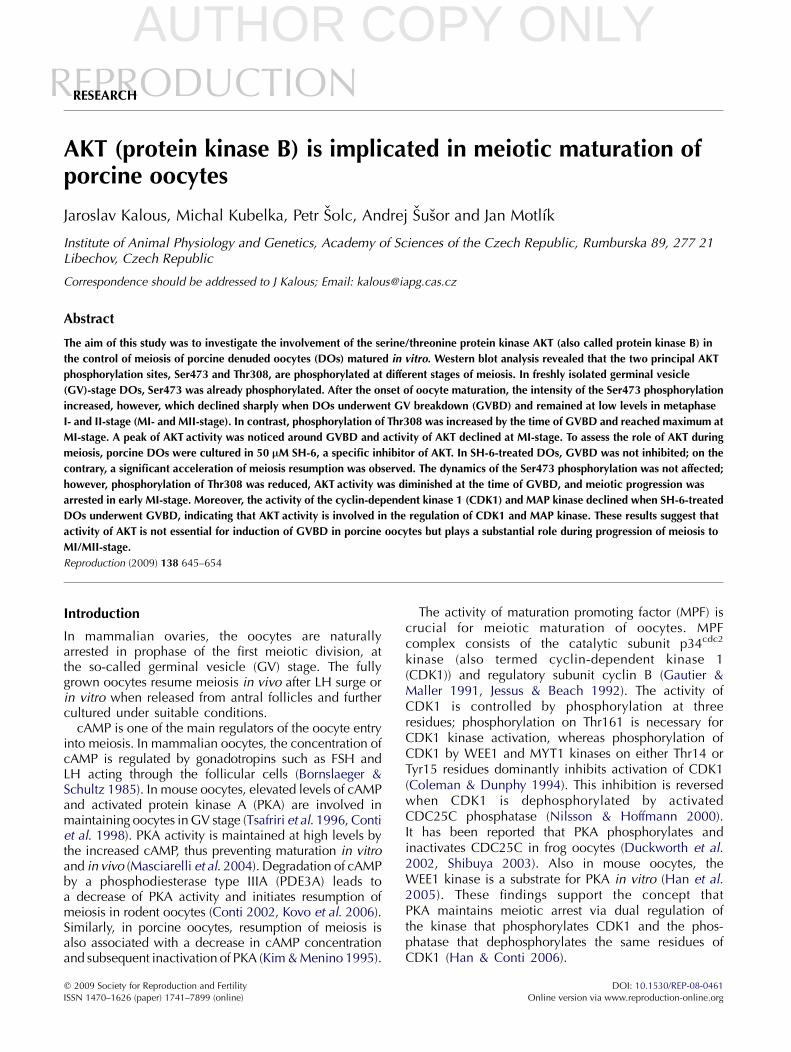
Figure 1 Progression of meiosis in porcine DOs cultured in controlconditions. Isolated porcine OCCs were deprived of cumulus cells, andDOs oocytes were subsequently cultured in vitro in control medium.GV, germinal vesicle; LD, late diakinesis; MI, metaphase-I; MII,metaphase-II. A total of 977 oocytes were scored.
646 J Kalous and others
Another kinase implicated in MPF regulation is aserine/threonine protein kinase B (PKB/AKT), a keydownstream effector of the phosphatidylinositol3-kinase (PI3K) pathway.
The PI3K–AKT pathway is activated by insulin, growthfactors, and adhesion to extracellular matrix or othercells. Activated PI3K converts its lipid substrate phos-phatidylinositol 4,5 biphosphate (PtdIns (4,5)P2) tophosphatidylinositol 3,4,5 triphosphate (PtdIns(3,4,5)P3). Consequently, AKT is recruited to the cellmembrane after binding PtdIns (3,4,5)P3 through its PHdomain, and membrane-bound AKT is activated byphosphorylation at two sites. PDK1 phosphorylatesThr308 residue located in the activation loop of thekinase (Stokoe et al. 1997, Kim et al. 2001), whereasmTORC2 was identified as the kinase responsible forphosphorylation of the Ser473 residue situated in thehydrophobic site (Sarbassov et al. 2005). The dual-specificity lipid phosphatase phosphatase and tensinhomolog deleted on chromosome 10 (PTEN) depho-sphorylates PI (3,4,5)P3 and in such a way negativelyregulates AKT kinase activity (Maehama et al. 2001).
It has been reported that PI3K/AKT pathway contrib-utes to cyclin B1 expression and CDK1 activation(Roberts et al. 2002). Data obtained on somatic cellsindicate that AKT promotes cell survival and participatesin the regulation of the G2/M transition. In epithelialcells, the activity of AKT is increased at the G2/Mtransition of the cell cycle, and it is necessary for thetimely progression through mitosis (Shtivelman et al.2002). The cell cycle arrest imposed by inhibitors of PI3Kcould be reversed by the expression of constitutivelyactive AKT (Shtivelman 2003). A G2/M cell cyclecheckpoint induced by DNA damage is surpassed byconstitutively activated AKT, and this effect has also beenobserved in cells lacking PTEN that limits activation ofAKT (Kandel et al. 2002). AKT was also reported topromote cell cycle progression at the G2/M transitionthrough WEE1 inactivation in HEK 293 cells (Katayamaet al. 2005). In HeLa cells, AKT phosphorylates theCDC25B phosphatase and regulates its intracellularlocalization (Baldin et al. 2003).
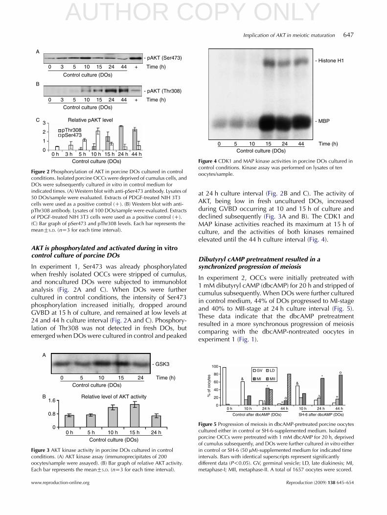
The glycogen synthase kinase-3 (GSK3), an importantsubstrate of AKT, is involved in numerous cellularprocesses, including regulation of the activity ofmicrotubule-associated proteins affecting formationand function of mitotic spindle (Moreno & Avila 1998).In mouse oocytes, pharmacological inhibition of GSK3causes abnormal meiotic spindle configuration, chro-matin organization, and bivalent chromatin segregation(Wang et al. 2003).
Experiments performed on starfish oocytes providedthe evidence of AKT as an initiator of the meiotic Mphase. It has been documented that AKT activatesmeiotic G2/M transition by phosphorylation andinactivation of protein kinase MYT1, which hasbeen shown to phosphorylate and inactivate MPF
Reproduction (2009) 138 645–654
(Okumura et al. 2002). Study on Xenopus oocytesrevealed that insulin-like growth factor 1 (IGF1)- orinsulin-induced meiotic maturation is mediatedby activation of PI3K and AKT (Liu et al. 1995,DeuterReinhardt et al. 1997). It has been shown thatexpression of constitutive active PI3K can activateMAPK and induce GV breakdown (GVBD) in Xenopusoocytes, and this effect is mediated through its lipidkinase activity and activation of AKT (Hehl et al. 2001).Furthermore, it was documented in Xenopus oocytesthat phosphorylation of PDE3A by AKT plays a role inmeiotic maturation induced by growth factors (Andersenet al. 2003). Also the data obtained in mouse oocytesindicate that activation of PDE3A by PKB-mediatedphosphorylation is implicated in the control of PDE3Aactivity and resumption of meiosis (Han et al. 2006).Suppression of AKT activity was accompanied with adecrease of CDK1 activity and postponed resumption ofmeiosis in mouse oocytes (Kalous et al. 2006). Similarly,in cattle oocytes, AKT is also involved in the regulation ofmeiotic metaphase I and II (MI/MII) transition (Tomek &Smiljakovic 2005).
The objectives of this study were to evaluate thedynamics of AKT phosphorylation and activation inporcine oocytes matured in vitro and to analyze possiblerole of AKT in the regulation of meiosis.
Results
Over 80% of porcine denuded oocytes reachedMII-stage during control culture
In experiment 1, fresh porcine oocyte–cumuluscomplexes (OCCs) were deprived of cumulus, anddenuded oocytes (DOs) were cultured in controlmedium subsequently. During 44 h of control culture,84% of DOs reached MII-stage (Fig. 1). However, at 24 hculture interval, the time course of meiotic maturationwas not synchronous as 18% of DOs retained GV, 27%exhibited late diakinesis stage (LD), 36% advanced toMI, and 19% to MII-stage (Fig. 1).
www.reproduction-online.org
AUTHOR COPY ONLY
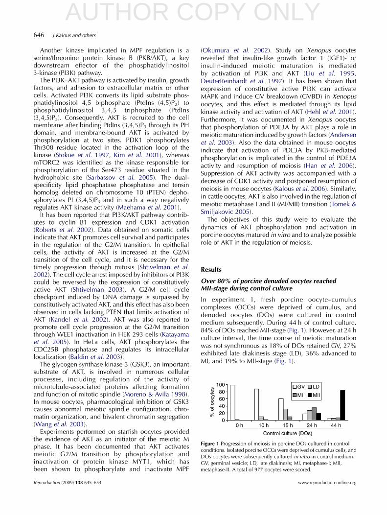
Figure 2 Phosphorylation of AKT in porcine DOs cultured in controlconditions. Isolated porcine OCCs were deprived of cumulus cells, andDOs were subsequently cultured in vitro in control medium forindicated times. (A) Western blot with anti-pSer473 antibody. Lysates of50 DOs/sample were evaluated. Extracts of PDGF-treated NIH 3T3cells were used as a positive control (C). (B) Western blot with anti-pThr308 antibody. Lysates of 100 DOs/sample were evaluated. Extractsof PDGF-treated NIH 3T3 cells were used as a positive control (C).(C) Bar graph of pSer473 and pThr308 levels. Each bar represents themeanGS.D. (nZ3 for each time interval).
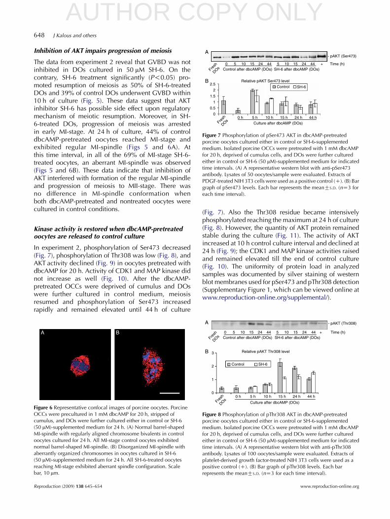
Figure 4 CDK1 and MAP kinase activities in porcine DOs cultured incontrol conditions. Kinase assay was performed on lysates of tenoocytes/sample.
Implication of AKT in meiotic maturation 647
AKT is phosphorylated and activated during in vitrocontrol culture of porcine DOs
In experiment 1, Ser473 was already phosphorylatedwhen freshly isolated OCCs were stripped of cumulus,and noncultured DOs were subjected to immunoblotanalysis (Fig. 2A and C). When DOs were furthercultured in control conditions, the intensity of Ser473phosphorylation increased initially, dropped aroundGVBD at 15 h of culture, and remained at low levels at24 and 44 h culture interval (Fig. 2A and C). Phosphory-lation of Thr308 was not detected in fresh DOs, butemerged when DOs were cultured in control and peaked
Figure 3 AKT kinase activity in porcine DOs cultured in controlconditions. (A) AKT kinase assay (immunoprecipitates of 200oocytes/sample were assayed). (B) Bar graph of relative AKT activity.Each bar represents the meanGS.D. (nZ3 for each time interval).
www.reproduction-online.org
at 24 h culture interval (Fig. 2B and C). The activity ofAKT, being low in fresh uncultured DOs, increasedduring GVBD occurring at 10 and 15 h of culture anddeclined subsequently (Fig. 3A and B). The CDK1 andMAP kinase activities reached its maximum at 15 h ofculture, and the activities of both kinases remainedelevated until the 44 h culture interval (Fig. 4).
Dibutyryl cAMP pretreatment resulted in asynchronized progression of meiosis
In experiment 2, OCCs were initially pretreated with1 mM dibutyryl cAMP (dbcAMP) for 20 h and stripped ofcumulus subsequently. When DOs were further culturedin control medium, 44% of DOs progressed to MI-stageand 40% to MII-stage at 24 h culture interval (Fig. 5).These data indicate that the dbcAMP pretreatmentresulted in a more synchronous progression of meiosiscomparing with the dbcAMP-nontreated oocytes inexperiment 1 (Fig. 1).
Figure 5 Progression of meiosis in dbcAMP-pretreated porcine oocytescultured either in control or SH-6-supplemented medium. Isolatedporcine OCCs were pretreated with 1 mM dbcAMP for 20 h, deprivedof cumulus subsequently, and DOs were further cultured in vitro eitherin control or SH-6 (50 mM)-supplemented medium for indicated timeintervals. Bars with identical superscripts represent significantlydifferent data (P!0.05). GV, germinal vesicle; LD, late diakinesis; MI,metaphase-I; MII, metaphase-II. A total of 1657 oocytes were scored.
Reproduction (2009) 138 645–654
AUTHOR COPY ONLY
Figure 7 Phosphorylation of pSer473 AKT in dbcAMP-pretreatedporcine oocytes cultured either in control or SH-6-supplementedmedium. Isolated porcine OCCs were pretreated with 1 mM dbcAMPfor 20 h, deprived of cumulus cells, and DOs were further culturedeither in control or SH-6 (50 mM)-supplemented medium for indicatedtime intervals. (A) A representative western blot with anti-pSer473antibody. Lysates of 50 oocytes/sample were evaluated. Extracts ofPDGF-treated NIH 3T3 cells were used as a positive control (C). (B) Bargraph of pSer473 levels. Each bar represents the meanGS.D. (nZ3 foreach time interval).
648 J Kalous and others
Inhibition of AKT impairs progression of meiosis
The data from experiment 2 reveal that GVBD was notinhibited in DOs cultured in 50 mM SH-6. On thecontrary, SH-6 treatment significantly (P!0.05) pro-moted resumption of meiosis as 50% of SH-6-treatedDOs and 39% of control DOs underwent GVBD within10 h of culture (Fig. 5). These data suggest that AKTinhibitor SH-6 has possible side effect upon regulatorymechanism of meiotic resumption. Moreover, in SH-6-treated DOs, progression of meiosis was arrestedin early MI-stage. At 24 h of culture, 44% of controldbcAMP-pretreated oocytes reached MI-stage andexhibited regular MI-spindle (Figs 5 and 6A). Atthis time interval, in all of the 69% of MI-stage SH-6-treated oocytes, an aberrant MI-spindle was observed(Figs 5 and 6B). These data indicate that inhibition ofAKT interfered with formation of the regular MI-spindleand progression of meiosis to MII-stage. There wasno difference in MI-spindle conformation whenboth dbcAMP-pretreated and nontreated oocytes werecultured in control conditions.
Kinase activity is restored when dbcAMP-pretreatedoocytes are released to control culture
In experiment 2, phosphorylation of Ser473 decreased(Fig. 7), phosphorylation of Thr308 was low (Fig. 8), andAKT activity declined (Fig. 9) in oocytes pretreated withdbcAMP for 20 h. Activity of CDK1 and MAP kinase didnot increase as well (Fig. 10). After the dbcAMP-pretreated OCCs were deprived of cumulus and DOswere further cultured in control medium, meiosisresumed and phosphorylation of Ser473 increasedrapidly and remained elevated until 44 h of culture
Figure 6 Representative confocal images of porcine oocytes. PorcineOCCs were precultured in 1 mM dbcAMP for 20 h, stripped ofcumulus, and DOs were further cultured either in control or SH-6(50 mM)-supplemented medium for 24 h. (A) Normal barrel-shapedMI-spindle with regularly aligned chromosome bivalents in controloocytes cultured for 24 h. All MI-stage control oocytes exhibitednormal barrel-shaped MI-spindle. (B) Disorganized MI-spindle withaberrantly organized chromosomes in oocytes cultured in SH-6(50 mM)-supplemented medium for 24 h. All SH-6-treated oocytesreaching MI-stage exhibited aberrant spindle configuration. Scalebar, 10 mm.
Reproduction (2009) 138 645–654
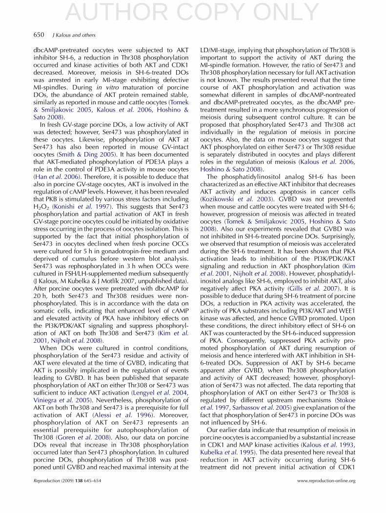
(Fig. 7). Also the Thr308 residue became intensivelyphosphorylated reaching the maximum at 24 h of culture(Fig. 8). However, the quantity of AKT protein remainedstable during the culture (Fig. 11). The activity of AKTincreased at 10 h control culture interval and declined at24 h (Fig. 9); the CDK1 and MAP kinase activities raisedand remained elevated till the end of control culture(Fig. 10). The uniformity of protein load in analyzedsamples was documented by silver staining of westernblot membranes used for pSer473 and pThr308 detection(Supplementary Figure 1, which can be viewed online atwww.reproduction-online.org/supplemental/).
Figure 8 Phosphorylation of pThr308 AKT in dbcAMP-pretreatedporcine oocytes cultured either in control or SH-6-supplementedmedium. Isolated porcine OCCs were pretreated with 1 mM dbcAMPfor 20 h, deprived of cumulus cells, and DOs were further culturedeither in control or SH-6 (50 mM)-supplemented medium for indicatedtime intervals. (A) A representative western blot with anti-pThr308antibody. Lysates of 100 oocytes/sample were evaluated. Extracts ofplatelet-derived growth factor-treated NIH 3T3 cells were used as apositive control (C). (B) Bar graph of pThr308 levels. Each barrepresents the meanGS.D. (nZ3 for each time interval).
www.reproduction-online.org
AUTHOR COPY ONLY
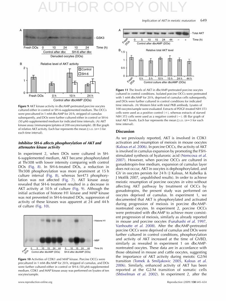
Figure 9 AKT kinase activity in dbcAMP-pretreated porcine oocytescultured either in control or SH-6-supplemented medium. The OCCswere precultured in 1 mM dbcAMP for 20 h, stripped of cumulussubsequently, and DOs were further cultured either in control or SH-6(50 mM)-supplemented medium for indicated time intervals. (A) AKTkinase assay (immunoprecipitates of 200 oocytes/sample). (B) Bar graphof relative AKT activity. Each bar represents the meanGS.D. (nZ3 foreach time interval).
Figure 11 The levels of AKT in dbcAMP-pretreated porcine oocytescultured in control conditions. Isolated porcine OCCs were pretreatedwith 1 mM dbcAMP for 20 h, deprived of cumulus cells subsequently,and DOs were further cultured in control conditions for indicatedtime intervals. (A) Western blot with total PKB antibody. Lysates of100 oocytes/sample were evaluated. Extracts of PDGF-treated NIH 3T3cells were used as a positive control (C), whereas extracts of starvedNIH 3T3 cells were used as a negative control (K). (B) Bar graph oftotal AKT levels. Each bar represents the meanGS.D. (nZ3 for eachtime interval).
Implication of AKT in meiotic maturation 649
Inhibitor SH-6 affects phosphorylation of AKT andattenuates kinase activity
In experiment 2, when DOs were cultured in SH-6-supplemented medium, AKT became phosphorylatedat Thr308 with lower intensity comparing with controlDOs (Fig. 8). In SH-6-treated DOs, a reduction inThr308 phosphorylation was more prominent at 15 hculture interval (Fig. 8), whereas Ser473 phosphory-lation was not affected (Fig. 7). AKT kinase assayrevealed that SH-6 treatment resulted in a decrease inAKT activity at 10 h of culture (Fig. 9). Although theinitial activation of histone H1 kinase and MAP kinasewas not prevented in SH-6-treated DOs, suppression ofactivity of these kinases was apparent at 24 and 44 hof culture (Fig. 10).
Figure 10 Activities of CDK1 and MAP kinase. Porcine OCCs wereprecultured in 1 mM dbcAMP for 20 h, stripped of cumulus, and DOswere further cultured either in control or SH-6 (50 mM)-supplementedmedium. CDK1 and MAP kinase assay was performed on lysates of tenoocytes/sample.
www.reproduction-online.org
Discussion
As we previously reported, AKT is involved in CDK1activation and resumption of meiosis in mouse oocytes(Kalous et al. 2006). In porcine OCCs, the activity of AKTis involved in cumulus expansion by promoting the FSH-stimulated synthesis of hyaluronic acid (Nemcova et al.2007). However, when porcine OCCs are cultured ingonadotropin-free medium, expansion of cumulus layerdoes not occur, AKT in oocytes is dephosphorylated, andGV in oocytes persists for 24 h ( J Kalous, M Kubelka &J Motlık 2007, unpublished results). In order to achievemeiotic resumption of porcine oocytes in vitro withoutaffecting AKT pathway by treatment of OCCs bygonadotropins, the present study was performed onoocytes deprived of cumulus. In experiment 1, wedocumented that AKT is phosphorylated and activatedduring progression of meiosis in porcine dbcAMP-nontreated oocytes. In experiment 2, porcine OCCswere pretreated with dbcAMP to achieve more consist-ent progression of meiosis, similarly as already reportedin mouse and porcine oocytes (Funahashi et al. 1997,Vanhoutte et al. 2008). After the dbcAMP-pretreatedporcine OCCs were deprived of cumulus and DOs werefurther cultured in control conditions, phosphorylationand activity of AKT increased at the time of GVBD,similarly as revealed in experiment 1 on dbcAMP-nontreated oocytes. These data are in accordance withthose obtained in mouse and cattle oocytes, suggestingthe importance of AKT activity during meiotic G2/Mtransition (Tomek & Smiljakovic 2005, Kalous et al.2006). Similarly, enhanced activity of AKT has beenreported at the G2/M transition of somatic cells(Shtivelman et al. 2002). In experiment 2, after the
Reproduction (2009) 138 645–654
AUTHOR COPY ONLY650 J Kalous and others
dbcAMP-pretreated oocytes were subjected to AKTinhibitor SH-6, a reduction in Thr308 phosphorylationoccurred and kinase activities of both AKT and CDK1decreased. Moreover, meiosis in SH-6-treated DOswas arrested in early MI-stage exhibiting defectiveMI-spindles. During in vitro maturation of porcineDOs, the abundance of AKT protein remained stable,similarly as reported in mouse and cattle oocytes (Tomek& Smiljakovic 2005, Kalous et al. 2006, Hoshino &Sato 2008).
In fresh GV-stage porcine DOs, a low activity of AKTwas detected; however, Ser473 was phosphorylated inthese oocytes. Likewise, phosphorylation of AKT atSer473 has also been reported in mouse GV-intactoocytes (Smith & Ding 2005). It has been documentedthat AKT-mediated phosphorylation of PDE3A plays arole in the control of PDE3A activity in mouse oocytes(Han et al. 2006). Therefore, it is possible to deduce thatalso in porcine GV-stage oocytes, AKT is involved in theregulation of cAMP levels. However, it has been revealedthat PKB is stimulated by various stress factors includingH2O2 (Konishi et al. 1997). This suggests that Ser473phosphorylation and partial activation of AKT in freshGV-stage porcine oocytes could be initiated by oxidativestress occurring in the process of oocytes isolation. This issupported by the fact that initial phosphorylation ofSer473 in oocytes declined when fresh porcine OCCswere cultured for 5 h in gonadotropin-free medium anddeprived of cumulus before western blot analysis.Ser473 was rephosphorylated in 3 h when OCCs werecultured in FSH/LH-supplemented medium subsequently(J Kalous, M Kubelka & J Motlık 2007, unpublished data).After porcine oocytes were pretreated with dbcAMP for20 h, both Ser473 and Thr308 residues were non-phosphorylated. This is in accordance with the data onsomatic cells, indicating that enhanced level of cAMPand elevated activity of PKA have inhibitory effects onthe PI3K/PDK/AKT signaling and suppress phosphoryl-ation of AKT on both Thr308 and Ser473 (Kim et al.2001, Nijholt et al. 2008).
When DOs were cultured in control conditions,phosphorylation of the Ser473 residue and activity ofAKT were elevated at the time of GVBD, indicating thatAKT is possibly implicated in the regulation of eventsleading to GVBD. It has been published that separatephosphorylation of AKT on either Thr308 or Ser473 wassufficient to induce AKT activation (Lengyel et al. 2004,Viniegra et al. 2005). Nevertheless, phosphorylation ofAKT on both Thr308 and Ser473 is a prerequisite for fullactivation of AKT (Alessi et al. 1996). Moreover,phosphorylation of AKT on Ser473 represents anessential prerequisite for autophosphorylation ofThr308 (Goren et al. 2008). Also, our data on porcineDOs reveal that increase in Thr308 phosphorylationoccurred later than Ser473 phosphorylation. In culturedporcine DOs, phosphorylation of Thr308 was post-poned until GVBD and reached maximal intensity at the
Reproduction (2009) 138 645–654
LD/MI-stage, implying that phosphorylation of Thr308 isimportant to support the activity of AKT during theMI-spindle formation. However, the ratio of Ser473 andThr308 phosphorylation necessary for full AKTactivationis not known. The results presented reveal that the timecourse of AKT phosphorylation and activation wassomewhat different in samples of dbcAMP-nontreatedand dbcAMP-pretreated oocytes, as the dbcAMP pre-treatment resulted in a more synchronous progression ofmeiosis during subsequent control culture. It can beproposed that phosphorylated Ser473 and Thr308 actindividually in the regulation of meiosis in porcineoocytes. Also, the data on mouse oocytes suggest thatAKT phosphorylated on either Ser473 or Thr308 residueis separately distributed in oocytes and plays differentroles in the regulation of meiosis (Kalous et al. 2006,Hoshino & Sato 2008).
The phosphatidylinositol analog SH-6 has beencharacterized as an effective AKT inhibitor that decreasesAKT activity and induces apoptosis in cancer cells(Kozikowski et al. 2003). GVBD was not preventedwhen mouse and cattle oocytes were treated with SH-6;however, progression of meiosis was affected in treatedoocytes (Tomek & Smiljakovic 2005, Hoshino & Sato2008). Also our experiments revealed that GVBD wasnot inhibited in SH-6-treated porcine DOs. Surprisingly,we observed that resumption of meiosis was acceleratedduring the SH-6 treatment. It has been shown that PKAactivation leads to inhibition of the PI3K/PDK/AKTsignaling and reduction in AKT phosphorylation (Kimet al. 2001, Nijholt et al. 2008). However, phosphatidyl-inositol analogs like SH-6, employed to inhibit AKT, alsonegatively affect PKA activity (Gills et al. 2007). It ispossible to deduce that during SH-6 treatment of porcineDOs, a reduction in PKA activity was accelerated, theactivity of PKA substrates including PI3K/AKT and WEE1kinase was affected, and hence GVBD promoted. Uponthese conditions, the direct inhibitory effect of SH-6 onAKT was counteracted by the SH-6-induced suppressionof PKA. Consequently, suppressed PKA activity pro-moted phosphorylation of AKT during resumption ofmeiosis and hence interfered with AKT inhibition in SH-6-treated DOs. Suppression of AKT by SH-6 becameapparent after GVBD, when Thr308 phosphorylationand activity of AKT decreased; however, phosphoryl-ation of Ser473 was not affected. The data reporting thatphosphorylation of AKT on either Ser473 or Thr308 isregulated by different upstream mechanisms (Stokoeet al. 1997, Sarbassov et al. 2005) give explanation of thefact that phosphorylation of Ser473 in porcine DOs wasnot influenced by SH-6.
Our earlier data indicate that resumption of meiosis inporcine oocytes is accompanied by a substantial increasein CDK1 and MAP kinase activities (Kalous et al. 1993,Kubelka et al. 1995). The data presented here reveal thatreduction in AKT activity occurring during SH-6treatment did not prevent initial activation of CDK1
www.reproduction-online.org
AUTHOR COPY ONLYImplication of AKT in meiotic maturation 651
and MAP kinase essential for induction of GVBD.However, when GVBD was accomplished, both CDK1and MAP kinase were inhibited in SH-6-treated porcineDOs. In starfish oocytes, AKT downregulates MYT1, aregulatory kinase of CDK1 and hence induces the initialactivation of cyclin B/CDK1 at the meiotic G2/M-phasetransition (Okumura et al. 2002). Our results indicate thatin SH-6-treated porcine DOs, induction of GVBD andMPF activation is either AKT-independent or reducedactivity of AKT is sufficient to support resumption ofmeiosis. We observed that in control culture, AKTbecame phosphorylated at Thr308 around GVBD. Incontrast, in SH-6-treated DOs, phosphorylation ofThr308 was reduced, and progression of meiosis wasarrested in early MI-stage. These data indicate thatphosphorylation of AKT at Thr308 is not implicated inthe events leading to the resumption of meiosis inporcine DOs; however, a full phosphorylation of theThr308 is likely to be essential for meiosis to progress tothe MI- and MII-stage. Similarly, inhibition of AKT incattle oocytes did not prevent GVBD; however, meiosiswas arrested in the MI-stage (Tomek & Smiljakovic 2005).
In mouse MI- and MII-stage oocytes, AKT phosphory-lated at Ser473 was localized with microtubules, whileThr308-phosphorylated AKT was present in pericentriolarmaterial (Hoshino et al. 2004). Activated AKT phosphory-lated at Thr308 is able to phosphorylate and inhibit GSK3during mitosis (Cross et al. 1995, Cohen & Frame 2001).Such AKT-mediated phosphorylation and activation ofGSK3 occur on the centrosome and spindle poles and isinvolved in stabilization of microtubules in this region(Wakefield et al. 2002). We suggest that suppression ofactivity of AKT in porcine DOs induced by SH-6 led to anincrease of GSK3 activity in centrosomal region. Conse-quently, progress of meiosis was interrupted, as assemblyof MI-spindles was disturbed and aberrant MI-spindleswere formed. In conclusion, our results on porcineoocytes indicate that AKT is involved in the regulationof CDK1 activity during meiosis, and activated AKT isnecessary for successful course of meiotic maturation.
Materials and Methods
Experimental design
The aim of the present study was to investigate the role of AKTin meiotic events occurring during in vitro maturation ofporcine oocytes. To avoid the necessity of inducing meiotic invitro progression in OCCs by gonadotropins and thus influenceAKT pathway in oocytes by hormonal stimulus, the study wasperformed on DOs maturing in gonadotropin-free medium.
In experiment 1, phosphorylation and activation of AKTduring control culture of dbcAMP-nontreated porcine DOswere investigated. Fresh OCCs were stripped of cumulus beforeculture, and DOs were further cultured in control medium for 3,5, 10, 15, 24, and 44 h. After culture, DOs were proceeded forwestern blot analysis, kinase assays, and assessment of nuclearmaturation. In experiment 2, the effect of AKT inhibitor SH-6
www.reproduction-online.org
upon AKT activity and meiotic progression in DOs wereanalyzed. To achieve more synchronous progression of meiosis,porcine OCCs were pretreated with 1 mM dbcAMP for 20 h,and OCCs were deprived of cumulus subsequently. The DOswere further cultured either in control or SH-6 (50 mM)-supplemented medium for 5, 10, 15, 24, and 44 h. After culture,DOs were subjected to phase-contrast and immunofluorescentmicroscopy, western blot analysis, and kinase assays.
Oocyte collection, culture, and evaluation
Ovaries, collected from slaughtered noncycling gilts, weretransported to the laboratory in physiological saline at 20 8C.The ovaries were briefly washed in physiological saline, andOCCs were aspirated from 2- to 5-mm follicles using a21-gauge needle attached to a syringe. M-199 medium(Sigma) supplemented with 5% BSA (Sigma), 0.68 mMglutamine, 100 mM sodium pyruvate, 200 mM cysteamine,100 IU/ml penicillin, and 0.1 mg/ml streptomycin (Sigma) wasused as a control medium. In experiment 1, the OCCs weredeprived of cumulus before culture, and DOs were furthercultured in nonsupplemented control medium. In experiment 2,the OCCs were precultured for 20 h in control mediumsupplemented with dbcAMP (1 mM final concentration;Calbiochem, San Diego, CA, USA). OCCs were stripped ofcumulus after preculture, and DOs were further cultured eitherin nonsupplemented control medium or in control mediumsupplemented with SH-6 (50 mM final concentration; Calbio-chem) for defined time intervals. All cultures were performed ina humidified atmosphere of 5% CO2 at 38.5 8C. At the end ofculture, the DOs were washed in physiological saline, and afterthe last wash, the oocytes were fixed for microscopy or stored atK80 8C until western blot or kinase assay analysis.
Western blotting
Oocytes were lysed in 10 ml SDS/PAGE sample buffer(62.5 mM Tris/HCl, pH 6.8, at 25 8C, 2% w/v SDS, 10%v/v glycerol, 0.01% w/v bromophenol blue, and 5% v/v2-mercaptoethanol). Samples were heated at 100 8C for 3 minand subjected to SDS/PAGE. After electrophoresis, proteinswere electrically transferred onto PVDF membrane (Immo-bilon-P; Millipore, Bedford, MA, USA) using a semi-dry blottingsystem. Subsequently, blotted membranes were blocked for 1 hin 5% (v/v) nonfat milk (Sigma) in T-TBS (Tris/HCl-bufferedsaline (pH 7.6) with 0.05% Tween 20). T-TBS was used for allfollowing incubations and washes. Blocked membranes wereincubated overnight at 4 8C with a PKB-specific primaryantibody diluted 1:1000 in T-TBS with 5% nonfat milk (pAKTSer473 (SC-7985), pAKT Thr308 (SC-16646), or AKT1 antibody(SC-5298)); all primary antibodies were purchased from SantaCruz Biotechnology (Santa Cruz, CA, USA). The blots were thenwashed (thrice for 5 min) in T-TBS and incubated for 1 h at roomtemperature (18–25 8C) with HRP-linked donkey anti-rabbit IgG(Amersham; 1:10 000, phospho-AKT detection) or rabbit anti-mouse IgG (Amersham; 1:15 000, total AKT protein detection),both diluted in T-TBS with 5% nonfat milk. After extensivewashing (three to five times for 5 min), the immunoreactivesignal was detected using ECL (Amersham International).
Reproduction (2009) 138 645–654
AUTHOR COPY ONLY652 J Kalous and others
Histone H1 assay
At each time interval, ten oocytes/sample were collected,washed (4!) in PBS, and transferred into 3 ml PBS in Eppendorftubes. Samples were immediately frozen on solid CO2 andstored at K80 8C until assays were performed. The histone H1and myelin basic protein (MBP) kinase activities were measuredas previously described (Motlik et al. 1998); H1 and MBPare substrates for CDK1 and MAPK respectively. Data fromautoradiograms were analyzed using Scion Image software(Scion Corporation, Frederick, MD, USA).
AKT kinase assay
Eppendorf tubes with frozen oocytes (200 DOs in each tube)were placed on ice, and 200 ml 1! ice-cold cell lysis buffer(catalog no. 9803; Cell Signaling Technology, Beverly, MA, USA)containing 1 mM phenylmethylsulphonyl fluoride was added toeach tube; oocytes were lysed by several cycles of freezing andthawing. After centrifugation (14 000 g for 10 min at 4 8C), thesupernatant was removed and 3 ml AKT1 MAB (SC-5298; SantaCruz Biotechnology) was added, and then the samples wereincubated for 1 h at 4 8C. Protein A Sepharose (Sigma) (200 ml ofthe suspension) was added, and the mixture was incubated at4 8C overnight. The pellet was collected by low-speed centri-fugation and washed twice with ice-cold cell lysis buffer (catalogno. 9803; Cell Signaling Technology) and ice-cold kinase buffer(catalog no. 9802; Cell Signaling Technology). The washed pelletwas assayed for PKB activity as follows: the reaction was startedby adding 30 ml kinase buffer (25 mM Tris/HCl, pH 7.5, 5 mMb-glycerophosphate, 2 mM dithiothreitol, 0.1 mM Na3VO4,10 mM MgCl2, and 200 mM ATP) to each sample together with2 mg GSK3 fusion protein (Cell Signaling Technology) as anexternal substrate for AKT and 500 mCi/ml [g-32P]ATP(10 mCi/ml; Amersham). The reaction was performed for30 min at 30 8C and terminated byadding 30 ml 4! concentratedSDS/PAGE sample buffer and boiled for 3 min. After SDS/PAGE(15% w/v PAGE) (Laemmli 1970), the gels were stained withCoomassie Blue R250 (Serva, Heidelberg, Germany), destainedovernight, dried, and autoradiographed. Subsequently, the gelwas scanned with a Fujifilm BAS-2500 Phosphoimager, and thedata were analyzed by Scion Image software.
Morphological analyses of oocytes
To check the current stage of maturation at the time of samplecollection, an aliquot of 30–40 DOs was mounted onmicroscope slides with Vaseline (Sigma) strips, covered withcover glass, and fixed in solution ethanol:acetic acid (3:1 v/v)for 48 h. Staining was performed with 2% orcein in 50%aqueous acetic acid and 1% sodium citrate. The slides werethen placed in 40% acetic acid and observed with a phase-contrast NU Zeiss microscope.
Immunocytochemistry
Denuded zona-intact oocytes were washed in PBS, fixed for 1 hin 3.7% paraformaldehyde (16005, Aldrich), washed in PBS,and stored in PBS with 1% sodium azide. Such prepared oocytes
Reproduction (2009) 138 645–654
were washed for 30 min in PBS containing 1% Triton-X (T9284,Sigma–Aldrich) and subsequently blocked in 2% BSA fraction V(Sigma) for 2 h in PBS. The oocytes were incubated overnight at4 8C with mouse monoclonal anti-acetylated tubulin antibody(T7451, Sigma, diluted 1:500). The excess primary antibody wasremoved by extensive washing in PBS containing 0.2%BSA/0.05% saponin (16109, Sigma). Oocytes were thenincubated with donkey anti-mouse antibody conjugated withAlexa Flour-594 (Invitrogen, diluted 1:800) in PBS/0.2%BSA/0.05% saponin for 60 min at room temperature. As acontrol, oocytes were incubated only with secondary antibody.The well-washed oocytes were mounted in mounting mediumwith DAPI (H-1500; Vector Laboratories, Peterborough, UK)and examined using confocal microscopy (Leica).
Statistical analyses
Statistical analysis of all data was performed using NCSS 2000computer software (NCSS, Kaysville, UT, USA). Error sectionsin graphs express confidence limits. The frequencies of meiosisprogression were pooled from three replicate experiments, andcumulative results were subjected to c2 analysis. Differenceswere considered significant at P!0.05. A total of 2634 oocyteswere subjected to morphological analyses. The results of thewestern blotting and AKT kinase assays were quantified byScion Image software and expressed as the meansGS.D. of threeindependent experiments. The data were normalized to themean value by mean centering.
Declaration of interest
The authors declare that there is no conflict of interest thatwould prejudice the impartiality of this scientific work.
Funding
This work was supported by the Academy of Sciences of theCzech Republic (program IRP IAPG No. AV0Z50450515) andGrant Agency of the Czech Republic (grant numbers204/06/1297, 524/07/1087, 523/03/0857).
References
Alessi DR, Andjelkovic M, Caudwell B, Cron P, Morrice N, Cohen P &Hemmings BA 1996 Mechanism of activation of protein kinase B byinsulin and IGF-I. EMBO Journal 15 6541–6551.
Andersen CB, Sakaue H, Nedachi T, Kovacina KS, Clayberger C, Conti M &Roth RA 2003 Protein kinase B/Akt is essential for the insulin- but notprogesterone-stimulated resumption of meiosis in Xenopus oocytes.Biochemical Journal 369 227–238.
Baldin V, Theis-Febvre N, Benne C, Froment C, Cazales M, Burlet-Schiltz O&Ducommun B 2003 PKB/Akt phosphorylates the CDC25B phosphataseand regulates its intracellular localization. Biology of the Cell 95547–554.
Bornslaeger EA & Schultz RM 1985 Regulation of mouse oocytematuration: effect of elevating cumulus cell cAMP on oocyte cAMPlevels. Biology of Reproduction 33 698–704.
Cohen P & Frame S 2001 The renaissance of GSK3. Nature Reviews.Molecular Cell Biology 2 769–776.
Coleman TR & DunphyWG 1994 Cdc2 regulatory factors. Current Opinionin Cell Biology 6 877–882.
www.reproduction-online.org
AUTHOR COPY ONLYImplication of AKT in meiotic maturation 653
Conti M 2002 Specificity of the cyclic adenosine 3 0,5 0-monophosphatesignal in granulose cell function. Biology of Reproduction 671653–1661.
Conti M, Andersen CB, Richard FJ, Sitsukawa K & Tsafriri A 1998 Roleof cyclic nucleotide phosphodiesterases in resumption of meiosis.Molecular and Cellular Endocrinology 145 9–14.
Cross DA, Alessi DR, Cohen P, Andjelkovich M & Hemmings BA 1995Inhibition of glycogen synthase kinase-3 by insulin mediated by proteinkinase B. Nature 378 785–789.
DeuterReinhardt M, Apell G, Pot D, Klippel A, Williams LT &Kavanaugh WM 1997 SIP/SHIP inhibits Xenopus oocyte maturationinduced by insulin and phosphatidylinositol 3-kinase. Molecular andCellular Biology 17 2559–2565.
Duckworth BC, Weaver JS & Ruderman JV 2002 G2 arrest in Xenopusoocytes depends on phosphorylation of cdc25 by protein kinase A. PNAS99 16794–16799.
Funahashi H, Cantley TC & Day BN 1997 Synchronization of meiosis inporcine oocytes by exposure to dibutyryl cyclic adenosine mono-phosphate improves developmental competence following in vitrofertilization. Biology of Reproduction 57 49–53.
Gautier J & Maller JL 1991 Cyclin B in Xenopus oocytes: implications forthe mechanism of pre-MPF activation. EMBO Journal 10 177–182.
Gills JJ, Castillo SS, Zhang Ch, Petukhov PA, Memmott RM,Hollingshead M, Warfel N, Han J, Kozikowski AP & Dennis PA 2007Phosphatidylinositol ether lipid analogues that inhibit AKT alsoindependently activate the stress kinase, p38, through MKK3/6-indepen-dent and -dependent mechanisms. Journal of Biological Chemistry 28227020–27029.
Goren I, Muller E, Pfeischifter J & Frank S 2008 Thr308 determines Akt1localization in insulin-stimulated keratinocites. Biochemical andBiophysical Research Communications 372 103–107.
Han SJ & Conti M 2006 New pathways from PKA to the Cdc2/cyclin Bcomplex in oocytes: Wee1B as a potential PKA substrate. Cell Cycle 5227–231.
Han SJ, Chen R, ParonettoMP & Conti M 2005 Wee1B is an oocyte-specifickinase involved in the control of meiotic arrest in the mouse. CurrentBiology 15 1670–1676.
Han SJ, Vaccari S, Nedachi T, Andersen CB, Kovacina KS, Roth RA &Conti M 2006 Protein kinase B/Akt phosphorylation of PDE3Aand its role in mammalian oocyte maturation. EMBO Journal 255716–5725.
Hehl S, Stoyanov B, Oehrl W, Schonnherr R, Wetzker R & Heinemann SF2001 Phosphoinositide 3-kinase-C induces Xenopus oocyte maturationvia lipid kinase activity. Biochemical Journal 360 691–698.
Hoshino Y & Sato E 2008 Protein kinase B (PKB/Akt) is required for thecompletion of meiosis in mouse oocytes. Developmental Biology 314215–223.
Hoshino Y, Yokoo M, Yoshida N, Sasada H, Matsumoto H & Sato E 2004Phosphatidylinositol 3-kinase and Akt participate in the FSH-inducedmeiotic maturation of mouse oocytes. Molecular Reproduction andDevelopment 69 77–86.
Jessus C & Beach D 1992 Oscillation of MPF is accompanied by periodicassociation between cdc25 and cdc2-cyclin B. Cell 68 323–332.
Kalous J, Kubelka M, Rimkevicova Z, Guerrier P & Motlik J 1993 Okadaicacid accelerates germinal vesicle breakdown and overcomes cyclohex-imide- and 6-dimethylaminopurine block in cattle and pig oocytes.Developmental Biology 157 448–454.
Kalous J, Solc P, Baran V, Kubelka M, Schultz RM & Motlik J 2006 PKB/Aktis involved in resumption of meiosis in mouse oocytes. Biology of theCell 98 111–123.
Kandel ES, Skeen J, Majewski N, Di Cristofano A, Pandolfi PP &Feliciano CS 2002 Activation of Akt/protein kinase B overcomes aG(2)/M cell cycle checkpoint induced by DNA damage. Molecular andCellular Biology 22 7831–7841.
Katayama K, Fujita N & Tsuruo T 2005 Akt/protein kinase B-dependentphosphorylation and inactivation of WEE1Hu promote cell cycleprogression at G2/M transition. Molecular and Cellular Biology 255725–5737.
Kim NH &Menino AR 1995 Effects of stimulators of protein kinases A and Cand modulators of phosphorylation on plasminogen activator activity inporcine oocyte–cumulus cell complexes during in vitro maturation.Molecular Reproduction and Development 40 364–370.
www.reproduction-online.org
Kim S, Jee K, Kim D, Koh H& Chung J 2001 Cyclic AMP inhibits Akt activityby blocking the membrane localization of PDK1. Journal of BiologicalChemistry 276 12864–12870.
Konishi H, Matsuzaki H, Tanaka M, Takemura Y, Kuroda S, Ono Y &Kikkawa U 1997 Activation of protein kinase B (Akt/RAC-protein kinase)by cellular stress and its association with heat shock protein Hsp27. FEBSLetters 410 493–498.
Kovo M, Kandli-Cohen M, Ben-Hamin M, Galiani D, Carr DW & Dekel N2006 An active protein kinase A (PKA) is involved in meiotic arrest of ratgrowing oocytes. Reproduction 132 33–43.
Kozikowski AP, Sun H, Brognard J & Dennis PA 2003 Novel PIanalogues selectively block activation of the pro-survival serine/threonine kinase Akt. Journal of the American Chemical Society 1251144–1145.
KubelkaM, Rimkevicova Z, Guerrier P &Motlik J 1995 Inhibition of proteinsynthesis affects histone Hl kinase, but not chromosome condensationactivity, during the first meiotic division of pig oocytes. MolecularReproduction and Development 4 63–69.
Laemmli UK 1970 Cleavage of structural proteins during the assembly of thehead of bacteriophage T4. Nature 15 680–685.
Lengyel F, Vertes Z, Kovacs KA, Kornyei JL, Sumegi B & Vertes M 2004Expression and activation of Akt/protein kinase B in sexually immatureand mature rat uterus. Journal of Steroid Biochemistry and MolecularBiology 91 285–288.
Liu XJ, Sorisky A, Zhu L & Pawson T 1995 Molecular cloningof an amphibian insulin receptor substrate 1-like cDNA andinvolvement of phosphatidylinositol 3-kinase in insulin-inducedXenopus oocyte maturation. Molecular and Cellular Biology 153563–3570.
Maehama T, Taylor GS & Dixon JE 2001 PTEN and myotubularin: novelphosphoinositide phosphatases. Annual Review of Biochemistry 70247–279.
Masciarelli S, Horner K, Liu C, Park SH, Hinckley M, Hockmam S,Nedachi T, Jin C, Conti M & Manganiello V 2004 Cyclic nucleotidephosphodiesterase 3A-deficient mice as a model of female infertility.Journal of Clinical Investigation 114 196–205.
Moreno FJ & Avila J 1998 Phosphorylation of stathmin modulates itsfunction as a microtubule depolymerizing factor. Molecular and CellularBiochemistry 183 201–209.
Motlik J, Pavlok A, Kubelka M, Kalous J & Kalab P 1998 Interplay betweenCDC2 kinase and MAP kinase pathway during maturation of mammalianoocytes. Theriogenology 49 461–469.
Nemcova L, Nagyova E, Petlach M, Tomanek M & Prochazka R 2007Molecular mechanisms of insulin-like growth factor 1 promotedsynthesis and retention of hyaluronic acid in porcine oocyte–cumuluscomplexes. Biology of Reproduction 76 1016–1024.
Nijholt IM, Dolga AM, Ostroveanu A, Luiten PG, Schmidt M & Eisel UL2008 Neuronal AKAP150 coordinates PKA and Epac-mediated PKB/Aktphosphorylation. Cellular Signalling 20 1715–1724.
Nilsson I & Hoffmann I 2000 Cell cycle regulation by the Cdc25phosphatase family. Progress in Cell Cycle Research 4 107–114.
Okumura E, Fukuhara T, Yoshida H, Hanada S, Kozutsumi R, Mori M,Tachibana K & Kishimoto T 2002 Akt inhibits Myt1 in the signalingpathway that leads to meiotic G2/M transition. Nature Cell Biology 4111–116.
Roberts EC, Shapiro PS, Nahreini TS, Pages G, Pouyssegur J & Ahn NG2002 Distinct cell cycle timing requirements for extracellularsignal-regulated kinase and phosphoinositide 3-kinase signalingpathways in somatic cell mitosis. Molecular and Cellular Biology 227226–7241.
Sarbassov DD, Guertin DA, Ali SM & Sabatini DM 2005 Phosphorylationand regulation of Akt/PKB by the rictor–mTOR complex. Science 3071098–1101.
Shibuya EK 2003 G2 cell cycle arrest – a direct link between PKA andCdc25C. Cell Cycle 2 39–41.
Shtivelman E 2003 Promotion of mitosis by activated protein kinase B afterDNA damage involves Polo-like kinase 1 and checkpoint protein CHFR.Molecular Cancer Research 1 959–969.
Shtivelman E, Sussman J & Stokoe D 2002 A role for PI 3-kinase andPKB activity in the G2/M phase of the cell cycle. Current Biology 12919–924.
Reproduction (2009) 138 645–654
AUTHOR COPY ONLY654 J Kalous and others
Smith G & Ding J 2005 Insulin signaling through glycogen synthase kinase-3 regulates chromatin condensation and segregation in mouse oocytes.Proceedings of the Society for the Study of Reproduction, 38th AnnualMeeting, Quebec City, Quebec, Canada. Abstract M394.
Stokoe D, Stephens LR, Copeland T, Gaffney PR, Reese CB, Painter GF,Holmes AB, McCormick F & Hawkins PT 1997 Dual role ofphosphatidylinositol-3,4,5-trisphosphate in the activation of proteinkinase B. Science 277 567–570.
Tomek W & Smiljakovic T 2005 Activation of Akt (protein kinase B)stimulates metaphase I to metaphase II transition in bovine oocytes.Reproduction 130 423–430.
Tsafriri A, Chun SY, Zhang R, Hsueh AJ & Conti M 1996 Oocyte maturationinvolves compartmentalization and opposing changes of cAMP levels infollicular somatic and germ cells: studies using selective phosphodi-esterase inhibitors. Developmental Biology 178 393–402.
Vanhoutte L, Nogueira D, Gerris J, Dhont M & De Sutter P 2008 Effect oftemporary nuclear arrest by phosphodiesterase 3-inhibitor on morpho-logical and functional aspects of in vitro matured mouse oocytes.Molecular Reproduction and Development 75 1021–1030.
Reproduction (2009) 138 645–654
Viniegra JG, Martınez N, Modirassari P, Losa JH, Parada Cobo C, Lobo VK,Luquero CI, Alvarez-Vallina L, Ramon y Cajal S, Rojas JM et al. 2005 Fullactivation of PKB/Akt in response to insulin or ionizing radiation ismediated through ATM. Journal of Biological Chemistry 280 4029–4036.
Wakefield JG, Stephens DJ & Tavare JM 2002 A role of glycogen synthasekinase-3 in mitotic spindle dynamics and chromosome alignment.Journal of Cell Science 116 637–646.
Wang X, Liu X, Dunn R, Ohl D & Smith G 2003 Glycogen synthasekinase-3 regulates mouse oocyte homologue segregation. MolecularReproduction and Development 64 96–105.
Received 5 May 2008
First decision 9 December 2008
Revised manuscript received 6 July 2009
Accepted 24 July 2009
www.reproduction-online.org


