Embed Size (px)
Citation preview
Cell Calcium 36 (2004) 515–524
NAADP triggers the fertilization potential in starfish oocytes
Francesco Mocciaa, Dmitry Lim a, Keiichiro Kyozukab, Luigia Santellaa,∗a Laboratory of Cell Biology, Stazione Zoologica “Anton Dohrn”, Villa Comunale I-80121, Naples, Italy
b Asamushi Marine Biological Station, Aomori 039-3501, Japan
Received 2 March 2004; accepted 21 May 2004
Abstract
In invertebrates oocytes or eggs, the fertilization or activation potential establishes the fast electrical block to polyspermy and, in somespecies, provides the Ca2+ influx which contributes to the following intracellular Ca2+ wave. In echinoderms, the molecule triggering theactivation potential is still unknown. The aim of this study was to assess whether nicotinic acid-adenine dinucleotide phosphate (NAADP)elicited the fertilization potential in starfish oocytes. The changes in membrane potential induced by the sperm were measured in oocytesheld at a low resting potential, so that the Ca2+-action potential was inactivated and only the initial slower depolarization caused by thesperm could be studied. Decreasing extracellular Na+ concentration did not prevent the onset of the fertilization potential, while removal ofexternal Ca2+ abolished it. The pre-incubation with SK&F 96365 and verapamil and the pre-injection of BAPTA inhibited the fertilizationpotential, while the injection of heparin only reduced its duration. The biophysical and pharmacological properties of the sperm-eliciteddepolarization were similar to those displayed by the NAADP-activated Ca2+-mediated current recently described in starfish oocytes.Indeed, the desensitization of NAADP-receptors prevented the onset of the fertilization potential. Taken together, these data suggest thatNAADP could trigger the fertilization potential in starfish oocytes.© 2004 Elsevier Ltd. All rights reserved.
Keywords:Starfish; Fertilization; Oocyte; Calcium; NAADP
1. Introduction
At fertilization, both oocytes and eggs undergo severalionic responses, including a change in the membrane poten-tial (the so-called “fertilization” or “activation potential”)[1], an increase in intracellular Ca2+ concentration ([Ca2+]i )[2,3], and a raise in intracellular pH[4]. In invertebratesand amphibians, the activation potential consists in asperm-induced depolarization, that may be amplified byan action potential driven by voltage-gated Ca2+ chan-nels [5–8] and which provides the fast electrical block topolyspermy[9]. Indeed, in the species where such a mech-anism is missing, such as mammals[10] and fish[11], fer-tilization triggers an hyperpolarizing response[12,13]. Theshift in membrane potential is accompanied by an increasein [Ca2+]i , which takes the form of a single Ca2+ transientor repetitive Ca2+ oscillations, depending on the species[2,3]. For instance, in echinoderms and frog, a single Ca2+wave propagates across the oocyte upon sperm attachment
∗ Corresponding author. Tel.:+39 081 5833289; fax:+39 081 7641355.E-mail address:[email protected] (L. Santella).
[14–16], while, in mammals and ascidians, fertilizationtriggers a series of periodic Ca2+ spikes[17,18]. The rise in[Ca2+]i is required to activate the oocytes by restarting thecell cycle and promoting the exocytosis of cortical granules,which establishes the slow block to polyspermy[2,3]. Theintracellular Ca2+ response is mainly sustained by Ca2+ re-lease from the inositol 1,4,5-trisphosphate (InsP3) receptors(InsP3Rs) of the endoplasmic reticulum (ER)[2,3]. How-ever, the contribution of nicotinic acid-adenine dinucleotidephosphate (NAADP)- and ryanodine (RyRs)-sensitive storeshas been recently pointed out[16,19–21]. In echinoderms,such as sea urchin and starfish, the Ca2+ wave is usuallypreceded by a transient ring of brightness at the oocytesurface, the so-called “cortical flash”[14,22], which is dueto Ca2+ influx during the action potential that accompaniesthe sperm-induced depolarization[5,6,8], although in thestarfish Astropecten auranciacusthe “cortical ring” mayfollow the Ca2+ sweep[23]. Therefore, beside providing thefast block to polyspermy, the fertilization potential mediatesan influx of Ca2+ which may participate to the intracellularCa2+ signal. In addition, in molluscs and echiurans, theCa2+ required for the onset of the Ca2+ wave at fertilizationenters primarily through the voltage-gated Ca2+ channels
0143-4160/$ – see front matter © 2004 Elsevier Ltd. All rights reserved.doi:10.1016/j.ceca.2004.05.004
516 F. Moccia et al. / Cell Calcium 36 (2004) 515–524
activated during the activation potential[24,25]. The mech-anism by which the sperm initiates the depolarization is,however, still uncertain. In sea urchin eggs, prior injectionof NAADP to desensitize the NAADP-receptors abolishedthe fertilization-induced “cortical flash”[26]. In agreementwith the role played by NAADP in the activation of seaurchin eggs, a dramatic increase in NAADP concentrationoccurs during fertilization, that is largely due to productionin sperm upon contacting egg jelly[26,27].
Starfish oocytes undergo monospermic fertilizationafter a maturation process induced by the hormone1-methyladenine (1-MA), which is revealed by the break-down of the nucleus (germinal vesicle) and the subsequentintermixing of the nucleoplasm with the cytoplasm[28].Both in immature and mature oocytes of the two species ofstarfish that we have studied, i.e.Asterina pectiniferaandA.auranciacus, a cortical Ca2+ flash is produced by NAADPfollowing the influx of extracellular Ca2+ [16,23]. Indeed,the response to NAADP is absent or very low upon re-moval of external Ca2+ [29]. The main difference betweenthe two species is that, in the former, the Ca2+ influx ispropagated inside the cytosol by the activation of InsP3Rsand RyRs through a mechanism of Ca2+-induced Ca2+ re-lease[16,29], while in the latter it remains localized in thecortex [23]. The NAADP-sensitive cortical ring is due tothe activation of a Ca2+-mediated membrane current[30].Such a current is mainly brought by Ca2+, but not Na+,is insensitive to the inhibition of InsP3Rs and RyRs, butis strongly affected by the preincubation with thapsigarginand by the preinjection of the Ca2+ chelator, BAPTA[30].These features suggest that NAADP triggers a highly local-ized, cortical Ca2+ increase from a thapsigargin-sensitivestore that is tightly coupled to the NAADP-dependent mem-brane channels. The activation of the latter would, then,require both the cortical Ca2+ pulse and NAADP itself[30]. In a recent report, it has been suggested that the corti-cal flash observed at fertilization in mature oocytes is dueto the activation of NAADP-receptors, while the InsP3Rswould be responsible for the propagation of the follow-ing Ca2+ wave [16]. Such a model implies that NAADPcould elicit the activation potential in starfish oocytes,but the electrophysiological evidence of this hypothesis ismissing.
The aim of the present study was to investigate whetherthe Ca2+ current induced by NAADP was responsible forthe fertilization potential in starfish oocytes fromA. au-ranciacusby impaling the cells with a single-electrode andmeasuring the changes in membrane potential induced bythe sperm by employing both Ca2+ imaging and electro-physiological techniques, as shown elsewhere[30]. Our re-sults provide the first evidence that the fertilization potentialin starfish oocytes can be triggered by NAADP, thus sug-gesting that NAADP-induced Ca2+ influx is necessary toactivate starfish oocytes at fertilization and indicating a fur-ther physiological role for this recently discovered secondmessenger.
2. Materials and methods
2.1. Preparation of oocytes
Starfish (A. auranciacus) were collected during the breed-ing season in February–June in the gulf of Naples and keptin running natural sea water (16◦C). Immature oocytes (con-taining the germinal vesicle, nucleus) were dissected fromthe ovaries and kept in artificial sea water (ASW) containingin mM: 500 NaCl, 8 KCl, 10 CaCl2, 12 MgCl2, 2.5 NaHCO3,pH 8.0 titrated with NaOH), washed several times with it,and kept for 40–50 min before use. Oocytes in which thebreakdown of the germinal vesicle occurred spontaneouslywere discarded.
2.2. Microinjections and Ca2+ imaging
The calcium fluorescent dye Oregon Green 488 BAPTA-1coupled to a 10 kDa dextran (OGBD, Molecular Probes,Eugene, OR, USA) was injected by pressure, using an Ep-pendorf Transjector 5246, at a concentration of 5 mg/mlin the injection buffer (IB; 450 mM potassium chloride,10 mM HEPES, pH 7.4) into the cytoplasm of immatureoocytes. As the volume of the injected substances corre-sponded to 1–2% of the oocyte volume, the final concentra-tion of OGBD was 50–100�g/ml. Cytosolic Ca2+ changeswere measured by using a cooled CCD camera (Micro-Max, Princeton Instruments Inc., Trenton, NJ) mountedon a Zeiss Axiovert 200 microscope with a Plan-Neofluar20×/0.50 objective. Fluorescence images were processedwith a MetaMorph Imaging System software (UniversalImaging Corporation, West Chester, PA). To exclude vari-ations of fluorescence intensity, the signals were correctedfor variations in dye concentration by normalizing the flu-orescence against baseline fluorescence and the imageswere displayed in terms of the relative fluorescenceFrel= [(ft − f0)/f0], where ft is the recorded fluorescence andf0 is the resting fluorescence before sperm addition. Allthe experiments were carried out at room temperature (20–23◦C).
2.3. Electrophysiological measurements
Most of the records were obtained with a single electrodewhich was inserted into an immature oocytes as, just afterthe germinal vesicle breakdown, the membrane is so elas-tic that a good implement is difficult[6,31,32]. Therefore,1-MA was applied to an impaled immature oocyte and in-seminated 50–60 min later, when the germinal vesicle haddisappeared. Nevertheless, to study the effect of heparinand BAPTA (Sigma, St. Louis, MO, USA) on the onset ofthe fertilization potential, the oocytes were impaled at theend of GVBD. The rate of successful impalements was,however, very low and obtaining the measurements froma significant number of cells (no less than five for eachcondition) required a lot of efforts. Heparin and BAPTA
F. Moccia et al. / Cell Calcium 36 (2004) 515–524 517
were injected at the doses of 50 mg/ml and 200 mM, re-spectively, the final concentrations into the oocytes being500�g/ml and 2 mM. In order to measure the membranepotential, micro-electrodes were pulled from borosilicateglass (filament type, 1.2 mm o.d.; Clark Electromedical,UK), filled with 450 mM KCl and had a resistance of15–20 M�. Membrane potentials were recorded through aheadstage connected to an Axoclamp-2B amplifier (AxonInstruments Inc., Union City, CA) in bridge mode[33].For the measurement of the voltage-dependent currents,electrodes with a lower resistance (2–3 M�) were pulled,while the AxoClamp-2B amplifier was used in the discon-tinuous single-electrode voltage-clamp mode and operatedat a sampling rate of 0.8–2 kHz after optimization for theRC characteristics of the cells[30,34]. The current–voltage(I–V) relationship was measured by applying voltage-stepsof 1 s to potentials ranging between−100 to 30 mVwith an increment of 1 s from an holding potential of−70 mV. Experiments were carried out at room temper-ature (20–23◦C). Pooled data are given as mean± S.E.and the significance of differences between the averageswas evaluated by Student’st-test for paired or unpairedobservations.P < 0.05 was considered significant. Whenreported,�V is the difference between the resting value ofVm and the value of the membrane potential measured atthe threshold of activation or at the peak of the fertilizationpotential.
2.4. Insemination
The oocytes were inseminated by carefully dropping 10�lof sperm suspension near the cells. In most of the experi-ments, the resting potential of mature oocytes was depolar-ized and resulted in a slow electrophysiological response tothe sperm which could favor polyspermy (seeSection 3).As the probability of sperm entry depends on the spermconcentration[6], the sperm were diluted to a concentration(105/ml) which was shown to prevent polyspermy. On theother hand, when restingVm was more positive than−5 mV,the strong membrane depolarization could prevent sperm at-tachment[6]. Therefore, we carefully increased the spermconcentration until a monospermic response could be mea-sured.
2.5. Solutions
The oocytes were always bathed in ASW, whose com-position is given above. To investigate the Na+ perme-ability of the fertilization potential, 495 mM NaCl wasreplaced by an equimolar amount of choline chloride(Low-NaSW). For experiments in absence of external Ca2+,the oocytes were transferred to a solution containing in mM(CaFSW): 500 NaCl, 8 KCl, 12 MgCl2, 2.5 NaHCO3, 2EGTA, pH 8.0 titrated with NaOH. All the chemicals wereof analytical grade and obtained from Sigma ChemicalCo.
3. Results
3.1. Fertilization-induced Ca2+ wave in starfish oocytes
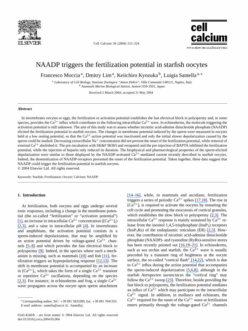
The increase in [Ca2+]i induced by the sperm has beenrecorded with a cooled CCD camera in oocytes pre-injectedwith OGBD. In 20 out of 37 oocytes, the first detectableCa2+ signal was the cortical flash, which mirrored the ac-tivation of voltage-gated Ca2+ channels[22,35] and oc-curred 35.6± 6.9 s (n = 17) after sperm addition to thebath (Fig. 1A and B). In these cells, a point-source waveof Ca2+ started 2.3± 0.2 s (n = 20) after the cortical flash(Fig. 1A and B) from the site of sperm interaction and trav-elled to the antipode of the oocyte within 1–2 min, reachinga peak amplitude of 0.81± 0.02 (n = 16) (Fig. 1C and D).The duration of the Ca2+ elevation was of about 12 min(Fig. 1C), although in several oocytes the Ca2+ signal didnot completely decay to the baseline during the period underobservation (up to 20 min; data not shown). In 11 oocytes,the Ca2+ wave preceded the cortical flash by 3.3± 0.5 s(n = 11; not shown) and initiated 46.0± 9.3 s after spermaddition (n = 13; not shown), a value which is not signifi-cantly different from the latency of the signal starting withthe cortical flash (P = 0.37). Also the height and the kinet-ics of the Ca2+ response were similar to those displayed bythe oocytes where the Ca2+ sweep followed the Ca2+ ring(not shown). Although unusual, such a pattern of increasein [Ca2+]i has already been observed in oocytes fromA.auranciacus[23]. Finally, no cortical flash was detectablein the remaining six cells, although both the peak fluores-cence and the time course of the Ca2+ wave did not differfrom those of the oocytes which displayed the cortical flash(not shown). In all the oocytes, the fertilization membraneappeared approximately 2–5 min after the onset of the waveand was fully elevated after 12–15 min (Fig. 1A).
3.2. Changes in the voltage-dependent K+ and Ca2+currents during hormone-induced maturation of starfishoocytes
The fertilization potential was measured by impaling im-mature oocytes with a single microelectrode and recordingthe electrophysiological response to the sperm 50–60 minlater, when the germinal vesicle had disappeared. The meanmembrane potential (Vm) of immature oocytes was−69.4± 0.9 mV (n = 50), which is not different from the valuereported in a previous paper[30], whereas the specificmembrane resistance was equal to 0.545± 0.025 M� cm−2
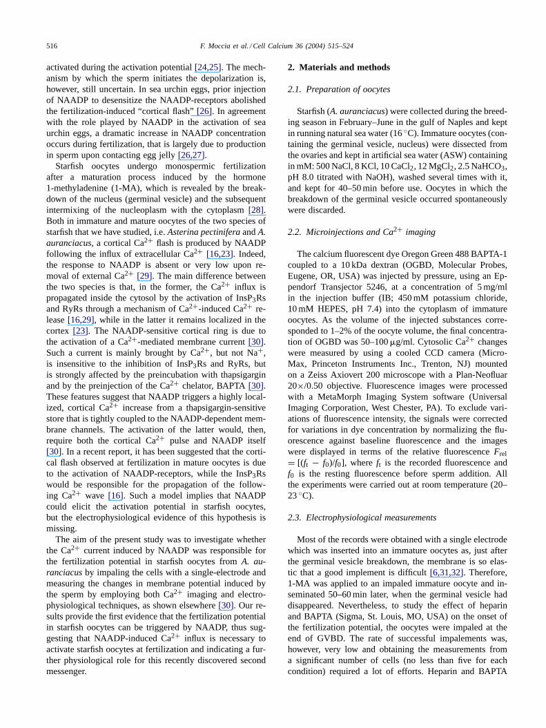
(n = 5). TheI–V relationship of the immature oocytes wasmeasured by applying a step-like protocol from−100 to+30 mV to cells held at−70 mV. The restingI–V showed aclear inward rectification at potentials lower than−70 mV,a transient inward Ca2+ current between−50 and−20 mV,and a transient outward K+ current at positive potentials(Fig. 2A and C). All of these currents have been extensivelystudied in starfish oocytes and we did not proceed in their
518 F. Moccia et al. / Cell Calcium 36 (2004) 515–524
Fig. 1. Ca2+ wave observed at fertilization in starfish oocytes. Sequence of the fluorescence images showing the onset and the propagation of the Ca2+sweep induced by the sperm in starfish oocytes (A and B). In order to better illustrate the spreading of the Ca2+ signal from the point of sperm–oocytecontact to the antipode of the cell, in Panel A, the relative fluorescent images have been overlapped with the corresponding light transmitted images.Thefertilization membrane is first visible 2.45 min after the start of the recording and fully elevated after 14.45 min. (C) Graph of the relative fluorescence ofthe Ca2+ indicator, measured from a region of interest (ROI) around the oocyte (as indicated by the blue circle drawn around the oocyte), which offersa numerical equivalent of the pseudocolors shown in Panel B as function of the time. (D) The ROIs were positioned inside the oocyte to graphicallyillustrate the pattern of propagation of the Ca2+ wave: in according with the relative fluorescent images in Panels A and B, the Ca2+ sweep propagatedfrom the entry point of the sperm (blue trace) to the centre of the oocyte (purple trace) and the antipode (dark green trace). (For interpretation of thereferences to color in this figure legend, the reader is referred to the web version of the article.)
further characterization[31,36–40]. The addition of 1-MA(10�M) resulted in a decrease in the amplitude of both theK+ currents and in an increase in the peak of the Ca2+ cur-rent. This feature is clearly shown inFig. 2B and Cand hasbeen also shown in oocytes fromA. pectinifera[31] andLep-tasterias hexactis[37]. In agreement with the data reportedin the latter studies, the disappearance of the inward recti-fier K+ current raised the specific resistance of the oocytesto 25.4± 8.1 M� cm−2 (n = 5). It has been shown that, asa consequence of the increase in membrane resistance, anyslight change in the inward current during maturation couldresult in a large potential change[6,31,36]. Indeed, both theleakage current due to electrode penetration and the increasein Na+ permeability occurring upon 1-MA addition[41]may shift the resting potential of mature oocytes to more de-polarized value than those measured at the immature stage[6,31,36]. A similar jump in the membrane potential of theoocytes fromA. auranciacusfollowing 1-MA addition isshown inFig. 2D. A slow depolarization started 10–20 minafter 1-MA addition and culminated in one or more spikesdue to the activation of the voltage-gated Ca2+ channels,which broughtVm to the average value of−13.8± 1.9 mV(n = 49; ranging from−40 to +3 mV). In 12 out of 30oocytes, replacement of external Na+ with an equimolaramount of choline caused a recovery ofVm to −70.1 ±0.8 mV, suggesting that an increase in Na+ permeabilitycould occur during maturation and depolarize the mem-
brane in a fraction of cells. In the other 18 oocytes, removalof external Na+ shifted the resting potential of mature cellsto −27.5 ± 3.6 mV, which is significantly more negativethan the value ofVm measured in ASW (−11.4± 2.4 mV;P = 0.002), but more depolarized thanVm recorded at theimmature stage (−69.5 ± 2.1 mV; P = 0.0000081). It is,therefore, conceivable that the leakage current due to elec-trode penetration may contribute to the jump in membranepotential measured upon 1-MA addition in the majority ofthe oocytes, as suggested by Miyazaki et al.[6,31].
3.3. Ca2+ entry is responsible for the fertilization potentialin starfish oocytes
In starfish oocytes[6], the activation potential lacks theCa2+-action potential when the restingVm is too small,due to the inactivation of voltage-gated Ca2+ channels[42].The slow depolarization observed under these conditionsrepresents the first step of the response to the sperm[5,6],which leads to the threshold of activation of the regen-erative process whenVm is maintained at more negativevalues. The initial depolarization is evidently mediated by asperm–oocyte interaction[43] and provides a valuable toolto assess the trigger of the activation potential in starfishwithout any contamination by the voltage-sensitive actionpotential. Therefore, we focussed our attention on the shiftin Vm which occurred at fertilization in depolarized oocytes
F. Moccia et al. / Cell Calcium 36 (2004) 515–524 519
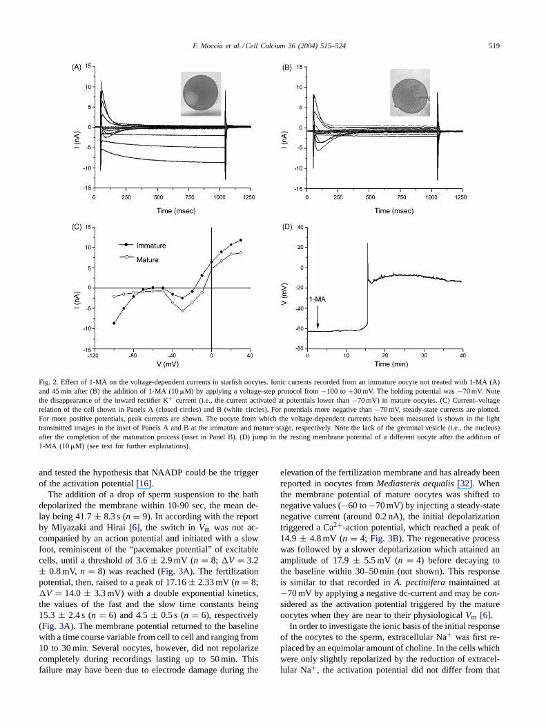
Fig. 2. Effect of 1-MA on the voltage-dependent currents in starfish oocytes. Ionic currents recorded from an immature oocyte not treated with 1-MA (A)and 45 min after (B) the addition of 1-MA (10�M) by applying a voltage-step protocol from−100 to+30 mV. The holding potential was−70 mV. Notethe disappearance of the inward rectifier K+ current (i.e., the current activated at potentials lower than−70 mV) in mature oocytes. (C) Current–voltagerelation of the cell shown in Panels A (closed circles) and B (white circles). For potentials more negative than−70 mV, steady-state currents are plotted.For more positive potentials, peak currents are shown. The oocyte from which the voltage-dependent currents have been measured is shown in the lighttransmitted images in the inset of Panels A and B at the immature and mature stage, respectively. Note the lack of the germinal vesicle (i.e., the nucleus)after the completion of the maturation process (inset in Panel B). (D) jump in the resting membrane potential of a different oocyte after the addition of1-MA (10�M) (see text for further explanations).
and tested the hypothesis that NAADP could be the triggerof the activation potential[16].
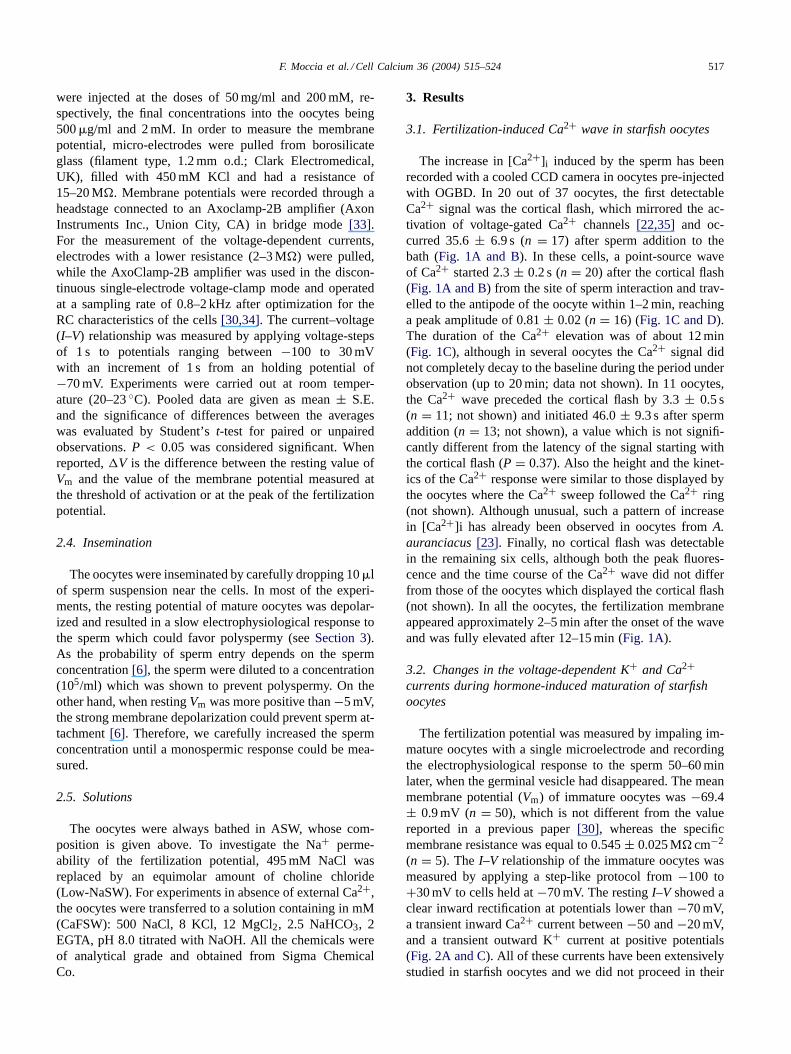
The addition of a drop of sperm suspension to the bathdepolarized the membrane within 10-90 sec, the mean de-lay being 41.7± 8.3 s (n = 9). In according with the reportby Miyazaki and Hirai[6], the switch inVm was not ac-companied by an action potential and initiated with a slowfoot, reminiscent of the “pacemaker potential” of excitablecells, until a threshold of 3.6± 2.9 mV (n = 8; �V = 3.2± 0.8 mV, n = 8) was reached (Fig. 3A). The fertilizationpotential, then, raised to a peak of 17.16± 2.33 mV (n = 8;�V = 14.0 ± 3.3 mV) with a double exponential kinetics,the values of the fast and the slow time constants being15.3 ± 2.4 s (n = 6) and 4.5± 0.5 s (n = 6), respectively(Fig. 3A). The membrane potential returned to the baselinewith a time course variable from cell to cell and ranging from10 to 30 min. Several oocytes, however, did not repolarizecompletely during recordings lasting up to 50 min. Thisfailure may have been due to electrode damage during the
elevation of the fertilization membrane and has already beenreported in oocytes fromMediasteris aequalis[32]. Whenthe membrane potential of mature oocytes was shifted tonegative values (−60 to−70 mV) by injecting a steady-statenegative current (around 0.2 nA), the initial depolarizationtriggered a Ca2+-action potential, which reached a peak of14.9± 4.8 mV (n = 4; Fig. 3B). The regenerative processwas followed by a slower depolarization which attained anamplitude of 17.9± 5.5 mV (n = 4) before decaying tothe baseline within 30–50 min (not shown). This responseis similar to that recorded inA. pectiniferamaintained at−70 mV by applying a negative dc-current and may be con-sidered as the activation potential triggered by the matureoocytes when they are near to their physiologicalVm [6].
In order to investigate the ionic basis of the initial responseof the oocytes to the sperm, extracellular Na+ was first re-placed by an equimolar amount of choline. In the cells whichwere only slightly repolarized by the reduction of extracel-lular Na+, the activation potential did not differ from that
520 F. Moccia et al. / Cell Calcium 36 (2004) 515–524
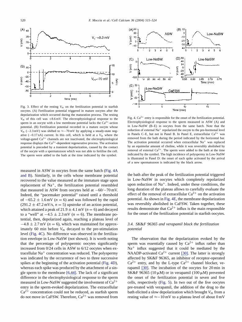
Fig. 3. Effect of the restingVm on the fertilization potential in starfishoocytes. (A) Fertilization potential triggered in mature oocytes after thedepolarization which occurred during the maturation process. The restingVm of this cell was+8.6 mV. The electrophysiological response to thesperm in an oocyte with a low membrane potential lacks the Ca2+-actionpotential. (B) Fertilization potential recorded in a mature oocyte whoseVm (−2.3 mV) was shifted to≈−70 mV by applying a steady-state neg-ative (−0.17 nA) current. In this cell, which is held at aVm where thevoltage-gated Ca2+ channels are not inactivated, the electrophysiologicalresponse displays the Ca2+-dependent regenerative process. The activationpotential is preceded by a transient depolarization, caused by the contactof the oocyte with a spermatozoon which was not able to fertilise the cell.The sperm were added to the bath at the time indicated by the symbol.
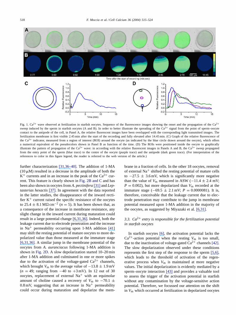
measured in ASW in oocytes from the same batch (Fig. 4Aand B). Similarly, in the cells whose membrane potentialrecovered to the value measured at the immature stage uponreplacement of Na+, the fertilization potential resembledthat measured in ASW from oocytes held at−60/−70 mV.Indeed, the “pacemaker potential” raised until a thresholdof −65.2± 1.6 mV (n = 6) and was followed by the rapid(291.2± 47.2 mV/s,n = 5) upstroke of an action potential,which attained a peak of 21.9± 4.1 mV (n = 6) and decayedto a “well” at −4.5 ± 2.3 mV (n = 6). The membrane po-tential, then, depolarized again, reaching a plateau level of−4.8± 2.7 mV (n = 6), which was maintained for approx-imately 60 min beforeVm decayed to the pre-stimulationlevel (Fig. 4C). No difference was observed in the fertiliza-tion envelope in Low-NaSW (not shown). It is worth notingthat the percentage of polyspermic oocytes significantlyincreased from 0/24 cells in ASW to 6/12 oocytes when ex-tracellular Na+ concentration was reduced. The polyspermywas indicated by the occurrence of two to three successivespikes at the beginning of the activation potential (Fig. 4D),whereas each spike was produced by the attachment of a sin-gle sperm to the membrane[6,44]. The lack of a significantdifference in the electrophysiological response to the spermmeasured in Low-NaSW suggested the involvement of Ca2+entry in the sperm-evoked depolarization. The extracellularCa2+ concentration could not be reduced, as starfish spermdo not move in CaFSW. Therefore, Ca2+ was removed from
Fig. 4. Ca2+ entry is responsible for the onset of the fertilization potential.Electrophysiological response to the sperm measured in ASW (A) andin Low-NaSW (B–E) in oocytes from the same batch. Note that thereduction of external Na+ repolarized the oocyte to the pre-hormonal levelin Panels C–E, but not in Panel B. In Panel E, extracellular Ca2+ wasremoved from the bath during the period indicated by the horizontal bar.The activation potential occurred when extracellular Na+ was replacedby an equimolar amount of choline, while it was reversibly abolished byremoval of external Ca2+. The sperm were added to the bath at the timeindicated by the symbol. The high incidence of polyspermy in Low-NaSWis illustrated in Panel D: the onset of each spike activated by the arrivalof a new spermatozoon is indicated by the black arrow.
the bath after the peak of the fertilization potential triggeredin Low-NaSW in oocytes which completely repolarizedupon reduction of Na+. Indeed, under these conditions, thelong duration of the plateau allows to carefully evaluate theeffect of the removal of extracellular Ca2+ on the activationpotential. As shown inFig. 4E, the membrane depolarizationwas reversibly abolished in CaFSW. Taken together, theseresults demonstrate that Ca2+ influx is the main responsiblefor the onset of the fertilization potential in starfish oocytes.
3.4. SK&F 96365 and verapamil block the fertilizationpotential
The observation that the depolarization evoked by thesperm was essentially caused by Ca2+ influx rather thanNa+ influx suggested that it could be mediated by theNAADP-activated Ca2+ current[30]. The latter is stronglyaffected by SK&F 96365, an inhibitor of receptor-operatedCa2+ entry, and by the L-type Ca2+ channel blocker, ve-rapamil [30]. The incubation of the oocytes for 20 min inSK&F 96365 (10�M) or in verapamil (100�M) preventedthe onset of the fertilization potential in seven and fivecells, respectively (Fig. 5). In two out of the five oocytespre-treated with verapamil, the addition of the drug to thebath elicited a slow depolarization which broughtVm from aresting value of≈−10 mV to a plateau level of about 0 mV
F. Moccia et al. / Cell Calcium 36 (2004) 515–524 521
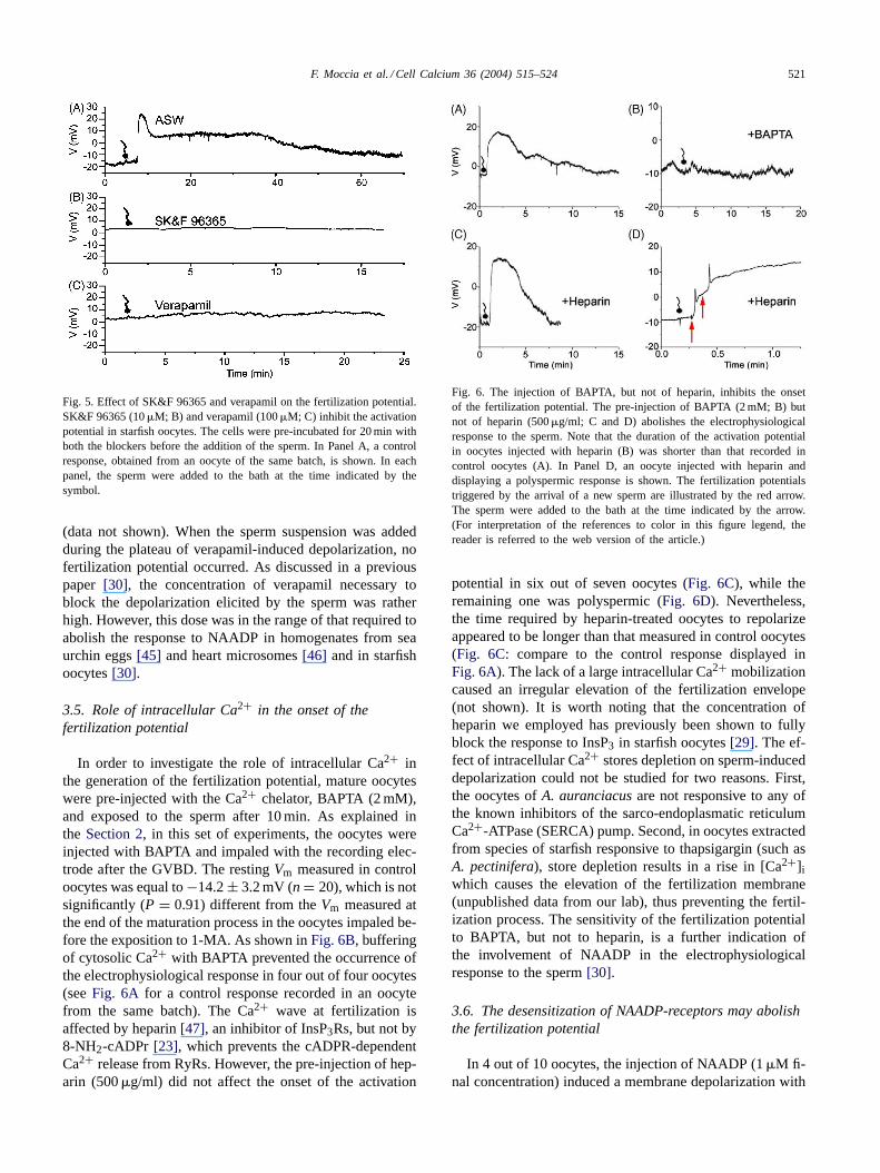
Fig. 5. Effect of SK&F 96365 and verapamil on the fertilization potential.SK&F 96365 (10�M; B) and verapamil (100�M; C) inhibit the activationpotential in starfish oocytes. The cells were pre-incubated for 20 min withboth the blockers before the addition of the sperm. In Panel A, a controlresponse, obtained from an oocyte of the same batch, is shown. In eachpanel, the sperm were added to the bath at the time indicated by thesymbol.
(data not shown). When the sperm suspension was addedduring the plateau of verapamil-induced depolarization, nofertilization potential occurred. As discussed in a previouspaper [30], the concentration of verapamil necessary toblock the depolarization elicited by the sperm was ratherhigh. However, this dose was in the range of that required toabolish the response to NAADP in homogenates from seaurchin eggs[45] and heart microsomes[46] and in starfishoocytes[30].
3.5. Role of intracellular Ca2+ in the onset of thefertilization potential
In order to investigate the role of intracellular Ca2+ inthe generation of the fertilization potential, mature oocyteswere pre-injected with the Ca2+ chelator, BAPTA (2 mM),and exposed to the sperm after 10 min. As explained inthe Section 2, in this set of experiments, the oocytes wereinjected with BAPTA and impaled with the recording elec-trode after the GVBD. The restingVm measured in controloocytes was equal to−14.2± 3.2 mV (n = 20), which is notsignificantly (P = 0.91) different from theVm measured atthe end of the maturation process in the oocytes impaled be-fore the exposition to 1-MA. As shown inFig. 6B, bufferingof cytosolic Ca2+ with BAPTA prevented the occurrence ofthe electrophysiological response in four out of four oocytes(seeFig. 6A for a control response recorded in an oocytefrom the same batch). The Ca2+ wave at fertilization isaffected by heparin[47], an inhibitor of InsP3Rs, but not by8-NH2-cADPr [23], which prevents the cADPR-dependentCa2+ release from RyRs. However, the pre-injection of hep-arin (500�g/ml) did not affect the onset of the activation
Fig. 6. The injection of BAPTA, but not of heparin, inhibits the onsetof the fertilization potential. The pre-injection of BAPTA (2 mM; B) butnot of heparin (500�g/ml; C and D) abolishes the electrophysiologicalresponse to the sperm. Note that the duration of the activation potentialin oocytes injected with heparin (B) was shorter than that recorded incontrol oocytes (A). In Panel D, an oocyte injected with heparin anddisplaying a polyspermic response is shown. The fertilization potentialstriggered by the arrival of a new sperm are illustrated by the red arrow.The sperm were added to the bath at the time indicated by the arrow.(For interpretation of the references to color in this figure legend, thereader is referred to the web version of the article.)
potential in six out of seven oocytes (Fig. 6C), while theremaining one was polyspermic (Fig. 6D). Nevertheless,the time required by heparin-treated oocytes to repolarizeappeared to be longer than that measured in control oocytes(Fig. 6C: compare to the control response displayed inFig. 6A). The lack of a large intracellular Ca2+ mobilizationcaused an irregular elevation of the fertilization envelope(not shown). It is worth noting that the concentration ofheparin we employed has previously been shown to fullyblock the response to InsP3 in starfish oocytes[29]. The ef-fect of intracellular Ca2+ stores depletion on sperm-induceddepolarization could not be studied for two reasons. First,the oocytes ofA. auranciacusare not responsive to any ofthe known inhibitors of the sarco-endoplasmatic reticulumCa2+-ATPase (SERCA) pump. Second, in oocytes extractedfrom species of starfish responsive to thapsigargin (such asA. pectinifera), store depletion results in a rise in [Ca2+]iwhich causes the elevation of the fertilization membrane(unpublished data from our lab), thus preventing the fertil-ization process. The sensitivity of the fertilization potentialto BAPTA, but not to heparin, is a further indication ofthe involvement of NAADP in the electrophysiologicalresponse to the sperm[30].
3.6. The desensitization of NAADP-receptors may abolishthe fertilization potential
In 4 out of 10 oocytes, the injection of NAADP (1�M fi-nal concentration) induced a membrane depolarization with
522 F. Moccia et al. / Cell Calcium 36 (2004) 515–524
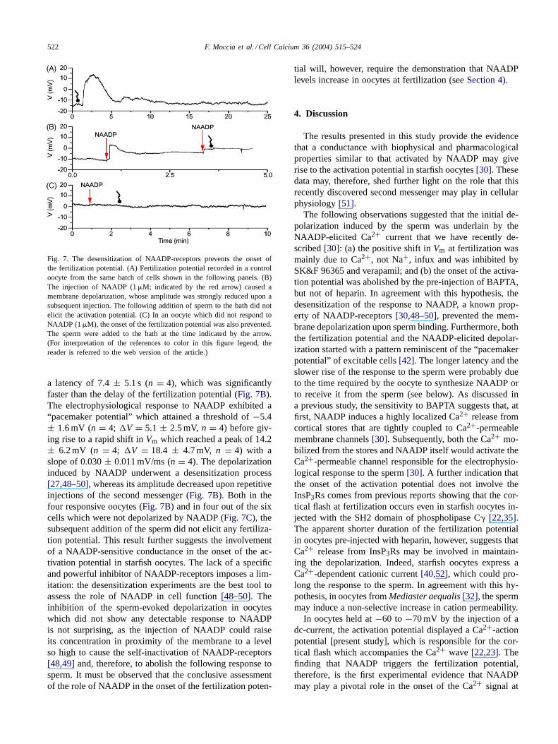
Fig. 7. The desensitization of NAADP-receptors prevents the onset ofthe fertilization potential. (A) Fertilization potential recorded in a controloocyte from the same batch of cells shown in the following panels. (B)The injection of NAADP (1�M; indicated by the red arrow) caused amembrane depolarization, whose amplitude was strongly reduced upon asubsequent injection. The following addition of sperm to the bath did notelicit the activation potential. (C) In an oocyte which did not respond toNAADP (1�M), the onset of the fertilization potential was also prevented.The sperm were added to the bath at the time indicated by the arrow.(For interpretation of the references to color in this figure legend, thereader is referred to the web version of the article.)
a latency of 7.4± 5.1 s (n = 4), which was significantlyfaster than the delay of the fertilization potential (Fig. 7B).The electrophysiological response to NAADP exhibited a“pacemaker potential” which attained a threshold of−5.4± 1.6 mV (n = 4; �V = 5.1 ± 2.5 mV, n = 4) before giv-ing rise to a rapid shift inVm which reached a peak of 14.2± 6.2 mV (n = 4; �V = 18.4 ± 4.7 mV, n = 4) with aslope of 0.030± 0.011 mV/ms (n = 4). The depolarizationinduced by NAADP underwent a desensitization process[27,48–50], whereas its amplitude decreased upon repetitiveinjections of the second messenger (Fig. 7B). Both in thefour responsive oocytes (Fig. 7B) and in four out of the sixcells which were not depolarized by NAADP (Fig. 7C), thesubsequent addition of the sperm did not elicit any fertiliza-tion potential. This result further suggests the involvementof a NAADP-sensitive conductance in the onset of the ac-tivation potential in starfish oocytes. The lack of a specificand powerful inhibitor of NAADP-receptors imposes a lim-itation: the desensitization experiments are the best tool toassess the role of NAADP in cell function[48–50]. Theinhibition of the sperm-evoked depolarization in oocyteswhich did not show any detectable response to NAADPis not surprising, as the injection of NAADP could raiseits concentration in proximity of the membrane to a levelso high to cause the self-inactivation of NAADP-receptors[48,49] and, therefore, to abolish the following response tosperm. It must be observed that the conclusive assessmentof the role of NAADP in the onset of the fertilization poten-
tial will, however, require the demonstration that NAADPlevels increase in oocytes at fertilization (seeSection 4).
4. Discussion
The results presented in this study provide the evidencethat a conductance with biophysical and pharmacologicalproperties similar to that activated by NAADP may giverise to the activation potential in starfish oocytes[30]. Thesedata may, therefore, shed further light on the role that thisrecently discovered second messenger may play in cellularphysiology[51].
The following observations suggested that the initial de-polarization induced by the sperm was underlain by theNAADP-elicited Ca2+ current that we have recently de-scribed[30]: (a) the positive shift inVm at fertilization wasmainly due to Ca2+, not Na+, infux and was inhibited bySK&F 96365 and verapamil; and (b) the onset of the activa-tion potential was abolished by the pre-injection of BAPTA,but not of heparin. In agreement with this hypothesis, thedesensitization of the response to NAADP, a known prop-erty of NAADP-receptors[30,48–50], prevented the mem-brane depolarization upon sperm binding. Furthermore, boththe fertilization potential and the NAADP-elicited depolar-ization started with a pattern reminiscent of the “pacemakerpotential” of excitable cells[42]. The longer latency and theslower rise of the response to the sperm were probably dueto the time required by the oocyte to synthesize NAADP orto receive it from the sperm (see below). As discussed ina previous study, the sensitivity to BAPTA suggests that, atfirst, NAADP induces a highly localized Ca2+ release fromcortical stores that are tightly coupled to Ca2+-permeablemembrane channels[30]. Subsequently, both the Ca2+ mo-bilized from the stores and NAADP itself would activate theCa2+-permeable channel responsible for the electrophysio-logical response to the sperm[30]. A further indication thatthe onset of the activation potential does not involve theInsP3Rs comes from previous reports showing that the cor-tical flash at fertilization occurs even in starfish oocytes in-jected with the SH2 domain of phospholipase C� [22,35].The apparent shorter duration of the fertilization potentialin oocytes pre-injected with heparin, however, suggests thatCa2+ release from InsP3Rs may be involved in maintain-ing the depolarization. Indeed, starfish oocytes express aCa2+-dependent cationic current[40,52], which could pro-long the response to the sperm. In agreement with this hy-pothesis, in oocytes fromMediaster aequalis[32], the spermmay induce a non-selective increase in cation permeability.
In oocytes held at−60 to −70 mV by the injection of adc-current, the activation potential displayed a Ca2+-actionpotential [present study], which is responsible for the cor-tical flash which accompanies the Ca2+ wave[22,23]. Thefinding that NAADP triggers the fertilization potential,therefore, is the first experimental evidence that NAADPmay play a pivotal role in the onset of the Ca2+ signal at
F. Moccia et al. / Cell Calcium 36 (2004) 515–524 523
fertilization in starfish oocytes, as suggested in a previousreport [16]. The long duration of the activation potentialfurther suggests the involvement of Ca2+ entry in sustainingthe intracellular Ca2+ sweep. In molluscs and echiurans,the Ca2+ response to the sperm consists in an initial corticalflash which spreads inwardly to the center and primarilydepends on Ca2+ influx through the voltage-gated Ca2+channels[24,25]. This pattern is clearly different from thepoint-source Ca2+ wave occurring in starfish oocytes, whosepropagation certainly rely on InsP3Rs[16,22], but highlightsthe role that extracellular Ca2+ may play at fertilization.The interaction between NAADP-receptors and InsP3Rs inshaping the sperm-induced Ca2+ signal has also been foundin ascidian oocytes[19] and sea urchin eggs[26,53]. In thelatter, the desensitization of NAADP-receptors prevents theinitial cortical Ca2+ flash and shortens the duration of thefollowing Ca wave[26,53]. As pointed out above, in seaurchin, the fertilization potential consists in a depolarizationwhich brings the membrane to the threshold of activationof the Ca2+-action potential[5]. The nature of the stimuluscausing the initial depolarization is still unknown[54]. AsNAADP elicits the Ca2+ ring [26], it is conceivable that theNAADP-dependent current may be involved in the onsetof the fertilization potential also in sea urchin eggs. Thecurrent immediately activated by the sperm–egg contact insea urchin, however, reverts polarity at about+20 mV [55],a potential which is significantly less positive than+50 mV,the reversal potential of the NAADP-stimulated current[30].
The biophysical properties of the NAADP-activated cur-rent are quite different from those exhibited by the fertil-ization currents recorded in other invertebrates. Indeed, theactivation potential is mediated by a non-selective cation cur-rent gated by ADP-ribose in ascidian oocytes (ADPr)[56]and by a Cl− current activated by Ca2+ released from theInsP3Rs in frog oocytes[7]. Furthermore, in molluscs andechiurans, where the Ca2+ influx during the activation poten-tial sustains the intracellular Ca2+ wave[24,25], the initialdepolarization is due to the opening of sperm-gated mem-brane channels which are mainly permeable to Na+ [57,58].The source of NAADP for the onset of the fertilization po-tential in starfish oocytes remains to be elucidated. NAADPcould be synthesized by the oocytes after the binding ofsperm or could be injected into the cells by the sperm itself.In according with the latter hypothesis, NAADP has beenshown to increase in sperm upon contact with the jelly layerand to be, then, released into the eggs in sea urchin[26,59].
In conclusion, this report challenged for the first timethe possibility that NAADP may be involved in the onsetof the activation potential in starfish oocytes. The resultsshown indicate the initiation of the fertilization potentialrequires the activation of an ionic conductance with bio-physical and pharmacological properties similar to thatactivated by NAADP in these cells[30]. In order to pro-vide the clear-cut evidence that the fertilization potential iselicited by NAADP, however, it will be necessary to inda-gate whether it goes up in sperm and eggs at fertilization
[26]. In a previous study, it has been suggested that the ini-tiation of the Ca2+ response to the sperm in starfish oocytesdepends on the stimulation of NAADP-receptors, while thepropagation of the Ca2+ wave throughout the cytoplasmrelies on the InsP3Rs [16]. The future aim of our work willbe to assess this hypothesis by the simultaneous measure-ment of the changes in the membrane potential and in theCa2+ signal at fertilization.
Acknowledgements
We thank Dr. F. Tanzi for critical reading of themanuscript. The help of the Marine Resource Service ofthe Stazione Zoologica for maintaining the starfish is alsogratefully acknowledged. This work was supported bythe Italian Ministry of University and Scientific Research(PRIN 2002–2004).
References
[1] L.A. Jaffe, N.L. Cross, Electrical regulation of sperm–egg fusion,Annu. Rev. Physiol. 48 (1986) 191–200.
[2] S.A. Stricker, Comparative biology of calcium signaling during fer-tilization and egg activation in animals, Dev. Biol. 211 (1999) 157–176.
[3] L.L. Runft, L.A. Jaffe, L.M. Mehlmann, Egg activation at fertiliza-tion: where it all begins, Dev. Biol. 245 (2002) 237–254.
[4] D. Ben-Yosef, R. Shalgi, Early ionic events in activation of themammalian egg, Rev. Reprod. 3 (1998) 96–103.
[5] E.L. Chambers, J. de Armendi, Membrane potential, action poten-tial and activation potential of eggs of the sea urchinLytechinusvariegatus, Exp. Cell Res. 122 (1979) 203–218.
[6] S.I. Miyazaki, S. Hirai, Fast polyspermy block and activation po-tential. Correlated changes during oocyte maturation of a starfish, J.Physiol. 70 (1979) 327–340.
[7] D. Kline, R. Nuccitelli, The wave of activation current in the Xenopusegg, Dev. Biol. 111 (1985) 471–487.
[8] D. Kline, L.A. Jaffe, R.T. Kado, A calcium-activated sodium con-ductance contributes to the fertilization potential in the egg of thenemertean wormCerebratulus lacteus, Dev. Biol. 117 (1986) 184–193.
[9] L.A. Jaffe, Fast block to polyspermy in sea urchin eggs is electricallymediated, Nature 261 (1976) 68–71.
[10] S.I. Miyazaki, Fertilization potential and calcium transients in mam-malian eggs, Dev. Growth Differ. 30 (1988) 603–610.
[11] R. Nuccitelli, The fertilization potential is not necessary for the blockto polyspermy or the activation of development in the medaka egg,Dev. Biol. 76 (1980) 499–504.
[12] S.I. Miyazaki, Y. Igusa, Fertilization potential in golden hamster eggsconsists of recurring hyperpolarizations, Nature 290 (1981) 702–704.
[13] S.T. Homa, K. Swann, A cytosolic sperm factor triggers calciumoscillations and membrane hyperpolarizations in human oocytes,Hum. Reprod. 9 (1994) 2356–23661.
[14] S.S. Shen, W.R. Buck, Sources of calcium in sea urchin eggs duringthe fertilization response, Dev. Biol. 157 (1993) 157–169.
[15] R.A. Fontanilla, R. Nuccitelli, Characterization of the sperm-inducedcalcium wave in Xenopus eggs using confocal microscopy, Biophys.J. 75 (1998) 2079–2087.
[16] D. Lim, K. Kyozuka, G. Gragnaniello, E. Carafoli, L. Santella,NAADP+ initiates the Ca2+ response during fertilization of starfishoocytes, FASEB J. 15 (2001) 2257–2267.
524 F. Moccia et al. / Cell Calcium 36 (2004) 515–524
[17] S.I. Miyazaki, H. Shirakawa, K. Nakada, Y. Honda, Essential roleof the inositol 1,4,5-trisphosphate receptor/Ca+ release channel inCa+ waves and Ca+ oscillations at fertilization of mammalian eggs,Dev. Biol. 158 (1993) 62–78.
[18] R. Dumollard, C. Sardet, Three different calcium wave pacemakersin ascidian eggs, J. Cell Sci. 114 (2001) 2471–2481.
[19] M. Albrieux, H.C. Lee, M. Villaz, Calcium signaling by cyclic ADP-ribose NAADP, and inositol trisphosphate are involved in distinctfunctions in ascidian oocytes, J. Biol. Chem. 273 (1998) 14566–14574.
[20] R. Kuroda, K. Kontani, Y. Kanda, T. Katada, T. Nakano, Y. Satoh,N. Suzuki, H. Kuroda, Increase of cGMP cADP-ribose and inositol1,4,5-trisphosphate preceding Ca2+ transients in fertilization of seaurchin eggs, Development 128 (2001) 4405–4414.
[21] C. Leckie, R. Empson, A. Becchetti, J. Thomas, A. Galione, M.Whitaker, The NO pathway acts late during the fertilization responsein sea urchin eggs, J. Biol. Chem. 278 (2003) 12247–12254.
[22] D.J. Carroll, C.S. Ramarao, L.M. Mehlmann, S. Roche, M. Terasaki,L.A. Jaffe, Calcium release at fertilization in starfish eggs is mediatedby phospholipase C�, J. Cell Biol. 138 (1997) 1303–1311.
[23] G.A. Nusco, D. Lim, P. Sabala, L. Santella, Ca2+ response to cADPrduring maturation and fertilization of starfish oocytes, Biochem.Biophys. Res. Commun. 290 (2002) 1015–1021.
[24] R. Deguchi, M. Morisawa, Extracellular Ca2+ is predominantly usedfor cytoplasmic and nuclear Ca+ increases in fertilized oocytes ofthe marine bivalveMactra chinensis, Development 122 (1996) 3651–3660.
[25] J.L. Stephano, M.C. Gould, The intracellular calcium increase atfertilization in Urechis caupooocytes: activation without waves, Dev.Biol. 191 (1997) 53–68.
[26] G.C. Churchill, J.S. O’Neill, R. Masgrau, S. Patel, J.M. Thomas,A.A. Genazzani, A. Galione, Sperm deliver a new second messenger:NAADP, Curr. Biol. 13 (2003) 247–251.
[27] H.C. Lee, Calcium signaling: NAADP ascends as a new messenger,Curr. Biol. 13 (2003) R186–R188.
[28] L. Santella, K. Kyozuka, Effects of 1-methyladenine on nuclear Ca+transients and meiosis resumption in starfish oocytes are mimickedby the nuclear injection of inositol 1,4,5-trisphosphate and cADP-ribose, Cell Calcium 22 (1997) 11–20.
[29] L. Santella, K. Kyozuka, A.A. Genazzani, L. De Riso, E. Carafoli,Nicotinic acid adenine dinucleotide phosphate-induced Ca2+ release.Interactions among distinct Ca2+ mobilizing mechanisms in starfishoocytes, J. Biol. Chem. 275 (2002) 8301–8306.
[30] F. Moccia, D. Lim, D.G.A. Nusco, E. Ercolano, L. Santella, NAADPactivates a Ca+ current that is dependent on F-actin cytoskeleton,FASEB J. 17 (2003) 1907–1909.
[31] S.I. Miyazaki, H. Ohmori, S. Sasaki, Potassium rectifications of thestarfish oocyte membrane and their changes during oocyte maturation,J. Physiol. 246 (1975) 55–78.
[32] J.B. Lansman, Voltage-clamp study of the conductance activated atfertilization in the starfish egg, J. Physiol. 345 (1983) 353–372.
[33] D.J. Adams, A.B. Smith, C.I. Schroeder, T. Yasuda, R.J. Lewis,Omega-conotoxin CVID inhibits a pharmacologically distinctvoltage-sensitive calcium channel associated with transmitter releasefrom preganglionic nerve terminals, J. Biol. Chem. 278 (2003) 4057–4062.
[34] I. Inoue, I. Tsutsui, N.J. Abbott, E.R. Brown, Ionic currents in isolatedand in situ squid Schwann cells, J. Physiol. 541 (2002) 769–778.
[35] A.F. Giusti, D.J. Carroll, Y.A. Abassi, M. Terasaki, K.R. Foltz, L.A.Jaffe, Requirement of a Src family kinase for initiating calciumrelease at fertilization in starfish eggs, J. Biol. Chem. 274 (1999)29318–29322.
[36] S.I. Miyazaki, H. Ohmori, S. Sasaki, Action potential and non-linearcurrent-voltage relation in starfish oocytes, J. Physiol. 246 (1975)37–54.
[37] W.J. Moody, J.B. Lansman, Developmental regulation of Ca2+and K+ currents during hormone-induced maturation of starfish
oocytes, Proc. Natl. Acad. Sci. U.S.A. 80 (1983) 3096–3100.
[38] W.J. Moody, M.M. Bosma, Hormone-induced loss of surface mem-brane during maturation of starfish oocytes: differential effects onpotassium and calcium channels, Dev. Biol. 112 (1985) 396–404.
[39] L. Simoncini, W.J. Moody, Changes in voltage-dependent currentsand membrane area during maturation of starfish oocytes: speciesdifferences and similarities, Dev. Biol. 138 (1990) 194–201.
[40] F. Moccia, G.A. Nusco, D. Lim, E. Ercolano, G. Gragnaniello, E.R.Brown, L. Santella, Ca2+ signalling and membrane current activatedby cADPr in starfish oocytes, Pflügers Arch. 446 (2003) 541–552.
[41] M. Dòree, Hormonal control of meiosis in starfish oocytes. Calciumion release induced by 1-methyladenine increases membrane perme-ability to sodium ions, Exp. Cell Res. 131 (1981) 115–121.
[42] B. Hille, in: Sinauer (Ed.), Ionic Channels of Excitable Membranes,Sunderland, 2002.
[43] B. Dale, L.J. De Felice, V. Taglietti, Membrane noise and conductanceincrease during single spermatozoon–egg interactions, Nature 275(1978) 217–219.
[44] B. Dale, L. Santella, Sperm–oocyte interaction in the sea-urchin, J.Cell Sci. 74 (1985) 153–167.
[45] A.A. Genazzani, M. Mezna, D.M. Dickey, F. Michelangeli, T.F.Walseth, A. Galione, Pharmacological properties of the Ca2+-releasemechanism sensitive to NAADP in the sea urchin egg, Br. J. Phar-macol. 121 (1996) 1489–1495.
[46] J. Bak, R.A. Billington, G. Timar, A.C. Dutton, A.A. Genazzani,NAADP receptors are present and functional in the heart, Curr. Biol.11 (2001) 987–990.
[47] S.A. Stricker, Time-lapse confocal imaging of calcium dynamics instarfish embryos, Dev. Biol. 170 (1995) 496–518.
[48] J.M. Cancela, G.C. Churchill, A. Galione, Coordination of agonist-induced Ca2+-signalling patterns by NAADP in pancreatic acinarcells, Nature 398 (1999) 74–76.
[49] I. Berg, B.V. Potter, G.W. Mayr, A.H. Guse, Nicotinic acid adeninedinucleotide phosphate (NAADP+) is an essential regulator of T-lymphocyte Ca2+-signaling, J. Cell Biol. 150 (2000) 581–588.
[50] J.D. Johnson, S. Misler, Nicotinic acid-adenine dinucleotidephosphate-sensitive calcium stores initiate insulin signaling in humanbeta cells, Proc. Natl. Acad. Sci. U.S.A. 99 (2002) 14566–14571.
[51] J.M. Cancela, G. Charpentier, O.H. Petersen, Co-ordination of Ca2+signalling in mammalian cells by the new Ca2+-releasing messengerNAADP, Pflügers Arch. 446 (2003) 322–327.
[52] J.B. Lansman, Calcium current and calcium-activated inward currentin the oocyte of the starfishLeptasterias hexactis, J. Physiol. 390(1987) 397–413.
[53] C.M. Perez-Terzic, E.N. Chini, S.S. Shen, T.P. Sousa, D. Clapham,Ca2+ release triggered by nicotinate adenine dinucleotide phosphatein intact sea urchin eggs, Biochem J. 312 (1995) 955–959.
[54] T. Mohri, P.I. Ivonnet, E.L. Chambers, Effect on sperm-induced acti-vation current and increase of cytosolic Ca2+ by agents that modifythe mobilization of [Ca2+]i . I. Heparin and pentosan polysulfate,Dev. Biol. 172 (1995) 139–157.
[55] J.W. Lynn, D.H. McCulloh, E.L. Chambers, Voltage clamp studiesof fertilization in sea urchin eggs. II. Current patterns in relation tosperm entry, nonentry and activation, Dev. Biol. 128 (1988) 305–323.
[56] M. Wilding, G.L. Russo, A. Galione, M. Marino, B. Dale, ADP-ribose gates the fertilization channel in ascidian oocytes, Am. J.Physiol. Cell Physiol. 275 (1998) C1277–C1283.
[57] T. Togo, K. Osanai, M. Morisawa, Existence of three mechanismsfor blocking polyspermy in oocytes of the musselMytilus Edulis,Biol. Bull. 189 (1995) 330–339.
[58] L.A. Jaffe, M. Gould-Somero, L. Holland, Ionic mechanism of thefertilization potential of the marine wormUrechis caupo(Echiura),J. Gen. Physiol. 73 (1979) 469–492.
[59] R.A. Billington, A. Ho, A.A. Genazzani, Nicotinic acid adenine din-ucleotide phosphate (NAADP) is present at micromolar concentra-tions in sea urchin spermatozoa, J. Physiol. 544 (2002) 107–112.


