Embed Size (px)
Citation preview
Journal of Magnetic Resonance 192 (2008) 252–257
Contents lists available at ScienceDirect
Journal of Magnetic Resonance
journal homepage: www.elsevier .com/ locate/ jmr
NMR observation of Tau in Xenopus oocytes
Jean-Franc�ois Bodart a,d, Jean-Michel Wieruszeski b, Laziza Amniai b, Arnaud Leroy b,c, Isabelle Landrieu b,Arlette Rousseau-Lescuyer a, Jean-Pierre Vilain a, Guy Lippens b,*
a Laboratoire de Régulation des Signaux de division, EA4020, Building SN3, Université des Sciences et Technologies de Lille, 59655 Villeneuve d’Ascq, Franceb Glycobiologie structurale et fonctionnelle, CNRS UMR8576, Université des Sciences et Technologies de Lille, 59655 Villeneuve d’Ascq, Francec Faculté de Pharmacie à Châtenay-Malabry (Paris XI), Chatenay-Malabry Cedex, Franced Institut de Recherche Interdisciplinaire, CNRS FRE 2963, Nanosystèmes biologiques, Université des Sciences et Technologies de Lille, 59655 Villeneuve d’Ascq, France
a r t i c l e i n f o
Article history:Received 11 October 2007Revised 25 February 2008Available online 14 March 2008
Keywords:NMRIn-cellXenopus oocytesFicollCell survivalTauPhosphorylation
1090-7807/$ - see front matter � 2008 Elsevier Inc. Adoi:10.1016/j.jmr.2008.03.006
* Corresponding author. Fax: +33 3 20 43 65 55.E-mail address: [email protected] (G. Lipp
a b s t r a c t
The observation by NMR spectroscopy of microinjected 15N-labelled proteins into Xenopus laevis oocytesmight open the way to link structural and cellular biology. We show here that embedding the oocytesinto a 20% Ficoll solution maintains their structural integrity over extended periods of time, allowingfor the detection of nearly physiological protein concentrations. We use these novel conditions to studythe neuronal Tau protein inside the oocytes. Spectral reproducibility and careful comparison of the spec-tra of Tau before and after cell homogenization is presented. When injecting Tau protein into immatureoocytes, we show that both its microtubule association and different phosphorylation events can bedetected.
� 2008 Elsevier Inc. All rights reserved.
1. Introduction the link between structural and cell biology. Very recently, two
The possibility to observe biological macromolecules within theliving cell by NMR spectroscopy might be able to bridge the gap be-tween structural and cell biology. When we first observed theosmoregulated periplasmic glucans (OPGs) of the Burkholderiasolanacearum bacteria by in-cell NMR [1], the absence of a cryo-probe at that moment and the concomitant problems of magneticsusceptibility when working at higher cell density forced us to ap-ply High Resolution Magic Angle Spinning (HRMAS) NMR to recorda reproducible high quality NMR spectrum [2]. Simultaneously, thegroup of V. Dötch combined bacterial protein overexpression witha cryoprobe to observe the first proteins in the cell [3]. However,contrary to our experimental system where synthesis of the endo-geneous OPG molecules is physiologically high when the bacteriaare grown in a low osmolar medium, the overexpression of pro-teins inside bacteria invariably leads to non-physiological concen-trations, and the cell mostly represents a medium of realisticviscosity and crowding. Although a number of studies have re-cently succeeded to obtain novel biological insights through in-cellNMR [4,5], the bacterial cell and the requirement of significantoverexpression remain a stringent limit on the establishment of
ll rights reserved.
ens).
groups have shown that microinjection of small model proteins(GB1 and ubiquitin) in Xenopus oocytes could be combined withsolution NMR to obtain workable spectra within an eucaryotic cellmodel [6,7]. With 200 oocytes inside the NMR active volume, andtypical injection volumes of 50 nl for the estimated 1 ll volume ofthe oocyte, the millimolar stock solutions of the model proteingave rise to protein concentrations that could be observed on atime scale of 1 h. A decrease in amount of protein injected wouldevidently approach the experimental set-up to a more realisticin vivo situation, but requires measuring times that scale as thesquare of the inverse protein concentration.
Our goal was to explore different sample preparations to mon-itor lower protein concentrations inside the oocytes by NMR spec-troscopy. We are particularly interested in the behaviour of Tau, aneuronal protein involved in microtubule stabilization but equallythe major part of the intracellular tangles that form inside the neu-rons of Alzheimer diseased (AD) patients. Oocytes indeed havebeen previously used as a model system to evaluate the effect ofspecific Tau mutations on the meiotic spindle formation and M-phase entry following progesterone stimulation [8]. Moreover,Xenopus oocytes contain many of the kinases that are potentiallyinvolved in the generation of pathological Tau phosphorylationpatterns, and the AT100 Alzheimer specific epitope can be gener-ated upon maturation with progesterone [9]. In the latter experi-
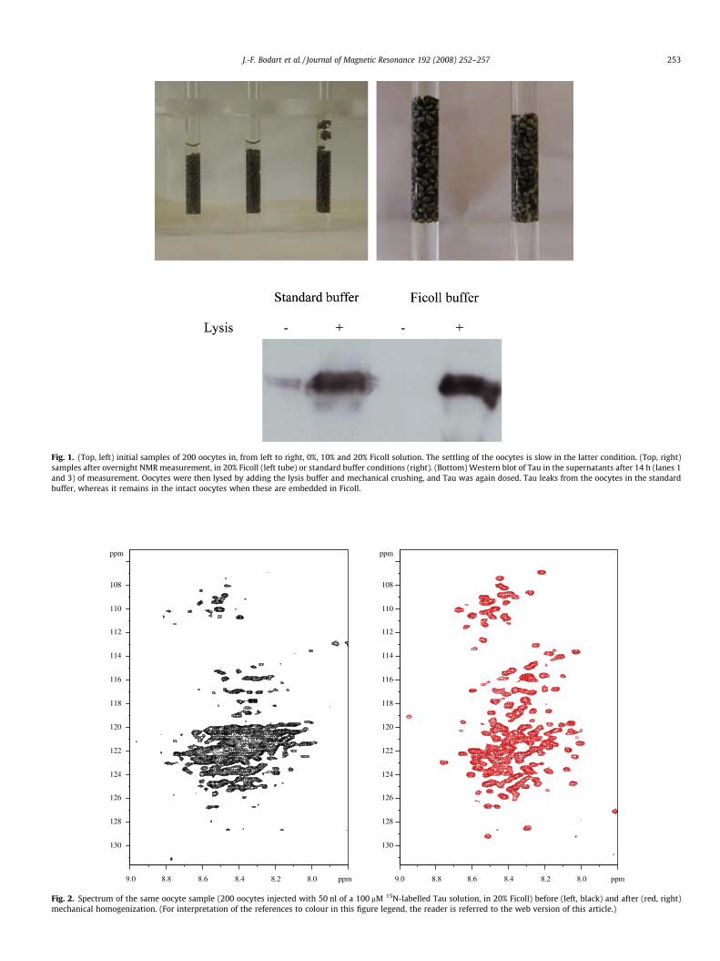
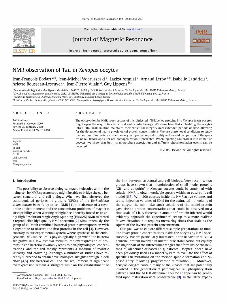
Fig. 1. (Top, left) initial samples of 200 oocytes in, from left to right, 0%, 10% and 20% Ficoll solution. The settling of the oocytes is slow in the latter condition. (Top, right)samples after overnight NMR measurement, in 20% Ficoll (left tube) or standard buffer conditions (right). (Bottom) Western blot of Tau in the supernatants after 14 h (lanes 1and 3) of measurement. Oocytes were then lysed by adding the lysis buffer and mechanical crushing, and Tau was again dosed. Tau leaks from the oocytes in the standardbuffer, whereas it remains in the intact oocytes when these are embedded in Ficoll.
ppm
8.08.28.48.68.89.0 ppm
108
110
112
114
116
118
120
122
124
126
128
130
108
110
112
114
116
118
120
122
124
126
128
130
ppm
8.08.28.48.68.89.0 ppm
Fig. 2. Spectrum of the same oocyte sample (200 oocytes injected with 50 nl of a 100 lM 15N-labelled Tau solution, in 20% Ficoll) before (left, black) and after (red, right)mechanical homogenization. (For interpretation of the references to colour in this figure legend, the reader is referred to the web version of this article.)
J.-F. Bodart et al. / Journal of Magnetic Resonance 192 (2008) 252–257 253
254 J.-F. Bodart et al. / Journal of Magnetic Resonance 192 (2008) 252–257
ments, though, the maximal amount of Tau injected led to a 5 lMintracellular concentration, which is nearly one order of magnitudelower than the one used in the previous NMR studies, and hencewould require a measurement over 100 h to obtain a comparablesignal to noise (S/N) ratio. Tau moreover is not exactly a model pro-tein, but is with its 441 amino acids a challenge even for solutionNMR. Its intrinsically disordered nature spectrum moreover leads
ppm
8.08.28.48.68.89.0 ppm
8.08.28.48.68.89.0 ppm
108
110
112
114
116
118
120
122
124
126
128
130
ppm
108
110
112
114
116
118
120
122
124
126
128
130
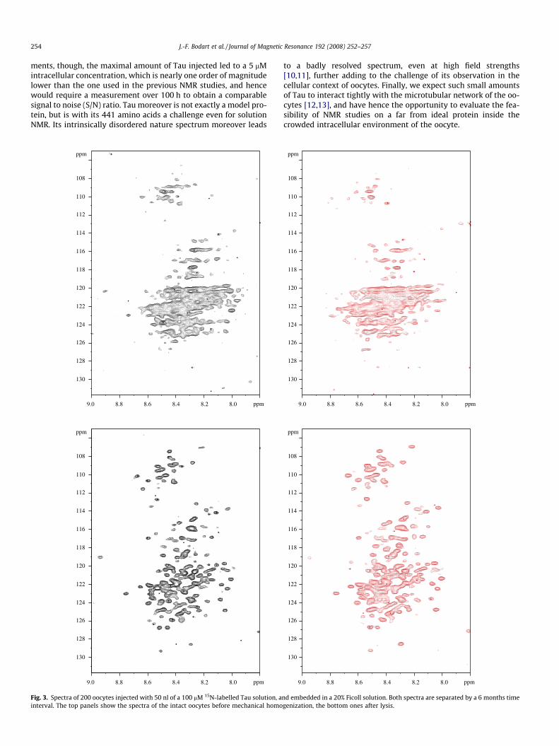
Fig. 3. Spectra of 200 oocytes injected with 50 nl of a 100 lM 15N-labelled Tau solution, ainterval. The top panels show the spectra of the intact oocytes before mechanical homo
to a badly resolved spectrum, even at high field strengths[10,11], further adding to the challenge of its observation in thecellular context of oocytes. Finally, we expect such small amountsof Tau to interact tightly with the microtubular network of the oo-cytes [12,13], and have hence the opportunity to evaluate the fea-sibility of NMR studies on a far from ideal protein inside thecrowded intracellular environment of the oocyte.
8.08.28.48.68.89.0 ppm
8.08.28.48.68.89.0 ppm
ppm
108
110
112
114
116
118
120
122
124
126
128
130
ppm
108
110
112
114
116
118
120
122
124
126
128
130
nd embedded in a 20% Ficoll solution. Both spectra are separated by a 6 months timegenization, the bottom ones after lysis.
ppm
108
110
J.-F. Bodart et al. / Journal of Magnetic Resonance 192 (2008) 252–257 255
2. Experimental
15N-labelled Tau protein was produced by bacterial overexpres-sion and purified as described previously [10,11]. We made a100 lM stock solution in a deuterated Tris–d11 25 mM buffer, NaCl25 mM, 300 lM DTT, pH 6.8, that was subsequently used for injec-tion. DTT-containing buffer solutions were observed to exert notoxic effect on oocytes, as previously reported [14].
Full-grown stage VI oocytes were obtained by defolliculationafter a 45–60 min treatment with collagenase A (2% collagenaseA, 1% SBTI), and kept at 14 �C until injection. A positive displace-ment digital pipette (Nichyrio) was used to inject 50 nl of a100 lM 15N-labelled Tau solution in every oocyte, leading to a5 lM intracellular concentration. The 200 oocytes were broughtin the Ficoll solution, and introduced delicately into a Shigemi tube(without the plunger), in order for them to occupy the active vol-ume of the proton coil, i.e. roughly 300 ll. For the HRMAS rotor,we filled to rotor with the Ficoll solution, and introduced the oo-cytes manually one by one.
Mechanical homogenization of the oocyte sample was obtainedby adding 300 ll of lysis buffer (60 mM b-glycerophosphate,15 mM paranitrophenylphosphate, 25 mM MOPS, 15 mM EGTA,15 mM MgCl2, 2 mM DTT, 1 mM sodium orthovanadate, 1 mM NaFand proteases inhibitors, pH 7.2) to the oocyte sample (200 oocytesin the Ficoll solution, for a total volume of 300 ll), manually crushingthe oocytes by a plunger, and centrifuging the cell lysate for 10 minin an Eppendorf bench centrifuge at 14,500 rpm. The pH of the super-natant was readjusted to 6.8 before recording the NMR spectrum.
NMR spectroscopy was performed on a Bruker Avance 600 MHzspectrometer equipped with a cryogenic probe head for the liquidstate experiments, and on a Bruker Avance 800 MHz spectrometerwith a 1H/13C/15N HRMAS probe for the HRMAS experiments. HSQCspectra were recorded with a standard Bruker pulse sequence, with1k � 128 complex points. Spectra were Fourier transformed to2k � 1k points, after zero filling and multiplication with a p/4 orp/2 shifted square sine bell function. After measurement, 12 ll ofthe supernatant was loaded on a SDS gel, and the amount of Taureleased in the supernatant was monitored by Western blottingwith a Tau polyclonal antibody. As a control, we injected the same200 � 50 nl = 10 ll of Tau solution in 300 ll of buffer, and did a se-rial dilution of this sample to estimate on the Western the amountof released Tau.
8.08.28.48.68.89.2 9.0 ppm
112
114
116
118
120
122
124
126
128
130
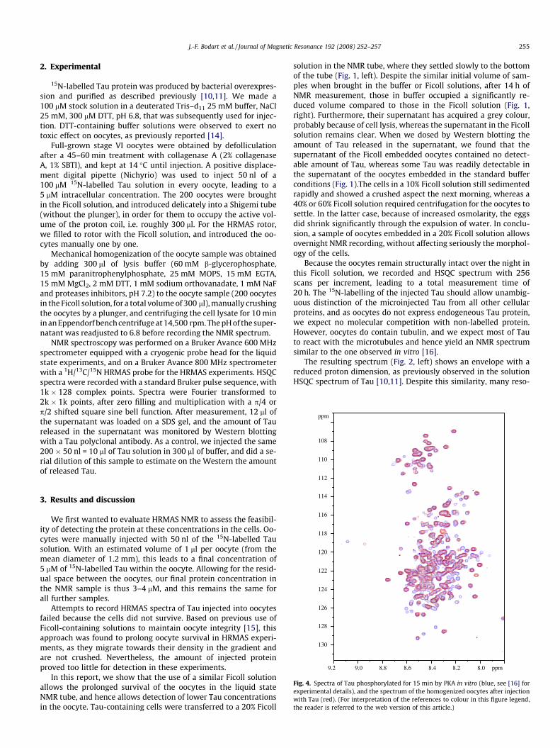
Fig. 4. Spectra of Tau phosphorylated for 15 min by PKA in vitro (blue, see [16] forexperimental details), and the spectrum of the homogenized oocytes after injectionwith Tau (red). (For interpretation of the references to colour in this figure legend,the reader is referred to the web version of this article.)
3. Results and discussion
We first wanted to evaluate HRMAS NMR to assess the feasibil-ity of detecting the protein at these concentrations in the cells. Oo-cytes were manually injected with 50 nl of the 15N-labelled Tausolution. With an estimated volume of 1 ll per oocyte (from themean diameter of 1.2 mm), this leads to a final concentration of5 lM of 15N-labelled Tau within the oocyte. Allowing for the resid-ual space between the oocytes, our final protein concentration inthe NMR sample is thus 3–4 lM, and this remains the same forall further samples.
Attempts to record HRMAS spectra of Tau injected into oocytesfailed because the cells did not survive. Based on previous use ofFicoll-containing solutions to maintain oocyte integrity [15], thisapproach was found to prolong oocyte survival in HRMAS experi-ments, as they migrate towards their density in the gradient andare not crushed. Nevertheless, the amount of injected proteinproved too little for detection in these experiments.
In this report, we show that the use of a similar Ficoll solutionallows the prolonged survival of the oocytes in the liquid stateNMR tube, and hence allows detection of lower Tau concentrationsin the oocyte. Tau-containing cells were transferred to a 20% Ficoll
solution in the NMR tube, where they settled slowly to the bottomof the tube (Fig. 1, left). Despite the similar initial volume of sam-ples when brought in the buffer or Ficoll solutions, after 14 h ofNMR measurement, those in buffer occupied a significantly re-duced volume compared to those in the Ficoll solution (Fig. 1,right). Furthermore, their supernatant has acquired a grey colour,probably because of cell lysis, whereas the supernatant in the Ficollsolution remains clear. When we dosed by Western blotting theamount of Tau released in the supernatant, we found that thesupernatant of the Ficoll embedded oocytes contained no detect-able amount of Tau, whereas some Tau was readily detectable inthe supernatant of the oocytes embedded in the standard bufferconditions (Fig. 1).The cells in a 10% Ficoll solution still sedimentedrapidly and showed a crushed aspect the next morning, whereas a40% or 60% Ficoll solution required centrifugation for the oocytes tosettle. In the latter case, because of increased osmolarity, the eggsdid shrink significantly through the expulsion of water. In conclu-sion, a sample of oocytes embedded in a 20% Ficoll solution allowsovernight NMR recording, without affecting seriously the morphol-ogy of the cells.
Because the oocytes remain structurally intact over the night inthis Ficoll solution, we recorded and HSQC spectrum with 256scans per increment, leading to a total measurement time of20 h. The 15N-labelling of the injected Tau should allow unambig-uous distinction of the microinjected Tau from all other cellularproteins, and as oocytes do not express endogeneous Tau protein,we expect no molecular competition with non-labelled protein.However, oocytes do contain tubulin, and we expect most of Tauto react with the microtubules and hence yield an NMR spectrumsimilar to the one observed in vitro [16].
The resulting spectrum (Fig. 2, left) shows an envelope with areduced proton dimension, as previously observed in the solutionHSQC spectrum of Tau [10,11]. Despite this similarity, many reso-
256 J.-F. Bodart et al. / Journal of Magnetic Resonance 192 (2008) 252–257
nances observable in the solution spectrum of free Tau are missing.Of special interest is the comparison with the spectrum of the samesample after mechanical homogenization. Cross-peaks clearly be-come better defined after lysis (Fig. 2, right), pointing to intracellu-lar viscosity or interaction with cellular partners as possiblebroadening mechanisms. Still, even the spectrum after homogeni-zation does still not show all cross-peaks previously described forthe full-length Tau protein, but rather resembles the spectrum ofTau bound to tubulin [16] (Fig. S1). When considering that oocyteshave a cellular tubulin concentration of about 20 lM but are de-void of endogeneous Tau [17], and with a 1:3 stoichiometry ofTau to preformed microtubules (MTs) [18], we can consider thatindeed all injected Tau becomes MT associated. However, on thebasis of the in-cell spectrum, we conclude that other cellular fac-tors equally compete for the same Tau. From these spectra, we thus
ppm
9.2 9.1 ppm
119
120
118
ppm
8.08.28.48.68.89.2 9.0 ppm
108
110
112
114
116
118
120
122
124
126
128
130
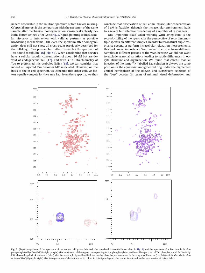
Fig. 5. (Top) comparison of the spectrum of the oocyte cell lysate (left, red; the threphosphorylated by PKA/Gsk3b (right, purple). (Bottom) zoom of the region correspondinPKA shows the pSer214 resonance (blue), that becomes split by unidentified but nearbyaction of Gsk3b (purple, right). (For interpretation of the references to colour in this fig
conclude that observation of Tau at an intracellular concentrationof 5 lM is feasible, although the intracellular environment leadsto a severe but selective broadening of a number of resonances.
One important issue when working with living cells is thereproducibility of the spectra. In the perspective of recording mul-tiple spectra on different samples, in order to reconstruct triple res-onance spectra or perform intracellular relaxation measurements,this is of crucial importance. We thus recorded spectra on differentsamples at different periods of the year, because we did not wantto exclude seasonal variations leading to subtle differences in oo-cyte structure and organization. We found that careful manualinjection of the same 15N-labelled Tau solution at always the sameposition in the equatorial unpigmented ring under the pigmentedanimal hemisphere of the oocyte, and subsequent selection ofthe ‘‘best” oocytes (in terms of minimal visual deformation and
ppm
9.2 9.1 ppm
118
119
120
ppm
8.08.28.48.68.89.2 9.0 ppm
108
110
112
114
116
118
120
122
124
126
128
130
shold is twofold lower than in Fig. 3) and the spectrum of a Tau sample in vitrog to the phosphorylated residues. The spectrum of Tau phosphorylated for 5 min byphosphorylation events in the oocyte cell interior (red, left) as it is after the in vitroure legend, the reader is referred to the web version of this article.)
J.-F. Bodart et al. / Journal of Magnetic Resonance 192 (2008) 252–257 257
cytoplasm leak) led to the best results. Still, whereas the reproduc-ibility of the overall aspect of the spectrum improved by this pro-cedure (Fig. 3), it was not sufficient to exploit the cross-peakintensity in a quantitative way. As a concrete example, the cross-peak at [8.9 ppm, 120.3 ppm] that we assigned to a phosphoryla-tion event (see below) is clearly visible in one spectrum, whereasit was below the noise level in the second one. After mechanicalhomogenization of both oocyte samples, though, the reproducibil-ity of the spectra improved, and a similar cross-peak at a slightlydifferent nitrogen frequency became visible in both spectra. Wethus conclude that the recording of a series of spectra on differentsamples with variation of one parameter (relaxation delay, 13C evo-lution, . . .) will not be straightforward. Variation not only comesfrom the protein, but might equally stem from variations in thephysicochemical nature of the intracellular environment.
In the in-cell spectrum, we did detect some novel signals thatwe assign to phosphorylated residues of Tau. Assignment of thephosphorylation sites by triple resonance spectroscopy on oocytesinjected with 13C/15N Tau proved below the limit of sensibility.Mechanical homogenization of the cells led to better quality spec-tra (Fig. 3), with a slight change in the resonance frequency of thephosphor-peak, but the amount of protein in 200 oocytes still wastoo low for 3D triple resonance spectroscopy. Comparison with thespectra of Tau phosphorylated in vitro by PKA [19] showed that themajor site observed on the in vivo spectrum before or after homog-enization (Fig. 2, peak at [8.9 ppm, 120.3 ppm]) was not generatedby this kinase (Fig. 4).
Another enzyme that was reported as active in the immatureoocyte is Gsk3b [20]. We obtained a clone of this kinase, expressedand purified it. The latter was used as a second candidate kinase toobtain a standard for the identification of the in vivo phosphoryla-tion pattern (A.L. and G.L., manuscript in preparation) BecauseGsk3b needs a priming to be efficient, we used our PKA phosphor-ylated sample as a substrate for subsequent phosphorylation byPKA. When we compare the spectrum of PKA/Gsk3b phosphory-lated Tau with the one of the oocyte cell lysate (Fig. 5), it is clearthat the major in vivo peak does not overlap with any of the peaksin the in vitro generated spectrum. However, when we lower thethreshold of the in vivo spectrum, and especially when we lookat the spectrum of the cell lysate, we observe a second less inten-sive peak at 9.14 ppm, 119.1 ppm (Fig. 5). A zoom of this peak, andcomparison with the PKA pSer214 correlation, suggests thatin vivo, PKA indeed does phosphorylate the Ser214 position, butthat other unidentified phosphorylation events influence this reso-nance position in a similar way as Gsk3b.
We conclude from the present study that the embedding of theoocytes in a 20% Ficoll solution prior to NMR recording signifi-cantly enhances the time span over which the cells remain their in-tact morphology, such that spectral recording on physiologicallyrelevant protein concentrations becomes feasible. Evidently, thenatively unfolded nature of Tau leads to sharper lines than for afolded protein of the same or even smaller molecular weight, andthe same minimal concentration of 5 lM might therefore not beattainable with every protein.
For the case of the neuronal Tau protein that we here have stud-ied, NMR allows the detection of its interaction with tubulin andother cellular factors, but equally of novel phosphorylation events.The reproducibility of the spectra does depend on the precise phys-iological state of the oocytes, and is thus not easily assessed. How-ever, mechanical homogenization and subsequent cytoskeletondisruption improves the situation, and might thus be the methodof choice to evaluate post-translational modifications of microin-jected proteins.
Acknowledgments
We thank Dr. L. Buée (Lille, France) for a generous gift of thepolyclonal Tau antibody. The NMR facility used in this study wasfunded by the Région Nord-Pas de Calais (France), the CNRS, theUniversities of Lille 1 and Lille 2, and the Institut Pasteur de Lille.L.A. holds a doctoral fellowship from the CNRS and the RegionNord/Pas de Calais. Part of this work was financed by the FrenchANR grant ‘‘Tau: tubulin”.
Appendix A. Supplementary data
Supplementary data associated with this article can be found, inthe online version, at doi:10.1016/j.jmr.2008.03.006.
References
[1] G. Lippens, J.-P. Bohin, Structural diversity of the osmoregulated perplasmicglucans of gram-negative bacteria by a combined genetics and nuclearmagnetic approach, in: M. Pons (Ed.), NMR in Supramolecular Chemistry,Nato Advanced Research Series, vol. 191, Kluwer Academic, Dordrecht, TheNetherlands, 1999.
[2] J.-M. Wieruszeski, A. Bohin, J.-P. Bohin, G. Lippens, In vivo detection of theosmoregulated periplasmic glucan of Ralstonia solanancearum by NMR, J.Magn. Reson. 151 (2001) 118–123.
[3] Z. Serber, A.T. Keatinge-Clay, R. Ledwidge, A.E. Kelly, S.M. Miller, V. Dotsch,High-resolution macromolecular NMR spectroscopy inside living cells, J. Am.Chem. Soc. 123 (2001) 2446–2447.
[4] Z. Serber, P. Selenko, R. Hänsel, S. Recker, F. Löhr, J.E. Ferrell, G. Wagner, V.Dötsch, Investigating macromolecules inside cultured and injected cells by in-cell NMR spectroscopy, Nat. Protoc. 1 (2006) 2701–2709.
[5] M.M. Dedmon, C.N. Patel, G.B. Young, et al., FlgM gains structure in living cells,Proc. Natl. Acad. Sci. USA 99 (2002) 12681–12684.
[6] P. Selenko, Z. Serber, B. Gadea, J. Ruderman, G. Wagner, Quantitative NMRanalysis of the protein G B1 domain in Xenopus laevis egg extracts and intactoocytes, Proc. Natl. Acad. Sci. USA 103 (2006) 11904–11909.
[7] T. Sakai, H. Tochio, T. Tenno, Y. Ito, T. Kokubo, H. Hiroaki, H.J. Shirakawa, In-cellNMR spectroscopy of proteins inside Xenopus laevis oocytes, J. Biomol. NMR 36(2006) 179–188.
[8] P. Delobel, S. Flament, M. Hamdane, R. Jakes, A. Rousseau, A. Delacourte, J.-P.Vilain, M. Goedert, L. Buee, Functional characterization of FTDP-17 tau genemutations through their effects on Xenopus oocyte maturation, J. Biol. Chem.277 (2002) 9199–9205.
[9] P. Delobel, S. Flament, M. Hamdane, A. Delacourte, J.-P. Vilain, L. Buee,Modelling Alzheimer-specific abnormal Tau phosphorylation independently ofGSK3beta and PKA kinase activities, FEBS Lett. 516 (2002) 151–155.
[10] G. Lippens, J.M. Wieruszeski, A. Leroy, C. Smet, A. Sillen, L. Buée, I. Landrieu,Proline directed random coil chemical shift values as a tool for the NMRassignment of the Tau phosphorylation sites, Chembiochem 5 (2004) 73–78.
[11] C. Smet, A. Leroy, A. Sillen, J.M. Wieruszeski, I. Landrieu, G. Lippens, Acceptingits random coil nature allows a partial NMR assignment of the neuronal Tauprotein, Chembiochem 5 (2004) 1639–1646.
[12] D.W. Cleveland, S.Y. Hwo, M.W. Kirschner, Physical and chemical properties ofpurified tau factor and the role of tau in microtubule assembly, J. Mol. Biol. 116(1977) 227–247.
[13] A.H. Lockwood, Tubulin assembly protein: immunochemical andimmunofluorescent studies on its function and distribution in microtubulesand cultured cells, Cell 13 (1978) 613–627.
[14] W.T. Matten, G.F. Vande Woude, Microinjection into Xenopus oocytes, MethodsEnzymol. 254 (1995) 458–466 .
[15] H.P. Richter, H.C. Hoock, B. Neumcke, Morphological and electrophysiologicalproperties of centrifuged stratified Xenopus oocytes, Biol. Cell 84 (1995) 129–138.
[16] A. Sillen, P. Barbier, I. Landrieu, S. Lefebvre, J.M. Wieruszeski, A. Leroy, V.Peyrot, G. Lippens, NMR investigation of the interaction between the neuronalprotein tau and the microtubules, Biochemistry 46 (2007) 3055–3064.
[17] D.L. Gard, M.W. Kirschner, Microtubule assembly in cytoplasmic extracts ofXenopus oocytes and eggs, J. Cell Biol. 105 (1987) 2191–2201.
[18] V. Makrides, M.R. Massie, S.C. Feinstein, J. Lew, Evidence for two distinctbinding sites for tau on microtubules, Proc. Natl. Acad. Sci. USA 101 (2004)6746–6751.
[19] I. Landrieu, L. Lacosse, A. Leroy, J.M. Wieruszeski, X. Trivelli, A. Sillen, N. Sibille,H. Schwalbe, K. Saxena, T. Langer, G. Lippens, NMR analysis of a Tauphosphorylation pattern, J. Am. Chem. Soc. 128 (2006) 3575–3583.
[20] M. Sarkissian, R. Mendez, J.D. Richter, Progesterone and insulin stimulation ofCPEB-dependent polyadenylation is regulated by Aurora A and glycogensynthase kinase-3, Genes Dev. 18 (2004) 48–61.


![Michel parameters and [tau] neutrino helicity from decay correlations in Z--\u003e[tau]+[tau]](https://img.pdfslide.net/doc/110x75/6350c359f55d98549a09e91e/michel-parameters-and-tau-neutrino-helicity-from-decay-correlations-in-z-u003etautau.jpg)


