Embed Size (px)
Citation preview
1
Comparative Antiviral (HIV) Photoactivity Of Metalized meso-Tetraphenylsulfonated Porphyrins
Franklin Vargas*a, Tamara Zoltana, Liliana Padróna, Carla Izzoa, Verónica Lópeza, Lubimar
Gómeza, Flor Pujolb, Hector Rangelb, Domingo Garzazob and Rona Fabbrob
a Laboratorio de Fotoquímica, Centro de Química, Instituto Venezolano de Investigaciones Científicas I.V.I.C., Caracas 1020-A , Venezuela. Apartado 21827, e-mail: [email protected]
b Laboratorio de Virología Molecular, Centro de Microbiología y Biología Celular, Instituto Venezolano de Investigaciones Científicas I.V.I.C., Caracas, Venezuela
ABSTRACT
We have carried out the study of photochemical properties of a series of synthetic meso-tetraphenylsulfonated porphyrins (TPPMS4) metalized Cu(II), Zn(II), Pd(II), Mn(II), Fe(III), Ni(II) and Co(II) for the optimization of their clinical applications as antiviral against human immunodeficiency virus (HIV-1) and the study of the in vitro antiviral photoinactivation mechanisms with future application for blood sterilization. Keywords: meso-Tetraphenylsulfonated porphyrins, HIV, Metallo porphyrins, Photoinactivation, Phototherapy
1. INTRODUCTION
Photochemical studies of new photosensitizer for the photodynamic therapy (PDT), like they are Pheophytin, Radachlorin, Mg-chlorophyll-a and -b, among other natural compounds; they have been carried out in our laboratory 1-4. The most extensively studied photosensitizers are porphyrins that were identified over 150 years ago. One of their good features is minimal toxicity in the dark and a lack of pharmacological interactions with other drugs, making PDT a safe procedure in oncological combined treatments (PDT and drug or radiation). For a long time most preclinical studies were dominated by the use of hematoporphyrin derivatives. The best clinical experience has been obtained with Photofrin which is a mixture of monomers, dimers and oligomers derived from hematoporphyrin. As it is very well-known the singlet oxygen is a reactive species of oxygen able to promote harmful processes at cellular level, such as: lipid peroxidation, damage of cellular membrane and cellular death. The photosensitizers are derived of porphyrins and chlorophylls, originating high yields of 1O2,
. OH and in vitro phototoxicity. Some of them have complete pre-clinical investigations, showing significant advantages for their use in dynamic anti-viral and anti-tumoral phototherapy 5-9. The PDT is a novel treatment for cancer and other abnormal tissue degeneration that employs a photosensitizer and visible light to produce singlet oxygen and other reactive oxygen species that lead to subcellular damage at the sites where the photosensitizer accumulates. Alternatively, an excited photosensitizer may react directly with biomolecules to form free radicals that further react with molecular oxygen producing the superoxide radical anion, hydrogen peroxide or hydroxyl radical. The superoxide radical anion is generated, for instance, by excitation of porphyrins in the presence of reducing substances. The objective of this study was to investigate the ability of Cu(II), Zn(II), Pd(II), Mn(II), Fe(III), Ni(II) and Co(II) complexes of meso-tetraphenylsulfonated porphyrins (TPPMS4) to generate the reactive oxygen species (. OH, 1O2) upon irradiation (UVA-Vis) 10.
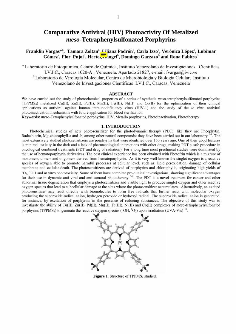
Figure 1. Structure of TPPMS4 studied.
2
The ability of these compounds generated .OH by cell-free systems that use the system luminol-chemiluminescence, were investigated. These compounds are also capable of producing singlet oxygen by energy transfer when they are irradiated with UV-A and visible light in the presence of molecular oxygen. We have carried out the optimization of their clinical applications as antiviral against human immunodeficiency virus (HIV-1) and blood sterilization by means of in vitro viability and ELISA assay.
2. MATERIALS AND METHODS 2.1. Chemicals and materials All analytical or HPLC grade solvents were obtained from Merck (Darmstadt, Germany). Tetraphenylporphyrin (TPP), histidine, rose bengal, luminol, isoluminol, horseradish peroxidase (HRP), 1-(4,5-dimethylthiazol-2yl)-3,5-diphenylformazan (MTT), RPMI-1640 medium, nicotinamide adenine dinucleotide phosphate reduced form (NADPH), hydrogen peroxide, 30 wt. % solution in water and p-nitrosodimethyl aniline were purchased from Sigma (St. Louis, MO, USA). Phosphate-buffered saline solution (PBS) pH 7.4 (0.01 M phosphate buffer and 0.135 M NaCl).
UV-Vis spectrophotometry of the TPP and TPPSM solution was followed using a Milton-Roy Spectronic 3000 array instrument (Milton Roy Company-USA) and also Perkin Elmer Lambda-2 UV-Vis spectrophotometer (USA). The fluorescence spectra were registered with a Shimadzu RF 1501 spectrofluorophotometer.
The structures of the isolated products were elucidated by 1H NMR and 13C NMR (Brucker Aspect 3000, 300 and 100 MHZ respectively), FT I.R. (Nicolet DX V 5.07) and mass spectra (Varian Saturn 2000) in connection with a Varian chromatograph equipped with a 30-m capillary (CP-Sil, 8CB-MS). Elemental analyses were performed by the “Laboratorio Nacional de Análisis Químico” – IVIC – Centro de Química, Caracas, Venezuela in a Fisions Instrument EA-1108. All preparative irradiations (1.6 x 10⁻² M) were monitored by liquid chromatography (HPLC, Waters Delta Prep 4000) equipped with an analytic and a preparative C18-Bondapak column. 2.2. Synthesis and characterization of the metalized porphyrins and sulphonation. All the metalized porphyrins were prepared by chemical synthesis. Absorption, emission, 1H-, 13C-NMR, EPR were compared with theses parent compound in the literature 11-15. EPR were register by mean a Brucker EMX (Germany) at 28 ºC..and compared with some reported 11.
The inclusion of metal in TPP was carried out heating the porphyrin with a metal salt (divalent metal ions) in chloroform, methanol or acetic acid. 2.3. General procedure of the sulfonation reaction. In a flask of 5 mL, equipped with magnetic, coolant agitator and trap of humidity, 10 mg is placed (0.02 mmol) of metallotetraphenylporphyrin (TPPM) and 1 mL of H2SO4 (96%) and it is allowed to react at 60 ºC during 1 hr. The reaction mixture allows cooling down, it is diluted with cold water (50 mL), and it is neutralized with Na2CO3. Extraction with CH2Cl2 was necessary to eliminate the TPP in excess 13. The aqueous phase was evaporated to dryness and methanol was added to dissolve the sulfonated product. The reaction was monitoring to different intervals of time by TLC and HPLC. The separation of the sulfonated products was made by means of column chromatography (reverse phase, Polygoprep 100-50 and Bondapak C18 column) with elution gradients of H2O:MeOH. The purification of each one of the compounds required more than one separation in column. 2.4. Irradiation All processes of irradiation were carried out using a illuminator Cole Palmer 41720-series keeping a distance of 10 cm between the lamp surface and the solution, varying the time periods of exposure at 37 ºC under continuous shaking., with a emission maximum in UVA-Vis 320-600 nm (3.3 mW/cm2, 45.575 Lux/seg) (radiation dose 4.5 J/cm
2) as measured with a
model of UVX Digital Radiometer after 1 h continued illumination. 2.5. Quantum yields The relative quantum yields of fluorescence for TPPMS complexes were determined at room temperature either by comparing the corrected fluorescence intensity of the these complexes in ethanol-H2O with that of rhodamine B (at a concentration of 1 x 10⁻⁶ M in ethanol; fluorescence quantum yield, 0.69) or with that of quinine bisulfate in 0.05 M H2SO4 (fluorescence quantum yield, 0.55) 16. 2.6. Singlet oxygen generation. Indirectly, photosensitized degradation of histidine was measured in the presence of 0.25, 0.50, 1.0, and 1.5 x 10⁻⁵ M solution of TPP and TPPMS4 17. These solutions were mixed with an equal quantity of L-histidine solution at 0.60 to 0.74 mM in phosphate buffer 0.01 M, pH 7.4. Samples of this mixture were irradiated at time intervals from 60 to 180 min. with the respective controls being protected from light. The concentration of histidine was determined by a colorimetric reaction using phosphate buffer, sulfanilic acid, sodium nitrite, sodium carbonate and ethanol as reagents. The optic density was read
3
on a spectrophotometer at 440 nm against a blank reagent, a modified Pauly reaction and by bleaching of p-nitrosodimethylaniline18, 19. Another "trap" method has been successfully used to detect generated 1O2 in a variety of samples. This method is based upon following the consumption of a chemical trap (Furfuryl alcohol, FFA) that react with singlet oxygen. The consumption of FFA was followed by HPLC using a 90:10 H20/CH3CN mobile phase composition. The detection
wavelength used for monitoring FFA consumption was at 222 nm. Rose bengal, a well known 1O2 sensitizer, was used as a
standard for comparison with TPP and TPPSM for 1O2 formation, under identical conditions of photolysis19, 20. 2.7. Generation and detection of others reactive oxygen species Chemiluminescence (CL) was generated in cell-free systems; H2O2-induced CL (as a blank): H2O2 (3.5 mM) was added to phosphate buffered saline solution (PBS, 10 mM KH2PO4 and 150 mM NaCl, pH 7.2) and luminol (250 µM, prepared daily in 2 M NaOH and diluted with PBS). The TPPMS4-induced CL at different concentrations was dispensed after irradiation in presence of NADPH. The generated CL at 37 ºC was measured continuously for 10 min in a Luminoskan Ascent luminometer (ThermoLabsystems, Finland) in a 96-well Thermo Labsystems Microtiter plate21-23. 2.8. Cells and virus The MT4 cells were cultivated in RPMI-1640 medium supplemented with 10% PBS, and penicillin/streptomycin. Cultures were subcultured every 3 days and for the drugs assay the cells were harvested in the day two of cultured in order to use the exponential phase of growth. The HIV-1 (HXB2) was obtained through the acquired immune deficiency syndrome-AIDS research and reference reagent program, division of AIDS, National Institute of Allergy and Infectious Diseases-NIAID, National Institutes of Health-NIH. Human T-Lymphotropic Virus Types I & II - HTLV-IIIB/H9 from Dr. Robert Gallo24. 2.9. Citotoxicity assay To determine the cellular toxicity (IC50) of the compounds, we evaluate different concentration of them in MT4 cell culture. The cells were seeded in 96 well/plate at a density of 30.000 cell/weel, the different drugs dilution were added to each well and illuminated with 3.3 mW/cm2, 45.575 Lux/seg (radiation dose 4.5 J/cm
2) for 30 min, control were established
with drugs without illumination and without drug illuminated. After 72, 96 and 120 h. the cultures were evaluated with MTT cell proliferation assay to establish the fraction of death cells in each concentration25. 2.10. Viral inhibition The MT4 cells were seeded in 24 well/plate at a density of 120000 cells/well and the different compound were added at IC50 level or below, then were infected with HIV-1 IIIB at a moi of 0.1 then illuminated or not. 12 h later the supernatant were changed and the cells washed out with PBS tree times and reseeded in a final volume of 3 ml. The virus production was evaluated at 72, 96 and 120 h by collection and replaced of 100 µl of supernatant. The virus determination was performed by p24 detection in a no commercial ELISA. The results were expressed in relative to the control 25. 2.11. Statistical treatment of results At least three independent experiments were performed except where indicated otherwise. The results are expressed as a mean ± S.E.M. derived from 3-4 observations. The level of significance accepted was p ≤ 0.05.
3. RESULTS 3.1. Preparation and determination of the matalized porphyrin complexes Absorption, emission, 1H-, 13C-NMR, EPR spectra of the sulfonated metalloporphyrin complexes, were compared with theses authentic or similar parent compound in the literature11-15, 26. All the metalized porphyrins contain divalent metal, except for Fe, which was determined to have valence III with Cl as contra-ion. 3.2. Formation of ROS The generation of .OH was determined by means of chemiluminescence of luminol after the irradiation (λ= 400-900 nm) of the metallic complexes Cu(II), Zn(II), Pd(II), Mn(II), Fe(III), Ni(II) and Co(II) of meso-tetraphenylsulfonated porphyrins in presence of NADPH. The formation of peroxidic species that activate to the luminol showed the following tendency: TPPPdS4 > TPPFeS4 > TPPMnS4 > TPPZnS4 > TPPCuS4 > TPPS4 > TPPNiS4 > TPPCoS4.
The detection of 1O2 was determined by the test of histidine and p-nitrosodimethylaniline relative to Rose Bengal. The best yields in generation of 1O2 were those of ZnTPPS4> PdTPPS4> CuTPPS4. The quantum yield for singlet oxygen generation were estimated in relation to Rose bengal (0.79), TPPZnS4 (0.37), TPPPdS4 (0.14), TPPCuS4 (0.12), TPPMnS4 (0.08), TPPS4 (0.07), TPPCoS4 (0.06), TPPFeS4 (0.04), TPPNiS4 (0.03) and corroborated with the literature27.
4
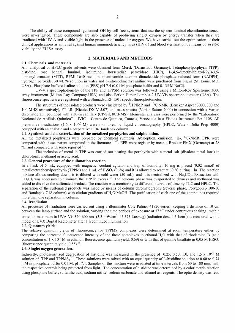
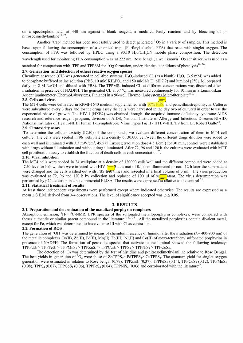
3.3. Antiviral photoactivity The cytotoxicity (MTT-CellTiter96 non radiative cell proliferation assay) with and without light, of MT4 cells free virus stayed on 70% of cellular viability to concentrations of 10-3, 10-4 and 10-5 M of metalized porphyrins. The quantification of these results is carrying out by means of citotoxicity and ELISA assay. On infected HIV-cells the inactivation of viral proliferation was effective depending on the TPPMS4 and for consequence of the mechanism which this predominantly acts.
The following graphs show the antiviral action of the complexes TPPFeS4, TPPMnS4, TPPPdS4 and TPPZnS4 on the production of HIV-1. It is important to stand out that the cellular viability stays during the irradiation process. The other metallic complexes as TPPCuS4, TPPNiS4 and TPPCoS4 didn't show appreciable antiviral action when were irradiated in presence of cultivated HIV-1.
0
20
40
60
80
100
120
dark Irradiated dark irradiated dark irradiated
72 h 96 h 120 h
Incubation time
Vira
l pro
duct
ion
(%)
TPPFeS4 10-3 M TPPFeS4 10-4 M Control
Figure 2. Effects of TPPFeS4 on the viral production (HIV-1) under irradiated and dark conditions by Elisa assay.
0
20
40
60
80
100
120
dark irradiated dark irradiated dark irradiated
72 h 96 h 120 h
Incubation time
Viab
ility
(%)
TPPFeS4 10-3 M TPPFeS4 10-4 M Control
Figure 3. Effects of TPPFeS4 on the cellular viability under irradiated and dark conditions by viability assay.
5
0
20
40
60
80
100
120
dark irradiated dark irradiated dark irradiated
72 h 96 h 120 h
Incubation time
Vira
l pro
duct
ion
(%)
TPPMnS4 10-3 M TPPMnS4 10-4 Control
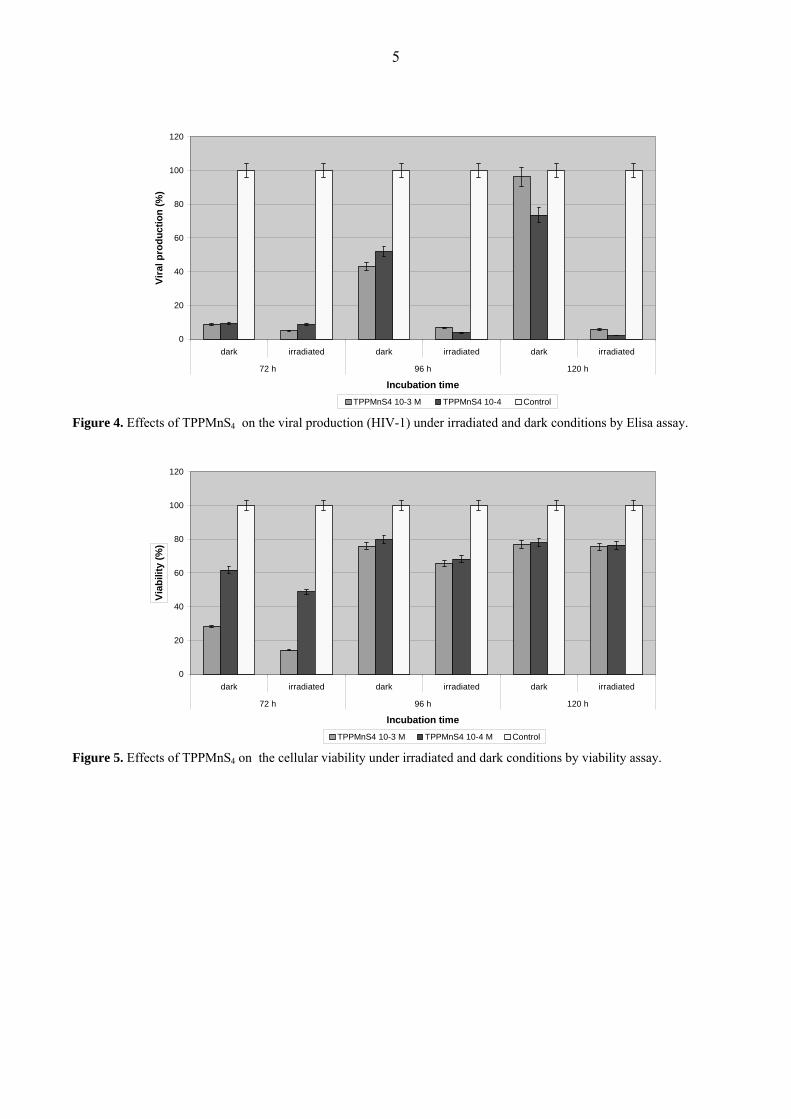
Figure 4. Effects of TPPMnS4 on the viral production (HIV-1) under irradiated and dark conditions by Elisa assay.
0
20
40
60
80
100
120
dark irradiated dark irradiated dark irradiated
72 h 96 h 120 h
Incubation time
Viab
ility
(%)
TPPMnS4 10-3 M TPPMnS4 10-4 M Control
Figure 5. Effects of TPPMnS4 on the cellular viability under irradiated and dark conditions by viability assay.
6
0
20
40
60
80
100
120
dark irradiated dark irradiated dark irradiated
72 h 96 h 120 h
Incubation time
Vira
l pro
duct
ion
(%)
TPPPdS4 10-3 M TPPPdS4 10-4 M Control
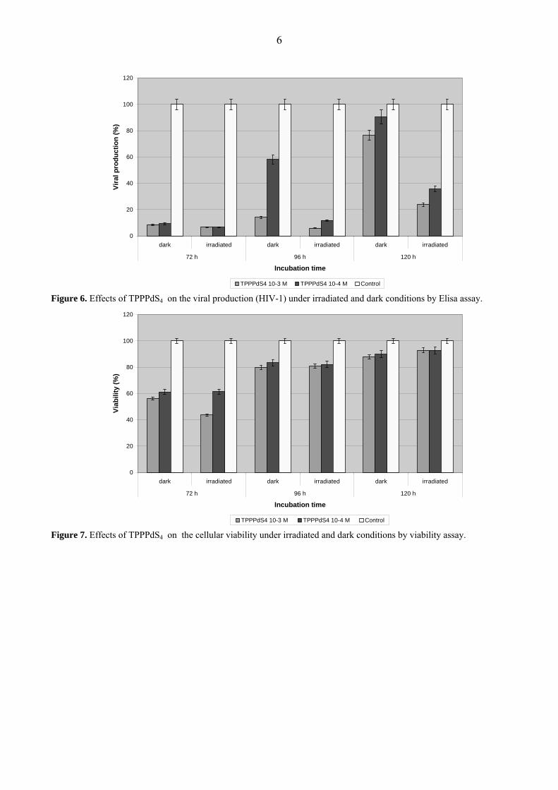
Figure 6. Effects of TPPPdS4 on the viral production (HIV-1) under irradiated and dark conditions by Elisa assay.
0
20
40
60
80
100
120
dark irradiated dark irradiated dark irradiated
72 h 96 h 120 h
Incubation time
Viab
ility
(%)
TPPPdS4 10-3 M TPPPdS4 10-4 M Control
Figure 7. Effects of TPPPdS4 on the cellular viability under irradiated and dark conditions by viability assay.
7
0
20
40
60
80
100
120
dark irradiated dark irradiated dark irradiated
72 h 96 h 120 h
Incubation time
Vira
l pro
duct
ion
(%)
TPPZnS4 10-3 M TPPZnS4 10-4 M Control
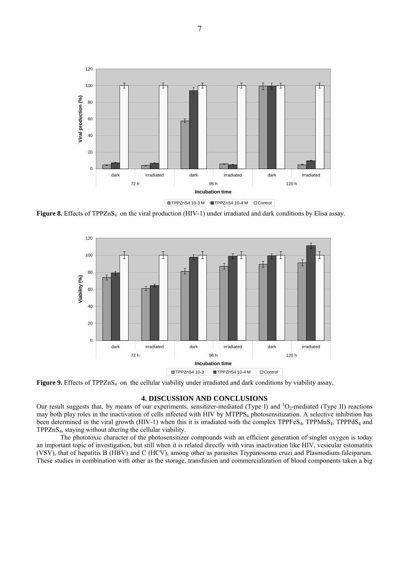
Figure 8. Effects of TPPZnS4 on the viral production (HIV-1) under irradiated and dark conditions by Elisa assay.
0
20
40
60
80
100
120
dark irradiated dark irradiated dark irradiated
72 h 96 h 120 h
Incubation time
Viab
ility
(%)
TPPZnS4 10-3 TPPZnS4 10-4 M Control
Figure 9. Effects of TPPZnS4 on the cellular viability under irradiated and dark conditions by viability assay.
4. DISCUSSION AND CONCLUSIONS Our result suggests that, by means of our experiments, sensitizer-mediated (Type I) and 1O2-mediated (Type II) reactions may both play roles in the inactivation of cells infected with HIV by MTPPS4 photosensitization. A selective inhibition has been determined in the viral growth (HIV-1) when this it is irradiated with the complex TPPFeS4, TPPMnS4, TPPPdS4 and TPPZnS4, staying without altering the cellular viability.
The phototoxic character of the photosensitizer compounds with an efficient generation of singlet oxygen is today an important topic of investigation, but still when it is related directly with virus inactivation like HIV, vesicular estomatitis (VSV), that of hepatitis B (HBV) and C (HCV), among other as parasites Trypanosoma cruzi and Plasmodium falciparum. These studies in combination with other as the storage, transfusion and commercialization of blood components taken a big
8
importance in the processes of sterilization of plasma and others blood derivates 28, 29. Nevertheless, the considerations of the toxic effects to other cellular and immunologic systems take us to investigate about the selectivity and the factors of efficiency of these processes. In spite of the implementation of severe measures to prevent the transmission of pathogen agents by way of sanguine transfusion, a high risk still exists due to the residual permanence of these agents. It makes necessary the development of new and better sterilization methods that inactive a wide range of pathogens. Some of these methods, very promissory, are that of the direct photochemical or photosensitized sterilization.
5. REFERENCES 1. Vargas, F., Y. Díaz, V. Yartsev, A. Marcano and A. Lappa, “Photophysical properties of a novel PDT photosensitizer radachlorin in different media”, Ciencia 1, pp. 70-77, 2004. 2. Vargas, F., C. Rivas, T. Zoltan, L. Padrón and C. Izzo, “Fototerapia: aplicaciones clínicas”, abcMedicus, Medicina pp. 1-9, 2005. http://www.abcmedicus.com/articulo/medicos /2/id/421/pagina/1/fototerapia_aplicaciones_clinicas.html. 3. Vargas, F. and C. Rivas (2004) Las aplicaciones clínicas de fototerapia. Dermatología Venezolana. 42, 4-8. 4. Vargas, F., T. Zoltan, C. Rivas and F. Salazar, “Photoinduced apoptosis by photosensitizer drugs”, for invitation to publish in the series: New Cell Apoptosis Research. Nova Science Publishers, Inc. Hauppauge, NY 11788, U.S.A. 2006 (in press). 5. Hynninen, P. H. and E. S. Nyman, “Research advances in the use of tetrapyrrolic photosensitizers for photodynamic therapy”, J. Photochem. Photobiol. B: Biol. 73, pp. 1-28, 2004. 6. Almeida, R. D., B. J. Manadas, A. P Carvalho and C. B Duarte, “Intracellular signaling mechanisms in photodynamic therapy”, Biochim Biophys Acta 1704, pp. 59–86, 2004. 7. Vzorov, A. N., D. W. Dixon, J. S. Trommel, L. G. Marzilli and R. W. Comons, “Inactivation of Human Immunodeficiency Virus type 1 by porphyrins”, Antimicrob. Agents Chemother. pp. 3917-3925, 2002. 8. Müller-Breitkreutz, K., H. Mohr, K. Briviba and H. Sies, “Inactivation of viruses by chemically and photochemically generated singlet molecular oxygen”, J. Photochem. Photobiol. B: Biol. 30, 63-70, 1995. 9. North, J., H. Neyndorff, D. King and J. Levy, “Viral inactivation in blood and red cell concentrates with benzoporphyrin derivative”, Blood Cells 18, pp. 129-140, 1992. 10. Braun, A. M. and E. Oliveros, “Applications of singlet oxygen reactions: mechanistic and kinetic investigations”, Pure Appl. Chem. 62, pp.1-29, 1990. 11. Fuhrhop J-H. and K. M. Smith, Laboratory methods in porphyrin and metalloporphyrin reseach, Elsevier, Amsterdam, The Netherlands, 1975. 12. Smith, K. M. Porphyrin and Metalloporphyrin, Elsevier, Amsterdam, The Netherlands, 1975. 13. H. G. Ortega, “Porfirinas Solubles en Agua: Síntesis, Homoasociación y Propiedades Fotofísicas de Porfirinas Sulfonadas 5,15-Difenilsustituidas”, Thesis Ph.D Universitat Barcelona, Barcelona, Spain, 2003. 14. Morgan, A. R., N. H. Petousis and J. E. van Lier, “Synthesis and photodynamic activity of some tetraaziporphyrin derivatives”, Eur. J. Med. Chem. 32, pp. 21-26, 1997. 15. Horváth, O., R. Huszánk, Z. Valicsek and G. Lendvay, “Photophysics and photochemistry of kinetically labile, water-soluble porphyrin complexes”, Coord. Chem. Rev. 250, pp. 1792-1803, 2006. 16. Calvert, J. G. and J. N. Pitts, “Experimental methods in photochemistry”, in: J.G. Calvert, J.N. Pitts (Eds.), Photochemistry, Wiley, New York, pp. 783-804, 1966. 17. Lovell W. W., D. J. Sanders, “ Screening test for phototoxins using solutions of simple biochemicals”,Toxic in vitro 4, pp. 318-320, 1990. 18. Kraljic I., S. El Mohsni, “A new method for the detection of singlet oxygen in aqueous solutions”, Photochem. Photobio.l 28, pp. 577-581, 1978. 19. Haag W. R., J. Hoigne, E. Gassman and A. D. Braun, “Singlet oxygen in surface water-part I: furfuryl alcohol as a trapping agent”, Chemosphere 13, pp. 631-640, 1984. 20. Allen J. M., C. J. Gossett and S. K. Allen, “Photochemical formation of singlet molecular oxygen (1O2) in illuminated aqueous solutions of p-aminobenzoic acid (PABA) ”, J. Photochem. Photobiol. B: Biol. 32, pp. 33-37, 1996. 21. Lundqvist, H. and C. Dahlgren, “Isoluminol-enhanced chemiluminescence: a sensitive method to study the release of superoxide anion from human neutrophils”, Free Rad. Biol. Med. 20, pp. 785-792, 1996. 22. Vargas, F., C. Rivas, Y. Díaz, N. Contreras, A. Silva, L. Ojeda, M. Velásquez and G. Fraile, “Antioxidant properties of dipyridamole as assessed by chemiluminescence”, Pharmazie 58, pp. 817-823, 2003. 23. Yildiz, G., A. T. Demiyürek, I. Sahin-Erdemli and I. Kanzik, “Comparison of antioxidant activities of aminoguanidine, methylguanidine and guanidine by luminol-enhanced chemiluminescence”, Br. J. Pharmacol. 124, pp. 905-910, 1998.

9
24. Popovic, M., M. G. Sarngadharan, E. Read and R. C. Gallo, “Detection, isolation, and continuous production of cytopathic retroviruses (HTLV-III) from patients with AIDS and pre-AIDS”, Science 224, pp. 497−500, 1984. 25. Motsman, T., “Rapid colorimetric assay for cellular growth and survival: application to proliferation and citotoxicity assays”, J. Inmunol. Methods 65, pp. 55-63, 1986. 26. Yushmanov, V. E., H. Imasato, T. T. Tominaga and M. Tabak, “HNMR and electronic absorption spectroscopy of paramagnetic water-soluble mesotetraarylsubstituted cationic and anionic metalloporphyrins”, J. Inorg. Biochem. 61, pp. 233-250, 1996. 27. Redmond, R. W. and J. Gamlin, “A compilation of singlet oxygen yields from biologically relevant molecules”, Photochem. Photobiol. 70, pp. 391-475, 1999. 28. Burnouf, T. and M. Radosevich, “Reducing the risk of infection from plasma products: specific preventative strategies”, Blood Reviews 14, pp. 94-110, 2000. 29. Moor, A. C. E., A. van der Veen, T. M. Dubbelman, J. VanSteveninck and A. Brand, “Photodynamic sterilization of red cells and its effect on contaminating white cells: viability and mechanism of cell death”, Transfusion 39, pp. 599-607, 1999.


