Embed Size (px)
Citation preview
© 2015 Li et al. This work is published by Dove Medical Press Limited, and licensed under Creative Commons Attribution – Non Commercial (unported, v3.0) License. The full terms of the License are available at http://creativecommons.org/licenses/by-nc/3.0/. Non-commercial uses of the work are permitted without any further
permission from Dove Medical Press Limited, provided the work is properly attributed. Permissions beyond the scope of the License are administered by Dove Medical Press Limited. Information on how to request permission may be found at: http://www.dovepress.com/permissions.php
OncoTargets and Therapy 2015:8 703–711
OncoTargets and Therapy Dovepress
submit your manuscript | www.dovepress.com
Dovepress 703
O r i g i n a l r e s e a r c h
open access to scientific and medical research
Open access Full Text article
http://dx.doi.org/10.2147/OTT.S76370
apoptosis of hela cells induced by a new targeting photosensitizer-based PDT via a mitochondrial pathway and er stress
Donghong li1
lei li2
Pengxi li1
Yi li3
Xiangyun chen1
1state Key laboratory of Trauma, Burn and combined injury, The second Department of research institute of surgery, 2The First Department of research institute of surgery, 3cancer center, Daping hospital, Third Military Medical University, chongqing, People’s republic of china
Abstract: Photodynamic therapy (PDT) is emerging as a viable treatment for many cancers.
To decrease the cutaneous photosensitivity induced by PDT, many attempts have been made to
search for a targeting photosensitizer; however, few reports describe the molecular mechanism of
PDT mediated by this type of targeting photosensitizer. The present study aimed to investigate the
molecular mechanism of PDT induced by a new targeting photosensitizer (PS I), reported previously
by us, on HeLa cells. Apoptosis is the primary mode of HeLa cell death in our system, and apoptosis
occurs in a manner dependent on concentration, irradiation dose, and drug–light intervals. After
endocytosis mediated by the folate receptor, PS I was primarily localized to the mitochondria and
the endoplasmic reticulum (ER) of HeLa cells. PS I PDT resulted in rapid increases in intracellular
reactive oxygen species (ROS) production and Ca2+ concentration, both of which reached a peak
nearly simultaneously at 15 minutes, followed by the loss of mitochondrial membrane potential
at 30 minutes, release of cytochrome c from mitochondria into the cytoplasm, downregulation of
Bcl-2 expression, and upregulation of Bax expression. Meanwhile, activation of caspase-3, -9,
and -12, as well as induction of C/EBP homologous protein (CHOP) and glucose-regulated protein
(GRP78), in HeLa cells after PS I PDT was also detected. These results suggest that apoptosis
of HeLa cells induced by PS I PDT is not only triggered by ROS but is also regulated by Ca2+
overload. Mitochondria and the ER serve as the subcellular targets of PS I PDT, the effective
activation of which is responsible for PS I PDT-induced apoptosis in HeLa cells.
Keywords: folate-PEG-chlorin, folate receptor positive cells, cell death model, mechanism
IntroductionPhotodynamic therapy (PDT) is emerging as a viable treatment option for many
cancers. PDT is based on light-induced activation of a photosensitizer, which results
in subsequent in situ production of reactive oxygen species (ROS), the latter directly
destroying the cells that accumulate in the photosensitizer.1
Despite the promising effect of PDT in the treatment of oncologic disease becoming
increasingly clear, there is also growing awareness of its side effects. The long-lasting
cutaneous photosensitivity after irradiation is a major side effect that severely impacts the
applicability of PDT. To optimize PDT as a useful therapeutic option for malignant tumors,
a number of attempts have been made to improve photosensitizer targeting by conjugat-
ing the photosensitizer to tumor-specific molecules, such as epidermal growth factor,
monoclonal antibodies, carrier proteins, carbohydrates, and hydrophilic polymers.2–6
Because the folate receptor is overexpressed in human epithelial cancer cells but is
absent from most normal cells and because folic acid can specifically bind to the folate
receptor with high affinity,7 we previously synthesized a targeting photosensitizer, PS I,
by conjugating folic acid with chlorin, an analog of meta-tetrahydroxyphenylchlorin
correspondence: Donghong liThe second Department of research institute of surgery, Daping hospital, Third Military Medical University, chongqing 400042, People’s republic of chinaTel +86 23 6875 7423Fax +86 23 6875 7421email [email protected]
Journal name: OncoTargets and TherapyArticle Designation: Original ResearchYear: 2015Volume: 8Running head verso: Li et alRunning head recto: Apoptosis of HeLa cells by a new targeting photosensitizer-based PDTDOI: http://dx.doi.org/10.2147/OTT.S76370
OncoTargets and Therapy 2015:8submit your manuscript | www.dovepress.com
Dovepress
Dovepress
704
li et al
(mTHPC), using polyethylene glycol (PEG) as the linker
(Figure 1A). Human cervical carcinoma HeLa cells strongly
express the folate receptor, which has been confirmed by
our previous study8 and another report;9 therefore, the HeLa
cell line was used as one of the test cell lines to investigate
the targeting of PS I in our studies. We found that this new
photosensitizer targeted HeLa cells much more effectively
than the original, chlorin, and its water solubility was sig-
nificantly improved.8
Although many studies generally focused on the devel-
opment of targeting photosensitizers have been reported,
the mechanism of cell death induced by these targeting
photosensitizers remains to be elucidated. It is well known
that the mechanism of PDT-mediated cell death is dependent
not only on the cell line but also on the properties of the
photosensitizer used.10 The primary aims of this study are to
clarify the death pathway induced by PS I PDT in HeLa cells
and to determine whether the structural modifications in PS I,
especially the introduction of macromolecules (such as PEG)
and tumor-targeting compounds (in this case, folic acid), could
affect its cell death mechanism. To answer these questions,
the production of ROS, 1) the changes in intracellular Ca2+
concentration ([Ca2+]i) and mitochondrial membrane potential
(MMP), 2) cytochrome c release, 3) expression levels of B cell
lymphoma-2 (Bcl-2), Bcl-2 Associated X Protein (BAX),
CCAAT/enhancer-binding protein (c/EBP) homologous
protein (CHOP), and glucose-regulated protein (GRP78), and
4) the activation of caspase-3, -9, and -12 were investigated
during the 48-hour period post-PDT in this study.
Materials and methodschemicals and antibodiesCell culture materials were purchased from Costar (Dutscher,
Brumath, France). Fetal bovine serum (FBS), penicillin,
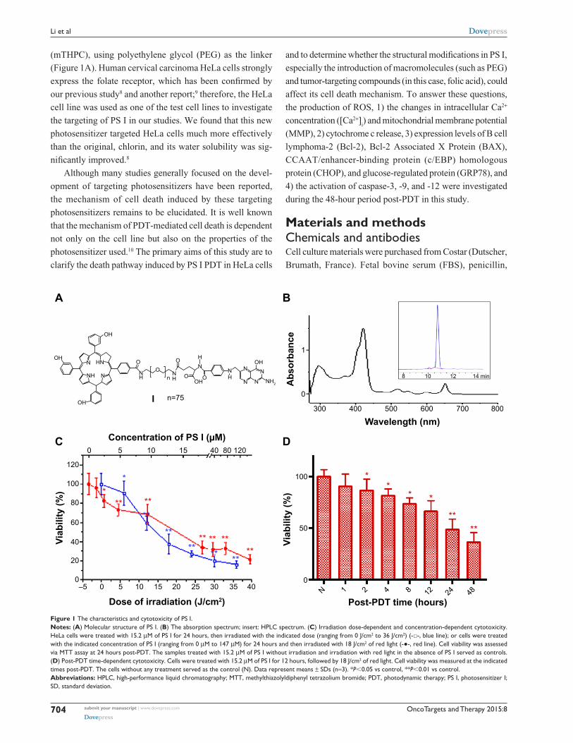
Figure 1 The characteristics and cytotoxicity of Ps i.Notes: (A) Molecular structure of Ps i. (B) The absorption spectrum; insert: hPlc spectrum. (C) irradiation dose-dependent and concentration-dependent cytotoxicity. hela cells were treated with 15.2 μM of Ps i for 24 hours, then irradiated with the indicated dose (ranging from 0 J/cm2 to 36 J/cm2) (-□-, blue line); or cells were treated with the indicated concentration of Ps i (ranging from 0 μM to 147 μM) for 24 hours and then irradiated with 18 J/cm2 of red light (-●-, red line). cell viability was assessed via MTT assay at 24 hours post-PDT. The samples treated with 15.2 μM of Ps i without irradiation and irradiation with red light in the absence of Ps i served as controls. (D) Post-PDT time-dependent cytotoxicity. cells were treated with 15.2 μM of Ps i for 12 hours, followed by 18 J/cm2 of red light. cell viability was measured at the indicated times post-PDT. The cells without any treatment served as the control (n). Data represent means ± sDs (n=3). *P,0.05 vs control, **P,0.01 vs control.Abbreviations: hPlc, high-performance liquid chromatography; MTT, methylthiazolyldiphenyl tetrazolium bromide; PDT, photodynamic therapy; Ps i, photosensitizer i; sD, standard deviation.
OncoTargets and Therapy 2015:8 submit your manuscript | www.dovepress.com
Dovepress
Dovepress
705
apoptosis of hela cells by a new targeting photosensitizer-based PDT
streptomycin, Dulbecco’s Modified Eagle’s Medium
(DMEM), and Dulbecco’s phosphate buffered saline (DPBS)
were purchased from Hyclone (South Logan, UT, USA).
Trypsin, rhodamine 123 (R123), methylthiazolyldiphenyl
tetrazolium bromide (MTT), 4′,6-diamidino-2-phenylindole
(DAPI), dimethyl sulfoxide (DMSO), and fluorescein iso-
thiocyanate (FITC)-conjugated propidium iodide (PI) were
purchased from Sigma-Aldrich. Annexin V–PI Apoptosis
Detection Kit was purchased from BD Biosciences (San
Jose, CA, USA). MitoTracker Green FM, LysoTracker
Yellow HCK-123, ER-Tracker Blue-White DPX, 2′,7′-dichlorodihydrofluorescein diacetate (H
2DCFDA), and
fluo-4-(acetoxymethyl) ester (Fluo-4-AM) were obtained
from Molecular Probes (Invitrogen, Carlsbad, CA, USA).
Antibodies against cytochrome c, caspase-3, caspase-9,
caspase-12, Bcl-2, Bax, CHOP, GRP78, and glyceraldehyde-
3-phosphate dehydrogenase were purchased from Abcam
(Cambridge, UK).
cell cultureThe human cervical carcinoma HeLa cell line was obtained
from the Chinese Academy of Sciences’ Shanghai Cell
Library, cultured in DMEM supplemented with 10% FBS,
penicillin (100 μg/mL), and streptomycin (100 μg/mL) under
5% CO2 at 37°C in a humidified incubator, and routinely
subcultured every 3 days.
PDT protocolThe novel targeting photosensitizer I (PS I), with a maximum
absorption peak at 650 nm and a purity .98% (Figure 1B),
was synthesized in our laboratory. A stock solution (1 mg/mL)
was prepared in FBS-free DMEM and stored covered in
aluminum foil at 4°C. The cultured cells were incubated with
the indicated concentrations of PS I for the indicated times
at 37°C in the dark. Subsequently, the cells were washed
with DPBS three times, and the photosensitized cells were
exposed to 600–700 nm red light generated by a KDH150B
red-light therapy instrument (Kedian Co, Beijing, China)
to activate PS I. During irradiation, the temperature never
exceeded 25°C±2°C.
cytotoxicity evaluationCell viability was measured using the MTT assay. HeLa cells
at a density of 5×10⁴ cells per well were incubated with PS I
at 37°C in detachable 96-well culture plates in the dark and
then exposed to red light. After incubation for 24 hours, the
cells were incubated in 0.5 mg/mL MTT solution for 4 hours
at 37°C, and the resulting insoluble purple formazan crystals
were dissolved in DMSO. The absorbance at 492 nm was
measured using a microplate reader model 450 (Bio-Rad,
USA). Cell viability was proportional to the A492
value and
was expressed as a percentage of the cell viability of control
cells, and the cell viability in the control group (irradiation
dose was 0 J/cm2) represented the dark cytotoxicity. For
each concentration or irradiation dose, three trials were
performed.
Detection of cytoskeletal and Dna damageHeLa cells were inoculated in 3 cm plates at a density of 1×10⁵
cells per plate. After incubation for 10 hours in 15.2 μM of
PS I, cells were stained with FITC–phalloidin and DAPI,
then irradiated with 18 J/cm2 of red light. The untreated
cells were used as control. Twelve hours after irradiation,
cells were fixed with 4% paraformaldehyde for 10 minutes
and permeabilized with 0.1% Triton X-100. After washing
with DPBS, the cells were incubated in FITC–phalloidin for
60 minutes, followed by incubation in DAPI for 10 minutes.
Samples were mounted under glass cover slides and observed
under a laser confocal microscope (TCS SP5, Leica).11
assessment of apoptosis in hela cellsHeLa cells were inoculated in 3 cm plates at a density of
5×10⁵ cells per plate. At 24 hours or the indicated time
post-PDT, HeLa cells were harvested and double-stained
with Annexin V and PI using an Annexin V–PI apoptosis
detection kit, according to the manufacturer’s instructions.
Stained cells were analyzed using a FACSCalibur flow
cytometer (BD Biosciences). At least 10,000 events were
collected for each sample.
subcellular localization of photosensitizerDuring the final 30 minutes of PS I incubation, cells were co-
loaded with ER-Tracker™ Blue-White DPX, MitoTracker®
Green FM, and LysoTracker® Yellow-HCK-123 at a final
concentration of 500 nM to label the endoplasmic reticulum
(ER), mitochondria, and lysosomes, respectively. Double-
stained cells were observed using a laser confocal microscope
(TCS SP5, Leica). Organelle probes were excited using a
488 nm argon laser (MitoTracker® and LysoTracker®) and
a 405 nm diode laser (ER-Tracker). PS I was excited using
a helium/neon laser at 488 nm.
Quantification of intracellular ROS levelsBriefly, after incubation in 15.2 μM of PS I for 12 hours, HeLa
cells were incubated in 10 μM of H2DCFDA in culture medium
OncoTargets and Therapy 2015:8submit your manuscript | www.dovepress.com
Dovepress
Dovepress
706
li et al
lacking phenol red for 10 minutes and irradiated, followed
immediately by continuous confocal scanning at an excitation
wavelength of 488 nm with a 510–530 nm bandpass barrier
filter to measure the change in fluorescence intensity.
Measurement of MMPBriefly, PS I-treated HeLa cells were loaded with 2 μM
of R123 for 30 minutes. After gentle rinsing with DPBS,
stained cells were irradiated and immediately monitored via
continuous confocal scanning using the same filter settings
as used for H2DCFDA.
Measurement of intracellular ca2+ levelsIn brief, the photosensitized cells were incubated in 2 μM of
Fluo-4-AM in Ca2+-free medium (2 mM of ethylene glycol
tetraacetic acid was added to chelate any residual Ca2+ in the
medium) for 30 minutes and irradiated. The stained cells
were immediately observed via continuous confocal scanning
using the same filter settings as used for ROS.
Western blot assayHeLa cells were lysed using radio-immunoprecipitation assay
(RIPA) buffer 24 hours after PDT. Equivalent amounts of
proteins were electrophoresed on 10% polyacrylamide gels
and transferred to polyvinylidene fluoride membrane. After
blocking with 5% skim milk for 1 hour, primary antibodies
were bound overnight at 4°C. Each membrane was probed
with horseradish peroxidase-labeled goat anti-rabbit IgG
antibody for 1.5 hours. The grayscale image of each labeled
protein band was analyzed using Quantity One software
(version 4.4.0; Bio-Rad).
statistical analysisThe data are presented as means ± standard deviation.
Student’s t-test was used for statistical analysis. The differ-
ence was considered statistically significant when P,0.05.
SPSS software was used.
ResultsPhotodynamic activity of Ps i in hela cellsThe chemical structure, absorption spectrum, and
high-performance liquid chromatography spectrum of PS I are
shown in Figure 1A and B. As shown in Figure 1C, when the
irradiation dose was zero, the viability of HeLa cells was 100%,
which indicated that the dark cytotoxicity of PS I to HeLa cells
was very low. Similarly, the irradiation alone (the concentra-
tion of PS I was zero) was noncytotoxic to HeLa cells. The
cytotoxicity induced by PS I PDT positively correlated with
the irradiation dose (Figure 1C, blue line, R=0.962; P=0.001)
and the concentration of PS I (Figure 1C, red line, R=0.763;
P=0.017). Meanwhile, cytotoxicity also occurred in a time-
dependent manner (Figure 1D). Cell viability was significantly
reduced after 2 hours (P,0.05) and 4–48 hours (P,0.01) of
incubation in 15.2 μM of PS I with PDT.
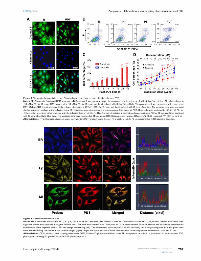
changes in F-actin and Dna structure following PDTTo investigate the PDT effects induced by PS I on the
cytoskeleton and DNA structure, cells were stained using
FITC–PI and DAPI to visualize F-actin and DNA, respec-
tively. In the control group (N), a network of F-actin filaments
extending throughout the cell and nuclei displaying a regular,
oval morphology could be observed. However, following PS
I PDT, the fluorescence originating from the labeled filaments
became weaker, and the nuclei became more circular and
smaller (Figure 2A), indicating that after PDT, the cytoskel-
eton of HeLa cells lost its F-actin structure.
apoptosis and necrosis of hela cells induced by Ps i PDTThe percentage of apoptotic and necrotic cells was quanti-
fied via Annexin V and PI double staining. Compared to the
control group, the numbers of apoptotic and necrotic cells
just slightly increased in both the irradiation-alone group and
the PS I-without-light group, but the number significantly
increased in the PTD group, especially the number of apop-
totic cells (Figure 2B). As the post-PDT time progressed,
the number of apoptotic cells clearly increased (Figure 2C).
Meanwhile, an irradiation dose-dependent (Figure 2D,
red lines) and a concentration-dependent (Figure 2D, blue
lines) apoptotic effect was observed. The quantities of both
apoptotic and necrotic cells positively correlated with the
irradiation dose (apoptosis Ra=0.966; necrosis R
n=0.989)
and the concentration of PS I (apoptosis Ra=0.996; necrosis
Rn=0.983). However, the percentage of apoptotic cells was
higher than that of necrotic cells under all conditions.
localization of Ps i in hela cellsAs displayed in Figure 3, diffuse cytoplasmic red staining
was detected, indicating that after entry into cells, PS I
localized to the cytoplasm around the nucleus of HeLa cells.
No accumulation of PS I was observed inside the nucleus.
Meanwhile, the merged stained images and fluorescence
intensity profiles revealed that the red fluorescent signal of
PS I exhibited a perfect overlap with the green fluorescent
signal of mitochondria and the blue fluorescent signal of ER,
OncoTargets and Therapy 2015:8 submit your manuscript | www.dovepress.com
Dovepress
Dovepress
707
apoptosis of hela cells by a new targeting photosensitizer-based PDT
Figure 2 changes in the cytoskeleton and Dna and apoptotic characteristics of hela cells after PDT.Notes: (A) changes in F-actin and Dna structures. (B) Results of flow cytometry analysis. N, untreated cells; Ir, only treated with 18 J/cm2 of red light; Ps, only incubated in 15.2 μM of Ps i for 12 hours; PDT, treated with 15.2 μM of Ps i for 12 hours and then irradiated with 18 J/cm2 of red light. The apoptotic cells were measured at 24 hours post-PDT. (C) Post-PDT time dependence. hela cells were incubated in 15.2 μM of Ps i for 12 hours and then irradiated with 18 J/cm2 of red light. The apoptotic cells were measured via flow cytometry analysis at the indicated times. (D) irradiation dose dependence and concentration dependence of PDT. hela cells were incubated in 15.2 μM of Ps i for 12 hours; they were then either irradiated with the indicated dose of red light (red lines) or were incubated in the indicated concentration of Ps i for 12 hours and then irradiated with 18 J/cm2 of red light (blue lines). The apoptotic cells were measured at 24 hours post-PDT. Data represent means ± sDs (n=3). *P,0.05 vs control; **P,0.01 vs control.Abbreviations: FITC, fluorescein isothiocyanate; Ir, irradiation; PDT, photodynamic therapy; PI, propidium iodide; PS I, photosensitizer I; SD, standard deviation.
Figure 3 subcellular localization of Ps i.Notes: hela cells were incubated in Ps i (15.2 μM, 24 hours) at 37°c and then Mito Tracker green FM, lysoTracker Yellow hcK-123, and er-Tracker Blue-White DPX organelle probes were included during the final 0.5 hour. The cells were washed with DPBS prior to CLSM measurement. The first, second, and third rows represent the fluorescence of the organelle probes, PS I, and merge, respectively (left). The fluorescence intensity profiles of PS I (red lines) and the organelle probe (blue and green lines) were examined along the arrows in the confocal images (right). images are representative of those obtained from three independent experiments. scale bar, 20 μm.Abbreviations: clsM, confocal laser scanning microscopy; DPBs, Dulbecco’s phosphate buffered saline; er, endoplasmic reticulum; ly, lysosomes; Mi, mitochondria; PDT, photodynamic therapy; Pi, propidium iodide; Ps i, photosensitizer i.
OncoTargets and Therapy 2015:8submit your manuscript | www.dovepress.com
Dovepress
Dovepress
708
li et al
but only a partial overlap with the yellow fluorescent signal
of lysosomes, suggesting that both mitochondria and ER
of the HeLa cells are the preferential sites of intracellular
accumulation of PS I after 24-hour incubation.
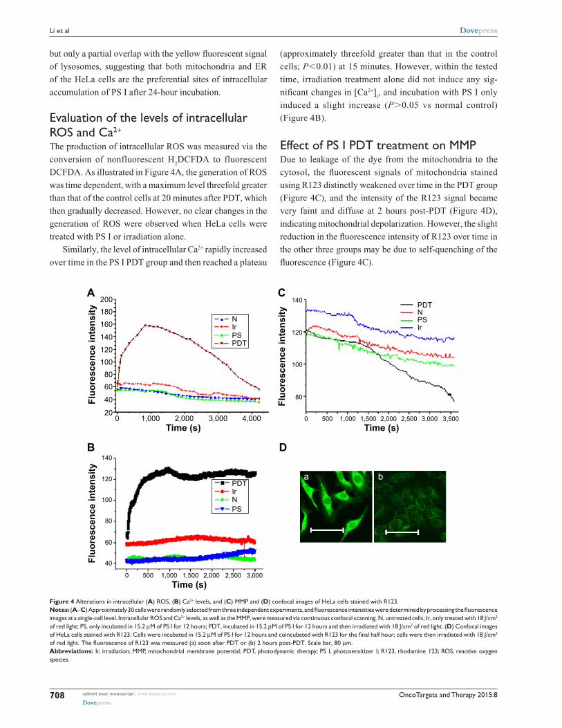
evaluation of the levels of intracellular rOs and ca2+
The production of intracellular ROS was measured via the
conversion of nonfluorescent H2DCFDA to fluorescent
DCFDA. As illustrated in Figure 4A, the generation of ROS
was time dependent, with a maximum level threefold greater
than that of the control cells at 20 minutes after PDT, which
then gradually decreased. However, no clear changes in the
generation of ROS were observed when HeLa cells were
treated with PS I or irradiation alone.
Similarly, the level of intracellular Ca2+ rapidly increased
over time in the PS I PDT group and then reached a plateau
(approximately threefold greater than that in the control
cells; P,0.01) at 15 minutes. However, within the tested
time, irradiation treatment alone did not induce any sig-
nificant changes in [Ca2+]i, and incubation with PS I only
induced a slight increase (P.0.05 vs normal control)
(Figure 4B).
effect of Ps i PDT treatment on MMPDue to leakage of the dye from the mitochondria to the
cytosol, the fluorescent signals of mitochondria stained
using R123 distinctly weakened over time in the PDT group
(Figure 4C), and the intensity of the R123 signal became
very faint and diffuse at 2 hours post-PDT (Figure 4D),
indicating mitochondrial depolarization. However, the slight
reduction in the fluorescence intensity of R123 over time in
the other three groups may be due to self-quenching of the
fluorescence (Figure 4C).
Figure 4 alterations in intracellular (A) rOs, (B) ca2+ levels, and (C) MMP and (D) confocal images of hela cells stained with r123.Notes: (A–C) Approximately 30 cells were randomly selected from three independent experiments, and fluorescence intensities were determined by processing the fluorescence images at a single-cell level. intracellular rOs and ca2+ levels, as well as the MMP, were measured via continuous confocal scanning. n, untreated cells; ir, only treated with 18 J/cm2 of red light; Ps, only incubated in 15.2 μM of Ps i for 12 hours; PDT, incubated in 15.2 μM of Ps i for 12 hours and then irradiated with 18 J/cm2 of red light. (D) confocal images of hela cells stained with r123. cells were incubated in 15.2 μM of PS I for 12 hours and coincubated with R123 for the final half hour; cells were then irradiated with 18 J/cm2 of red light. The fluorescence of R123 was measured (a) soon after PDT or (b) 2 hours post-PDT. Scale bar, 80 μm.Abbreviations: ir, irradiation; MMP, mitochondrial membrane potential; PDT, photodynamic therapy; Ps i, photosensitizer i; r123, rhodamine 123; rOs, reactive oxygen species.
OncoTargets and Therapy 2015:8 submit your manuscript | www.dovepress.com
Dovepress
Dovepress
709
apoptosis of hela cells by a new targeting photosensitizer-based PDT
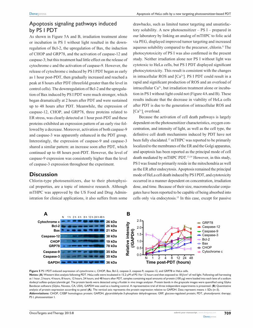
apoptosis signaling pathways induced by Ps i PDTAs shown in Figure 5A and B, irradiation treatment alone
or incubation in PS I without light resulted in the down-
regulation of Bcl-2, the upregulation of Bax, the induction
of CHOP and GRP78, and the activation of caspase-12 and
caspase-3; but this treatment had little effect on the release of
cytochrome c and the activation of caspase-9. However, the
release of cytochrome c induced by PS I PDT began as early
as 1 hour post-PDT, then gradually increased and reached a
peak at 8 hours after PDT (threefold greater than the level in
control cells). The downregulation of Bcl-2 and the upregula-
tion of Bax induced by PS I PDT were much stronger, which
began dramatically at 2 hours after PDT and were sustained
up to 48 hours after PDT. Meanwhile, the expression of
caspase-12, CHOP, and GRP78, three proteins related to
ER stress, was clearly detected at 1 hour post-PDT and these
proteins exhibited an expression pattern of an early rise fol-
lowed by a decrease. Moreover, activation of both caspase-9
and caspase-3 was apparently enhanced in the PDT group.
Interestingly, the expression of caspase-9 and caspase-3
shared a similar pattern: an increase soon after PDT, which
continued up to 48 hours post-PDT. However, the level of
caspase-9 expression was consistently higher than the level
of caspase-3 expression throughout the experiment.
DiscussionChlorin-type photosensitizers, due to their photophysi-
cal properties, are a topic of intensive research. Although
mTHPC was approved by the US Food and Drug Admin-
istration for clinical applications, it also suffers from some
drawbacks, such as limited tumor targeting and unsatisfac-
tory solubility. A new photosensitizer – PS I – prepared in
our laboratory by linking an analog of mTHPC to folic acid
via PEG, displayed improved tumor targeting and increased
aqueous solubility compared to the precursor, chlorin.8 The
photocytotoxicity of PS I was also confirmed in the present
study. Neither irradiation alone nor PS I without light was
cytotoxic to HeLa cells, but PS I PDT displayed significant
photocytotoxicity. This result is consistent with the changes
in intracellular ROS and [Ca2+]. PS I PDT could result in a
rapid and significant production of ROS and an overload of
intracellular Ca2+, but irradiation treatment alone or incuba-
tion in PS I without light could not (Figure 4A and B). These
results indicate that the decrease in viability of HeLa cells
after PDT is due to the generation of intracellular ROS and
[Ca2+]i overload.
Because the activation of cell death pathways is largely
dependent on the photosensitizer characteristics, oxygen con-
centration, and intensity of light, as well as the cell type, the
definitive cell death mechanisms induced by PDT have not
been fully elucidated.12 mTHPC was reported to be primarily
localized to the membranes of the ER and the Golgi apparatus,
and apoptosis has been reported as the principal mode of cell
death mediated by mTHPC PDT.13,14 However, in this study,
PS I was found to primarily reside in the mitochondria as well
as the ER after endocytosis. Apoptosis remained the principal
mode of HeLa cell death induced by PS I PDT, and cytotoxicity
occurred in a manner dependent on concentration, irradiation
dose, and time. Because of their size, macromolecular conju-
gates have been reported to be capable of being absorbed into
cells only via endocytosis.15 In this case, except for passive
Figure 5 Ps i PDT-induced expression of cytochrome c, chOP, Bax, Bcl-2, caspase-3, caspase-9, caspase-12, and grP78 in hela cells.Notes: (A) Western blot analysis following PDT. hela cells were incubated in 15.2 μM of Ps i for 12 hours and then exposed to 18 J/cm2 of red light. Following cell harvesting at 1 hour, 2 hours, 4 hours, 8 hours, 12 hours, 24 hours, and 48 hours after PDT, samples containing equal amounts of protein (100 μg) were loaded into each lane of a sodium dodecyl sulfate–polyacrylamide gel. The protein bands were detected using a Kodak in vivo image analyzer. Protein bands in the grayscale images were quantified using Glyko Bandscan software (glyko, novato, ca, Usa). gaPDh was used as a loading control. a representative trial of three independent experiments is presented. (B) Quantitative analysis of protein expression according to panel (A). The vertical axis represents the protein expression relative to gaPDh. Data represent means ± sDs (n=3).Abbreviations: chOP, c/eBP homologous protein; gaPDh, glyceraldehyde-3-phosphate dehydrogenase; grP, glucose-regulated protein; PDT, photodynamic therapy; Ps i, photosensitizer i.
OncoTargets and Therapy 2015:8submit your manuscript | www.dovepress.com
Dovepress
Dovepress
710
li et al
endocytosis, there was likely active endocytosis mediated
by the folate receptor.8,16 It is reasonable that the lipophilic
photosensitizer tends to accumulate in membrane organelles
such as mitochondria or lysosomes. On the other hand, other
reports indicate that the aggregation state of intracellular pho-
tosensitizers affects their localization, with more aggregation-
prone species tending toward lysosomal localization.17 Due to
the amphiphilicity of the PEG linker, a clear decrease in the
aggregation of PS I has been observed previously.8 Therefore,
we hypothesize that PEGylation leads PS I to localize more at
the mitochondria and the ER than at the lysosomes.
The fact that PS I was primarily localized at the mito-
chondria and the ER prompted us to further examine
mitochondrial and ER stress-associated apoptosis. There is
increasing evidence that mitochondria play a critical role
in the apoptotic cascade, and the release of cytochrome c
from the mitochondria into the cytosol is a hallmark of cells
undergoing apoptosis.18 In the present study, a rapid loss of
MMP was observed after PDT, which was followed by the
time-dependent release of cytochrome c. The opening of
mitochondrial membrane permeability transition pores, which
results in the dissipation of the MMP, has been proposed as the
principal mechanism for the release of cytochrome c.19 Accu-
mulating evidence suggests that Bcl-2, residing in the mito-
chondrial outer membrane and the ER membrane, performs
the unique functional role of antiapoptosis via stabilization of
the mitochondrial membrane.20,21 PS I PDT induced an almost
twofold and time-dependent downregulation in Bcl-2 protein
expression and a marked increase in the expression of Bax,
a proapoptotic protein. During apoptosis, Bax oligomerizes
in the mitochondrial outer membrane and likely disrupts its
integrity, freeing proapoptotic proteins such as cytochrome c,
which allows for activation of caspase-9.22
Otherwise, intracellular Ca2+ overload attracted our atten-
tion. Intracellular Ca2+ overload may be due to the influx of
extracellular Ca2+ or the release of Ca2+ from different intra-
cellular stores (eg, the ER, the primary intracellular Ca2+
store) following photoinduced injury.9 ER stress-induced
apoptotic cell death represents a novel mitochondrial-inde-
pendent intrinsic apoptotic pathway.23,24 To specify the effect
of ER stress on HeLa cell apoptosis induced by PS I PDT,
the expression levels of caspase-12, CHOP, and GRP78 were
monitored via Western blot analysis. Caspase-12 is an impor-
tant caspase involved in ER-induced apoptosis.25 CHOP is
an ER-stress-inducible transcription factor and is considered
a major trigger of ER-stress-induced apoptosis by reducing
the expression of Bcl-2.26 GRP78 is an ER chaperone and a
key regulator of ER stress response signaling27 In our study,
the activation of caspase-12 and the induction of CHOP
and GRP78 shared the same response pattern, namely, an
early increase followed by a decrease, as oxidative stress is
induced by PS I PDT. Additionally, as CHOP was induced,
the level of Bcl-2 expression was reduced following PDT
(Figure 5A and B). All of these results probably reflect
the initiation of the ER-stress-specific caspase cascade of
apoptosis. Interestingly, although both irradiation treatment
alone and incubation in PS I without light could not induce
the generation of ROS or [Ca2+]i overload, they induced the
ER stress response to some extent through the activation of
caspase-12 and the induction of CHOP and GRP78.
Activation of both caspase-9 and caspase-3 were observed
soon after PDT, suggesting that PS I PDT may induce apop-
totic cell death via a caspase-dependent pathway. However,
it is worth noting that the level of caspase-9 expression
was consistently higher than that of caspase-3 throughout
our experiments (Figure 5A and B). It is well known that
caspase-9 is an initiator caspase of apoptosis and can be
activated by mitochondrial events and ER stress,28 but
caspase-3 is a downstream substrate of caspase-9 and a key
player in the execution phase of apoptosis. Thus, the activa-
tion of caspase-9 should induce the activation of caspase-3.
Therefore, we hypothesize that the difference in expression
of caspase-9 and caspase-3 could be due to the induction of
GRP78, as some reports have indicated that overexpression
of GRP78 could reduce apoptosis and mitigate cytosolic Ca2+
overload induced by ER stressors in some cell types.29
ConclusionIn summary, after active endocytosis mediated by the folate
receptor, PS I is principally localized at the mitochondria
and the ER of HeLa cells. Apoptosis is the primarily pre-
ferred mechanism of HeLa cell death in our system. The rapid
generation of ROS in HeLa cells following PDT provoked
mitochondrial damage and ER stress, which, in turn, acti-
vated the intrinsic apoptotic pathway. Specifically, compared
to mTHPC, after the structural modifications reported by
our research group, the mitochondria became the primary
site of PS I localization and the targeting of PS I for folate
receptor-positive cells was obviously improved; however,
the cell death mode induced by PS I PDT was similar to that
induced by mTHPC PDT.
AcknowledgmentWe are grateful for the financial support from the National
Natural Science Foundation of China (project number
21072227).
OncoTargets and Therapy
Publish your work in this journal
Submit your manuscript here: http://www.dovepress.com/oncotargets-and-therapy-journal
OncoTargets and Therapy is an international, peer-reviewed, open access journal focusing on the pathological basis of all cancers, potential targets for therapy and treatment protocols employed to improve the management of cancer patients. The journal also focuses on the impact of management programs and new therapeutic agents and protocols on
patient perspectives such as quality of life, adherence and satisfaction. The manuscript management system is completely online and includes a very quick and fair peer-review system, which is all easy to use. Visit http://www.dovepress.com/testimonials.php to read real quotes from published authors.
OncoTargets and Therapy 2015:8 submit your manuscript | www.dovepress.com
Dovepress
Dovepress
Dovepress
711
apoptosis of hela cells by a new targeting photosensitizer-based PDT
DisclosureThe authors report no conflicts of interest in this work.
References 1. Song J, Chen Q, Xing D. Enhanced apoptotic effects by downregulating
Mcl-1: evidence for the improvement of photodynamic therapy with celecoxib. Exp Cell Res. 2013;19:1491–1504.
2. Gijsens A, Missiaen L, Merlevede W, de Witte P. Epidermal growth factor-mediated targeting of chlorin e6 selectively potentiates its pho-todynamic activity. Cancer Res. 2000;60:2197–2202.
3. St Denis TG, Hamblin MR. Synthesis, bioanalysis and biodistribution of photosensitizer conjugates for photodynamic therapy. Bioanalysis. 2013;5:1099–1114.
4. Stefflova K, Li H, Chen J, Zheng G. Peptide-based pharmacomodulation of a cancer-targeted optical imaging and photodynamic therapy agent. Bioconjug Chem. 2007;18:379–388.
5. Hu Z, Rao B, Chen S, Duanmu J. Targeting tissue factor on tumor cells and angiogenic vascular endothelial cells by factor VII-targeted verteporfin photodynamic therapy for breast cancer in vitro and in vivo in mice. BMC Cancer. 2010;10:235–248.
6. McCarthy JR, Bhaumik J, Merbouh N, Weissleder R. High-yielding syntheses of hydrophilic, conjugatable chlorins and bacteriochlorins. Org Biomol Chem. 2009;7:3430–3436.
7. Vlahov IR, Leamon CP. Engineering folate-drug conjugates to target cancer: from chemistry to clinic. Bioconjug Chem. 2012;23: 1357–1369.
8. Li D, Li P, Lin H, Jiang Z, Guo L, Li B. A novel chlorin-PEG-folate conjugate with higher water solubility, lower cytotoxicity, better tumor targeting and photodynamic activity. J Photochem Photobiol B. 2013; 127:28–37.
9. Zhang Q, Xiang G, Zhang Y, et al. Increase of doxorubicin sensi-tivity for folate receptor positive cells when given as the prodrug N-(phenylacetyl)doxorubicin in combination with folate-conjugated PGA. J Pharm Sci. 2006;95:2266–2275.
10. Buytaert E, Dewaele M, Agostinis P. Molecular effectors of multiple cell death pathways initiated by photodynamic therapy. Biochim Biophys Acta. 2007;1776:86–107.
11. Szurko A, Rams M, Sochanik A. Spectroscopic and biological studies of a novel synthetic chlorin derivative with prospects for use in PDT. Bioorg Med Chem. 2009;17:8197–8205.
12. Robertson CA, Evans DH, Abrahamse H. Photodynamic therapy (PDT): a short review on cellular mechanisms and cancer research applications for PDT. J Photochem Photobiol B. 2009;96:1–8.
13. Teiten MH, Bezdetnaya L, Morlière P, Santus R, Guillemin F. Endoplas-mic reticulum and Golgi apparatus are the preferential sites of Foscan1 localization in cultured tumor cells. Br J Cancer. 2003;88:146–152.
14. Senge MO, Brandt JC. Temoporfin (Foscan, 5,1015,20-Tetra (m-hydroxyphenyl)chlorin) – a second-generation photosensitizer. Photochem Photobiol. 2011;87:1240–1296.
15. Duncan R, Spreafico F. Polymer conjugates. Pharmacokinetic consid-erations for design and development. Clin Pharmacokinet. 1994;27: 290–306.
16. Low PS, Kularatne SA. Folate-targeted therapeutic and imaging agents for cancer. Curr Opin Chem Biol. 2009;13:256–262.
17. Hamblin MR, Miller JL, Rizvi I, Ortel B, Maytin EV, Hasan T. Pegy-lation of a chlorine6 polymer conjugate increases tumor targeting of photosensitizer. Cancer Res. 2001;61:7155–7162.
18. Zamzami N, Kroemer G. Apoptosis: mitochondrial membrane permeabilization – the whole story? Curr Biol. 2003;13:R71–R73.
19. Mroz P, Yaroslavsky A, Kharkwal GB, Hamblin MR. Cell death pathways in photodynamic therapy of cancer. Cancer. 2011;3:2516–2539.
20. He GF, Bian ML, Zhao YW, Xiang Q, Li HY, Xiao C. A study on the mechanism of 5-aminolevulinic acid photodynamic therapy in vitro and in vivo in cervical cancer. Oncol Rep. 2009;21:861–868.
21. Chen X, Zhao P, Chen F, Li L, Luo R. Effect and mechanism of 5-aminolevulinic acid-mediated photodynamic therapy in esophageal cancer. Lasers Med Sci. 2011;26:69–78.
22. Panzarini E, Inguscio V, Dini L. Timing the multiple cell death pathways initiated by rose Bengal acetate photodynamic therapy. Cell Death Dis. 2011;2:e169.
23. Zhou Z, Yang H, Zhang Z. Role of calcium in phototoxicity of 2-butylamino-2-demethoxy-hypocrellin A to human gastric cancer MGC-803 cells. Biochim Biophys Acta. 2003;1593:191–200.
24. Rao RV, Castro-Obregon S, Frankowski H, et al. Coupling endoplasmic reticulum stress to the cell death program. An Apaf-1-independent intrinsic pathway. J Biol Chem. 2002;277:21836–21842.
25. Chung PS, He P, Shin JI, Hwang HJ, Lee SJ, Ahn JC. Photodynamic therapy with 9-hydroxypheophorbide α on AMC-HN-3 human head and neck cancer cells. Cancer Biol Ther. 2009;8:1343–1351.
26. Szegezdi E, Logue SE, Gorman AM, Samali A. Mediators of endo-plasmic reticulum stress-induced apoptosis. EMBO Rep. 2006;7: 880–885.
27. Firczuk M, Gabrysiak M, Barankiewicz J, et al. GRP78-targeting sub-tilase cytotoxin sensitizes cancer cells to photodynamic therapy. Cell Death Dis. 2013;4:e741.
28. Morishima N, Nakanishi K, Takenouchi H, Shibata T, Yasuhiko Y. An endoplasmic reticulum stress-specific caspase cascade in apoptosis. Chtochrome c-independent activation of caspase-9 bycaspase-12. J Bio Chem. 2002;277:34287–34294.
29. He P, Ahn JC, Shin JI, Chung PS. Photoactivation of 9-hydroxypheo-phorbide α triggers apoptosis through the reactive oxygen species-mediated mitochondrial pathway and endoplasmic reticulum stress in AMC-HN-3 laryngeal cancer cells. Int J Oncol. 2010;36:801–808.


