Embed Size (px)
Citation preview

Benefits and Harms of Treatment Options for Esophageal Adenocarcinoma and
Precancerous Conditions: An Overview of Systematic Reviews
June 2019
Ottawa Evidence Review and Synthesis Centre (ERSC)
ERSC Principal Investigators: David Moher, Julian Little
ESRC Scientific Lead: Adrienne Stevens
Project oversight: Candyce Hamel
ERSC Project Staff: Nadera Ahmadzai, Micere Thuku, Kusala Pussegoda, Andrew Beck, Becky
Skidmore Knowledge Synthesis Group, Ottawa Methods Centre
Ottawa Hospital Research Institute; School of Epidemiology and Public Health, University of Ottawa
Ottawa, Ontario, Canada
ERSC Clinical Experts & Methodological Collaborators
Avijit Chatterjee, Lorenzo Ferri, Brian Hutton, Donna Maziak, Beverley Shea
CTFPHC Working Group Chair
Stéphane Groulx
CTFPHC Working Group Members
Scott Klarenbach, Harminder Singh, Brett Thombs, Brenda Wilson
CTFPHC Working Group External Clinical Experts
Paul James Belletrutti, Laura Targownik
PHAC Global Health and Guidelines Division
Marion Doull, Heather Limburg
Suggested Citation: Ahmadzai N, Hamel C, Thuku M, Pussegoda K, Beck A, Skidmore B,
Maziak D, Chatterjee A, Ferri L, Hutton B, Shea B, Little J, Stevens A. 2019. Benefits and
Harms of Treatment Options for Esophageal Adenocarcinoma and Precancerous Conditions: An
Overview of Systematic Reviews. Evidence Review Synthesis Centre: Ottawa Hospital Research
Institute, Ottawa, Ontario.

Protocol Registration
PROSPERO CRD42018084825
Corresponding Author: Nadera Ahmadzai
Author Contribution
NA, CH, AB, KP, and MT participated in screening, and data extraction/verification. NA, CH,
AS, and AB drafted the report. BSk developed the search strategy and provided text for the review.
All authors (NA, CH, MT, KP, AB, BSk, DM, AC, LF, BH, BS, JL, AS) critically reviewed the
overview and provided methodological or clinical expertise.
Acknowledgements
We would like to acknowledge the contribution of Raymond Daniel, who managed the citations.
We would also like to acknowledge the Public Health Agency of Canada Science Lead (Heather
Limburg), the Canadian Task Force for Preventive Health Care members and the external clinical
experts, Paul James Belletrutti, and Laura Targownik who critically reviewed the report and
provided clinical expertise.
Declaration of Funding
Funding for this systematic review was provided by the Public Health Agency of Canada, with
funds distributed by the Nova Scotia Health Research Foundation. This funding supported the
development of the protocol, execution of search strategies, collection of the data, data
management, analyses, and writing of the systematic review technical report.
Role of Funder
The funder provided feedback on the protocol and draft overview, but was not involved in the
study selection, data extraction, or analysis and will not be involved in a decision to seek
publication.

Contents
Abstract ........................................................................................................................................... 1
Abbreviations/Glossary................................................................................................................... 3
1 Introduction .............................................................................................................................. 4
1.1 Objective ........................................................................................................................... 4
1.2 Background ....................................................................................................................... 4
2. Methods................................................................................................................................... 6
2.1 Key question ..................................................................................................................... 7
2.2 Inclusion and exclusion criteria ........................................................................................ 7
2.4 Literature search................................................................................................................ 8
2.5 Study selection .................................................................................................................. 9
2.6 Data extraction and management ...................................................................................... 9
2.7 Quality assessment of reviews .......................................................................................... 9
2.8 Analysis........................................................................................................................... 10
2.9 Rating the certainty of the evidence................................................................................ 10
2.10 Changes from the protocol ............................................................................................ 12
3 Results .................................................................................................................................... 12
3.1 Summary of the literature search .................................................................................... 12
3.2 Results ............................................................................................................................. 13
Outcomes ...................................................................................................................................... 16
1 Pharmacological therapy vs Placebo ..................................................................................... 17
1.1 Celecoxib vs Placebo ...................................................................................................... 17
2 Pharmacological Therapies vs Pharmacological Therapies ................................................... 17
2.1 Omeprazole vs Histamine Type 2 Receptor Antagonists ............................................... 17
3 Chemical ablative technique combined with pharmacological therapy vs Pharmacological
therapy alone ............................................................................................................................. 18
3.1 Photodynamic Therapy (PDT) + Omeprazole vs Omeprazole alone ............................. 18
4 Surgery combined with + thermal ablative techniques vs Surgery combined with
surveillance ............................................................................................................................... 20
4.1 Anti-reflux surgery (Nissen fundoplication) + Argon plasma coagulation (APC) vs Anti-
reflux surgery (Nissen fundoplication) + Surveillance (endoscopic) ................................... 20
5 Thermal Ablative Techniques combined with Pharmacological Therapy vs Pharmacological
Therapy ..................................................................................................................................... 21
5.1 Radiofrequency ablation (RFA) + Proton Pump Inhibitor (PPI) vs PPI alone ............... 21
6 Surgery vs Pharmacological Therapies .................................................................................. 22

6.1 Anti-reflux surgery (Nissen Fundoplication) vs H2 receptor agonist / Omeprazole ...... 22
7 Chemical ablative techniques with different treatment parameters ....................................... 23
7.1 PDT with 5-aminolevulinic acid (ALA-5) vs PDT with porfimer sodium ..................... 23
7.2 Photodynamic therapy with different treatment parameters ........................................... 23
8 Thermal Ablative Technique vs Surveillance (endoscopic) .................................................. 24
8.1 Radiofrequency ablation (RFA) vs surveillance (endoscopic) ....................................... 24
9 Thermal Ablative Technique + Pharmacological Therapy vs Thermal Ablative Technique +
Pharmacological Therapy ......................................................................................................... 25
9.1 Argon Plasma Coagulation (APC) + Proton Pump Inhibitor (PPI) vs Multipolar
Electrocoagulation (MPEC) + Proton Pump Inhibitor (PPI) ................................................ 25
9.2 Multipolar Electrocoagulation (MPEC) vs Argon Plasma Coagulation (APC) ............. 26
10 Thermal Ablative Technique vs Chemical Ablative Technique + Pharmacological Therapy
................................................................................................................................................... 26
10.1 Photodynamic Therapy (PTD) vs Argon Plasma Coagulation (APC) + Proton Pump
Inhibitor (PPI) ....................................................................................................................... 26
11 Mechanical ablative technique vs Thermal ablative technique ........................................... 29
11.1 Endoscopic mucosal resection (EMR) vs Radiofrequency ablation (RFA) ................. 29
4. Discussion ............................................................................................................................. 30
Summary of Main Results and Quality of the Evidence Ratings ......................................... 30
Evidence Considerations and Future Research ..................................................................... 31
Conclusions ........................................................................................................................... 34
References ..................................................................................................................................... 35
Table 1. Characteristics of Included Systematic Reviews ............................................................ 46
Table 2. Outcomes and comparisons per systematic review (and primary study) ........................ 49
Figure 2. Primary studies and conditions overlap among the systematic reviews........................ 51
Figure 3. Map of Systematic Reviews and Primary RCTs ........................................................... 52
Evidence Set 1: Pharmacological therapy vs Placebo .................................................................. 53
Evidence Set 1.1 Celecoxib vs Placebo: Results table ............................................................. 53
Evidence Set 1.1 Celecoxib vs Placebo: GRADE domains table ............................................. 54
Evidence Set 2: Pharmacological therapy vs Pharmacological therapy ....................................... 55
Evidence Set 2.1 Omeprazole vs H2RA: Results table ............................................................ 55
Evidence Set 2.1 Omeprazole vs H2RA: GRADE domains table ............................................ 56
Evidence Set 3: Chemical ablative technique combined with pharmacological therapy vs
Pharmacological therapy alone ..................................................................................................... 57
Evidence Set 3.1 PDT + Omeprazole vs Omeprazole alone: Results table .............................. 57
Evidence Set 3.1 PDT + Omeprazole vs Omeprazole alone: GRADE domains table ............. 60

Evidence Set 4: Surgery combined with thermal ablative technique vs Surgery combined with
surveillance ................................................................................................................................... 64
Evidence Set 4.1 Anti-reflux surgery (Nissen Fundoplication) + APC vs Anti-reflux surgery
(Nissen Fundoplication) + Surveillance (endoscopic): Results table ....................................... 64
Evidence Set 4.1 Anti-reflux surgery (Nissen Fundoplication) + APC vs Anti-reflux surgery
(Nissen Fundoplication) + Surveillance (endoscopic): GRADE domains table ....................... 66
Evidence Set 5: Thermal ablative techniques combined with pharmacological therapy vs
Pharmacological therapy ............................................................................................................... 68
Evidence Set 5.1 RFA + PPI vs PPI: Results table ................................................................... 68
Evidence Set 5.1 RFA + PPI vs PPI: GRADE domains table .................................................. 71
Evidence Set 6: Surgery vs Pharmacological therapy .................................................................. 74
Evidence Set 6.1 Anti-reflux surgery vs H2 receptor antagonist/Omeprazole: Results table .. 74
Evidence Set 6.1 Anti-reflux surgery vs H2 receptor antagonist/Omeprazole: GRADE
domains table ............................................................................................................................ 76
Evidence Set 7: Chemical ablative technique vs Chemical ablative technique ............................ 78
Evidence Set 7.1 PDT (5-ALA) vs PDT (Photofrin): Results table ......................................... 78
Evidence Set 7.1 PDT (5-ALA) vs PDT (Photofrin): GRADE domains table ......................... 79
Evidence Set 7.2 PDT with different treatment parameters: Results table ............................... 80
Evidence Set 7.2 PDT with different treatment parameters: GRADE domains table .............. 81
Evidence Set 8: Thermal ablative technique vs Surveillance ....................................................... 82
Evidence Set 8.1 RFA vs Surveillance (endoscopic): Results table ......................................... 82
Cumulative disease progression rates to EAC reported ............................................................ 82
Evidence Set 8.1 RFA vs Surveillance (endoscopic): GRADE domains table ........................ 84
Evidence Set 9: Thermal ablative technique combined with pharmacological therapy vs Thermal
ablative technique combined with pharmacological therapy ........................................................ 86
Evidence Set 9.1 APC + PPI vs MPEC + PPI: Results table.................................................... 86
Evidence Set 9.1 APC + PPI vs MPEC +PPI: GRADE domains table .................................... 87
Evidence Set 9.2 MPEC + PPI vs APC + PPI: Results table .................................................... 88
Evidence Set 9.2 MPEC vs APC: GRADE domains table ....................................................... 89
Evidence Set 10: Thermal ablative technique vs Chemical ablative technique combined with
pharmacological therapy ............................................................................................................... 90
Evidence Set 10.1 PDT vs APC + PPI: results table ................................................................ 90
Evidence Set 10.1 PDT vs APC + PPI: GRADE domains table .............................................. 94
Evidence Set 11: Mechanical ablative technique vs Thermal ablative technique ........................ 98
Evidence Set 11.1 EMR vs RFA............................................................................................... 98
Evidence Set 11.1 EMR vs RFA: GRADE domains table ..................................................... 101

Supplementary Tables ................................................................................................................. 104
Supplementary Table 1. AMSTAR ratings for included systematic reviews ......................... 105
Supplementary Table 2. Risk of bias/Methodological Assessments of Primary Studies ....... 107
Appendices .................................................................................................................................. 114
Appendix 1. PICOS table ........................................................................................................ 115
Appendix 2. List of treatment options .................................................................................... 117
Appendix 3. PRESS ................................................................................................................ 118
Appendix 4. Search strategies ................................................................................................. 121
Appendix 5. Screening forms ................................................................................................. 124
Title and abstract screening form ........................................................................................ 124
Full-text screening form ...................................................................................................... 125
Appendix 6. AMSTAR checklist ............................................................................................ 127
Appendix 7. List of excluded reviews at full text ................................................................... 129
Appendix 8. List of potentially relevant trials ........................................................................ 190
Appendix 9. Characteristics of primary studies in included reviews...................................... 191
Appendix 10. Evaluation of overlap of studies and concordance of results among reviews.. 194

1
Abstract
Background: Esophageal adenocarcinoma (EAC) is the most common type of esophageal
cancer in Canada. It usually develops in the lower third of the esophagus, in the area where
Barrett’s esophagus (BE) occurs. Incidence rates of esophageal cancer has doubled in both men
(1.8 to 3.5 per 100,000) and women (0.2 to 0.5 per 100,000) from 1986 to 2006 respectively,
with an average annual increase of 3.9% and 3.6%, respectively. Five-year survival of EAC is
low among both men and women, with a rate of 14%, mostly due to late-stage diagnosis, where
cancer has metastasized or spread to other organs. Those diagnosed early with asymptomatic
EAC have better survival than those diagnosed with symptomatic disease, over 50% of whom
will require palliative measures at diagnosis. The prevention of EAC via screening, early
diagnosis, and treatment of precancerous conditions such as BE, and low- and high-grade
dysplasia, if effective, would offer a strategy for reducing mortality and improving long term
survival and quality of life of those affected.
Objective: The aim of this project was to examine the evidence on the treatment options for
stage 1 EAC and precancerous conditions (BE and/or dysplasia), using an overview of reviews
approach. The results from this overview will be used to inform the Canadian Task Force on
Preventive Health Care (CTFPHC) during their development of guideline recommendations on
screening for EAC.
Methods: A protocol for this review was registered with PROSPERO (CRD42018084825). A
detailed search of MEDLINE, Embase and the Cochrane Library from inception to October 2018
was carried out by an experienced information specialist and peer reviewed by another senior
information specialist. Grey literature was also searched. Quality assessment was performed with
AMSTAR, with additional guidance using AMSTAR 2. Full-text screening and quality
assessment were carried out independently by two reviewers, and disagreements were resolved
through discussion or third-party adjudication. Data extraction, risk of bias (using the Cochrane
risk of bias tool), and evaluation of the certainty of the body of evidence (using the GRADE
domains as a guide) was performed by one reviewer and verification was carried out by a second
senior reviewer.
Results: After removing duplicates, 3,761 bibliographic records were screened on title and
abstract. Of these, 2,754 were excluded. Among 1,007 articles screened based on full-text, 995
records were excluded, leaving eleven included systematic reviews (SRs). Of these, five were in
adults with BE with or without dysplasia, three in BE patients with high-grade dysplasia and
intramucosal cancer, and two in BE patients with low-grade dysplasia. The SRs were published
between 2008 and 2018 and included 25 articles reporting results of randomized control trials
published between 1996 and 2014. Trials included from nine to 208 participants and most
included fewer than 100. There was overlap of primary studies across included reviews. The risk
of bias of the primary trials were assessed with various tools (e.g., Cochrane risk of bias, Jadad,
Downs and Black, Critical Appraisal Skills Programme checklist) and rated as unclear or high
risk of bias. The AMSTAR rating was low for two reviews and critically low for the remaining
nine SRs. The quality of evidence was low or very low for most outcomes. The findings were
based on a few trials with small sample sizes, and most outcomes were based on a single study.

2
Survival, quality of life, psychological effects, and overtreatment were not reported in any of the
SRs.
Limitations: There was no limitation on language in the search, however, only English and
French language reviews were considered for inclusion. Few analyses were discordant within
and across reviews. The methodological issues pertaining to the quality of the source reviews
was another major limitation of this overview. Poor data presentation, and incomplete reporting
and description of primary source data within and between the reviews was another limitation.
Reduction and regression were reported differently, which made it difficult to combine and
compare outcomes across studies and reviews.
Conclusions: Many treatment modalities for BE have been evaluated, but there are few small
studies for each and most had low or very low quality of evidence. Due to several limitations,
including the low or critically low quality of the reviews themselves there is uncertainty in
understanding the effectiveness of these treatments. Large multicentre trials with longer follow-
up are needed.

3
Abbreviations/Glossary
AMSTAR A MeaSurement Tool to Assess Systematic Reviews
APC Argon Plasma Coagulation
ARD
BE
Absolute Risk Difference
Barrett’s Esophagus
CI Confidence Interval
COMET Core Outcome Measures in Effectiveness Trials
CTFPHC Canadian Task Force for Preventive Health Care
CADTH Canadian Agency for Drugs and Technologies in Health
DSR Distiller Systematic Review
EAC Esophageal adenocarcinoma
EMR Endoscopic mucosal resection
ESCC Esophageal squamous cell carcinoma
GERD Gastroesophageal reflux disease
GRADE Grading of Recommendations, Assessment, Development and Evaluation
HGD High-grade dysplasia
H2RA Histamine Type 2 Receptor Antagonists
LGD Low-grade dysplasia
MD Mean Difference
MPEC Multipolar Electrocoagulation
NR Not Reported
OR Odds Ratio
PICOS Population, Interventions, Comparisons, Outcomes, Study design
PPI Proton Pump Inhibitor
PRESS Peer Review of Electronic Search Strategies
PRISMA Preferred Reporting Items for Systematic Reviews and Meta-Analyses
PTD Photodynamic Therapy
RCT Randomized Controlled Trial
RFA Radiofrequency Ablation
ROB Risk of Bias
RR Risk Ratio
SD Standard Deviation
SR Systematic Review

4
1 Introduction
1.1 Objective
The Canadian Task Force for Preventive Health Care (CTFPHC) is undertaking a series of
systematic evaluations of the evidence to inform the development of a clinical practice guideline
regarding the effectiveness of screening adults for esophageal adenocarcinoma (EAC) and
associated precancerous lesions (Barrett’s Esophagus (BE) and dysplasia).
Two systematic reviews (not yet published) (protocols1,2 available at https://osf.io/ty926 and
https://osf.io/pzyej) were undertaken by the Ottawa Research and Synthesis Center. They have
synthesized the evidence on the benefits and harms of screening, as well as the patient
preferences and values in relation to screening. However, sufficient direct evidence on the
effectiveness of screening was not identified. Consequently, the present project on treatment
effectiveness will be used as linked evidence to inform the CTFPHC guideline recommendation
on EAC screening.
The purpose of this project is to examine the evidence on the treatment options for stage 1 EAC
and precancerous conditions (BE and/or dysplasia), using an overview of reviews approach.
1.2 Background
Prevalence and burden
There are two main types of esophageal cancer, EAC where malignant cells form in the tissues
of the lower third of the esophagus, primarily in glandular cells where BE also develops3, and
esophageal squamous cell carcinoma (ESCC) where malignant cells form in the squamous cells
of the esophagus. Globally, there were approximately 52,000 cases of EAC in 2012.4 Nearly
50% of EAC cases occurred in North America and northwestern Europe.5 From 1986-2006, EAC
incidence in males increased by 3.9% (1.8 to 3.5 per 100,000) and 3.6% in females (0.2 to 0.5
per 100,000) per year in Canada.5 About 20% of EAC cases are diagnosed at an early stage.6
Treatment with surgery at early stage leads to a five-year survival rate of 90%.6 The overall very
low five-year survival rate of EAC (14%) maybe attributable to a higher late stage diagnosis
(39.9% at stage IV).7 Increases in incidence of EAC may be dependent on the increasing
prevalence of related risk factors such as obesity and gastroesophageal reflux disease (GERD).5
Other risk factors for the development of EAC are BE, male sex, age older than 50 years, white
ethnicity, current or past smoking history, and a family history of BE or EAC.3,5,8,9
Although GERD and BE are two risk factors for EAC, not every person diagnosed with EAC
will have experienced GERD or have been diagnosed with BE. Approximately 10% of people
with GERD will develop BE10,11 and there is some evidence of progression from GERD to BE,
to low-/high-grade dysplasia, and to EAC. The annual incidence of EAC among BE patients has
been reported to range between 0.3-0.6%.12
BE is the most critical precancerous condition for EAC. In BE, the tissue lining the esophagus
transforms into tissue resembling the lining of the intestines. Generally, this transformation is
called intestinal metaplasia, and in the esophagus, it is called BE. It is currently not known how
the transformation occurs; however, it has been suggested that the acid regurgitation associated

5
with GERD may assist changes at the cellular level.13 One Canadian study reported the
prevalence of BE at 2.4% among primary care patients experiencing dyspepsia.14 The prevalence
of BE among those who undergo an esophagogastroduodenoscopy (EGD) (also known as upper
GI endoscopy) for any reason is between 1% and 2%, and between 5% and 15% among those
who receive an EGD for symptoms of GERD.15 Most patients with non-dysplastic BE or only
low-grade dysplasia (cellular change) will not develop cancer. However, incidence of carcinoma
has been reported as high as 1 in 52 patient-years, corresponding to 1,920 carcinomas per
100,000 BE patients, compared to annual incidence of 15 esophageal cancers in the general
population (whether dysplasia was present was not indicated).16,17 It has also been found that the
longer the length of BE (e.g., short segment vs. long segment) the higher the risk for EAC.18
Treatment
The goal of treatment for BE and/or low- or high-grade dysplasia is to slow or halt GERD
symptoms, reduce mucosal inflammation, control dysplasia, and prevent progression to
adenocarcinoma.10 The treatments for EAC depend on the stage of the disorder (0 to 4). For stage
0, the disease is considered precancerous and is synonymous with high-grade dysplasia.
Endoscopic therapies [e.g., radiofrequency ablation (RFA) or endoscopic mucosal resection
(EMR)] are typically performed, followed by endoscopic surveillance.19 For stage 1, the disease
is generally treated with mechanical methods to remove tissue (e.g., endoscopic mucosal
resection) followed by an ablative technique to destroy any remaining abnormal areas in the
esophagus lining.19
There are four main categories for managing and/or treating the conditions of interest (i.e., stage
1 EAC, BE, or dysplasia): (1) pharmacological therapies; (2) surveillance (endoscopic); (3)
endoscopic or endoscopic-assisted therapies; and (4) surgery (see Appendices 1 and 2). These
strategies may overlap with some of the conditions of interest. For example, proton pump
inhibitor therapy (PPI) is not a treatment for EAC but may reduce the risk of developing
dysplasia and EAC among people with BE. These therapies may also be used in combination
(e.g., pharmacological therapy and surveillance procedures for BE) depending on the disease
progression.
There are several types of pharmacological therapies used for treatment, such as PPIs and
Histamine 2 receptor antagonists (H2RA). These therapies decrease the production of stomach
acid, which helps reduce acid reflux-related symptoms, allows for healing, and improves GERD
symptoms.20
Surveillance strategies, such as high-definition white light endoscopy and chromoendoscopy, are
generally considered for patients with BE and are used to monitor progression and assist in the
detection of dysplastic and malignant lesions. These strategies use various technologies that help
visualize and detect lesions early.21,22
The endoscopic or endoscopic assisted therapies, such as endoscopic mucosal resection and
radiofrequency ablation, intend to destroy affected tissue and encourage the growth of new
healthy tissue in the esophagus.

6
About 20% of cases of EAC are diagnosed during an early stage where the cancer is limited to
the mucosa or submucosa.6 Treatment with surgery during this stage can be done with
endoscopic eradication therapies or esophagectomy. An esophagectomy performed during the
early stage of EAC leads to a five-year survival rate of 90%, but this procedure has a mortality
rate of 2% and a major morbidity (e.g., unexpected return to operating room, anastomotic leak,
reintubation, pneumonia, renal failure23) rate of up to 10%.6
The prevention of EAC via screening for Barrett’s esophagus, surveillance of patients with
known Barrett’s esophagus for dysplasia, and the non-invasive eradication of high-risk lesions, if
effective, could offer a strategy for reducing mortality and improving long term survival and
quality of life of those affected.
Current recommendations
Several international organizations such as the American College of Physicians,24 the American
Gastroenterological Association,25 the American College of Gastroenterology,9 the National
Institute for Health and Care Excellence,26 the Society for Thoracic Surgeons,27 and the National
Comprehensive Cancer Network28 have guidelines addressing the management and treatment
options for EAC, BE, and low- and high-grade dysplasia. The National Institute for Health and
Care Excellence’s guideline on endoscopy treatments for people aged 18 and over with BE and
high-grade dysplasia or intramucosal cancer (T1A) includes recommendations on various types
of endoscopic treatments. One of the recommendations is to consider endoscopic therapies (e.g.,
endoscopic mucosal resection or ablative therapies) as an alternative to esophagectomy,
considering individual patient preferences and general health.29 The American
Gastroenterological Association’s guideline issued a position statement on the management of
BE. It recommends endoscopic mucosal resection for patients with dysplasia in BE associated
with a visible mucosal irregularity to determine the T stage of the neoplasia.9
We are not aware of any national recommendations based on a SR of the evidence regarding
treatment for EAC and precancerous conditions in Canada. A few provincial organizations10,30
(e.g., Alberta Health Services and the British Columbia Provincial Health Authority) have
published recommendations on treatment and management, but none are based on a SR of the
evidence.
Several relevant clinical practice organizations9,25,27,31 have also published recommendations or
statements addressing the management and treatment options for EAC, BE, and low- and high-
grade dysplasia, but none are based on a SR of the evidence.
2. Methods
This overview was developed, conducted, and prepared according to the Cochrane Handbook of
Systematic Reviews of Interventions chapter on overviews32 and other overview methodology
publications.33–37
The protocol for this overview was registered with PROSPERO (CRD42018084825) and is
available on the CTFPHC website and Open Science Framework (https://osf.io/mxceb/).

7
Any amendments made to the protocol when conducting the overview have been outlined in this
manuscript.
2.1 Key question
Key Question 1: What is the effectiveness (benefits and harms) of treatment for stage 1 EAC and
precancerous conditions (BE and low- and high-grade dysplasia) in adults?
2.2 Inclusion and exclusion criteria
A narrative of the inclusion and exclusion criteria is provided below and the PICOS (Population,
Interventions, Comparison, Outcomes, Study design) table can be found in Appendix 1.
Population
The population of interest for this overview were adults (≥18 years) with stage 1 EAC or
precancerous condition (nondysplastic BE, BE with low- or high-grade dysplasia). We did not
use a predefined method for diagnosis (e.g., histopathological exams, ICD code) and relied on
how it was defined in the SRs. Similarly, the presence of chronic GERD was deemed as per the
review authors’ definitions, whether it was reported or not. SRs with participants diagnosed with
other gastro-esophageal conditions (e.g., gastric cancer, esophageal atresia, and other life-
threatening esophageal conditions) were excluded.
Interventions
All management/treatment strategies for stage 1 EAC or precancerous conditions (BE, low- or
high- grade dysplasia) were considered, including: 1) pharmacological therapies; 2) surveillance
methods (endoscopic); 3) endoscopic or endoscopic assisted therapies; and 4) surgery (Appendix
2). We excluded follow-up diagnostic tests, such as 24-hour esophageal pH test or tests for staging
purposes, such as computerized tomography and magnetic resonance imaging.
Comparisons
We included SRs that compared treatment with no management/treatment, any other
management/treatment strategies, or a combination of management/treatment strategies.
Outcomes
To measure treatment effectiveness, the following outcomes were considered by the CTFPHC
EAC working group as critical and important for decision making. These outcomes were drawn
from the CTFPHC EAC working group outcome rating and validation with patients as part of the
SR on the benefits and harms of screening for EAC. Additionally, other relevant outcomes were
identified during the data extraction phase (e.g., eradication). This is further described in the
amendments to the protocol section.
The screening outcomes of interest that are considered critical for decision-making are:
1. All-cause mortality and EAC-related mortality (1, 5, 10 years, or as available)
2. Survival (1, 5, 10 years, or as available)
3. Progression from non-dysplastic BE to BE with dysplasia, progression from low-grade to
high-grade dysplasia, progression to EAC. The following outcomes were added post-hoc
to include the reverse of progression: complete eradication of intestinal metaplasia/BE,

8
complete eradication of dysplasia, complete eradication of high-grade dysplasia,
complete eradication of neoplasia, reduction/regression of BE in length (cm) and in area
(%). Treatment failure (no ablation, no eradication) and EAC recurrence were also added.
4. Life threatening, severe, or medically significant consequences (e.g.,
requiring/prolonging hospitalization)
Outcomes considered important for decision-making are:
5. Quality of life (validated scales only)
6. Major or minor medical procedures
7. Psychological effects (e.g., anxiety, stress)
8. Overtreatment
Study design
SRs of randomized controlled trials (RCTs) were included. To be defined as a SR, a review must
have met all four of the following criteria: (1) searched at least one database; (2) reported its
selection criteria; (3) conducted quality or risk of bias assessment on included studies; and (4)
provided a list and synthesis of included studies. SRs that identified observational studies were
included if results from RCTs were provided separately.
Settings
Any setting was considered.
Timing
There were no limitations set for publication dates.
Language
There were no language restrictions in the electronic searches; however, only English articles
were considered for inclusion at full-text.
2.4 Literature search
The search strategy was developed and tested through an iterative process by an experienced
medical information specialist in consultation with the review team. Another senior information
specialist peer reviewed the strategy prior to execution according to the Peer Review of
Electronic Search Strategies (PRESS) checklist (Appendix 3).38 Using the OVID platform, we
searched OVID MEDLINE Epub Ahead of Print, In-Process & Other Non-Indexed Citations,
Ovid MEDLINE, and Embase Classic + Embase. We also searched the Cochrane Library on
Wiley, including the Database of Systematic Reviews, Database of Abstracts of Reviews of
Effects, and Health Technology Assessment databases. All searches from inception were updated
and run on October 29-30, 2018.
Strategies utilized a combination of controlled vocabulary (e.g., “Barrett Esophagus”,
“Esophageal Neoplasms”, “Meta Analysis”) and keywords (e.g., Barrett’s dysplasia, esophageal
cancer, systematic review). Vocabulary and syntax were adjusted across databases. There were
no language or date restrictions but when possible, animal-only records and opinion pieces were
removed from the results.
The completed peer-reviewed search strategy can be found in Appendix 4.

9
We performed a targeted grey literature search based on the Canadian Agency for Drugs and
Technologies in Health (CADTH)’s Grey Matters Checklist (https://www.cadth.ca/sites/default/files/pdf/Grey-Matters_A-Practical-Search-Tool-for-Evidence-Based-
Medicine.doc). Additional references were sought through hand-searching the bibliographies of
SRs and clinical practice guidelines.
2.5 Study selection
Results from the search strategies were uploaded into Reference Manager39 and duplicates across
searches were identified and removed. The remaining citations were then uploaded into Distiller
Systematic Review (DistillerSR) Software©40 for the title and abstract screening and full-text
screening (Appendix 5).
A screening pilot was performed prior to full screening of titles and abstracts (50 titles and
abstracts) and full-text screening (25 reviews). Two reviewers independently assessed the titles
and abstracts for eligible SR using the liberal accelerated method41 where only one reviewer is
required to include citations for further assessment at full-text screening and two reviewers are
needed to exclude a citation. Citations were reviewed in random order and reviewers were
unaware if a citation had already been assessed.
The full-text articles of potentially relevant citations were retrieved for full-text screening and
two reviewers independently assessed the article for relevancy. Any disagreements were resolved
through discussion and if needed, a third reviewer.
Full-text articles that were not available electronically were ordered through the University of
Ottawa’s interlibrary loan service. Articles that were not received within 30 days were excluded
with the reason provided. For articles with abstracts only, a search was performed to locate any
full-text publications. Those that were not available as full-texts were excluded.
2.6 Data extraction and management
Data were extracted by one reviewer using a data extraction form developed a priori and verified
by a second reviewer. Any discrepancies were resolved through discussion and if needed, a third
reviewer. Data were extracted as they were synthesized and/or reported in the included reviews.
No additional information from the primary studies was extracted or assessed and quality control
was not performed to verify the accuracy of the reviews’ data on the included studies.
Full data extraction included the general characteristics of the review (author, year, country,
funding source, conflict of interest, and PICOS); characteristics of included studies (e.g.,
intervention, outcomes, and risk of bias); methodological features (e.g., study designs included,
databases, last search date, methods for the quality assessment of primary studies); and results
(e.g., number of included studies, total number of participants, and review findings).
2.7 Quality assessment of reviews
The quality of the included SRs was assessed using the AMSTAR measurement tool42
(Appendix 6). Two reviewers assessed the quality of each included SR independently. Any
discrepancies were resolved through discussion and if needed, a third reviewer. We used the

10
AMSTAR 243 approach to come up with final assessments of quality of conduct, including
consideration of four critical domains (i.e., 1. Was an a priori design provided? 2. Was a
comprehensive literature search performed? 3. Was a list of studies provided? 4. Was the
likelihood of publication bias assessed? see Supplementary Table 1). A senior reviewer
categorized the quality as high, moderate, low, or critically low, using the criteria below, with
another senior reviewer verifying these categorizations:
- High quality: ≤1 non-critical weakness
- Moderate quality: >1 non-critical weakness and no critical flaw
- Low: one critical flaw
- Critically low: >1 critical flaws
2.8 Analysis
The characteristics of all included reviews are presented in tables and summarized narratively. The
results presented in evidence sets 1-11 may omit some results due to overlap. In the case of overlap
where outcome data was the same in multiple reviews, the review with the highest methodological
quality or with the most complete outcome data was included; the additional reviews are listed in
Table 2 and mentioned in the Notes column of the evidence sets.
Odds ratios (OR) were commonly used in SRs and absolute risk differences (ARDs) were
calculated accordingly. Where SR authors did not provide an OR, a relative risk (RR) was
calculated based on the results and the ARD was calculated based on the RR. In instances where
the RR did not approximate the OR reported in the SR, we inserted the RR in the notes column in
the evidence set; however, the ARDs were calculated based on the OR.
We determined the extent of overlap of evidence across reviews by outcome for each comparison
using the corrected covered area (CCA) method.44
2.9 Rating the certainty of the evidence
The CTFPHC endorses the use of GRADE methodology to provide a transparent assessment of
the strength and quality (also known as ‘certainty’) of evidence from very low to high certainty.
As there are no published methods for performing GRADE for overviews of reviews, we have
used the five domains as a guide: 1) study limitations (i.e., risk of bias); 2) indirectness; 3)
inconsistency; 4) imprecision; and 5) other considerations (i.e., publication bias and
comprehensiveness of the search).45 The certainty of the evidence for each outcome, in each
review, was rated by one reviewer and verified by a second reviewer. Any discrepancies were
resolved through consensus.
As none of the included reviews used GRADE to evaluate the body of evidence, we performed
these assessments using the reported information in the reviews and did not access the primary
studies for any additional information, as was pre-specified in the protocol.
When undertaking domain assessments, we considered an approach with sufficient face validity
to align with GRADE guidance. We have elaborated on considerations and decisions, below. As
with existing GRADE guidance, each GRADE domain was judged as possessing no serious

11
limitations (no rating down), serious limitations (rating down by one), or very serious limitations
(rating down by two).
Study limitations domain
The GRADE Handbook outlines several criteria that are likely to result in biased results in
randomized trials: randomization/concealment; blinding; attrition; selective reporting; and other
limitations, such as the use of unvalidated outcome measures for patient-reported outcomes.46
Different critical appraisal criteria were used across reviews, including the Cochrane ROB tool.
Unlike other tools, the Cochrane ROB tool addresses the GRADE criteria directly. Since the
Cochrane ROB criteria correspond perfectly to the GRADE criteria, we have elected to present
all critical appraisal information across reviews according to those criteria, to facilitate
judgements for the study limitations domain.
To optimize the use of relevant information for a given study, we considered available Cochrane
ROB information, either as a primary source or together with assessments made with another
tool, to inform a judgement. We regarded study-level Cochrane ROB assessments as relevant to
any reporting of a study. Details on how this information was considered is provided by outcome
in the footnotes of the GRADE evidence sets 1-11.
In cases where information relevant to the study limitations criteria was not available or not
reported in a way to enable its use, the study limitations domain was labelled as ‘unclear’, and no
judgement was made on whether to down-rate (see final GRADE rating for further details).
In a few cases where the body of evidence was a mix of abstract and full report information (e.g.,
Evidence Set 7.2), we provide a range of potential assessments, reflective of the uncertainty in
the collective risk of bias information. Aligning with assessments made in an included Cochrane
review, conference abstracts were deemed to possess very serious limitations due to their
preliminary nature (also used for abstract data alone), and the ROB information for the full
reports was provided in aggregate in these cases, making it uncertain to know the contribution of
an individual study in the analysis.
Indirectness domain
Evaluating directness was more difficult, owing to our reliance on review authors’ reporting of
study information. When evaluating indirectness, two factors were considered:
- Country of conduct: Of particular importance, as the delivery of care in some
jurisdictions may not be directly applicable to the Canadian context and, therefore, may
impact the understanding of the applicability of treatment effectiveness. This could
impact pharmacological treatment as it may impact accessibility to the regimens. It may
also impact procedural and surgical treatment, as there may be differences in training or
equipment used. Therefore, when the country of conduct was included it was assessed
against the Canadian context to determine if down-rating was necessary. For example, if
a trial was conducted in the USA, care delivery for these interventions was thought not to
differ from the Canadian context, so down-rating did not occur. If information on the
country of conduct was missing, indirectness was not rated, but labelled as ‘unclear’.

12
- Other gastroesophageal conditions (GE): None of the included SRs provided any
information as to whether the participants had other GE conditions, an a priori
determined exclusion criterion. Although important to note in the GRADE tables as
having been considered, it was judged to have minimal effect, and indirectness was not
down-rated.
Imprecision
Imprecision was judged based on GRADE default thresholds for optimal information size (300
events for dichotomous outcomes and 400 patients for continuous outcomes) and interpretation
of confidence intervals according to whether results include no effect, appreciable benefit, and/or
appreciable harm (benefit/harm threshold RR<0.75 and RR>1.25, along with consideration of
the absolute confidence interval). Clinical significance of estimates was difficult to determine for
many outcomes and addressed in the Discussion section.
Final GRADE rating
As all primary studies in the reviews were RCTs, each outcome started with a high level of
certainty. If there was sufficient down-rating to very low certainty (i.e., three levels of down-
rating) among domains with sufficient information for assessment, any unclear domain(s) would
be inconsequential as no further rating changes are possible. However, if the certainty of the
evidence was low, moderate, or high after rating the domains with sufficient information, an
unclear domain may impact the certainty of the evidence. For example, if the rating (based on
GRADE domains with known evidence) was a low level of certainty, and there was one domain
that was unclear, having sufficient information to rate the domain could result in one of two
situations: 1. A rating of no serious limitations would result in a final level of certainty of low
(no change); or 2. A rating of serious or very serious would result in a final level of certainty of
very low (one additional down-rating). To reflect this uncertainty, we have provided the range of
possible certainty rating (i.e., very low to low).
2.10 Changes from the protocol
As noted above in the outcomes section of 2.3, data for additional relevant outcomes were
extracted and included in this overview. Outcomes defined a priori only included progression;
however, as the review is on treatment, other outcomes such as eradication/regression, reduction,
and recurrence were considered relevant as well. It was stated in the protocol that AMSTAR
assessments would be done by one reviewer, with verification by a second reviewer. However,
these assessments were done independently, in duplicate, with conflicts resolved through
discussion or with a third reviewer. We used the AMSTAR 2 approach, relating to the four
critical domains, to come up with final categorization of the quality of conduct as noted in
section 2.7.
3 Results
3.1 Summary of the literature search
The database search (from inception to October 2018) yielded 4,374 citations, and a grey
literature search identified an additional 45 records. After 658 duplicates were removed, 3,761
unique records were screened based on the title and abstract. Of these, 2,754 studies were

13
excluded while 1,007 records passed to full-text screening. Among these, 996 publications were
excluded based on full-text screening and eleven SRs met all eligibility criteria and were
included in this overview (Figure 1).
Figure 1 - PRISMA diagram for EAC Treatment Overview
Appendix 7 provides a list of excluded reviews at full-text with reasons. We did not identify any
ongoing SRs; however, a list of ongoing trials is provided in Appendix 8.
3.2 Results
Key Question 1: What is the effectiveness (benefits and harms) of treatment for stage 1 EAC and
precancerous conditions (BE and low- and high-grade dysplasia) in adults?

14
3.2.1 Characteristics of included reviews
The review characteristics of the eleven included SRs are shown in Table 1,47–57 Briefly, the
populations included in these reviews were adults with BE with or without dysplasia in five SRs,47–
50,57 BE patients with low grade dysplasia (LGD) in three SRs,51,52,56 and BE with high grade
dysplasia (HGD), or intramucosal cancer in three SRs.50,53–55 One review, Fayter et al.,50 primarily
reported results narratively.
SRs were published between 2008 and 2018. Five SRs included RCTs only,47–50 , six included
both observational and RCT study designs in patients with BE, dysplasia and/or stage 1 EAC.
The sample size of included RCTs across the SRs ranged from nine to 208 participants, with
most studies including fewer than 100. One included SR reported on an ongoing RCT with no
results57, and should be tracked (Appendix 8).
A total of 25 articles reporting results of RCTs were included across the ten reviews with
available data (Appendix 9). The number of included primary RCTs within a review ranged
from one55 to 16.49 Some of the individual trials were represented in more than one review since
the reviews did not have mutually exclusive eligibility criteria (Figures 2 and 3). The number of
included RCTs in each SR is reported in Table 1.
3.2.2 AMSTAR rating
Two of the ten included SRs with available data were rated as low quality49,51 and the remaining
eight as critically low (Supplementary Table 1). All ten SRs had at least one of the four critical
flaws stated in the method’s section: only three SRs reported a priori design, four performed a
comprehensive literature search, one provided a list of included and excluded studies, and four
assessed the likelihood of publication bias.
3.2.3. Risk of bias assessment of primary RCTs:
Among the ten reviews, risk of bias of the primary RCTs was assessed by various tools including
Cochrane risk of bias in one SR,49 Jadad score in three SRs,47,48,52 Newcastle-Ottawa scale in two
SRs,53,55 Downs and Black in two SRs,51,54, assessment guided by combination of Cochrane risk
of bias tool and the Critical Appraisal Skills Programme (CASP) checklist in one SR,56and an
unspecified checklist in one SR.50 All SRs reported only study specific assessments across all
outcomes, with only one review indicating that the assessments were the same across all
outcomes.49 The risk of bias assessment varied across primary studies with the majority of
studies rated as unclear or high risk of bias. The most common reasons for overall assessments of
unclear risk of bias in Rees 2010 were associated with the domains of selection bias,
performance bias, and attrition bias; there was a lack of clarity as to whether the sequence
generation and allocation concealment were carried out, if blinding was used, and if complete
outcomes data were reported. Of the 16 RCTs included in Rees 2010,49 only one was rated as
low risk of bias,58 Fayter 201050 did not report outcome and study specific assessments. Overall,
most of trials in this review did not clearly report study methods. There was lack of clarity with
regards to randomization in almost 80% of trials, allocation concealment in approximately 90%
of studies, and use of blinding in about 62% of RCTs.50 Of the two reviews using Downs and

15
Black tool based on sums, one54 reported rating for individual items pertaining to risk of bias
assessment (scored one for randomization and attrition bias, and zero for allocation concealment
and blinding both primary trials59,60); and the other did not.51 Rating for risk of bias items such as
randomization, allocation concealment, blinding, attrition and selective outcome reporting were
not distinguishable in two SRs using Newcastle-Ottawa scale,53,55 Of the three SRs47,48,52 using
Jadad score, only one48 reported item specific rating in which 69% of the trials had unclear
randomization and allocation concealment, and 85% did not use blinding. Risk of bias
assessment in Pandey 2018 was reported as being guided by the Cochrane risk of bias tool and
CASP checklist, however, it was unclear if the actual tools were used.56 Specifically, the SR
reported a rating from one to four (one being the highest quality) and the two included RCTs
were rated as one but there was no information on specific domains or how they reached such
rating.56 Supplementary Table 2 provides detailed quality assessments of the included RCTs.
3.2.4. Certainty of the body of evidence
As none of the SRs reported a GRADE assessment, we assessed the certainty of the body of
evidence based on the information reported in the reviews using the GRADE domains as a guide.
Briefly, evidence available for most outcomes was rated as very low certainty or ‘very low to
low certainty’. The range in rating for some outcomes reflects our uncertainty in final rating of
evidence due to lack of sufficient information in the SRs pertaining to primary studies to address
GRADE domains as stated in the methods section (section 2.9). Detailed GRADE domain
assessments are presented in Evidence Sets 1-11 (GRADE domains tables) and discussed later in
the report.
3.2.5 Comparisons
The included SRs compared 11 different treatment group comparisons based on the four broad
treatment group types (i.e., pharmacological therapies, surveillance, endoscopic or endoscopic
assisted therapies, and surgery; Appendix 2). Detailed information on all comparisons, primary
studies providing data, sample size for each arm, and outcomes are presented in Evidence sets 1-
11 (results tables), and these comparisons include:
Evidence
Set
Treatment group comparisons Specific therapy comparisons
1 Pharmacological therapy vs Placebo 1.1 Celecoxib vs Placebo
2 Pharmacological therapy vs
Pharmacological therapy
2.1 Omeprazole vs Histamine Type 2
Receptor Antagonists
3 Chemical ablative techniques
combined with pharmacological
therapy vs Pharmacological therapy
3.1 Photodynamic therapy +
Omeprazole vs Omeprazole
4 Surgery combined with thermal
ablative technique vs Surgery
combined with surveillance
4.1 Anti-reflux surgery (Nissen
fundoplication) +Argon plasma
coagulation vs Anti-reflux surgery
(Nissen fundoplication)
+Surveillance (endoscopic)

16
Evidence
Set
Treatment group comparisons Specific therapy comparisons
5 Thermal ablative technique combined
with pharmacological therapy vs
Pharmacological therapy
5.1 Radiofrequency ablation + Proton
pump inhibitor vs Proton pump
inhibitor
6 Surgery vs Pharmacological therapy 6.1 Anti-reflux surgery (Nissen
fundoplication) vs
H2RA/Omeprazole
7 Chemical ablative technique vs
Chemical ablative technique
7.1 Photodynamic therapy using 5-
ALA vs Photodynamic therapy using
Photofrin
7.2 Photodynamic therapy with
different treatment parameters
8 Thermal ablative technique vs
Surveillance
8.1 Radiofrequency ablation vs
Surveillance (endoscopic)
9 Thermal ablative technique combined
with Pharmacological therapy vs
Thermal ablative technique combined
with Pharmacological therapy
9.1 Argon plasma coagulation +
Proton pump inhibitor vs Multipolar
electrocoagulation + Proton pump
inhibitor
9.2 evaluates 9.1 but reversed
treatment and comparison groups
10 Thermal ablative technique vs
Chemical ablative technique combined
with Pharmacological therapy
10.1 Photodynamic therapy vs Argon
plasma coagulation + Proton pump
inhibitor
11 Mechanical ablative technique vs
Thermal ablative technique
11.1 Endoscopic mucosal resection
vs Radiofrequency ablation
Where possible, all treatment parameters were included in the comparison, but not all reviews
described all facets of the treatment. For example, one review reported that both study groups
received pharmacological therapy in addition to treatment,49 while another review did not
include this detail for the same primary study.51
Tables 1 and 2 provide additional details of all primary studies included in each SR, and which
treatment comparisons provided results in each SR. All primary studies within a review that
provided outcome data but were not included in the evidence sets are displayed by italicized font
in Table 2.
Outcomes
Throughout the Evidence Sets 1-11, the word “significance” refers to statistical significance
unless stated otherwise.
Twenty-two sets of comparisons had overlapping data across reviews (Appendix 10). In most
cases, included studies overlapped completely, according to corrected covered area (CCA)
calculations. In few cases was there discordance among reviews.

17
1 Pharmacological therapy vs Placebo
1.1 Celecoxib vs Placebo
One SR49 with one included primary RCT61 reported on the COX-2 inhibitor, Celecoxib (200 mg
twice daily for up to two years) compared to placebo. Evidence Set 1.1: Results table provides
details results for each outcome. Overall, there was no difference between the groups in the
celecoxib and placebo arms.
Not presented in the results table but presented narratively in the SR, review authors stated that
the primary trial authors did not report any statistical difference for the following outcomes: the
area of Barrett’s esophagus segment at 12 months, and in the reduction in the number of patients
progressing from intestinal metaplasia to dysplasia between baseline and one-year. In addition,
review authors reported “no statistical difference in the number of patients” with complete
eradication of dysplasia at 12 months, and with bleeding in each group.
All-cause mortality: There is discordant reporting of this outcome within the review, where the
text reports two deaths in the trial, but the forest plot reports three deaths in each group. Based on
the information in the analysis, there was no difference in the number of deaths between the
groups.
Progression to adenocarcinoma at one-year: There were three cases of EAC reported in each
group, with no overall difference in treatment effects.
Overall, the certainty of the evidence for all-cause mortality was considered low due to serious
concerns in the study limitations (risk of bias) and imprecision domains. Progression to EAC at
one year was considered very low due to serious concerns in the study limitations (risk of bias)
domain and very serious concern in the imprecision domain (Evidence Set 1.1: GRADE
domains table).
2 Pharmacological Therapies vs Pharmacological Therapies
2.1 Omeprazole vs Histamine Type 2 Receptor Antagonists
One systematic review49 reported data from three primary studies62–64 on regression of BE
(dysplasia status was not given) in terms of change in length and change in area. The table of
results is provided in Evidence Set 2.1: Results table with results of the GRADE domains in
Evidence Set 2.1: GRADE domains table.
One included primary study was an abstract, with no full publication.62 The three studies had
differences with regards to drug dosage and regimens. Weinstein 1996 and Peters 1999
compared slightly different treatment regimens of omeprazole to ranitidine (omeprazole 40mg
twice daily for one year followed by omeprazole 40 mg one daily for a year compared to
ranitidine 150 mg for two years in Weinstein 1996;64 omeprazole 40 mg twice daily to ranitidine
150 mg twice daily for two years in Peters 199963 and Caldwell 1996 compared omeprazole (20
mg once daily) to Cimetidine (400 mg three times daily) for two years.62

18
Reduction in length (cm) of BE at 12 months: The meta-analysis of three studies demonstrated
no difference between the compared groups, and the pooled effect estimate remained non-
significant when the analysis was restricted to a subgroup who received a higher dose of
omeprazole.63,64 Both the overall and subgroup meta-analyses showed significant heterogeneity
(I² statistic = 62.6% and 60%, respectively) that might be due to differences in the drug dosage
and regimens in at least one of the analyses.
Reduction in area (%) of BE: The meta-analysis of two studies showed a reduction with
omeprazole that was statistically significant at 12 months; however, the change is small.63,64
The certainty of the evidence was very low in both main and subgroup analyses for reduction in
length (cm) of BE based on serious concerns in the study limitations (risk of bias), imprecision,
and inconsistency domains. There was insufficient information to judge indirectness (i.e.,
country of conduct) for these outcomes, however this domain would have no impact on the final
level of certainty as it was already at very low.
The certainty of the evidence was initially considered low for reduction in area (%) of BE based
on serious concerns in the study limitations (risk of bias) and imprecision domains. This level of
certainty was changed to a range of ‘very low to low’, as there was insufficient information on
indirectness (i.e., country of conduct). If sufficient information were available, the evidence may
not have been rated down, in which case the final rating would be low. However, if there were
serious or very serious concerns with indirectness, then the final rating would be very low
(Evidence Set 2.1. GRADE domains table).
3 Chemical ablative technique combined with pharmacological therapy vs
Pharmacological therapy alone
3.1 Photodynamic Therapy (PDT) + Omeprazole vs Omeprazole alone
Two unique65,66 trials (from three studies)65–67 reported across four SRs47–50 compared combined
photodynamic therapy and omeprazole to omeprazole alone in patients with BE. Overholt 200765
provided five-year follow-up data for progression to EAC, with Overholt 200567 providing two-
year follow-up data for other outcomes for the same trial participants. Evidence Set 3.1 provides
details for each outcome and for GRADE domains. Most outcomes were reported by one study
each with relatively small sample sizes.
All-cause mortality: Two studies reported on this outcome. One study67 used PDT with 5-ALA
and the other66 used PDT with porfimer sodium; no follow-up time is reported. The study by
Ackroyd et al.66 observed no deaths, and Overholt et al.67 reported no statistically significant
difference between groups, but this was based on few observed events (n=3).
Progression to EAC: Two studies evaluated this outcome, with two67 and five-year65 follow-up
data, respectively on the same population. At both two- (PDT + omeprazole: 18/138;
omeprazole: 20/70; OR 0.38 (95%CI 0.18 to 0.77)) and five- (PDT + omeprazole: 21/138;
omeprazole: 20/70; RR 0.53 (95%CI 0.31 to 0.91)) years, there was a statistically lower
progression from BE to cancer with combined therapy than with omeprazole alone.

19
Progression from non-dysplastic to dysplastic BE: One RCT reported66 that the progression to
dysplastic BE was statistically lower with combined therapy, with no events observed in that
group, and 12 events (of 18 participants) observed in the omeprazole group. Follow-up time was
not reported.
Eradication of dysplasia: Data discrepancies observed between two reviews48,49 were reported
for both studies66,67 that addressed this outcome. Based on the information presented, it is unclear
why this discrepancy occurred but it could be due to how the outcome was defined and/or
reporting error. However, both reviews show higher eradication with combined therapy.
Eradication of high-grade dysplasia: One review48 provides data among those with HGD from
the same studies as the eradication of dysplasia outcome. It is unclear why more participants
experienced eradication of HGD than dysplasia in general, as the denominators are the same.
There was higher eradication with PDT combined with Omeprazole.
Eradication of BE: One study reported that eradication of BE by five years was statistically
greater with combined therapy (PDT + omeprazole: 72/138; omeprazole: 5/70; OR 14.18
(95%CI 5.38 to 37.37)).65
Reduction/regression of BE: One study with 36 participants reported this outcome in three
reviews using four measures of reduction/regression.48–50 Statistically significant reductions in
both length and area were observed with combined therapy66 in two reviews.48,49 Fayter et al.50
provided results of evidence of regression (not further described), with much higher percentage
of those in the combined group experiencing regression (89% vs 11%).
Treatment failure of BE: A meta-analysis of two studies showed fewer absolute treatment
failures with combined therapy.66,67 No relative effect measure was reported for this meta-
analysis.
Stricture formation: Statistically significantly more strictures formed with combined therapy
(49/138) compared to the omeprazole treatment group (0/70) in one study.67
Seven outcomes (all-cause mortality, progression from IM to dysplasia, reduction in length (cm)
of BE at 12 months, reduction in area (%) of BE at 12 months, area of regression of BE,
evidence of regression, and treatment failure) were rated as very low certainty as there was
serious or very serious concern in the study limitations (risk of bias) and imprecision domains.
Area of regression of BE also had serious concern in the other considerations domain (i.e.,
publication bias and comprehensiveness of the search). Additionally, treatment failure had
serious concerns in the imprecision and other considerations (i.e., comprehensiveness of the
search) domains. For all seven outcomes, there was insufficient information to judge indirectness
(i.e., country of conduct), however this would have no impact on the final level of certainty
which was already at very low.
The certainty of the evidence for remaining seven outcomes (progression to cancer at the latest
possible time point, progression to cancer at 5 years, complete eradication of dysplasia at 2 years,

20
dysplasia eradication, eradication of HGD, complete eradication of BE over the course of the
study, and stricture formation) had serious concerns in the study limitations (risk of bias) and
imprecision domains resulting in an initial rating of low certainty. This level of certainty was
changed to a range of ‘very low to low’, as there was insufficient information on indirectness
(i.e., country of conduct). If sufficient information were available, the evidence may not have
been rated down, in which case the final rating would be low. However, if there were serious or
very serious concerns with indirectness, then the final rating would be very low (Evidence Set
3.1. GRADE domains table).
4 Surgery combined with + thermal ablative techniques vs Surgery combined
with surveillance
4.1 Anti-reflux surgery (Nissen fundoplication) + Argon plasma coagulation (APC) vs Anti-
reflux surgery (Nissen fundoplication) + Surveillance (endoscopic)
Three systematic reviews47–49 reported data from a single trial with two publications68,69 on
progression to EAC, progression to high grade dysplasia, progression from intestinal metaplasia
to dysplasia, eradication of BE, ablation of BE and treatment failure. Ackroyd 200469 was a
short-term follow up of the patients, with longer-term follow up presented in Bright 2007.68 This
trial compared APC ablation (at 60 W for a maximum of six sessions at four-weekly intervals)
with standard surveillance consisting of a repeat upper GI endoscopy at one year of patients with
BE after anti-reflux surgery (surgical fundoplication). A table of results and GRADE domains
can be found in Evidence Set 4.1.
Progression to EAC: No patients progressed to cancer.
Progression to HGD (from LGD): Based on sparse events (only two instances in the
surveillance group) in one RCT68 from one SR48, no difference between the treatment effects was
observed.
Progression from intestinal metaplasia to dysplasia: One trial68 provided five-year follow -up
data, and reported no difference between the two groups, although this was based on two cases of
progression (both in the surveillance group).49,68
Complete eradication of BE: The effect estimate favoured APC68 at 12 months. The data need
to be interpreted with caution because of the very low quality of evidence due to imprecision and
study limitations and uncertainty from authors’ reporting whether the data represent one or five
years of follow-up. Additionally, the data presented in the forest plot differed from the data in the
text.49,68
Complete ablation (among those with histological change): No difference was observed
between the treatment groups in one study46 included in one review.,67
Treatment failure (no ablation of BE): One RCT69 included in one review47 reported that no
difference was observed between the compared groups and the quality of evidence was low due
to imprecision.

21
The certainty of the evidence was very low for all outcomes. There were very serious concerns in
the study limitations (risk of bias) domain, and serious or very serious concerns in the
imprecision domain. For the progression to HDG and treatment failure at one year outcomes,
there were serious concerns in the other considerations domain (i.e., comprehensiveness of the
search). There was insufficient information to judge indirectness (i.e., country of conduct) for
these outcomes, however this would have no impact on the final level of certainty as they were
already rated as very low (Evidence Set 4.1 GRADE domains table).
5 Thermal Ablative Techniques combined with Pharmacological Therapy vs
Pharmacological Therapy
5.1 Radiofrequency ablation (RFA) + Proton Pump Inhibitor (PPI) vs PPI alone
Three systematic reviews49,51,56 reported data from a single trial70 on progression to EAC,
progression to high grade dysplasia, complete clearance of dysplasia, complete eradication of
BE, treatment failure and stricture formation. A table of results can be found in Evidence Set
4.1. Rees 2010 referred to a publication by Shaheen et al. 2009, with an incorrect publication
year (2008). This SR included patients with both low- and high-grade dysplasia, labelled the
comparison as RFA versus sham, and commented that all patients were followed by an extensive
surveillance protocol and high dose proton pump inhibitor. However, Qumseya 2017, and
Pandey 2018 included Shaheen 2009, but restricted their reporting to patients with low-grade
dysplasia and the comparison was labelled as RFA vs surveillance.
Progression to EAC: Five participants progressed to EAC at five years or at the latest timepoint
of follow-up, (RFA+PPI: 1/84; PPI: 4/43)49 resulting in no difference between the compared
treatments. Among those with LGD, none progressed to EAC over the follow-up period.49,51
Progression to higher grades of dysplasia: A reduction in progression to higher grades of
dysplasia was reported with the RFA treatment.49 However, when the outcome was restricted to
progression to high grade dysplasia among patients with low grade dysplasia, no difference was
observed.51,56
Complete clearance of intestinal metaplasia: One study reported a statistically significant
difference favouring RFA was observed (RFA+PPI:34/42 PPI: 1/22; RR 17.81, 95%CI 2.61-
121.54).56
Complete clearance of dysplasia: A favourable treatment effect with RFA was observed at 12
months (RFA+PPI: 72/84; PPI: 9/43; OR 22.67 (95%CI 8.72 to 58.94)).49 The treatment effect
was not lost when the outcome was restricted to patients with LGD comparing incomplete
clearance between the groups (RFA+PPI:4/42; PPI: 17/22; OR 0.03, 95%CI 0.01-0.13).56
Complete eradication of BE: A statistically significant difference favouring RFA was observed
at 12 months (RFA+PPI:65/84; PPI: 1/43; OR 143.53, 95%CI 18.53-1113.87).49 The opposite of
complete eradication, treatment failure, was reported by De Souza 2014 (see below).
22
Treatment failure (no ablation of BE): De Souza 201447 showed higher rate of treatment
failure in the PPI treatment group compared to the RFA + PPI group (RFA+PPI: 19/84; PPI:
42/43).
Stricture formation: There was no difference between treatment effects.49
Perforations: There were no instances of perforation reported.56
Bleeding: One study participant developed bleeding, but data was not presented per arm.56
The certainty of the evidence was low for four outcomes (progression to EAC at five years,
progression to higher grades of dysplasia, complete eradication of dysplasia at 12 months, and
complete eradication of BE at 12 months) due to serious concerns in the study limitations (risk of
bias) and imprecision domains. Stricture formation was rated as very low certainty due to serious
concerns in study limitations (risk of bias) and very serious concerns in imprecision. The
certainty of the evidence was very low for the remaining nine outcomes due to serious concerns
in the study limitations (risk of bias) and other considerations (publication bias and/or
comprehensiveness of the search) domains, with serious or very concerns the imprecision
domain. (Evidence Set 5.1 GRADE domains table).
6 Surgery vs Pharmacological Therapies
6.1 Anti-reflux surgery (Nissen Fundoplication) vs H2 receptor agonist / Omeprazole
Two systematic reviews48,49 reported data from a single trial71 on all-cause mortality, progression
to EAC, progression to dysplasia, complete eradication of dysplasia, and complete eradication of
BE. The primary study, Parrilla 2003, compared Nissen fundoplication (n=53) to H2 receptor
antagonist which was then converted to the proton pump inhibitor Omeprazole (n=40) part way
through the study. Although most outcomes report on all patients randomized to surgery, only 49
of the 58 patients were considered to have successful surgery.
All-cause mortality: No death was reported in either group.49
Progression to EAC: Few participants progressed to EAC, with two in each group (not
statistically significant).49
Progression to dysplasia from intestinal metaplasia: Rees 201049 reported a significant
difference in incidence of progression to dysplasia, with less progression in the surgical
treatment group compared with the pharmacological treatment group. Although Li et al.48
included the same primary study, the incidence in the surgery group differed from Rees et al, and
demonstrated no significant difference between the groups.48,49 Because different data were
reported for the intervention groups, this led to discordant results between reviews.
Complete eradication of dysplasia: Although some participants experienced eradication of
dysplasia (surgery: 5/58, H2 receptor antagonist/omeprazole: 3/43) at five-year follow-up, this
was not statistically different between treatment groups.49

23
Complete eradication of BE: None of the participants experienced complete eradication at five
years in either treatment group.49
The certainty of the evidence was very low for all outcomes based on very serious concerns in
the study limitations (risk of bias) domain and serious or very serious concerns for the
imprecision domain. Progression from non-dysplastic BE to BE with dysplasia also had serious
concerns for the other considerations (i.e., publication bias and the comprehensiveness of the
search) domain. There was insufficient information to judge indirectness (i.e., country of
conduct), however this would have no impact on the final level of certainty as it was already at
very low. (Evidence Set 6.1 GRADE domains table).
7 Chemical ablative techniques with different treatment parameters
7.1 PDT with 5-aminolevulinic acid (ALA-5) vs PDT with porfimer sodium
One trial,72 as reported in Rees et al. 2010,49 comparing PDT using 5-ALA to porfimer sodium
(Photofrin) was included, but preliminary data were available only in abstract form as shown in
Evidence Set 7.1.
Eradication of high-grade dysplasia: The preliminary results based on an abstract showed no
statistically significant difference between the treatment groups (preliminary results included 14
patients in each treatment group, with recruitment not yet complete).72
Stricture formation: These preliminary results showed no difference between treatment groups.
The certainty of the evidence for both outcomes was very low based on very serious concerns in
the study limitations (risk of bias) domain and serious concerns in the imprecision domain. There
was insufficient information to judge indirectness (i.e., country of conduct) for these outcomes,
however, this would have no impact on the final level of certainty as it was already at very low.
(Evidence Set 7.1 GRADE domains table). A caution in the understanding of the results of this
study should be applied until a full report with complete patient recruitment is made available.
7.2 Photodynamic therapy with different treatment parameters
A SR by Fayter et al.50 compared three primary studies,73–75 one of which was an abstract.73
Evidence Set 7.2 provides review results and GRADE domains descriptions.
These three primary studies compared different parameters in the PDT treatment. These
parameters included ALA-PDT at 30 mg/kg or 60 mg/kg at 4- or 6-hour incubation times or with
fractionated illumination, ALA-PDT with varying doses of light and comparing red or green
light, and ALA-PDT with red light vs ALA-PDT with green light at 30 or 60 mg/kg.
Results are provided narratively and can be found in Evidence Set 7.2: Results table. Generally,
higher doses and red light had lower cancer risk and lower rates of adenocarcinoma.73 These
results were considered significant, but were taken from an abstract, so should be interpreted
with caution.

24
The certainty of the evidence for cancer risk was rated as very low based on very serious concern
in study limitations (risk of bias) domain as this rating was informed by a publication in abstract
form, and serious concern in the imprecision domain.
The certainty of the evidence for reduction in BE and perforations was initially rated as low
based on serious concerns in the study limitations (risk of bias) domain and imprecision domain.
This level of certainty was changed to a range of ‘very low to low’, as there was insufficient
information on indirectness (i.e., country of conduct). If sufficient information were available,
the evidence may not have been rated down, in which case the final rating would be low.
However, if there were serious or very serious concerns, then the final rating would be very low.
The certainty of the evidence for lower rates of adenocarcinoma and strictures was rated as very
to low based on a range of serious to very serious risk in the study limitations (risk of bias)
domain. This range was given as the information was based on an abstract report and because the
information was provided in aggregate among all included studies. There was also serious
concern in the imprecision domain. This level of certainty can be further affected as there was
insufficient information on indirectness (i.e., country of conduct). If sufficient information were
available, and the study limitation (risk of bias) domain was a serious concern, the evidence may
not have been rated down, in which case the final rating would be low. However, if there was a
combination of serious or very serious concern for study limitations (risk of bias) and/or serious
or very serious concerns with indirectness, then the final rating would be very low (Evidence Set
7.2 GRADE domains table).
8 Thermal Ablative Technique vs Surveillance (endoscopic)
8.1 Radiofrequency ablation (RFA) vs surveillance (endoscopic)
One trial76 reported in two systematic reviews51,56 compared RFA to surveillance in patients with
BE with low-grade dysplasia. These reviews also included another primary study by Shaheen et
al.70; however, results from this study are presented in Evidence Set 5.1 as another review49
states that both treatment groups also received pharmacological therapy.
Progression to EAC: Data are reported as cumulative progression and progression per patient-
year. Authors report data for each group but do not compare data between groups. Few events
were observed.
Progression from low-grade to high-grade dysplasia: Qumseya 2017 reported data as
cumulative progression and progression per patient-year. Few events were observed and none
within the RFA group.51 Authors reported within group comparison but did not compare the
treatment effect between groups.51 Pandey 2017 demonstrated a marginally statistically
significant results favouring RFA (RFA:0/68, Surveillance: 18/68; RR 0.03, 95%CI 0.00-0.44).56
Although Pandey and Qumseya reported discrepant data for the surveillance group in the number
of patients with progression to HGD, 18 and 12, respectively, effect estimates are similar
between reviews.

25
Complete eradication of dysplasia: RFA resulted in fewer patients with incomplete eradication
(RFA+PPI:4/42; PPI: 17/22; OR 0.03, 95%CI 0.01-0.13).56
Complete eradication of intestinal metaplasia: A favourable treatment effect was observed
with RFA (RFA:54/60, Surveillance: 0/68; RR 123.30, 95%CI 7.78-1954.10).56
Stricture formation: Eight strictures were formed among the study population; however, data
was not reported per arm.56
Perforations: None of the study patients developed perforations.56
Bleeding: One study participant developed bleeding, but data was not reported per group.56
The certainty of evidence was rated as very low for progression to EAC: cumulative progression
over the follow-up period due to very serious concerns in the imprecision domain and serious
concern in the other considerations (i.e., publication bias was detected) domain. Progression to
high-grade dysplasia was also rated as very low based on serious concerns in the inconsistency,
imprecision, and other considerations (i.e., publication bias was detected) domains. There was
insufficient information to judge the study limitations (risk of bias) domain, as the tools used did
not map well to risk of bias criteria. However, this would have no impact on the final level of
certainty as it was already at very low.
All remaining outcomes were initially rated as low certainty based on serious concerns in the
imprecision domain and other considerations (i.e., publication bias and/or comprehensiveness of
the search) domain. This level of certainty was changed to a range of ‘very low to low’, as there
was insufficient information on the study limitations (risk of bias) domain, as the presentation of
the Downs and Black did not map well to the risk of bias criteria or it was not clear what
assessment tool was used and how scores were derived. If sufficient information were available,
the evidence may not have been rated down, in which case the final rating would be low.
However, if there were serious or very serious concerns, then the final rating would be very low
(Evidence Set 8.1 GRADE domains tables).
9 Thermal Ablative Technique + Pharmacological Therapy vs Thermal
Ablative Technique + Pharmacological Therapy
Three systematic reviews47–49 reported on two primary studies,58,77 and four outcomes: all-cause
mortality, complete ablation, treatment failure, and stricture formation. A table of results can be
found in Evidence Set 9.1 and Evidence Set 9.2. All reviews compare the same two
interventions, but reversed the intervention and comparison group. As such they are presented as
two sub-evidence sets.
9.1 Argon Plasma Coagulation (APC) + Proton Pump Inhibitor (PPI) vs Multipolar
Electrocoagulation (MPEC) + Proton Pump Inhibitor (PPI)
One SR49 reported on two primary studies,58,77 with few events of all-cause mortality and
stricture formation in either group.

26
All-cause mortality: There were no instances of mortality in either treatment group among the
48 participants.
Stricture formation: Only one participant, of the 19 in the APC treatment group experienced
stricture formation.
The certainty of the evidence for all-cause mortality was initially rated as low based on serious
concerns in the study limitations (risk of bias) and imprecision domains. This level of certainty
was changed to a range of ‘very low to low’, as there was insufficient information on
indirectness (i.e., country of conduct). If sufficient information were available, the evidence may
not have been rated down, in which case the final rating would be low. However, if there were
serious or very serious concerns with indirectness, then the final rating would be very low.
The certainty of the evidence was considered very low for stricture formation based on serious
concerns in the study limitations (risk of bias) domain and very serious concerns in the
imprecision domain. There was insufficient information to judge indirectness (i.e., country of
conduct) for this outcome, however this would have no impact on the final level of certainty as it
was already very low (Evidence Set 9.1 GRADE domains table).
9.2 Multipolar Electrocoagulation (MPEC) vs Argon Plasma Coagulation (APC)
Two SRs47,48 report MPEC vs APC in two primary studies.58,77 Outcomes reported are complete
ablation of BE and the opposite of that, treatment failure. Both outcomes are presented as one
review provided the pooled odds ratio (OR 2.01, 95%CI 0.77 to 5.23) for histological complete
ablation48 and the other provided the pooled risk difference (RD -0.14, 95%CI -0.33 to 0.05)47
for treatment failure.
The certainty of the evidence for both outcomes were judged as very low based on serious
concerns in the study limitation (risk of bias), imprecision, and other considerations (i.e.,
publication bias and/or the comprehensiveness of the search) domains. There was insufficient
information to judge indirectness (i.e., country of conduct) for both outcomes, however this
would have no impact on the final level of certainty as it was already very low (Evidence Set 9.2
GRADE domains table).
10 Thermal Ablative Technique vs Chemical Ablative Technique +
Pharmacological Therapy
10.1 Photodynamic Therapy (PTD) vs Argon Plasma Coagulation (APC) + Proton Pump
Inhibitor (PPI)
Five systematic reviews47–50,52 reported on six primary studies.78–83 A table of results can be
found in Evidence Set 10.1. Some reviews included primary studies that were abstracts only
(e.g. Zoepf 200382).

27
There were many differences between the SRs and the primary studies within the SRs. For
example, Rees 201049 reported the comparison groups as PDT vs APC + PPI, while Li 200848
and De Souza 201447 reported the comparison groups has PTD vs APC. An assumption has been
made that because these reviews referenced the same primary studies, the comparison groups are
the same. There was also heterogeneity between therapy types. For example, Rees 201049 and
Fayter 201050 reported that the primary studies of Hage 200479 and Kelty 200480 used 5-ALA
with the PDT, whereas that of Ragunath 200581 used Porfimer sodium. In addition, review
authors stated that these three studies each differed in their drug dosing and light delivery
regimens.49 Lastly, the participants who were included in the analyses also differed. For
example, Rees et al. included all BE patients regardless of level of dysplasia, while Almond et al.
only included those with low-grade dysplasia.
All-cause mortality: One SR49 reported on three studies,79–81 with a combined incidence of all-
cause mortality of one in the PDT group and none in the APC + PPI group. The single death was
reported in the Hage 200479 study.
Progression to EAC: One SR52 reported on three studies79,81,83 and reported one incident case of
cancer by 12 months in the PDT group. Almond et al.52 included only participants with low-
grade dysplasia which reduced the sample size within some of the primary studies.
Progression to high-grade dysplasia: One SR52 reported no events of progression to high-grade
dysplasia among a small number of participants (n=17) in two primary studies.79,81
Eradication of dysplasia: Rees 201049 and Almond 201452 show discrepant data for the PDT
group in Ragunath et al.81 The number of patient experiencing complete eradication of dysplasia
was reported as 10/13 in Rees 2010, and 8/11 in Almond 2014. As Almond et al. included only
those with low-grade dysplasia, it might be that the two additional participants in Rees et al. had
high-grade dysplasia, although this is not clearly reported. Both treatment regimens provided
high levels of eradication.
Eradication/regression/reduction of BE: Five SRs reported on PDT vs APC+PPI and how it
affected BE.47–50,52 These reviews reported the outcomes in several ways: complete ablation of
BE, eradication of BE, reduction of BE (length, surface reduction), and treatment failure (no
ablation).
Complete ablation of BE: One SR48 reported histologically complete ablation of BE in three
primary studies.78–80 Combined results show a statistically significant treatment effect for APC +
PPI over PDT (OR 3.46, 95%CI 1.67-7.18).
Eradication of BE: Three systematic reviews49,50,52 provided results on complete eradication of
BE, with high level of heterogeneity among studies. Hage 200479 and Kelty 200480, which
compared PDT using ALA-5 reported high levels of eradication, whereas Ragunath 200581
reported no eradication in Rees 200849 and two instances of eradication in each treatment group
in Almond 2010.52 Determining concordance of results across reviews was difficult due to the
differences in how information was reported.

28
Reduction in BE: There was little difference in the amount of reduction in the three reviews.48–50
Only one review by Fayter 2010 described narratively that those receiving APC had statistically
significantly better results for BE surface reduction than those who received single-dose PDT.79
Treatment failure: De Souza et al.47 reported on treatment failure (the opposite of complete
eradication) among three primary studies.79–81 These are the same three studies Rees et al.49
included in the complete eradication outcome. Although there was a higher level of treatment
failure in the PDT treatment group, due to a high level of heterogeneity between studies and
some discrepancy in the size of treatment groups with Rees 201049, these results should be
interpreted with caution.
Eradication of intestinal metaplasia: One review52 reports on one study81, reporting no
difference between treatments, with two participants experiencing eradication of IM in each
group.
Stricture: Both Rees 201049 and Almond 201452 reported on stricture, with Rees 2010 including
three primary studies and Almond et al. only including one. Ragunath 200581 was the primary
study reported in both reviews, with discordance in the number of those experiencing stricture
and those in each treatment group. This might be because Almond et al. only included those with
low-grade dysplasia. Neither review reported any difference between treatment groups.
Overall, the certainty of the evidence for 14 of the 16 outcomes were considered very low. The
concerns in domains were varied. For all-cause mortality, cancer incidence, progression to HGD,
complete eradication of dysplasia at 12 months (Rees 2010), complete eradication of dysplasia at
12 months (both primary studies in Almond 2014 (Hage 2004 and Ragunath 2005)),
histologically complete ablation of BE, complete eradication of BE at 12 months, complete
eradication of intestinal metaplasia, BE surface reduction, length of regression (median),
treatment failure, stricture formation (Rees 2010) and stricture formation (Almond 2014), this
was due to serious or very serious concerns in the study limitations (risk of bias) and the
imprecision domains. For some of these outcomes, the other considerations (i.e., publication bias
and/or comprehensiveness of the search) domain was also a serious concern (cancer incidence,
progression to HGD, complete eradication of dysplasia at 12 months (both primary studies in
Almond 2014 (Hage 2004 and Ragunath 2005)), histologically complete ablation of BE,
complete eradication of intestinal metaplasia, length of regression (median), treatment failure
and stricture formation (Almond 2014)). For all-cause mortality, cancer incidence, progression to
HGD, complete eradication of dysplasia at 12 months (Rees 2010), complete eradication of
dysplasia at 12 months (both primary studies in Almond 2014 (Hage 2004 and Ragunath 2005)),
histologically complete ablation of BE, complete eradication of BE at 12 months, BE surface
reduction, reduction in length, treatment failure, and stricture formation (Rees 2010), there was
insufficient information to judge indirectness (i.e., country of conduct), however this would have
no impact on the final level of certainty as it was already very low. There was no concern in
indirectness for complete eradication of intestinal metaplasia, reduction in length (cm) of BE at
12 months, length of regression (median), and stricture formation (Almond 2014).
The certainty of the evidence for reduction in length was very low to low. This was due to a
range of very serious to serious concerns in the study limitations (risk of bias) domain, as the

29
information was based on an abstract report. Further, there was serious concern in the
imprecision domain. There was insufficient information to judge indirectness (i.e., country of
conduct). If sufficient information were available and there was only serious concern in the study
limitations (risk of bias) domain, the evidence may not have been rated down, in which case the
final rating would be low. However, if there were a combination of serious or very serious
concerns in the study limitations (risk of bias) and/or serious or very serious concerns with
indirectness, then the final rating would be very low.
The certainty of the evidence for reduction in length (cm) of BE at 12 months was considered
low based on serious concerns in the study limitations (risk of bias) and imprecision domains
(Evidence Set 10.1 GRADE domains table).
11 Mechanical ablative technique vs Thermal ablative technique
11.1 Endoscopic mucosal resection (EMR) vs Radiofrequency ablation (RFA)
Three SRs included patients with BE and intramucosal neoplasia (i.e., early stage
adenocarcinoma).53–55 Although both Fujii-Lau et al.54 and Chadwick et al.53 include Shaheen
201160 as an included study, because only one of the treatment groups was considered relevant
for those reviews, neither reported the results from the placebo group. Therefore, results from
Shaheen 201160 are not presented in Evidence Set 11.1. All three reviews provided results for
both treatment groups for the primary study of van Vilsteren 2011,59 although all three reviews
also label the treatment groups differently (e.g., stepwise EMR vs focal EMR + RFA, EMR vs
RFA, complete EMR vs RFA).
Patients were given treatment for BE with dysplasia or early neoplasia and complete eradication
was measured after treatment. In addition, a follow-up of these patients was done to measure
recurrence rates of cancer, dysplasia and intestinal metaplasia.
Eradication of cancer: Both EMR and RFA eradicated neoplasia in most cases (EMR: 100%;
RFA: 96%), with no difference between treatments.55
Eradication of dysplasia: Dysplasia was eradicated completely in almost all participants at the
end of the treatment and at follow-up. Only one participant in the RFA group did not have
complete eradication at the end of treatment and follow-up.53
Eradication of intestinal metaplasia: Almost all participants experienced complete eradication
of intestinal metaplasia, although there was slight discordance among the percentages reported in
the two reviews.53,55
Recurrence of cancer: Only one participant in the EMR treatment group experienced recurrence
of cancer.54
Recurrence of dysplasia: No participant experienced recurrence of dysplasia.54

30
Recurrence of intestinal metaplasia: Two participants in each treatment group experience
recurrence of intestinal metaplasia.54 Desai 201755 reported that three participants in the EMR
group experience a recurrence but did not provide any results for the RFA group.
Bleeding: Two SRs53,55 reported on bleeding, with some data discrepancies, but overall
concordant results. Desai 201755 compared to Chadwick et al.,53 reported one additional
participant who experienced bleeding in each treatment group.
Perforations: One SR53 reported that among the 25 participants in the EMR group, only one
participant experience perforations. No one in the RFA group experienced this outcome.
Strictures: Most participants receiving EMR treatment experienced strictures (22 of 25, 88%)
compared to only three of 22 (14%) in the RFA group. Review authors did not provide effect
estimates, but a risk ratio of 6.45 (95% CI 2.23 to 18.66) for EMR compared to RFA was
calculated using these data.55
Stenosis requiring treatment: Almost all participants receiving EMR experienced stenosis
requiring treatment (88%, 22/25), with only three of 21 (14%) experiencing stenosis in the RFA
group.53 This difference was statistically significant with a calculated risk ratio of 6.45 (95%CI
2.23-18.65) for EMR compared with RFA.
The certainty of the evidence was rated as very low among 14 of the 15 outcomes. For complete
eradication of neoplasia, complete eradication of dysplasia (end of treatment), complete
eradication of dysplasia with no recurrence at follow-up, complete eradication of intestinal
metaplasia, complete eradication of intestinal metaplasia (end of treatment), complete eradication
of intestinal metaplasia with no recurrence at follow-up, early neoplasia recurrence, recurrence of
IM (follow-up), number of perforations, strictures and stenosis requiring treatment this was due
to serious concerns in the study limitations (risk of bias), imprecision, and other considerations
(publication bias and/or comprehensiveness of the search) domains. Acute bleeding
endoscopically treated and bleeding had serious concerns in the study limitations (risk of bias)
and other considerations (publication bias and/or comprehensiveness of the search) domains,
with very serious concerns in the imprecision domain. Intestinal metaplasia recurrence was rated
as very low due to serious concerns in the study limitations (risk of bias) domain and very
serious concerns in the imprecision domain. The certainty of the evidence was rated as low for
dysplasia recurrence after achieving complete eradication based on serious concerns in the study
limitations (risk of bias) and imprecision domains (Evidence Set 11.1 GRADE domains table).
4. Discussion
Summary of Main Results and Quality of the Evidence Ratings
Esophageal cancer, although lower in incidence relative to other cancers, has a higher mortality
rate, partly due to a more advanced stage at diagnosis, when the cancer is widely spread to other
vital organs and is incurable. This makes the consideration of whether to invest in screening
services important. Because there was little direct evidence on the effectiveness of screening to

31
inform the CTFPHC guideline, an overview of SRs on treatment modalities for early stage EAC
and precancerous lesions was undertaken to provide linked evidence.
Eleven systematic reviews addressed modalities for treating BE, with or without dysplasia, of
which three reviews included data on participants with early-stage adenocarcinoma. Those
modalities covered pharmacological therapy, various ablative techniques, surgery, and some
combinations thereof, with a mix of statistically significant and non-significant results, meaning
that treatment may show an effect on some outcomes and little to no effect on others. However,
there were few studies, all with small sample sizes by outcome, and for many outcomes, only
one study provided results, thereby providing little information with which to gauge the certainty
of the evidence.
Accordingly, the quality of the evidence for treating BE, dysplasia, and early-stage cancer was
low or very low across the comparisons and outcomes, indicating uncertainty that the observed
effects would be representative of the true underlying effect. Poor reporting was a barrier in
assessing all domains. Additionally, items within tools such as the Jadad score and Downs &
Black do not directly translate to considerations that GRADE guidance suggests for assessing
risk of bias.
Evidence Considerations and Future Research
The current limited evidence originated from small RCTs with unclear or high risk of bias with
short follow-up times, comparing some of the pre-specified interventions in BE adults with only
one small RCT in EAC patients. Where overlapping reviews addressed the same comparison and
outcome, most reported the same studies and provided similar results.
Of the ten included reviews, only three were recently conducted, with search strategies being run
between 2015 and 2017. As of the time of writing of this paper, most of the remaining reviews
ran their most recent searches five to ten years ago. Trials were dated 1996 through 2011, except
for one published in 2014. Depending on comparison, it is likely, that additional evidence has
accumulated in those areas, even for the more recently conducted reviews.
Treatments have also changed over time. For instance, Photodynamic therapy, although assessed
in Evidence sets 7 and 10 by four SRs, is used less frequently, according to clinical expert
experience. There were some treatment options listed in Appendix 1 that were not evaluated
(e.g., cryotherapy, endoscopic submucosal resection). This might be because they are considered
newer techniques or less relevant options to include in a SR.
Most records (68%) were excluded during our screening phase due to not meeting the pre-
defined SR definition.84 Reason for exclusion were mainly lack of quality assessment of primary
studies and not a study design of interest (either a narrative review or clinical practice guideline
based on a non-systematic literature review). Consequently, there is a chance that our
conclusions may not be reflective of the totality of relevant, existing evidence. Updating the
evidence base is an important research agenda item. Among those that did meet the pre-defined
definition, some were excluded because they only included observational studies, or did not
separate results of RCTs from observational studies. Additionally, there were 102 records that

32
were not retrievable (i.e., not available through open access journals or through interlibrary
loans). There is a chance that some of these records may have met the inclusion criteria
and provided additional concordant or discordant results with those already included. Of those, it
is unknown how many would have met our criteria, but likely few given the relative proportion
of reviews that were included in this overview.
Although there were several results that were considered statistically significant, small sample
sizes, few studies, and lack of clear outcome reporting may limit consideration of clinical
significance. Discussions with clinical experts among the authorship team provided input on
whether results (statistically significant or not) should be considered clinically significant and
meaningful. Studies that provided results on all-cause mortality tended to report low incidence.
Although there may have been few deaths and no difference between treatment groups, experts
felt that informing patients of this finding is important. Experts also felt that a small reduction in
area of BE at 12 months was clinically significant because there are not many helpful therapies
(e.g., Evidence Set 2.1, Reduction in area (%) of BE). Other outcomes, such as progression from
non-dysplastic to dysplastic BE (Evidence Set 3.1) was based on one study, and experts felt that
there was a tendency toward clinical significance, but this was hard to determine based on a
single study. Similarly, small study sizes may provide low precision (wide confidence intervals),
which limits interpretation of clinical significance (e.g., Evidence Set 4.1, Complete eradication
of BE). Lastly, experts were unable to determine the clinical significance for some outcomes due
to insufficient or unclear information about how outcomes were measured (e.g. Evidence Set
3.1, Reduction/regression of BE).
The quality of conduct of the included reviews was poor. Two of the ten reviews were rated as
low quality on AMSTAR; the other eight were considered critically low quality due to
significant flaws in four critical domains. To be able to evaluate the validity of a given review, it
is critical that a comprehensive literature search is performed, a list of studies is provided, and
potential for publication bias is assessed.43 If these three elements are not provided when
updating the evidence, important improvements are needed to ensure that investment made to
develop syntheses for decision-making and guideline development minimize review-level biases
and are conducted to the highest of standards. As per our protocol, we used the AMSTAR tool to
inform quality; we direct review authors to use the more recently published AMSTAR-2 to
inform their systematic review conduct. None of the included reviews undertook GRADE
assessments.
In conducting GRADE assessments, we used the available information as reported in the
systematic reviews without seeking additional information from the primary studies (as per our
protocol). We could not assess study limitations (risk of bias) or indirectness domains in some
instances due to insufficiently reported relevant information in the reviews. Further, to validly
evaluate the study limitations domain according to the criteria outlined by GRADE, we used
information across reviews as best as possible, particularly that of Cochrane ROB study-level
assessments, to inform our judgements. However, challenges exist when information by-study is
not provided or when abstracts are included; in these cases, we reported a range of possible
assessments for that domain. We encourage authors of future SRs to perform GRADE and
transparently report information for each domain.

33
Further, researchers should be aware of and follow the PRISMA statement to ensure complete
and transparent reporting of their systematic reviews. We encountered issues of incomplete
reporting in relation to the reviews’ account of their included studies. For example, reviews did
not often report on the setting or geographic location of the studies they included; this is an
important aspect of understanding whether the results are applicable to a given jurisdiction as per
the availability of key aspects of implementing treatment modalities. As mentioned above,
reviews were lacking in their description of the study populations, such as potential co-
morbidities (i.e., other GE conditions). Treatment dosage and patient follow-up times were not
adequately described across outcomes, which is important in understanding the similarity of
comparisons across studies. Sufficient description of the study populations, interventions,
comparators, outcome definitions, and other characteristics is necessary in understanding the
applicability of the findings of studies. We encountered reporting issues in those domains that
led to conflicting understanding of a given study’s information, which necessitated us to make
judgement decisions on what information we felt was the most representative, especially for
outcomes data.
Most of the trials from the included reviews included a small number of participants and had
short follow-up times. Without reviewing the primary studies, it is difficult to know whether
study size is due to barriers to recruitment and/or retention issues. Multicenter trials are needed
to increase the power of the evidence base. The lack of a longer patient follow-up time to inform
outcomes may be explained by patient retention issues or the cost of following patients long-
term.
Not all outcomes that were considered critical or important were considered. Only one review49
reported on mortality, and five of ten reviews reported on progression to cancer, although at
different time periods. Survival, quality of life, psychological effects, and overtreatment were not
reported in any of the included reviews. Additional outcomes that were reported have been
reported using several different methods. For example, BE was reported as complete eradication,
regression or reduction (e.g., regression in cm, regression in area), making it difficult to combine
and compare results across studies. One review47 reported the outcome as “treatment failure”
which is the opposite of eradication but provides another opportunity for reporting core
outcomes across reviews. A quick search by our research team on the Core Outcome Measures in
Effectiveness Trials (COMET) web-based repository of core outcome sets (http://www.comet-
initiative.org/studies/search) revealed none for BE; the one available for esophageal cancer was
in relation to chemotherapy, radiation therapy, and surgery, presumably for later-stage cancers.
Although we developed our outcomes list a priori with review by various stakeholders, it would
be a worthwhile endeavour to formally develop a core outcomes list to inform the conduct of
future trials. The outcomes used in this review could be used to start discussions on developing
core outcomes in this area. Due to the lack of a core outcome list, the pre-specified protocol
missed some of the outcomes that were added post-hoc as we encountered them during screening
and assessment of the reviews. Those core outcomes sets can help with consistency of outcome
definition and terminology, an issue that was encountered in our review of the literature.
This overview was conducted specifically to inform a clinical practice guideline on screening of
patients with chronic GERD for EAC. Given the poor reporting of trials in the identified reviews,
it is unclear how many of those had a previous diagnosis of chronic GERD. We did not specify

34
this as a criterion in our eligibility criteria as we suspected this may not have been reported by
review authors. It is therefore, unknown what proportion of study population patients had such a
previous diagnosis, nor whether those with chronic GERD who then developed BE would be
systematically different than patients in the studies included in the systematic reviews.
The strength of this overview lies in the development of a protocol before conducting the
overview, use of comprehensive search strategies that were peer-reviewed before
implementation, and conducting GRADE assessments where possible. Future SRs may consider
conducting network meta-analyses of available treatment options, if there are additional primary
studies in this area that have been conducted since the last search date of the included reviews.
Conclusions
To our knowledge, this is the first overview of treatment options for EAC and precancerous
conditions. Many treatment modalities for BE have been evaluated in the SR literature, but
available evidence is of low or very low quality for most outcomes. Due to several limitations
(poorly reported low-quality SRs, unclear or high risk of bias trials with small sample sizes, few
studies per treatment modality), there is uncertainty in the effectiveness of these treatments.
Large multicentre trials with longer follow-up are needed.

35
References
1. Hamel, C. et al. Effectiveness of screening for esophageal adenocarcinoma and precancerous
conditions (dysplasia and Barrett’s esophagus) in patients with chronic gastroesophageal
reflux disease with or without other risk factors: protocol for a systematic review.
PROSPERO CRD42017049993 (2017).
2. Hamel, C. et al. Patient values and preferences in relation to screening for esophageal
adenocarcinoma and precancerous conditions (dysplasia and Barrett’s esophagus): protocol
for a systematic review. PROSPERO CRD42017050014 (2017).
3. Domper Arnal, M. J., Ferrández Arenas, Á. & Lanas Arbeloa, Á. Esophageal cancer: Risk
factors, screening and endoscopic treatment in Western and Eastern countries. World J.
Gastroenterol. 21, 7933–7943 (2015).
4. Rubenstein, J. H. & Shaheen, N. J. Epidemiology, Diagnosis, and Management of
Esophageal Adenocarcinoma. Gastroenterology 149, 302-317.e1 (2015).
5. Otterstatter, M. C. et al. Esophageal cancer in Canada: trends according to morphology and
anatomical location. Can. J. Gastroenterol. J. Can. Gastroenterol. 26, 723–727 (2012).
6. Wani, S. et al. Comparison of endoscopic therapies and surgical resection in patients with
early esophageal cancer: a population-based study. Gastrointest. Endosc. 79, 224-232.e1
(2014).
7. Canadian Cancer Statistics Advisory Committee. Canadian Cancer Statistics 2018.
(Canadian Cancer Society, 2018).
8. Kamangar, F., Dores, G. M. & Anderson, W. F. Patterns of cancer incidence, mortality, and
prevalence across five continents: defining priorities to reduce cancer disparities in different
geographic regions of the world. J. Clin. Oncol. Off. J. Am. Soc. Clin. Oncol. 24, 2137–2150
(2006).

36
9. Shaheen, N. J., Falk, G. W., Iyer, P. G., Gerson, L. B. & American College of
Gastroenterology. ACG Clinical Guideline: Diagnosis and Management of Barrett’s
Esophagus. Am. J. Gastroenterol. 111, 30–50; quiz 51 (2016).
10. Alberta Health Services. Management of patients with early esophageal cancer, dysplastic
and non-dysplastic Barrett’s esophagus: Clinical Practice Guideline. (2014). Available at:
https://www.albertahealthservices.ca/assets/info/hp/cancer/if-hp-cancer-guide-gi011-barretts-
esophagus.pdf. (Accessed: 31st May 2018)
11. Sharma, P., Falk, G. W., Sampliner, R., Jon Spechler, S. & Wang, K. Management of
nondysplastic Barrett’s esophagus: where are we now? Am. J. Gastroenterol. 104, 805–808
(2009).
12. Schoofs, N., Bisschops, R. & Prenen, H. Progression of Barrett’s esophagus toward
esophageal adenocarcinoma: an overview. Ann. Gastroenterol. 30, 1–6 (2017).
13. Spechler, S. J. & Souza, R. F. Barrett’s esophagus. N. Engl. J. Med. 371, 836–845 (2014).
14. Veldhuyzen van Zanten, S. J. O. et al. The prevalence of Barrett’s oesophagus in a cohort of
1040 Canadian primary care patients with uninvestigated dyspepsia undergoing prompt
endoscopy. Aliment. Pharmacol. Ther. 23, 595–599 (2006).
15. Runge, T. M., Abrams, J. A. & Shaheen, N. J. Epidemiology of Barrett’s Esophagus and
Esophageal Adenocarcinoma. Gastroenterol. Clin. North Am. 44, 203–231 (2015).
16. Sikkema, M., de Jonge, P. J. F., Steyerberg, E. W. & Kuipers, E. J. Risk of esophageal
adenocarcinoma and mortality in patients with Barrett’s esophagus: a systematic review and
meta-analysis. Clin. Gastroenterol. Hepatol. Off. Clin. Pract. J. Am. Gastroenterol. Assoc. 8,
235–244; quiz e32 (2010).

37
17. Hameeteman, W., Tytgat, G. N., Houthoff, H. J. & van den Tweel, J. G. Barrett’s esophagus:
development of dysplasia and adenocarcinoma. Gastroenterology 96, 1249–1256 (1989).
18. Pohl, H. et al. Length of Barrett’s oesophagus and cancer risk: implications from a large
sample of patients with early oesophageal adenocarcinoma. Gut 65, 196–201 (2016).
19. American Cancer Society. Treating Esophageal Cancer by Stage. Available at:
https://www.cancer.org/cancer/esophagus-cancer/treating/by-stage.html. (Accessed: 31st
May 2018)
20. El-Serag, H. B. et al. Proton pump inhibitors are associated with reduced incidence of
dysplasia in Barrett’s esophagus. Am. J. Gastroenterol. 99, 1877–1883 (2004).
21. Sami, S. S. et al. High definition versus standard definition white light endoscopy for
detecting dysplasia in patients with Barrett’s esophagus. Dis. Esophagus Off. J. Int. Soc. Dis.
Esophagus 28, 742–749 (2015).
22. Trivedi, P. J. & Braden, B. Indications, stains and techniques in chromoendoscopy. QJM
Mon. J. Assoc. Physicians 106, 117–131 (2013).
23. Wright, C. D. et al. Predictors of major morbidity and mortality after esophagectomy for
esophageal cancer: a Society of Thoracic Surgeons General Thoracic Surgery Database risk
adjustment model. J. Thorac. Cardiovasc. Surg. 137, 587–595; discussion 596 (2009).
24. Shaheen, N. J. et al. Upper endoscopy for gastroesophageal reflux disease: best practice
advice from the clinical guidelines committee of the American College of Physicians. Ann.
Intern. Med. 157, 808–816 (2012).
25. American Gastroenterological Association et al. American Gastroenterological Association
medical position statement on the management of Barrett’s esophagus. Gastroenterology
140, 1084–1091 (2011).

38
26. National Institute for Health and Care Excellence (NICE). Gastro-oesophageal reflux disease
and dyspepsia in adults: investigation and management: Clinical guideline. Available at:
https://www.nice.org.uk/guidance/cg184/resources/gastrooesophageal-reflux-disease-and-
dyspepsia-in-adults-investigation-and-management-pdf-35109812699845. (Accessed: 15th
June 2018)
27. Fernando, H. C. et al. The Society of Thoracic Surgeons practice guideline series: guidelines
for the management of Barrett’s esophagus with high-grade dysplasia. Ann. Thorac. Surg.
87, 1993–2002 (2009).
28. Ajani, J. A. et al. Esophageal and esophagogastric junction cancers. J. Natl. Compr. Cancer
Netw. JNCCN 9, 830–887 (2011).
29. National Institute for Health and Care Excellence (NICE). Barrett’s oesophagus: ablative
therapy. (2010). Available at: https://www.nice.org.uk/guidance/cg106/chapter/1-
Guidance#endoscopic-therapies. (Accessed: 15th June 2018)
30. BC Cancer. Esophageal and Esophagogastric Junction Cancer Management Guideline.
(2013). Available at: http://www.bccancer.bc.ca/health-professionals/clinical-
resources/cancer-management-guidelines/gastrointestinal/esophageal-esophagogastric-
junction#Treatment-Options-by-Stage. (Accessed: 31st May 2018)
31. Ajani, J. A. et al. Esophageal and esophagogastric junction cancers, version 1.2015. J. Natl.
Compr. Cancer Netw. JNCCN 13, 194–227 (2015).
32. Higgins, J. & Green, S. Cochrane Handbook for Systematic Reviews of Interventions Version
5.1.0. [Internet] http://handbook.cochrane.org/. (2011).
33. McKenzie, J. E. & Brennan, S. E. Overviews of systematic reviews: great promise, greater
challenge. Syst. Rev. 6, 185 (2017).

39
34. Ballard, M. & Montgomery, P. Risk of bias in overviews of reviews: a scoping review of
methodological guidance and four-item checklist. Res. Synth. Methods 8, 92–108 (2017).
35. Pollock, A., Campbell, P., Brunton, G., Hunt, H. & Estcourt, L. Selecting and implementing
overview methods: implications from five exemplar overviews. Syst. Rev. 6, 145 (2017).
36. Pollock, M., Fernandes, R. M., Becker, L. A., Featherstone, R. & Hartling, L. What guidance
is available for researchers conducting overviews of reviews of healthcare interventions? A
scoping review and qualitative metasummary. Syst. Rev. 5, 190 (2016).
37. Hartling, L., Vandermeer, B. & Fernandes, R. M. Systematic reviews, overviews of reviews
and comparative effectiveness reviews: a discussion of approaches to knowledge synthesis.
Evidence‐Based Child Health Cochrane Rev. J. 9, 486–494 (2014).
38. McGowan, J. et al. PRESS peer review of electronic search strategies: 2015 guideline
statement. J. Clin. Epidemiol. 75, 40–46 (2016).
39. Thomson Reuters. Reference Manager 12. (2011).
40. Evidence Partners. DistillerSR. (2011).
41. Khangura, S., Konnyu, K., Cushman, R., Grimshaw, J. & Moher, D. Evidence summaries:
the evolution of a rapid review approach. Syst. Rev. 1, 10 (2012).
42. Shea, B. J. et al. Development of AMSTAR: a measurement tool to assess the
methodological quality of systematic reviews. BMC Med. Res. Methodol. 7, 10 (2007).
43. Shea, B. J. et al. AMSTAR 2: a critical appraisal tool for systematic reviews that include
randomised or non-randomised studies of healthcare interventions, or both. Bmj 358, j4008
(2017).
40
44. Pieper, D., Antoine, S.-L., Mathes, T., Neugebauer, E. A. M. & Eikermann, M. Systematic
review finds overlapping reviews were not mentioned in every other overview. J. Clin.
Epidemiol. 67, 368–375 (2014).
45. Atkins, D. et al. Grading quality of evidence and strength of recommendations. BMJ 328,
1490 (2004).
46. Schünemann, H, Brożek, J, Guyatt, G & Oxman, A. GRADE handbook for grading quality
of evidence and strength of recommendations. (2013).
47. De Souza, T. F. et al. Systematic review and meta-analysis of endoscopic ablative treatment
of Barrett’s esophagus. Rev. Gastroenterol. Peru Organo Of. Soc. Gastroenterol. Peru 34,
217–224 (2014).
48. Li, Y.-M., Li, L., Yu, C.-H., Liu, Y.-S. & Xu, C.-F. A systematic review and meta-analysis
of the treatment for Barrett’s esophagus. Dig. Dis. Sci. 53, 2837–2846 (2008).
49. Rees, J. R., Lao-Sirieix, P., Wong, A. & Fitzgerald, R. C. Treatment for Barrett’s
oesophagus. Cochrane Database Syst. Rev. CD004060 (2010).
doi:10.1002/14651858.CD004060.pub2
50. Fayter, D., Corbett, M., Heirs, M., Fox, D. & Eastwood, A. A systematic review of
photodynamic therapy in the treatment of pre-cancerous skin conditions, Barrett’s
oesophagus and cancers of the biliary tract, brain, head and neck, lung, oesophagus and skin.
Health Technol. Assess. Winch. Engl. 14, 1–288 (2010).
51. Qumseya, B. J. et al. Disease Progression in Barrett’s Low-Grade Dysplasia With
Radiofrequency Ablation Compared With Surveillance: Systematic Review and Meta-
Analysis. Am. J. Gastroenterol. 112, 849–865 (2017).

41
52. Almond, L. M., Hodson, J. & Barr, H. Meta-analysis of endoscopic therapy for low-grade
dysplasia in Barrett’s oesophagus. Br. J. Surg. 101, 1187–1195 (2014).
53. Chadwick, G. et al. Systematic review comparing radiofrequency ablation and complete
endoscopic resection in treating dysplastic Barrett’s esophagus: a critical assessment of
histologic outcomes and adverse events. Gastrointest. Endosc. 79, 718-731.e3 (2014).
54. Fujii-Lau, L. L. et al. Recurrence of intestinal metaplasia and early neoplasia after
endoscopic eradication therapy for Barrett’s esophagus: a systematic review and meta-
analysis. Endosc. Int. Open 5, E430–E449 (2017).
55. Desai, M. et al. Efficacy and safety outcomes of multimodal endoscopic eradication therapy
in Barrett’s esophagus-related neoplasia: a systematic review and pooled analysis.
Gastrointest. Endosc. 85, 482-495.e4 (2017).
56. Pandey, G., Mulla, M., Lewis, W. G., Foliaki, A. & Chan, D. S. Y. Systematic review and
meta-analysis of the effectiveness of radiofrequency ablation in low grade dysplastic
Barrett’s esophagus. Endoscopy 50, 953–960 (2018).
57. Codipilly, D. C. et al. The Effect of Endoscopic Surveillance in Patients With Barrett’s
Esophagus: A Systematic Review and Meta-analysis. Gastroenterology 154, 2068-2086.e5
(2018).
58. Dulai, G. S., Jensen, D. M., Cortina, G., Fontana, L. & Ippoliti, A. Randomized trial of argon
plasma coagulation vs. multipolar electrocoagulation for ablation of Barrett’s esophagus.
Gastrointest. Endosc. 61, 232–240 (2005).
59. van Vilsteren, F. G. I. et al. Stepwise radical endoscopic resection versus radiofrequency
ablation for Barrett’s oesophagus with high-grade dysplasia or early cancer: a multicentre
randomised trial. Gut 60, 765–773 (2011).

42
60. Shaheen, N. J. et al. Durability of radiofrequency ablation in Barrett’s esophagus with
dysplasia. Gastroenterology 141, 460–468 (2011).
61. Heath, E. I. et al. Secondary chemoprevention of Barrett’s esophagus with celecoxib: results
of a randomized trial. J. Natl. Cancer Inst. 99, 545–557 (2007).
62. Caldwell, M., Byrne, P., Walsh, T. & Hennessy, T. A randomised trial on the effect of acid
suppression on regression of Barrett’s oesophagus. Gastroenterology 110, A74 (1996).
63. Peters, F. T. et al. Endoscopic regression of Barrett’s oesophagus during omeprazole
treatment; a randomised double blind study. Gut 45, 489–494 (1999).
64. Weinstein, W. et al. Omeprazole-induced regression of Barrett’s esophagus: A 2 year,
randomized, controlled double blind trial. 110, A294 (1996).
65. Overholt, B. F. et al. Five-year efficacy and safety of photodynamic therapy with Photofrin
in Barrett’s high-grade dysplasia. Gastrointest. Endosc. 66, 460–468 (2007).
66. Ackroyd, R. et al. Photodynamic therapy for dysplastic Barrett’s oesophagus: a prospective,
double blind, randomised, placebo controlled trial. Gut 47, 612–617 (2000).
67. Overholt, B. F. et al. Photodynamic therapy with porfimer sodium for ablation of high-grade
dysplasia in Barrett’s esophagus: international, partially blinded, randomized phase III trial.
Gastrointest. Endosc. 62, 488–498 (2005).
68. Bright, T. et al. Randomized trial of argon plasma coagulation versus endoscopic
surveillance for barrett esophagus after antireflux surgery: late results. Ann. Surg. 246, 1016–
1020 (2007).
69. Ackroyd, R., Tam, W., Schoeman, M., Devitt, P. G. & Watson, D. I. Prospective randomized
controlled trial of argon plasma coagulation ablation vs. endoscopic surveillance of patients
with Barrett’s esophagus after antireflux surgery. Gastrointest. Endosc. 59, 1–7 (2004).

43
70. Shaheen, N. J. et al. Radiofrequency ablation in Barrett’s esophagus with dysplasia. N. Engl.
J. Med. 360, 2277–2288 (2009).
71. Parrilla, P. et al. Long-term results of a randomized prospective study comparing medical
and surgical treatment of Barrett’s esophagus. Ann. Surg. 237, 291–298 (2003).
72. Mackenzie, G. et al. Preliminary results of a randomised controlled trial into the safety and
efficacy of ala versus photofrin photodynamic therapy for high grade dysplasia in Barrett’s
oesophagus. Gut 57, A14 (2008).
73. Mackenzie, G. et al. Low Incidence of Esophageal Adenocarcinoma Following Optimal
Regimen of ALA PDT for High Grade Dysplasia in Barrett’s Esophagus. Gastrointest.
Endosc. 65, AB132 (2007).
74. Mackenzie, G. D. et al. Optimal conditions for successful ablation of high-grade dysplasia in
Barrett’s oesophagus using aminolaevulinic acid photodynamic therapy. Lasers Med. Sci. 24,
729–734 (2009).
75. Kelty, C. J., Ackroyd, R., Brown, N. J., Brown, S. B. & Reed, M. W. R. Comparison of high-
vs low-dose 5-aminolevulinic acid for photodynamic therapy of Barrett’s esophagus. Surg.
Endosc. 18, 452–458 (2004).
76. Phoa, K. N. et al. Radiofrequency ablation vs endoscopic surveillance for patients with
Barrett esophagus and low-grade dysplasia: a randomized clinical trial. JAMA 311, 1209–
1217 (2014).
77. Sharma, P. et al. A randomised controlled trial of ablation of Barrett’s oesophagus with
multipolar electrocoagulation versus argon plasma coagulation in combination with acid
suppression: long term results. Gut 55, 1233–1239 (2006).

44
78. Hage, M. et al. Molecular evaluation of ablative therapy of Barrett’s oesophagus. J. Pathol.
205, 57–64 (2005).
79. Hage, M. et al. 5-aminolevulinic acid photodynamic therapy versus argon plasma
coagulation for ablation of Barrett’s oesophagus: a randomised trial. Gut 53, 785–790
(2004).
80. Kelty, C. J. et al. Endoscopic ablation of Barrett’s oesophagus: a randomized-controlled trial
of photodynamic therapy vs. argon plasma coagulation. Aliment. Pharmacol. Ther. 20,
1289–1296 (2004).
81. Ragunath, K. et al. Endoscopic ablation of dysplastic Barrett’s oesophagus comparing argon
plasma coagulation and photodynamic therapy: a randomized prospective trial assessing
efficacy and cost-effectiveness. Scand. J. Gastroenterol. 40, 750–758 (2005).
82. Zoepf, T. et al. Photodynamic therapy (PDT) versus argon plasma, coagulation (APC) for
ablative therapy of Barrett’s esophagus. in 57, AB139 (Gastrointest Endosc, 2003).
83. Zöpf, T. et al. [Photodynamic therapy of dysplasias and early carcinomas in Barrett
esophagus with a diode laser system--a pilot study]. Med. Klin. Munich Ger. 1983 96, 212–
216 (2001).
84. Robinson, K. A. et al. Integration of existing systematic reviews into new reviews:
identification of guidance needs. Syst. Rev. 3, 60 (2014).
85. Luman, W., Lessels, A. M. & Palmer, K. R. Failure of Nd-YAG photocoagulation therapy as
treatment for Barrett’s oesophagus--a pilot study. Eur. J. Gastroenterol. Hepatol. 8, 627–630
(1996).
86. Ackroyd, R., Brown, N. J. & Reed, M. Photodynamic therapy (PDT) for Barrett’s
oesophagus: establishing optimal treatment parameters. Eur J Surg Oncol 22, 551 (1996).

45
87. Boghossian, T. A. et al. Deprescribing versus continuation of chronic proton pump inhibitor
use in adults. Cochrane Database Syst. Rev. 3, CD011969 (2017).
88. Farrell, B. et al. Deprescribing proton pump inhibitors: Evidence-based clinical practice
guideline. Can. Fam. Physician Med. Fam. Can. 63, 354–364 (2017).
89. Standards of Practice Committee et al. Endoscopic eradication therapy for patients with
Barrett’s esophagus-associated dysplasia and intramucosal cancer. Gastrointest. Endosc. 87,
907-931.e9 (2018).

46
Table 1. Characteristics of Included Systematic Reviews
Author
Year,
Country
Funding
COI
Date of last search;
Databases searched
Included studies
Total population of SR Primary Studies† Comparisons
(number of trials) Outcomes
AMSTAR
Rating
Pandey
201856, UK
Funding: NR
COI: None
May 2017;
Ovid MEDLINE,
EMBASE, and Web of
Science
2 RCTs, 6 observational
cohort studies (3
prospective design)
619 Adult patients diagnosed
with low grade
Dysplasia (Barrett’s
esophagus-associated low
grade dysplasia receiving
RFA)
Phoa 2014, Shaheen
2009 • RFA vs surveillance
(n=2)
• Progression to high
grade dysplasia
• Complete eradication of
intestinal metaplasia
• Complete eradication of
dysplasia
• Stricture formation
• Perforation
Critically Low
Codipilly
201857, USA
Funding:
Public Health
Service
award, NIH
award, and
NIH grant
COI: Yes,
Declared
September 2017;
MEDLINE,
Cochrane CENTRAL,
SCOPUS, Web of Science,
PubMed, and
Ovid EMBASE
1 ongoing RCT, 1 Case-
control, 17 cohort studies
(included in the quantitative
synthesis + additional 3
cohort excluded from the
quantitative analysis)
3,400 BE patients (1700 in
each group) in one ongoing
RCT, the Barrett’s
Oesophagus Surveillance
Study (BOSS), in BE
patients
BOSS trial • Surveillance versus No
Surveillance (n=1
ongoing RCT)
Not applicable as it
included an ongoing RCT
with no results available
Critically Low
Almond
201452, UK
Funding: NR
COI: None
declared
January 2013; MEDLINE,
Embase
6 RCTs (37* studies:
cohort, case series): 3 RCTs
providing data
90 patients with a diagnosis
of low-grade dysplasia using
any form of endoscopic
therapy. Of these, 36 patients
provided comparative data.
Bright 2007, Dulai
2005, Hage 2004,
Ragunath 2005,
Shaheen 2011, Zopf
2001
• PDT vs APC (n=3)
• MPEC vs NR (n=1)
• APC vs NR (n=1)
• RFA vs NR (n=1)
• Incident cancers
• Progression to HGD
Critically low
Chadwick
201453, UK
Funding: NR
COI: None
declared
January 2013; PubMed,
Embase, Cochrane Library
3 RCTs (22 total studies:
cohort)
47 adults with Barrett’s
esophagus with HGD or
intramucosal cancer
n=42 in Shaheen 2009 and
n=61 in Shaheen 2011
(update)
Shaheen 2011 (follow-
up of Shaheen 2009),
van Vilsteren 2011
Only RFA group data is
presented in Shaheen
2009 and 2011
• Complete EMR + Triple
therapy vs RFA + Triple
therapy (n=1)
• RFA + PPI vs sham +
PPI (n=2)
• Recurrence of
intramucosal cancer
• Complete eradication
of: dysplasia, intestinal
metaplasia with no
recurrence
Critically low
De Souza
201447, Brazil
NR; Pubmed, Embase,
LILACS, Cochrane Library
649 adults with Barrett’s
esophagus comparing
various modalities of
Ackroyd 2000, Ackroyd
2004, Dulai 2005, Hage
2004, Kelty 2004,
• PDT vs APC (n=3)
• MPEC vs APC (n=2)
• PDT vs PPI (n=2)
• Treatment failure Critically low

47
Author
Year,
Country
Funding
COI
Date of last search;
Databases searched
Included studies
Total population of SR Primary Studies† Comparisons
(number of trials) Outcomes
AMSTAR
Rating
Funding: NR
COI: NR
9 RCTs endoscopic therapy for BE or
endoscopic ablation
treatment vs PPI.
Overholt 2005,
Ragunath 2005,
Shaheen 2009, Sharma
2006
• APC vs PPI (n=1)
• RFA vs PPI (n=1)
Desai 201755,
USA
Funding: NR
COI: None
declared
June 2016; PubMed,
Embase, Cochrane Library,
Web of Science
1 RCT (20 studies: cohorts)
47 patients with Barrett’s
esophagus related neoplasia
(HGD/EAC) who underwent
either f-EMR + RFA or
stepwise (or complete) EMR
with intent of complete
eradication of BE related
neoplasia.
van Vilsteren 2011 • Stepwise (complete)
EMR vs focal-EMR +
RFA (n=1)
• Recurrence of: EAC,
dysplasia, intestinal
metaplasia
• Complete eradication
of: neoplasia, intestinal
metaplasia
Critically low
Fayter
201050,
UK
Funding:
NIHR
COI: NR
October 2008; MEDLINE,
Embase, CINAHL,
PASCAL, LILACS,
Cochrane Library
11 RCTs
594 adults with Barrett’s
esophagus, adenocarcinoma
(no data of interest on EAC
population)
Ackroyd 1996, Ackroyd
2000, Hage 2004, Kelty
2004, Kelty 2004b,
Mackenzie 2007,
Mackenzie 2008,
Mackenzie 2009,
Overholt 2007,
Ragunath 2005, Zoepf
2003
• ALA-PDT vs placebo
PDT (n=2)
• ALA-PDT vs APC (n=3)
• PDT with porfimer
sodium vs APC (n=1)
• PDT with porfimer
sodium + PPI vs PPI
alone (n=1)
• PDT delivery
comparisons (n=4)
• All-cause mortality
• Eradication of dysplasia
• Complete
ablation/remission of:
dysplasia, BE
• Reduction/regression
of: BE
• Progression to cancer
• Many outcomes were
reported narratively
Critically low
Fujii-Lau
201754, USA
Funding: NR
COI: (1)
May 2016; PubMed,
Embase, Web of Science
2 RCTs (39 studies: cohort,
case series)
22 patients who achieved
complete eradication of
intestinal metaplasia after
treatment with endoscopic
eradication therapies (EMR,
RFA or a combination of
both)
Shaheen 2011, van
Vilsteren 2011
Only RFA group data is
presented in Shaheen
2011
• Stepwise complete EMR
vs RFA (n=1)
• RFA vs sham (n=1)
• Recurrence of
esophageal cancer
• Complete eradication
of: dysplasia, intestinal
metaplasia with no
recurrence
Critically low
Li 200848,
China
Funding: NR
COI: NR
Date NR; Pubmed, Embase,
Cochrane Library
13 RCTs; however: 12 of
them are providing data
747 patients who had BE
validated by pathology
review who were treated
with therapeutic treatment
modalities.
Ackroyd 2000, Bright
2007 (update of
Ackroyd 2004), Dulai
2005, Hage 2004, Hage
2005, Kelty 2004,
Peters 1999, Overholt
2007 (update of
Overholt 2005), Parrilla
2003, Ragunath 2005,
Sharma 2006
• Anti-reflux surgery vs
Omeprazole (n=1)
• PPI vs H2 Receptor
Antagonists (n=1)
• PDT vs PPI (n=3)
• Anti-reflux surgery
+APC vs Anti-reflux
surgery + surveillance
(n=2)
• APC vs PDT (n=4)
• Progression to: cancer,
dysplasia, HGD
• Eradication of:
dysplasia, HGD
• Complete ablation of
BE
• Regression of BE
(length, area)
Critically low

48
Author
Year,
Country
Funding
COI
Date of last search;
Databases searched
Included studies
Total population of SR Primary Studies† Comparisons
(number of trials) Outcomes
AMSTAR
Rating
• APC vs MPEC (n=2)
Qumseya
201751, USA
Funding: No
financial
support
COI: (2)
December 2015; Medline,
Embase, Cochrane Library
2 RCTs (19 studies:
prospective studies, four
national registries, and
retrospective analyses)
199 patients with Barrett’s
esophagus with LGD treated
with RFA (with or without
EMR) or surveillance.
Phoa 2014, Shaheen
2009 • RFA vs surveillance
(n=2)
• Progression to cancer
• Progression to HGD
Low
Rees 201049,
UK
Medical
Research
Council
COI: (3)
June 2008; MEDLINE,
Embase, Cochrane Library
16 RCTs: 15 providing data
Overholt 2007 was used to
supplement Overholt 2005.
1074 adults whom the
diagnosis of BE has been
established both
endoscopically and
confirmed histologically,
regardless of the status of
dysplasia.
Ackroyd 2000, Bright
2007, Caldwell 1996,
Dulai 2005, Hage 2004,
Heath 2007, Luman
1996, Kelty 2004,
Overholt 2005,
Mackenzie 2008,
Parrilla 2003, Peters
1999, Ragunath 2005,
Shaheen 2008, Sharma
2006, Weinstein 1996
• PPI vs H2RA (n=3)
• Celecoxib vs placebo
(n=1)
• Surgery vs PPI/ H2RA
(n=1)
• APC vs surveillance
(n=1)
• APC w/ PPI vs MPEC w/
PPI (n=2)
• APC w/ PPI vs PDT
(n=3)
• PDT w/ PPI vs PPI (n=2)
• PDT (5-ALA) vs PDT
(Porfimer sodium) (n=1)
• RFA w/PPI vs PPI (n=1)
• All-cause mortality
• Progression to: cancer,
dysplasia
• Complete eradication
of: dysplasia, BE
• Reduction/regression
of: BE (length, area)
Low
† italicized studies do not provide any data in the results of this overview of reviews.
(1) Authors have received funding from CSA Medical, Covidien, C2Therapeutic, CDx Medical, and Interpace Diagnostics
(2) Authors have received funding from Olympus, Ninepoint Medical, Medtronic, Cook Inc, Boston Scientific, Medtronic, C2Therapeutics, Erbe Medical
(3) Authors have received funding from Medical Research Council, Royal College of Surgeons of Edinburgh, Cancer Research UK, Astra Zeneca. Past collaborations with Merck
and GlaxoSmithKline
* SR authors state that four studies were identified from a single publication and the original references could not be obtained.

49
Table 2. Outcomes and comparisons per systematic review (and primary study)
Evidence Set 1.1 Evidence Set 2.1 Evidence Set 3.1 Evidence Set 4.1 Evidence Set 5.1 Evidence Set 6.1 Evidence Set 7.1
Celecoxib vs
placebo
Omeprazole vs
H2RA
PDT +
Omeprazole vs
Omeprazole
Anti-reflux
surgery + APC
vs Anti-reflux
surgery + Surveillance
RFA + PPI vs PPI Anti-reflux surgery
vs Omeprazole /
H2RA
PDT (ALA-5) vs
PDT (Photofrin)
All-cause mortality Rees 201049 (61) Rees 201049 (66,67) Rees 201049 (71)
Progression to EAC Rees 201049 (61) Rees 201049 (67)
Li 200848 (65)
Fayter 201050 (65)
Rees 201049 (68) Rees 201049 (70)
Qumseya 201751 (70)
Rees 201049 (71)
Li 200848 (71)
Progression to HGD Li 200848 (68)
Progression to
dysplasia
Rees 201049 (66)
Li 200848 (68) Rees 201049 (70)
Qumseya 201751 (70) Pandey 201856(70)
Rees 201049 (71)
Li 200848 (71)
Eradication of
neoplasia
Eradication of
dysplasia
Rees 201049 (66,67) Li 200848 (66,67)
Rees 201049 (71)
Eradication of HGD Li 200848 (65)
Fayter 201050 (65)b
Rees 201049 (71)
Fayter 201050 (72)
Complete clearance of
dysplasia
Rees 201049 (70)
Pandey 201856 (70)c
Eradication of BE Rees 201049 (65)
Li 200848 (65)
Rees 201049 (68)
Li 200848 (68)
Rees 201049 (70) Rees 201049 (71)
Reduction/ regression
in BEa
Rees 201049 (62–64)
Li 200848 (63)
Rees 201049 (66)
Li 200848 (66) Fayter 201050 (66)
Complete clearance of
intestinal metaplasia
Pandey 201856 (70)
Recurrence of EAC
Recurrence of intestinal
metaplasia
Treatment failure (no
ablation)
De Souza 201447 (66,67) De Souza 201447
(69)
De Souza 201447 (70)
Serious adverse
reaction
Stricture formation Rees 201049 (67)
Fayter 201050 (65)
Rees 20104947 (70) Rees 201047 (68)
Bleeding Pandey 201856 (70)
Perforations Pandey 201856 (70)
Stenosis requiring
treatment
APC: Argon Plasma Coagulation; BE: Barrett’s Esophagus; EAC: esophageal adenocarcinoma; H2RA: H2 Receptor Antagonists; PDT: Photodynamic Therapy; PPI: Proton Pump Inhibitor
a: could include reduction in length (cm), reduction in area (%) or regression; b: outcome evaluated was maintaining complete ablation; c: subset of patients

50
Evidence Set 7.2 Evidence Set 8.1 Evidence Set 9.1 Evidence Set 9.2 Evidence Set 10.1 Evidence Set 11.1
PDT w/ different
treatment parameters RFA vs Surveillance
APC + PPI vs
MPEC + PPI
MPEC + PPI vs
APC + PPI PDT vs APC EMR vs RFA
All-cause mortality Rees 201049 (58) Rees 201049 (79–81)
Progression to EAC Fayter 201050 (73,74)
Qumseya 201751 (76)
Almond 201452 (79,81,83,)
Progression to HGD Qumseya 201751 (76) Pandey 201856 (76)
Almond 201452 (79,81)
Progression to
dysplasia
Eradication of
neoplasia
Desai 201755 (59)
Eradication of
dysplasia
Pandey 201856 (76) Rees 201049 (81) Almond 201452 (79,81)
Fayter 201050 (81)
Li 200848 (81)
Chadwick 201453 (59)
Eradication of BE† Rees 201049 (79–81)
Fayter 201050 (80)
Complete ablation of
BE
Rees 201049 (58,77)
Li 200848 (58,77)
Li 200848 (78–80)
Fayter 201050 (79)
Reduction/ regression
in BEa
Fayter 201050 (75) Rees 201049 (81) Li 200848 (81)
Fayter 201050 (79,82)
Eradication of
intestinal metaplasia
Pandey 201856 (76) Almond 201452 (81) Desai 201755 (59) Chadwick 201453 (59)
Recurrence of EAC Fujii-Lau 201754 (59)
Recurrence of
dysplasia
Fujii-Lau 201754 (59)
Recurrence of IM Fujii-Lau 201754 (59)
Desai 201755 (59)
Treatment failure (no
ablation)
De Souza 201447 (58,77) De Souza 201447 (79–81)
Stricture formation Fayter 201050 (73,75) Pandey 201856 (76) Rees 201049 (77) Rees 201049 (79–81) Almond 201452 (81)
Fayter 201050 (80,81)
Desai 201755 (59)
Bleeding Pandey 201856 (76) Chadwick 201453 (59) Desai 201755 (59)
Perforations Fayter 201050 (75) Pandey 201856 (76) Chadwick 201453 (59)
Desai 201755 (59)
Stenosis requiring
treatment
Chadwick 201453 (59)
APC: Argon Plasma Coagulation; BE: Barrett’s Esophagus; EAC: esophageal adenocarcinoma; IM: Intestinal metaplasia; MPEC: Multipolar Electrocoagulation; PDT: Photodynamic Therapy; PPI:
Proton Pump Inhibitor; RFA: Radiofrequency Ablation;
† eradication and complete ablation of BE are considered the same outcome, however a distinction has been made in Evidence Set 10.1 as the included studies and results differ between Rees 2010 and Li 2008

51
Figure 2. Primary studies and conditions overlap among the systematic reviews

52
Figure 3. Map of Systematic Reviews and Primary RCTs
Li 200848
- Hage 2005
⬧ Peters 199963⬧
⬧ Overholt 200567⬧
⬧ Ackroyd 200066⬧
⬧ Parrilla 200371⬧
⬧ Mackenzie 200872⬧
⬧ Shaheen 200970⬧
⬧ Dulai 200558⬧
⬧ Sharma 200677⬧
⬧ Hage 200479⬧
⬧ Kelty 2004a80⬧
⬧ Ragunath 200581⬧
⬧ Bright 200768⬧
⬧ Overholt 200765⬧
⬧ van Vilsteren 201159⬧
Rees 201049
- Heath 200761
- Caldwell 199662
- Weinstein 199664
- Luman 199685*
De Souza 201447
- Ackroyd 200469
Pandey 201856
Qumseya 201751
Fayter 201050
- Zoepf 200382
- Kelty 2004b75
- Mackenzie 200773
- Mackenzie 200974
- Ackroyd 199686*
Almond 201452
- Zopf 200183
- Shaheen 201160*
- Dulai 200558*
⬧ Phoa 201476⬧
Chadwick 201453
Shaheen 200970*
Desai 201755
Fujii-Lau 201754
Shaheen 201160*
Systematic reviews
- Primary study found only in one systematic review ⬧ Primary studies in multiple systematic reviews
* Included RCT in SR, but did not provide any outcome data in this overview
Note: dashed lines are only to provide clearer pathways to primary studies from each review when lines are overlapping.

53
Evidence Set 1: Pharmacological therapy vs Placebo
Evidence Set 1.1 Celecoxib vs Placebo: Results table
Based on one primary study: Heath 2007, USA61
1.1 Celecoxib vs Placebo
Author
Year Outcome
Results: n/N Effect estimate
(95% CI)
Absolute
Risk Difference
(ARD)
AMSTAR† &
GRADE‡ Notes Study,
Country Celecoxib Placebo
All-cause mortality
Rees
201049
All-cause mortality Heath
200761, USA
3/49
See note
3/51
See note
See note See note AMSTAR: Low
Certainty: Low
In the text two deaths are
reported. We are unsure if the
reported estimate from the
forest plot belongs to
Progression to EAC at one
year (see next row).
Progression to EAC
Rees
201049
Progression to
EAC at one year
Heath
200761, USA
3/49
(6.1%)
3/51
(5.9%)
OR 1.04
(0.20, 5.44)
ARD with intervention:
2 more per 1,000 (from
46 fewer to 195 more);
Risk with control: 59
per 1,000
AMSTAR: Low
Certainty: Very
low
All six patients who
progressed to EAC had a
baseline diagnosis of HGD.
† see Supplementary table 1 for further details on AMSTAR domain ratings ‡ see Evidence Set 1: GRADE domains table for further details on GRADE domain ratings

54
Evidence Set 1.1 Celecoxib vs Placebo: GRADE domains table
1.1 Celecoxib vs Placebo
Review
Studies
Outcome Study
Limitations
Indirectness Inconsistency Imprecision Other considerations Certainty Importance
Rees 201049
Heath 200761
All-cause
mortality
Serious • some concerns
due to assessments
judged as unclear1
No serious
limitations • it is unknown if
patients had other
GE conditions2
No serious
limitations • one study
Serious • small sample
size (n=100)3
No serious limitations • SR did not assess for
publication bias. Although
small study, comprehensive search and included search for
unpublished literature.
Low Critical
Rees 201049
Heath 200761
Progression to
EAC at one-
year
Serious • some concerns
due to assessments judged as unclear1
No serious
limitations • it is unknown if
patients had other
GE conditions2
No serious
limitations • one study
Very serious • small sample
size (n=100), wide CI
including only
six events4
No serious limitations • SR did not assess for
publication bias. Although small studies, comprehensive
search and included search for
unpublished literature.
Very low Critical
1 Sequence generation, allocation concealment, and blinding all judged as unclear
2 The review does not report if the patients had other GE conditions, which was part of the exclusion criteria for this overview, but the effect on treatment is expected to be minimal
3 Based on few events/small study sample (sample size<rule of thumb of n=300 events) 4 Based on few events, CI for relative effects include both appreciable benefit and harm, and absolute CI reasonably includes appreciable benefit and harm

55
Evidence Set 2: Pharmacological therapy vs Pharmacological therapy
Evidence Set 2.1 Omeprazole vs H2RA: Results table
Based on three primary studies: Caldwell 199661; Peters 199962; Weinstein 199663
2.1 Omeprazole vs Histamine Type 2 Receptor Antagonists (H2RA)
Review
Author
Year
Outcome
Results: Mean (SD) Effect estimate
(95% CI)
Absolute
Risk Difference
AMSTAR† &
GRADE‡ Notes Study,
Country Omeprazole H2RA
Reduction/regression of Barrett’s Esophagus (BE) ϯ
Rees 201049
Reduction in
length (cm) of BE
at 12 months
Caldwell
199662, NR
0.7 (3.3)
(n=10)
-1.7 (4.6)
(n=10)
Pooled MD -0.42
(-1.65, 0.82)
Mean difference 0.42
lower (1.65 lower to
0.82 higher);
Risk with control:
The mean reduction
in length (cm) ranged
from -1.7 to 0.53
AMSTAR: Low
Certainty: Very
low
Weinstein and Peters
compare different
treatment regimens of
Omeprazole to Ranitidine
and Caldwell compares
Omeprazole 20 mg
once/day to Cimetidine1
3x/day.
Weinstein: Omeprazole2
2x/day for one year
followed by 40 mg 1/day
for one year compared to
Ranitidine 150 mg 2/day
for two years.
Peters: Omeprazole3
2x/day compared to
Ranitidine 150 mg 2x/day
for two years.
Peters 199963,
NR
-5.6 (10.86)
(n=26)
0.53 (13.86)
(n=27)
Weinstein
199664, NR
-0.4 (3.32)
(n=50)
0.2 (3.16)
(n=40)
Rees 201049
Reduction in
length (cm) of BE
at 12 months
(subgroup
analysis including
higher doses of
omeprazole)
Peters 199963,
NR
-5.6 (10.86)
(n=26)
0.53 (13.86)
(n=27)
Pooled MD -0.81
(-2.13, 0.50)
Mean difference 0.81
lower (2.13 lower to
0.5 higher);
Risk with control:
The mean reduction
in length (cm) ranged
from 0.2 to 0.53
AMSTAR: Low
Certainty: Very
low
Weinstein
199664, NR
-0.4 (3.32)
(n=50)
0.2 (3.16)
(n=40)
Rees 201049
Reduction in area
(%) of BE at 12
months
Peters 199963,
NR
5.2 (11.22)
(n=26)
0.8 (11.22)
(n=27)
Pooled MD 4.06
(0.08, 8.04)
Mean difference
4.06% higher (0.08%
higher to 8.04%
higher);
Risk with control:
The mean reduction
in area (%) ranged
from 0.5 to 0.8.
AMSTAR: Low
Certainty: Very
low to low
Li 2008 reports the area of
regression for Peters 1999;
concordance for these
data. Weinstein
199664, NR
4.3 (12.02)
(n=50)
0.5 (13.28)
(n=40)
Bolded effect estimates refer to statistically significant results. † see Supplementary table 1 for further details on AMSTAR domain ratings. ‡ see Evidence Set 2: GRADE domains table for further
details on GRADE domain ratings. Ϯ post-hoc outcome
1 discrepant values reported: 400 mg in one instance and 300 mg in another
2 discrepant values reported: 40 mg in one instance and 80 mg in another 3 discrepant values reported: 40 mg in one instance and 20 mg in another

56
Evidence Set 2.1 Omeprazole vs H2RA: GRADE domains table
2.1 Omeprazole vs Histamine Type 2 Receptor Antagonists (H2RA)
Review /
Studies
Comparison Outcome Study
Limitations
Indirectness Inconsistency Imprecision Other
considerations
Certainty Importance
Rees 201049
Caldwell
199662 (abstract) Peters
199963
Weinstein
199664
Omeprazole
vs histamine
type 2
receptor
antagonists
(ranitidine or
cimetidine)
Reduction
in length
(cm) of
BE at 12
months
Serious • some concerns
due to assessments
judged as unclear1
Unclear • country of conduct
not reported which
may affect the delivery of care3
• it is unknown if
patients had other GE
conditions4
Serious • substantial
heterogeneity
(I2=63%, p=0.07) unaccounted for,
little overlap of
Cis, variation in
direction of effect
estimates
Serious • small sample
size (n=143)5
No serious
limitations • SR did not assess for
publication bias.
Although small studies, comprehensive
search and included
search for unpublished literature.
Very Low Critical;
identified post
hoc
Rees 201049
Peters
199963
Weinstein
199664
Omeprazole
(40 mg) vs
histamine type
2 receptor
antagonists
(ranitidine)
Subgroup
including only
higher dose
omeprazole
Reduction
in length
(cm) of
BE at 12
months
Serious • some concerns
due to assessments judged as unclear2
Unclear • country of conduct
not reported which may affect the delivery
of care3
• it is unknown if patients had other GE
conditions4
Serious • moderate
heterogeneity (I2=60%, p=0.11)
unaccounted for,
variations in magnitude of
effect estimates
Serious • small sample
size (n=143)5
No serious
limitations • SR did not assess for
publication bias.
Although small studies, comprehensive
search and included
search for unpublished literature.
Very Low Critical;
identified post
hoc
Rees 201049
Peters
199963
Weinstein
199664
Omeprazole
vs histamine
type 2
receptor
antagonists
(ranitidine)
Reduction
in area
(%) of BE
at 12
months
Serious • some concerns due to assessments
judged as unclear2
Unclear • country of conduct not reported which
may affect the delivery
of care3 • it is unknown if
patients had other GE
conditions4
No serious
limitations
Serious • small sample sizes (n=143)5
No serious
limitations • SR did not assess for publication bias.
Although small
studies, comprehensive search and included
search for unpublished
literature.
Very low
to low
Critical;
identified post
hoc
1 Majority of evidence coming from 2/3 studies (85% of evidence), which were unclear for allocation concealment and whether attrition-related concerns exist; one of the studies (>50% of evidence) additionally unclear for randomization sequence, any differences in care provided or sought, and blinding of outcomes assessors. Assessments from remaining study was not included, as was published
in abstract form and accounted for 15% of pooled evidence.
2 Unclear for allocation concealment and whether attrition-related concerns exist in both studies; one of the studies (>60% of evidence) additionally unclear for randomization sequence, any differences
in care provided or sought, and blinding of outcomes assessors.
3 Country of conduct may affect the delivery of care in light of potential contextual influences such as a change in the accessibility to the regimens
4 The review does not report if the patients had other GE conditions, which was part of the exclusion criteria for this overview, but the effect on treatment is expected to be minimal 5 Based on low study sample (sample size<rule of thumb of n=400 people)

57
Evidence Set 3: Chemical ablative technique combined with pharmacological therapy vs
Pharmacological therapy alone
Evidence Set 3.1 PDT + Omeprazole vs Omeprazole alone: Results table
Based on three primary studies (one an update of another): Ackroyd 200066; Overholt 200567; Overholt 2007 (update of 2005)65
3.1 Photodynamic therapy (PDT) + Omeprazole vs Omeprazole alone
Author
Year Outcome
Results: n/N (%); Mean (SD) Effect
estimate
(95%CI)
Absolute Risk Difference
(ARD)
AMSTAR† &
GRADE‡ Notes
Study, Country PDT +
Omep. Omep.
All-cause mortality
Rees 201049
All-cause
mortality
Overholt 200567,
NR§
2/138
(1.4%)
1/70
(1.4%)
OR 1.01
(0.09, 11.39)
ARD with intervention: 0
fewer per 1,000 (from 13
fewer to 127 more);
Risk with control: 14 per
1,000
AMSTAR: Low
Certainty: Very
low
Overholt 2005 used PDT with
5-ALA and Ackroyd used
PDT with porfimer sodium
Ackroyd 200066,
NR
0/18 0/18 OR not
estimable
n/a
Progression to EAC
Rees 201049
Progression
to cancer at
latest
possible
time point
(up to 2
years)
Overholt 200567,
NR§
18/138
(13%)
20/70
(29%)
OR 0.38
(0.18, 0.77)
ARD with intervention:
154 fewer per 1,000 (from
50 fewer to 219 fewer);
Risk with control: 286 per
1,000
AMSTAR: Low
Certainty: Very
low to low
Li 2008 reports the same
results at 2 years.
Li 200848 Progression
to cancer (at
5 years)
Overholt 200765,
NR§
21/138
(15%)
20/70
(29%)
NR
**RR 0.53
(0.31, 0.91)
ARD with intervention:
134 fewer per 1,000 (from
26 fewer to 197 fewer);
Risk with control: 286 per
1,000
AMSTAR:
Critically low
Certainty: Very
low to low
Fayter 2010 reports that after
5 years of follow-up, the rate
of patients who progressed to
cancer in PDT+Omeprazole
was significantly lower than
in Omeprazole alone (p =
0.027).
Progression from non-dysplastic BE to BE with dysplasia
Rees 201049
Progression
from
intestinal
metaplasia
to dysplasia
Ackroyd 200066,
NR
0/18 12/18
(67%)
OR 0.01
(0.00, 0.27)
ARD with intervention:
647 fewer per 1,000 (from
--- to 316 fewer);
Risk with control: 667 per
1,000
AMSTAR: Low
Certainty: Very
low
Same results reported in Li
2008.
Eradication of dysplasia ϯ

58
3.1 Photodynamic therapy (PDT) + Omeprazole vs Omeprazole alone
Author
Year Outcome
Results: n/N (%); Mean (SD) Effect
estimate
(95%CI)
Absolute Risk Difference
(ARD)
AMSTAR† &
GRADE‡ Notes
Study, Country PDT +
Omep. Omep.
Rees 201049
Complete
eradication
of dysplasia
at two-years
Ackroyd 200066,
NR
*6/18
(33%)
*0/18
Pooled OR
9.13 (4.42,
18.86)
ARD with intervention:
426 more per 1,000 (from
248 more to 594 more);
Risk with control: 114 per
1,000
AMSTAR: Low
Certainty: Very
low to low
Ackroyd used PDT with 5-
ALA and Overholt 2005 used
PDT with porfimer sodium. Overholt 200567,
NR§
81/138
(59%)
10/70
(14%)
Li 200848 Dysplasia
eradication
Ackroyd 200066,
NR
*18/18
(100%)
*6/18
(33.3%)
**RR 2.85
(1.52, 5.33)
ARD with intervention:
617 more per 1,000 (from
173 more to 1,000 more);
Risk with control: 333 per,
1,000
AMSTAR:
Critically low
Certainty: Very
low to low
Although data differences
with what was reported in
Rees 2010 for Ackroyd 2000,
overall concordance between
reviews.
Overholt 200567,
NR§
81/138
(59%)
10/70
(14.3%)
**RR 4.11
(2.28, 7.42)
ARD with intervention:
444 more per 1,000 (from
183 more to 917 more);
Risk with control: 143 per
1,000
Li 200848 Eradication
of High
Grade
Dysplasia
Overholt 200567,
NR§
106/138
(77%)
27/70
(39%)
**RR 1.99
(1.46, 2.71)
ARD with intervention:
382 more per 1,000 (from
177 more to 660 more);
Risk with control: 386 per
1,000
AMSTAR:
Critically low
Certainty: Very
low to low
Fayter 2010 reported that the
probability of maintaining
complete ablation of HGD
was 48% in PDT + Omep.
compared to 4% in Omep.
alone (p<0.0001) at the end of
5-year follow up period.
Same denominator as Li 2008
eradication of dysplasia, but
unclear why there were more
in the numerator.
Eradication of Barrett’s Esophagus (BE) ϯ
Rees 201049
Complete
eradication
of BE over
the course of
the study (5
years)
Overholt 200765,
Overholt 200567,
NR§
72/138
(52%)
5/70
(7.1%)
OR 14.18
(5.38, 37.37)
ARD with intervention:
450 more per 1,000 (from
221 more to 670 more);
Risk with control: 71 per
1,000
AMSTAR: Low
Certainty: Very
low to low
Reported as complete ablation
in Li 2008 but as complete
eradication in Rees 2010.
Results are concordant.
Reduction/regression of Barrett’s Esophagus (BE) ϯ
Rees 201049
Reduction in
length (cm)
Ackroyd 200066,
NR
1.11
(1.23)
(n=18)
0.11
(0.32)
(n=18)
MD 1.00
(0.41, 1.59)
MD 1 cm higher (0.41 cm
higher to 1.59 cm higher);
AMSTAR: Low
Certainty: Very
low

59
3.1 Photodynamic therapy (PDT) + Omeprazole vs Omeprazole alone
Author
Year Outcome
Results: n/N (%); Mean (SD) Effect
estimate
(95%CI)
Absolute Risk Difference
(ARD)
AMSTAR† &
GRADE‡ Notes
Study, Country PDT +
Omep. Omep.
of BE at 12
months
Risk with control: mean
was 0.11 cm
Rees 201049
Reduction in
area (%) of
BE at 12
months
Ackroyd 200066,
NR
31.11
(20.25)
(n=18)
1.11
(3.23)
(n=18)
MD 30.00
(20.53, 39.47)
MD 30% higher (20.53%
higher to 39.47% higher);
Risk with control: mean
1.11%
AMSTAR: Low
Certainty: Very
low
Li 200848 Area of
regression of
BE
Ackroyd 200066,
NR
Median
(range):
30%
(0-60%)
Median
(range):
0%
(0-10%)
NR Not estimable AMSTAR:
Critically low
Certainty: Very
low
SR did not refer what specific
type of PPI was evaluated,
however it includes the same
primary studies as Rees 2010.
Fayter
201050
Evidence of
regression
Ackroyd 200066,
NR
89% 11% NR Not estimable AMSTAR:
Critically low
Certainty: Very
low
Reduction is reported as 30%
vs 0%, which corroborates
with the median reported in Li
2008.
Treatment failure ϯ
De Souza
201447
Treatment
Failure (no
ablation of
BE)
Ackroyd 200066,
NR
4/18
(22%)
18/18
(100%)
Pooled RD
-0.49
(-0.58, -0.39),
I2 = 89%
**RR 0.49
(0.41 to 0.59)
ARD with intervention:
487 fewer per 1,000 (391
fewer to 563 fewer)
Risk with control: 955 per
1,000
AMSTAR:
Critically low
Certainty: Very
low
SR did not refer what specific
type of PPI was evaluated,
however it includes the same
primary studies as Rees 2010.
Analysis uses fixed effects.
Random effects would be RR
0.40 (0.17 to 0.92), I2=77%.
Overholt 200567,
NR§
71/138
(51%)
66/70
(94%)
Stricture formation ϯ
Rees 201049
Stricture
formation
Overholt 200567,
NR§
49/138
(36%)
0/70 OR 77.98
(4.73, 1286.52)
Not estimable AMSTAR: Low
Certainty: Very
low to low
Fayter 2010 reported (from
Overholt 2007) that 36% of
PDT patients developed
oesophageal strictures, but
that 94% of those with
strictures were stricture free in
the initial phase of the trial. † see Supplementary table 1 for further details on AMSTAR domain ratings
‡ see Evidence Set 3: GRADE domains table below for further details on GRADE domain
ratings ϯ post-hoc outcome
*discrepant data
**the effect estimate was not reported in the original SR but calculated by the overview team
§ Overholt 2005 has been an international trial but it is not known which countries were involved. Overholt
2007 was a 5 year follow up of the same patients.
Bolded effect estimates refer to statistically significant results. Underlined first author name, publication year refers to a unique study included in more than one review.

60
Evidence Set 3.1 PDT + Omeprazole vs Omeprazole alone: GRADE domains table
3.1 Photodynamic therapy (PDT) + Omeprazole vs Omeprazole alone
Review Studies Outcome Study
Limitations
Indirectness Inconsistency Imprecision Other
considerations
Certainty Importance
Rees 201049
Overholt 200567
Ackroyd 200066
All-cause
mortality
Serious • some concerns
due to assessments
judged as unclear1
Unclear • country of conduct
not reported which
may affect the delivery of care12
• it is unknown if
patients had other
GE conditions13
No serious
limitations • CI only 1 study;
few events across
studies
Very serious • small sample
sizes (n=244),
wide CI including only
three events in
total14
No serious limitations • SR did not assess for
publication bias. Although
small studies, comprehensive search and
included search for
unpublished literature.
Very low Critical
Rees 201049
Overholt 200567
Progression
to cancer at
latest
possible time
point (up to
2 years)
Serious • some concerns
due to assessments
judged as unclear2
Unclear • country of conduct
not reported which
may affect the delivery of care12
• it is unknown if
patients had other GE conditions13
No serious
limitations • one study
Serious • small sample
size (n=208)15
No serious limitations • SR did not assess for
publication bias. Although
small studies, comprehensive search and
included search for
unpublished literature.
Very low to
low
Critical
Li 200848
Overholt 200765*
Progression
to cancer (at
5 years)
Serious • some concerns
due to assessments
judged as unclear3
Unclear • country of conduct
not reported which
may affect the
delivery of care12
• it is unknown if
patients had other GE conditions13
No serious
limitations • one study
Serious • small sample
size (n=208)15
No serious limitations • SR did not assess for
publication bias. Although
small studies,
comprehensive search and
included search for
unpublished literature.
Very low to
low
Critical
Rees 201049
Ackroyd 200066
Progression
from
intestinal
metaplasia to
dysplasia
Very serious • some concerns
due to assessments
judged as unclear and high risk4
Unclear • country of conduct
not reported which
may affect the delivery of care12
• it is unknown if
patients had other GE conditions13
No serious
limitations • one study
Serious • small sample
size (n=36)15
No serious limitations • SR did not assess for
publication bias. Although
small studies, comprehensive search and
included search for
unpublished literature.
Very low Critical
Rees 201049
Ackroyd 200066
Overholt 200567
Complete
eradication
of dysplasia
at two-years
Serious • some concerns
due to assessments
judged as unclear5
Unclear • country of conduct
not reported which
may affect the delivery of care12
• it is unknown if
patients had other GE conditions13
No serious
limitations
Serious • small sample
size (n=244)15
No serious limitations • SR did not assess for
publication bias. Although
small studies, comprehensive search and
included search for
unpublished literature.
Very low to
low
Critical;
identified post
hoc

61
3.1 Photodynamic therapy (PDT) + Omeprazole vs Omeprazole alone
Review Studies Outcome Study
Limitations
Indirectness Inconsistency Imprecision Other
considerations
Certainty Importance
Li 200848
Overholt 200567
Ackroyd 200066
Dysplasia
eradication
Serious • some concerns
due to assessments judged as unclear6
Unclear • country of conduct
not reported which may affect the
delivery of care12
• it is unknown if patients had other
GE conditions13
No serious
limitations
Serious • small sample
size (n=244)15
No serious limitations • SR did not assess for
publication bias. Although small studies,
comprehensive search and
included search for unpublished literature.
Very low to
low
Critical;
identified post
hoc
Li 200848
Overholt 200567
Eradication
of high-
grade
dysplasia
Serious • some concerns
due to assessments judged as unclear7
Unclear • country of conduct
not reported which may affect the
delivery of care12
• it is unknown if patients had other
GE conditions13
No serious
limitations • one study
Serious • small sample
size (n=208)15
No serious limitations • SR did not assess for
publication bias. Although small studies,
comprehensive search and
included search for unpublished literature.
Very low to
low
Critical;
identified post
hoc
Rees 201049
Overholt 200765 (update from
Overholt 200567)
Complete
eradication
of BE over
the course of
the study (5
years)
Serious • some concerns
due to assessments judged as unclear8
Unclear • country of conduct
not reported which may affect the
delivery of care12
• it is unknown if patients had other
GE conditions13
No serious
limitations • one study
Serious • small sample
sizes (n=208)15
No serious limitations • SR did not assess for
publication bias. Although small studies,
comprehensive search and
included search for unpublished literature.
Very low to
low
Critical;
identified post
hoc
Rees 201049
Ackroyd 200066
Reduction in
length (cm)
of BE at 12
months
Very serious • some concerns
due to assessments judged as unclear
and high-risk4
Unclear • country of conduct
not reported which may affect the
delivery of care12
• it is unknown if patients had other
GE conditions13
No serious
limitations • one study
Serious • small sample
size (n=36)16
No serious limitations • SR did not assess for
publication bias. Although small studies,
comprehensive search and
included search for unpublished literature.
Very low Critical;
identified post
hoc
Rees 201049
Ackroyd 200066
Reduction in
area (%) of
BE at 12
months
Very serious • some concerns
due to assessments judged as unclear
and high-risk4
Unclear • country of conduct
not reported which may affect the
delivery of care12
• it is unknown if patients had other
GE conditions13
No serious
limitations • one study
Serious • small sample
size (n=36)16
No serious limitations • SR did not assess for
publication bias. Although small studies,
comprehensive search and
included search for unpublished literature.
Very low Critical;
identified post
hoc
Li 200848
Ackroyd 200066
Area of
regression of
BE
Very serious • some concerns
due to assessments
judged as unclear and high-risk9
Unclear • country of conduct
not reported which
may affect the delivery of care12
No serious
limitations • one study
Serious • small sample
size (n=36)17
Serious • SR did not assess for
publication bias, and the
review did not perform
Very low Critical;
identified post
hoc

62
3.1 Photodynamic therapy (PDT) + Omeprazole vs Omeprazole alone
Review Studies Outcome Study
Limitations
Indirectness Inconsistency Imprecision Other
considerations
Certainty Importance
• it is unknown if patients had other
GE conditions13
comprehensive or grey lit searches
Fayter 201050
Ackroyd 200066
Evidence of
regression
Very serious • some concerns
due to assessments
judged as unclear and high-risk10
Unclear • country of conduct
not reported which
may affect the delivery of care12
• it is unknown if
patients had other
GE conditions13
No serious
limitations • one study; no
quantitative
sufficient data
Serious • small sample
size based on
information from Rees 2010
(n=36)18
No serious limitations • SR did not assess for
publication bias. Although
small studies, comprehensive search and
included search for
unpublished literature.
Very low Critical
De Souza 201447
Overholt 200567
Ackroyd 200066
Treatment
failure
Serious • some concerns
due to assessments
judged as unclear11
Unclear • country of conduct
not reported which
may affect the delivery of care12
• it is unknown if
patients had other GE conditions13
Serious • considerable
heterogeneity
(I2=89%, p=0.003)
Serious • small sample
size (n=244)15
Serious • comprehensive search not
undertaken and uncertain
about grey literature search
Very low Critical;
identified post
hoc
Rees 201049
Overholt 200567
Stricture
formation
Serious • some concerns
due to assessments
judged as unclear2
Unclear • country of conduct
not reported which
may affect the delivery of care12
• it is unknown if
patients had other GE conditions13
No serious
limitations • one study
Serious • small sample
size (n=208)15
No serious limitations • SR did not assess for
publication bias. Although
small studies, comprehensive search and
included search for
unpublished literature.
Very low to
low
Critical;
identified post
hoc
1 Both studies were unclear in relation to adequate sequence generation and attrition. One study (85% of evidence) was additionally unclear for allocation concealment and blinding in relation to care
provided or sought. (Rees 2010 assessments) 2 Unclear in relation to whether adequate sequence generation, concealment used, systematic differences in the care provided (providers) or sought (patients), blinding of outcome assessors, and if
attrition-related concerns. (Rees 2010 assessments)
3 Review authors (Li 2008) used Jadad tool indicating that randomization methods and allocation concealment unclear and no blinding took place, which is likely if the same and additional domains were assessed as unclear by the review authors (Rees 2010) of the 2005 version of this study. Authors did not make Jadad assessments specific to this outcome, but is reasonable to infer there may be
concerns as per the assessments for the 2005 version.
4 Review authors state at unclear risk of bias in relation to sequence generation and attrition and at high risk from outcomes in this study not being clearly outlined. Given description of concealment, this is likely also unclear. Presumably, the latter refers to inadequate description or definition of outcomes. (Rees 2010 assessments)
5 Both studies were unclear in relation to adequate sequence generation and attrition. One study (85% of evidence) was additionally unclear for allocation concealment and blinding in relation to care
provided or sought; the other study was deemed at high risk due to selective reporting (outcomes not clearly outlined) but contributed a minority of the evidence. (Rees 2010 assessments) 6 Assessments for both studies (Li 2008 review) made using Jadad tool, thus at least unclear from sequence generation and attrition. This information is corroborated by the assessments made in the
Rees 2010 review, with an additional unclear assessment for allocation concealment. May be reasonable to assume that unclear judgements made for blinding (performance and detection bias) in the
other review (Rees 2010) would have been deemed the same for this outcome. 7 Assessments (Li 2008 review) made using Jadad tool, thus at least unclear from sequence generation, concealment, and attrition. May be reasonable to assume that unclear judgements made for
blinding (performance and detection bias) for other outcomes in the study (Rees 2010 review) would pertain to this outcome.
8 Using assessments reported in two reviews (Li 2008 and Rees 2010), the risk of bias related to sequence generation, concealment, care provided or sought, assessment of outcomes, and attrition is unclear.

63
9 Assessments (Li 2008 review) made using Jadad tool, thus at least unclear from sequence generation and concealment. May be reasonable to assume assessments made for other outcomes in the Rees 2010 review would pertain to this outcome: unclear attrition, high risk of selective reporting from outcomes not being clearly outlined in the study.
10 Review authors (Fayter 2010) provide information for all studies as an aggregate. From Rees 2010, the study is unclear for sequence generation and likely also for concealment as per description. It is
likely that if unclear attrition and high risk for selective reporting for other outcomes, these would pertain to this outcome. 11 Assessments made in De Souza 2014 review not adequately detailed. Using assessments made in Rees 2010, both studies were unclear for sequence generation. One study (85% of evidence) was
additionally unclear for allocation concealment; may be reasonable to assume that unclear judgements made for blinding (performance and detection bias) for other outcomes in the study would pertain
to this outcome. 12 Country of conduct may affect the delivery of care in light of potential contextual influences such as a change in the accessibility to the regimens (for pharmacological treatment), and differences in
training or equipment (procedural treatment).
13 The reviews do not report if the patients had other GE conditions, which was part of the exclusion criteria for this overview, but the effect on treatment is expected to be minimal 14 Based on few events, CI for relative effects include both appreciable benefit and harm, and absolute CI reasonably includes appreciable benefit and harm.
15 Based on few events/ small study sample (sample size<rule of thumb of n=300 events) 16 Based on small study sample (less than rule of thumb of n=400 people) 17 Likely low study sample based on other outcomes for same study (sample size<rule of thumb of 400 people). Ranges overlap between groups, but extent of dispersion difficult to compare.
18 As likely low study sample based on other outcomes for same study (sample size<rule of thumb of n=400 people).
* Overholt 2005 is an international trial and Overholt 2007 presents the five-year follow up of the patients.

64
Evidence Set 4: Surgery combined with thermal ablative technique vs Surgery combined with
surveillance
Evidence Set 4.1 Anti-reflux surgery (Nissen Fundoplication) + APC vs Anti-reflux surgery (Nissen
Fundoplication) + Surveillance (endoscopic): Results table
Based on two primary studies: Bright 2007 (update of Ackroyd 2004)68; Ackroyd 200469
4.1 Anti-reflux surgery (Nissen fundoplication) + Argon plasma coagulation (APC)
vs Anti-reflux surgery (Nissen fundoplication) + Surveillance (endoscopic)
Author
Year Outcome
Results: n/N (%)
Effect estimate (95%
CI)
Absolute
Risk Difference (ARD)
AMSTAR† &
GRADE‡ Notes
Study, Country
Anti-
reflux
surgery
+ APC
Anti-
reflux
surgery
+ Surv.
Progression to EAC
Rees 201049 Progression to
EAC
Bright
200768,NR
NR NR NR Not estimable AMSTAR: Low
Certainty: Very
low
The review reports
qualitatively that “none of
the patients progressed
to adenocarcinoma”.
Progression from low- to high-grade dysplasia
Li 200848 Progression to
HGD
Bright 200768,
NR
0/20
(0%)
2/20
(10%)
NR
**RR 0.20
(0.01, 3.92)
ARD with intervention:
80 fewer per 1,000
(from 99 fewer to 292
more);
Risk with control: 100
per 1,000
AMSTAR:
Critically low
Certainty: Very
low
Progression from IM to dysplasia
Rees 201049 Progression to
dysplasia at 5
years
Bright 200768,
NR
0/19 2/20
(10%)
**RR 0.21
(0.01, 4.11)
ARD with intervention:
79 fewer per 1,000
(from 99 fewer to 311
more);
Risk with control: 100
per 1,000
AMSTAR: Low
Certainty: Very
low
Bright 2007 is an update
of Ackroyd 2004 (data not
presented).
Eradication of Barrett’s Esophagus (BE)
Rees 201049 Complete
eradication of
BE at 12
months
Bright 200768,
NR
14/20
(70%)
0/20 OR 91.46
(4.77, 1754.50)
Not estimable AMSTAR: Low
Certainty: Very
low
The data presented in the
forest plot differs from
that in the text (APC:
11/19 vs Surv: 3/20).
Although bannered as 12

65
4.1 Anti-reflux surgery (Nissen fundoplication) + Argon plasma coagulation (APC)
vs Anti-reflux surgery (Nissen fundoplication) + Surveillance (endoscopic)
Author
Year Outcome
Results: n/N (%)
Effect estimate (95%
CI)
Absolute
Risk Difference (ARD)
AMSTAR† &
GRADE‡ Notes
Study, Country
Anti-
reflux
surgery
+ APC
Anti-
reflux
surgery
+ Surv.
months follow-up in the
forest plot, data may
represent five years, based
on study description.
Li 200848
Complete
ablation
(among those
with
histological
change)
Bright 200768,
NR
8/20
(40%)
3/20
(15%)
**RR 2.67
(0.82, 8.62)
ARD with intervention:
251 more per 1,000
(from 27 fewer to 1,000
more);
Risk with control: 150
per 1,000
AMSTAR:
Critically low
Certainty: Very
low
Bright 2007 is an update
of Ackroyd 2004 (data not
presented).
Treatment failure ϯ
De Souza
201447
Treatment
failure (no
ablation of BE)
at one year
Ackroyd 200469,
NR
6/19
(32%)
10/20
(50%)
ARR was not reported
but only 95% CI (-
0.119, 0.487), p>0.05
** RR 0.63
(0.29,1.40)
ARD with intervention:
185 fewer per 1,000
(from 355 fewer to 200
more);
Risk with control:500
per 1,000
AMSTAR:
Critically low
Certainty: Very
low
† see Supplementary table 1 for further details on AMSTAR domain ratings ‡ see Evidence Set 4.1: GRADE domains table for further details on GRADE domain ratings
ϯ post-hoc outcome **the effect estimate was not reported in the original SR but calculated by the overview team
Note: Comparison in Rees 2010 and Li 2008 is labelled as APC vs surveillance; however, in De Souza 2014 as APC vs PPI. The assumption has been made that as it refers to the same study and has the
same study group sizes, that it is the same comparison. It is assumed that surgery [(Anti-reflux surgery (Nissen Fundoplication)] was administered in the patients before randomization into APC vs. surveillance.
Bolded effect estimates refer to statistically significant results.
Underlined first author name, publication year refers to a unique study included in more than one review.

66
Evidence Set 4.1 Anti-reflux surgery (Nissen Fundoplication) + APC vs Anti-reflux surgery (Nissen
Fundoplication) + Surveillance (endoscopic): GRADE domains table
4.1 Anti-reflux surgery (Nissen Fundoplication) + Argon plasma coagulation (APC)
vs Anti-reflux surgery (Nissen Fundoplication) + Surveillance (endoscopic) Review
Studies
Outcome Study
Limitations
Indirectness Inconsistency Imprecision Other considerations Certainty Importance
Rees
201049
Bright
200768
Progression to
EAC Very serious • some concerns due
to assessments
judged as unclear and high-risk1
Unclear • country of conduct not
reported which may
affect the delivery of care4
• it is unknown if patients
had other GE conditions5
No serious
limitation • one study
Serious • small sample
size (n=40)6
No serious limitations • SR did not assess for
publication bias. Although
small studies, comprehensive search and included search for
unpublished literature.
Very low Critical
Li 200848
Bright
200768
Progression to
HGD
Very serious • some concerns due to assessments
judged as unclear
and high-risk2
Unclear • country of conduct not reported which may
affect the delivery of
care4 • it is unknown if patients
had other GE conditions5
No serious
limitations • one study
Very serious • few events, small sample
size (n=40)7
Serious • SR did not assess for publication bias, and the
review did not perform
comprehensive or grey lit searches
Very low Critical
Rees
201049
Bright
200768
Progression
from IM to
dysplasia
Very serious • some concerns due
to assessments judged as unclear
and high-risk1
Unclear • country of conduct not
reported which may affect the delivery of
care4
• it is unknown if patients had other GE conditions5
No serious
limitations • one study
Very serious • sparse events,
small sample size (n=40)7
No serious limitations • SR did not assess for
publication bias. Although small studies, comprehensive
search and included search for
unpublished literature
Very low Critical
Rees
201049
Bright
200768
Complete
eradication of
BE at 12 months
Very serious • some concerns due
to assessments
judged as unclear and high-risk1
Unclear • country of conduct not
reported which may
affect the delivery of care4
• it is unknown if patients
had other GE conditions5
No serious
limitations • one study
Serious • small sample
sizes (n=40)8
No serious limitations • SR did not assess for
publication bias. Although
small studies, comprehensive search and included search for
unpublished literature.
Very low Critical;
identified post
hoc
Li 200848
Bright
200768
Complete
ablation (among
those with
histological
change)
Very serious • some concerns due
to assessments
judged as unclear
and high-risk2
Unclear • country of conduct not
reported which may
affect the delivery of
care4 • it is unknown if patients
had other GE conditions5
No serious
limitations • one study
Serious • small sample
size (n=40)9
No serious limitations • SR did not assess for
publication bias. Although
small studies, comprehensive
search and included search for unpublished literature.
Very low Critical;
identified post
hoc
De Souza
201447
Treatment
failure at one
year
Very serious • some concerns due
to assessments
Unclear • country of conduct not
reported which may
No serious
limitations • one study
Very serious • small sample
size (n=40)10
Serious • comprehensive search not
undertaken and uncertain
about grey literature search
Very low Critical;
identified post
hoc

67
4.1 Anti-reflux surgery (Nissen Fundoplication) + Argon plasma coagulation (APC)
vs Anti-reflux surgery (Nissen Fundoplication) + Surveillance (endoscopic) Review
Studies
Outcome Study
Limitations
Indirectness Inconsistency Imprecision Other considerations Certainty Importance
Ackroyd
200469
judged as unclear and high-risk3
affect the delivery of care4
• it is unknown if patients
had other GE conditions5 1 High risk of bias for performance bias, detection bias, and selective reporting (outcomes not clearly outlined in methodology); unclear sequence generation and potentially allocation concealment as per authors’ description. (Rees 2010 assessment).
2 Assessments (Li 2008) made using Jadad tool so at least unclear for randomization method and concealment; this information is corroborated by the assessments made in the Rees 2010 review. May
be reasonable to assume that the high risk of performance and detection biases for the outcomes addressed in the Rees 2010 assessments would pertain here and corroborate the Jadad assessment of no
blinding. Rees 2010 also identified selective reporting (see footnote 1) issues that could reasonably apply to this outcome.
3 Assessments in De Souza 2014 review not adequately detailed. Using assessments from Rees 2010 and Li 2008, sequence generation is unclear, likely also for concealment; high risk for performance
and detection bias and selective reporting could apply to this outcome. 4 Country of conduct may affect the delivery of care in light of potential contextual influences such as differences in training or equipment.
5 The reviews do not report if the patients had other GE conditions, which were part of the exclusion criteria for this overview, but the effect on treatment is expected to be minimal
6 Likely low study sample based on other outcomes for the same study (sample size<rule of thumb of n=300 events) 7 Based on few events, CI for relative effects include both appreciable benefit and harm, and absolute CI reasonably includes appreciable benefit and harm
8 Based on low study sample (sample size<rule of thumb of n=300 events)
9 Based on low study sample (sample size<rule of thumb of n=300 events) and including little to no absolute effect and appreciable benefit 10 Based on few events, CI for relative effects include both appreciable benefit and harm, and absolute CI reasonably includes appreciable benefit and harm
*Bright 2007 is a follow-up to Ackroyd 2004

68
Evidence Set 5: Thermal ablative techniques combined with pharmacological therapy vs
Pharmacological therapy
Evidence Set 5.1 RFA + PPI vs PPI: Results table
Based on one primary study: Shaheen 2009, USA70
5.1 Radiofrequency ablation (RFA) + Proton Pump Inhibitor (PPI) vs Proton Pump Inhibitor (PPI)
Author
Year Outcome
Results: n/N (%) Effect estimate
(95% CI)
Absolute
Risk Difference (ARD)
AMSTAR† &
GRADE‡ Notes
Study, Country RFA +
PPI PPI
Progression to EAC
Rees 201049 Progression to
cancer at five
years or latest
time point
Shaheen 200970,
USA
1/84
(1.2%)
4/43
(9.3%)
OR 0.12
(0.01, 1.09)
ARD with intervention:
81 fewer per 1,000 (from
92 fewer to 8 more);
Risk with control: 93 per
1,000
AMSTAR: Low
Certainty: Low
Includes both low- and
high-grade dysplasia.
Qumseya
201751
Cumulative
progression to
EAC over
follow up
Shaheen 200970,
USA
0/42 0/21 Not reported¥
Not estimable AMSTAR: Low
Certainty: Very
low
Comparison labelled as
RFA vs surveillance in
SR, but assumption that it
is the same study, but only
reporting on those with
LGD, as stated in review.
Progression from non-dysplastic BE to BE with dysplasia
Rees 201049 Progression to
higher grades
of dysplasia
Shaheen 200970,
USA
3/84
(3.6%)
7/43
(16%)
OR 0.19
(0.05, 0.78)
ARD with intervention:
127 fewer per 1,000
(from 31 fewer to 153
fewer);
Risk with control: 163
per 1,000
AMSTAR: Low
Certainty: Low
Qumseya
201751
Progression to
high-grade
dysplasia
Shaheen 200970,
USA
2/42
(4.8%)
3/21*
(14.3%)
**RR 0.33
(0.06, 1.84)
Event rates¥¥
ARD with intervention:
91 fewer per 1,000 (from
133 fewer to 92 more);
Risk with control: 143
per 1,000
AMSTAR: Low
Certainty: Very
low
Comparison labelled as
RFP vs surveillance in SR,
but assumption that it is
the same study, but only
reporting on those with
LGD, as stated in review.
Qumseya
201751
Progression to
high-grade
dysplasia (per
patient-year)
(among those
with LGD)
Shaheen 200970,
USA
0.0238 0.1429 Not reported¥¥¥
Not estimable AMSTAR: Low
Certainty: Very
low
Comparison labelled as
RFA vs surveillance in
SR, but assumption that it
is the same study, but only
reporting on those with
LGD, as stated in review.

69
5.1 Radiofrequency ablation (RFA) + Proton Pump Inhibitor (PPI) vs Proton Pump Inhibitor (PPI)
Author
Year Outcome
Results: n/N (%) Effect estimate
(95% CI)
Absolute
Risk Difference (ARD)
AMSTAR† &
GRADE‡ Notes
Study, Country RFA +
PPI PPI
Pandey
201856
Progression to
high-grade
dysplasia
Shaheen 200970,
USA
2/42
(4.8%)
3/22*
(13.6%)
OR 0.32
(0.05, 2.06)
ARD with intervention:
93 fewer per 1,000 (from
130 fewer to 145 more);
Risk with control: 136
per 1,000
AMSTAR:
Critically Low
Certainty: Very
low
The forest plot (figure 5)
of Pandey et al., 2018
refers to progression to
high grade dysplasia
(HGD) or cancer;
however, the
corresponding numbers in
Table 2 of the review
labelled as progression to
HGD only.
Discrepant denominator
compared to Qumseya
2017.
Complete clearance of dysplasia ϯ
Rees 201049
Complete
eradication of
dysplasia at 12
months
Shaheen 200970,
USA
72/84
(86%)
9/43
(21%)
OR 22.67
(8.72, 58.94)
ARD with intervention:
648 more per 1,000
(from 488 more to 730
more);
Risk with control: 209
per 1,000
AMSTAR: Low
Certainty: Low
RR 4.10 (3.33, 4.49),
calculated from the OR
Pandey
201856
Complete
eradication of
dysplasia
Shaheen 200970,
USA
38/42
(90%)
5/22
(23%)
**RR 3.98
(1.83 to 8.66)
ARD with intervention:
677 more per 1,000
(from 189 more to 1,000
more);
Risk with control: 227
per 1,000
AMSTAR:
Critically Low
Certainty:
Very low
Standardized Mean
Difference=90.50;
95%CI=90.40, 90.60;
SE=0.05
Evaluated only LGD
patients.
Complete eradication of BE at 12 months ϯ
Rees 201049
Complete
eradication of
BE at 12
months
Shaheen 200970,
USA
65/84
(77%)
1/43
(2.3%)
OR 143.68
(18.53, 1113.87)
ARD with intervention:
751 more per 1,000
(from 283 more to 940
more);
Risk with control: 23 per
1,000
AMSTAR: Low
Certainty: Low
Result for PPI group in
text differs from forest
plot (0/43).
RR 33.27 (13.16, 41.44),
calculated from the OR
Complete clearance of intestinal metaplasia
Pandey
201856
Complete
eradication of
Shaheen 200970,
USA
34/42
(81.0%)
1/22
(4.6%)
**RR 17.81
(2.61, 121.54)
ARD with intervention:
764 more per 1,000
AMSTAR:
Critically Low
Standardized Mean
Difference=81.00;

70
5.1 Radiofrequency ablation (RFA) + Proton Pump Inhibitor (PPI) vs Proton Pump Inhibitor (PPI)
Author
Year Outcome
Results: n/N (%) Effect estimate
(95% CI)
Absolute
Risk Difference (ARD)
AMSTAR† &
GRADE‡ Notes
Study, Country RFA +
PPI PPI
intestinal
metaplasia
(from 73 more to 1,000
more);
Risk with control: 45 per
1,000
Certainty:
Very low
95%CI=80.88, 81.12;
SE=0.06
Treatment failure ϯ
De Souza
201447
Treatment
Failure (no
ablation of BE)
at one year
Shaheen 200970,
USA
19/84
(23%)
42/43
(98%)
ARI 0.751
(0.651, 0.851)
**RR 0.23
(0.16, 0.34)
ARD with intervention:
752 fewer per 1,000 (645
fewer to 820 fewer)
Risk with control: 977
per 1,000
AMSTAR:
Critically low
Certainty:
Very low
Labelled as RFA vs PPI in
review, but assumption has
been made that it is the
same comparator as in
Rees 2010.
Stricture formation ϯ
Rees 201049
Stricture
formation
Shaheen 200970,
USA
5/84
(6.0%)
0/43 OR 6.02
(0.33, 111.44)
Not estimable AMSTAR: Low
Certainty: Very
low
Perforations
Pandey
201856
Perforations Shaheen 200970,
USA
No instances of perforation were reported in total 84 patients. AMSTAR:
Critically Low
Certainty: Very
low
Bleeding
Pandey
201856
Bleeding Shaheen 200970,
USA
One event (1/84) was reported, but data was not presented per arm. AMSTAR:
Critically Low
Certainty: Very
low
† see Supplementary table 1 for further details on AMSTAR domain ratings ‡ see Evidence Set 4.2: GRADE domains table for further details on GRADE domain ratings
ϯ post-hoc outcome
**the effect estimate was not reported in the original SR, but calculated by the overview team ¥ Between groups comparison was not reported but only event rate per arm was provided: RFA: 0.002, 95% CI -0.012, 0.017, p-value 0.752. Surveillance: 0.005, 95% CI –0.025, 0.034, p-value 0.752 ¥¥ Between groups comparison was not reported but only event rate per arm was provided: RFA: 0.048, 95% CI 0.012,0.171, p-value 0.000 Surveillance: 0.143, 95% CI 0.047, 0.361, p-value 0.004 ¥¥¥ Between groups comparison was not reported but only event rate per arm was provided: RFA: 0.048, 95% CI -0.018, 0.114, p-value 0.157. Surveillance: 0.143, 95% CI -0.019, 0.305, p-value 0.083
Note: Shaheen 2009 is cited as Shaheen 2008 in Rees 2010, but refers to the same study as that in De Souza 2014. In Rees 2010, the comparison is labelled as RFA vs sham in the text description, but in
the tables of included studies, it states that all participants were given PPI. Bolded effect estimates refer to statistically significant results.
Underlined first author name, publication year refers to a unique study included in more than one review.

71
Evidence Set 5.1 RFA + PPI vs PPI: GRADE domains table
5.1 Radiofrequency ablation (RFA) + Proton Pump Inhibitor (PPI) vs Proton Pump Inhibitor (PPI)
Review
Study
(ies)
Outcome Study
Limitations
Indirectness Inconsistency Imprecision Other considerations Certainty Importance
Rees
201049
Shaheen
200970
Progression to
EAC at five years or latest
time point
Serious • some concerns
due to assessments
judged as unclear1
No serious
limitations • it is unknown if patients had other GE
conditions5
No serious
limitations • one study
Serious • small sample size
(n=117)6
No serious limitations • SR did not assess for
publication bias. Although
small studies, comprehensive
search and included search for
unpublished literature.
Low Critical
Qumseya 201751
Shaheen 200970
Cumulative progression to
EAC over
follow up (among those
with LGD)
Serious
• some concerns
due to assessments judged as unclear2
No serious
limitations • it is unknown if
patients had other GE
conditions5
No serious
limitations • one study
Serious • sparse number of
events, small sample size
(n=63)7
Serious • publication bias detected
Very low Critical
Rees
201049
Shaheen
200970
Progression to
higher grades
of dysplasia
Serious
• some concerns due to assessments
judged as unclear1
No serious
limitations • it is unknown if
patients had other GE conditions5
No serious
limitations • one study
Serious
• small sample size (n=117)7
No serious limitations
• SR did not assess for publication bias. Although
small studies, comprehensive
search and included search for unpublished literature
Low Critical
Qumseya
201751
Shaheen
200970
Progression to
high-grade dysplasia
Serious • some concerns
due to assessments
judged as unclear2
No serious
limitations • it is unknown if patients had other GE
conditions5
No serious
limitations • one study
Very serious • small sample size
(n=63)8
Serious • publication bias detected
Very low Critical
Qumseya 201751
Shaheen 200970
Progression to high-grade
dysplasia (per
person/year)
Serious • some concerns
due to assessments judged as unclear2
No serious
limitations • it is unknown if
patients had other GE
conditions5
No serious
limitations • one study
Serious • small sample size
(n=63), unclear number of events9
Serious
• publication bias detected
Very low Critical
Pandey
201856
Shaheen 200970
Progression to
high-grade
dysplasia
Serious • some concerns
due to assessments judged as unclear3
No serious
limitations • it is unknown if
patients had other GE
conditions5
No serious
limitations • one study
Very serious • fewer events,
small sample size (n=64)8
Serious • publication bias was assessed
but for a composite outcome (progression to HGD or cancer)
and included only two RCTs
and one observational study not relevant to overview. Small
studies; search for unpublished
research was not reported in the review.
Very low Critical

72
5.1 Radiofrequency ablation (RFA) + Proton Pump Inhibitor (PPI) vs Proton Pump Inhibitor (PPI)
Review
Study
(ies)
Outcome Study
Limitations
Indirectness Inconsistency Imprecision Other considerations Certainty Importance
Pandey
201856
Shaheen
200970
Complete eradication of
intestinal
metaplasia
Serious • some concerns
due to assessments judged as unclear3
No serious
limitations • it is unknown if
patients had other GE conditions5
No serious
limitations
• one study
Serious • small sample size
(n=64)7
Serious • publication bias was assessed
but included only two RCTs and four observational studies
not relevant to overview; Small
studies; search for unpublished research was not reported in the
review.
Very low Critical
Rees 201049
Shaheen 200970
Complete eradication of
dysplasia at 12
months
Serious • some concerns
due to assessments judged as unclear1
No serious
limitations • it is unknown if
patients had other GE conditions5
No serious
limitations
• one study
Serious
• small sample size
(n=117)7
No serious limitations • SR did not assess for
publication bias. Although small studies, comprehensive
search and included search for
unpublished literature.
Low Critical;
identified
post hoc
Pandey
201856
Shaheen
200970
Complete
eradication of
dysplasia
Serious • some concerns due to assessments
judged as unclear3
No serious
limitations • it is unknown if
patients had other GE conditions5
No serious
limitations
• one study
Serious • small sample size (n=64)7
Serious • publication bias was assessed but included only two RCTs
and four observational studies
not relevant to overview; Small studies; search for unpublished
research was not reported in the
review.
Very low Critical
Rees
201049
Shaheen
200970
Complete
eradication of
BE at 12 months
Serious • some concerns due to assessments
judged as unclear1
No serious
limitations • it is unknown if
patients had other GE conditions5
No serious limitations • one study
Serious • small sample sizes (n=117)7
No serious limitations • SR did not assess for publication bias. Although
small studies, comprehensive
search and included search for unpublished literature.
Low Critical;
identified
post hoc
De Souza
201447
Shaheen
200970
Treatment
failure at one year
Serious
• some concerns
due to assessments
judged as unclear4
No serious
limitations • it is unknown if patients had other GE
conditions5
No serious
limitations
• one study
Serious • small sample size
(n=117)7
Serious • comprehensive search not
undertaken and uncertain about
grey literature search
Very low Critical;
identified
post hoc
Rees
201049
Shaheen
200970
Stricture
formation Serious • some concerns
due to assessments
judged as unclear1
No serious
limitations • it is unknown if patients had other GE
conditions5
No serious
limitations • one study
Very serious
• small sample size
(n=117) and very
wide CI due to zero events in one
group10
No serious limitations
• SR did not assess for
publication bias. Although
small studies, comprehensive search and included search for
unpublished literature.
Very low Critical;
identified
post hoc
Pandey
201856
Perforations Serious
• some concerns due to assessments
judged as unclear3
No serious
limitations
No serious
limitations • one study
Serious • small sample size
(n=64)7
Serious • publication bias was not
assessed for this outcome.
Search for unpublished research
Very low Critical

73
5.1 Radiofrequency ablation (RFA) + Proton Pump Inhibitor (PPI) vs Proton Pump Inhibitor (PPI)
Review
Study
(ies)
Outcome Study
Limitations
Indirectness Inconsistency Imprecision Other considerations Certainty Importance
Shaheen 200970
• it is unknown if patients had other GE
conditions5
was not stated in search strategy.
Pandey
201856
Shaheen
200970
Bleeding Serious • some concerns
due to assessments judged as unclear3
No serious
limitations • it is unknown if
patients had other GE
conditions5
No serious
limitations
• one study
Serious • small sample size
(n=64)7
Serious • publication bias was not
assessed for this outcome. Search for unpublished research
was not stated in search strategy
Very low Critical
1 Allocation concealment unclear. (Rees 2010 assessment) 2 Assessments in Qumseya 2017 review do not map well to study limitations criteria. Using study-level assessments made in the Rees 2010 review, allocation concealment is at an unclear risk of bias.
3 Assessments as performed or reported in Pandey 2018 are of limited application to the study limitations domain. Using study-level assessment made in the Rees 2010 review, allocation concealment is
at an unclear risk of bias. 4 Although the De Souza 2017 assessment yielded a Jadad score of 5, the Rees 2010 review assessment reported this study at an unclear risk of bias for allocation concealment.
5 The reviews do not report if the patients had other GE conditions, which was part of the exclusion criteria for this overview, but the effect on treatment is expected to be minimal
6 Based on low study sample (sample size<rule of thumb of n=300 events) and including little to no absolute effect and appreciable benefit 7 Based on few events/small study sample (sample size<rule of thumb of n=300 events)
8 Based on few events, CI for relative effects include both appreciable benefit and harm, and absolute CI reasonably includes appreciable benefit and harm
9 Based on low study sample (sample size<rule of thumb of n=400 people) 10 Based on few events and CI for relative effects include both appreciable benefit and harm. Absolute CI unknown.

74
Evidence Set 6: Surgery vs Pharmacological therapy
Evidence Set 6.1 Anti-reflux surgery vs H2 receptor antagonist/Omeprazole: Results table
Based on one primary study: §Parrilla 200371
6.1 Anti-reflux surgery (Nissen Fundoplication) vs H2 receptor agonist/Omeprazolea
Author
Year Outcome
Results: n/N (%) Effect estimate
(95% CI)
Absolute
Risk Difference (ARD)
AMSTAR† &
GRADE‡ Notes Study,
Country
Surgery H2RA/
Omeprazole
All-cause mortality
Rees 201049 Mortality Parrilla
200371, NR
0 0 Not estimable Not estimable AMSTAR: Low
Certainty: Very
low
Progression to EAC
Rees 201049 Progression to
cancer
Parrilla
200371, NR
2/53
(3.8%)
2/40
(5%)
OR 0.75
(0.10, 5.53)
ARD with intervention: 12
fewer per 1,000 (from 45
fewer to 175 more);
Risk with control: 50 per
1,000
AMSTAR: Low
Certainty: Very
low
Li 2008 provided similar results,
with slight difference in the total
N, but this did not change the
overall effect estimate or ARD.
Progression from non-dysplastic BE to BE with dysplasia
Rees 201049 Progression to
dysplasia from
intestinal
metaplasia
Parrilla
200371, NR
1/44*
(2.3%)
8/40
(20%)
OR 0.09
(0.01, 0.78)
ARD with intervention:
178 fewer per 1,000 (from
37 fewer to 198 fewer);
Risk with control: 200 per
1,000
AMSTAR: Low
Certainty: Very
low
De novo dysplasia 3/58 in the
surgery group and 8/43 in
omeprazole group.
Li 200848 Progression
from non-
dysplastic BE to
BE with
dysplasia
Parrilla
200371, NR
3/53*
(5.7%)
8/40
(20%)
NR
**RR 0.28
(0.08, 1.00)
ARD with intervention:
144 fewer per 1,000 (from
0 fewer to 184 fewer);
Risk with control: 200 per
1,000
AMSTAR:
Critically low
Certainty: Very
low
1/49 de novo dysplasia among
those with successful surgery,
8/40 among the omeprazole
group. The difference was
statistically significant.
Complete eradication of dysplasia ϯ
Rees 201049
Complete
eradication of
dysplasia at 5-
years
Parrilla
200371, NR
5/58
(8.6%)
3/43
(7.0%)
OR 1.26
(0.28, 5.58)
ARD with intervention: 17
more per 1,000 (from 49
fewer to 225 more);
Risk with control: 70 per
1,000
AMSTAR: Low
Certainty: Very
low
49 of the 58 patients were
considered to have successful
surgery
Complete eradication of Barrett’s Esophagus (BE) ϯ

75
6.1 Anti-reflux surgery (Nissen Fundoplication) vs H2 receptor agonist/Omeprazolea
Author
Year Outcome
Results: n/N (%) Effect estimate
(95% CI)
Absolute
Risk Difference (ARD)
AMSTAR† &
GRADE‡ Notes Study,
Country
Surgery H2RA/
Omeprazole
Rees 201049 Complete
eradication of
BE at 5 years
Parrilla
200371, NR
0/53 0/40 Not estimable Not estimable AMSTAR: Low
Certainty: Very
low
† see Supplementary table 1 for further details on AMSTAR domain ratings
‡ see Evidence Set 5: GRADE domain table for further details on GRADE domain ratings
ϯ post-hoc outcome **the effect estimate was not reported in the original SR but calculated by the overview team
a patients prior to 1992 were given H2RA (ranitidine) and then converted to omeprazole
Bolded effect estimates refer to statistically significant results.
Underlined first author name, publication year refers to a unique study included in more than one review.
§the median (range) age was reported as 50(12-78) in medical arm, and 43(10-71) in surgical group in Li 2008 but according to Rees 2010’s only adults were eligible in the review.
*discrepant data

76
Evidence Set 6.1 Anti-reflux surgery vs H2 receptor antagonist/Omeprazole: GRADE domains table
6.1 Anti-reflux surgery (Nissen Fundoplication) vs H2 receptor antagonist/ Omeprazolea
Review
Studies
Outcome Study
Limitations
Indirectness Inconsistency Imprecision Other
considerations
Certainty Importance
Rees
201049
Parrilla
200371
All-cause
mortality
Very serious • some concerns
due to assessments
judged as unclear and high risk1
Unclear • country of conduct not
reported which may affect
the delivery of care3 • it is unknown if patients
had other GE conditions4
No serious
limitations • one study
Serious • small sample size
(although sample is
not mentioned for this outcome, but it is
apparent from another
outcome in this study
(n=101))5
No serious limitations • SR did not assess for
publication bias. Although
small studies, comprehensive search and
included search for
unpublished literature.
Very low Critical
Rees
201049
Parrilla
200371
Progression to
cancer
Very serious • some concerns
due to assessments
judged as unclear and high-risk1
Unclear • country of conduct not
reported which may affect
the delivery of care3 • it is unknown if patients
had other GE conditions4
No serious
limitations • one study
Very serious • small sample size
(n=101), wide CI
including only four events in total6
No serious limitations • SR did not assess for
publication bias. Although
small studies, comprehensive search and
included search for
unpublished literature.
Very low Critical
Rees
201049
Parrilla
200371
Progression
from non-
dysplastic BE
to BE with
dysplasia
Very serious • some concerns due to assessments
judged as unclear
and high-risk1
Unclear • country of conduct not reported which may affect
the delivery of care3
• it is unknown if patients
had other GE conditions4
No serious
limitations • one study
Serious • small sample size (n=101)7
No serious limitations • SR did not assess for publication bias. Although
small studies,
comprehensive search and
included search for
unpublished literature.
Very low Critical
Li 200848
Parrilla
200371
Progression
from non-
dysplastic BE
to BE with
dysplasia
Very serious • some concerns
due to assessments judged as unclear
and high-risk2
Unclear • country of conduct not
reported which may affect the delivery of care3
• it is unknown if patients
had other GE conditions4
No serious
limitations • one study
Serious • small sample size
(n=101)8
Serious
• SR did not assess for
publication bias, and the
review did not perform
comprehensive or grey
lit searches
Very low Critical
Rees
201049
Parrilla
200371
Complete
eradication of
dysplasia at 5
years
Very serious • some concerns due to assessments
judged as unclear
and high-risk1
Unclear • country of conduct not reported which may affect
the delivery of care3
• it is unknown if patients
had other GE conditions4
No serious
limitations • one study
Very serious • small sample size (n=101), wide CI with
only eight events in
total6
No serious limitations • SR did not assess for publication bias. Although
small studies,
comprehensive search and
included search for
unpublished literature.
Very low Critical;
identified post
hoc
Rees
201049
Parrilla
200371
Complete
eradication of
BE at 5 years
Very serious • some concerns
due to assessments judged as unclear
and high-risk1
Unclear • country of conduct not
reported which may affect the delivery of care3
• it is unknown if patients
had other GE conditions4
No serious
limitations • one study
Serious • few events, small
sample size (n=101)7
No serious limitations • SR did not assess for
publication bias. Although small studies,
comprehensive search and
included search for unpublished literature.
Very low Critical;
identified post
hoc

77
1 Authors (Rees 2010) report that due to the nature of the study blinding (performance bias) was impossible and deemed it as unclear risk. However, two other studies in this review with the same explanation were deemed as high risk. This is corroborated by the no blinding assessment (Jadad) in the Li 2008 review; with unclear risk of bias for attrition (Rees 2010).
2 High risk for lack of blinding in Jadad assessment (Li 2008 review) reasonably corroborated by high risk of performance and detection biases (Rees 2010 review) for other outcomes in this study. The
unclear risk of bias for attrition for other outcomes in this study may also pertain to this outcome. 3 Country of conduct may affect the delivery of care in light of potential contextual influences such as a change in the accessibility to the regimens (for pharmacological treatment), and differences in
training or equipment (surgical treatment).
4 The reviews do not report if the patients had other GE conditions, which was part of the exclusion criteria for this overview, but the effect on treatment is expected to be minimal 5 Likely low study sample based on other outcomes for the same study (sample size<rule of thumb of n=300 events)
6 Based on few events, CI for relative effects include both appreciable benefit and harm, and absolute CI reasonably includes appreciable benefit and harm
7 Based on few events/ small study sample (sample size<rule of thumb of n=300 events) 8 Based on few events/ small study sample (sample size<rule of thumb of n=300 events). Relative and absolute CIs include the possibility of little to no effect.

78
Evidence Set 7: Chemical ablative technique vs Chemical ablative technique
Evidence Set 7.1 PDT (5-ALA) vs PDT (Photofrin): Results table
Based on one primary study in abstract: Mackenzie 200872
7.1 Photodynamic therapy (PDT) using 5-ALA vs Photodynamic therapy (PDT) using Photofrin
Author
Year Outcome
Results: n/N Effect estimate
(95% CI)
Absolute
Risk Difference (ARD)
AMSTAR† &
GRADE‡ Notes
Study 5-ALA Photofrin
Eradication of high-grade dysphagia ϯ
Rees 201049
Eradication of
high-grade
dysphagia
Mackenzie
2008
(abstract)72,
NR
14/14
(100%)
9/14
(64%)
OR 16.79
(0.83, 340.08)
ARD not calculated, as
the data were from an
abstract.
AMSTAR: Low
Certainty: Very
low
The trial reported
preliminary data only,
as recruitment is not
yet complete.
Reported as remission
in Fayter 2010.
RR 1.51 (0.93, 1.55),
calculated from the
OR Stricture formation ϯ
Rees 201049
Stricture
formation
Mackenzie
2008
(abstract)72,
NR
1/16
(6.3%)
6/16
(28%)
OR 0.11
(0.01, 1.07)
ARD not calculated, as
the data were from an
abstract.
AMSTAR: Low
Certainty: Very
low
† see Supplementary table 1 for further details on AMSTAR domain ratings
‡ see Evidence Set 7.1: GRADE domains table for further details on GRADE domain ratings
ϯ post-hoc outcome

79
Evidence Set 7.1 PDT (5-ALA) vs PDT (Photofrin): GRADE domains table
7.1 Photodynamic therapy (PDT) using 5-ALA vs Photodynamic therapy (PDT) using Photofrin
Review
Studies
Outcome Study
Limitations
Indirectness Inconsistency Imprecision Other considerations Certainty Importance
Rees
201049
Mackenzie
2008
(abstract)72
Eradication
of high-
grade
dysplasia
Very serious • abstract1
Unclear • country of conduct not
reported which may affect
the delivery of care2 • it is unknown if patients
had other GE conditions3
No serious
limitations • one study
Serious • small sample
sizes (n=32)
and very wide CI4
No serious limitations • SR did not assess for
publication bias. Although
small studies, comprehensive search and included search for
unpublished literature.
Very low Critical;
identified post
hoc
Rees
201049
Mackenzie
2008
(abstract)72
Stricture
formation
Very serious • abstract1
Unclear • country of conduct not
reported which may affect the delivery of care2
• it is unknown if patients
had other GE conditions3
No serious
limitations • one study
Serious • small sample
size (n=32)4
No serious limitations • SR did not assess for
publication bias. Although small studies, comprehensive
search and included search for
unpublished literature.
Very low Critical;
identified post
hoc
1 Authors identify high risk in ‘other bias’ domain because in abstract form.
2 Country of conduct may affect the delivery of care in light of potential contextual influences such as differences in training or equipment
3 The reviews do not report if the patients had other GE conditions, which was part of the exclusion criteria for this overview, but the effect on treatment is expected to be minimal 4 Based on few events/ small study sample (sample size<rule of thumb of n=300 events). CI includes little to no effect and appreciable benefit.

80
Evidence Set 7.2 PDT with different treatment parameters: Results table
Based on three primary studies: Kelty 2004b73; Mackenzie 2007(abstract)73; Mackenzie 200974
7.2 PDT with different treatment parameters
Author
Year Outcome
Results AMSTAR† &
GRADE‡ Notes
Study Narrative results
Multiple outcomes reported narratively ϯ
Fayter
201050
Cancer risk at 36
months follow-
up
Mackenzie
2007
(abstract)73
Patients with HGD receiving high-dose ALA–PDT (60
mg/kg) and high-dose red light (1000 J/cm) had a
significant decrease in cancer risk compared with
treatment groups with lower doses of photosensitiser
and/or lower light doses (3% risk vs 24% risk).
AMSTAR:
Critically low
Certainty: Very
low
ALA–PDT with varying doses of light
and comparing red or green light
(abstract only)73
Fayter
201050
Lower rates of
adenocarcinoma
Mackenzie
2007
(abstract)73
Mackenzie
200974
ALA red light was associated with lower rates of
adenocarcinoma than green light (8% vs 45%, p < 0.05).73
60-mg ALA red light was statistically significantly more
successful than 30-mg ALA red light (p=0.03) and 30-mg
ALA green light (p=0.005).74
AMSTAR:
Critically low
Certainty: Very
low to low
ALA–PDT with varying doses of light
and comparing red or green light
(abstract only)73
Discrepancies in what comparisons
are: the description compares ALA–
PDT with red light vs ALA with green
light at 30 or 60 mg/kg, however the
results compare 60-mg red light to 30-
mg redlight and 60-mg red light to 30-
mg green light74
Fayter
201050
Reductions in
BE
Kelty 2004b75 Among patients with no dysplasia (5 patients per group),
30-mg/kg and fractionated groups showed the greatest
reductions in Barrett’s epithelium (results not statistically
significant).
AMSTAR:
Critically low
Certainty: Very
low to low
ALA–PDT at 30 mg/kg or 60 mg/kg at
4- or 6-hour incubation times or with
fractionated illumination75
Fayter
201050 Stricture Kelty 2004b75
Mackenzie
2007
(abstract)73
No patients developed strictures. AMSTAR:
Critically low
Certainty: Very
low to low
ALA–PDT with varying doses of light
and comparing red or green light
(abstract only)73
Fayter
201050
Perforation Kelty 2004b75 Reported no major side effects in terms of perforations. AMSTAR:
Critically low
Certainty: Very
low to low
ALA–PDT at 30 mg/kg or 60 mg/kg at
4- or 6-hour incubation times or with
fractionated illumination75
† see Supplementary table 1 for further details on AMSTAR domain ratings
‡ see Evidence Set 7.2: GRADE domains table for further details on GRADE domain ratings
ϯ post-hoc outcome

81
Evidence Set 7.2 PDT with different treatment parameters: GRADE domains table
7.2 PDT with different treatment parameters
Review Studies Outcome Study
Limitation
s
Indirectness Inconsistency Imprecision Other
considerations
Certainty Importanc
e
Fayter
201050
Mackenzie 200773 (abstract)
Cancer risk Very serious • abstract1
Unclear • country of conduct not
reported which may affect the delivery of care3
• it is unknown if patients
had other GE conditions4
No serious
limitations • one study
Serious • small sample
size (n=72)5
No serious limitations • SR did not assess for
publication bias. Although small studies,
comprehensive search
and included search for unpublished literature.
Very low Critical
Fayter
201050
Mackenzie 200773
(abstract) Mackenzie
200974
Lower rates of
adenocarcinoma
Very serious
to serious1,2
Unclear • country of conduct not
reported which may affect the delivery of care
• it is unknown if patients
had other GE conditions4
No serious
limitations
Serious • small sample
size (n=101)5
No serious limitations • SR did not assess for
publication bias. Although small studies,
comprehensive search
and included search for unpublished literature.
Very low to
low
Critical
Fayter
201050
Kelty 2004b75
Reductions in
BE
Serious2
Unclear • country of conduct not
reported which may affect
the delivery of care3 • it is unknown if patients
had other GE conditions4
No serious
limitations • one study
Serious • small sample
size (n=10)5
No serious limitations • SR did not assess for
publication bias.
Although small studies, comprehensive search
and included search for
unpublished literature.
Very low to
low
Critical; post
hoc
Fayter
201050
Kelty 2004b75
Mackenzie 200773 (abstract)
Strictures Very serious
to serious1,2
Unclear • country of conduct not reported which may affect
the delivery of care3
• it is unknown if patients had other GE conditions4
No serious
limitations
Serious • small sample size (n=29)5
No serious limitations • SR did not assess for publication bias.
Although small studies,
comprehensive search and included search for
unpublished literature.
Very low to
low
Critical
Fayter
201050
Kelty 2004b75
Perforation Serious2
Unclear • country of conduct not
reported which may affect the delivery of care3
• it is unknown if patients
had other GE conditions4
No serious
limitations • one study
Serious • small sample
size (n=25)5
No serious limitations • SR did not assess for
publication bias. Although small studies,
comprehensive search
and included search for unpublished literature.
Very low to
low
Critical
1 Information provided was only in aggregate; assessments made on a study available only in abstract form and as per Rees 2010 assessment, can be indicated at high risk for ‘other bias’ domain
2 Information provided was only in aggregate with sequence generation unclear for 80% of the included studies, concealment allocation unclear for 90% of the included studies, and blinding was unclear
in 62% of the included studies 3 Country of conduct may affect the delivery of care in light of potential contextual influences such as differences in training or equipment
4 The review does not report if the patients had other GE conditions, which was part of the exclusion criteria for this overview
5 Based on few events/ small study sample (sample size<rule of thumb of n=300 events). Insufficient reporting of information to judge extent of imprecision of data.

82
Evidence Set 8: Thermal ablative technique vs Surveillance
Evidence Set 8.1 RFA vs Surveillance (endoscopic): Results table
Based on one primary study: Phoa 201476
8.1 Radiofrequency ablation (RFA) vs Surveillance (endoscopic)
Author Year Outcome Results: n/N; n/year Effect estimate
(95% CI)
Absolute
Risk Difference
AMSTAR† &
GRADE‡
Notes
Study,
Country
RFA Surv.
Progression to EAC
Qumseya 201751 Progression to
EAC:
Cumulative
progression
over the follow
up period
Phoa 201476,
Netherlands
1/68
(1.5%)
6/68
(8.8%)
**RR 0.17
(0.02, 1.35)
ARD with intervention: 73
fewer per 1,000 (from 86
fewer to 31 more);
Risk with control: 88 per
1,000
AMSTAR: Low
Certainty: Very
low
Cumulative disease
progression rates to
EAC reported
Qumseya 201751 Progression to
EAC:
progression per
patient-year
Phoa 201476,
Netherlands
0.00501 0.03852 Not reported¥
Not estimable AMSTAR: Low
Certainty: Very
low to low
Incidence rate of
disease progression to
EAC reported
Progression from low-grade to high-grade dysplasia (HGD)
Qumseya 201751 Progression to
high-grade
dysplasia
Phoa 201476,
Netherlands
0/68 12/68
(18%)
**RR 0.04
(0.00 to 0.66)
Event rate¥¥
ARD with intervention: 169
fewer per 1,000 (60 fewer to
---)
Risk with control: 176 per
1,000
AMSTAR: Low
Certainty: Very
low
Cumulative disease
progression rates to
HGD reported in
review but do not
provide a difference
between groups Pandey 201856 Progression to
high-grade
dysplasia
Phoa 201476,
Netherlands
0/68 18/68
(26%)
**RR 0.03
(0.00, 0.44)
ARD with intervention: 257
fewer per 1,000 (from 148
fewer to ---);
Risk with control: 265 per
1,000
AMSTAR:
Critically Low
Certainty: Very
low to low
The forest plot (figure
5) of Pandey et al.,
2018 refers to
progression to HGD or
cancer; however,
Table 2 documents
only 1 EAC event in
RFA group only.
Qumseya 201751 Progression to
high-grade
dysplasia (per
patient-year)
Phoa 201476,
Netherlands
0 0.07704 Not reported
Not reported¥¥¥
Not estimable AMSTAR: Low
Certainty: Very
low to low
Incidence rate of
disease progression to
HGD reported but do
not provide a
difference between
groups Eradication of dysplasia

83
8.1 Radiofrequency ablation (RFA) vs Surveillance (endoscopic)
Author Year Outcome Results: n/N; n/year Effect estimate
(95% CI)
Absolute
Risk Difference
AMSTAR† &
GRADE‡
Notes
Study,
Country
RFA Surv.
Pandey 201856 Complete
eradication of
dysplasia
Phoa 201476,
Netherlands
62/63
(98%)
19/68
(28%)
**RR 3.52
(2.40, 5.17)
ARD with intervention: 704
more per 1,000 (391 more to
1,000 more);
Risk with control: 279 per
1,000
AMSTAR:
Critically Low
Certainty: Very
low to low
Standardized Mean
Difference=98.4;
95%CI=98.63, 98.44);
SE=0.02
Eradication of intestinal metaplasia
Pandey 201856 Complete
eradication of
intestinal
metaplasia
Phoa 201476,
Netherlands
54/60
(90%)
0/68 **RR 123.30
(7.78, 1954.10)
Not estimable AMSTAR:
Critically Low
Certainty: Very
low to low
Standardized Mean
Difference=90.00;
95%CI=89.92, 90.08);
SE=0.04
Stricture formation
Pandey 201856 Stricture
formation
Phoa 201476,
Netherlands
8 events were reported, but data was not presented per arm. AMSTAR:
Critically Low
Certainty: Very
low to low
Perforations
Pandey 201856 Perforations Phoa 201476,
Netherlands
No instances of perforation were reported among the 68 patients. AMSTAR:
Critically Low
Certainty: Very
low to low
Bleeding
Pandey 201856 Bleeding Phoa 201476,
Netherlands
One event in total (1/68) was reported, but data was not presented per arm. AMSTAR:
Critically Low
Certainty: Very
low to low
† see Supplementary table 1 for further details on AMSTAR domain ratings
‡ see Evidence Set 8.1: GRADE domains table for further details on GRADE domain ratings
ϯ post-hoc outcome **the effect estimate was not reported in the original SR but calculated by the overview team ¥ Between groups comparison was not reported but only event rate per arm was provided: RFA: 0.015, 95%CI 0.002, 0.097, p-value 0.000, Surveillance: 0.088, 95%CI 0.040, 0.183, p-value 0.000 ¥¥ RFA: 0.007, 95% CI 0.00,0.105, p-value 0.001, Surveillance: 0.176, 95% CI 0.103,0.296, p-value 0.000
¥¥¥ Between groups comparison was not reported but only event rate per arm was provided: RFA: 0.003, 95% CI -0.004, 0.009, p-value 0.480, Surveillance: 0.077, 95% CI 0.033, 0.121, p-value
0.001Bolded effect estimates refer to statistically significant results.
Underlined first author name, publication year refers to a unique study included in more than one review.

84
Evidence Set 8.1 RFA vs Surveillance (endoscopic): GRADE domains table
8.1 Radiofrequency ablation (RFA) vs Surveillance (endoscopic)
Review
Studies
Outcome Study
Limitations
Indirectness Inconsistency Imprecision Other considerations Certainty Importance
Qumseya
201751
Phoa 201476
Progression
to EAC:
cumulative
progression
over the
follow-up
period
Unclear • insufficient
information to
judge1
No serious
limitations • it is unknown if
patients had other
GE conditions3
No serious
limitations
• one study
Very serious • small sample size
(n=136) with only
seven events in total4
Serious • publication bias detected
Very low Critical
Qumseya
201751
Phoa 201476
Progression
to EAC:
progression
per patient
per year
Unclear • insufficient
information to judge1
No serious
limitations • it is unknown if
patients had other
GE conditions3
No serious
limitations
• one study
Serious • zero/ unclear
number of events (yet incidence rates
generated), small
sample size (n=136)5
Serious • publication bias detected
Very low to
low
Critical
Qumseya
201751
Phoa 201476
Progression
to high-
grade
dysplasia
Unclear • insufficient
information to
judge1
No serious
limitations • it is unknown if patients had other
GE conditions3
Serious • considerable
heterogeneity
(I2=79%, p<0.001). No overlap of CI.
No detailed
information provided to account
for heterogeneity
Serious • small sample size
(n=136)6
Serious • publication bias detected
Very low Critical
Pandey 201856
Phoa 201476
Progression
to high-
grade
dysplasia
Unclear • insufficient
information to judge2
No serious
limitations
• it is unknown if
patients had other
GE conditions3
No serious
limitations
• one study
Serious
• small sample size
(n=136)6
Serious
• publication bias was
assessed for a composite outcome (progression to HGD
or cancer) and included only
two RCTs and one observational study not
relevant to overview. Small
studies; search for unpublished research was not
reported in the review.
Very low to
low
Critical
Qumseya
201751
Phoa 201476
Progression
to high-
grade
dysplasia:
progression
per patient
per year
Unclear • insufficient
information to judge1
No serious
limitations • it is unknown if
patients had other
GE conditions3
No serious
limitations
• one study
Serious • small sample size
(n=136)7
Serious • publication bias detected
Very low to
low
Critical

85
8.1 Radiofrequency ablation (RFA) vs Surveillance (endoscopic)
Review
Studies
Outcome Study
Limitations
Indirectness Inconsistency Imprecision Other considerations Certainty Importance
Pandey 201856
Phoa 201476
Complete
eradication
of dysplasia
Unclear • insufficient
information to judge2
No serious
limitations • it is unknown if
patients had other
GE conditions3
No serious
limitations • one study
Serious • small sample size
(n=136)6
Serious • publication bias assessed
that included only two RCTs and four observational studies
not relevant to overview;
Small studies; search for unpublished research was not
reported in the review.
Very low to
low
Critical
Pandey 201856
Phoa 201476
Complete
eradication
of intestinal
metaplasia
Unclear • insufficient
information to judge2
No serious
limitations • it is unknown if
patients had other
GE conditions3
No serious
limitations
• one study
Serious • small sample size
(n=136)6
Serious • publication bias assessed but
included only two RCTs and with 4 observational studies
not relevant to overview;
Small studies; search for unpublished research was not
reported in the review.
Very low to
low
Critical
Pandey 201856
Phoa 201476
Stricture
formation
Unclear • insufficient
information to judge2
No serious
limitations • it is unknown if
patients had other
GE conditions3
No serious
limitations • one study
Serious • small sample size
(n=136) with only eight events in
total6
Serious • publication bias was not
assessed for this outcome. Search for unpublished
research was not reported.
Very low to
low
Critical
Pandey 201856
Phoa 201476
Perforations Unclear • insufficient information to
judge2
No serious
limitations • it is unknown if
patients had other GE conditions3
No serious
limitations • one study
Serious • small sample size (n=136) with zero
events in total6
Serious • publication bias was not assessed for this outcome.
Search for unpublished
research was not reported.
Very low to
low
Critical
Pandey 201856
Phoa 201476
Bleeding Unclear • insufficient
information to
judge2
No serious
limitations • it is unknown if patients had other
GE conditions3
No serious
limitations • one study
Serious
• small sample size
(n=136) with only
one event in total6
Serious • publication bias was not
assessed for this outcome.
Search for unpublished research was not reported.
Very low to
low
Critical
1 Presentation of Downs and Black does not map well to risk of bias criteria. No additional information available for use. 2 Unclear if the Cochrane risk of bias tool and the Critical Appraisal Skills Programme checklist were actually used, and how 1-4 ranking was determined. No additional information available for use.
3 The reviews do not report if the patients had other GE conditions, which was part of the exclusion criteria for this overview, but the effect on treatment is expected to be minimal
4 Based on few events, CI for relative effects include both appreciable benefit and harm, and absolute CI reasonably includes appreciable benefit and harm. 5 Based on low study sample (sample size<rule of thumb of n=400 people)
6 Based on few events/ small study sample (sample size<rule of thumb of n=300 events)
7 Likely low study sample based on other outcomes for the same study (sample size<rule of thumb of n=400 people)

86
Evidence Set 9: Thermal ablative technique combined with pharmacological therapy vs
Thermal ablative technique combined with pharmacological therapy
Evidence Set 9.1 APC + PPI vs MPEC + PPI: Results table
Based on two primary studies: Dulai 200558; Sharma 200677
9.1 Argon plasma coagulation (APC) + Proton Pump Inhibitor (PPI) vs Multipolar electrocoagulation (MPEC) + PPI
Author
Year
Outcome Results: n/N Effect estimate
(95% CI)
Absolute
Risk Difference (ARD)
AMSTAR† &
GRADE‡
Notes
Study,
Country
APC MPEC
All-cause mortality
Rees 201049 All-cause
mortality
Dulai 200558,
NR
0/24 0/24 OR not estimable Not estimable AMSTAR: Low
Certainty: Very
low to low
Stricture formation ϯ
Rees 201049 Stricture
formation
Sharma
200677, NR
1/19
(5.3%)
0/12 OR 2.03
(0.08, 53.87)
Not estimable AMSTAR: Low
Certainty: Very
low
† see Supplementary table 1 for further details on AMSTAR domain ratings ‡ see Evidence Set 9.1: GRADE domains table for further details on GRADE domain ratings
ϯ post-hoc outcome

87
Evidence Set 9.1 APC + PPI vs MPEC +PPI: GRADE domains table
9.1 Argon plasma coagulation (APC) + PPI vs Multipolar electrocoagulation (MPEC) + PPI
Review
Studies
Outcome Study
Limitations
Indirectness Inconsistency Imprecision Other
considerations
Certainty Importance
Rees
201049
Dulai
200558
All-cause
mortality
Serious • concern due to
unclear
assessment1
Unclear • country of conduct not
reported which may affect
the delivery of care3 • it is unknown if patients
had other GE conditions4
No serious
limitations • one study
Serious • small sample
size (n=52), CI
not estimable, but likely wide5
No serious limitations • SR did not assess for
publication bias. Although
small study, comprehensive search and
included search for
unpublished literature.
Very low to
low
Critical
Rees
201049
Sharma
200677
Stricture
formation
Serious • some concerns due to
assessments
judged as unclear2
Unclear • country of conduct not reported which may affect
the delivery of care3
• it is unknown if patients had other GE conditions4
No serious
limitations • one study
Very serious • small sample size (n=35) and
very wide CI
with only one event in total6
No serious limitations • SR did not assess for publication bias. Although
small studies,
comprehensive search and included search for
unpublished literature.
Very Low Critical;
identified post
hoc
1 Although review authors rated this study at a low risk of bias, the support for judgement provided for allocation concealment did not meet the Cochrane criterion and should be deemed unclear. (Rees 2010 review)
2 Sequence generation, blinding (performance and detection bias), and attrition unclear (Rees 2010 review)
3 Country of conduct may affect the delivery of care in light of potential contextual influences such as a change in the accessibility to the regimens (for pharmacological treatment), and differences in training or equipment (procedural treatment).
4 The review does not report if the patients had other GE conditions, which was part of the exclusion criteria for this overview, but the effect on treatment is expected to be minimal
5 Based on few events/ small study sample (sample size<rule of thumb of n=300 events) 6 Based on few events and CI for relative effects include both appreciable benefit and harm. Absolute CI unknown.

88
Evidence Set 9.2 MPEC + PPI vs APC + PPI: Results table
Based on two primary studies: Dulai 200558; Sharma 200677
9.2 Multipolar electrocoagulation (MPEC) vs Argon plasma coagulation (APC)
Author
Year
Outcome Results: n/N Effect estimate
(95% CI)
Absolute
Risk Difference (ARD)
AMSTAR† &
GRADE‡
Notes
Study,
Country
MPEC +
PPI
APC + PPI
Complete ablation of Barrett’s esophagus
Li 200848 Histological
complete
ablation of BE
Dulai 200558,
NR
21/26
(81%)
17/26 (65%) Pooled OR 2.01
(0.77, 5.23)
ARD with intervention:
140 more per 1,000
(from 62 fewer to 260
more);
Risk with control: 644
per 1,000
AMSTAR:
Critically low
Certainty: Very
low
Rees 2010 reports as
complete eradication and
provides percentage data
only and does not clearly
provide follow-up time,
with discordant results for
Dulai 2015.
Sharma
200677, NR
12/16
(75%)
12/19 (63%)
Treatment failure ϯ
De Souza
201447
Treatment
Failure (no
ablation of BE)
Dulai 200558,
NR
5/26
(19%)
9/26
(35%)
Pooled RD -0.14
(-0.33, 0.05),
I2: 0%
**RR 0.61
(0.30, 1.22)
ARD with intervention:
139 more per 1,000 (249
fewer to 78 more)
Risk with control: 356
per 1,000
AMSTAR:
Critically low
Certainty: Very
low
Sharma
200677, NR
4/16
(25%)
7/19
(37%)
† see Supplementary table 1 for further details on AMSTAR domain ratings ‡ see Evidence Set 9.2: GRADE domains table for further details on GRADE domain ratings
ϯ post-hoc outcome
Underlined first author name, publication year refers to a unique study included in more than one review.

89
Evidence Set 9.2 MPEC vs APC: GRADE domains table
Review
Studies
Outcome Study
Limitations
Indirectness Inconsistency Imprecision Other
considerations
Certainty Importance
Li 200848
Dulai
200558
Sharma
200677
Ablation Serious • some concerns
due to assessments judged as unclear1
Unclear • country of conduct not
reported which may affect the delivery of care3
• it is unknown if patients
had other GE conditions4
No serious
limitations • overlap of CIs,
I2=0%
Serious • small sample size
(n=87)5
Serious • SR did not assess for
publication bias, and the review did not perform
comprehensive or grey
lit searches
Very Low Critical;
identified post
hoc
De Souza
201447
Dulai
200558
Sharma
200677
Treatment
failure
Serious • some concerns
due to assessments
judged as unclear2
Unclear • country of conduct not
reported which may affect
the delivery of care3
• it is unknown if patients had other GE conditions4
No serious
limitation • I2=0%, overlap of
CIs
Serious • small sample size
(n=87)5
Serious • comprehensive search
not undertaken and
uncertain about grey
literature search
Very Low Critical;
identified post
hoc
1 Unclear assessment between the two studies in relation to sequence generation (Sharma 2006) and allocation concealment (Dulai 2006; based on authors’ supporting text in Rees 2010). It is unclear whether the lack of blinding detected in both studies using the Jadad tool corresponds directly to assessments of performance and detection biases.
2 Assessments made in De Souza 2014 review not adequately detailed. It is reasonable to consider that judgements made in footnote 1 pertain to this outcome.
3 Country of conduct may affect the delivery of care in light of potential contextual influences such as differences in training or equipment 4 The review does not report if the patients had other GE conditions, which was part of the exclusion criteria for this overview, but the effect on treatment is expected to be minimal
5 Based on few events/ small study sample (sample size<rule of thumb of n=300 events). CI includes little to no effect and appreciable benefit.

90
Evidence Set 10: Thermal ablative technique vs Chemical ablative technique combined with
pharmacological therapy
Evidence Set 10.1 PDT vs APC + PPI: results table
Based on five primary studies: Hage 200479; Hage 200578; Ragunath 200581; Zoepf 200382 (abstract); Zopf 200183
10.1 Photodynamic therapy (PDT) vs Argon plasma coagulation (APC) + Proton Pump Inhibitor (PPI)
Author
Year Outcome
Results: n/N; Mean (SD) Effect estimate
(95% CI)
Absolute
Risk Difference (ARD)
AMSTAR† &
GRADE‡ Notes Study,
Country PDT
APC +
PPI
All-cause mortality
Rees 201049
All-cause
mortality
Hage 200479,
NR
1/26
(3.8%)
0/14 OR 1.71
(0.07, 44.65)
Not estimable
AMSTAR: Low
Certainty: Very
low
Hage 2005 and Kelty 2004
use 5-ALA PDT and
Ragunath 2005 uses
porfimer sodium. Kelty 200480,
NR
0/34 0/34 OR not
estimable
Ragunath
200581, UK
0/13 0/13 OR not
estimable
Progression to EAC
Almond
201452
Cancer
incidence
Zopf 200183,
NR
0/4 0/5 Not estimable
Not estimable
AMSTAR:
Critically low
Certainty: Very
low
Hage 200479,
NR
0/5 0/3 Not estimable
Ragunath
200581, UK
1/11
(9%)
0/9 **RR 2.50
(0.11, 54.87)
Progression to high-grade dysplasia
Almond
201452
Progression to
high-grade
dysplasia
Hage 200479,
NR
0/4 0/5
Not estimable Not estimable
AMSTAR:
Critically low
Certainty: Very
low
Ragunath
200581, UK
0/5 0/3
Eradication of dysplasia ϯ
Rees 201049
Complete
eradication of
dysplasia at 12
months
Ragunath
200581, UK
*10/13
(77%)
6/9
(67%)
OR 1.67
(0.25, 11.07)
ARD with intervention:
103 more per 1,000
(from 333 fewer to 290
more);
Risk with control: 667
per 1,000
AMSTAR: Low
Certainty: Very
low
Fayter 2010 reports that
dysplasia eradication was
statistically significantly
better at 4 months, but not
at 12 months, with PDT.
Li 2008 provides
concordant data.
Almond
201452
Complete
eradication of
dysplasia at 12
months
Hage 200479,
NR
5/5
(100%)
3/3
(100%)
NR
**RR 1.00
(0.64, 1.56)
ARD with intervention: 0
fewer per 1,000 (from
360 fewer to 560 more);
AMSTAR:
Critically low
Certainty: Very
low

91
10.1 Photodynamic therapy (PDT) vs Argon plasma coagulation (APC) + Proton Pump Inhibitor (PPI)
Author
Year Outcome
Results: n/N; Mean (SD) Effect estimate
(95% CI)
Absolute
Risk Difference (ARD)
AMSTAR† &
GRADE‡ Notes Study,
Country PDT
APC +
PPI
Risk with control: 1,000
per 1,000
Ragunath
200581, UK
*8/11
(73%)
6/9
(67%)
NR
**RR 1.09
(0.61, 1.96)
ARD with intervention:
60 more per 1,000 (from
260 fewer to 640 more);
Risk with control: 667
per 1,000
AMSTAR:
Critically low
Certainty: Very
low
Complete ablation of Barrett’s Esophagus (BE)
Li 200848 Histologically
complete
ablation of BE
Kelty 200480,
NR
13/35
(37%)
26/37
(70%)
**RR 0.51
(0.34, 0.77)
ARD with intervention:
289 fewer per 1,000
(from 136 fewer to 390
fewer);
Risk with control: 590
per 1,000
AMSTAR:
Critically low
Certainty: Very
low
Results in review are
presented as APC+PPI vs
PDT but restructured here
to align with this table’s
presentation. OR favours
APC+PPI treatment.
The comparator is labelled
as ALA-PDT and the
intervention is labelled as
APC alone in Li 2008.
Fayter 2010 reports no
significant difference in
rates of complete ablation
between groups (Hage
2004).
Hage 200479,
NR
5/26
(19%)
5/14
(36%)
Hage 200578,
NR
4/19
(21%)
5/10
(50%)
Eradication of Barrett’s Esophagus (BE)
Rees 201049
Complete
eradication of
BE at 12 months
Hage 200479,
NR
18/21
(86%)
8/12
***(67%)
Pooled OR 0.31
(0.00, 32.60),
I2=91.5%
Moderate baseline risk:
***ARD with
intervention: 284 fewer
per 1,000 (from --- to
315 more)
Risk with control: 670
per 1,000
High baseline risk:
***ARD with
intervention: 61 fewer
per 1,000 (from --- to 29
more)
AMSTAR: Low
Certainty: Very
low
Fayter 2010 reports that
treatment led to complete
reversal of the columnar
segment to squamous
epithelium in 50% of
patients receiving ALA–
PDT and 97% of patients
receiving APC (p <
0.0001).
Kelty 200480,
NR
17/34
(50%)
33/34
***(97%)
Ragunath
200581, UK
*0/13 *0/13

92
10.1 Photodynamic therapy (PDT) vs Argon plasma coagulation (APC) + Proton Pump Inhibitor (PPI)
Author
Year Outcome
Results: n/N; Mean (SD) Effect estimate
(95% CI)
Absolute
Risk Difference (ARD)
AMSTAR† &
GRADE‡ Notes Study,
Country PDT
APC +
PPI
Risk with control: 970
per 1,000 Reduction of Barrett’s Esophagus (BE) ϯ
Rees 201049
Reduction in
length (cm) of
BE at 12
months
Ragunath
200581, UK
2.31 (1.75)
(n=13)
3.22 (1.3)
(n=13)
MD -0.91
(-2.10, 0.28)
MD 0.91cm lower
(2.1cm lower to 0.28cm
higher);
Risk with control: The
mean reduction in length
(cm) of BE at 12 months
was 3.22cm
AMSTAR: Low
Certainty: Low
Fayter
201050
BE surface
reduction
Hage 200479,
NR
Both the group receiving fractionated-dose PDT with ALA and the group
receiving APC had statistically significantly better results in terms of
Barrett’s oesophagus surface reduction than the group receiving single-
dose PDT. Differences between fractionated-dose PDT and APC were not
significant.
AMSTAR:
Critically low
Certainty: Very
low
Compares two different
PDT doses to APC.
Li 200848 Length of
regression
(median)
(endoscopic
change)
Ragunath
200581, UK
57% (4 month)
60% (12 months)
65% (4 months)
56% (12 months)
Not estimable Not estimable AMSTAR:
Critically low
Certainty: Very
low
Data provided by authors
was the median percentage
regression of BE without
additional information.
Fayter
201050
Reduction in
length
Zoepf 200382,
NR
90% reduction for those undergoing ALA–PDT treatment than those
receiving APC but fewer treatments were used for APC.
AMSTAR:
Critically low
Certainty: Very
low to low
Zoepf 2003 abstract only.
Eradication of intestinal metaplasia
Almond
201452
Complete
eradication of
intestinal
metaplasia
Ragunath
200581, UK
2/11 2/9 NR
**RR 0.82
(0.14, 4.71)
ARD with intervention:
40 fewer per 1,000 (from
191 fewer to 824 more);
Risk with control: 222
per 1,000
AMSTAR:
Critically low
Certainty: Very
low
Treatment failure (no ablation) ϯ
De Souza
201445
Treatment
Failure (no
ablation of BE)
Hage 200479,
NR
1/13
(7.8%)
3/14
(21%) Pooled RD 0.14
(0.02, 0.27),
I2=82%Ϯ
Low baseline risk:
ARD with intervention:
79 more per 1,000 (14
more to 174 more)
AMSTAR:
Critically low
Certainty: Very
low
Opposite of complete
eradication of BE (reported
above by Rees 2010), with
the same three primary

93
10.1 Photodynamic therapy (PDT) vs Argon plasma coagulation (APC) + Proton Pump Inhibitor (PPI)
Author
Year Outcome
Results: n/N; Mean (SD) Effect estimate
(95% CI)
Absolute
Risk Difference (ARD)
AMSTAR† &
GRADE‡ Notes Study,
Country PDT
APC +
PPI
Kelty 200480,
NR
18/35
(51%)
4/37
(11%)
**RR 1.72
(1.13, 2.61)
Risk with control: 110
per 1,000
High baseline risk:
ARD with intervention:
612 more per 1,000
(from 110 more to 1,000
more)
Risk with control: 850
per 1,000
studies, with some
discordance in group size.
The review labelled the
comparator as APC alone.
PDT in Hage and Kelty
were ALA-PDT but
porfimer sodium PDT in
Ragunath 2005.
Ragunath
200581, UK
11/13
(85%)
11/13
(85%)
Stricture formation ϯ
Rees 201049
Stricture
formation
Hage 2004 79,
NR
0/26 1/14
(7.1%)
Pooled OR 0.51
(0.11, 2.44)
ARD with intervention:
31 fewer per 1,000 (from
58 fewer to 81 more);
Risk with control: 66 per
1,000
AMSTAR: Low
Certainty: Very
low
Fayter 2010 reported that
no major side effects in
terms of perforations or
strictures occurred in the
trials (from Kelty 2004
only).
Kelty 200480,
NR
0/34 1/34
(2.9%)
Ragunath
200581, UK
*2/13
(15%)
*2/13
(15%)
Almond
201452
Stricture Ragunath
200581, UK
*2/11
(18%)
*1/9
(11%)
NR
** RR 1.64
(0.18, 15.26)
ARD with intervention:
71 more per 1,000 (from
91 fewer to 1,000 more);
Risk with control: 111
per 1,000
AMSTAR:
Critically low
Certainty: Very
low
Almond 2014 included only
those with LGD.
† see Supplementary table 1 for further details on AMSTAR domain ratings
‡ see Evidence Set 10.1: GRADE domains table for further details on GRADE domain ratings
ϯ post-hoc outcome *discrepant data
Ϯ SR authors seem to have double counted the Hage 2004 data, therefore the MD and I2 may be different from what is presented. The ARD is calculated based on the RR (based on the three studies).
**the effect estimate was not reported in the original SR but calculated by the overview team *** The ARD was not estimable for the pooled estimate because the lower 95% CI is 0.00. The calculated ARDs are, therefore, shown according to moderate and high baseline control group rates.
Bolded effect estimates refer to statistically significant results.
Underlined first author name, publication year refers to a unique study included in more than one review.

94
Evidence Set 10.1 PDT vs APC + PPI: GRADE domains table
10.1 Photodynamic therapy (PDT) vs Argon plasma coagulation (APC) + Proton Pump Inhibitor (PPI)
Review
Studies
Outcome Study
Limitations
Indirectness Inconsistency Imprecision Other considerations Certainty Importance
Rees 201049
Hage 200479
Kelty 200480
Ragunath
200581
All-cause
mortality
Serious • some concern
due to assessments
judged as unclear1
Unclear • country of conduct not
reported in two RCTs
which may affect the delivery of care13
• it is unknown if patients
had other GE conditions14
No serious
limitations • CI only 1 study;
few events across
studies
Very serious • small sample
size (n=134),
very wide CI with only one
event in total15
No serious limitations • SR did not assess for
publication bias. Although
small studies, comprehensive search and included search
for unpublished literature.
Very low Critical
Almond
201452
Zӧpf 200183
Hage 200479
Ragunath
200581
Cancer
incidence
Serious • some concern due to assessments
judged as unclear2
Unclear • country of conduct not reported in two RCTs
which may affect the
delivery of care13 • it is unknown if patients
had other GE conditions14
No serious
limitations • CI not estimable because of zero
events but is
likely to overlap among studies.
Serious • small sample size (n=37)16
Serious • SR did not assess for publication bias, and the
review did not perform
comprehensive or grey lit searches
Very low Critical
Almond
201452
Hage 200479
Ragunath
200581
Progression to
HGD
Serious • some concern due to assessments
judged as unclear3
Unclear • country of conduct not reported in one RCT which
may affect the delivery of
care13
• it is unknown if patients
had other GE conditions14
No serious
limitations • CI not estimable
because of zero events but is
likely to overlap between studies.
Serious • small sample sizes (n=17).
No events.16
Serious • SR did not assess for publication bias, and the
review did not perform
comprehensive or grey lit
searches
Very low Critical
Rees 201049
Ragunath
200581
Complete
eradication of
dysplasia at
12 months
Serious • some concerns due to assessments
judged as unclear4
Unclear • country of conduct not reported which may affect
the delivery of care13
• it is unknown if patients had other GE conditions14
No serious
limitations • one study
Very serious • small sample size (n=22),
wide CI17
No serious limitations • SR did not assess for publication bias. Although
small studies, comprehensive
search and included search for unpublished literature.
Very low Critical;
identified post
hoc
Almond
201452
Hage 200479
Complete
eradication of
dysplasia at
12 months
Very serious • concerns due to
assessments
judged as unclear and high risk5,6
Unclear • country of conduct not
reported which may affect
the delivery of care13 • it is unknown if patients
had other GE conditions14
No serious
limitations • one study, insufficient data
Very serious • small sample
(n=28)18
Serious • SR did not assess for
publication bias, and the
review did not perform comprehensive or grey lit
searches
Very low Critical
Almond
201452
Ragunath
200581
Complete
eradication of
dysplasia at
12 months
Serious • some concerns due to assessments
judged as unclear6
Unclear • country of conduct not reported which may affect
the delivery of care13
• it is unknown if patients had other GE conditions14
No serious
limitations • one study
Very serious • small sample (n=26)17
Serious • SR did not assess for publication bias, and the
review did not perform
comprehensive or grey lit searches
Very low Critical
Li 200848
Histologically
complete
Very serious Unclear No serious
limitations
Serious Serious Very low Critical

95
10.1 Photodynamic therapy (PDT) vs Argon plasma coagulation (APC) + Proton Pump Inhibitor (PPI)
Review
Studies
Outcome Study
Limitations
Indirectness Inconsistency Imprecision Other considerations Certainty Importance
Kelty 200480
Hage 200479
Hage 200578
ablation of
BE
• some concerns due to assessments
judged as unclear and high risk7
• country of conduct not reported which may affect
the delivery of care13
• it is unknown if patients had other GE conditions14
• overlap of CIs, I2=0%
• small sample size (n=141)16
• SR did not assess for publication bias, and the
review did not perform
comprehensive or grey lit searches
Rees 201049
Hage 200479
Kelty 200480
Ragunath
200581
Complete
eradication of
BE at 12
months
Serious • some concerns due to assessments
judged as unclear8
Unclear • country of conduct not reported in one RCT which
may affect the delivery of
care13
• it is unknown if patients
had other GE conditions14
Serious • considerable heterogeneity
(I2=91%,
p=0.00061)
unaccounted for,
no overlap of CIs, variation in effect
estimates
Very serious • small sample size (n=134)
and very wide
CI17
No serious limitations • SR did not assess for publication bias. Although
small studies, comprehensive
search and included search
for unpublished literature.
Very low Critical;
identified post
hoc
Almond
201452
Ragunath
200581
Complete
Eradication-
Intestinal
Metaplasia
Serious • some concerns
due to assessments
judged as unclear6
No serious limitations • it is unknown if patients
had other GE conditions14
No serious
limitations • one study
Very serious • small sample
size (n=20)
only four events in total17
Serious • SR did not assess for
publication bias, and the
review did not perform comprehensive or grey lit
searches
Very low Critical
Rees 201049
Ragunath
200581
Reduction in
length (cm) of
BE at 12
months
Serious • some concerns
due to assessments judged as unclear4
No serious limitations • it is unknown if patients
had other GE conditions14
No serious
limitations • one study
Serious • small sample
size (n=26), wide CI19
No serious limitations • SR did not assess for
publication bias. Although small studies, comprehensive
search and included search
for unpublished literature.
Low Critical;
identified post
hoc
Fayter
201050
Hage 200479
BE surface
reduction
Very serious • some concerns due to assessments
judged as unclear
and high risk9
Unclear • country of conduct not reported which may affect
the delivery of care13
• it is unknown if patients had other GE conditions14
No serious
limitations • one study,
insufficient data
Serious • small sample size (n=40)16
No serious limitations • SR did not assess for publication bias. Although
small studies, comprehensive
search and included search for unpublished literature.
Very low Critical
Li 200848
Ragunath
200581
Length of
regression
(median)
(endoscopic
change)
Serious • some concern
due to assessments
judged as unclear10
No serious limitations • it is unknown if patients
had other GE conditions14
No serious
limitations • one study
Serious • small sample
size (n=26)20
Serious
• SR did not assess for
publication bias, and the review did not perform
comprehensive or grey lit
searches
Very low Critical
identified post
hoc
Fayter
201050
Zoepf
200382 (abstract)
Reduction in
length
Very serious to
serious • abstract11
Unclear • country of conduct not reported which may affect
the delivery of care13
• it is unknown if patients had other GE conditions14
No serious
limitations • one study
Serious • small sample size (n=20)20
No serious limitations • SR did not assess for publication bias. Although
small studies, comprehensive
search and included search for unpublished literature.
Very low
to low
Critical

96
10.1 Photodynamic therapy (PDT) vs Argon plasma coagulation (APC) + Proton Pump Inhibitor (PPI)
Review
Studies
Outcome Study
Limitations
Indirectness Inconsistency Imprecision Other considerations Certainty Importance
De Souza
201445
Hage 200479
Kelty 200480
Ragunath
200581
Treatment
failure (no
ablation of
BE)
Serious • some concerns
due to assessments judged as unclear12
Unclear • country of conduct not
reported in two RCTs which may affect the
delivery of care13
• it is unknown if patients had other GE conditions14
Serious • considerable
heterogeneity (I2=82%,
p=0.0007) and
some variation in overlap of CI
Serious • small sample
size (n=125)16
Serious • comprehensive search not
undertaken and uncertain about grey literature search
Very low Critical;
identified post
hoc
Rees 201049
Hage 200479
Kelty 200480
Ragunath
200581
Stricture
formation
Serious • some concerns
due to assessments
judged as unclear8
Unclear • country of conduct not
reported in two RCTs
which may affect the delivery of care13
• it is unknown if patients
had other GE conditions14
No serious
limitations
Very serious • small sample
size (n=134)17
No serious limitations • SR did not assess for
publication bias. Although
small studies, comprehensive search and included search
for unpublished literature.
Very low Critical;
identified post
hoc
Almond
201452
Ragunath
200581
Stricture
formation
Serious • some concerns
due to assessments judged as unclear6
No serious limitations • it is unknown if patients
had other GE conditions14
No serious
limitations • one study
Very serious • few events,
small sample size (n=20)17
Serious • SR did not assess for
publication bias, and the review did not perform
comprehensive or grey lit
searches
Very low Critical;
identified post
hoc
1 80% of evidence from studies at unclear risk of bias: allocation concealment, performance bias, and attrition common between studies; remaining study at high risk of bias because of blinding
(performance bias). We did not consider detection bias at risk because of the objective nature of the outcome. (Rees 2010 assessment)
2 Assessments made in the Almond 2014 review not adequately described. Using assessments made in Rees 2010, 75% of evidence (two studies) is at unclear risk for allocation concealment, and assessment made for unclear risk of attrition bias for other outcomes may apply to this outcome. Additionally, 22% of evidence (one study) was unclear for sequence generation and high risk for
performance and detection bias.
3 Assessments made in the Almond 2014 not adequately described. Using assessments made in Rees 2010, evidence at unclear risk for allocation concealment, and assessment made for unclear risk of attrition bias for other outcomes may apply to this outcome. Additionally, around half of evidence was unclear for sequence generation and high risk for performance and detection bias.
4 Allocation concealment, blinding and attrition unclear (Rees 2010 assessment)
5 High risk for performance and detection bias, unclear remaining domains (Rees 2010 assessment) 6 Assessments made in the Almond 2014 review not adequately described. Using assessments made in Rees 2010, unclear risk for allocation concealment; unclear assessments for performance and
detection bias and attrition may be applicable to this outcome.
7 As the primary assessments are made with the Jadad tool (Li 2008 review), information from the Cochrane assessment (Rees 2010 review) was also used for context. Information on Hage 2005 is limited with only Jadad assessment available; it might reasonable to infer study conduct was similar as Hage 2004. High risk of performance and detection bias in 49% of the evidence, with high or
unclear in the remainder. Allocation concealment is unclear across the evidence base, and the randomization method is unclear in 49% of the evidence.
8 80% of evidence from studies at unclear risk of bias: allocation concealment, performance and detection bias, and attrition common between studies; remaining study at high risk of bias because of blinding (performance and detection bias). (Rees 2010 assessment).
9 Assessments made in the Fayter 2010 review were provided in aggregate. Using assessments from Rees 2010, high risk of performance bias and detection bias seem reasonable to consider for this
outcome. Remaining domains were at unclear risk. 10 Based on both Jadad (Li 2008) and Cochrane (Rees 2010) assessments, unclear risk for allocation concealment and performance and detection biases.
11 Assessments made on a study available only in abstract form; the risk of bias could be serious or very serious but unlikely to be at low risk; information provided was only in aggregate.
12 Assessments made in the De Souza review not adequately described. Using assessments made in the Rees 2010 review, 80% of evidence from studies at unclear risk of bias: allocation concealment, performance and detection bias, and attrition common between studies; remaining study at high risk of bias because of blinding (performance and detection bias).
13 Country of conduct may affect the delivery of care in light of potential contextual influences such as a change in the accessibility to the regimens (for pharmacological treatment), and differences in
training or equipment (procedural treatment). 14 The reviews do not report if the patients had other GE conditions, which was part of the exclusion criteria for this overview, but the effect on treatment is expected to be minimal

97
15 Based on few events and CI for relative effects include both appreciable benefit and harm. Absolute CI unknown. 16 Based on few events/ small study sample (sample size<rule of thumb of n=300 events)
17 Based on few events, CI for relative effects include both appreciable benefit and harm, and absolute CI reasonably includes appreciable benefit and harm.
18 Based on few events/ small study sample (sample size<rule of thumb of n=300 events). CI includes little to no effect and appreciable benefit. 19 Based on low study sample (sample size<rule of thumb of n=400 people).
20 Based on few events/ small study sample (sample size<rule of thumb of n=300 events). Insufficient reporting of information to judge extent of imprecision of data.

98
Evidence Set 11: Mechanical ablative technique vs Thermal ablative technique
Evidence Set 11.1 EMR vs RFA
Based on one primary study in abstract: van Vilsteren 201159
11.1 Endoscopic mucosal resection (EMR) vs Radiofrequency ablation (RFA)
Author
Year Outcome
Results: n/N (%) Effect estimate
(95% CI)
Absolute
Risk Difference (ARD)
AMSTAR† &
GRADE‡ Notes Study,
Country EMR RFA
Eradication of cancer ϯ
Desai 201755 Complete
eradication of
neoplasia
van Vilsteren
201159, The
Netherlands/
Germany
25/25
(100%)
NR/22
(96%)
NR Not estimable AMSTAR:
Critically low
Certainty: Very
low
Comparisons labelled as
stepwise (complete)
EMR compared to
focal-EMR + RFA.
Same primary study as
in Chadwick 2014 and
Fujii-Lau 2017.
Eradication of dysplasia ϯ
Chadwick
201453
Complete
eradication of
dysplasia (end
of treatment)
van Vilsteren
201159, The
Netherlands/
Germany
25/25
(100%)
21/22
(95%)
NR
**RR 1.05
(0.93, 1.18)
ARD with intervention:
48 more per 1,000 (from
67 fewer to 172 more);
Risk with control: 955
per 1,000
AMSTAR:
Critically low
Certainty: Very
low
Intervention labelled as
EMR and comparator as
RFA.
Chadwick
201453
Complete
eradication of
dysplasia with
no recurrence at
follow-up
van Vilsteren
201159, The
Netherlands/
Germany
25/25
(100%)
21/22
(95%)
NR
**RR 1.05
(0.93, 1.18)
ARD with intervention:
48 more per 1,000 (from
67 fewer to 172 more);
Risk with control: 955
per 1,000
AMSTAR:
Critically low
Certainty: Very
low
Eradication of intestinal metaplasia (IM) ϯ
Desai 201755 Complete
eradication of
IM
van Vilsteren
201159, The
Netherlands/
Germany
*NR/25
(92%)
*NR/22
(92%)
NR Not estimable AMSTAR:
Critically low
Certainty: Very
low
Text and table differ on
CE-IM rates for s-EMR
group (20/25 in text).
Chadwick
201453
Complete
eradication of
IM (end of
treatment)
van Vilsteren
201159, The
Netherlands/
Germany
*24/25
(96%)
*21/22
(95%)
NR
**RR 1.01
(0.89, 1.14)
ARD with intervention:
10 more per 1,000 (from
105 fewer to 134 more);
Risk with control: 955
per 1,000
AMSTAR:
Critically low
Certainty: Very
low
*Percentages are
discrepant between
reviews but results
overall are concordant.

99
11.1 Endoscopic mucosal resection (EMR) vs Radiofrequency ablation (RFA)
Author
Year Outcome
Results: n/N (%) Effect estimate
(95% CI)
Absolute
Risk Difference (ARD)
AMSTAR† &
GRADE‡ Notes Study,
Country EMR RFA
Chadwick
201453
Complete
eradication of
IM with no
recurrence at
follow- up
van Vilsteren
201159, The
Netherlands/
Germany
24/25 (96%)
21/22
(95%)
NR
**RR 1.01
(0.89, 1.14)
ARD with intervention:
10 more per 1,000 (from
105 fewer to 134 more);
Risk with control: 955
per 1,000
AMSTAR:
Critically low
Certainty: Very
low
Recurrence of EAC
Fujii-Lau
201754
Early neoplasia
recurrence after
complete
eradication
van Vilsteren
201159, The
Netherlands/
Germany
1/25 0/22 Not estimable
**RR 2.65
(0.11, 62.00)
Not estimable AMSTAR:
Critically low
Certainty: Very
low
Intervention labelled as
stepwise complete
endoscopic resection (s-
EMR) in this review.
Recurrence of dysplasia ϯ
Fujii-Lau
201754
Dysplasia
recurrence after
achieving
complete
eradication
van Vilsteren
201159, The
Netherlands/
Germany
0/25 0/22 Not estimable¥
Not estimable AMSTAR:
Critically low
Certainty: Low
Recurrence after
achieving complete
eradication of IM
following endoscopic
eradication therapy
Recurrence of intestinal metaplasia (IM) ϯ
Desai 201755 Recurrence of
IM (follow up)
van Vilsteren
201159, The
Netherlands/
Germany
3/25
(12%)
NR/22 NR Not estimable AMSTAR:
Critically low
Certainty: Very
low
Fujii-Lau
201754
IM recurrence
van Vilsteren
201159, The
Netherlands/
Germany
2/25 2/22 **RR 0.88
(0.14, 5.73)
Incidence rate¥¥
ARD with intervention:
11 fewer per 1,000 (from
78 fewer to 430 more)
Risk with control: 91 per
1,000
AMSTAR:
Critically low
Certainty: Very
low
Recurrence after
achieving complete
eradication of IM
following endoscopic
eradication therapy
Bleeding ϯ
Chadwick
201453
Acute bleeding
endoscopically
treated
van Vilsteren
201159, The
Netherlands/
Germany
5/25 (20%)* 2/22
(9.1%)*
NR
**RR 2.20
(0.47, 10.23)
ARD with intervention:
109 more per 1,000
(from 48 fewer to 839
more);
Risk with control: 91 per
1,000
AMSTAR:
Critically low
Certainty: Very
low
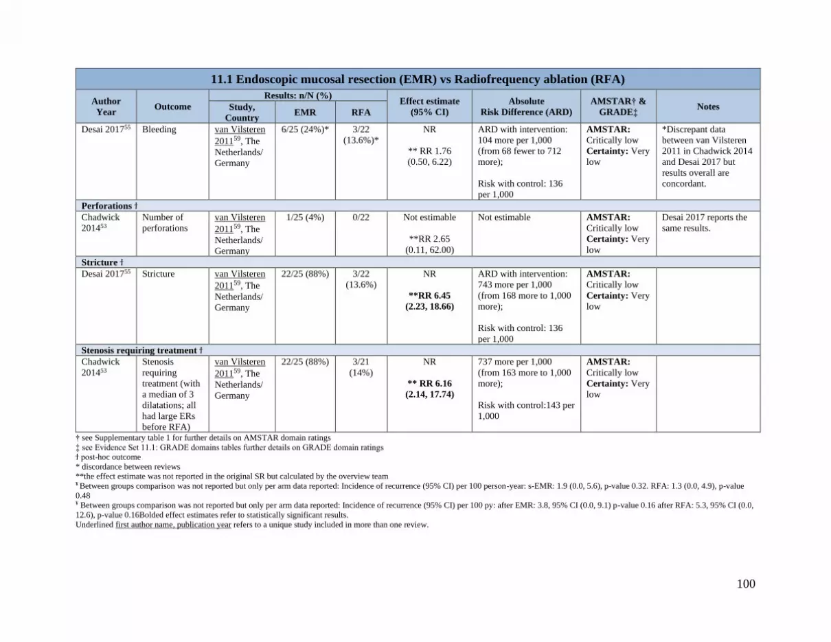
100
11.1 Endoscopic mucosal resection (EMR) vs Radiofrequency ablation (RFA)
Author
Year Outcome
Results: n/N (%) Effect estimate
(95% CI)
Absolute
Risk Difference (ARD)
AMSTAR† &
GRADE‡ Notes Study,
Country EMR RFA
Desai 201755 Bleeding van Vilsteren
201159, The
Netherlands/
Germany
6/25 (24%)*
3/22
(13.6%)*
NR
** RR 1.76
(0.50, 6.22)
ARD with intervention:
104 more per 1,000
(from 68 fewer to 712
more);
Risk with control: 136
per 1,000
AMSTAR:
Critically low
Certainty: Very
low
*Discrepant data
between van Vilsteren
2011 in Chadwick 2014
and Desai 2017 but
results overall are
concordant.
Perforations ϯ
Chadwick
201453
Number of
perforations
van Vilsteren
201159, The
Netherlands/
Germany
1/25 (4%) 0/22 Not estimable
**RR 2.65
(0.11, 62.00)
Not estimable AMSTAR:
Critically low
Certainty: Very
low
Desai 2017 reports the
same results.
Stricture ϯ
Desai 201755 Stricture van Vilsteren
201159, The
Netherlands/
Germany
22/25 (88%) 3/22
(13.6%)
NR
**RR 6.45
(2.23, 18.66)
ARD with intervention:
743 more per 1,000
(from 168 more to 1,000
more);
Risk with control: 136
per 1,000
AMSTAR:
Critically low
Certainty: Very
low
Stenosis requiring treatment ϯ
Chadwick
201453
Stenosis
requiring
treatment (with
a median of 3
dilatations; all
had large ERs
before RFA)
van Vilsteren
201159, The
Netherlands/
Germany
22/25 (88%) 3/21
(14%)
NR
** RR 6.16
(2.14, 17.74)
737 more per 1,000
(from 163 more to 1,000
more);
Risk with control:143 per
1,000
AMSTAR:
Critically low
Certainty: Very
low
† see Supplementary table 1 for further details on AMSTAR domain ratings
‡ see Evidence Set 11.1: GRADE domains tables further details on GRADE domain ratings ϯ post-hoc outcome
* discordance between reviews
**the effect estimate was not reported in the original SR but calculated by the overview team ¥ Between groups comparison was not reported but only per arm data reported: Incidence of recurrence (95% CI) per 100 person-year: s-EMR: 1.9 (0.0, 5.6), p-value 0.32. RFA: 1.3 (0.0, 4.9), p-value
0.48 ¥ Between groups comparison was not reported but only per arm data reported: Incidence of recurrence (95% CI) per 100 py: after EMR: 3.8, 95% CI (0.0, 9.1) p-value 0.16 after RFA: 5.3, 95% CI (0.0,
12.6), p-value 0.16Bolded effect estimates refer to statistically significant results.
Underlined first author name, publication year refers to a unique study included in more than one review.

101
Evidence Set 11.1 EMR vs RFA: GRADE domains table
11.1 Endoscopic mucosal resection (EMR) vs Radiofrequency ablation (RFA)
Review
Studies
Outcome Study
Limitations
Indirectness Inconsistency Imprecision Other
considerations
Certainty Importance
Desai 201755
van Vilsteren
201159
Complete
eradication of
neoplasia
Serious • mix of risk of bias
across outcomes,
some information missing1
No serious limitations • it is unknown if
patients had other GE
conditions3
No serious
limitations • one study
Serious • small sample
size (n=47)4
Serious • although review
performed grey literature
searches, a comprehensive search was not performed
Very low Critical
Chadwick
201453
van Vilsteren
201159
Complete
eradication of
dysplasia (end
of treatment)
Serious • mix of risk of bias
across outcomes,
some information missing2
No serious limitations • it is unknown if
patients had other GE
conditions3
No serious
limitations • one study
Serious • small sample
size (n=47)5
Serious • SR did not assess for
publication bias, and
although the review performed grey literature
searches, a comprehensive
search was not performed
Very low Critical
Chadwick
201453
van Vilsteren
201159
Complete
eradication of
dysplasia with
no recurrence
at follow-up
Serious • mix of risk of bias across outcomes,
some information
missing2
No serious limitations • it is unknown if patients had other GE
conditions3
No serious
limitations • one study
Serious • small sample size (n=47)5
Serious • SR did not assess for publication bias, and
although the review
performed grey literature searches, a comprehensive
search was not performed
Very low Critical
Desai 201755
van Vilsteren
201159
Complete
eradication of
intestinal
metaplasia
Serious • mix of risk of bias
across outcomes, some information
missing1
No serious limitations • it is unknown if
patients had other GE conditions3
No serious
limitations • one study
Serious • small sample
size (n=47)4
Serious • although review
performed grey literature searches, a comprehensive
search was not performed
Very low Critical
Chadwick
201453
van Vilsteren
201159
Complete
eradication of
intestinal
metaplasia
(end of
treatment)
Serious • mix of risk of bias
across outcomes, some information
missing2
No serious limitations • it is unknown if
patients had other GE conditions3
No serious
limitations • one study
Serious • small sample
size (n=47)5
Serious • SR did not assess for
publication bias, and although the review
performed grey literature
searches, a comprehensive search was not performed
Very low Critical
Chadwick
201453
van Vilsteren
201159
Complete
eradication of
intestinal
metaplasia
with no
recurrence at
follow-up
Serious • mix of risk of bias
across outcomes,
some information missing2
No serious limitations • it is unknown if
patients had other GE
conditions3
No serious
limitations • one study
Serious • small sample
size (n=47)5
Serious • SR did not assess for
publication bias, and
although the review performed grey literature
searches, a comprehensive
search was not performed
Very low Critical
Fujii-Lau
201754
Early
neoplasia
recurrence
Serious • mix of risk of bias
across outcomes,
No serious limitations • it is unknown if
patients had other GE
conditions3
No serious
limitations • one study
Serious • small sample
size (n=47)
Serious • publication bias detected
Very low Critical; post
hoc

102
11.1 Endoscopic mucosal resection (EMR) vs Radiofrequency ablation (RFA)
Review
Studies
Outcome Study
Limitations
Indirectness Inconsistency Imprecision Other
considerations
Certainty Importance
van Vilsteren
201159
after complete
eradication
some information missing2
with only one event in total4
Fujii-Lau
201754
van Vilsteren
201159
Dysplasia
recurrence
after
achieving
complete
eradication
Serious • mix of risk of bias
across outcomes,
some information missing2
No serious limitations • it is unknown if
patients had other GE
conditions3
No serious
limitations • one study
Serious • small sample
size (n=47)
with no events4
No serious limitations • not detected by review
authors
Low Critical; post
hoc
Desai 201755
van Vilsteren
201159
Recurrence of
IM (follow-
up)
Serious • mix of risk of bias
across outcomes, some information
missing1
No serious limitations • it is unknown if
patients had other GE conditions3
No serious
limitations • one study
Serious • small sample
size (n=47)4
Serious • although review
performed grey literature searches, a comprehensive
search was not performed
Very low Critical; post
hoc
Fujii-Lau
201754
van Vilsteren
201159
Intestinal
Metaplasia
recurrence
Serious • mix of risk of bias
across outcomes, some information
missing2
No serious limitations • it is unknown if
patients had other GE conditions3
No serious
limitations • one study
Very serious • small sample
size (n=47) with only four
events in total6
No serious limitations • not detected by review
authors
Very low Critical; post
hoc
Chadwick
201453
van Vilsteren
201159
Acute
bleeding
endoscopicall
y treated
Serious • mix of risk of bias
across outcomes,
some information
missing2
No serious limitations • it is unknown if
patients had other GE
conditions3
No serious
limitations • one study
Very serious • small sample
size (n=47)
with only seven
events in total6
Serious • SR did not assess for
publication bias, and
although the review
performed grey literature
searches, a comprehensive search was not performed
Very Low Critical
Desai 201755
van Vilsteren
201159
Bleeding Serious • mix of risk of bias
across outcomes,
some information missing1
No serious limitations • it is unknown if
patients had other GE
conditions3
No serious
limitations • one study
Very serious • small sample
size (n=47)
with only nine events in total6
Serious • although review
performed grey literature
searches, a comprehensive search was not performed
Very Low Critical
Chadwick
201453
van Vilsteren
201159
Number of
perforations
Serious • mix of risk of bias
across outcomes,
some information
missing2
No serious limitations • it is unknown if
patients had other GE
conditions3
No serious
limitations • one study
Serious • small sample
size (n=47)
with only one
event in total4
Serious • SR did not assess for
publication bias, and
although the review
performed grey literature
searches, a comprehensive
search was not performed
Very Low Critical
Desai 201755
van Vilsteren
201159
Stricture Serious • mix of risk of bias across outcomes,
some information
missing1
No serious limitations • it is unknown if patients had other GE
conditions3
No serious
limitations • one study
Serious • small sample size (n=47)5
Serious • although review performed grey literature
searches, a comprehensive
search was not performed
Very Low Critical; post
hoc

103
11.1 Endoscopic mucosal resection (EMR) vs Radiofrequency ablation (RFA)
Review
Studies
Outcome Study
Limitations
Indirectness Inconsistency Imprecision Other
considerations
Certainty Importance
Chadwick
201453
van Vilsteren
201159
Stenosis
requiring
treatment
Serious • mix of risk of bias
across outcomes, some information
missing2
No serious limitations • it is unknown if
patients had other GE conditions3
No serious
limitations • one study
Serious • small sample
size (n=47)5
Serious • SR did not assess for
publication bias, and although the review
performed grey literature
searches, a comprehensive search was not performed
Very Low Critical
1 Information provided in Desai 2017 review not adequately detailed and information only given for the s-EMR group Tools other than Cochrane used in other reviews for this study; information from
across reviews considered together. Unclear sequence generation (Chadwick 2014); concealment and blinding of patients (performance bias) could be at either high or unclear risk based on reporting
(Chadwick 2014, Fujii-Lau 2017).
2 No reviews evaluating this study used the Cochrane risk of bias tool; therefore, information was considered from across reviews together. Unclear randomization method (Chadwick 2014);
concealment and blinding of patients (performance bias) could be at either high or unclear risk based on reporting (Fujii-Lau 2017; Chadwick 2014). 3 The reviews do not report if the patients had other GE conditions, which was part of the exclusion criteria for this overview, but the effect on treatment is expected to be minimal
4 Based on few events/ small study sample (sample size<rule of thumb of n=300 events). Insufficient reporting of information to judge extent of imprecision of data.
5 Based on few events/ small study sample (sample size<rule of thumb of n=300 events). 6 Based on few events, CI for relative effects include both appreciable benefit and harm, and absolute CI reasonably includes appreciable benefit and harm.

104
Supplementary Tables
Supplementary Table 1. AMSTAR ratings for included systematic reviews
Supplementary Table 2. Risk of bias/Methodological Assessments of Primary Studies

105
Supplementary Table 1. AMSTAR ratings for included systematic reviews
Fujii-Lau
201754
Desai
201755
Qumseya
201751
De Souza
201447
Almond
201452
Chadwick
201453
Fayter
201050
Rees
201049
Li 200848 Pandey
201856
Codipilly
201857
Confidence Critically
low
(total score
7; 2
critical
domains)
Critically
low
(total score
4; 3
critical
domains)
Low
(total
score 8; 1
critical
domain)
Critically
low
(total score
2; 4
critical
domains)
Critically
low
(total score
2; 4
critical
domains)
Critically
low
(total score
4; 4 critical
domains)
Critically
low
(total
score 6; 2
critical
domains)
Low
(total
score 8; 1
critical
domain)
Critically
low (total
score 3; 4
critical
domains)
Critically
Low
(total score
3; 1
critical
domain)
Critically
low
(total
score 6; 2
critical
domains)
1.Was an 'a priori'
design provided
No No Yes No No No Yes Yes No No No
2. Was there
duplicate study
selection and data
extraction?
Yes No Yes No No Yes Yes Yes No Can’t tell Yes
3. Was a
comprehensive
literature search
performed?
Yes No Yes No No No Yes Yes No Can’t tell Yes
4. Was the status of
publication (i.e., grey
literature) used as an
inclusion criterion?
Yes Yes Can’t
answer
Can’t
answer
No Yes Yes Yes No Can’t tell Can’t tell
5. Was a list of
studies (included and
excluded) provided?
No No No No No No No Yes No No No
6. Were the
characteristics of the
included studies
provided?
Yes Yes Yes Yes Yes Yes Yes Yes Yes Yes Yes
7. Was the scientific
quality of the
included studies
assessed and
documented?
Yes Yes Yes Yes Yes Yes Yes Yes Yes Yes Yes
8. Was the scientific
quality of the
included studies used
appropriately in
formulating
conclusions?
No No Yes No Can’t
answer
No No No No No Yes

106
Fujii-Lau
201754
Desai
201755
Qumseya
201751
De Souza
201447
Almond
201452
Chadwick
201453
Fayter
201050
Rees
201049
Li 200848 Pandey
201856
Codipilly
201857
Confidence Critically
low
(total score
7; 2
critical
domains)
Critically
low
(total score
4; 3
critical
domains)
Low
(total
score 8; 1
critical
domain)
Critically
low
(total score
2; 4
critical
domains)
Critically
low
(total score
2; 4
critical
domains)
Critically
low
(total score
4; 4 critical
domains)
Critically
low
(total
score 6; 2
critical
domains)
Low
(total
score 8; 1
critical
domain)
Critically
low (total
score 3; 4
critical
domains)
Critically
Low
(total score
3; 1
critical
domain)
Critically
low
(total
score 6; 2
critical
domains)
9. Were the methods
used to combine the
findings of the
studies appropriate?
Yes No Yes No Can’t
answer
Can’t
answer
Not
applicable
Yes Yes No Yes
10. Was the
likelihood of
publication bias
assessed?
Yes Yes Yes No No No No No No Yes N/A
11. Was the conflict
of interest included?
Can’t
answer
No Can’t
answer
No No No No Can’t
answer
Can’t
answer
No No
Note: Highlighted texts demonstrate the critical domains

107
Supplementary Table 2. Risk of bias/Methodological Assessments of Primary Studies
Study
(Review)
Notes Outcome Sequence
generation/
randomization
method
Allocation
concealment
Blinding*† Attrition† Selective
reporting
Other
Ackroyd
200066
(Rees 2010)49
Cochrane RoB tool used. Outcome-
specific assessments were reported as pertaining to all outcomes.
All-cause mortality; Progression
from IM to dysplasia; Complete eradication of dysplasia at two
years; Reduction in length (cm)
of BE at 12 months; Reduction in area (%) of BE at 12 months
Unclear Low Low Unclear High Low
Ackroyd
200066
(Li 2008)48
Jadad score = 4/5. No specification on
who was blinded (e.g., patients,
physicians, outcome assessors, statisticians). Explanation provided for
withdrawals and dropouts, but no
information on the total number of withdrawals/ dropouts and relative
number between arms.
Dysplasia eradication; area of
regression of BE Unclear Sealed envelops Double blind Yes n/a n/a
Ackroyd 200066
(De Souza 2014)47
Jadad score = 3/5, with no details on specific items.
Treatment failure n/a n/a n/a n/a n/a n/a
Ackroyd
200066
(Fayter
2010)50
Use of an adopted checklist (not
specified). Neither the outcome specific nor study specific assessments was
reported (i.e., provided in aggregate
among all included studies).
Evidence of regression Unclear for
almost 80% of
the studies
Unclear for almost 90% of
the studies
Unclear for almost 62%
of the studies
Not carried out in
almost 10% of the studies and
unclear for almost
20% of the studies
n/a n/a
Ackroyd 200469
(Li 2008)48
Jadad score = 2/5. Explanation provided for withdrawals and dropouts,
but no information on the total number
of withdrawals/ dropouts and relative number between arms.
Area of regression of BE Unclear Sealed envelops None Yes n/a n/a
Ackroyd
200469
(De Souza
2014)47
Jadad score = 2/5, with no details on
specific items. Treatment failure at one year n/a n/a n/a n/a n/a n/a
Bright 200768
(Rees 2010)49
Cochrane RoB tool used. Outcome-specific assessments were reported as
pertaining to all outcomes.
Progression to EAC; Progression to dysplasia at 5 years; Complete
eradication of BE at 12 months
Unclear Low (sealed
opaque
envelops)
High Low High Low
Bright 200768
(Li 2008)48
Jadad score = 2/5. Explanation provided for withdrawals and dropouts,
but no information on the total number
of withdrawals/ dropouts and relative number between arms.
Progression to HDG; Complete
ablation (among those with
histological change)
Unclear Unclear None Yes n/a n/a

108
Study
(Review)
Notes Outcome Sequence
generation/
randomization
method
Allocation
concealment
Blinding*† Attrition† Selective
reporting
Other
Caldwell
199662 (abstract)
(Rees 2010)49
Cochrane RoB tool used. Outcome-
specific assessments were reported as pertaining to all outcomes.
Reduction in length (cm) of BE
at 12 months Unclear Unclear Unclear Unclear Unclear High
Dulai 200558
(Rees 2010)49
Cochrane RoB tool used. Outcome-
specific assessments were reported as
pertaining to all outcomes.
All-cause mortality Low
Low (sealed
opaque
envelopes)
Low Low Low Low
Dulai 200558
(Li 2008)48
Jadad score = 2/5. Explanation provided for withdrawals and dropouts,
but no information on the total number
of withdrawals/ dropouts and relative number between arms.
Histological complete ablation Unclear Unclear None Yes n/a n/a
Dulai 200558
(De Souza
2014)47
Jadad score = 2/5, with no details on
specific items. Treatment failure n/a n/a n/a n/a n/a n/a
Hage 200479
(Rees 2010)49
Cochrane RoB tool used. Outcome-
specific assessments were reported as pertaining to all outcomes.
All-cause mortality; Complete
eradication of BE at 12 months; Stricture formation
Unclear Unclear High Unclear Unclear Unclear
Hage 200479
(Li 2008)48
Jadad score = 2/5. Explanation
provided for withdrawals and dropouts,
but no information on the total number
of withdrawals/ dropouts and relative
number between arms.
Histologically complete ablation
of BE Unclear Unclear None Yes n/a n/a
Hage 200479
(De Souza 2014)47
Jadad score = 2/5, with no details on
specific items. Treatment failure (no ablation of
BE) n/a n/a n/a n/a n/a n/a
Hage 200479
(Almond 2014)52
Jadad score = 1/5 , with no details on
specific items.
Cancer incidence; Progression to HGD; Complete eradication of
dysplasia at 12 months
n/a n/a n/a n/a n/a n/a
Hage 200479
(Fayter
2010)50
Use of an adopted checklist (not
specified). Neither the outcome specific nor study specific assessments was
reported (i.e., provided in aggregate
among all included studies).
BE surface reduction Unclear for
almost 80% of
the studies
Unclear for almost 90% of
the studies
Unclear for almost 62%
of the studies
Not carried out in
almost 10% of the studies and
unclear for almost
20% of the studies
n/a n/a
Hage 200578
(Li 2008)48
Jadad score = 2/5. Explanation provided for withdrawals and dropouts,
but no information on the total number
of withdrawals/ dropouts and relative number between arms.
Histologically complete ablation of BE
Unclear Unclear None Yes n/a n/a

109
Study
(Review)
Notes Outcome Sequence
generation/
randomization
method
Allocation
concealment
Blinding*† Attrition† Selective
reporting
Other
Heath 200761
(Rees 2010)49
Cochrane RoB tool used. Outcome-
specific assessments were reported as pertaining to all outcomes.
All-cause mortality; Progression
to EAC at one year Unclear Unclear Unclear Low Low Low
Kelty 200480
(Rees 2010)49
Cochrane RoB tool used. Outcome-
specific assessments were reported as pertaining to all outcomes.
All-cause mortality; Complete
eradication of BE at 12 months; Stricture formation
Low Unclear Unclear Unclear Unclear Unclear
Kelty 200480
(Li 2008)48
Jadad score = 3/5. Explanation
provided for withdrawals and dropouts,
but no information on the total number of withdrawals/ dropouts and relative
number between arms.
Histologically complete ablation
of BE Low Unclear None Yes n/a n/a
Kelty 200480
(De Souza
2014)47
Jadad score = 3/5, with no details on specific items. Treatment failure (no ablation of
BE) n/a n/a n/a n/a n/a n/a
Kelty 2004b73
(Fayter
2010)50
Use of an adopted checklist (not specified). Neither the outcome specific
nor study specific assessments was
reported (i.e., provided in aggregate among all included studies).
Reductions in BE; Perforations or strictures
Unclear for
almost 80% of
the studies
Unclear for
almost 90% of
the studies
Unclear for
almost 62%
of the studies
Not carried out in almost 10% of the
studies and
unclear for almost 20% of the studies
n/a n/a
Mackenzie
200773
(abstract)
(Fayter 2010)50
Use of an adopted checklist (not
specified). Neither the outcome specific
nor study specific assessments was
reported (i.e., provided in aggregate among all included studies).
Cancer risk; Lower rates of adenocarcinoma; Stricture
Unclear for
almost 80% of
the studies
Unclear for
almost 90% of
the studies
Unclear for
almost 62%
of the studies
Not carried out in
almost 10% of the
studies and
unclear for almost 20% of the studies
n/a n/a
Mackenzie 200872
(abstract)
(Rees 2010)49
Cochrane RoB tool used. Outcome-specific assessments were reported as
pertaining to all outcomes. It is unclear
why there was a high-risk rating under the “other” domain.
Eradication of HGD; Stricture formation
Unclear Unclear Unclear Unclear Unclear
High
(published in
abstract)
Mackenzie
200974
(Fayter
2010)50
Use of an adopted checklist (not
specified). Neither the outcome specific
nor study specific assessments was reported (i.e., provided in aggregate
among all included studies).
Lower rates of adenocarcinoma
Unclear for
almost 80% of
the studies
Unclear for
almost 90% of
the studies
Unclear for
almost 62%
of the studies
Not carried out in almost 10% of the
studies and
unclear for almost
20% of the studies
n/a n/a
Overholt
200567
(Rees 2010)49
Cochrane RoB tool used. Outcome-
specific assessments were reported as
pertaining to all outcomes. Support for judgement points to another study.
All-cause mortality; Progression
to cancer at latest possible time
point; Complete eradication of dysplasia at two years; Stricture
formation; Complete eradication
of BE over the course of the study (5 years)
Unclear Unclear Unclear Unclear Unclear Unclear

110
Study
(Review)
Notes Outcome Sequence
generation/
randomization
method
Allocation
concealment
Blinding*† Attrition† Selective
reporting
Other
Overholt
200567
(Li 2008)48
Jadad score = 2/5. Explanation
provided for withdrawals and dropouts, but no information on the total number
of withdrawals/ dropouts and relative
number between arms.
Dysplasia eradication;
Eradication of HGD Unclear Unclear None Yes n/a n/a
Overholt
200567
(De Souza
2014)47
Jadad score = 2/5, with no details on
specific items.
Treatment failure n/a n/a n/a n/a n/a n/a
Overholt
200765
(Li 2008)48
Jadad score = 2/5. Explanation
provided for withdrawals and dropouts, but no information on the total number
of withdrawals/ dropouts and relative
number between arms
Progression to cancer at 5 years Unclear Unclear None Yes n/a n/a
Parrilla 200371
(Rees 2010)49
Cochrane RoB tool used. Outcome-
specific assessments were reported as
pertaining to all outcomes.
Mortality; Progression to cancer; Progression to dysplasia from
intestinal metaplasia; Complete
eradication of dysplasia at 5 years; Complete eradication of
BE at 5 years
Low Low
Unclear
(nature of
study made blinding
impossible;
interpreted as high risk)
Unclear Low Low
Parrilla 200371
(Li 2008)48
Jadad score = 3/5. Explanation
provided for withdrawals and dropouts, but no information on the total number
of withdrawals/ dropouts and relative
number between arms.
Progression from non-dysplastic
BE to BE with dysplasia Low
Sealed
envelopes None Yes n/a n/a
Peters 199963
(Rees 2010)49
Cochrane RoB tool used. Outcome-specific assessments were reported as
pertaining to all outcomes.
Reduction in length (cm) of BE at 12 months; Reduction in area
(%) of BE at 12 months
Low Unclear Low Unclear Low Unclear
Phoa 201476
(Qumseya
2017)51
Downs & Black 23 (Good). Based on sums, the tool may have been modified.
Items relevant to risk of bias tool
cannot be distinguished due to reporting.
(Poor quality if the score was <15, fair
quality if the score was 15–19, and good quality if the score was >20.)
Progression to EAC: cumulative
progression over the follow-up period; Progression to EAC:
progression/patient-year;
Progression to HGD; Progression to HDG:
progression/patient-year
n/a n/a n/a n/a n/a n/a
Black & Downs:
Reporting: 11 External validity: 3
Interval validity, bias: 4 Internal validity, confounding: 5
Power: 0
Phoa 201476
(Pandey 2018)56
Quality assessment was guided by the
Cochrane RoB tool and Critical
Appraisal Skills Programme (CASP) checklist. They quality was ranked
from 1 to 4. This RCT was ranked as 1
(highest quality). Per outcome assessment was not provided.
Progression to HDG; Complete
eradication of IM; Complete eradication of dysplasia;
Stricture formation;
Perforations; Bleeding
NR NR NR NR NR NR

111
Study
(Review)
Notes Outcome Sequence
generation/
randomization
method
Allocation
concealment
Blinding*† Attrition† Selective
reporting
Other
Ragunath
200581
(Rees 2010)49
Cochrane RoB tool used. Outcome-
specific assessments were reported as pertaining to all outcomes.
All-cause mortality; Complete
eradication of dysplasia at 12 months; Complete eradication of
BE at 12 months; Reduction in
length (cm) of BE at 12 months; Stricture formation
Low Unclear Unclear Unclear Low Low
Ragunath
2005
(Li 2008)
Jadad score = 3/5. Explanation
provided for withdrawals and dropouts,
but no information on the total number
of withdrawals/ dropouts and relative
number between arms.
Length of regression (median)
(endoscopic change) Low Unclear None Yes n/a n/a
Ragunath 200581
(De Souza 2014) 47
Jadad score = 3/5, with no details on specific items.
Treatment failure (no ablation of BE)
n/a n/a n/a n/a n/a n/a
Ragunath
200581
(Almond
2014)52
Jadad score = 3/5, with no details on
specific items.
Cancer incidence; Progression to
HGD; Complete eradication of dysplasia at 12 months;
Complete eradication of IM;
Stricture
n/a n/a n/a n/a n/a n/a
Shaheen
200970
(Rees 2010)47
Cochrane RoB tool used. Outcome-
specific assessments were reported as
pertaining to all outcomes.
Progression to EAC at 5-years
(or latest time point);
Progression to higher grades of dysplasia; Complete eradication
of dysplasia at 12 months;
Complete eradication of BE at 12 months; Stricture formation
Low Unclear Low Low Low Low
Shaheen
200970
(De Souza
2014)47
Jadad score = 5/5, with no details on
specific items.
Treatment failure at one year n/a n/a n/a n/a n/a n/a
Shaheen 200970
(Qumseya 2017)51
Downs & Black 27. Based on sums, the tool may have been modified. Items
relevant to risk of bias tool cannot be
distinguished due to reporting. (Poor quality if the score was <15, fair
quality if the score was 15–19, and
good quality if the score was >20.)
Cumulative progression to EAC
over follow-up (among those with LGD); Progression to HGD
n/a n/a n/a n/a n/a n/a
Black & Downs: Reporting: 12
External validity: 3
Interval validity, bias: 5 Internal validity, confounding: 6
Power: 1
Shaheen 200970
(Pandey 2018)56
Quality assessment was guided by the Cochrane RoB tool and Critical
Appraisal Skills Programme (CASP)
checklist. They ranked quality from 1 to 4. This study was ranked as 1 (highest
Progression to HGD; Complete; Eradication of intestinal
metaplasia; Complete
eradication of dysplasia; Perforations; Bleeding
NR NR NR NR NR NR

112
Study
(Review)
Notes Outcome Sequence
generation/
randomization
method
Allocation
concealment
Blinding*† Attrition† Selective
reporting
Other
quality). Per outcome assessment was
not provided.
Sharma 200677
(Rees 2010) 49
Cochrane RoB tool used. Outcome-
specific assessments were reported as
pertaining to all outcomes. Stricture formation Unclear Low Unclear Unclear Low Low
Sharma 200677
(Li 2008)48
Jadad score = 2/5. Explanation
provided for withdrawals and dropouts,
but no information on the total number of withdrawals/ dropouts and relative
number between arms.
Histological complete ablation
Unclear
(“according to
BE length”)
Sealed
Envelope None Yes n/a n/a
Sharma 200677
(De Souza
2014) 47
Jadad score = 1/5, with no details on specific items.
Treatment failure n/a n/a n/a n/a n/a n/a
van Vilsteren 201159
(Chadwick 2014)53
Newcastle-Ottawa scale. No final score, information provided by domain. No
indication that outcome-specific items
were addressed by item – one assessment provided.
Complete eradication of dysplasia (end of treatment);
Complete eradication of
dysplasia with no recurrence at follow-up; Complete eradication
of IM (end of treatment);
Complete eradication of IM with no recurrence at follow-up;
Acute bleeding endoscopically
treated; Number of perforations; Stenosis requiring treatment
Authors state randomized but
not method
n/a
Performance:
unknown
Detection: low risk
Adequate follow-
up rate n/a
Groups compara
ble.
Other consider
ations?
NOS:
Representativeness of cohort: 1
Selection of non-exposed cohort: 1
Ascertainment of exposure: 1 Demonstration outcome of interest not present at start: 1
Comparability of cohorts on the basis of the design or analysis: 1
Assessment of outcome: 1 Was the follow-up long enough for outcomes to occur: 1
Adequacy of follow-up cohorts: 1
Reporting: 11 External validity: 3
Interval validity, bias: 4
Internal validity, confounding: 5 Power: 0
van Vilsteren
201159
(Desai 2017)55
Newcastle-Ottawa scale. Score = 7. No
indication that outcome-specific items
were addressed by item – one assessment provided. Complete eradication of
neoplasia; Complete eradication
of IM; Recurrence of IM (follow up); Bleeding; Stricture
n/a n/a n/a n/a n/a n/a
NOS:
Representativeness of cohort: 1 Selection of non-exposed cohort: not reported
Ascertainment of exposure: 1
Demonstration outcome of interest not present at start: 1 Comparability of cohorts on the basis of the design or analysis: 1
Assessment of outcome: 1
Was the follow-up long enough for outcomes to occur: 1 Adequacy of follow-up cohorts: 1

113
Study
(Review)
Notes Outcome Sequence
generation/
randomization
method
Allocation
concealment
Blinding*† Attrition† Selective
reporting
Other
van
Vilsteren59
(Fujii-Lau
2017)54
Downs and Black. Rating for individual
items reported. Those pertaining to risk of bias assessments are provided here.
Early neoplasia recurrence after complete eradication; Dysplasia
recurrence after achieving
complete eradication; IM recurrence
Score = 1 for random method
Score = 0 for concealment
Score = 0 for patient
blinding
Score = 0 for
blinding of
outcome
assessors
Score = 1 for loss to follow-up
n/a n/a
Weinstein 199664
(Rees 2010) 49
Cochrane RoB tool used. Outcome-specific assessments were reported as
pertaining to all outcomes.
Reduction in length (cm) of BE
at 12 months; Reduction in area (%) of BE at 12 months
Unclear Unclear Unclear Unclear Unclear Unclear
Zopf 200183
(Almond
2014)52
Jadad score = 1/5, with no details on specific items.
Cancer incidence n/a n/a n/a n/a n/a n/a
Zoepf 200382
(Fayter 2010)50
Use of an adopted checklist (not
specified). Neither the outcome specific
nor study specific assessments was reported (i.e., provided in aggregate
among all included studies).
Reduction in length
Unclear for
almost 80% of the studies
Unclear for
almost 90% of the studies
Unclear for
almost 62% of the studies
Not carried out in
almost 10% of the
studies and unclear for almost
20% of the studies
n/a n/a
Abbreviation: n/a=not available.
*Performance and detection bias
†Outcome-specific assessments.
** Rees 2010 used data from Overholt 2007 to supplement data in Overholt 2005.

114
Appendices
Appendix 1 List of treatment options
Appendix 2 PICOS table
Appendix 3 PRESS
Appendix 4 Search strategies
Appendix 5 Screening forms
Appendix 6 AMSTAR checklist
Appendix 7 List of excluded reviews at full text
Appendix 8 List of potentially relevant ongoing reviews or trials
Appendix 9 Characteristics of primary studies included in reviews
Appendix 10 Evaluation of overlap of studies and concordance of results among reviews

115
Appendix 1. PICOS table
Inclusion Exclusion
Population Adults (≥18 years old) with stage 1 EAC, BE, or low- or high-grade dysplasia, with or without chronic GERD
as defined in the systematic reviews.
Those diagnosed with
other gastro-esophageal
conditions.
Interventions Treatment for stage 1 EAC, low- or high-grade dysplasia or BE including:
Pharmacological therapies such as: PPI, H2 receptor antagonists, Cox-2 inhibitors, Prokinetics and antacids,
NSAIDs;
Surveillance methods such as: Esophagogastroduodenoscopy (EGD)*† plus biopsy₺, EGD† plus biopsy plus
adjunct techniques‡ (high-definition/high-resolution white light endoscopy, chromoendoscopy, electronic
chromoendoscopy, autofluorescensce imaging, confocal laser endomicroscopy, light scattering spectroscopy,
diffuse reflectance spectroscopy;
Endoscopic or Endoscopic Assisted therapies such as: Ablative techniques (thermal or chemical), and
mechanical methods (EMR, ESD or combined options)
Surgery, including fundoplication and esophagectomy
Any follow-up diagnostic
tests, such 24-hour
esophageal pH test or any
test for staging purposes,
such as CT and MRI
Comparators No management/treatment compared to another management/treatment regimen
Outcomes Mortality - all-cause and EAC-related (1, 5 and 10 years, or as available) †
Survival (1, 5 and 10 years, or as available) †
Progression from non-dysplastic BE to BE with dysplasia, progression from low-grade to high-grade
dysplasia, progression to EAC
Life threatening, severe, or medically significant consequences (such as requiring hospitalization or
prolongation of hospitalization; disabling (limiting self-care or activities of daily living)
Quality of life (validated scales only; e.g. SF-36, WHOQUAL)
Major or minor medical procedures
Psychological effects (e.g., anxiety, stress)
Overtreatment
Post-hoc outcomes:
Complete eradication of: intestinal metaplasia/BE, dysplasia, high-grade dysplasia, neoplasia
Reduction/regression of BE: in length (cm), in area (%)
Treatment Failure (no ablation)
EAC recurrence
†from the time of allocation to screening or control arm
Timing No limits
Settings Any setting
Study designs Systematic reviews of randomized controlled trials (RCTs)*
SRs that combine results
from RCTs with non-

116
Inclusion Exclusion
*Systematic reviews that combine RCT and non-RCTs will be included if results for RCTs are provided
separately from non-RCT studies.
RCTs, controlled before-
after, interrupted times
series, cohort studies, case-
control studies, cross-
sectional studies, case
series, case reports, and
other publication types
(editorials, commentaries,
notes, letter, opinions) or
SRs that only include non-
RCT and observational
studies.
Language No language restrictions in the search, however only English articles will be included at full-text.
Databases Medline, Embase, Cochrane (CDSR, DARE, HTA)
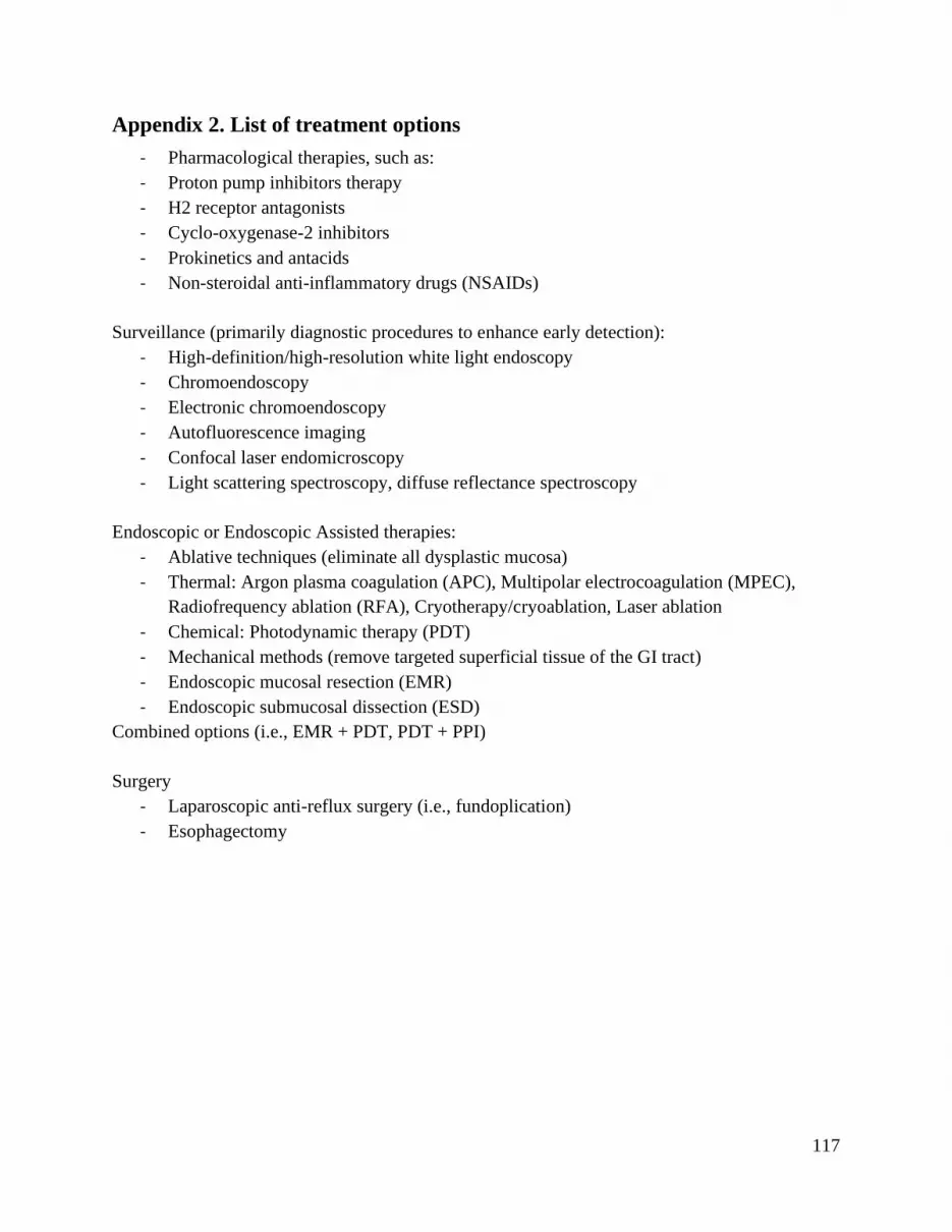
117
Appendix 2. List of treatment options
- Pharmacological therapies, such as:
- Proton pump inhibitors therapy
- H2 receptor antagonists
- Cyclo-oxygenase-2 inhibitors
- Prokinetics and antacids
- Non-steroidal anti-inflammatory drugs (NSAIDs)
Surveillance (primarily diagnostic procedures to enhance early detection):
- High-definition/high-resolution white light endoscopy
- Chromoendoscopy
- Electronic chromoendoscopy
- Autofluorescence imaging
- Confocal laser endomicroscopy
- Light scattering spectroscopy, diffuse reflectance spectroscopy
Endoscopic or Endoscopic Assisted therapies:
- Ablative techniques (eliminate all dysplastic mucosa)
- Thermal: Argon plasma coagulation (APC), Multipolar electrocoagulation (MPEC),
Radiofrequency ablation (RFA), Cryotherapy/cryoablation, Laser ablation
- Chemical: Photodynamic therapy (PDT)
- Mechanical methods (remove targeted superficial tissue of the GI tract)
- Endoscopic mucosal resection (EMR)
- Endoscopic submucosal dissection (ESD)
Combined options (i.e., EMR + PDT, PDT + PPI)
Surgery
- Laparoscopic anti-reflux surgery (i.e., fundoplication)
- Esophagectomy

118
Appendix 3. PRESS
PRESS Guideline 2015— Search Submission & Peer Review Assessment83 Searcher’s Name: Becky Skidmore E-mail: [email protected]
Date submitted: 7 Mar 2017 Date needed by: ASAP (Mar 9, if possible) Note to peer reviewers – please enter your information in the Peer Review Assessment area
Remember: this peer review only pertains to your MEDLINE search strategy. Search question (Describe the purpose of the search) Title: Benefits and Harms of Treatment for Barrett’s Esophagus: An Overview of Systematic Reviews Question: What is the evidence for the benefits and harms of treatment for Barrett’s esophagus (BE) on reducing EAC, EAC related and all-cause mortality, and improving quality of life? PICO format (Outline the PICO for your question, i.e., the Patient, Intervention, Comparison and Outcome)
P: Adults ≥18 years with Barrett’s Esophagus (BE)
I: Treatment strategies for BE including: pharmacological therapies, surveillance methods and endoscopic therapies
C: One treatment method vs. another treatment method
O: Effectiveness of treatment for BE. Primary/critical outcomes are: all-cause mortality and cancer-related mortality (1,5,10 year as available), survival, incidence of EAC, low- and high-grade dysplasia, stage at diagnosis, life-threatening or medically-significant consequences
(see also PICO table in Protocol for more details)
Inclusion criteria (List any inclusion criteria, such as age groups, study designs, to be included) Systematic reviews Exclusion criteria (List any exclusion criteria, such as study designs, to be excluded) Was a search filter applied? (Remember this pertains only to the MEDLINE strategy)
Yes No If yes, which one? Cochrane hedge: PUBMED clinical query: Haynes/McKibbon et al: SIGN (Scottish): CRD (UK): Robinson and Dickerson: Other: Modified CADTH, have added in network meta-analyses
MEDLINE search interface used EBSCO OVID PubMED Other ___________________
Has the search strategy been adapted (i.e., subject heading and terms reviewed) for other databases? Please check all that apply.
Ageline
AMED
C2-SPCTRE
CINAHL
Cochrane Database of Systematic Reviews (CDSR; Cochrane Reviews)
Cochrane Central Register of Controlled Trials (CENTRAL; Clinical Trials)
Cochrane Methodology Register (CMR; Methods Studies)
Cochrane Library (all databases)
Database of Abstracts of Reviews of Effects (DARE; Other Reviews)
Embase
ERIC
LILACS (Latin American and Caribbean Health Sciences Literature)
MEDLINE
PsycINFO
PreMEDLINE
Cochrane HTA
Other
Other

119
Other notes or comments that you feel would be useful for the peer reviewer?
Group has decided to revert back to the original search that included both BE and EAC search terms Please paste your MEDLINE strategy here: Database: Ovid MEDLINE(R) Epub Ahead of Print, In-Process & Other Non-Indexed Citations, Ovid MEDLINE(R) Daily and Ovid MEDLINE(R) <1946 to Present> Search Strategy: -------------------------------------------------------------------------------- 1 Barrett Esophagus/ (7075) 2 (Barrett* adj1 (esophag* or oesophag* or epitheli* or metaplasi* or syndrome?)).tw,kw. (8135) 3 1 or 2 (9350) 4 ((Barrett* or esophag* or oesophag* or pharynx-esophag* or gastro-esophag* or gastro-oesophag*) adj3 (dysplasia* or dysplastic* or precancer* or pre-cancer* or premalignan* or pre-malignan*)).tw,kw. (2138) 5 3 or 4 (9835) 6 Esophageal Neoplasms/ (43936) 7 exp Esophagus/ and exp Neoplasms/ (9850) 8 ((esophag* or oesophag* or pharynx-esophag*) adj3 (neoplas* or cancer* or tumour* or tumor* or carcinoma* or malignan* or metasta* or oncolog* or adenoma* or adenocarcinoma* or adeno-carcinoma* or carcinosarcoma* or carcino-sarcoma*)).tw,kw. (42666) 9 or/6-8 (57539) 10 5 or 9 (61194) 11 exp Infant/ not (exp Adult/ and exp Infant/) (761289) 12 exp Child/ not (exp Adult/ and exp Child/) (1081083) 13 10 not (11 or 12) (60742) 14 exp Animals/ not (exp Animals/ and Humans/) (4327457) 15 13 not 14 (59461) 16 (comment or editorial or interview or news).pt. (1171650) 17 (letter not (letter and randomized controlled trial)).pt. (950923) 18 15 not (16 or 17) (56366) 19 limit 18 to systematic reviews (1429) 20 meta analysis.pt. (75191) 21 exp meta-analysis as topic/ (15514) 22 (meta-analy* or metanaly* or metaanaly* or met analy* or integrative research or integrative review* or integrative overview* or research integration or research overview* or collaborative review*).tw,kw. (112084) 23 (systematic review* or systematic overview* or evidence-based review* or evidence-based overview* or (evidence adj3 (review* or overview*)) or meta-review* or meta-overview* or meta-synthes* or "review of reviews" or technology assessment* or HTA or HTAs).tw,kw. (136923) 24 exp Technology assessment, biomedical/ (9954) 25 (cochrane or health technology assessment or evidence report).jw. (14812) 26 (network adj (MA or MAs)).tw,kw. (2) 27 (NMA or NMAs).tw,kw. (1409) 28 indirect comparison?.tw,kw. (1208) 29 (indirect treatment* adj1 comparison?).tw,kw. (119) 30 (mixed treatment* adj1 comparison?).tw,kw. (349) 31 (multiple treatment* adj1 comparison?).tw,kw. (74) 32 (multi-treatment* adj1 comparison?).tw,kw. (0) 33 simultaneous comparison?.tw,kw. (396) 34 mixed comparison?.tw,kw. (13) 35 or/20-34 (243608) 36 18 and 35 (1247) 37 19 or 36 (1644) ***************************

120
Peer Review Assessment [For peer reviewers only]
Peer reviewer’s name: Kaitryn Campbell
Press #: N/A
E-mail: [email protected]
Date completed: 7 Mar 2017
Please select the one most appropriate answer for each element
Adequate Adequate with revisions*
Needs revision*
1. Translation of the research question X
2. Boolean and proximity operators X
3. Subject headings X
4. Natural language / free-text X
5. Spelling, syntax and line numbers X
6. Limits and filters X
7. Search strategy adaptations X
* Provide an explanation or example for “Adequate with revisions” and “needs revision”: Other Comments (please limit to 3-5 sentences): Well done and straight forward. No changes or suggestions.
121
Appendix 4. Search strategies
Database: Embase Classic+Embase <1947 to 2018 October 26>, Ovid MEDLINE(R) ALL <1946 to October 25, 2018> Search Strategy: -------------------------------------------------------------------------------- 1 Barrett Esophagus/ (23026) 2 (Barrett* adj1 (esophag* or oesophag* or epitheli* or metaplasi* or syndrome?)).tw,kf. (22348) 3 1 or 2 (27331) 4 ((Barrett* or esophag* or oesophag* or pharynx-esophag* or gastro-esophag* or gastro-oesophag*) adj3 (dysplasia* or dysplastic* or precancer* or pre-cancer* or premalignan* or pre-malignan*)).tw,kf. (5923) 5 3 or 4 (28574) 6 Esophageal Neoplasms/ (54194) 7 exp Esophagus/ and exp Neoplasms/ (37262) 8 ((esophag* or oesophag* or pharynx-esophag*) adj3 (neoplas* or cancer* or tumour* or tumor* or carcinoma* or malignan* or metasta* or oncolog* or adenoma* or adenocarcinoma* or adeno-carcinoma* or carcinosarcoma* or carcino-sarcoma*)).tw,kf. (115152) 9 or/6-8 (144916) 10 5 or 9 (156979) 11 exp Infant/ not (exp Adult/ and exp Infant/) (1653036) 12 exp Child/ not (exp Adult/ and exp Child/) (3188154) 13 10 not (11 or 12) (155770) 14 exp Animals/ not (exp Animals/ and Humans/) (16928885) 15 13 not 14 (118125) 16 (comment or editorial or interview or news).pt. (1857143) 17 (letter not (letter and randomized controlled trial)).pt. (2039050) 18 15 not (16 or 17) (112822) 19 limit 18 to systematic reviews [Limit not valid in Embase; records were retained] (53850) 20 meta analysis.pt. (93528) 21 exp meta-analysis as topic/ (55716) 22 (meta-analy* or metanaly* or metaanaly* or met analy* or integrative research or integrative review* or integrative overview* or research integration or research overview* or collaborative review*).tw,kf. (322057) 23 (systematic review* or systematic overview* or evidence-based review* or evidence-based overview* or (evidence adj3 (review* or overview*)) or meta-review* or meta-overview* or meta-synthes* or "review of reviews" or technology assessment* or HTA or HTAs).tw,kf. (382261) 24 exp Technology assessment, biomedical/ (23572) 25 (cochrane or health technology assessment or evidence report).jw. (38344) 26 (network adj (MA or MAs)).tw,kf. (16) 27 (NMA or NMAs).tw,kf. (4256) 28 indirect* compar*.tw,kf. (4481) 29 (indirect treatment* adj1 compar*).tw,kf. (605) 30 (mixed treatment* adj1 compar*).tw,kf. (1228) 31 (multiple treatment* adj1 compar*).tw,kf. (312) 32 (multi-treatment* adj1 compar*).tw,kf. (4) 33 simultaneous* compar*.tw,kf. (2157) 34 mixed comparison?.tw,kf. (53)

122
35 or/20-34 (671928) 36 18 and 35 (2302) 37 19 or 36 (54111) 38 37 use medall [MEDLINE RECORDS] (2016) 39 Barrett esophagus/ (23026) 40 (Barrett* adj1 (esophag* or oesophag* or epitheli* or metaplasi* or syndrome?)).tw,kw. (22711) 41 39 or 40 (27498) 42 esophagus dysplasia/ (864) 43 exp esophagus/ and dysplasia/ (1609) 44 ((Barrett* or esophag* or oesophag* or pharynx-esophag* or gastro-esophag* or gastro-oesophag*) adj3 (dysplasia* or dysplastic* or precancer* or pre-cancer* or premalignan* or pre-malignan*)).tw,kw. (6007) 45 or/42-44 (7478) 46 41 or 45 (29142) 47 exp esophagus tumor/ (77710) 48 exp esophagus/ and exp neoplasm/ (37262) 49 ((esophag* or oesophag* or pharynx-esophag*) adj3 (neoplas* or cancer* or tumour* or tumor* or carcinoma* or malignan* or metasta* or oncolog* or adenoma* or adenocarcinoma* or adeno-carcinoma* or carcinosarcoma* or carcino-sarcoma*)).tw,kw. (115398) 50 or/47-49 (152799) 51 46 or 50 (164217) 52 exp juvenile/ not (exp juvenile/ and exp adult/) (2337309) 53 exp Infant/ not (exp Adult/ and exp Infant/) (1653036) 54 exp Child/ not (exp Adult/ and exp Child/) (3188154) 55 or/52-54 (3908908) 56 51 not 55 (162843) 57 exp animal experimentation/ or exp models animal/ or exp animal experiment/ or nonhuman/ or exp vertebrate/ (47785712) 58 exp human/ or exp human experimentation/ or exp human experiment/ (37610583) 59 57 not 58 (10176834) 60 56 not 59 (159227) 61 editorial.pt. (1053946) 62 letter.pt. not (letter.pt. and randomized controlled trial/) (2034134) 63 60 not (61 or 62) (152899) 64 meta-analysis/ (244188) 65 "systematic review"/ (181694) 66 "meta analysis (topic)"/ (38725) 67 (meta-analy* or metanaly* or metaanaly* or met analy* or integrative research or integrative review* or integrative overview* or research integration or research overview* or collaborative review*).tw,kw. (324815) 68 (systematic review* or systematic overview* or evidence-based review* or evidence-based overview* or (evidence adj3 (review* or overview*)) or meta-review* or meta-overview* or meta-synthes* or "review of reviews" or technology assessment* or HTA or HTAs).tw,kw. (385401) 69 biomedical technology assessment/ (22465) 70 (cochrane or health technology assessment or evidence report).jw. (38344) 71 (network adj (MA or MAs)).tw,kw. (16) 72 (NMA or NMAs).tw,kw. (4274) 73 indirect* compar*.tw,kw. (4543)

123
74 (indirect treatment* adj1 compar*).tw,kw. (607) 75 (mixed treatment* adj1 compar*).tw,kw. (1251) 76 (multiple treatment* adj1 compar*).tw,kw. (317) 77 (multi-treatment* adj1 compar*).tw,kw. (4) 78 simultaneous* compar*.tw,kw. (2157) 79 mixed comparison?.tw,kw. (54) 80 or/64-79 (731158) 81 63 and 80 (4759) 82 81 use emczd [EMBASE RECORDS] (3269) 83 38 or 82 [BOTH DATABASES] (5285) 84 2018*.dt. (1068337) 85 38 and 84 [MEDLINE UPDATE RECORDS] (190) 86 2018*.dc. (1460954) 87 82 and 86 [EMBASE UPDATE RECORDS] (361) 88 85 or 87 [BOTH DATABASES - UPDATE RECORDS] (551) 89 remove duplicates from 88 (428) 90 89 use medall [MEDLINE UNIQUE UPDATE RECORDS] (184) 91 89 use emczd [EMBASE UNIQUE UPDATE RECORDS] (244) *************************** Cochrane Date Run: 30/10/2018 03:06:00 ID Search Hits #1 MeSH descriptor: ["Barrett Esophagus"] explode all trees 207 #2 (Barrett* next (esophag* or oesophag* or epitheli* or metaplasi* or syndrome*)):ti,ab,kw 470 #3 #1 or #2 470 #4 ((Barrett* or esophag* or oesophag* or (pharynx next esophag*) or (gastro next esophag*) or (gastro next oesophag*)) near/3 (dysplasia* or dysplastic* or precancer* or (pre next cancer*) or premalignan* or (pre next malignan*))):ti,ab,kw 195 #5 #3 or #4 532 #6 MeSH descriptor: ["Esophageal Neoplasms"] explode all trees 1308 #7 MeSH descriptor: [Esophagus] explode all trees 267 #8 ((esophag* or oesophag* or (pharynx next esophag*)) near/3 (neoplas* or cancer* or tumour* or tumor* or carcinoma* or malignan* or metasta* or oncolog* or adenoma* or adenocarcinoma* or (adeno next carcinoma*) or carcinosarcoma* or (carcino next sarcoma*))):ti,ab,kw 3387 #9 {or #6-#8} 3457 #10 #5 or #9 3673 #11 MeSH descriptor: [Infant] explode all trees 14928 #12 MeSH descriptor: [Child] explode all trees 832 #13 #10 not (#11 or #12) with Cochrane Library publication date Between Jan 2018 and Oct 2018, in Cochrane Reviews, Cochrane Protocols 1

124
Appendix 5. Screening forms
Title and abstract screening form
1. Is this record a review (addresses multiple studies within)? (exclude primary studies such as
RCTs, cohort, case-control, cross-sectional, case series, case reports, and editorials/
commentaries/ opinion pieces, and protocols)
Notes:
Include clinical practice guidelines and scoping reviews of interest at this level.
Yes (include)
Unclear (include)
No (exclude)
2. Does the review describe a management/treatment regimen for EAC and/or BE and/or low-
or high-grade dysplasia? (i.e., pharmacological, surveillance, surgical/mechanical or
chemotherapy/radiation, surgery)?
Notes:
If review is not directly on management/treatment (e.g., prognostic factors), please exclude it (also exclude
reviews that only consider dietary intakes, physical activities, smoking etc.).
If an otherwise eligible review does not specify the type of esophageal cancer (EAC or ESCC), please include
it under "unclear" at this level. If it is only on ESCC, exclude it.
Include reviews that are on cancers of esophagogastric junction (that is located at the borderline between
esophagus and stomach.
If a review only focuses on chemotherapy/immunotherapy and/or radiation therapy, please exclude it.
Yes (include)
Unclear (include)
No (exclude)
3. Does the review discuss adults (≥18 years)?
Yes (include)
Unclear (include)
No (exclude)
Additional notes (optional)
125
Full-text screening form
1. Is this record a systematic review of RCTs (or provide a separate analysis for RCTs)?
Note: In order to fulfill the SR definition, the record must meet all of the following criteria:1) searched at least one
database; 2) reported selection criteria; 3) reported quality appraisal; 4) provided a list and synthesis of included
studies.
Notes:
If a review claims assessing risk of bias, but it does not report some details of QA, it would not satisfy the “QA”
condition for question 1. As such it should be excluded (please see #2 below for explanation of details).
If a review does not use a specific tool for assessing the quality of included primary studies but just “generic”
assessment of risk of bias, please include it as long as it reports some details of QA results. If they just state "low
risk of bias" for overall body of evidence for example, you can't consider that sufficient as you have no idea how
they determined that. If they provide at least final rating per study (e.g., low risk, high risk, scoring etc.) +/-
additional details, please include it.
If the record is a clinical practice guideline, please exclude it under Q1; however, if it is based on an SR, please
check if they have referenced the original SR. If yes, please look the SR up and assess if it meets our eligibility
criteria. If yes, please send the note to Nadera with the citation/reference for the SR. If you are unable to locate the
full-text of the referenced SR, please request Raymond to locate it for you and keep Nadera copied.
Yes (include)
No (exclude)
Can't tell because abstract only
Full-text not available in English
Full-text not available (other reasons)
2. Does the SR discuss adults with EAC (stage 1 only), BE, low or high-grade dysplasia?
Note:
If an SR has mix of children and adults with no separate analyses for adults, please exclude it but make a note of it
in the "additional note" section.
Cancer type: If an SR included mix of esophageal cancer types [esophageal adenocarcinoma (EAC) and esophageal
squamous cell carcinoma (ESCC or SCC)], with no separate analysis for EAC, please exclude under question 2 “No
(exclude)” option.
Cancer stage: If an SR included mix EAC stages e.g., 0, I, II, III with no separate analysis for stages 0 and/or I.
Please exclude it under question 2 under “No (exclude)”” option.
If an SR does not report the type of esophageal cancer (EAC or SCC), or EAC’s stage (0, I, II, III) and there is no
other clue to know if it was EAC with stage 0 and/or I, please exclude it by choosing “unclear” option under
question 2 and write the reason in the box.
Yes (include)
No (exclude)
Unclear (please specify what is unclear)
3. Does the review describe a management/treatment regimen for EAC (stage 1) and/or BE
and/or low- or high-grade dysplasia? (i.e., pharmacological, surveillance,
surgical/mechanical or surgery)?

126
Note:
If it addresses another type of treatment/management not listed above, please consult Nadera.
We are not interested in:
perioperative protocols before/ after surgery etc.
Reconstruction after esophagectomy e.g., gastric tube vs whole stomach etc.
Palliation given it is not provided in stage 1
Please do include:
different techniques of the same intervention e.g., different surgery techniques/procedures of the esophagectomy
Yes (include)
No (exclude)
Unclear (please specify what is unclear)
4. Does the SR compare one management/treatment strategy to another
management/treatment strategy or to no management/treatment?
Yes (include)
No (exclude)
5. Is there any other reason to exclude this SR?
Yes exclude (please specify in the box)
No, include
Additional notes (optional)
Optional question for "eligible SRs that includes RCTs but with no sufficient separate data for
RCTs". In order to conduct a separate synthesis for RCTs, one would need to go to the primary
studies.

127
Appendix 6. AMSTAR checklist
1. Was an 'a priori' design provided?
The research question and inclusion criteria should be established before the conduct of the review. Note: Need to refer to a protocol, ethics approval, or pre-determined/a priori published research objectives to score a “yes.”
Yes No Can't answer Not applicable
2. Was there duplicate study selection and data extraction?
There should be at least two independent data extractors and a consensus procedure for disagreements should be in place. Note: 2 people do study selection, 2 people do data extraction, consensus process or one person checks the other’s work.
Yes
No Can't answer Not applicable
3. Was a comprehensive literature search performed?
At least two electronic sources should be searched. The report must include years and databases used (e.g., Central, EMBASE, and MEDLINE). Key words and/or MESH terms must be stated and where feasible the search strategy should be provided. All searches should be supplemented by consulting current contents, reviews, textbooks, specialized registers, or experts in the particular field of study, and by reviewing the references in the studies found. Note: If at least 2 sources + one supplementary strategy used, select “yes” (Cochrane register/Central counts as 2 sources; a grey literature search counts as supplementary).
Yes No Can't answer
Not applicable
4. Was the status of publication (i.e. grey literature) used as an inclusion criterion?
The authors should state that they searched for reports regardless of their publication type. The authors should state
whether or not they excluded any reports (from the systematic review), based on their publication status, language etc. Note: If review indicates that there was a search for “grey literature” or “unpublished literature,” indicate “yes.” SIGLE database, dissertations, conference proceedings, and trial registries are all considered grey for this purpose. If searching a source that contains both grey and non-grey, must specify that they were searching for grey/unpublished lit.
Yes
No Can't answer
Not applicable
5. Was a list of studies (included and excluded) provided?
A list of included and excluded studies should be provided. Note: Acceptable if the excluded studies are referenced. If there is an electronic link to the list but the link is dead, select “no.”
Yes No Can't answer Not applicable
6. Were the characteristics of the included studies provided?
In an aggregated form such as a table, data from the original studies should be provided on the participants, interventions
and outcomes. The ranges of characteristics in all the studies analyzed e.g., age, race, sex, relevant socioeconomic data, disease status, duration, severity, or other diseases should be reported. Note: Acceptable if not in table format as long as they are described as above.
Yes No
Can't answer
Not applicable
7. Was the scientific quality of the included studies assessed and documented?
'A priori' methods of assessment should be provided (e.g., for effectiveness studies if the author(s) chose to include only randomized, double-blind, placebo-controlled studies, or allocation concealment as inclusion criteria); for other types of studies alternative items will be relevant.
Yes No
Can't answer Not applicable

128
Note: Can include use of a quality scoring tool or checklist, e.g., Jadad scale, risk of bias, sensitivity analysis, etc., or a description of quality items, with some kind of result for EACH study (“low” or “high” is fine, as long as it is clear which studies scored “low” and which scored “high”; a summary score/range for all studies is not acceptable).
8. Was the scientific quality of the included studies used appropriately in formulating conclusions?
The results of the methodological rigor and scientific quality should be considered in the analysis and the conclusions of the review, and explicitly stated in formulating recommendations. Note: Might say something such as “the results should be interpreted with caution due to poor quality of included studies.” Cannot score “yes” for this question if scored “no” for question 7.
Yes No Can't answer Not applicable
9. Were the methods used to combine the findings of studies appropriate?
For the pooled results, a test should be done to ensure the studies were combinable, to assess their homogeneity (i.e., Chi-squared test for homogeneity, I2). If heterogeneity exists a random effects model should be used and/or the clinical
appropriateness of combining should be taken into consideration (i.e., is it sensible to combine?). Note: Indicate “yes” if they mention or describe heterogeneity, i.e., if they explain that they cannot pool because of heterogeneity/variability between interventions.
Yes
No Can't answer Not applicable
10. Was the likelihood of publication bias assessed?
An assessment of publication bias should include a combination of graphical aids (e.g., funnel plot, other available tests) and/or statistical tests (e.g., Egger regression test, Hedges-Olken). Note: If no test values or funnel plot included, score “no”. Score “yes” if mentions that publication bias could not be assessed because there were fewer than 10 included studies.
Yes No Can't answer
Not applicable
11. Was the conflict of interest included?
Potential sources of support should be clearly acknowledged in both the systematic review and the included studies. Note: To get a “yes,” must indicate source of funding or support for the systematic review AND for each of the included studies.
Yes
No Can't answer Not applicable
Additional notes (in italics) made by Michelle Weir, Julia Worswick, and Carolyn Wayne based on conversations with Bev Shea and/or Jeremy Grimshaw in June and October 2008 and July and September 2010.

129
Appendix 7. List of excluded reviews at full text
Reason # 1: Not being a systematic review of RCTs (or provide a separate analysis for RCTs), (681
records).
Reason # 2: Not discussing adults with EAC (stage 1 only), BE, low or high-grade dysplasia (98 records).
Reason # 3: Not describing a management/treatment regimen for EAC (stage 1) and/or BE and/or low- or
high-grade dysplasia? (i.e., pharmacological, surveillance, surgical/mechanical or surgery), (2 records).
Reason # 4: Not comparing one management/treatment strategy to another management/treatment
strategy or to no management/treatment (4 records)
Reason # 5: Non-English language (109 records)
Reason # 6: Full text unavailable (102 records)
S# Reference Exclusion
Criteria
1 Taioli E, Schwartz RM, Lieberman-Cribbin W, Moskowitz G, van Gerwen M, Flores R.
Quality of Life after Open or Minimally Invasive Esophagectomy in Patients With
Esophageal Cancer—A Systematic Review. InSeminars in thoracic and cardiovascular
surgery 2017 Sep 1 (Vol. 29, No. 3, pp. 377-390). WB Saunders.
Reason #1
2 Joo MK, Park JJ, Chun HJ. Additional benefits of routine drugs on gastrointestinal cancer:
statins, metformin, and proton pump inhibitors. Digestive Diseases. 2018;36(1):1-4..
Reason #1
3 Aurello P, Sirimarco D, Mangogna LM, Nigri G, Valabrega S, D’Angelo F, Ramacciato G.
Esophagectomy with Esophagocoloplasty for Malignancies: Indications, Technique (with
Video), and Results. Systematic Review of the Literature. Journal of Gastrointestinal Surgery.
2017 Sep 1;21(9):1557-61.
Reason #1
4 Kataoka K, Nakamura K, Mizusawa J, Kato K, Eba J, Katayama H, Shibata T, Fukuda H.
Surrogacy of progression-free survival (PFS) for overall survival (OS) in esophageal cancer
trials with preoperative therapy: Literature-based meta-analysis. European Journal of Surgical
Oncology (EJSO). 2017 Oct 1;43(10):1956-61.
Reason #1
5 Law R, Prabhu A, Fujii-Lau L, Shannon C, Singh S. Stent migration following endoscopic
suture fixation of esophageal self-expandable metal stents: a systematic review and meta-
analysis. Surgical endoscopy. 2018 Feb 1;32(2):675-81.
Reason #2
6 Ma Y, Wu X, Yu J, Zhu J, Pen X, Meng X. Can polysaccharide K improve therapeutic
efficacy and safety in gastrointestinal cancer? a systematic review and network meta-analysis.
Oncotarget. 2017 Oct 24;8(51):89108.
Reason #2
7 He YM, Yu C, Li WB, Li ZP, Xu N. Evaluation of short‐term effectiveness of eight targeted
agents combined with chemotherapy for treating esophageal‐gastric junction adenocarcinoma:
A network meta-analysis. Journal of cellular biochemistry. 2018 Jan;119(1):1183-92.
Reason #2
8 Karstens KF, Izbicki JR, Reeh M. Does the Margin Matter in Esophageal Cancer. Digestive
surgery. 2018;35(3):196-203.
Reason #1
9 Thomas T, Loke Y, Beales IL. Systematic review and meta-analysis: use of statins is
associated with a reduced incidence of oesophageal adenocarcinoma. Journal of
gastrointestinal cancer. 2017 Jul 10:1-3.
Reason #1
10 Chen Y, Zhu HP, Wang T, Sun CJ, Ge XL, Min LF, Zhang XW, Jia QQ, Yu J, Yang JQ,
Allgayer H. What is the optimal radiation dose for non-operable esophageal cancer?
Dissecting the evidence in a meta-analysis. Oncotarget. 2017 Oct 24;8(51):89095.
Reason #2
11 Büyükkaramikli NC, Blommestein HM, Riemsma R, Armstrong N, Clay FJ, Ross J, Worthy
G, Severens J, Kleijnen J, Al MJ. Ramucirumab for treating advanced gastric cancer or
Reason #1

130
gastro-oesophageal junction adenocarcinoma previously treated with chemotherapy: an
evidence review group perspective of a NICE single technology appraisal.
PharmacoEconomics. 2017 Dec 1;35(12):1211-21.
12 Niezink AG, de Jong RA, Muijs CT, Langendijk JA, Widder J. Pulmonary Function Changes
After Radiotherapy for Lung or Esophageal Cancer: A Systematic Review Focusing on Dose‐Volume Parameters. The oncologist. 2017 Oct 1;22(10):1257-64.
Reason #1
13 PDQ Screening and Prevention Editorial Board. Esophageal Cancer Screening (PDQ): Health
Professional Version. 2002.
Reason #1
14 PDQ Adult Treatment Editorial Board Esophageal Cancer Treatment (PDQ): Health
Professional Version. 2002.
Reason #1
15 Tomizawa Y, Konda VJ, Coronel E, Chapman CG, Siddiqui UD. Efficacy, Durability, and
Safety of Complete Endoscopic Mucosal Resection of Barrett Esophagus. Journal of clinical
gastroenterology. 2018 Mar 1;52(3):210-6.
Reason #1
16 Schlottmann F, Patti MG, Shaheen NJ. Endoscopic treatment of high-grade dysplasia and
early esophageal cancer. World journal of surgery. 2017 Jul 1;41(7):1705-11.
Reason #1
17 Iams WT, Villaflor VM. Neoadjuvant Treatment for Locally Invasive Esophageal Cancer.
World journal of surgery. 2017 Jul 1;41(7):1719-25.
Reason #1
18 Schlottmann F, Patti MG, Shaheen NJ. From Heartburn to Barrett’s Esophagus, and Beyond.
World journal of surgery. 2017 Jul 1;41(7):1698-704.
Reason #1
19 Loots E, Sartorius B, Madiba TE, Mulder CJ, Clarke DL. Is clinical research in oesophageal
cancer in South Africa in crisis? A systematic review. World journal of surgery. 2017 Mar
1;41(3):810-6.
Reason #1
20 Metcalfe C, Avery K, Berrisford R, Barham P, Noble SM, Fernandez AM, Hanna G, Goldin
R, Elliott J, Wheatley T, Sanders G. Comparing open and minimally invasive surgical
procedures for oesophagectomy in the treatment of cancer: the ROMIO (Randomised
Oesophagectomy: Minimally Invasive or Open) feasibility study and pilot trial. Health
technology assessment (Winchester, England) 2016; 20 (48): 1-68.
Reason #1
21 Mönig SP, Schiffmann LM. Resection of advanced esophagogastric adenocarcinoma:
Extended indications. Der Chirurg; Zeitschrift fur alle Gebiete der operativen Medizen. 2016
May;87(5):398-405.
Reason #5
22 Noordman BJ, Wijnhoven BP, Lagarde SM, Biermann K, van der Gaast A, Spaander MC,
Valkema R, van Lanschot JJ. Active surveillance in clinically complete responders after
neoadjuvant chemoradiotherapy for esophageal or junctional cancer. Diseases of the
Esophagus. 2017 Dec 1;30(12):1-8.
Reason #1
23 Irino T, Tsekrekos A, Coppola A, Scandavini CM, Shetye A, Lundell L, Rouvelas I. Long-
term functional outcomes after replacement of the esophagus with gastric, colonic, or jejunal
conduits: a systematic literature review. Diseases of the esophagus: official journal of the
International Society for Diseases of the Esophagus. 2017 Dec;30(12):1-1.
Reason #1
24 Pence K, Correa AM, Chan E, Khaitan P, Hofstetter W, Kim MP. Management of esophageal
gastrointestinal stromal tumor: review of one hundred seven patients. Diseases of the
esophagus: official journal of the International Society for Diseases of the Esophagus. 2017
Dec;30(12):1-5.
Reason #1
25 Somerville M, Pitt M. Surveillance of Barrett's oesophagus: do we yet know whether it is
worthwhile?. Frontline gastroenterology. 2010 Jul 1;1(2):88-93.
Reason #1
26 van Workum F, Berkelmans GH, Klarenbeek BR, Nieuwenhuijzen GA, Luyer MD, Rosman
C. McKeown or Ivor Lewis totally minimally invasive esophagectomy for cancer of the
esophagus and gastroesophageal junction: systematic review and meta-analysis. Journal of
thoracic disease. 2017 Jul;9(Suppl 8):S826-33.
Reason #2

131
27 Donohoe CL, Reynolds JV. Neoadjuvant treatment of locally advanced esophageal and
junctional cancer: the evidence-base, current key questions and clinical trials. Journal of
thoracic disease. 2017 Jul;9(Suppl 8):S697-S704.
Reason #1
28 Klevebro F, Ekman S, Nilsson M. Current trends in multimodality treatment of esophageal
and gastroesophageal junction cancer–Review article. Surgical oncology. 2017 Sep
1;26(3):290-5.
Reason #1
29 Liu Y, Kou C, Su Y, Zhang Y, You Y, Zhang L, Wang M, Fu Y, Ren X, Yang Y. accelerated
or hyperfractionated radiotherapy for esophageal carcinoma: a meta-analysis of randomized
controlled trials. OncoTargets and therapy. 2017;10:2971-81.
Reason #1
30 Wang X, Miao C, Chen Z, Li W, Yuan S, Yu J, Hu X. Can involved-field irradiation replace
elective nodal irradiation in chemoradiotherapy for esophageal cancer? A systematic review
and meta-analysis. OncoTargets and therapy. 2017;10:2087-95.
Reason #1
31 Liu Y, Mu Y, Zhang A, Ren S, Wang W, Xie J, Zhang Y, Zhou C. Cytokine-induced killer
cells/dendritic cells and cytokine-induced killer cells immunotherapy for the treatment of
esophageal cancer in China: a meta-analysis. OncoTargets and therapy. 2017;10:1897-1908.
Reason #2
32 Lv L, Hu W, Ren Y, Wei X. Minimally invasive esophagectomy versus open esophagectomy
for esophageal cancer: a meta-analysis. OncoTargets and therapy. 2016;9:6751-62.
Reason #2
33 Ter Veer E, Ngai LL, Van Valkenhoef G, Mohammad NH, Anderegg MC, van Oijen MG,
van Laarhoven HW. Capecitabine, 5-fluorouracil and S-1 based regimens for previously
untreated advanced oesophagogastric cancer: A network meta-analysis. Scientific reports.
2017 Aug 2;7(1):7142.
Reason #2
34 Schlottmann F, Patti MG. Current concepts in treatment of Barrett’s esophagus with and
without dysplasia. Journal of Gastrointestinal Surgery. 2017 Aug 1;21(8):1354-60.
Reason #1
35 Duan X, Yu Z. Neoadjuvant chemoradiotherapy combined with operation vs. operation alone
for resectable esophageal cancer: Meta-analysis on randomized controlled trials. Zhonghua
wei chang wai ke za zhi= Chinese journal of gastrointestinal surgery. 2017 Jul;20(7):809-15.
Reason #5
36 Du D, Song T, Liang X, Fang M, Wu S. Concurrent chemoradiotherapy with elective lymph
node irradiation for esophageal cancer: a systemic review and pooled analysis of the
literature. Diseases of the esophagus : official journal of the International Society for Diseases
of the Esophagus 2017; 30 (2): 1-9.
Reason #1
37 Dumonceau JM, Deprez PH, Jenssen C, Iglesias-Garcia J, Larghi A, Vanbiervliet G, Aithal
GP, Arcidiacono PG, Bastos P, Carrara S, Czakó L. Indications, results, and clinical impact of
endoscopic ultrasound (EUS)-guided sampling in gastroenterology: European Society of
Gastrointestinal Endoscopy (ESGE) Clinical Guideline - Updated January 2017. Endoscopy
2017 Jul 1;49(7):695-714.
Reason #1
38 Li S, Liu H, Diao C, Wang X, Gao M, Li Z, Song L, Gao X, Han J, Wang F, Li W. Prognosis
of surgery combined with different adjuvant therapies in esophageal cancer treatment: a
network meta-analysis. Oncotarget. 2017 May 30;8(22):36339-353.
Reason #2
39 Yan R, Dang C. Meta-analysis of Transhiatal Esophagectomy in carcinoma of
esophagogastric junction, does it have an advantage?. International Journal of Surgery. 2017
Jun 1;42:183-90.
Reason #2
40 Qumseya BJ, Wolfsen HC. The Role of Endoscopic Ultrasound in the Management of
Patients with Barrett's Esophagus and Superficial Neoplasia. Gastrointestinal Endoscopy
Clinics. 2017 Jul 1;27(3):471-80.
Reason #1
41 Zhu Y, Liu M, Yun X, Wang D, Bai Y, Zhang G, Ji B, Jing C. Meta-Analysis for the
Therapeutic Effect of Neoadjuvant Therapy in Resectable Esophageal Cancer. Pathology &
Oncology Research. 2017 Jul 1;23(3):657-63.
Reason #2

132
42 Sun L, Zhang Z, Xu J, Xu G, Liu X. Dietary fiber intake reduces risk for Barrett's esophagus
and esophageal cancer. Critical reviews in food science and nutrition. 2017 Sep
2;57(13):2749-57.
Reason #3
43 Van DD, Honoré P, Collignon J, Polus M, Loly C, Mutijima E, De AR, Coucke PA, Louis E,
Martinive P. Comprehensive therapeutic strategy for localized esophageal cancer. Second
part: interest of multimodal approaches with or without surgery. Revue medicale de Liege.
2017 Apr;72(4):168-74.
Reason #5
44 Mansour NM, El-Serag HB, Anandasabapathy S. Barrett’s esophagus: best practices for
treatment and post-treatment surveillance. Annals of cardiothoracic surgery. 2017
Mar;6(2):75-87.
Reason #1
45 Visser E, Franken IA, Brosens LA, Ruurda JP, van Hillegersberg R. Prognostic gene
expression profiling in esophageal cancer: a systematic review. Oncotarget. 2017 Jan
17;8(3):5566-5577.
Reason #2
46 Zhao Y, Guo C, Hu H, Zheng L, Ma J, Jiang L, Zhao E, Li H. Folate intake, serum folate
levels and esophageal cancer risk: an overall and dose-response meta-analysis. Oncotarget.
2017 Feb 7;8(6):10458-69.
Reason #2
47 Zhu H, Luo H, Zhu X, Hu X, Zheng L, Zhu X. Pyruvate kinase M2 (PKM2) expression
correlates with prognosis in solid cancers: a meta-analysis. Oncotarget. 2017 Jan 3;8(1):1628-
40.
Reason #2
48 Parry K, Ruurda JP, van der Sluis PC, van Hillegersberg R. Current status of laparoscopic
transhiatal esophagectomy for esophageal cancer patients: a systematic review of the
literature. Diseases of the Esophagus. 2017;30(1):1-7.
Reason #1
49 Van DD, Honoré P, Collignon J, Polus M, Loly C, Mutijima E, De AR, Coucke PA, Louis E,
Martinive P. Comprehensive therapeutic strategy for localized esophageal cancer. Revue
medicale de Liege. 2017 Feb;72(2):58-63.
Reason #5
50 Chen HL, Shen WQ, Liu K. Radioactive self-expanding stents for palliative management of
unresectable esophageal cancer: a systematic review and meta-analysis. Diseases of the
Esophagus. 2017 May 1;30(5):1-6.
Reason #2
51 Luo M, Yang Y, Luo D, Liu L, Zhang Y, Xiao F, Yang J, Zhang C, Fu S, Luo Z. Tumor
necrosis factor-alpha promoter polymorphism 308 G/A is not significantly associated with
esophageal cancer risk: a meta-analysis. Oncotarget. 2016 Nov 29;7(48):79901-79913.
Reason #1
52 Zhang H, Huang Z, Zou X, Liu T. Bevacizumab and wound-healing complications: a
systematic review and meta-analysis of randomized controlled trials. Oncotarget. 2016 Dec
13;7(50):82473-81.
Reason #2
53 Deng HY, Wang WP, Wang YC, Hu WP, Ni PZ, Lin YD, Chen LQ. Neoadjuvant
chemoradiotherapy or chemotherapy? A comprehensive systematic review and meta-analysis
of the options for neoadjuvant therapy for treating oesophageal cancer. European Journal of
Cardio-Thoracic Surgery. 2017 Mar 1;51(3):421-31.
Reason #2
54 Fuccio L, Mandolesi D, Farioli A, Hassan C, Frazzoni L, Guido A, de Bortoli N, Cilla S,
Pierantoni C, Violante FS, Bazzoli F. Brachytherapy for the palliation of dysphagia owing to
esophageal cancer: A systematic review and meta-analysis of prospective studies.
Radiotherapy and Oncology. 2017 Mar 1;122(3):332-9.
Reason #2
55 Cowley A, Bath-Hextall F, Cooper J. Interventions for healthcare professionals, organizations
and patients to enhance quality of life for people diagnosed with palliative esophagogastric
cancer: a systematic review. JBI database of systematic reviews and implementation reports.
2017 Mar 1;15(3):840-52..
Reason #1
56 Nieto T, Tomlinson CL, Dretzke J, Bayliss S, Dilworth M, Beggs AD, Tucker O. Epigenetic
biomarkers in progression from non-dysplastic Barrett's oesophagus to oesophageal
adenocarcinoma: a systematic review protocol. BMJ open. 2016 Dec 1;6(12):e013361.
Reason #1

133
57 Leng XF, Zhu Y, Wang GP, Jin J, Xian L, Zhang YH. Accuracy of ultrasound for the
diagnosis of cervical lymph node metastasis in esophageal cancer: a systematic review and
meta-analysis. Journal of thoracic disease. 2016 Aug;8(8):2146-2157.
Reason #1
58 Wu T, Zhang W, Yang G, Li H, Chen Q, Song R, Zhao L. HMGB1 overexpression as a
prognostic factor for survival in cancer: a meta-analysis and systematic review. Oncotarget.
2016 Aug 2;7(31):50417-27.
Reason #1
59 Braunschweig C, Liang H, Sheean P. Indications for administration of parenteral nutrition in
adults. Nutrition in clinical practice. 2004 Jun;19(3):255-62.
Reason #1
60 Chen XP, Xu DF, Xu WH, Ma ZC, Yao J, Fu SM. Association Studies of CYP1A1 Exon7
Polymorphism and-GSTM1 Interaction with Esophageal Cancer Risk: a Meta-Analysis in the
Chinese Population. Clinical laboratory. 2016 Sep;62(9):1795-802.
Reason #1
61 Morita FH, Bernardo WM, Ide E, Rocha RS, Aquino JC, Minata MK, Yamazaki K, Marques
SB, Sakai P, de Moura EG. Narrow band imaging versus lugol chromoendoscopy to diagnose
squamous cell carcinoma of the esophagus: a systematic review and meta-analysis. BMC
cancer. 2017 Dec;17(1):54.
Reason #1
62 Wang L, Zhu C, Ma X, Shen K, Li H, Hu Y, Guo L, Zhang J, Li P. Impact of enhanced
recovery program on patients with esophageal cancer in comparison with traditional care.
Supportive Care in Cancer. 2017 Feb 1;25(2):381-9.
Reason #1
63 Rayner CJ, Gatenby P. Effect of antireflux surgery for Barrett's esophagus: long-term results.
Minerva chirurgica. 2016 Jun;71(3):180-91.
Reason #1
64 Zagari RM, Eusebi LH, Rabitti S, Cristoferi L, Vestito A, Pagano N, Bazzoli F. Prevalence of
upper gastrointestinal endoscopic findings in the community: A systematic review of studies
in unselected samples of subjects. Journal of gastroenterology and hepatology. 2016
Sep;31(9):1527-38.
Reason #3
65 Montazeri Z, Nyiraneza C, El-Katerji H, Little J. Waterpipe smoking and cancer: systematic
review and meta-analysis. Tobacco control. 2017 Jan 1;26(1):92-7.
Reason #2
66 Ku GY, Ilson DH. Adjuvant therapy in esophagogastric adenocarcinoma: controversies and
consensus. Gastrointestinal cancer research: GCR. 2012 May;5(3):85.
Reason #1
67 Verma V, Lin SH, Simone CB. Clinical outcomes and toxicities of proton radiotherapy for
gastrointestinal neoplasms: a systematic review. Journal of gastrointestinal oncology. 2016
Aug;7(4):644.
Reason #1
68 Cho YK. How to improve the quality of screening endoscopy in Korea: National Endoscopy
Quality Improvement Program. Clinical endoscopy. 2016 Jul;49(4):312-317.
Reason #1
69 Shaheen NJ, Falk GW, Iyer PG, Gerson LB. Corrigendum: ACG Clinical Guideline:
Diagnosis and Management of Barrett's Esophagus. The American journal of
gastroenterology. 2016 Jul 1;111(7):1077.
Reason #1
70 Parikh M, Liu J, Vieira D, Tzimas D, Horwitz D, Antony A, Saunders JK, Ude-Welcome A,
Goodman A. Preoperative endoscopy prior to bariatric surgery: a systematic review and meta-
analysis of the literature. Obesity surgery. 2016 Dec 1;26(12):2961-6.
Reason #1
71 Macías-García F, Domínguez-Muñoz JE. Update on management of Barrett's esophagus.
World journal of gastrointestinal pharmacology and therapeutics. 2016 May 6;7(2):227-234.
Reason #1
72 He Q, Zhang M, Zhang J, Zhong S, Liu Y, Shen J, He J, Jiang L, Yang C, Zeng Y, Guo M.
Predictive value of BRCA1 expression on the efficacy of chemotherapy based on anti-
microtubule agents: a pooled analysis across different malignancies and agents. Annals of
translational medicine. 2016 Mar;4(6):110.
Reason #1
73 Hoeben A, Polak J, Van De Voorde L, Hoebers F, Grabsch HI, de Vos-Geelen J. Cervical
esophageal cancer: a gap in cancer knowledge. Annals of Oncology. 2016 Apr 26;27(9):1664-
74..
Reason #1

134
74 Fan M, Lin Y, Pan J, Yan W, Dai L, Shen L, Chen K. Survival after neoadjuvant
chemotherapy versus neoadjuvant chemoradiotherapy for resectable esophageal carcinoma: A
meta‐analysis. Thoracic cancer. 2016 Mar;7(2):173-81.
Reason #1
75 Fokas E, Rödel C. Definitive, preoperative, and palliative radiation therapy of esophageal
cancer. Visceral Medicine. 2015;31(5):347-53.
Reason #1
76 Metzger R, Schütze F, Mönig S. Evidence-based operative details in esophageal cancer
treatment: Surgical approach, lymphadenectomy, anastomosis. Visceral Medicine.
2015;31(5):337-40.
Reason #1
77 Zheng Z, Cai J, Yin J, Zhang J, Zhang ZT, Wang KL. Transthoracic versus abdominal-
transhiatal resection for treating Siewert type II/III adenocarcinoma of the esophagogastric
junction: a meta-analysis. International journal of clinical and experimental medicine.
2015;8(10):17167-17182.
Reason #1
78 Qiao Y, Hyder A, Bae SJ, Zarin W, O'neill TJ, Marcon NE, Stein L, Thein HH. Surveillance
in patients with Barrett’s esophagus for early detection of esophageal adenocarcinoma: a
systematic review and meta-analysis. Clinical and translational gastroenterology. 2015
Dec;6(12):e131.
Reason #1
79 Burmeister BH. Role of radiotherapy in the pre-operative management of carcinoma of the
esophagus. World journal of gastrointestinal oncology. 2015 Jan 15;7(1):1-5.
Reason #1
80 Yachimski P, Hur C. Evidence-based endoscopic management of Barrett’s esophagus.
Gastroenterology report. 2014 Sep 17;3(1):54-62.
Reason #1
81 Komeda Y, Bruno M, Koch A. EMR is not inferior to ESD for early Barrett’s and EGJ
neoplasia: an extensive review on outcome, recurrence and complication rates. Endoscopy
international open. 2014 Jun;2(2):E58-64.
Reason #1
82 Yim HB. Self-expanding metallic stents and self-expanding plastic stents in the palliation of
malignant oesophageal dysphagia. Annals of palliative medicine. 2014 Apr 24;3(2):41-6.
Reason #1
83 Purwar P, Bambarkar S, Jiwnani S, Karimundackal G, Laskar SG, Pramesh CS.
Multimodality management of esophageal cancer. Indian Journal of Surgery. 2014 Dec
1;76(6):494-503.
Reason #1
84 Gatenby P, Soon Y. Barrett’s oesophagus: Evidence from the current meta-analyses. World
journal of gastrointestinal pathophysiology. 2014 Aug 15;5(3):178-187.
Reason #1
85 Nagaraja V, Cox MR, Eslick GD. Safety and efficacy of esophageal stents preceding or
during neoadjuvant chemotherapy for esophageal cancer: a systematic review and meta-
analysis. Journal of gastrointestinal oncology. 2014 Apr;5(2):119-126.
Reason #1
86 Kawakubo H, Takeuchi H, Kitagawa Y. Current status and future perspectives on minimally
invasive esophagectomy. The Korean journal of thoracic and cardiovascular surgery. 2013
Aug;46(4):241-248.
Reason #1
87 Sabirin J, Abd Rahman M, Rajan P. Changing trends in oesophageal endoscopy: a systematic
review of transnasal oesophagoscopy. ISRN otolaryngology 2013: 586973.
Reason #1
88 Lin JL. T1 esophageal cancer, request an endoscopic mucosal resection (EMR) for in-depth
review. Journal of thoracic disease. 2013 Jun;5(3):353 -356.
Reason #1
89 Cho JW. The role of endoscopic ultrasonography in T staging: early gastric cancer and
esophageal cancer. Clinical endoscopy. 2013 May;46(3):239 -242.
Reason #1
90 Melis M, Weber J, Shridhar R, Hoffe S, Almhanna K, Karl RC, Meredith KL. Body mass
index and perioperative complications after oesophagectomy for adenocarcinoma: a
systematic database review. BMJ open. 2013 Jan 1;3(5):e001336.
Reason #1
91 Gouda BP, Nelson T, Bhoyrul S. Revisional surgery after Heller myotomy for treatment of
achalasia: A comparative analysis focusing on operative approach. Indian Journal Of Surgery.
2012 Aug 1;74(4):309-13.
Reason #1

135
92 Hancock S, Bowman E, Prabakaran J, Benson M, Agni R, Pfau P, Reichelderfer M, Weiss J,
Gopal D. Use of i-Scan endoscopic image enhancement technology in clinical practice to
assist in diagnostic and therapeutic endoscopy: a case series and review of the literature.
Diagnostic and therapeutic endoscopy. 2012: 193570.
Reason #1
93 Zhu Y, Terry NG, Wax A. Development of angle-resolved low coherence interferometry for
clinical detection of dysplasia. Journal of carcinogenesis. 2011;10:19.
Reason #1
94 Wagner TD, Khushalani N, Yang GY. Clinical T2N0M0 carcinoma of thoracic esophagus.
Journal of thoracic disease. 2010 Mar;2(1):36-42.
Reason #1
95 Bird-Lieberman EL, Lao-Sirieix P, Saeed I, Khoo D, Burnham R, Fitzgerald R. The definition
and management of Barrett’s oesophagus: a case report, review of the literature and a
suggestion for the future. BMJ case reports. 2009 Jan 1;2009:bcr0720080450.
Reason #1
96 Wilson KS, Barnett JB, Shah A, Khoo KE. The BC Cancer Agency Compassionate Access
Program: outcome analysis of patients with esophagogastric cancer. Current Oncology. 2009
Sep;16(5):9 -14.
Reason #1
97 Ilson DH. Esophageal cancer chemotherapy: recent advances. Gastrointestinal cancer
research: GCR. 2008 Mar;2(2):85-92.
Reason #1
98 D’Amico TA. Outcomes after surgery for esophageal cancer. Gastrointestinal cancer research:
GCR. 2007 Sep;1(5):188-196.
Reason #1
99 Gillham CM, Reynolds J, Hollywood D. Predicting the response of localised oesophageal
cancer to neo-adjuvant chemoradiation. World journal of surgical oncology. 2007
Dec;5(1):97.
Reason #1
100 Raimondi AM. Use of preoperative glucocorticoid to risk reduction of complications after
esophagectomy by esophagus carcinoma: meta-analysis. Revista Brasileira de terapia
intensiva. 2006 Dec;18(4):366-73.
Reason #5
101 Bonino JA, Sharma P. Barrett esophagus. Current opinion in gastroenterology. 2004 Jul
1;20(4):375-80.
Reason #1
102 van Lanschot JJ, Aleman BM, Richel DJ. Esophageal carcinoma: surgery, radiotherapy, and
chemotherapy. Current opinion in gastroenterology. 2002 Jul 1;18(4):490-5.
Reason #1
103 Vellayappan BA, Soon YY, Ku GY, Leong CN, Lu JJ, Tey J. Chemoradiotherapy versus
chemoradiotherapy plus surgery for esophageal cancer. The Cochrane Library. 2017 Jan 1; 8:
CD010511.
Reason #2
104 Liu B, Bo Y, Wang K, Liu Y, Tang X, Zhao Y, Zhao E, Yuan L. Concurrent neoadjuvant
chemoradiotherapy could improve survival outcomes for patients with esophageal cancer: a
meta-analysis based on random clinical trials. Oncotarget. 2017 Mar 21;8(12):20410-17.
Reason #1
105 Tomasello G, Petrelli F, Ghidini M, Pezzica E, Passalacqua R, Steccanella F, Turati L, Sgroi
G, Barni S. Tumor regression grade and survival after neoadjuvant treatment in gastro-
esophageal cancer: A meta-analysis of 17 published studies. European Journal of Surgical
Oncology (EJSO). 2017 Sep 1;43(9):1607-16.
Reason #2
106 Ronellenfitsch U, Schwarzbach M, Hofheinz R, Kienle P, Nowak K, Kieser M, Slanger TE,
Burmeister B, Kelsen D, Niedzwiecki D, Schuhmacher C. Predictors of overall and
recurrence-free survival after neoadjuvant chemotherapy for gastroesophageal
adenocarcinoma: Pooled analysis of individual patient data (IPD) from randomized controlled
trials (RCTs). European Journal of Surgical Oncology (EJSO). 2017 Aug 1;43(8):1550-8.
Reason #1
107 Pisarska M, Małczak P, Major P, Wysocki M, Budzyński A, Pędziwiatr M. Enhanced
recovery after surgery protocol in oesophageal cancer surgery: Systematic review and meta-
analysis. PloS one. 2017 Mar 28;12(3):e0174382.
Reason #2

136
108 Li Y. Strategy and prospective of enhanced recovery after surgery for esophageal cancer.
Zhonghua wei chang wai ke za zhi= Chinese journal of gastrointestinal surgery. 2016
Sep;19(9):965-70.
Reason #5
109 Giugliano DN, Berger AC, Rosato EL, Palazzo F. Total minimally invasive esophagectomy
for esophageal cancer: approaches and outcomes. Langenbeck's archives of surgery. 2016 Sep
1;401(6):747-56.
Reason #1
110 Labenz, J. Barrett's esophagus. Der Internist 2016 Nov 1;57(11):1079-1092. Reason #5
111 May, A. Endoscopic therapy of esophageal diseases. Der Internist 2016 Aug 1; 57(8):740-
747.
Reason #5
112 Zhang W, Yu D, Peng J, Xu J, Wei Y. Gastric-tube versus whole-stomach esophagectomy for
esophageal cancer: A systematic review and meta-analysis. PloS one. 2017 Mar
7;12(3):e0173416.
Reason #2
113 Noronha V, Patil VM, Joshi A, Chougule A, Banavali S, Prabhash K. Potential role of
metronomic chemotherapy in the treatment of esophageal and gastroesophageal cancer.
Cancer letters. 2017 Aug 1;400:267-75.
Reason #1
114 Xu D, Li G, Li H, Jia F. Comparison of IMRT versus 3D-CRT in the treatment of esophagus
cancer: a systematic review and meta-analysis. Medicine. 2017 Aug;96(31):e7685.
Reason #1
115 Crucitti P, Mangiameli G, Petitti T, Condoluci A, Rocco R, Gallo IF, Longo F, Rocco G.
Does prophylactic ligation of the thoracic duct reduce chylothorax rates in patients
undergoing oesophagectomy? A systematic review and meta-analysis. European Journal of
Cardio-Thoracic Surgery. 2016 Apr 10;50(6):1019-24.
Reason #1
116 Tan WK, di Pietro M, Fitzgerald RC. Past, present and future of Barrett's oesophagus.
European Journal of Surgical Oncology (EJSO). 2017 Jul 1;43(7):1148-60.
Reason #1
117 Chan DL, Sjoquist KM, Goldstein D, Price TJ, Martin AJ, Bang YJ, Kang YK, Pavlakis N.
The effect of anti-angiogenic agents on overall survival in metastatic oesophago-gastric
cancer: A systematic review and meta-analysis. PloS one. 2017 Feb 21;12(2):e0172307.
Reason #2
118 Bartley AN, Washington MK, Ventura CB, Ismaila N, Colasacco C, Benson III AB, Carrato
A, Gulley ML, Jain D, Kakar S, Mackay HJ., Streutker C, Tang L, Troxell M, Ajani JA.
HER2 Testing and Clinical Decision Making in Gastroesophageal Adenocarcinoma:
Guideline From the College of American Pathologists, American Society for Clinical
Pathology, and American Society of Clinical Oncology. Archives of pathology & laboratory
medicine 2016; 140 (12): 1345-1363.
Reason #1
119 Spaander MC, Baron TH, Siersema PD, Fuccio L, Schumacher B, Escorsell À, Garcia-Pagán
JC, Dumonceau JM, Conio M, De Ceglie A, Skowronek J. Esophageal stenting for benign
and malignant disease: European Society of Gastrointestinal Endoscopy (ESGE) Clinical
Guideline. Endoscopy. 2016;48(10):939-48.
Reason #1
120 Lv XH, Wang CH, Xie Y. Efficacy and safety of submucosal tunneling endoscopic resection
for upper gastrointestinal submucosal tumors: a systematic review and meta-analysis. Surgical
endoscopy. 2017 Jan 1;31(1):49-63.
Reason #2
121 Zhang XC, Li QL, Yu YF, Yao LQ, Xu MD, Zhang YQ, Zhong YS, Chen WF, Zhou PH.
Diagnostic efficacy of endoscopic ultrasound-guided needle sampling for upper
gastrointestinal subepithelial lesions: a meta-analysis. Surgical endoscopy. 2016 Jun
1;30(6):2431-41.
Reason #1
122 Qumseya BJ, Wani S, Desai M, Qumseya A, Bain P, Sharma P, Wolfsen H. Adverse events
after radiofrequency ablation in patients with Barrett’s esophagus: a systematic review and
meta-analysis. Clinical Gastroenterology and Hepatology. 2016 Aug 1;14(8):1086-95.
Reason #4

137
123 Hu Q, Sun TT, Hong J, Fang JY, Xiong H, Meltzer SJ. Proton pump inhibitors do not reduce
the risk of esophageal adenocarcinoma in patients with Barrett’s esophagus: a systematic
review and meta-analysis. PloS one. 2017 Jan 10;12(1):e0169691.
Reason #1
124 Cardoso J, Mesquita M, Pereira AD, Bettencourt-Dias M, Chaves P, Pereira-Leal JB. CYR61
and TAZ Upregulation and Focal Epithelial to Mesenchymal Transition May Be Early
Predictors of Barrett’s Esophagus Malignant Progression. PloS one. 2016 Sep
1;11(9):e0161967.
Reason #1
125 Krishnamoorthi R, Singh S, Ragunathan K, Katzka DA, Wang KK, Iyer PG. Risk of
recurrence of Barrett’s esophagus after successful endoscopic therapy. Gastrointestinal
endoscopy. 2016 Jun 1;83(6):1090-106.
Reason #1
126 Oliveira JF, Moura EG, Bernardo WM, Ide E, Cheng S, Sulbaran M, Santos CM, Sakai P.
Prevention of esophageal stricture after endoscopic submucosal dissection: a systematic
review and meta-analysis. Surgical endoscopy. 2016 Jul 1;30(7):2779-91.
Reason #2
127 Hu Y, Li Z, Mi DH, Cao N, Zu SW, Wen ZZ, Yu XL, Qu Y. Chemoradiation combined with
regional hyperthermia for advanced oesophageal cancer: a systematic review and meta-
analysis. Journal of clinical pharmacy and therapeutics. 2017 Apr;42(2):155-64.
Reason #2
128 Guo W, Ma X, Yang S, Zhu X, Qin W, Xiang J, Lerut T, Li H. Combined thoracoscopic-
laparoscopic esophagectomy versus open esophagectomy: a meta-analysis of outcomes.
Surgical endoscopy. 2016 Sep 1;30(9):3873-81.
Reason #2
129 Kauppila JH, Xie S, Johar A, Markar SR, Lagergren P. Meta‐analysis of health‐related quality
of life after minimally invasive versus open oesophagectomy for oesophageal cancer. British
Journal of Surgery. 2017 Aug;104(9):1131-40.
Reason #2
130 Calvet X. Oesophageal diseases: gastroesophageal reflux disease, Barrett's disease, achalasia
and eosinophilic oesophagitis. Gastroenterologia y hepatologia. 2015 Sep;38:49-55.
Reason #5
131 Martínek J, Akiyama JI, Vacková Z, Furnari M, Savarino E, Weijs TJ, Valitova E, der Horst
S, Ruurda JP, Goense L, Triadafilopoulos G. Current treatment options for esophageal
diseases. Annals of the New York Academy of Sciences. 2016 Oct 1;1381(1):139-51.
Reason #1
132 Lv HW, Li Y, Zhou MH, Cheng JW, Xing WQ. Remnant lymph node metastases after
neoadjuvant therapy and surgery in patients with pathologic T0 esophageal carcinoma impact
on prognosis: A systematic review and meta-analysis. Medicine. 2017 Jun;96(26):e7342.
Reason #2
133 Mishra MV, Aggarwal S, Bentzen SM, Knight N, Mehta MP, Regine WF. Establishing
evidence-based indications for proton therapy: an overview of current clinical trials.
International Journal of Radiation Oncology* Biology* Physics. 2017 Feb 1;97(2):228-35.
Reason #1
134 Ter Veer E, Haj Mohammad N, van Valkenhoef G, Ngai LL, Mali RM, Anderegg MC, van
Oijen MG, van Laarhoven HW. The efficacy and safety of first-line chemotherapy in
advanced esophagogastric cancer: a network meta-analysis. Journal of the National Cancer
Institute. 2016 Aug 30;108(10):djw166.
Reason #2
135 Iwakiri K, Kinoshita Y, Habu Y, Oshima T, Manabe N, Fujiwara Y, Nagahara A, Kawamura
O, Iwakiri R, Ozawa S, Ashida K. Evidence-based clinical practice guidelines for
gastroesophageal reflux disease 2015. Journal of gastroenterology. 2016 Aug 1;51(8):751-67.
Reason #1
136 Xi M, Lin SH. Recent advances in intensity modulated radiotherapy and proton therapy for
esophageal cancer. Expert review of anticancer therapy. 2017 Jul 3;17(7):635-46.
Reason #1
137 Feuerstein JD, Castillo NE, Akbari M, Belkin E, Lewandowski JJ, Hurley CM, Lloyd S,
Leffler DA, Cheifetz AS. Systematic analysis and critical appraisal of the quality of the
scientific evidence and conflicts of interest in practice guidelines (2005–2013) for Barrett’s
esophagus. Digestive diseases and sciences. 2016 Oct 1;61(10):2812-22.
Reason #2
138 Garg PK, Sharma J, Jakhetiya A, Goel A, Gaur MK. Preoperative therapy in locally advanced
esophageal cancer. World journal of gastroenterology. 2016 Oct 21;22(39):8750-59.
Reason #1

138
139 Freedberg DE, Kim LS, Yang YX. The risks and benefits of long-term use of proton pump
inhibitors: expert review and best practice advice from the American Gastroenterological
Association. Gastroenterology. 2017 Mar 1;152(4):706-15.
Reason #1
140 Thrift AP, Anderson LA, Murray LJ, Cook MB, Shaheen NJ, Rubenstein JH, El-Serag HB,
Vaughan TL, Schneider JL, Whiteman DC, Corley DA. Nonsteroidal anti-inflammatory drug
use is not associated with reduced risk of Barrett’s esophagus. The American journal of
gastroenterology. 2016 Nov;111(11):1528-35.
Reason #1
141 Pasquali S, Yim G, Vohra RS, Mocellin S, Nyanhongo D, Marriott P, Geh JI, Griffiths EA.
Survival after neoadjuvant and adjuvant treatments compared to surgery alone for resectable
esophageal carcinoma. Annals of surgery. 2017 Mar 1;265(3):481-91.
Reason #2
142 Tomasello G, Ghidini M, Barni S, Passalacqua R, Petrelli F. Overview of different available
chemotherapy regimens combined with radiotherapy for the neoadjuvant and definitive
treatment of esophageal cancer. Expert review of clinical pharmacology. 2017 Jun
3;10(6):649-60.
Reason #1
143 Sun L, Zhao F, Zeng Y, Yi C. Risks and Benefits of Multimodal Esophageal Cancer
Treatments: A Meta-Analysis. Medical science monitor: international medical journal of
experimental and clinical research. 2017;23:889-910.
Reason #2
144 Hussain Z, Diamantopoulos A, Krokidis M, Katsanos K. Double-layered covered stent for the
treatment of malignant oesophageal obstructions: systematic review and meta-analysis. World
journal of gastroenterology. 2016 Sep 14;22(34):7841-50.
Reason #1
145 Tapia Rico G, Townsend AR, Klevansky M, Price TJ. Liver metastases resection for gastric
and esophageal tumors: is there enough evidence to go down this path?. Expert review of
anticancer therapy. 2016 Dec 1;16(12):1219-25.
Reason #1
146 Lin G, Han SY, Xu YP, Mao WM. Increasing the interval between neoadjuvant
chemoradiotherapy and surgery in esophageal cancer: a meta-analysis of published studies.
Diseases of the esophagus : official journal of the International Society for Diseases of the
Esophagus 2016;29(8):1107-1114.
Reason #1
147 Yibulayin W, Abulizi S, Lv H, Sun W. Minimally invasive oesophagectomy versus open
esophagectomy for resectable esophageal cancer: a meta-analysis. World journal of surgical
oncology. 2016 Dec;14(1):304.
Reason #2
148 Butskiy O, Rahmanian R, White RA, Durham S, Anderson DW, Prisman E. Revisiting the
gastric pull‐up for pharyngoesophageal reconstruction: A systematic review and meta‐analysis
of mortality and morbidity. Journal of surgical oncology. 2016 Dec;114(8):907-14.
Reason #1
149 Xiong WL, Li R, Lei HK, Jiang ZY. Comparison of outcomes between minimally invasive
oesophagectomy and open oesophagectomy for oesophageal cancer. ANZ journal of surgery.
2017 Mar;87(3):165-70.
Reason #2
150 Suda K, Nakauchi M, Inaba K, Ishida Y, Uyama I. Minimally invasive surgery for upper
gastrointestinal cancer: Our experience and review of the literature. World journal of
gastroenterology. 2016 May 21;22(19):4626.
Reason #1
151 Kopp HG, Hofheinz RD. Targeted treatment of esophagogastric cancer. Oncology research
and treatment. 2016;39(12):788-94.
Reason #1
152 Roviello G, Polom K, Roviello F, Marrelli D, Multari AG, Paganini G, Pacifico C, Generali
D. Targeting VEGFR-2 in Metastatic Gastric Cancer: Results From a Literature-Based Meta-
Analysis. Cancer investigation. 2017 Mar 16;35(3):187-94.
Reason #2
153 Ghidini M, Petrelli F, Hahne JC, De Giorgi A, Toppo L, Pizzo C, Ratti M, Barni S,
Passalacqua R, Tomasello G. Clinical outcome and molecular characterization of brain
metastases from esophageal and gastric cancer: a systematic review. Medical Oncology. 2017
Apr 1;34(4):62.
Reason #1

139
154 Peng J, Cai J, Niu ZX, Chen LQ. Early enteral nutrition compared with parenteral nutrition
for esophageal cancer patients after esophagectomy: a meta-analysis. Diseases of the
Esophagus. 2016 Jun 1;29(4):333-41.
Reason #2
155 Wang DB, Sun ZY, Deng LM, Zhu DQ, Xia HG, Zhu PZ. Neoadjuvant Chemoradiotherapy
Improving Survival Outcomes for Esophageal Carcinoma: An Updated Meta-analysis.
Chinese medical journal. 2016 Dec 20;129(24):2974-82.
Reason #2
156 Haisley KR, Hart KD, Fischer LE, Kunio NR, Bakis G, Tieu BH, Schipper PH, Sheppard BC,
Hunter JG, Dolan JP. Increasing tumor length is associated with regional lymph node
metastases and decreased survival in esophageal cancer. The American Journal of Surgery.
2016 May 1;211(5):860-6.
Reason #1
157 Jankowski J, Bennett C, Jankowski JA. Management of Barrett esophagus: a practical guide
for clinicians based on the BADCAT and BoB CAT recommendations. Pol Arch Med Wewn.
2015 Jan 1;125(10):765-70.
Reason #1
158 Ben HS, Bahri M, Dhouib F, Daoud J. Role of postoperative chemoradiotherapy in the
therapeutic management of adenocarcinomas of the stomach and oesogastric junction. Cancer
radiotherapie: journal de la Societe francaise de radiotherapie oncologique. 2016
Dec;20(8):830-2.
Reason #5
159 Gao P, Tsai C, Yang Y, Xu Y, Zhang C, Wang L, Liu H, Wang Z. Intraoperative radiotherapy
in gastric and esophageal cancer: meta-analysis of long-term outcomes and complications.
Minerva medica 2017;108(1):74-83.
Reason #1
160 Martin-Richard M, Beveridge RD, Arrazubi V, Alsina M, Guzmán MG, Custodio AB, Gómez
C, Muñoz FL, Pazo R, Rivera F. SEOM Clinical Guideline for the diagnosis and treatment of
esophageal cancer (2016). Clinical and Translational Oncology. 2016 Dec 1;18(12):1179-86.
Reason #1
161 Aurello P, Magistri P, Berardi G, Petrucciani N, Sirimarco D, Antolino L, Nigri G, D'angelo
F, Ramacciato G. Transthoracically or transabdominally: how to approach adenocarcinoma of
the distal esophagus and cardia. A meta-analysis. Tumori Journal. 2016 Jul;102(4):352-60.
Reason #1
162 Bustamante FAC, Hourneaux de Moura EG, Bernardo W, Sallum RA, Ide E, Baba E. Surgery
versus endoscopic therapies for early cancer and high-grade dysplasia in the esophagus: a
systematic review. Arquivos de gastroenterologia. 2016 Mar;53(1):10-9.
Reason #1
163 Straatman J, Joosten PJ, Terwee CB, Cuesta MA, Jansma EP, van der Peet DL. Systematic
review of patient-reported outcome measures in the surgical treatment of patients with
esophageal cancer. Diseases of the Esophagus. 2016 Oct 1;29(7):760-72.
Reason #2
164 Qureshi MA, Khan S, Ujjan ID, Iqbal A, Khan R, Khan BA. Quantitative Analyses of
Esophageal Cancer Research in Pakistan. Asian Pacific Journal of Cancer Prevention.
2016;17(7):3117-22.
Reason #1
165 Ter Veer E, Mohammad NH, van Valkenhoef G, Ngai LL, Mali RM, van Oijen MG, van
Laarhoven HW. Second-and third-line systemic therapy in patients with advanced
esophagogastric cancer: a systematic review of the literature. Cancer and Metastasis Reviews.
2016 Sep 1;35(3):439-56.
Reason #2
166 van Rossum PS, Goense L, Meziani J, Reitsma JB, Siersema PD, Vleggaar FP, van Vulpen
M, Meijer GJ, Ruurda JP, van Hillegersberg R. Endoscopic biopsy and EUS for the detection
of pathologic complete response after neoadjuvant chemoradiotherapy in esophageal cancer: a
systematic review and meta-analysis. Gastrointestinal endoscopy. 2016 May 1;83(5):866-79.
Reason #1
167 Meghelli L, Narducci F, Mariette C, Piessen G, Vanseymortier M, Leblanc E, Collinet P,
Duhamel A, Penel N. Reporting adverse events in cancer surgery randomized trials: a
systematic review of published trials in oesophago-gastric and gynecological cancer patients.
Critical reviews in oncology/hematology. 2016 Aug 1;104:108-14.
Reason #1

140
168 Ikebe M, Morita M, Yamamoto M, Toh Y. Neoadjuvant therapy for advanced esophageal
cancer: the impact on surgical management. General thoracic and cardiovascular surgery.
2016 Jul 1;64(7):386-94.
Reason #1
169 Bushan K, Sharma S. Neoadjuvant chemotherapy and surgery versus surgery alone in
resectable esophageal cancer. Indian journal of cancer. 2015 Jul 1;52(3):413-416.
Reason #1
170 Thosani N, Dayyeh BK, Sharma P, Aslanian HR, Enestvedt BK, Komanduri S, Manfredi M,
Navaneethan U, Maple JT, Pannala R, Parsi MA. ASGE Technology Committee systematic
review and meta-analysis assessing the ASGE Preservation and Incorporation of Valuable
Endoscopic Innovations thresholds for adopting real-time imaging–assisted endoscopic
targeted biopsy during endoscopic surveillance of Barrett’s esophagus. Gastrointestinal
endoscopy. 2016 Apr 1;83(4):684-98.
Reason #1
171 Allemann P, Mantziari S, Wagner D, Digklia A, Ozsahin E, De BB, Dorta G, Godat S,
Montserrat F, Sempoux C, Brunel C. Curative treatment for esophageal cancer: results of a
multidisciplinary consensus. Revue medicale suisse. 2016 Jun;12(523):1165-9.
Reason #5
172 Song H, Zhu J, Lu D. Molecular‐targeted first‐line therapy for advanced gastric cancer. The
Cochrane Library. 2016 Jan 1;7:CD011461.
Reason #2
173 Chuong MD, Hallemeier CL, Jabbour SK, Yu J, Badiyan S, Merrell KW, Mishra MV, Li H,
Verma V, Lin SH. Improving outcomes for esophageal cancer using proton beam therapy.
International Journal of Radiation Oncology* Biology* Physics. 2016 May 1;95(1):488-97..
Reason #1
174 Gao A, Wang L, Li J, Li H, Han Y, Ma X, Sun Y. Prognostic Value of Perineural Invasion in
Esophageal and Esophagogastric Junction Carcinoma: A Meta-Analysis. Disease markers
2016;2016:7340180.
Reason #1
175 Gurusamy KS, Pallari E, Midya S, Mughal M. Laparoscopic versus open transhiatal
oesophagectomy for oesophageal cancer. The Cochrane database of systematic reviews 2016
Jan 1; 3: CD011390.
Reason #1
176 Best LM, Mughal M, Gurusamy KS. Non-surgical versus surgical treatment for oesophageal
cancer. The Cochrane database of systematic reviews 2016 Mar 29; 3: CD011498.
Reason #2
177 Janowitz T, Thuss-Patience P, Marshall A, Kang JH, Connell C, Cook N, Dunn J, Park SH,
Ford H. Chemotherapy vs supportive care alone for relapsed gastric, gastroesophageal
junction, and oesophageal adenocarcinoma: a meta-analysis of patient-level data. British
journal of cancer. 2016 Feb;114(4):381-387.
Reason #1
178 Chen X, Deng L, Jiang X, Wu T. Chinese herbal medicine for oesophageal cancer. The
Cochrane database of systematic reviews 2016; (1): CD004520.
Reason #2
179 Rodham P, Batty JA, McElnay PJ, Immanuel A. Does minimally invasive oesophagectomy
provide a benefit in hospital length of stay when compared with open oesophagectomy?.
Interactive cardiovascular and thoracic surgery. 2015 Dec 15;22(3):360-7.
Reason #1
180 Maret-Ouda J, Konings P, Lagergren J, Brusselaers N. Antireflux surgery and risk of
esophageal adenocarcinoma: a systematic review and meta-analysis. Annals of surgery. 2016
Feb 1;263(2):251-7.
Reason #2
181 Beales IL, Dearman L, Vardi I, Loke Y. Reduced risk of Barrett’s esophagus in statin users:
case–control study and meta-analysis. Digestive diseases and sciences. 2016 Jan 1;61(1):238-
46.
Reason #1
182 Koyanagi K, Ozawa S, Tachimori Y. Minimally invasive esophagectomy performed with the
patient in a prone position: a systematic review. Surgery today. 2016 Mar 1;46(3):275-84.
Reason #1
183 Ge L, Mao L, Tian JH, Shi FY, Lou LL, Qiu X, Li JL, Yang KH. Network meta-analysis on
selecting Chinese medical injections in radiotherapy for esophageal cancer.China journal of
Chinese materia medica 2015;40(18):3674-3681.
Reason #5

141
184 Chung CS, Lee YC, Wu MS. Prevention strategies for esophageal cancer: Perspectives of the
East vs. West. Best Practice & Research Clinical Gastroenterology. 2015 Dec 1;29(6):869-83.
Reason #1
185 Messager M, Pasquer A, Duhamel A, Caranhac G, Piessen G, Mariette C. Laparoscopic
gastric mobilization reduces postoperative mortality after esophageal cancer surgery: a French
Nationwide Study. Annals of surgery. 2015 Nov 1;262(5):817-23.
Reason #1
186 Porschen R, Buck A, Fischbach W, Gockel I, Görling U, Grenacher L, Hollerbach S,
Hölscher A, Körber J, Messmann H, Meyer HJ. S3-Leitlinie Diagnostik und Therapie der
Plattenepithelkarzinome und Adenokarzinome des Ösophagus (Langversion 1.0–September
2015, AWMF-Registernummer: 021/023OL). Zeitschrift für Gastroenterologie. 2015
Nov;53(11):1288-347.
Reason #5
187 Yamoah K, Showalter TN, Ohri N. Radiation therapy intensification for solid tumors: a
systematic review of randomized trials. International Journal of Radiation Oncology*
Biology* Physics. 2015 Nov 15;93(4):737-45.
Reason #1
188 Ruurda JP, Van Der Sluis PC, Van Der Horst S, Van Hilllegersberg R. Robot‐assisted
minimally invasive esophagectomy for esophageal cancer: a systematic review. Journal of
surgical oncology. 2015 Sep;112(3):257-65.
Reason #1
189 Huang WK, Tu HT, See LC. Aspirin use on incidence and mortality of gastrointestinal
cancers: current state of epidemiological evidence. Current pharmaceutical design. 2015 Oct
1;21(35):5108-15.
Reason #1
190 Straughan DM, Azoury SC, Bennett RD, Pimiento JM, Fontaine JP, Toloza EM. Robotic-
assisted esophageal surgery. Cancer Control. 2015 Jul;22(3):335-9.
Reason #1
191 Zhou C, Ma G, Li X, Li J, Yan Y, Liu P, He J, Ren Y. Is minimally invasive esophagectomy
effective for preventing anastomotic leakages after esophagectomy for cancer? A systematic
review and meta-analysis. World journal of surgical oncology. 2015 Dec;13(1):269.
Reason #2
192 Yu JP, Liu YJ, Tao YL, Ruan RW, Cui Z, Zhu SW, Shi W. Prevention of esophageal stricture
after endoscopic submucosal dissection: a systematic review. World journal of surgery. 2015
Dec 1;39(12):2955-64.
Reason #2
193 Pimentel-Nunes P, Dinis-Ribeiro M, Ponchon T, Repici A, Vieth M, De Ceglie A, Amato A,
Berr F, Bhandari P, Bialek A, Conio M. Endoscopic submucosal dissection: European society
of gastrointestinal endoscopy (ESGE) guideline. Endoscopy. 2015 Sep;47(09):829-54.
Reason #1
194 Mohammad NH, Ter Veer E, Ngai L, Mali R, van Oijen MG, van Laarhoven HW. Optimal
first-line chemotherapeutic treatment in patients with locally advanced or metastatic
esophagogastric carcinoma: triplet versus doublet chemotherapy: a systematic literature
review and meta-analysis. Cancer and Metastasis Reviews. 2015 Sep 1;34(3):429-41.
Reason #2
195 Gemmill EH, Humes DJ, Catton JA. Systematic review of enhanced recovery after gastro-
oesophageal cancer surgery. The Annals of The Royal College of Surgeons of England. 2015
Apr;97(3):173-9.
Reason #1
196 Huang TC, Hsu CH, Lin CC, Tu YK. Systematic review and network meta-analysis:
neoadjuvant chemoradiotherapy for locoregional esophageal cancer. Japanese journal of
clinical oncology. 2015 Aug 5;45(11):1023-8.
Reason #2
197 Zhou C, Zhang L, Wang H, Ma X, Shi B, Chen W, He J, Wang K, Liu P, Ren Y. Superiority
of minimally invasive oesophagectomy in reducing in-hospital mortality of patients with
resectable oesophageal cancer: a meta-analysis. PLoS One. 2015 Jul 21;10(7):e0132889.
Reason #2
198 Kinoshita Y, Ishimura N, Ishihara S. Revision process and present task: evidence-based
clinical practice guidelines for gastroesophageal reflux disease. Nihon rinsho. Japanese
journal of clinical medicine. 2015 Jul;73(7):1190-4.
Reason #5

142
199 Markar SR, Wiggins T, Antonowicz S, Zacharakis E, Hanna GB. Minimally invasive
esophagectomy: Lateral decubitus vs. prone positioning; systematic review and pooled
analysis. Surgical oncology. 2015 Sep 1;24(3):212-9.
Reason #1
200 Zhu LL, Yuan L, Wang H, Ye L, Yao GY, Liu C, Sun NN, Li XJ, Zhai SC, Niu LJ, Zhang
JB. A meta-analysis of concurrent chemoradiotherapy for advanced esophageal cancer. PloS
one. 2015 Jun 5;10(6):e0128616.
Reason #1
201 Niaz SK, Quraishy MS, Taj MA, Abid S, Alam A, Nawaz AA, Hasnain S, Shah A, Khan IM,
Memon AR, Zuberi BF. Guidelines on gastroesophageal reflux disease. J Pak Med Assoc.
2015 May 1;65(5):532-41.
Reason #1
202 Kidane B, Coughlin S, Vogt K, Malthaner R. Preoperative chemotherapy for resectable
thoracic esophageal cancer. The Cochrane database of systematic reviews 2015; (5):
CD001556.
Reason #2
203 Li F, Li T, Liu L, Lv J, Song Y, Li C, Diao P. Concurrent versus sequential
chemoradiotherapy for esophageal cancer among Chinese population: a meta-analysis.
Tumori Journal. 2015 Jul;101(4):353-9.
Reason #2
204 Fu T, Bu ZD, Li ZY, Zhang LH, Wu XJ, Wu AW, Shan F, Ji X, Dong QS, Ji JF. Neoadjuvant
chemoradiation therapy for resectable esophago-gastric adenocarcinoma: a meta-analysis of
randomized clinical trials. BMC cancer. 2015 Dec;15(1):322.
Reason #1
205 Bennett C, Moayyedi P, Corley DA, DeCaestecker J, Falck-Ytter Y, Falk G, Vakil N, Sanders
S, Vieth M, Inadomi J, Aldulaimi D. BOB CAT: a large-scale review and Delphi consensus
for management of Barrett’s esophagus with no dysplasia, indefinite for, or low-grade
dysplasia. The American journal of gastroenterology. 2015 May;110(5):662-683.
Reason #1
206 Labenz J, Koop H, Tannapfel A, Kiesslich R, Hölscher AH. The epidemiology, diagnosis, and
treatment of Barrett’s carcinoma. Deutsches Ärzteblatt International. 2015 Mar;112(13):224.-
234.
Reason #1
207 Coats M, Shimi SM. Cholecystectomy and the risk of alimentary tract cancers: a systematic
review. World journal of gastroenterology: WJG. 2015 Mar 28;21(12):3679-3693.
Reason #1
208 Aprile G, Ongaro E, Del Re M, Lutrino SE, Bonotto M, Ferrari L, Rihawi K, Cardellino GG,
Pella N, Danesi R, Fasola G. Angiogenic inhibitors in gastric cancers and gastroesophageal
junction carcinomas: A critical insight. Critical reviews in oncology/hematology. 2015 Aug
1;95(2):165-78.
Reason #1
209 Tian X, Zhou JG, Zeng Z, Shuai T, Yi LJ, Ma L, Wang Y, Cao H, Song GM. Cetuximab in
patients with esophageal cancer: a systematic review and meta-analysis of randomized
controlled trials. Medical Oncology. 2015 Apr 1;32(4):127.
Reason #2
210 Schneider JL, Corley DA. A review of the epidemiology of Barrett's oesophagus and
oesophageal adenocarcinoma. Best Practice & Research Clinical Gastroenterology. 2015 Feb
1;29(1):29-39.
Reason #1
211 Li A, Huang X, Song Y, Chen X, Sun J, Xu H, Wang Z. Anti-epidermal growth factor
receptor-targeted therapy in upper gastrointestinal tract cancers: a meta-analysis. Growth
Factors. 2015 Mar 4;33(2):113-27.
Reason #2
212 Jang R, Darling G, Wong RK. Multimodality approaches for the curative treatment of
esophageal cancer. Journal of the National Comprehensive Cancer Network. 2015 Feb
1;13(2):229-38.
Reason #1
213 Ajani JA, D’Amico TA, Almhanna K, Bentrem DJ, Besh S, Chao J, Das P, Denlinger C,
Fanta P, Fuchs CS, Gerdes H. Esophageal and esophagogastric junction cancers, version
1.2015. Journal of the National Comprehensive Cancer Network. 2015 Feb 1;13(2):194-227.
Reason #1

143
214 Cools-Lartigue J, Spicer J, Ferri LE. Current status of management of malignant disease:
current management of esophageal cancer. Journal of Gastrointestinal Surgery. 2015 May
1;19(5):964-72.
Reason #1
215 Markar SR, Wiggins T, Ni M, Steyerberg EW, Van Lanschot JJ, Sasako M, Hanna GB.
Assessment of the quality of surgery within randomised controlled trials for the treatment of
gastro-oesophageal cancer: a systematic review. The Lancet Oncology. 2015 Jan 1;16(1):e23-
31.
Reason #2
216 Kumagai K, Rouvelas I, Tsai JA, Mariosa D, Lind PA, Lindblad M, Ye W, Lundell L,
Schuhmacher C, Mauer M, Burmeister BH. Survival benefit and additional value of
preoperative chemoradiotherapy in resectable gastric and gastro-oesophageal junction cancer:
a direct and adjusted indirect comparison meta-analysis. European Journal of Surgical
Oncology (EJSO). 2015 Mar 1;41(3):282-94.
Reason #1
217 Kallam A, Alsop BR, Sharma P. Limitations of endoscopic ablation in Barrett’s esophagus.
Expert review of gastroenterology & hepatology. 2015 Apr 3;9(4):487-96..
Reason #1
218 Park CH, Kim EH, Kim HY, Roh YH, Lee YC. Clinical outcomes of endoscopic submucosal
dissection for early stage esophagogastric junction cancer: a systematic review and meta-
analysis. Digestive and Liver Disease. 2015 Jan 1;47(1):37-44.
Reason #1
219 Moehler M, Baltin CT, Ebert M, Fischbach W, Gockel I, Grenacher L, Hölscher AH, Lordick
F, Malfertheiner P, Messmann H, Meyer HJ. International comparison of the German
evidence-based S3-guidelines on the diagnosis and multimodal treatment of early and locally
advanced gastric cancer, including adenocarcinoma of the lower esophagus. Gastric Cancer.
2015 Jul 1;18(3):550-63.
Reason #1
220 Jiang L, Yang KH, Guan QL, Chen Y, Zhao P, Tian JH. Survival benefit of neoadjuvant
chemotherapy for resectable cancer of the gastric and gastroesophageal junction. Journal of
clinical gastroenterology. 2015 May 1;49(5):387-94.
Reason #1
221 Hategan M, Cook N, Prewett S, Hindmarsh A, Qian W, Gilligan D. Trimodality therapy and
definitive chemoradiotherapy for esophageal cancer: a single-center experience and review of
the literature. Diseases of the Esophagus. 2015 Oct 1;28(7):612-8.
Reason #1
222 Subramanian CR, Triadafilopoulos G. Endoscopic treatments for dysplastic Barrett’s
esophagus: resection, ablation, what else?. World journal of surgery. 2015 Mar 1;39(3):597-
605.
Reason #1
223 Moorcraft SY, Smyth EC, Cunningham D. Adjuvant or neoadjuvant therapy for operable
esophagogastric cancer?. Gastric Cancer. 2015 Jan 1;18(1):1-10.
Reason #1
224 Main BG, Strong S, McNair AG, Falk SJ, Crosby T, Blazeby JM. Reporting outcomes of
definitive radiation-based treatment for esophageal cancer: a review of the literature. Diseases
of the Esophagus. 2015 Mar 1;28(2):156-63.
Reason #1
225 Encheva E, Kolev N, Tonev A, Ignatov V, Shterev S, Petrov D, Zlatarov A, Koleva I,
Chaushev B, Kirilova T, Ivanov K. Radiotherapy in the multimodality management of
esophageal cancer. Khirurgiia. 2014(3):12-19.
Reason #1
226 Ma GW, Situ DR, Ma QL, Long H, Zhang LJ, Lin P, Rong TH. Three-field vs two-field
lymph node dissection for esophageal cancer: a meta-analysis. World journal of
gastroenterology: WJG. 2014 Dec 21;20(47):18022-18030.
Reason #1
227 Alzahrani AM, Bazarbashi SN, Rahal MM, Al-Shehri AS, Aljubran AH, Kandil MS, Zekri
JE, Al Olayan AA, Alsharm AA, Yamani NM, Alomary IS. Saudi Oncology Society clinical
management guideline series: Esophageal cancer 2014. Saudi Medical Journal.
2014;35(12):1545-1549.
Reason #1
228 Haverkamp L, Ruurda JP, Van Leeuwen MS, Siersema PD, van Hillegersberg R. Systematic
review of the surgical strategies of adenocarcinomas of the gastroesophageal junction.
Surgical oncology. 2014 Dec 1;23(4):222-28.
Reason #1

144
229 Van De Voorde L, Larue RT, Pijls M, Buijsen J, Troost EG, Berbée M, Sosef M, van Elmpt
W, Schraepen MC, Vanneste B, Oellers M. A qualitative synthesis of the evidence behind
elective lymph node irradiation in oesophageal cancer. Radiotherapy and Oncology. 2014
Nov 1;113(2):166-74.
Reason #1
230 Lin D, Leichman L. The current status of neoadjuvant therapy for esophageal cancer.
InSeminars in thoracic and cardiovascular surgery 2014 Jun 1 (Vol. 26, No. 2, pp. 102-109).
WB Saunders.
Reason #1
231 Tian GY, Miu M, Huang XE. Systematic analysis of pemetrexed-based chemoradiotherapy
for patients with locally advanced or metastatic esophageal cancer. Asian Pac J Cancer Prev.
2014 Jan 1;15(19):8475-78.
Reason #1
232 Graf D, Vallböhmer D, Knoefel WT, Budach W, Häussinger D. Multimodal treatment of
esophageal carcinoma. Deutsche medizinische Wochenschrift (1946). 2014
Oct;139(42):2141-47.
Reason #5
233 Yuan Y, Zeng X, Hu Y, Xie T, Zhao Y. Omentoplasty for oesophagogastrostomy after
oesophagectomy. The Cochrane database of systematic reviews 2014;(10):CD008446.
Reason #2
234 Zhang H, Wang J, Wang W, Zhou L, Chen J, Yang B, Xia Y, Jiang T. A meta-analysis of
esophagectomy: the comparative study of Ivor-Lewis operation and Sweet operation. Chinese
journal of gastrointestinal surgery 2014;17(9):892-897.
Reason #5
235 Castro PM, Ribeiro FP, Rocha AD, Mazzurana M, Alvarez GA. Hand-sewn versus stapler
esophagogastric anastomosis after esophageal ressection: sistematic review and meta-analysis.
ABCD. Arquivos Brasileiros de Cirurgia Digestiva (São Paulo). 2014 Sep;27(3):216-21.
Reason #2
236 Wei MT, Zhang YC, Deng XB, Yang TH, He YZ, Wang ZQ. Transthoracic vs transhiatal
surgery for cancer of the esophagogastric junction: a meta-analysis. World Journal of
Gastroenterology: WJG. 2014 Aug 7;20(29):10183-10192.
Reason #2
237 Yamasaki M, Miyata H, Miyazaki Y, Takahashi T, Kurokawa Y, Nakajima K, Takiguchi S,
Mori M, Doki Y. Perioperative therapy for esophageal cancer. General thoracic and
cardiovascular surgery. 2014 Sep 1;62(9):531-40.
Reason #1
238 Luo HQ, Han L, Jiang Y. Meta-analysis of six randomized control trials of chemotherapy plus
anti-HER monoclonal antibody for advanced gastric and gastroesophageal cancer. Asian Pac J
Cancer Prev. 2014 Jan 1;15(13):5343-8.
Reason #2
239 Deng J, Wang C, Xiang M, Liu F, Liu Y, Zhao K. Meta-analysis of postoperative efficacy in
patients receiving chemoradiotherapy followed by surgery for resectable esophageal
carcinoma. Diagnostic pathology. 2014 Dec;9(1):151.
Reason #2
240 Ballester V, Cruz-Correa M. Endoscopic surveillance of gastrointestinal premalignant lesions:
current knowledge and future directions. Current opinion in gastroenterology. 2014
Sep;30(5):477-83.
Reason #1
241 Yegin EG, Duman DG. Staging of esophageal and gastric cancer in 2014. Minerva medica.
2014 Oct;105(5):391-411.
Reason #1
242 Rackley T, Leong T, Foo M, Crosby T. Definitive chemoradiotherapy for oesophageal
cancer—a promising start on an exciting journey. Clinical oncology. 2014 Sep 1;26(9):533-
40.
Reason #1
243 Benedix F, Dalicho SF, Stübs P, Schubert D, Bruns C. Evidence base for minimally invasive
esophagectomy for esophageal cancer. Der Chirurg; Zeitschrift fur alle Gebiete der operativen
Medizen. 2014 Aug;85(8):668-74.
Reason #5
244 Gwynne S, Wijnhoven BP, Hulshof M, Bateman A. Role of chemoradiotherapy in
oesophageal cancer—adjuvant and neoadjuvant therapy. Clinical oncology. 2014 Sep
1;26(9):522-32.
Reason #1

145
245 Morrow E, Bushyhead D, Wassenaar E, Hinojosa M, Loviscek M, Pellegrini C, Oelschlager
B. The impact of laparoscopic anti-reflux surgery in patients with Barrett’s esophagus.
Surgical endoscopy. 2014 Dec 1;28(12):3279-84.
Reason #1
246 Sun F, Yuan P, Chen T, Hu J. Efficacy and complication of endoscopic submucosal dissection
for superficial esophageal carcinoma: a systematic review and meta-analysis. Journal of
cardiothoracic surgery. 2014 Dec;9(1):78.
Reason #1
247 Waxman I, González-Haba-Ruiz M, Vázquez-Sequeiros E. Endoscopic diagnosis and
therapies for Barrett esophagus. A review. Rev Esp Enferm Dig. 2014 Feb 1;106(2):103-19.
Reason #1
248 Guo HM, Zhang XQ, Chen M, Huang SL, Zou XP. Endoscopic submucosal dissection vs
endoscopic mucosal resection for superficial esophageal cancer. World journal of
gastroenterology: WJG. 2014 May 14;20(18):5540-47.
Reason #1
249 Küper MA, Eisner F, Königsrainer A, Glatzle J. Laparoscopic surgery for benign and malign
diseases of the digestive system: indications, limitations, and evidence. World Journal of
Gastroenterology: WJG. 2014 May 7;20(17):4883-91.
Reason #1
250 Migliore M, Rassl D, Criscione A. Longitudinal and circumferential resection margin in
adenocarcinoma of distal esophagus and cardia. Future Oncology. 2014 Apr;10(5):891-901.
Reason #1
251 Fuchs KH, Babic B, Breithaupt W, Dallemagne B, Fingerhut A, Furnee E, Granderath F,
Horvath P, Kardos P, Pointner R, Savarino E. EAES recommendations for the management of
gastroesophageal reflux disease. Surgical endoscopy. 2014 Jun 1;28(6):1753-73.
Reason #1
252 Liu Y, Han G, Wang G, Wan X, Ren Y, Cheng Y, Jiang Z. Proximal gastrectomy versus total
gastrectomy for adenocarcinoma of esophagogastric junction: a meta-analysis. Chinese
journal of gastrointestinal surgery 2014;17(4):373-377.
Reason #5
253 Bilici A. Treatment options in patients with metastatic gastric cancer: current status and future
perspectives. World Journal of Gastroenterology: WJG. 2014 Apr 14;20(14):3905. -3915.
Reason #1
254 Zhang S, Zhang XQ, Ding XW, Yang RK, Huang SL, Kastelein F, Bruno M, Yu XJ, Zhou D,
Zou XP. Cyclooxygenase inhibitors use is associated with reduced risk of esophageal
adenocarcinoma in patients with Barrett’s esophagus: a meta-analysis. British journal of
cancer. 2014 Apr;110(9):2378-88.
Reason #1
255 Wang J, Ge J, Zhang XH, Liu JY, Yang CM, Zhao SL. Endoscopic submucosal dissection
versus endoscopic mucosal resection for the treatment of early esophageal carcinoma: a meta-
analysis. Asian Pac J Cancer Prev. 2014 Jan 1;15(4):1803-6.
Reason #1
256 Zacherl J. The current evidence in support of multimodal treatment of locally advanced,
potentially resectable esophageal cancer. Digestive Diseases. 2014;32(1-2):171-5..
Reason #1
257 Gupta A, Attar BM, Koduru P, Murali AR, Go BT, Agarwal R. Utility of confocal laser
endomicroscopy in identifying high-grade dysplasia and adenocarcinoma in Barrett’s
esophagus: a systematic review and meta-analysis. European journal of gastroenterology &
hepatology. 2014 Apr 1;26(4):369-77.
Reason #1
258 Kumagai K, Rouvelas I, Tsai JA, Mariosa D, Klevebro F, Lindblad M, Ye W, Lundell L,
Nilsson M. Meta‐analysis of postoperative morbidity and perioperative mortality in patients
receiving neoadjuvant chemotherapy or chemoradiotherapy for resectable oesophageal and
gastro‐oesophageal junctional cancers. British Journal of Surgery. 2014 Mar;101(4):321-38.
Reason #1
259 Mallipeddi MK, Onaitis MW. The contemporary role of minimally invasive esophagectomy
in esophageal cancer. Current oncology reports. 2014 Mar 1;16(3):374.
Reason #1
260 Xiaohua Y, Zhenjiang Y, Weidong L, Pengcheng X, Sidong C. The non‐linear threshold
association between aspirin use and esophageal adenocarcinoma: results of a dose–response
meta‐analysis. Pharmacoepidemiology and drug safety. 2014 Mar;23(3):278-84.
Reason #1
261 Jacobs M, Macefield RC, Elbers RG, Sitnikova K, Korfage IJ, Smets EM, Henselmans I, van
Berge Henegouwen MI, de Haes JC, Blazeby JM, Sprangers MA. Meta-analysis shows
Reason #2

146
clinically relevant and long-lasting deterioration in health-related quality of life after
esophageal cancer surgery. Quality of life research. 2014 May 1;23(4):1155-76.
262 Filip B, Scarpa M, Cavallin F, Alfieri R, Cagol M, Castoro C. Minimally invasive surgery for
esophageal cancer: a review on sentinel node concept. Surgical endoscopy. 2014 Apr
1;28(4):1238-49.
Reason #1
263 Markar SR, Karthikesalingam A, Penna M, Low DE. Assessment of short-term clinical
outcomes following salvage esophagectomy for the treatment of esophageal malignancy:
systematic review and pooled analysis. Annals of surgical oncology. 2014 Mar 1;21(3):922-
31.
Reason #1
264 Fitzgerald RC, di Pietro M, Ragunath K, Ang Y, Kang JY, Watson P, Trudgill N, Patel P,
Kaye PV, Sanders S, O'donovan M. British Society of Gastroenterology. British Society of
Gastroenterology guidelines on the diagnosis and management of Barrett's oesophagus. Gut
2014;63(1):7-42.
Reason #1
265 Wu J, Pan YM, Wang TT, Gao DJ, Hu B. Endotherapy versus surgery for early neoplasia in
Barrett's esophagus: a meta-analysis. Gastrointestinal endoscopy. 2014 Feb 1;79(2):233-41.
Reason #1
266 Almond LM, Barr H. Management controversies in Barrett’s oesophagus. Journal of
gastroenterology. 2014 Feb 1;49(2):195-205.
Reason #1
267 Abbas H, Rossidis G, Hochwald SN, Ben-David K. Robotic esophagectomy: new era of
surgery. Minerva chirurgica. 2013 Oct;68(5):427-33.
Reason #1
268 Orditura M, Galizia G, Fabozzi A, Lieto E, Gambardella V, Morgillo F, Del Genio GM, Fei
L, Di Martino N, Renda A, Ciardiello F. Preoperative treatment of locally advanced
esophageal carcinoma. International journal of oncology. 2013 Dec 1;43(6):1745-53.
Reason #1
269 Stahl M, Mariette C, Haustermans K, Cervantes A, Arnold D, ESMO Guidelines Working
Group. Oesophageal cancer: ESMO Clinical Practice Guidelines for diagnosis, treatment and
follow-up. Annals of oncology. 2013 Oct 1;24(suppl_6):vi51-6.
Reason #1
270 Yang YS, Niu ZX, Chen LQ. Meta-analysis on reconstructions of posterior mediastinal route
and anterior mediastinal route after esophagectomy. Chinese journal of gastrointestinal
surgery 2013;16(9):846-852.
Reason #5
271 Ye T, Sun Y, Zhang Y, Zhang Y, Chen H. Three-field or two-field resection for thoracic
esophageal cancer: a meta-analysis. The Annals of thoracic surgery. 2013 Dec 1;96(6):1933-
41.
Reason #1
272 Mocanu SN, Ponz MC, Soler EM, Figuls MR, Folch MT. Influence of the type of thoracic
access on postesophagectomy respiratory complications. Cirugía Española (English Edition).
2013 Nov 1;91(9):563-73.
Reason #5
273 Sehdev A, Catenacci DV. Perioperative therapy for locally advanced gastroesophageal
cancer: current controversies and consensus of care. Journal of hematology & oncology. 2013
Dec;6(1):66.
Reason #1
274 Al Mansoor S, Ziske C, Schmidt-Wolf IG. Primary small cell carcinoma of the esophagus:
patient data metaanalysis and review of the literature. GMS German Medical Science.
2013;11.
Reason #1
275 Wang F, Wang YM, He W, Li XK, Peng FH, Yang XL, Fan QX. Chemoradiotherapy
followed by surgery could improve the efficacy of treatments in patients with resectable
esophageal carcinoma. Chinese medical journal 2013;126(16):3138-45.
Reason #2
276 Markar SR, Arya S, Karthikesalingam A, Hanna GB. Technical factors that affect
anastomotic integrity following esophagectomy: systematic review and meta-analysis. Annals
of surgical oncology. 2013 Dec 1;20(13):4274-81.
Reason #1
277 Spechler SJ. Barrett esophagus and risk of esophageal cancer: a clinical review. Jama. 2013
Aug 14;310(6):627-36.
Reason #1
147
278 Sharaf RN, Shergill AK, Odze RD, Krinsky ML, Fukami N, Jain R, Appalaneni V, Anderson
MA, Ben-Menachem T, Chandrasekhara V, Chathadi K. Endoscopic mucosal tissue sampling.
Gastrointestinal endoscopy. 2013 Aug 1;78(2):216-24.
Reason #1
279 Shridhar R, Imani-Shikhabadi R, Davis B, Streeter OA, Thomas CR. Curative treatment of
esophageal cancer; an evidenced based review. Journal of gastrointestinal cancer. 2013 Dec
1;44(4):375-84.
Reason #1
280 Ronellenfitsch U, Schwarzbach M, Hofheinz R, Kienle P, Kieser M, Slanger TE, Burmeister
B, Kelsen D, Niedzwiecki D, Schuhmacher C, Urba S. Preoperative chemo (radio) therapy
versus primary surgery for gastroesophageal adenocarcinoma: systematic review with meta-
analysis combining individual patient and aggregate data. European Journal of Cancer. 2013
Oct 1;49(15):3149-58.
Reason #1
281 Fokas E, Weiss C, Rödel C. The role of radiotherapy in the multimodal management of
esophageal cancer. Digestive Diseases. 2013;31(1):30-7.
Reason #1
282 Yang Z, Wu Q, Wang F, Ye X, Qi X, Fan D. A systematic review and meta-analysis of
randomized trials and prospective studies comparing covered and bare self-expandable metal
stents for the treatment of malignant obstruction in the digestive tract. International journal of
medical sciences. 2013;10(7):825-35.
Reason #2
283 Zhang CD, Zeng YJ, Li HW, Zhao ZM, Zhang JK, Dai DQ. Neoadjuvant chemotherapy for
nonmetastatic esophago-gastric adenocarcinomas: a systematic review and meta-analysis.
Cancer investigation. 2013 Jul 1;31(6):421-31.
Reason #1
284 Ronellenfitsch U, Schwarzbach M, Hofheinz R, Kienle P, Kieser M, Slanger TE, Jensen K.
Adenocarcinoma Meta-analysis Group Perioperative chemo(radio)therapy versus primary
surgery for resectable adenocarcinoma of the stomach, gastroesophageal junction, and lower
esophagus. The Cochrane database of systematic reviews 2013 Jan 1;(5):CD008107.
Reason #2
285 Kranzfelder M, Gertler R, Hapfelmeier A, Friess H, Feith M. Chylothorax after
esophagectomy for cancer: impact of the surgical approach and neoadjuvant treatment:
systematic review and institutional analysis. Surgical endoscopy. 2013 Oct 1;27(10):3530-8.
Reason #1
286 Dunbar KB. Endoscopic eradication therapy for mucosal neoplasia in Barrett's esophagus.
Current opinion in gastroenterology. 2013 Jul 1;29(4):446-53.
Reason #1
287 Liu J, Yue J, Xing L, Yu J. Present status and progress of neoadjuvant chemoradiotherapy for
esophageal cancer. Frontiers of medicine. 2013 Jun 1;7(2):172-9.
Reason #1
288 Orman ES, Li N, Shaheen NJ. Efficacy and durability of radiofrequency ablation for Barrett's
esophagus: systematic review and meta-analysis. Clinical Gastroenterology and Hepatology.
2013 Oct 1;11(10):1245-55.
Reason #4
289 Wong S, Hennequin C, Quero L. Brachytherapy for oesophageal cancer. Cancer
radiotherapie: journal de la Societe francaise de radiotherapie oncologique. 2013
Apr;17(2):159-61.
Reason #5
290 Forde PM, Kelly RJ. Chemotherapeutic and targeted strategies for locally advanced and
metastatic esophageal cancer. Journal of Thoracic Oncology. 2013 Jun 1;8(6):673-84.
Reason #1
291 Almhanna K, Shridhar R, Meredith KL. Neoadjuvant or adjuvant therapy for resectable
esophageal cancer: is there a standard of care?. Cancer Control. 2013 Apr;20(2):89-96.
Reason #1
292 Li X, Yang G, Li X, Zhang Y, Yang J, Chang J, Sun X, Zhou X, Guo Y, Xu Y, Liu J.
Traditional Chinese medicine in cancer care: a review of controlled clinical studies published
in Chinese. PloS one. 2013 Apr 3;8(4):e60338.
Reason #1
293 Sgourakis G, Gockel I, Lang H. Endoscopic and surgical resection of T1a/T1b esophageal
neoplasms: a systematic review. World Journal of Gastroenterology: WJG. 2013 Mar
7;19(9):1424-1437.
Reason #1

148
294 Zheng B, Zheng W, Zhu Y, Lin XY, Xu BH, Chen C. Role of adjuvant chemoradiotherapy in
treatment of resectable esophageal carcinoma: a meta-analysis.. Chinese medical journal
2013;126(6):1178-1182.
Reason #1
295 Sivarasan N, Smith G. Role of aspirin in chemoprevention of esophageal adenocarcinoma: A
meta‐analysis. Journal of digestive diseases. 2013 May;14(5):222-30.
Reason #1
296 Evans JA, Early DS, Chandraskhara V, Chathadi KV, Fanelli RD, Fisher DA, Foley KQ,
Hwang JH, Jue TL, Pasha SF, Sharaf R. The role of endoscopy in the assessment and
treatment of esophageal cancer. Gastrointestinal Endoscopy. 2013 Mar 1;77(3):328-34.
Reason #1
297 Uzunoglu FG, Reeh M, Kutup A, Izbicki JR. Surgery of esophageal cancer. Langenbeck's
archives of surgery. 2013 Feb 1;398(2):189-93.
Reason #1
298 Chan DS, Reid TD, Howell I, Lewis WG. Systematic review and meta‐analysis of the
influence of circumferential resection margin involvement on survival in patients with
operable oesophageal cancer. British Journal of Surgery. 2013 Mar;100(4):456-64.
Reason #1
299 Raja S, Rice TW, Rajeswaran J, Zhong J, Blackstone EH. Esophageal small-cell cancer: study
of a rare disease. Diseases of the Esophagus. 2013 Oct 1;26(7):690-5.
Reason #1
300 Schweigert M, Dubecz A, Stein HJ. Oesophageal cancer—an overview. Nature reviews
Gastroenterology & hepatology. 2013 Apr;10(4):230-244.
Reason #1
301 Uttley L, Campbell F, Rhodes M, Cantrell A, Stegenga H, Lloyd-Jones M. Minimally
invasive oesophagectomy versus open surgery: is there an advantage?. Surgical endoscopy.
2013 Mar 1;27(3):724-31.
Reason #1
302 Lordick F, Hölscher AH, Haustermans K, Wittekind C. Multimodal treatment of esophageal
cancer. Langenbeck's archives of surgery. 2013 Feb 1;398(2):177-87.
Reason #1
303 Watanabe M, Baba Y, Nagai Y, Baba H. Minimally invasive esophagectomy for esophageal
cancer: an updated review. Surgery today. 2013 Mar 1;43(3):237-44.
Reason #1
304 Bennett C, Vakil N, Bergman J, Harrison R, Odze R, Vieth M, Sanders S, Gay L, Pech O,
Longcroft–Wheaton G, Romero Y. Consensus statements for management of Barrett's
dysplasia and early-stage esophageal adenocarcinoma, based on a Delphi process.
Gastroenterology. 2012 Aug 1;143(2):336-46.
Reason #1
305 Jung HK, Hong SJ, Jo Y, Jeon SW, Cho YK, Lee KJ, Lee JS, Park HJ, Shin ES, Lee SH, Han
SU. Updated guidelines 2012 for gastroesophageal reflux disease. The Korean Journal of
Gastroenterology. 2012 Oct 1;60(4):195-218.
Reason #5
306 Lee JH, Cho YK, Jeon SW, Kim JH, Kim N, Lee JS, Bak YT. Guidelines for the treatment of
gastroesophageal reflux disease. The Korean Journal of Gastroenterology. 2011 Feb
1;57(2):57-66.
Reason #5
307 Xu XH, Peng XH, Yu P, Xu XY, Cai EH, Guo P, Li K. Neoadjuvant chemotherapy for
resectable esophageal carcinoma: a meta-analysis of randomized clinical trials. Asian Pacific
journal of cancer prevention. 2012;13(1):103-10.
Reason #2
308 Harrison DF. Surgical repair in hypopharyngeal and cervical esophageal cancer: analysis of
162 patients. Annals of Otology, Rhinology & Laryngology. 1981 Jul;90(4):372-5.
Reason #1
309 Jacobson BC, Hirota W, Baron TH, Leighton JA, Faigel DO. The role of endoscopy in the
assessment and treatment of esophageal cancer. Gastrointestinal endoscopy. 2003 Jun
1;57(7):817-22.
Reason #1
310 Lieberman D, Hamilton F. NIH-ADHF Workshop on Endoscopy Priorities: workshop
statement and recommendations. Gastrointestinal endoscopy. 1999 Mar 1;49(3):S3-4.
Reason #1
311 Xu Y, Yu X, Chen Q, Mao W. Neoadjuvant versus adjuvant treatment: which one is better for
resectable esophageal squamous cell carcinoma?. World journal of surgical oncology. 2012
Dec;10(1):173.
Reason #1

149
312 Ge L, Wang HJ, Yin D, Lei C, Zhu JF, Cai XH, Zhang GQ. Effectiveness of 5-flurouracil-
based neoadjuvant chemotherapy in locally-advanced gastric/gastroesophageal cancer: a
meta-analysis. World journal of gastroenterology: WJG. 2012 Dec 28;18(48):7384-7393.
Reason #2
313 Wolf DS, Dunkin BJ, Ertan A. Endoscopic radiofrequency ablation of Barrett's esophagus.
Surgical technology international 2012;22:83-89.
Reason #1
314 Villaflor VM, Allaix ME, Minsky B, Herbella FA, Patti MG. Multidisciplinary approach for
patients with esophageal cancer. World Journal of Gastroenterology: WJG. 2012 Dec
14;18(46):6737-6746.
Reason #1
315 Shaheen NJ, Weinberg DS, Denberg TD, Chou R, Qaseem A, Shekelle P. Upper endoscopy
for gastroesophageal reflux disease: best practice advice from the clinical guidelines
committee of the American College of Physicians. Annals of internal medicine. 2012 Dec
4;157(11):808-16..
Reason #1
316 Hanna GB, Arya S, Markar SR. Variation in the standard of minimally invasive
esophagectomy for cancer—systematic review. InSeminars in thoracic and cardiovascular
surgery 2012 Sep 1 (Vol. 24, No. 3, pp. 176-187). WB Saunders.
Reason #1
317 Evans JA, Early DS, Fukami N, Ben-Menachem T, Chandrasekhara V, Chathadi KV, Decker
GA, Fanelli RD, Fisher DA, Foley KQ, Hwang JH. The role of endoscopy in Barrett's
esophagus and other premalignant conditions of the esophagus. Gastrointestinal endoscopy.
2012 Dec 1;76(6):1087-94.
Reason #1
318 Almhanna K, Strosberg JR. Multimodality approach for locally advanced esophageal cancer.
World Journal of Gastroenterology: WJG. 2012 Oct 28;18(40):5679.
Reason #1
319 Bennett C, Green S, DeCaestecker J, Almond M, Barr H, Bhandari P, Ragunath K, Singh R,
Jankowski J. Surgery versus radical endotherapies for early cancer and high‐grade dysplasia
in Barrett's oesophagus. The Cochrane database of systematic reviews 2012 Jan
1;11:CD007334.
Reason #1
320 Dantoc M, Cox MR, Eslick GD. Evidence to support the use of minimally invasive
esophagectomy for esophageal cancer: a meta-analysis. Archives of Surgery. 2012 Aug
1;147(8):768-76.
Reason #1
321 Bremholm L, Funch-Jensen P, Eriksen J, Hendel L, Havelund T, Matzen P. Barrett's
esophagus. Diagnosis, follow-up and treatment. Danish medical journal. 2012
Aug;59(8):C4499-.
Reason #6
322 Sharma P, Savides TJ, Canto MI, Corley DA, Falk GW, Goldblum JR, Wang KK, Wallace
MB, Wolfsen HC. The American Society for Gastrointestinal Endoscopy PIVI (preservation
and incorporation of valuable endoscopic innovations) on imaging in Barrett's esophagus.
Gastrointestinal endoscopy. 2012 Aug 1;76(2):252-4.
Reason #1
323 Zhang J, Wang R, Liu S, Luketich JD, Chen S, Chen H, Schuchert MJ. Refinement of
minimally invasive esophagectomy techniques after 15 years of experience. Journal of
Gastrointestinal Surgery. 2012 Sep 1;16(9):1768-74.
Reason #1
324 Fovos A, Jarral O, Panagiotopoulos N, Podas T, Mikhail S, Zacharakis E. Does endoscopic
treatment for early oesophageal cancers give equivalent oncological outcomes as compared
with oesophagectomy? Best evidence topic (BET). International journal of surgery. 2012 Jan
1;10(9):415-20.
Reason #1
325 Mauguen A, Le Péchoux C, Saunders MI, Schild SE, Turrisi AT, Baumann M, Sause WT,
Ball D, Belani CP, Bonner JA, Zajusz A. Hyperfractionated or accelerated radiotherapy in
lung cancer: an individual patient data meta-analysis. Journal of Clinical Oncology. 2012 Aug
1;30(22):2788 -2797.
Reason #1
326 Chen L, Wang WJ, Cai RJ. Thoraco laparoscopic esophagectomy versus open
esophagectomy: a meta-analysis of outcomes. Chinese journal of gastrointestinal surgery
2012;15(6):603-607.
Reason #5

150
327 Mori K, Yamagata Y, Seto Y. Current topics on neoadjuvant chemotherapy for resectable
esophageal cancer. Gan to kagaku ryoho. Cancer & chemotherapy. 2012 Jun;39(6):863-65.
Reason #5
328 Yang K, Chen HN, Chen XZ, Lu QC, Pan L, Liu J, Dai B, Zhang B, Chen ZX, Chen JP, Hu
JK. Transthoracic resection versus non-transthoracic resection for gastroesophageal junction
cancer: a meta-analysis. Plos one. 2012 Jun 4;7(6):e37698.
Reason #2
329 Delaunoit T. Management of esophageal superficial tumors: non take away approaches. Acta
gastro-enterologica Belgica. 2012 Mar;75(1):5-8.
Reason #1
330 Pradhan C, Khan OA. Does thoracoscopic mobilisation of the oesophagus during oesophageal
resection result in improved outcomes as compared to open thoracotomy?. International
Journal of Surgery. 2012 Jan 1;10(5):232-5.
Reason #1
331 Blencowe NS, Strong S, McNair AG, Brookes ST, Crosby T, Griffin SM, Blazeby JM.
Reporting of short-term clinical outcomes after esophagectomy: a systematic review. Annals
of surgery. 2012 Apr 1;255(4):658-66.
Reason #1
332 Dantoc MM, Cox MR, Eslick GD. Does minimally invasive esophagectomy (MIE) provide
for comparable oncologic outcomes to open techniques? A systematic review. Journal of
gastrointestinal surgery. 2012 Mar 1;16(3):486-94.
Reason #1
333 Meyer HJ, Hölscher AH, Lordick F, Messmann H, Mönig S, Schumacher C, Stahl M, Wilke
H, Möhler M. Current S3 guidelines on surgical treatment of gastric carcinoma. Der Chirurg;
Zeitschrift fur alle Gebiete der operativen Medizen. 2012 Jan;83(1):31-7.
Reason #5
334 Pöttgen C, Stuschke M. Radiotherapy versus surgery within multimodality protocols for
esophageal cancer–a meta-analysis of the randomized trials. Cancer treatment reviews. 2012
Oct 1;38(6):599-604.
Reason #2
335 Narsule CK, Montgomery MM, Fernando HC. Evidence-based review of the management of
cancers of the gastroesophageal junction. Thoracic surgery clinics. 2012 Feb 1;22(1):109-21.
Reason #1
336 Liao LM, Vaughan TL, Corley DA, Cook MB, Casson AG, Kamangar F, Abnet CC, Risch
HA, Giffen C, Freedman ND, Chow WH. Nonsteroidal anti-inflammatory drug use reduces
risk of adenocarcinomas of the esophagus and esophagogastric junction in a pooled analysis.
Gastroenterology. 2012 Mar 1;142(3):442-52.
Reason #1
337 Hungin AP, Hill C, Molloy–Bland M, Raghunath A. Systematic review: patterns of proton
pump inhibitor use and adherence in gastroesophageal reflux disease. Clinical
gastroenterology and hepatology. 2012 Feb 1;10(2):109-16.
Reason #1
338 Scarpa M, Valente S, Alfieri R, Cagol M, Diamantis G, Ancona E, Castoro C. Systematic
review of health-related quality of life after esophagectomy for esophageal cancer. World
journal of gastroenterology: WJG. 2011 Nov 14;17(42):4660-4674.
Reason #1
339 Fang DC, Lin SR, Huang Q, Yu ZL, Yuan YZ, Chen MH, Bai WY, Chen XX, Zhang J, Li
YQ, Zhou LY. Chinese National Consensus on diagnosis and management of Barrett's
esophagus (BE): revised edition, June 2011, Chongqing, China. Journal of digestive diseases.
2011 Dec;12(6):415-9.
Reason #1
340 Thallinger CM, Raderer M, Hejna M. Esophageal cancer: a critical evaluation of systemic
second-line therapy. Journal of Clinical Oncology. 2011 Nov 7;29(35):4709-14.
Reason #1
341 Vallböhmer D, Brabender J, Grimminger P, Schröder W, Hölscher AH. Predicting response to
neoadjuvant therapy in esophageal cancer. Expert review of anticancer therapy. 2011 Sep
1;11(9):1449-55.
Reason #1
342 Ajani JA, Barthel JS, Bentrem DJ, D'Amico TA, Das P, Denlinger CS, Fuchs CS, Gerdes H,
Glasgow RE, Hayman JA, Hofstetter WL. Esophageal and esophagogastric junction cancers.
Journal of the National Comprehensive Cancer Network. 2011 Aug 1;9(8):830-87.
Reason #1
343 Vallböhmer D, Schröder W, Brabender J, Hölscher AH. Oesophageal cancer: current status of
multimodality therapy. Zentralblatt fur Chirurgie. 2011 Aug;136(4):312-6.
Reason #5

151
344 Gray NA, Odze RD, Spechler SJ. Buried metaplasia after endoscopic ablation of Barrett's
esophagus: a systematic review. The American journal of gastroenterology. 2011
Nov;106(11):1899-1909.
Reason #1
345 Matuschek C, Bölke E, Peiper M, Knoefel WT, Budach W, Erhardt A, Scherer A, Gerber PA,
Buhren BA, Gattermann N, Baldus SE. The role of neoadjuvant and adjuvant treatment for
adenocarcinoma of the upper gastrointestinal tract. European journal of medical research.
2011 Dec;16(6):265-274.
Reason #1
346 Boshier PR, Anderson O, Hanna GB. Transthoracic versus transhiatal esophagectomy for the
treatment of esophagogastric cancer: a meta-analysis. Annals of surgery. 2011 Dec
1;254(6):894-906.
Reason #2
347 Miyata H, Yamasaki M, Kurokawa Y, Takiguchi S, Nakajima K, Fujiwara Y, Mori M, Doki
Y. Multimodal treatment for resectable esophageal cancer. General thoracic and
cardiovascular surgery. 2011 Jul 1;59(7):461-6.
Reason #1
348 Palmes D, Brüwer M, Bader FG, Betzler M, Becker H, Bruch HP, Büchler M, Buhr H,
Ghadimi ΒM, Hopt UT, Konopke R. Diagnostic evaluation, surgical technique, and
perioperative management after esophagectomy: consensus statement of the German
Advanced Surgical Treatment Study Group. Langenbeck's archives of surgery. 2011 Aug
1;396(6):857-866.
Reason #1
349 Allum W, Blazeby JM, Griffin SM, Cunningham D, Jankowski JA, Wong R. Association of
Upper Gastrointestinal Surgeons of Great Britain and Ireland, the British Society of
Gastroenterology and the British Association of Surgical Oncology. Guidelines for the
management of oesophageal and gastric cancer. Gut. 2011 Nov;60(11):1449-72.
Reason #1
350 Sjoquist KM, Burmeister BH, Smithers BM, Zalcberg JR, Simes RJ, Barbour A, Gebski V,
Australasian Gastro-Intestinal Trials Group. Survival after neoadjuvant chemotherapy or
chemoradiotherapy for resectable oesophageal carcinoma: an updated meta-analysis. The
lancet oncology. 2011 Jul 1;12(7):681-92.
Reason #1
351 Gaddam S, Muthusamy R, Sharma P. The controversy regarding ablation for Barrett's
esophagus without dysplasia. Current opinion in gastroenterology. 2011 Jul 1;27(4):368-73.
Reason #1
352 Moehler M, Al-Batran SE, Andus T, Anthuber M, Arends J, Arnold D, Aust D, Baier P,
Baretton G, Bernhardt J, Boeing H. German S3-guideline" Diagnosis and treatment of
esophagogastric cancer". Zeitschrift fur Gastroenterologie. 2011 Apr;49(4):461-531.
Reason #5
353 Bystricky B, Okines AF, Cunningham D. Optimal therapeutic strategies for resectable
oesophageal or oesophagogastric junction cancer. Drugs. 2011 Mar 1;71(5):541-55.
Reason #1
354 D'Cunha J, Andrade RS, Maddaus MA. Surgical management of gastroesophageal reflux
disease/Barrett's esophagus. Minerva chirurgica. 2011 Feb;66(1):7-19.
Reason #6
355 Spechler SJ, Sharma P, Souza RF, Inadomi JM, Shaheen NJ. American Gastroenterological
Association technical review on the management of Barrett's esophagus. Gastroenterology.
2011 Mar 1;140(3):e18-52.
Reason #1
356 Whistance RN, Blazeby JM. Systematic review: quality of life after treatment for upper
gastrointestinal cancer. Current opinion in supportive and palliative care. 2011 Mar 1;5(1):37-
46.
Reason #1
357 Butler N, Collins S, Memon B, Memon MA. Minimally invasive oesophagectomy: current
status and future direction. Surgical endoscopy. 2011 Jul 1;25(7):2071-83.
Reason #1
358 Biere SS, Maas KW, Cuesta MA, Van Der Peet DL. Cervical or thoracic anastomosis after
esophagectomy for cancer: a systematic review and meta-analysis. Digestive surgery.
2011;28(1):29-35.
Reason #2

152
359 Hirst NG, Gordon LG, Whiteman DC, Watson DI, Barendregt JJ. Is endoscopic surveillance
for non‐dysplastic Barrett's esophagus cost‐effective? Review of economic evaluations.
Journal of gastroenterology and hepatology. 2011 Feb;26(2):247-54.
Reason #1
360 Wang F, Lv ZS, Fu YK. Nonsteroidal anti-inflammatory drugs and esophageal inflammation–
Barrett’s esophagus–adenocarcinoma sequence: a meta-analysis. Diseases of the Esophagus.
2011 Jul 1;24(5):318-24.
Reason #1
361 Rothwell PM, Fowkes FG, Belch JF, Ogawa H, Warlow CP, Meade TW. Effect of daily
aspirin on long-term risk of death due to cancer: analysis of individual patient data from
randomised trials. The Lancet. 2011 Jan 1;377(9759):31-41.
Reason #1
362 Bulsiewicz WJ, Shaheen NJ. The role of radiofrequency ablation in the management of
Barrett's esophagus. Gastrointestinal Endoscopy Clinics. 2011 Jan 1;21(1):95-109.
Reason #1
363 Mariette C, Piessen G, Briez N, Gronnier C, Triboulet JP. Oesophagogastric junction
adenocarcinoma: which therapeutic approach?. The lancet oncology. 2011 Mar 1;12(3):296-
305.
Reason #1
364 Clark J, Sodergren MH, Purkayastha S, Mayer EK, James D, Athanasiou T, Yang GZ, Darzi
A. The role of robotic assisted laparoscopy for oesophagogastric oncological resection; an
appraisal of the literature. Diseases of the Esophagus. 2011 May 1;24(4):240-50.
Reason #1
365 Colvin H, Dunning J, Khan OA. Transthoracic versus transhiatal esophagectomy for distal
esophageal cancer: which is superior?. Interactive cardiovascular and thoracic surgery. 2011
Feb 1;12(2):265-9.
Reason #1
366 McCann P, Stafinski T, Wong C, Menon D. The safety and effectiveness of endoscopic and
non-endoscopic approaches to the management of early esophageal cancer: a systematic
review. Cancer treatment reviews. 2011 Feb 1;37(1):11-62.
Reason #1
367 Radiofrequency ablation of nondysplastic or low-grade dysplastic Barrett's esophagus.
Technology Evaluation Center Assessment Program. Executive summary 2010; 25 (5): 1-3.
Reason #6
368 Carr SR, Jobe BA. Esophageal preservation in the setting of high-grade dysplasia and
superficial cancer. In Seminars in thoracic and cardiovascular surgery 2010 Jun 1 (Vol. 22,
No. 2, pp. 155-164). WB Saunders.
Reason #1
369 Menon D, Stafinski T, Wu H, Lau D, Wong C. Endoscopic treatments for Barrett's
esophagus: a systematic review of safety and effectiveness compared to esophagectomy.
BMC gastroenterology. 2010 Dec;10(1):111.
Reason #1
370 Campbell NP, Villaflor VM. Neoadjuvant treatment of esophageal cancer. World journal of
gastroenterology: WJG. 2010 Aug 14;16(30):3793-3803.
Reason #1
371 Sanchez A, Reza M, Blasco JA, Callejo D. Effectiveness, safety, and cost-effectiveness of
photodynamic therapy in Barrett’s esophagus: a systematic review. Diseases of the
Esophagus. 2010 Nov 1;23(8):633-40.
Reason #2
372 Muijs CT, Beukema JC, Pruim J, Mul VE, Groen H, Plukker JT, Langendijk JA. A systematic
review on the role of FDG-PET/CT in tumour delineation and radiotherapy planning in
patients with esophageal cancer. Radiotherapy and oncology. 2010 Nov 1;97(2):165-71.
Reason #1
373 Semlitsch T, Jeitler K, Schoefl R, Horvath K, Pignitter N, Harnoncourt F, Siebenhofer A. A
systematic review of the evidence for radiofrequency ablation for Barrett’s esophagus.
Surgical endoscopy. 2010 Dec 1;24(12):2935-43.
Reason #1
374 Sgourakis G, Gockel I, Radtke A, Dedemadi G, Goumas K, Mylona S, Lang H, Tsiamis A,
Karaliotas C. The use of self-expanding stents in esophageal and gastroesophageal junction
cancer palliation: a meta-analysis and meta-regression analysis of outcomes. Digestive
diseases and sciences. 2010 Nov 1;55(11):3018-30.
Reason #2
375 Kim RH, Takabe K. Methods of esophagogastric anastomoses following esophagectomy for
cancer: a systematic review. Journal of surgical oncology. 2010 May 1;101(6):527-33.
Reason #1

153
376 Malthaner R, Wong RK, Spithoff K, Gastrointestinal Cancer Disease Site Group of Cancer
Care Ontario's Program in Evidence-based Care. Preoperative or postoperative therapy for
resectable oesophageal cancer: an updated practice guideline. Clinical Oncology. 2010 May
1;22(4):250-6.
Reason #1
377 Lagarde SM, Vrouenraets BC, Stassen LP, van Lanschot JJ. Evidence-based surgical
treatment of esophageal cancer: overview of high-quality studies. The Annals of thoracic
surgery. 2010 Apr 1;89(4):1319-26.
Reason #1
378 Sgourakis G, Gockel I, Radtke A, Musholt TJ, Timm S, Rink A, Tsiamis A, Karaliotas C,
Lang H. Minimally invasive versus open esophagectomy: meta-analysis of outcomes.
Digestive diseases and sciences. 2010 Nov 1;55(11):3031-40.
Reason #1
379 Nagpal K, Ahmed K, Vats A, Yakoub D, James D, Ashrafian H, Darzi A, Moorthy K,
Athanasiou T. Is minimally invasive surgery beneficial in the management of esophageal
cancer? A meta-analysis. Surgical endoscopy. 2010 Jul 1;24(7):1621-9.
Reason #1
380 Sharma P, Kozarek R. Role of esophageal stents in benign and malignant diseases. The
American journal of gastroenterology. 2010 Feb;105(2):258.
Reason #1
381 Di Giulio E, Hassan C, Marmo R, Zullo A, Annibale B. Appropriateness of the indication for
upper endoscopy: a meta-analysis. Digestive and Liver Disease. 2010 Feb 1;42(2):122-6.
Reason #1
382 Jin HL, Zhu H, Ling TS, Zhang HJ, Shi RH. Neoadjuvant chemoradiotherapy for resectable
esophageal carcinoma: a meta-analysis. World journal of gastroenterology: WJG. 2009 Dec
21;15(47):5983.
Reason #1
383 Wilke H, Stahl M. Therapy in gastric cancer. From an oncological perspective. Der Chirurg;
Zeitschrift fur alle Gebiete der operativen Medizen. 2009 Nov;80(11):1023-7.
Reason #5
384 Lv J, Cao XF, Zhu B, Ji L, Tao L, Wang DD. Effect of neoadjuvant chemoradiotherapy on
prognosis and surgery for esophageal carcinoma. World journal of gastroenterology: WJG.
2009 Oct 21;15(39):4962.
Reason #2
385 Barbiere JM, Lyratzopoulos G. Cost-effectiveness of endoscopic screening followed by
surveillance for Barrett's esophagus: a review. Gastroenterology. 2009 Dec 1;137(6):1869-76.
Reason #1
386 Wijnhoven BP, van Lanschot JJ, Tilanus HW, Steyerberg EW, van der Gaast A. Neoadjuvant
chemoradiotherapy for esophageal cancer: a review of meta-analyses. World journal of
surgery. 2009 Dec 1;33(12):2606-14.
Reason #1
387 Marzo M, Carrillo R, Mascort JJ, Alonso P, Mearin F, Ponce J, Ferrándiz J, Fernández M,
Martínez G, Sáez M, Bonfill X. Management of patients with GERD. Clinical practice
guideline. 2008 update. Clinical Practice Guideline Working Group on GERD.
Gastroenterologia y hepatologia. 2009;32(6):431.
Reason #5
388 Sharma RR, London MJ, Magenta LL, Posner MC, Roggin KK. Preemptive surgery for
premalignant foregut lesions. Journal of Gastrointestinal Surgery. 2009 Oct 1;13(10):1874-87.
Reason #1
389 Fernando HC, Murthy SC, Hofstetter W, Shrager JB, Bridges C, Mitchell JD, Landreneau RJ,
Clough ER, Watson TJ. The Society of Thoracic Surgeons practice guideline series:
guidelines for the management of Barrett's esophagus with high-grade dysplasia. The Annals
of thoracic surgery. 2009 Jun 1;87(6):1993-2002.
Reason #1
390 Homs MY, Voest EE, Siersema PD. Emerging drugs for esophageal cancer. Expert opinion
on emerging drugs. 2009 Jun 1;14(2):329-39.
Reason #1
391 Matzinger O, Gerber E, Bernstein Z, Maingon P, Haustermans K, Bosset JF, Gulyban A,
Poortmans P, Collette L, Kuten A. EORTC-ROG expert opinion: radiotherapy volume and
treatment guidelines for neoadjuvant radiation of adenocarcinomas of the gastroesophageal
junction and the stomach. Radiotherapy and oncology. 2009 Aug 1;92(2):164-75.
Reason #1
392 Gatenby PA, Bann SD. Antireflux surgery. Minerva chirurgica. 2009 Apr;64(2):169-81. Reason #1

154
393 Verhage RJ, Hazebroek EJ, Boone J, Van Hillegersberg R. Minimally invasive surgery
compared to open procedures in esophagectomy for cancer: a systematic review of the
literature. Minerva chirurgica. 2009 Apr 1;64(2):135-146.
Reason #1
394 Biere SS, Cuesta MA, Van Der Peet DL. Minimally invasive versus open esophagectomy for
cancer: a systematic review and meta-analysis. Minerva Chir. 2009 Apr 1;64(2):121-133.
Reason #1
395 Wani S, Puli SR, Shaheen NJ, Westhoff B, Slehria S, Bansal A, Rastogi A, Sayana H, Sharma
P. Esophageal adenocarcinoma in Barrett's esophagus after endoscopic ablative therapy: a
meta-analysis and systematic review. The American journal of gastroenterology. 2009
Feb;104(2):502-513.
Reason #4
396 Abnet CC, Freedman ND, Kamangar F, Leitzmann MF, Hollenbeck AR, Schatzkin A. Non-
steroidal anti-inflammatory drugs and risk of gastric and oesophageal adenocarcinomas:
results from a cohort study and a meta-analysis. British journal of cancer. 2009
Feb;100(3):551-557.
Reason #1
397 Decker G, Coosemans W, De Leyn P, Decaluwé H, Nafteux P, Van Raemdonck D, Lerut T.
Minimally invasive esophagectomy for cancer. European journal of cardio-thoracic surgery.
2009 Jan 1;35(1):13-21.
Reason #1
398 Filip AG, Clichici S, Daicoviciu D, Olteanu D, Mureşan A, Dreve S. Photodynamic therapy–
indications and limits in malignant tumors treatment. Rom. J. Intern. Med. 2008;46(4):285-
293.
Reason #1
399 Watson TJ. Endoscopic resection for Barrett's esophagus with high-grade dysplasia or early
esophageal adenocarcinoma. InSeminars in thoracic and cardiovascular surgery 2008 Dec 1
(Vol. 20, No. 4, pp. 310-319). WB Saunders.
Reason #1
400 Ajani JA, Barthel JS, Bekaii-Saab T, Bentrem DJ, D'Amico TA, Fuchs CS, Gerdes H,
Hayman JA, Hazard L, Ilson DH, NCCN Esophageal. Cancer Panel Esophageal cancer.
Journal of the National Comprehensive Cancer Network: JNCCN 2008;6(9):818-849.
Reason #1
401 Juergens RA, Forastiere A. Combined modality therapy of esophageal cancer. Journal of the
National Comprehensive Cancer Network. 2008 Oct 1;6(9):851-61.
Reason #1
402 Odze RD. Update on the diagnosis and treatment of Barrett esophagus and related neoplastic
precursor lesions. Archives of pathology & laboratory medicine. 2008 Oct;132(10):1577-85.
Reason #1
403 Dent J, Sharma P, Tytgat G. Best practice & research. Clinical gastroenterology. Advances in
diagnostic assessment of the oesophageal mucosa. Preface. Best practice & research. Clinical
gastroenterology. 2008;22(4):551.
Reason #1
404 Crehange G, Bonnetain F, Chauffert B, Rat P, Bedenne L, Maingon P. Resectable
adenocarcinoma of the oesophagogastric junction care: which perioperative treatment?.
Cancer radiotherapie: journal de la Societe francaise de radiotherapie oncologique. 2008
Sep;12(5):365-73.
Reason #5
405 Vestermark LW, Sørensen P, Pfeiffer P. Chemotherapy to patients with metastatic carcinoma
of the esophagus and gastro-esophageal junction. A survey of a Cochrane review. Ugeskrift
for laeger. 2008 Feb;170(8):633-36.
Reason #5
406 Dunn J, Lovat L. Photodynamic therapy using 5-aminolaevulinic acid for the treatment of
dysplasia in Barrett's oesophagus. Expert opinion on pharmacotherapy. 2008 Apr 1;9(5):851-
58.
Reason #1
407 Wang KK, Sampliner RE. Updated guidelines 2008 for the diagnosis, surveillance and
therapy of Barrett's esophagus. The American journal of gastroenterology. 2008
Mar;103(3):788-97.
Reason #1
408 Dyer SM, Levison DB, Chen RY, Lord SJ, Blamey S. Systematic review of the impact of
endoscopic ultrasound on the management of patients with esophageal cancer. International
journal of technology assessment in health care. 2008 Jan;24(1):25-35.
Reason #1

155
409 Society for Surgery of the Alimentary Tract SSAT patient care guidelines. Surgical treatment
of esophageal cancer. Journal of gastrointestinal surgery. Journal of the Society for Surgery of
the Alimentary Tract. 2007; 11 (9): 1216-1218.
Reason #1
410 Society for Surgery of the Alimentary Tract SSAT patient care guidelines. Management of
Barrett's esophagus. Journal of gastrointestinal surgery. Journal of the Society for Surgery of
the Alimentary Tract. 2007; 11 (9): 1213-1215.
Reason #1
411 Facey K, Bradbury I, Laking G, Payne E. Overview of the clinical effectiveness of positron
emission tomography imaging in selected cancers. Health technology assessment -
Southhampton. 2007 Oct; 11 (44): iii-267.
Reason #1
412 Gemmill EH, McCulloch P. Systematic review of minimally invasive resection for gastro‐oesophageal cancer. British Journal of Surgery: Incorporating European Journal of Surgery
and Swiss Surgery. 2007 Dec;94(12):1461-7.
Reason #1
413 Xin W, Chen Z, Yang X, Wu T. WITHDRAWN: Medicinal herbs for esophageal cancer. The
Cochrane database of systematic reviews. 2007 Jan; (4): CD004520.
Reason #1
414 Fujita H, Tsubuku T, Tanaka T, Tanaka Y, Matono S, Shirouzu K. Guidelines for the
diagnosis and treatment of esophageal cancer: clinical efficacy and impact. Nihon Geka
Gakkai zasshi. 2007 Sep;108(5):246-52.
Reason #5
415 Boyer J, Laugier R, Chemali M, Arpurt JP, Boustiere C, Canard JM, Dalbies PA, Gay G,
Escourrou J, Napoleon B, Palazzo L. French Society of Digestive Endoscopy SFED
guideline: monitoring of patients with Barrett’s esophagus. Endoscopy. 2007 Sep;39(09):840-
42.
Reason #1
416 Raja SG, Salhiyyah K, Nagarajan K. Does neoadjuvant chemotherapy improve survival in
patients with resectable thoracic oesophageal cancer?. Interactive cardiovascular and thoracic
surgery. 2007 Oct 1;6(5):661-64.
Reason #1
417 Khan OA, Manners J, Rengarajan A, Dunning J. Does pyloroplasty following esophagectomy
improve early clinical outcomes?. Interactive cardiovascular and thoracic surgery. 2007 Apr
1;6(2):247-50.
Reason #1
418 Lin SR, Xu GM, Hu PJ, Zhou LY, Chen MH, Ke MY, Yuan YZ, Fang DC, Xiao SD. Chinese
consensus on gastroesophageal reflux disease (GERD):* October 2006, Sanya, Hainan
Province, China. Journal of digestive diseases. 2007 Aug;8(3):162-69.
Reason #1
419 Lichtenstein DR, Cash BD, Davila R, Baron TH, Adler DG, Anderson MA, Dominitz JA,
Gan SI, Harrison ME, Ikenberry SO, Qureshi WA. Role of endoscopy in the management of
GERD. Gastrointestinal endoscopy. 2007 Aug 1;66(2):219-24..
Reason #1
420 Chang EY, Morris CD, Seltman AK, O'rourke RW, Chan BK, Hunter JG, Jobe BA. The
effect of antireflux surgery on esophageal carcinogenesis in patients with Barrett esophagus: a
systematic review. Annals of surgery. 2007 Jul;246(1):11. -21.
Reason #1
421 Holstein V, Staël C, Norlund A, Lööf L. Dyspepsi och reflux. Systematisk litteraturöversikt
från SBU:[Dyspepsia and reflux. Systematic literature review from SBU]. Läkartidningen.
2007;104(18):1414-17.
Reason #5
422 Peters CJ, Fitzgerald RC. Systematic review: the application of molecular pathogenesis to
prevention and treatment of oesophageal adenocarcinoma. Alimentary pharmacology &
therapeutics. 2007 Jun;25(11):1253-69.
Reason #1
423 Graham AJ, Shrive FM, Ghali WA, Manns BJ, Grondin SC, Finley RJ, Clifton J. Defining the
optimal treatment of locally advanced esophageal cancer: a systematic review and decision
analysis. The Annals of thoracic surgery. 2007 Apr 1;83(4):1257-64.
Reason #2
424 Bourguet P, Planchamp F, Monteil J, Metges JP, Mitry E, Tubiana-Mathieu N.
Recommendations for clinical practice: use of TEP-FDG in cancer of the esophagus, stomach,
Reason #5

156
colon and rectum, anal canal, large intestine, pancreas and bile ducts, liver and endocrine
tumors (digestive system). Bulletin du cancer. 2007 Feb;94(2):212.218.
425 Schneider BJ, Urba SG. Preoperative chemoradiation for the treatment of locoregional
esophageal cancer: the standard of care?. InSeminars in radiation oncology 2007 Jan 1 (Vol.
17, No. 1, pp. 45-52). WB Saunders.
Reason #1
426 Lordick F, Ebert M, Stein HJ. Current treatment approach to locally advanced esophageal
cancer: is resection mandatory?. Future oncology (London, England). 2006;2(6):717-21.
Reason #1
427 Bauerfeind P, Mullhaupt B, Schofl R, Rosch T, Schwizer W, Wirth HP, Kullak-Ublick GA,
Fried M. Highlights in gastroenterology 2006. Praxis. 2006;95(46):1793-1804.
Reason #5
428 Huang WZ, Fu JH, Hu Y, Zhang X, Yang H. Meta-analysis of postoperative adjuvant
chemotherapy for localized esophageal carcinoma. Chinese journal of cancer.
2006;25(10):1303-06.
Reason #5
429 Siersema PD, Rosenbrand CJ, Bergman JJ, Goedhart C, Richel DJ, Stassen LP, Tilanus HW.
Guideline'Diagnosis and treatment of oesophageal carcinoma'. Nederlands tijdschrift voor
geneeskunde. 2006 Aug;150(34):1877-82.
Reason #5
430 Tachibana M, Kinugasa S, Shibakita M, Tonomoto Y, Hattori S, Hyakudomi R, Yoshimura
H, Dhar DK, Nagasue N. Surgical treatment of superficial esophageal cancer. Langenbeck's
archives of surgery. 2006 Aug 1;391(4):304-21.
Reason #1
431 Ajani J. Review of capecitabine as oral treatment of gastric, gastroesophageal, and esophageal
cancers. Cancer. 2006 Jul 15;107(2):221-31.
Reason #1
432 Lerut T, Coosemans W, Decker G, De Leyn P, Moons J, Nafteux P, Van Raemdonck D.
Diagnosis and therapy in advanced cancer of the esophagus and the gastroesophageal
junction. Current opinion in gastroenterology. 2006 Jul 1;22(4):437-41.
Reason #1
433 Gines A, Fernandez-Esparrach G, Pellise M, Llach-Osendino J, Mata A, Bordas JM. Impact
of endoscopic ultrasonography (EUS) and EUS-guided fine-needle aspiration (EUS-FNA) in
the management of patients with esophageal cancer. A critical review of the literature.
Gastroenterologia y hepatologia. 2006 May;29(5):314-19.
Reason #5
434 Ajani J, Bekaii-Saab T, D'Amico TA, Fuchs C, Gibson MK, Goldberg M, Hayman JA, Ilson
DH, Javle M, Kelley S, Kurtz RC. Esophageal cancer: Clinical Practice Guidelines in
oncology™. JNCCN Journal of the National Comprehensive Cancer Network. 2006
Apr;4(4):328-47.
Reason #1
435 Hirota WK, Zuckerman MJ, Adler DG, Davila RE, Egan J, Leighton JA, Qureshi WA, Rajan
E, Fanelli R, Wheeler-Harbaugh J, Baron TH. ASGE guideline: the role of endoscopy in the
surveillance of premalignant conditions of the upper GI tract. Gastrointestinal endoscopy.
2006 Apr 1;63(4):570-80.
Reason #1
436 Garside R, Pitt M, Somerville M, Stein K, Price A, Gilbert N. Surveillance of Barrett's
oesophagus: exploring the uncertainty through systematic review, expert workshop and
economic modelling. Health technology assessment (Winchester, England). 2006;10 (8): 1-iv.
Reason #1
437 Grover M, Strickland C, Kesler E, Crawford P. How should patients with Barrett's esophagus
be monitored?. The Journal of family practice. 2006;55(3):243-247.
Reason #1
438 Shah N, Sattar A, Benanti M, Hollander S, Cheuck L. Magnetic resonance spectroscopy as an
imaging tool for cancer: a review of the literature. The Journal of the American Osteopathic
Association. 2006 Jan 1;106(1):23-7.
Reason #1
439 Mearin F, Ponce J. Potent acid inhibition: summary of the evidence and clinical application.
Drugs. 2005 Nov 1;65(1):113-26.
Reason #1
440 Wolfsen HC. Endoprevention of esophageal cancer: endoscopic ablation of Barrett’s
metaplasia and dysplasia. Expert review of medical devices. 2005 Nov 1;2(6):713-23.
Reason #1

157
441 Mehta S, Johnson IT, Rhodes M. Systematic review: the chemoprevention of oesophageal
adenocarcinoma. Alimentary pharmacology & therapeutics. 2005 Nov;22(9):759-68.
Reason #1
442 Fibbe C, Keller J, Layer P. Short practice guideline for the management of gastroesophageal
reflux disease. Deutsche medizinische Wochenschrift (1946). 2005 Aug 26;130(34-35):1970-
1973.
Reason #5
443 Westerterp M, van Westreenen HL, Reitsma JB, Hoekstra OS, Stoker J, Fockens P, Jager PL,
Van Eck-Smit BL, Plukker JT, van Lanschot JJ, Sloof GW. Esophageal cancer: CT,
endoscopic US, and FDG PET for assessment of response to neoadjuvant therapy—
systematic review. Radiology. 2005 Sep;236(3):841-51.
Reason #1
444 von Rahden BH, Stein HJ. Staging and treatment of advanced esophageal cancer. Current
opinion in gastroenterology. 2005 Jul 1;21(4):472-77.
Reason #1
445 Sabharwal T, Morales JP, Irani FG, Adam A. Quality improvement guidelines for placement
of esophageal stents. Cardiovascular and interventional radiology. 2005 Apr 1;28(3):284-88.
Reason #1
446 Holscher AH., Bollschweiler E, Gutschow C, Malfertheiner P. Correct diagnosis for
indication in gastroesophageal reflux disease. Der Chirurg; Zeitschrift fur alle Gebiete der
operativen Medizen. 2005;76(4):345-352.
Reason #5
447 Harris RE, Beebe-Donk J, Doss H, Doss DB. Aspirin, ibuprofen, and other non-steroidal anti-
inflammatory drugs in cancer prevention: a critical review of non-selective COX-2 blockade.
Oncology reports. 2005 Apr 1;13(4):559-83.
Reason #1
448 Messmann H, Ell C, Fein M, Kiesslich R, Ortner M, Porschen R, Stolte M. Topic Complex
VI: Barrett esophagus. Zeitschrift fur Gastroenterologie 2005;43(2):184-190.
Reason #5
449 Armstrong D, Marshall JK, Chiba N, Enns R, Fallone CA, Fass R, Hollingworth R, Hunt RH,
Kahrilas PJ, Mayrand S, Moayyedi P. Canadian Consensus Conference on the management of
gastroesophageal reflux disease in adults–update 2004. Canadian Journal of Gastroenterology
and Hepatology. 2005;19(1):15-35.
Reason #1
450 Greer SE, Goodney PP, Sutton JE, Birkmeyer JD. Neoadjuvant chemoradiotherapy for
esophageal carcinoma: a meta-analysis. Surgery. 2005 Feb 1;137(2):172-77.
Reason #1
451 Harewood GC. Economic comparison of current endoscopic practices: Barrett’s surveillance
vs. ulcerative colitis surveillance vs. biopsy for sprue vs. biopsy for microscopic colitis.
Digestive diseases and sciences. 2004 Nov 1;49(11-12):1808-14.
Reason #1
452 Füessl HS. The best strategies against reflux disease. Acid stomach and the sequelae. MMW
Fortschritte der Medizin. 2004 Oct;146(44):4-6.
Reason #5
453 Malthaner RA, Wong RK, Rumble RB, Zuraw L. Neoadjuvant or adjuvant therapy for
resectable esophageal cancer: a clinical practice guideline. BMC cancer. 2004 Dec;4(1):67.
Reason #1
454 Malthaner RA, Wong RK, Rumble RB, Zuraw L. Neoadjuvant or adjuvant therapy for
resectable esophageal cancer: a systematic review and meta-analysis. BMC medicine. 2004
Dec;2(1):35.
Reason #1
455 Sharma P, McQuaid K, Dent J, Fennerty MB, Sampliner R, Spechler S, Cameron A, Corley
D, Falk G, Goldblum J, Hunter J. A critical review of the diagnosis and management of
Barrett’s esophagus: the AGA Chicago Workshop1. Gastroenterology. 2004 Jul 1;127(1):310-
30.
Reason #1
456 Iyer R, Wilkinson N, Demmy T, Javle M. Controversies in the multimodality management of
locally advanced esophageal cancer: evidence-based review of surgery alone and combined-
modality therapy. Annals of surgical oncology. 2004 Jul 1;11(7):665-73.
Reason #1
457 Fiorica F, Di Bona D, Schepis F, Licata A, Shahied L, Venturi A, Falchi AM, Craxi A,
Camma C. Preoperative chemoradiotherapy for oesophageal cancer: a systematic review and
meta-analysis. Gut. 2004 Jul 1;53(7):925-30.
Reason #1

158
458 Ectors N, Geboes K. Histopathological reporting of resected carcinomas of the oesophagus
and gastro-oesophageal junction. Acta gastro-enterologica Belgica. 2004;67(1):28-33.
Reason #1
459 Cesas A, Bagajevas A. Combined treatment of esophageal cancer: a review. Medicina
(Kaunas, Lithuania). 2004;40:161-65.
Reason #5
460 Stahl M. Adjuvant chemoradiotherapy in gastric cancer and carcinoma of the oesophago-
gastric junction. Oncology Research and Treatment. 2004;27(1):33-6.
Reason #1
461 Naughton P, Walsh TN. Pre-operative chemo-radiotherapy improves 3-year survival in people
with resectable oesophageal cancer: Abstracted from: Urschel JD, Vasan H. A meta-analysis
of randomized controlled trials that compared neoadjuvant chemo-radiation and surgery to
surgery alone for resectable esophageal cancer. Am J Surg 2003; 185: 538–43. Cancer
treatment reviews. 2004 Feb 1;30(1):141-4..
Reason #1
462 Harari PM, Ritter MA, Petereit DG, Mehta MP. Chemoradiation for upper aerodigestive tract
cancer: balancing evidence from clinical trials with individual patient recommendations.
Current problems in cancer. 2004;28(1):7-40.
Reason #1
463 Ajani J, D'Amico TA, Hayman JA, Meropol NJ, Minsky B. Esophageal cancer. Clinical
practice guidelines in oncology. Journal of the National Comprehensive Cancer Network:
JNCCN. 2003 Jan;1(1):14-27.
Reason #1
464 Hwang JJ. Locally advanced esophageal carcinoma: at the crossroads of evidence-based
medicine and patient care. Clinical advances in hematology & oncology: H&O. 2003
May;1(5):289-292.
Reason #1
465 Pacifico RJ, Wang KK, Wongkeesong LM, Buttar NS, Lutzke LS. Combined endoscopic
mucosal resection and photodynamic therapy versus esophagectomy for management of early
adenocarcinoma in Barrett’s esophagus. Clinical Gastroenterology and Hepatology. 2003 Jul
1;1(4):252-57.
Reason #1
466 Tachibana M, Kinugasa S, Yoshimura H, Dhar DK, Nagasue N. Extended esophagectomy
with 3-field lymph node dissection for esophageal cancer. Archives of Surgery. 2003 Dec
1;138(12):1383-9.
Reason #1
467 Nabeya Y, Ochiai T. Chemotherapy for esophageal cancer. Gan to kagaku ryoho. Cancer &
chemotherapy. 2003 Nov;30(12):1873-80.
Reason #5
468 Nguyen NT, Roberts P, Follette DM, Rivers R, Wolfe BM. Thoracoscopic and laparoscopic
esophagectomy for benign and malignant disease: lessons learned from 46 consecutive
procedures. Journal of the American College of Surgeons. 2003 Dec 1;197(6):902-13.
Reason #1
469 Corey KE, Schmitz SM, Shaheen NJ. Does a surgical antireflux procedure decrease the
incidence of esophageal adenocarcinoma in Barrett's esophagus? A meta-analysis. The
American journal of gastroenterology. 2003 Nov 1;98(11):2390-4.
Reason #1
470 Kara MA, Bergman JJ, Tytgat GN. Follow-up for high-grade dysplasia in Barrett's esophagus.
Gastrointestinal endoscopy clinics of North America. 2003 Jul;13(3):513-33.
Reason #1
471 Ask A, Albertsson M, Järhult J, Cavallin-Ståhl E. A systematic overview of radiation therapy
effects in oesophageal cancer. Acta Oncologica. 2003 Jan 1;42(5-6):462-73.
Reason #1
472 González-Pérez A, Rodríguez LA, López-Ridaura R. Effects of non-steroidal anti-
inflammatory drugs on cancer sites other than the colon and rectum: a meta-analysis. BMC
cancer. 2003 Dec;3(1):28.
Reason #1
473 Hölscher AH, Blum AL. Surgical and medical therapy for gastroesophageal reflux disease.
Consensus of pro/contra clinical debate. Zeitschrift fur Gastroenterologie. 2003
Aug;41(8):729-732.
Reason #5
474 Kaklamanos IG, Walker GR, Ferry K, Franceschi D, Livingstone AS. Neoadjuvant treatment
for resectable cancer of the esophagus and the gastroesophageal junction: a meta-analysis of
randomized clinical trials. Annals of surgical oncology. 2003 Aug 1;10(7):754-61.
Reason #1

159
475 Urschel JD, Vasan H. A meta-analysis of randomized controlled trials that compared
neoadjuvant chemoradiation and surgery to surgery alone for resectable esophageal cancer.
The American journal of surgery. 2003 Jun 1;185(6):538-43.
Reason #1
476 Jian R, Coffin B. Should we take into account Helicobacter pylori infection in a patient with
dyspeptic symptoms?. Gastroenterologie clinique et biologique. 2003 Mar;27(3 Pt 2):432-9.
Reason #5
477 Corley DA, Kerlikowske K, Verma R, Buffler P. Protective association of aspirin/NSAIDs
and esophageal cancer: a systematic review and meta-analysis. Gastroenterology. 2003 Jan
1;124(1):47-56.
Reason #1
478 Gerson LB, Triadafilopoulos G. Screening for esophageal adenocarcinoma: an evidence-
based approach. The American journal of medicine. 2002 Oct 15;113(6):499-505.
Reason #1
479 Craanen ME, Blok P, Meijer GA, Meuwissen SG. Surveillance in Barrett's oesophagus: a
critical reappraisal. Scandinavian Journal of Gastroenterology. 2002 Jan 1;37(236):4-8.
Reason #1
480 Shaheen NJ, Provenzale D, Sandler RS. Upper endoscopy as a screening and surveillance tool
in esophageal adenocarcinoma: a review of the evidence. The American journal of
gastroenterology. 2002 Jun;97(6):1319-1327.
Reason #1
481 Msika S, Hay JM. Value of radiotherapy and chemotherapy in treatment of operable cancers
of the esophagus. Journal de chirurgie. 2002 Feb;139(1):17-24.
Reason #5
482 La Vecchia C, Tavani A. A review of epidemiological studies on cancer in relation to the use
of anti-ulcer drugs. European journal of cancer prevention. 2002 Apr 1;11(2):117-23.
Reason #1
483 Shaheen N, Ransohoff DF. Gastroesophageal reflux, barrett esophagus, and esophageal
cancer: scientific review. Jama. 2002 Apr 17;287(15):1972-81.
Reason #1
484 Jolly K, Cheng KK, Langman MJ. NSAIDs and gastrointestinal cancer prevention. Drugs.
2002 Apr 1;62(6):945-56.
Reason #1
485 Kim HJ, Bains MS. Randomized clinical trials in esophageal cancer. Surgical Oncology
Clinics. 2002 Jan 1;11(1):89-109.
Reason #1
486 Blue Cross Blue Shield Association Technology Evaluation Center. FDG Positron Emission
Tomography for Evaluating Esophageal Cancer. Technol Eval Cent Assess. 2001;16:21.
Reason #6
487 Urschel JD, Blewett CJ, Bennett WF, Miller JD, Young JE. Handsewn or stapled
esophagogastric anastomoses after esophagectomy for cancer: meta-analysis of randomized
controlled trials. Diseases of the Esophagus. 2001 Oct 1;14(3-4):212-17.
Reason #1
488 Kelly S, Harris KM, Berry E, Hutton J, Roderick P, Cullingworth J, Gathercole L, Smith MA.
A systematic review of the staging performance of endoscopic ultrasound in gastro-
oesophageal carcinoma. Gut. 2001 Oct 1;49(4):534-39.
Reason #1
489 Urschel JD. Does the interponat affect outcome after esophagectomy for cancer?. Diseases of
the Esophagus. 2001 Apr 1;14(2):124-30.
Reason #1
490 Hulscher JB, Tijssen JG, Obertop H, van Lanschot JJ. Transthoracic versus transhiatal
resection for carcinoma of the esophagus: a meta-analysis. The Annals of thoracic surgery.
2001 Jul 1;72(1):306-13.
Reason #1
491 Seitz JF, Sarradet A, Francois E, Jacob JH, Ollivier JM, Rougier P, Roussel A, French
National Federation of Cancer (FNCLCC). Carcinoma of the oesophagus. British journal of
cancer 2001;84 Suppl 2:61-64.
Reason #1
492 Barr H, Dix AJ, Kendall C, Stone N. The potential role for photodynamic therapy in the
management of upper gastrointestinal disease. Alimentary pharmacology & therapeutics.
2001 Mar 6;15(3):311-21..
Reason #1
493 Sampliner RE. Reduction of acid exposure and regression of Barrett’s esophagus. Digestive
Diseases. 2000;18(4):203-7.
Reason #1

160
494 Wong R, Malthaner R. Esophageal cancer: a systematic review. Current problems in cancer.
2000 Nov 1;24(6):293-373.
Reason #1
495 Van Laethem JL, Baert F, Buset M, de Hemptine B, De Ronde T, Honoré P, Lerut T, Scaillet
P, Van Cutsem E, Belgian Working Group. Curative management of adenocarcinoma of the
oesophagus and oesogastric junction--current recommendations of the Belgian Working
Group. Acta gastro-enterologica Belgica (Ed. multilingue). 2000;63(3):304-6.
Reason #1
496 The Society for Surgery of the Alimentary Tract (SSAT), American Gastroenterological
Association (AGA), American Society for Gastrointestinal Endoscopy (ASGE) Consensus
Panel. Management of Barrett's esophagus. Journal of gastrointestinal surgery. Journal of the
Society for Surgery of the Alimentary Tract. 2000;4(2):115-116.
Reason #1
497 Boyer J, Robaszkiewicz M. Guidelines of the French Society of Digestive Endoscopy:
monitoring of Barrett's esophagus. Endoscopy. 2000 Jun;32(06):498-99.
Reason #1
498 Ibrahim NB. ACP. Best Practice No 155. Guidelines for handling oesophageal biopsies and
resection specimens and their reporting. Journal of clinical pathology. 2000;53(2):89-94.
Reason #1
499 Stigliano V, Assisi D, Fracasso P, Grassi A, Lapenta R, Casale V. Endoscopic follow-up in
oncological diseases of the gastrointestinal tract: the experience of the Regina Elena Cancer
Institute. Journal of experimental & clinical cancer research: CR. 1999 Dec;18(4):463-67.
Reason #6
500 Collard JM, Giuli R. Surgical and multimodal approaches to cancer of the oesophagus: state
of the art. Acta gastro-enterologica Belgica. 1999;62(3):272-82.
Reason #1
501 Lightdale CJ. Esophageal cancer. American College of Gastroenterology. The American
journal of gastroenterology. 1999;94(1):20-29.
Reason #1
502 Harris KM, Cullingworth J, Kelly S, National Coordinating Centre for Health Technology
Assessment, Southampton (United Kingdom);. Systematic review of endoscopic ultrasound in
gastro-oesophageal cancer. Health technology assessment (Winchester, England).
1998;2(18):i-134.
Reason #1
503 Pace F, Bianchi GP. Gastro-oesophageal reflux and Helicobacter pylori. Italian journal of
gastroenterology and hepatology. 1998 Oct;30:S289-93.
Reason #1
504 NCCN practice guidelines for upper gastrointestinal carcinomas. National Comprehensive
Cancer Network. Oncology (Williston Park, N.Y.). 1998;12(11A):179-223.
Reason #1
505 Coia, L. R., Minsky, B. D., John, M. J., Haller, D., Landry, J., Pisansky, T. M., Willet, C. G.,
Mahon, I., Owen, J., and Hanks, G. E. Patterns of care study decision tree and management
guidelines for esophageal cancer. American College of Radiology. Radiation medicine 1998;
16 (4): 321-327.
Reason #1
506 Micallef J, Macquart-Moulin G, Auquier P, Seitz JF, Moatti JP. Assessment of the quality of
life in the surgery of cancer of the esophagus: critical review of the literature. Bulletin du
cancer. 1998 Jul;85(7):644-50.
Reason #5
507 Sampliner RE. Practice guidelines on the diagnosis, surveillance, and therapy of Barrett's
esophagus. The American journal of gastroenterology. 1998 Jul;93(7):1028-1032.
Reason #1
508 Arnott SJ, Duncan W, Gignoux M, Girling DJ, Hansen HS, Launois B, Nygaard K, Parmar
MK, Roussel A, Spiliopoulos G, Stewart LA. Preoperative radiotherapy in esophageal
carcinoma: a meta-analysis using individual patient data (Oesophageal Cancer Collaborative
Group). International Journal of Radiation Oncology* Biology* Physics. 1998 Jun
1;41(3):579-83.
Reason #1
509 Law S, Wong J. The roles of multimodality treatment and lymphadenectomy in the
management of esophageal cancer. Chinese medical journal. 1997 Nov;110(11):819-25.
Reason #1
510 Ginsberg R, Roth J, Fergusson M. Esophageal cancer surgical practice guidelines. Esophageal
Cancer Practice Guideline Committee. Oncology (Williston Park, NY). 1997 Jul;11(7):1059-
1065.
Reason #1
161
511 Gaspar LE, Nag S, Herskovic A, Mantravadi R, Speiser B. American Brachytherapy Society
(ABS) consensus guidelines for brachytherapy of esophageal cancer. Clinical Research
Committee, American Brachytherapy Society, Philadelphia, PA. International journal of
radiation oncology, biology, physics. 1997 Apr;38(1):127-32.
Reason #1
512 Li H, Yao SC. Surgical treatment for carcinoma of the oesophagus in Chinese language
publications. British journal of surgery. 1997 Jun;84(6):855-57.
Reason #1
513 Goodnight J, Venook A, Ames M, Taylor C, Gilden R, Figlin RA. Practice guidelines for
esophageal cancer. The cancer journal from Scientific American. 1996;2(3A Suppl):S37-S43.
Reason #1
514 Zoller WG, Siebeck M, Schweiberer L. Endosonography of tumors of the upper
gastrointestinal tract: is routine use justified?. InLangenbecks Archiv fur Chirurgie.
Supplement. Kongressband. Deutsche Gesellschaft fur Chirurgie. Kongress 1996 (Vol. 113,
pp. 747-752).
Reason #5
515 Morgan G. Aspirin chemoprevention of colorectal and oesophageal cancers. An overview of
the literature and homeopathic explanation. European journal of cancer prevention: the
official journal of the European Cancer Prevention Organisation (ECP). 1996 Dec;5(6):439-
43.
Reason #1
516 Bhansali MS, Vaidya JS, Bhatt RG, Patil PK, Badwe RA, Desai PB. Chemotherapy for
carcinoma of the esophagus: a comparison of evidence from meta-analyses of randomized
trials and of historical control studies. Annals of oncology. 1996 Apr 1;7(4):355-59.
Reason #1
517 Kahrilas PJ. Gastroesophageal reflux disease. Jama. 1996 Sep 25;276(12):983-88. Reason #1
518 Millikan KW, Silverstein J, Hart V, Blair K, Bines S, Roberts J, Doolas A. A 15-year review
of esophagectomy for carcinoma of the esophagus and cardia. Archives of Surgery. 1995 Jun
1;130(6):617-24.
Reason #1
519 Katariya K, Harvey JC, Pina E, Beattie EJ. Complications of transhiatal esophagectomy.
Journal of surgical oncology. 1994 Nov;57(3):157-63.
Reason #1
520 Scheurlen CH, Sauerbruch T. Applications of endoscopy in esophageal diseases. German
Society of Digestive and Metabolic Diseases. Zeitschrift fur Gastroenterologie. 1994
Dec;32(12):707-712.
Reason #5
521 Hancock SL. Radiation or Surgery for Carcinoma of the Esophagus: The Role of Organ-
Conserving Therapy. InOrgan Conservation in Curative Cancer Treatment 1993 (Vol. 27, pp.
103-117). Karger Publishers.
Reason #1
522 Kumar MU, Ranganath T, Narayana BS, Swamy K, Keshava SL, Anantha N. Intraluminal
brachytherapy in oesophageal cancer: a simple afterloading technique. Clinical oncology.
1992 Jan 1;4(2):119-22.
Reason #1
523 John M, Flam M. Curative Treatment Approaches for Esophageal Cancer.
InRadiotherapy/Chemotherapy Interactions in Cancer Therapy 1992 (Vol. 26, pp. 83-94).
Karger Publishers.
Reason #1
524 The role of endoscopy in the surveillance of premalignant conditions of the upper
gastrointestinal tract. Guidelines for clinical application. Gastrointestinal endoscopy.
1988;34(3 Suppl):18S-20S.
Reason #1
525 Richter JE. A critical review of current medical therapy for gastroesophageal reflux disease.
Journal of clinical gastroenterology. 1986;8:72-80.
Reason #1
526 Wang J, Xie Y, Yao R. Gastric Cardia-fundic Cancer Mainly Manifesting as Esophageal-
gastric Submucosal Hematoma: Report of A Case and Review of Literature. Chinese Journal
of Gastroenterology. 2017;22(7):447-448.
Reason #5
527 Berretta S, Berretta M, Fiorica F, Di Francia R, Magistri P, Bertola G, Fisichella R,
Canzonieri V, Di Benedetto F, Tarantino G. Multimodal approach of advanced gastric cancer:
based therapeutic algorithm. Eur Rev Med Pharmacol Sci. 2016 Oct 1;20(19):4018-31.
Reason #1
162
528 Ma M, Gao XS, Gu X. Role of chemoradiation therapy as an initial treatment for esophageal
carcinoma: A meta-analysis. Radiotherapy and Oncology. 2017;123:S355.
Reason #6
529 Patil P, Luhmann A, Argenti F, Dolan R, Wilson M, Podda M, Shimi S, Alijani A. Do you
know a good tailor?-Hiatal resection in 2 stage oesophagectomy. Diseases of the
Esophagus.2016;29:86A-87A.
Reason #6
530 Huddy J, Wiggins T, Markar S, Athanasiou T, Hanna G. Effect of failure to complete therapy
on survival after neoadjuvant chemotherapyor chemoradiotherapy for resectable esophageal
cancer. Diseases of the Esophagus. 2016;29:28A.
Reason #6
531 Takeda, F. R., Mota, F., Sallum, R., Junior, U. R., and Cecconello, I. T2N0: Surgical or
neoadjuvant approach?-Metanalyses. Diseases of the Esophagus 2016; 29: 10A.
Reason #6
532 Yim G, Pasquali S, Vohra R, Mocellin S, Nyanhongo D, Marriott P, Geh I, Griffiths E. A
network meta-analysis of the treatment strategies for resectable oesophageal carcinoma.
Diseases of the Esophagus. 2016;29:40A.
Reason #6
533 Cheng H, Jing J, Zhao R, Wang CW. The efficacy of molecular targeted agents in the
treatment of gastroesophageal junction cancer: a systematic review and meta-analysis. Int J
Clin Exp Med. 2017 Jan 1;10(8):12193-98.
Reason #2
534 Hamade N, Sharma P. Ablation therapy for Barrett’s esophagus: new rules for changing
times. Current gastroenterology reports. 2017 Oct 1;19(10):48.
Reason #1
535 DaVee T, Ajani JA, Lee JH. Is endoscopic ultrasound examination necessary in the
management of esophageal cancer?. World journal of gastroenterology. 2017 Feb
7;23(5):751-762.
Reason #1
536 Becerril-Martinez G, Falk GL, D’Souza M. Esophageal stents during neoadjuvant therapy.
European Surgery. 2017 Aug 1;49(4):149-57.
Reason #1
537 Nishizawa T, Yahagi N. Endoscopic mucosal resection and endoscopic submucosal
dissection: technique and new directions. Current opinion in gastroenterology. 2017 Sep
1;33(5):315-19.
Reason #1
538 Smyth EC, Lagergren J, Fitzgerald RC, Lordick F, Shah MA, Lagergren P, Cunningham D.
Oesophageal cancer. Nature Reviews Disease Primers. 2017;3:17048.
Reason #1
539 Gronnier C, Mariette C, Bedenne L, Drouillard A, Raillat J, Bednarek C, D'Engremont C,
Dupont-Gossart AC, Fein F, Lepinoy A, Crehange G. Treatment of esophageal carcinoma.
Hepato-Gastro et Oncologie Digestive. 2016;23(5):414-434.
Reason #5
540 Mariette C, Gronnier C, Briez N. Carcinoma of the oesophago-gastric junction: which
curative therapeutic strategy?. Hépato-Gastro & Oncologie Digestive. 2010 Nov 1;17(6):486-
94.
Reason #5
541 Sharma N, Ho KY. The Medical Management of Gastro-Oesophageal Reflux Disease.
Inflammatory Intestinal Diseases. 2016;1(2):96-99.
Reason #1
542 Zhang YJ, Wang RT, Gu R, Li N, Wu S, Yue GJ, Bai YJ, Ma H. Preoperative chemoradiation
versus postoperative chemoradiation in locally advanced esophageal cancer:A Meta-analysis.
Chinese Journal of Cancer Prevention and Treatment. 2017;24(3):201-206.
Reason #5
543 Visrodia K, Zakko L, Wang KK. Radiofrequency Ablation of Barrett’s Esophagus: Efficacy,
Complications, and Durability. Gastrointestinal Endoscopy Clinics. 2017 Jul 1;27(3):491-501.
Reason #1
544 Tie Y, Ma X, Zhu C, Mao Y, Shen K, Wei X, Chen Y, Zheng H. Safety and efficacy of
nivolumab in the treatment of cancers: A meta‐analysis of 27 prospective clinical trials.
International journal of cancer. 2017 Feb 15;140(4):948-58.
Reason #2
545 Wani S, Muthusamy VR, Shaheen NJ, Yadlapati R, Wilson R, Abrams JA, Bergman J, Chak
A, Chang K, Das A, Dumot J. Development of quality indicators for endoscopic eradication
therapies in Barrett’s esophagus: the TREAT-BE (Treatment with Resection and Endoscopic
Reason #1

163
Ablation Techniques for Barrett’s Esophagus) Consortium. The American journal of
gastroenterology. 2017 Jul;112(7):1032.
546 Mizrak Kaya D, Harada K, Ajani JA. Advances in therapeutic options for esophageal and
esophagogastric junctional adenocarcinoma. Expert Opinion on Orphan Drugs. 2017 Jul
3;5(7):549-57.
Reason #1
547 Berry MF. The Role of Induction Therapy for Esophageal Cancer. Thoracic surgery clinics.
2016 Aug 1;26(3):295-304.
Reason #1
548 Whiteman DC, Kendall BJ. Barrett’s oesophagus: epidemiology, diagnosis and clinical
management. Med J Aust. 2016 Oct 3;205(7):317-24.
Reason #1
549 Ahmad NZ, Ahmed A. Erratum to: A meta-analysis of randomized controlled trials
comparing methylene blue-directed biopsies with random biopsies in the surveillance of
Barrett’s esophagus. Esophagus. 2011 Mar 1;8(1):79.
Reason #1
550 Giacopuzzi S, Bencivenga M, Weindelmayer J, Verlato G, de Manzoni G. Western strategy
for EGJ carcinoma. Gastric Cancer. 2017 Mar 1;20(1):60-68.
Reason #1
551 van Straten D, Mashayekhi V, de Bruijn HS, Oliveira S, Robinson DJ. Oncologic
photodynamic therapy: basic principles, current clinical status and future directions. Cancers.
2017 Feb 18;9(2):19.
Reason #1
552 Faherty D, O'Toole E, Killeen G, Murphy L, Nolan E, O'Rourke N. National clinical
guidelines for diagnosis, staging and treatment of patients with cancer. Irish Journal of
Medical Science. 2016;185(12 Supplement 1):S539.
Reason #6
553 Brimhall B, Wani S. Current Endoscopic Approaches for the Treatment of Barrett Esophagus.
Journal of clinical gastroenterology. 2017;51(1):2-11.
Reason #1
554 Izuishi K, Mori H. Recent strategies for treating stage IV gastric cancer: Roles of palliative
gastrectomy, chemotherapy, and radiotherapy. Journal of Gastrointestinal and Liver Diseases.
2016;25(1):87-94.
Reason #1
555 Luigiano C, Iabichino G, Eusebi LH, Arena M, Consolo P, Morace C, Opocher E,
Mangiavillano B. Outcomes of radiofrequency ablation for dysplastic Barrett’s esophagus: a
comprehensive review. Gastroenterology research and practice. 2016;2016: 4249510.
Reason #1
556 Lovat L. The new treatment paradigm for Barrett's dysplasia. Frontline gastroenterology.
2016;7(1):30-31.
Reason #1
557 Riesco-Martinez MC, Díaz-Serrano A, Gomez-Martin C, Alfonso JA, Cabrera ES, Garcia-
Carbonero R. The role of antiangiogenic therapy in advanced gastro-esophageal cancer: a
systematic review and meta-analysis. Annals of Oncology. 2016 Oct 1;27(suppl_6).
Reason #6
558 Jeong Y, Kim JH. Multimodality treatment for locally advanced esophageal cancers.
Gastrointestinal Intervention. 2015 Dec 24;4(2):99-107.
Reason #1
559 Oor JE, Wiezer MJ, Hazebroek EJ. Hiatal hernia after open versus minimally invasive
esophagectomy: a systematic review and meta-analysis. Annals of surgical oncology. 2016
Aug 1;23(8):2690-98.
Reason #2
560 Chung CS, Lo WC, Lee YC, Wu MS, Wang HP, Liao LJ. Image‐enhanced endoscopy for
detection of second primary neoplasm in patients with esophageal and head and neck cancer:
A systematic review and meta‐analysis. Head & neck. 2016 Apr;38(S1):E2343-49.
Reason #1
561 Yang XB, Yu LF. Clinical outcomes and endoscopic surveillance of gastroesophageal reflux
disease: a review. Int J Clin Exp Med. 2016 Jan 1;9(2):682-91.
Reason #1
562 Maret-Ouda J, El-Serag H, Lagergren J. Opportunities for preventing esophageal
adenocarcinoma. Cancer Prevention Research. 2016;9(11):828-834.
Reason #1
563 Knabe M, May A, Ell C. Endoscopic resection for patients with mucosal adenocarcinoma of
the esophagus. Minerva gastroenterologica e dietologica. 2016 Dec;62(4):281-95.
Reason #1

164
564 Liu FT, Zhu PQ, Luo HL, Zhang Y, Qiu C. Prognostic value of long non-coding RNA UCA1
in human solid tumors. Oncotarget. 2016 Sep 6;7(36):57991-58000.
Reason #1
565 Russell MC. Comparison of neoadjuvant versus a surgery first approach for gastric and
esophagogastric cancer. Journal of surgical oncology. 2016 Sep;114(3):296-303.
Reason #1
566 Zhang H. Magnitude of missed esophageal carcinoma-both squamous carcinoma and
adenocarcinoma after initial endoscopy: A systematic review and comprehensive analysis.
American Journal of Gastroenterology. 2016;111:S1239.
Reason #6
567 Zhang H. The efficiency and safety of endoscopic submucosal dissection vs mucosal resection
for early barrett's neoplasia: A systematic review and meta-analysis. American Journal of
Gastroenterology. 2016;111:S1241-S1242.
Reason #6
568 Cremonesi M, Gilardi L, Garibaldi C, Travaini L, Ferrari M, Ronchi S, Ciardo D, Botta F,
Baroni G, Grana C, Jereczek-Fossa BA, Orecchia R. Interim 18F-FDG-PET/CT for early
outcome prediction during chemoradiotherapy of thorax malignancies. Radiotherapy and
Oncology. 2016;119:S584-S585.
Reason #6
569 Yang LW, Wang HY, Guo CW, Han QS, Luan YC, Li ZF. the Value Of Possum In Surgery
Of Gastro-esophageal Cancer: meb16-h02. Basic & Clinical Pharmacology & Toxicology.
2016 Jun 1;118:23.
Reason #6
570 Sawas T, Shaker R, Saeian K. Tu1169 Safety and Efficacy of Endoscopic Mucosal Resection
Before RFA for Barrett's Dysplasia: Meta-Analysis. Gastrointestinal Endoscopy. 2016 May
1;83(5):AB563-64.
Reason #6
571 Qumseya BJ, Wolfsen HC, Wani S, Gendy S. 57 Significant Reduction in the Disease
Progression in Barrett's Esophagus Low-Grade Dysplasia Patients Treated With Endoscopic
Eradiation Therapy Compared With Surveillance Endoscopy: A Systematic Review and
Meta-Analysis. Gastrointestinal Endoscopy. 2016 May 1;83(5):AB121..
Reason #6
572 Guerra Estevez D, Campos Davila E, Alegre del Rey E, Villanueva Jimenez P, Marmesat
Rodas B, Marquez Fernandez E. Indirect comparison of ramucirumab and alternatives in
advanced or metastatic gastric adenocarcinoma. International Journal of Clinical Pharmacy.
2016 Apr;38(2): 584.
Reason #6
573 Sun YH, Lin SW, Hsieh CC. Mo1220 The Influence of Surgical Approach Methods in
Different Interval Overall Survival: A Meta-Analysis of Preoperative Chemoradiotherapy
Followed by Surgery With Surgery Alone in Resectable Esophageal Cancer.
Gastroenterology. 2016 Apr 1;150(4):S1225-26.
Reason #6
574 Janmaat V, Bruno MJ, Peppelenbosch M, Mathijssen R, van der Gaast A, Steyerberg E,
Kuipers EJ, Spaander M. Mo1154 Palliative Chemotherapy and Targeted Therapies for
Esophageal and Gastro-Esophageal Junction Cancer. Gastroenterology. 2016 Apr
1;150(4):S652.
Reason #6
575 Coletta M, Sarmed S, Fraquelli M, Casazza G, Conte D, Ragunath K. The accuracy of acetic
acid chromoendoscopy (AAC) for the diagnosis of specialized intestinal metaplasia (SIM) and
early neoplasia (EN) in patients with barrett's oesophagus (BO). Systematic review and meta-
analysis. Digestive and Liver Disease. 2016 Feb;48:e203.
Reason #6
576 Malgras B, Dico RL, Pautrat K, Dohan A, Boudiaf M, Pocard M, Soyer P. Gastrointestinal
stenting: Current status and imaging features. Diagnostic and interventional imaging. 2015
Jun 1;96(6):593-606.
Reason #2
577 Patel V, Burbridge RA. Endoscopic Approaches for Early-Stage Esophageal Cancer: Current
Options. Current oncology reports. 2015;17(1):1-6.
Reason #1
578 Huang PM, Chen CN. Therapeutic strategies for esophagogastric junction cancer. Formosan
Journal of Surgery. 2015 Dec 1;48(6):185-97.
Reason #1

165
579 Wang A, Shaukat A, Acosta RD, Bruining DH, Chandrasekhara V, Chathadi KV, Eloubeidi
MA, Fanelli RD, Faulx AL, Fonkalsrud L, Gurudu SR. Race and ethnicity considerations in
GI endoscopy. Gastrointestinal endoscopy. 2015 Oct 1;82(4):593-99..
Reason #1
580 Ashraf N, Hoffe S, Kim R. Locally advanced gastroesophageal junction tumor: a treatment
dilemma. The oncologist. 2015 Feb 1;20(2):134-42.
Reason #1
581 Sturm MB, Wang TD. Emerging optical methods for surveillance of Barrett's oesophagus.
Gut 2015;64(11):1816-1823.
Reason #1
582 Wilke TJ, Bhirud AR, Lin C. A Review of the Impact of Preoperative Chemoradiotherapy on
Outcome and Postoperative Complications in Esophageal Cancer Patients. American Journal
of Clinical Oncology: Cancer Clinical Trials. 2015;38(4):415-421.
Reason #1
583 Bollschweiler E, Holscher AH, Schmidt M, Warnecke-Eberz U. Neoadjuvant treatment for
advanced esophageal cancer: Response assessment before surgery and how to predict
response to chemoradiation before starting treatment. Chinese Journal of Cancer Research.
2015;27(3):221-230.
Reason #1
584 Cabezas-Camarero S, Puente J, Manzano A, Ayala E, Callata H, de la Rosa CA, González-
Larriba JL, Sotelo M, Díaz-Rubio E. Gastric and esophageal metastases in renal cell
carcinoma: systematic review and management options. InInternational Cancer Conference
Journal 2015 Jan 1. 4(1):1-12. Springer Japan.
Reason #1
585 Bauer K, Schroeder M, Porzsolt F, Henne-Bruns D. Comparison of international guidelines
on the accompanying therapy for advanced gastric cancer: reasons for the differences. Journal
of gastric cancer. 2015 Mar 1;15(1):10-18..
Reason #1
586 Chiarion-Sileni V, Valpione S, Pigozzo J. GI tumours. Which advances in esophageal
cancer?. European Journal of Oncology. 2015;20(1 Supplement):29-31.
Reason #1
587 Kim GH. Acid-suppressive medications and risk of esophageal adenocarcinoma in patients
with barrett's esophagus. Journal of neurogastroenterology and motility. 2015;21(2): 288-289.
Reason #1
588 Buscaglia JM. Issue Highlights. Clinical Gastroenterology and Hepatology.
2015;13(13):2205-2208.
Reason #1
589 Kuwano H, Nishimura Y, Oyama T, Kato H, Kitagawa Y, Kusano M, Shimada H, Takiuchi
H, Toh Y, Doki Y, Naomoto Y. Guidelines for diagnosis and treatment of carcinoma of the
esophagus April 2012 edited by the Japan Esophageal Society. Esophagus. 2015 Jan
1;12(1):1-30.
Reason #1
590 Rashid N, Elshaer M, Kosmin M, Riaz A. Current management of oesophageal cancer. British
Journal of Medical Practitioners. 2015 Mar 1;8(1).
Reason #1
591 Di Pietro M, Chan D, Fitzgerald RC, Wang KK. Screening for Barrett’s esophagus.
Gastroenterology. 2015 May 1;148(5):912-23..
Reason #1
592 Zeb MH, Baruah A, Kossak SK, Buttar NS. Chemoprevention in Barrett’s esophagus: current
status. Gastroenterology Clinics. 2015 Jun 1;44(2):391-413.
Reason #1
593 Anderson III WD, Strayer SM, Mull SR Common questions about the management of
gastroesophageal reflux disease. American Family Physician. 2015 May;91(10):692-697.
Reason #1
594 Peyre CG, Watson TJ. Surgical Management of Barrett's Esophagus. Gastroenterology clinics
of North America. 2015 Jun 1;44(2):459-471.
Reason #1
595 Dunbar KB, Souza RF, Spechler SJ. The effect of proton pump inhibitors on Barrett’s
esophagus. Gastroenterology Clinics. 2015 Jun 1;44(2):415-24.
Reason #1
596 Cuzick J, Thorat MA, Bosetti C, Brown PH, Burn J, Cook NR, Ford LG, Jacobs EJ,
Jankowski JA, La Vecchia C, Law M. Estimates of benefits and harms of prophylactic use of
aspirin in the general population. Annals of Oncology. 2014 Aug 5;26(1):47-57..
Reason #1

166
597 Jen MH, Mitchell S, Batson S, Liepa A, Cheng R, Hess L. 202psystematic review and meta-
analysis of recommended 2nd-line therapies for advanced gastric cancer (gc). Annals of
Oncology. 2015 Dec 1;26(suppl_9):ix60-1.
Reason #6
598 Krishnamoorthi R, Ragunathan K, Crews NR, Johnson ML, Wang KK, Singh S, Katzka DA,
Iyer PG. Tu1551 Risk of Recurrence of Barrett's Esophagus After Successful Endoscopic
Therapy: a Systematic Review and Meta-Analysis. Gastrointestinal Endoscopy. 2015 May
1;81(5):AB505-6.
Reason #6
599 Ragunathan K, Krishnamoorthi R, Nagarajan S, Iyer PG. Tu1538 Relationship Between
Baseline Histology and Recurrence Histology After Successful Endoscopic Ablation for
Barrett's Esophagus. Gastrointestinal Endoscopy. 2015 May 1;81(5):AB501.
Reason #6
600 Qumseya BJ, Qumseya A, Bain PA, Wolfsen HC. Sa1896 Subsquamous Intestinal Metaplasia
and Neoplasia After Radiofrequency Ablation for Barrett's Esophagus: A Systematic Review
and Meta-Analysis. Gastroenterology. 2015 Apr 1;148(4):S-349.
Reason #6
601 Paul S, Altorki N. Outcomes in the management of esophageal cancer. Journal of surgical
oncology. 2014 Oct;110(5):599-610.
Reason #1
602 Klysik M, Garg S, Pokharel S, Meier J, Patel N, Garg K. Challenges of imaging for cancer in
patients with diabetes and obesity. Diabetes technology & therapeutics. 2014 Apr
1;16(4):266-74.
Reason #1
603 De Paoli A, Di Bartolomeo M. Neoadjuvant therapy of gastroesophageal junction
adenocarcinoma: chemoradiotherapy or chemotherapy. European Journal of Oncology. 2014
Mar 1;19(1S):21-3.
Reason #6
604 Blom RL, Bogush T, Brücher BL, Chang AC, Davydov M, Dudko E, Leong T, Polotsky B,
Swanson PE, van Rossum PS, Ruurda JP. Therapeutic approaches to gastroesophageal
junction adenocarcinomas. Annals of the New York Academy of Sciences. 2014 Sep
1;1325(1):197-210.
Reason #1
605 Sami SS, Kaye P, Ragunath K. Endoscopic Tri-Modal Imaging (ETMI) With Optical
Magnification in the Detection of Barrett's Early Neoplasia. Video Journal and Encyclopedia
of GI Endoscopy. 2014 Jan 1;1(3-4):651-3.
Reason #1
606 Little AG, Lerut AE, Harpole DH, Hofstetter WL, Mitchell JD, Altorki NK, Krasna MJ. The
Society of Thoracic Surgeons practice guidelines on the role of multimodality treatment for
cancer of the esophagus and gastroesophageal junction. The Annals of thoracic surgery. 2014
Nov 1;98(5):1880-85.
Reason #1
607 Duan XF, Tang P, Yu ZT. Neoadjuvant chemoradiotherapy for resectable esophageal cancer:
an in-depth study of randomized controlled trials and literature review. Cancer biology &
medicine. 2014 Sep;11(3):191-201.
Reason #1
608 Philip R, Junainah S, Maharita AR. Transnasal oesophagoscopy: The feasibility and potential
impact on service provision of an office based procedure for upper gastrointestinal disorders
in Malaysia. Medical Journal of Malaysia. 2014;69(4):162-165.
Reason #1
609 Dunbar KB, Spechler SJ. Controversies in Barrett esophagus. Mayo Clinic Proceedings. 2014
Jul 1; 89(7):973-984.
Reason #1
610 PPIs and cancer risk in barrett's oesophagus. Drug and Therapeutics Bulletin. 2014;52(2):15. Reason #6
611 Cooper J, Bath-Hextall F, Cox K, Crosby V, Parsons S. Interventions for health care
professionals, organizations and patients to enhance quality of life for people diagnosed with
non-curative palliative esophago-gastric cancer: a systematic review protocol of the
quantitative evidence. JBI Database of Systematic Reviews and Implementation Reports.
2014 May 1;12(5):66-79.
Reason #1

167
612 He SL, Han B, Ma MJ, Wei N, Yang K, Zhang Y. Meta-analysis of randomised controlled
trials for route of reconstruction after esophagectomy for cancer. Chinese Journal of Cancer
Prevention and Treatment. 2014;21(9):705-710.
Reason #5
613 Zimmerman TG. Common questions about Barrett esophagus. American Family Physician.
2014;89(2):92-98.
Reason #1
614 Gockel I, Schroder W. Surgery for esophageal cancer. Onkologe. 2014;20(12):1195-1201. Reason #5
615 Wang, XJ, Zhang Z, Sun QC. Combined thoracoscopiclaparoscopic esophagectomy versus
open esophagectomy for esophageal cancer: A systematic review and metaanalysis. World
Chinese Journal of Digestology. 2014;22(3):375-382.
Reason #5
616 Meyers BF, Samson PP. EGJ and Esophageal Cancers: Choosing Induction Therapy so as to
Err on the Side of Overtreatment Rather Than Undertreatment When Staging Is Imperfect.
Oncology (United States). 2014; 28 (6): 8.
Reason #1
617 Peura DA, Wilcox CM. Aspirin and proton pump inhibitor combination therapy for
prevention of cardiovascular disease and Barrett's esophagus. Postgraduate medicine. 2014
Jan 1;126(1):87-96.
Reason #1
618 Chauhan SS, Dayyeh BK, Bhat YM, Gottlieb KT, Hwang JH, Komanduri S, Konda V, Lo
SK, Manfredi MA, Maple JT, Murad FM. Confocal laser endomicroscopy. Gastrointestinal
endoscopy. 2014 Dec 1;80(6):928-38.
Reason #1
619 Lin RJ, Hu WT, Luo W, Ma MJ, Zhang Y, Xu H, Han B. Postoperative complications and
quality of life in patients with esophageal cancer after esophagectomy using gastric tube: A
meta-analysis. Chinese Journal of Evidence-Based Medicine. 2014;14(8):950-957.
Reason #5
620 Grantzau T. Increased risk of second cancer after radiotherapy; A meta-analysis on 925,523
breast cancer patients. Radiotherapy and Oncology. 2014;111:S243-S244.
Reason #6
621 Li B, Cheng J, Kong L. Meta-analysis of neoadjuvant chemoradiotherapy for esophageal
carcinoma. Radiotherapy and Oncology. 2014;111:S26.
Reason #6
622 Zhong XQ, Sun G, Yang YS, Li ZY, Fu Y, Liu CW. autofluorescence videoendoscopy to
detect Barrett's associated neoplastic lesions in Be patients: A meta-analysis of diagnostic
operating characteristics: po-246. Journal of Digestive Diseases. 2014 Oct 1;15:105.
Reason #6
623 Abstract Submitted for the 79th Annual Scientific Meeting of the American College of
Gastroenterology. American Journal of Gastroenterology. 2014;109.
Reason #6
624 Cumberledge J, Mardini H, Pena L. Efficacy of radiofrequency ablation for barrett's
esophagus with low-grade dysplasia: A systematic review and a meta-analysis. American
Journal of Gastroenterology. 2014;109: S2-S3.
Reason #6
625 Chan KK, Delos Santos K, Shah K, Cramarossa G, Wong R. A Bayesian multiple-treatments
meta-analysis of neoadjuvant treatments for locally advanced, resectable esophageal cancer.
Journal of Clinical Oncology. 2014; 32(15 SUPPL. 1).
Reason #6
626 Gerson LB, Rutenberg M, Scott R. Sa1833 Incremental Benefit of Computer-Assisted Brush-
Biopsy (WATS3D) Compared to Standard Endoscopic Biopsy for Detection of Barrett's
Esophagus (BE) and Dysplasia: Systematic Review and Meta-Analysis. Gastroenterology.
2014 May 1;146(5):S-307.
Reason #6
627 Qumseya BJ, Brown J, Abraham M, White DL, Raimondo M, Wolfsen HC, Gupta N,
Vennalaganti P, Sharma P, Wallace MB. 1042 Role of Endoscopic Ultrasound in Barrett's
Esophagus With HGD or Cancer: a Systematic Review and Meta-Analysis. Gastrointestinal
Endoscopy. 2014 May 1;79(5):AB190.
Reason #6
628 Deng-hai MI, Zheng LI, Ke-hu YA. Hyperthermo-chemo-radiotherapy for esophageal
carcinoma: a Meta-analysis. Journal of Practical Oncology. 2013;1:009.
Reason #5

168
629 Reilly CM, Bruner DW, Mitchell SA, Minasian LM, Basch E, Dueck AC, Cella D, Reeve
BB. A literature synthesis of symptom prevalence and severity in persons receiving active
cancer treatment. Supportive care in cancer. 2013 Jun 1;21(6):1525-50.
Reason #1
630 Leo S, Accettura C, Gnoni A, Licchetta A, Giampaglia M, Mauro A, Saracino V, Carr BI.
Systemic treatment of gastrointestinal cancer in elderly patients. Journal of gastrointestinal
cancer. 2013 Mar 1;44(1):22-32.
Reason #1
631 Parasa S,Sharma P. Complications of gastro-oesophageal reflux disease. Best Practice and
Research: Clinical Gastroenterology. 2013;27(3):433-442.
Reason #1
632 Du Y, Xie D, Yang G, Ha M. Clinical efficacy of compound kushen injection combined with
radiotherapy in the treatment of esophageal carcinoma: a meta analysis. Anti-Tumor
Pharmacy. 2013;3(5):389-395.
Reason #5
633 Ku GY, Ilson DH. Chemotherapeutic options for gastroesophageal junction tumors. Seminars
in radiation oncology. 2013;23(1):24-30.
Reason #1
634 Knight G, Earle CC, Cosby R, Coburn N, Youssef Y, Malthaner R, Wong RK. Neoadjuvant
or adjuvant therapy for resectable gastric cancer: a systematic review and practice guideline
for North America. Gastric Cancer. 2013 Jan 1;16(1):28-40.
Reason #2
635 Chan FK. Endoscopic therapy as an alternative to radical surgery for early stage esophageal
adenocarcinoma with submucosal invasion?. Clinical Gastroenterology and Hepatology. 2013
Jun 1;11(6):587-8.
Reason #1
636 Khangura SK, Greenwald BD. Endoscopic management of esophageal cancer after definitive
chemoradiotherapy. Digestive diseases and sciences. 2013 Jun 1;58(6):1477-85.
Reason #1
637 Lim YC, Fitzgerald RC. Diagnosis and treatment of Barrett's oesophagus. British medical
bulletin. 2013 Sep 1;107(1).
Reason #1
638 Adenis A, Mirabel X, Mariette C. Is preoperative chemoradiation with paclitaxel and
carboplatin a new standard of treatment for esophageal cancer?. International Journal of
Radiation Oncology• Biology• Physics. 2013 May 1;86(1):16-7.
Reason #1
639 Gertler R, Feith M. Minimally Invasive Surgery for Malignancies of the Gastrointestinal
Tract: Esophagus-Contra Position. Viszeralmedizin: Gastrointestinal Medicine and Surgery.
2013 Jan 1;29(6):350-4
Reason #5
640 Chockalingam AP, Pitchumoni CS. Battle of the bulge and the burden of gastrointestinal
cancers. Pract Gastroenterol. 2013 Sep;37:15-24.
Reason #1
641 Li Z, Mi DH, Yang KH, Cao N, Tian JH, Wang DY, Ma B. Meta-analysis of chemotherapy
combined with thermotherapy for esophageal carcinoma. Chinese Journal of Cancer
Prevention and Treatment. 2013;20(1):61-66.
Reason #5
642 Meroni E, Fumo C. Endoscopic radiofrequency ablation (RFA) of Barrett's esophagus: HTA
approach. Giornale Italiano di Endoscopia Digestiva. 2013;36(3):207-214.
Reason #5
643 Di Pietro M, Fitzgerald RC. Research advances in esophageal diseases: Bench to bedside.
F1000Prime Reports. 2013;5:44.
Reason #1
644 Low DE, Bodnar A. Update on Clinical Impact, Documentation, and Management of
Complications Associated with Esophagectomy. Thoracic surgery clinics. 2013;23(4):535-
550.
Reason #1
645 Kim JJ. Upper gastrointestinal cancer and reflux disease. Journal of Gastric Cancer.
2013;13(2):79-85.
Reason #1
646 Schuhmacher C, Reim D, Novotny A. Neoadjuvant treatment for gastric cancer. Journal of
Gastric Cancer. 2013;13(2):73-78.
Reason #1
647 Cervantes A, Roda D, Tarazona N, Roselló S, Pérez-Fidalgo JA. Current questions for the
treatment of advanced gastric cancer. Cancer treatment reviews. 2013 Feb 1;39(1):60-7.
Reason #1

169
648 Triadafilopoulos G, Roorda AK, Akiyama J. Indications and safety of proton pump inhibitor
drug use in patients with cancer. Expert opinion on drug safety. 2013 Sep 1;12(5):659-72.
Reason #1
649 Zhang ZG, Wang XH, Yang KH, Zhang QN, Wang DY. Effectiveness and safety of
nedaplatin combined with 5-fluorouracil for advanced esophageal cancer: A meta-analysis.
Chinese Journal of Evidence-Based Medicine. 2013;13(1):39-46.
Reason #5
650 He SL, Han B, Ma MJ, Wei N, Yang K, Zhang Y. Systematic review of esophageal stents
plus radiotherapy in the treatment of patients with inoperable esophageal cancer. Cancer
Research and Clinic. 2013;25(9):619.
Reason #5
651 Sami S, Subramanian V, Ortiz-Fernandez-Sordo J, Saeed A, Ragunath K. The utility of
ultrathin endoscopy as a diagnostic tool for Barrett's oesophagus (BO). Systematic review and
meta-analysis. United European gastroenterology journal. 2013;1(1 SUPPL. 1):A318.
Reason #6
652 Uronis HE. Chemotherapy Alone is an Appropriate Preoperative Therapy for Locally
Advanced Esophageal Cancer. Gastrointestinal Cancer Research. 2013;6 (4 Supplement
1):S13.
Reason #6
653 20th International Congress of the European Association for Endoscopic Surgery, EAES
2012. Surgical Endoscopy and Other Interventional Techniques. 2013;27.
Reason #6
654 Ypsilantis E, Hamouda A, Abdulaal Y, Nisar A, Ali H. Enhanced recovery pathways after
surgery for oesophageal cancer: Promising results, limited evidence and the absence of
minimally invasive surgery. Surgical Endoscopy and Other Interventional Techniques.
2013;27:S144.
Reason #6
655 Wijnhoven B. Operative and non-operative mannagement of locally advanced oesophageal
cancer. Asia-Pacific journal of clinical oncology. 2013;9:78.
Reason #6
656 Yang JY, Guo DG, Xu QX. effectiveness and safety of Esd compared with Emr for early
esophageal cancer: A meta-analysis: pr0053. Journal of Gastroenterology and Hepatology.
2013 Oct 1;28:714.
Reason #6
657 Barkin JA, Sussman DA, Abreu MT. Chromoendoscopy and advanced imaging technologies
for surveillance of patients with IBD. Gastroenterology & hepatology. 2012 Dec;8(12):796.
Reason #1
658 Battafarano RJ. The Goal of Esophagogastrectomy for Patients With Esophageal Cancer:
Minimally Invasive or Maximally Effective?: Comment on “Evidence to Support the Use of
Minimally Invasive Esophagectomy for Esophageal Cancer”. Archives of Surgery. 2012 Aug
1;147(8):776-77.
Reason #1
659 Bevan R, Young C, Holmes P, Fortunato L, Slack R, Rushton L. Occupational cancer in
Britain - Gastrointestinal cancers: Liver, oesophagus, pancreas and stomach. British journal of
cancer. 2012;107(Suppl. 1):S33-S40.
Reason #1
660 Kwan V. Advances in gastrointestinal endoscopy. Internal Medicine Journal. 2012;42(2):116-
126.
Reason #1
661 Dent J, Becher A, Sung J, Zou D, Agréus L, Bazzoli F. Systematic review: patterns of reflux-
induced symptoms and esophageal endoscopic findings in large-scale surveys. Clinical
Gastroenterology and Hepatology. 2012 Aug 1;10(8):863-73.
Reason #1
662 Jarral OA, Purkayastha S, Athanasiou T, Darzi A, Hanna GB, Zacharakis E. Thoracoscopic
esophagectomy in the prone position. Surgical endoscopy. 2012 Aug 1;26(8):2095-103.
Reason #1
663 Jo Y. New Consensus on the Management of Barrett's Dysplasia and Early Stage Esophageal
Adenocarcinoma: Limited Evidence, but Best Available Guidance (Gastroenterology 2012;
143: 336-346). Journal of neurogastroenterology and motility. 2012 Oct;18(4):455-456.
Reason #1
664 Boyd M, Rubio E. The utility of stenting in the treatment of airway gastric fistula after
esophagectomy for esophageal cancer. Journal of bronchology & interventional pulmonology.
2012 Jul 1;19(3):232-36.
Reason #1

170
665 Rahma, O. E., Greten, T. F., and Duffy, A. Locally advanced cancer of the esophagus, current
treatment strategies, and future directions. Frontiers in oncology 2012; 2 MAY.
Reason #1
666 Sgourakis G, Gockel I, Lyros O, Lanitis S, Dedemadi G, Polotzek U, Karaliotas C, Lang H.
The use of neural networks in identifying risk factors for lymph node metastasis and
recommending management of t1b esophageal cancer. The American Surgeon. 2012 Feb
1;78(2):195-206.
Reason #1
667 Choi SE, Hur C. Screening and surveillance for Barrett’s esophagus: current issues and future
directions. Current opinion in gastroenterology. 2012 Jul;28(4):377.
Reason #1
668 Hu XJ, Zhang H, Shao GA, Wang SQ, Liu GF, Xu YY, Lu H. Antireflux stents versus
conventional stents for management of stenosis of distal esophageal and gastric cardia: An
systematic review. World Chinese Journal of Digestology. 2012;20(24):2270-2275.
Reason #5
669 Lin DD, Dong WG, Lv XG, Liu QS, Tian Z, Shi HX. Evidence-based treatment for a patient
with Barrett's esophagus. Chinese Journal of Evidence-Based Medicine. 2012;12(4):484-488.
Reason #5
670 Badakhshi H. Neoadjuvant concepts in multimodal therapy of gastrointestinal tumors. Journal
fur Gastroenterologische und Hepatologische Erkrankungen. 2012;10(3):14-19.
Reason #5
671 Wang YK, Hu HM, Hsu WH, Wu, DC, Kuo CH. From gastroesophageal reflux disease to
Barrett's esophagus. Journal of Internal Medicine of Taiwan. 2012;23(4):254-266.
Reason #5
672 Rice TW, Goldblum JR. Management of Barrett Esophagus with High-grade Dysplasia.
Thoracic surgery clinics. 2012;22(1):101-107.
Reason #1
673 Rajendra S, Sharma P. Management of Barrett's oesophagus and intramucosal oesophageal
cancer: A review of recent development. Therapeutic advances in gastroenterology.
2012;5(5):285-299.
Reason #1
674 Wang YM, Wang F, He W, Fan QX. Preoperative chemoradiotherapy followed by surgery
versus surgery alone for resectable oesophageal carcinoma: A meta-analysis. World Chinese
Journal of Digestology. 2012;20(32):3140-3153.
Reason #6
675 Hu XJ, Zhang H, Shao GA, Wang SQ, Liu GF, Wang LK, Xu YY. Clinical effects of
docetaxel chemotherapy combined with radiotherapy in the treatment of esophageal cancer: A
systematic review. World Chinese Journal of Digestology. 2012;20(33):3256-3265.
Reason #6
676 Krishnamoorthy R, Ragunath K, Thomas T. Efficacy and safety of extensive endomucosal
resection for complete Barrett's eradication in early Barrett's neoplasia: A meta-analysis. Gut.
2012;61:A374-A375.
Reason #6
677 Njei BM, Appiah J, Ditah IC, Birk JW. Chemoradiotherapy plus surgery versus surgery alone
for resectable esophageal cancer: A systematic review of randomized control trials. Journal of
Clinical Oncology. 2012 Feb 1;30(4_suppl):94-.
Reason #6
678 Maipang T. Carcinoma of the esophagus: Surgical perspectives. Journal of gastroenterology
and hepatology. 2012;27: 40.
Reason #1
679 Roy PK, Khan AS, Rivera RE, Antillon MR. Tu1743 Complete EMR for Barrett's Esophagus:
A Systematic Review. Gastrointestinal Endoscopy. 2012 Apr 1;75(4):AB508.
Reason #6
680 van der Gaast A. When is neoadjuvant radiochemotherapy the treatment of choice?. European
Journal of Cancer. 2012;48:S8.
Reason #6
681 Ang TL, De Angelis CG, Alvarez-Sanchez MV, Chak A, Chang KJ, Chen R, Eloubeidi M,
Herth FJ, Hirooka K, Irisawa A, Jin Z. EUS 2010 in Shanghai–Highlights and Scientific
Abstracts. Endoscopy. 2011 Nov;43(S 03):5-20.
Reason #1
682 Qumseya BJ, Wolfsen C, Wolfsen HC. Reflux disease and Barretts esophagus. Endoscopy.
2011; 43(11):962-965.
Reason #1

171
683 Kaifi JT, Gusani NJ, Jiang Y, Mackley HB, Dye CE, Mathew A, Kimchi ET, Reed MF,
Staveley-O'Carroll KF. Multidisciplinary management of early and locally advanced
esophageal cancer. Journal of clinical gastroenterology. 2011 May 1;45(5):391-99.
Reason #1
684 Bedenne L, Vincent J, Jouve JL. Is surgery always necessary in esophageal cancer?.
Esophagus. 2011 Mar 1;8(1):3-7.
Reason #1
685 Glasgow RE, Ilson DH, Hayman JA, Gerdes H, Mulcahy MF, Ajani JA. Modern approaches
to localized cancer of the esophagus. Journal of the National Comprehensive Cancer Network.
2011 Aug 1;9(8):902-11..
Reason #6
686 Sadiq A, Mansour KA. Esophageal cancer: recent advances. Thoracic Cancer. 2011
Aug;2(3):75-83.
Reason #1
687 Bhutani MS. The year of improved tissue acquisition, randomized trials, and endoscopic
ultrasound-guided therapy. Endoscopy. 2011 Nov;43(11):993-99.
Reason #1
688 Gopinath S. Minimally Invasive Esophagectomy (MIE): Techniques and Outcomes. World
Journal of Laparoscopic Surgery. 2011 Apr 19;4(1):53-58.
Reason #1
689 Ruhstaller T, Stahl M. Current treatment strategies of oesophageal carcinoma. memo-
Magazine of European Medical Oncology. 2011 Jun 1;4(2):66-69.
Reason #1
690 Bedenne L, Vincent J, Bonnetain F, Jouve JL. Early salvage surgery after failure of
chemoradiation in locally advanced thoracic esophageal cancer. Analysis of the non
randomized patients in the phase III trial FFCD 9102: Chemoradiation followed by surgery
compared with chemoradiation alone. Annals of Oncology. 2011;22.
Reason #6
691 Thosani N, Lunagariya A, Guha S, Bhutani M. Under Staging and Over Staging Rates of EUS
Between Squamous Cell Carcinoma and Adenocarcinoma While Evaluating for Submucosal
Invasion (T1b) of Superficial Esophageal Cancers: A Systematic Review and Meta-
analysis. American Journal of Gastroenterology 2011 Oct 1;106:S19-S20.
Reason #6
692 Stahl M. Pitfalls in multimodal treatment of esophageal cancer. Onkologie. 2011;34:113. Reason #6
693 Eisterer W. Esophageal cancer: Systemic treatment in theneoadjuvant and palliative setting.
Onkologie. 2011;34:112.
Reason #6
694 Ruhstaller T. Overview of current treatment strategies for locallyadvanced esophageal
carcinoma. Onkologie. 2011;34: 112.
Reason #6
695 Yaghoobi M, Bijarchi R, Hunt RH. Medical and Surgical Therapy in Barrett's Esophagus
(BE): Meta-Analyses Using Various Outcomes. Gastroenterology. 2011 May 1;140(5):S-219..
Reason #6
696 Win LL, Yuan Y, Hunt RH. Effect of proton pump inhibitors (PPIs) treatment on the length of
Barrett's esophagus: a systematic review of cohort studies. Gastroenterology. 2011 May
1;140(5):S-219.
Reason #6
697 Rougier P. Adjuvant treatment to surgical resection?-pre or peri-operative chemotherapy in
gastric and EGJ (cardia) cancer is it a new standard?. International journal of cancer.
2011;128 (2 Suppl. 1):11.
Reason #6
698 Mukherjee K, Chakravarthy AB, Goff LW, El-Rifai W. Esophageal adenocarcinoma:
treatment modalities in the era of targeted therapy. Digestive diseases and sciences. 2010 Dec
1;55(12):3304-14.
Reason #1
699 Zhu TF. Material characteristics and clinical application of esophageal stents for treating
advanced esophageal carcinoma complicated by malignant esophageal stenosis and
esophageal fistula. Journal of Clinical Rehabilitative Tissue Engineering Research.
2010;14(21):3899-3902.
Reason #6
700 Luo HT, Wang XH, Wei SH, Tian JH, Yang KH. Systematic review of external beam
radiotherapy plus brachytherapy in treatment of patients with inoperable esophageal cancer.
Chinese Journal of Cancer Prevention and Treatment. 2010;17(20):1665-1668.
Reason #6

172
701 Angst E, Hiatt JR, Gloor B, Reber HA, Hines OJ. Laparoscopic surgery for cancer: a
systematic review and a way forward. Journal of the American College of Surgeons. 2010 Sep
1;211(3):412-23.
Reason #1
702 He JD, Wang YP, Ouyang XB. Photodynamic therapy for Barrett's esophagus: A systematic
review. World Chinese Journal of Digestology. 2010;18(17):1815-1819.
Reason #5
703 Admad NZ, Ahmed A. A meta-analysis of randomized controlled trials comparing methylene
blue-directed biopsies with random biopsies in the surveillance of Barrett's esophagus.
Esophagus. 2010;7(4):207-213.
Reason #1
704 Forastiere AA. Multimodality treatment of esophagus cancer: Current status and future
perspectives in the United States. Esophagus. 2010;7(1):1-6.
Reason #1
705 Jankowska H, Hooper P, Jankowski JA. Aspirin chemoprevention of gastrointestinal cancer in
the next decade. Polskie Archiwum Medycyny Wewnetrznej 2010;120(10):407-412.
Reason #1
706 Attwood SE. Chemoprevention and Barrett's esophagus. Diseases of the Esophagus.
2010;23:8A.
Reason #6
707 Choudhary A, Szary NM, Kuwajima VK, Arif M, Hammoud GM, Bechtold ML, Ibdah JA.
S1482: Endoscopic Interventions for Barrett's Esophagus With Low-Grade Dysplasia to
Prevent Progression: A Meta-Analysis of Randomized Controlled Trials. Gastrointestinal
Endoscopy. 2010 Apr 1;71(5):AB173-74.
Reason #6
708 Thuss-Patience PC. Neoadjuvant and adjuvant therapy of gastric adenocarcinoma. Onkologie.
2010;33(6):88.
Reason #6
709 Ronellenfitsch U, Schwarzbach M, Hofheinz R, Kienle P, Hohenberger P, Jensen K, Kieser
M, Slanger TE, GE Adenocarcinoma Meta-analysis Group. Meta-analysis of preoperative
chemotherapy (CTX) versus primary surgery for locoregionally advanced adenocarcinoma of
the stomach, gastroesophageal junction, and lower esophagus (GE adenocarcinoma). Journal
of Clinical Oncology. 2010 May 20;28(15_suppl):4022.
Reason #6
710 Cao Y, Liao C, Tan A, Gao Y, Mo Z, Gao F. Meta-analysis of endoscopic submucosal
dissection versus endoscopic mucosal resection for tumors of the gastrointestinal tract.
Endoscopy. 2009 Sep;41(09):751-7.
Reason #1
711 Goetz M, Kiesslich R. Advances in confocal laser endomicroscopy for the diagnosis of
gastrointestinal diseases. Expert opinion on medical diagnostics. 2009 Sep 1;3(5):493-500.
Reason #1
712 Zhang L, Dong L, Liu J, Shi HY, Zhang J. Argon plasma coagulation for Barrett's esophagus:
A systematic review. Journal of Xi'an Jiaotong University (Medical Sciences).
2009;30(5):567-570.
Reason #5
713 Omloo JM, Law SY, Launois B, Le Prisé E, Wong J, van Berge Henegouwen MI, van
Lanschot JJ. Short and long-term advantages of transhiatal and transthoracic oesophageal
cancer resection. European Journal of Surgical Oncology (EJSO). 2009 Aug 1;35(8):793-97.
Reason #1
714 Kalaitzakis E, Meenan J. Controversies in the use of endoscopic ultrasound in esophageal
cancer staging. Scandinavian journal of gastroenterology. 2009 Jan 1;44(2):133-44.
Reason #1
715 Kwon RS, Adler DG, Chand B, Conway JD, Diehl DL, Kantsevoy SV, Mamula P, Rodriguez
SA, Shah RJ, Song LM, Tierney WM. High-resolution and high-magnification endoscopes.
Gastrointestinal endoscopy. 2009 Mar 1;69(3):399-407.
Reason #1
716 Moglia A, Menciassi A, Dario P, Cuschieri A. Capsule endoscopy: progress update and
challenges ahead. Nature Reviews Gastroenterology & Hepatology. 2009 Jun;6(6):353-61.
Reason #1
717 McCabe D, Ashford J, Wheeler-Hegland K, Frymark T, Mullen R, Musson N, Hammond CS,
Schooling T. Evidence-based systematic review: Oropharyngeal dysphagia behavioral
treatments. Part IV--impact of dysphagia treatment on individuals' postcancer treatments.
Journal of Rehabilitation Research & Development. 2009 Feb 1;46(2):205-214.
Reason #1

173
718 Gentry A, Acosta RD, Greenwald BD, Riddle MS, Young PE. The Utility of Endoscopic
Ultrasound in the Staging of Early Adenocarcinoma and High Grade Dysplasia of the
Esophagus: A Systematic Review. Gastrointestinal Endoscopy. 2009 Apr 1;69(5):AB361.
Reason #6
719 Vallbohmer D, Holscher AH, Brabender J, Metzger R, Monig S, Schmidt M, Dietlein M,
Bollschweiler E. Evaluation of response in the multimodality treatment of esophageal cancer
by [18F]-fluorodeoxyglucose-positron emission tomography: A systematic review and meta-
analysis. Journal of Clinical Oncology. 2009 May 20;27(15_suppl):e15520-.
Reason #6
720 Fletcher JW, Djulbegovic B, Soares HP, Siegel BA, Lowe VJ, Lyman GH, Coleman RE,
Wahl R, Paschold JC, Avril N, Einhorn LH. Recommendations on the use of 18F-FDG PET
in oncology. Journal of Nuclear Medicine. 2008 Mar 1;49(3):480-508.
Reason #1
721 Maldonado A, González-Alenda FJ, Alonso M, Sierra JM. PET-CT in clinical oncology.
Clinical and Translational Oncology. 2007 Aug 1;9(8):494-505.
Reason #1
722 Facey K, Bradbury I, Laking G, Payne E. Overview of the clinical effectiveness of positron
emission tomography imaging in selected cancers. Health Technology Assessment. 2007 Oct
1;11(44).
Reason #1
723 Brada M, Pijls-Johannesma M, De Ruysscher D. Proton therapy in clinical practice: current
clinical evidence. Journal of clinical oncology. 2007 Mar 10;25(8):965-70.
Reason #1
724 McLoughlin MT, Byrne MF. Endoscopic stenting-Where are we now and where can we go?.
World Journal of Gastroenterology: WJG. 2008 Jun 28;14(24):3798-3803
Reason #1
725 Pennathur A, Luketich JD. Resection for esophageal cancer: strategies for optimal
management. The Annals of thoracic surgery. 2008 Feb 1;85(2):S751-56.
Reason #1
726 Kaltenbach T, Sano Y, Friedland S, Soetikno R. American Gastroenterological Association
(AGA) Institute technology assessment on image-enhanced endoscopy. Gastroenterology.
2008 Jan 1;134(1):327-40.
Reason #1
727 Wani S, Sharma P. The role of chemoprevention in Barrett esophagus and esophageal
adenocarcinoma. Journal of clinical gastroenterology. 2007 Jul 1;41:S135-40.
Reason #1
728 Pazo RA, Lao J, Anton A. Neoadjuvant treatment of esophageal carcinoma. Cancer and
Chemotherapy Reviews. 2007;2(4):211-217.
Reason #6
729 Mariette C, Piessen G, Triboulet JP. Is there still a role for surgery in esophageal carcinoma in
2007?. Bulletin du cancer. 2007 Jan;94(1):63-69.
Reason #5
730 Papachristou GI, Baron TH. Use of stents in benign and malignant esophageal disease.
Reviews in gastroenterological disorders. 2007;7(2):74-88.
Reason #1
731 Paterson-Brown S. Surgical volume and clinical outcome. British Journal of Surgery.
2007;94(5):523-524.
Reason #1
732 Greil R, Stein HJ. Is it time to consider neoadjuvant treatment as the standard of care in
oesophageal cancer?. The Lancet Oncology. 2007 Mar 1;8(3):189-90.
Reason #1
733 Huang YQ. Current approaches to treating Barrett's esophagus. World Chinese Journal of
Digestology. 2007;15(24):2567-2571.
Reason #5
734 Greenberger NJ,Sharma P. Update in gastroenterology and hepatology. Annals of internal
medicine. 2006;145(4):294-298.
Reason #6
735 Das P, Fukami N, Ajani JA. Combined modality therapy of localized gastric and esophageal
cancers. Journal of the National Comprehensive Cancer Network. 2006 Apr 1;4(4):375-82.
Reason #1
736 Eickhoff A, Riemann JF. Clinical advances in gastroenterology. Deutsche Medizinische
Wochenschrift. 2006;131(25-26):1452-1455.
Reason #5
737 Ziegler R. Reflux disease: Obesity and diseases of the esophagus. Medizinische Monatsschrift
fur Pharmazeuten. 2006;29(6):233.
Reason #5
174
738 Law S, Wong J. Current management of esophageal cancer. Journal of Gastrointestinal
Surgery. 2005;9(2):291-310.
Reason #1
739 Freston J, Orlando R, Galmiche JP, Scheiman J, Asaka M, Chan, F. Discussion. Drugs.
2006;66(Suppl. 1):29-33.
Reason #1
740 McKian KP, Miller RC, Cassivi SD, Jatoi A. Curing patients with locally advanced
esophageal cancer: an update on multimodality therapy. Diseases of the Esophagus. 2006 Dec
1;19(6):448-53.
Reason #1
741 Pham CQ, Sadowski-Hayes LM, Regal RE. Prevalent prescribing of proton pump inhibitors:
prudent or pernicious?. P AND T. 2006;31(3):159.
Reason #1
742 Ran HM, Wang YP. Clinical evidence for treatment of Barrett esophagus. Chinese Journal of
Evidence-Based Medicine. 2006;6(12):912-917.
Reason #5
743 Schuchert MJ, McGrath K, Buenaventura PO. Barrett’s esophagus: diagnostic approaches and
surveillance. InSeminars in thoracic and cardiovascular surgery 2005 Dec 1 (Vol. 17, No. 4,
pp. 301-312). WB Saunders.
Reason #1
744 Rice TW, Mendelin JE, Goldblum JR. Barrett’s esophagus: pathologic considerations and
implications for treatment. InSeminars in thoracic and cardiovascular surgery 2005 Dec 1
(Vol. 17, No. 4, pp. 292-300). WB Saunders.
Reason #1
745 Polkowski M. Endoscopic ultrasonography. Endoscopy. 2006;38(1):16-21. Reason #1
746 Moore MJ. Late-course radiotherapy comparable to surgery for resectable esophageal cancer:
Comment. Oncology Report. 2006;(FALL):107.
Reason #6
747 Piraino A, Vita ML, Tessitore A, Cusumano G, Congedo MT, Margaritora S, Porziella V,
Meacci E, Cesario A, Granone P. Neoadjuvant therapy for esophageal cancer: surgical
considerations. Rays International Journal of Radiological Sciences 2006; 31 (1): 37-45.
Reason #1
748 Hill J, Hanstock Z, Lockwood B. Keeping cancer at bay the natural way. Pharmaceutical
Journal. 2006 Sep 2. 277(7416): 277-284.
Reason #1
749 Faybush EM, Sampliner RE. Randomized trials in the treatment of Barrett’s esophagus.
Diseases of the Esophagus. 2005 Oct 1;18(5):291-97.
Reason #1
750 Sampliner RE. Epidemiology, pathophysiology, and treatment of Barrett's esophagus:
Reducing mortality from esophageal adenocarcinoma. Medical Clinics of North America.
2005;89(2):293-312.
Reason #1
751 Orlando RC. Pathogenesis of reflux esophagitis and Barrett's esophagus. Medical Clinics of
North America. 2005;89(2):219-241.
Reason #1
752 Lerut T, Stamenkovic S, Coosemans W, Decker G, De Leyn P, Nafteux P, Van Raemdonck
D. Obstructive cancer of the oesophagus and gastroesophageal junction. European Journal of
Cancer Supplements. 2005 Oct 1;3(3):177-82.
Reason #1
753 High-quality evidence in oncology from 25th November to 16th December 2004: A summary.
Cancer treatment reviews. 2005;31(4):336-38.
Reason #1
754 van Meerten E, van der Gaast A. Systemic treatment for oesophageal cancer. European
Journal of Cancer. 2005 Mar 1;41(5):664-72.
Reason #1
755 Darling G, Deschamps C. Technical controversies in fundoplication surgery. Thoracic surgery
clinics. 2005 Aug 1;15(3):437-44.
Reason #1
756 Schneider HR. Is a proton pump inhibitor (PPI) the GP's gastroscopy?. South African Family
Practice. 2005 Mar 1;47(2):24-9.
Reason #1
757 Tew WP, Kelsen DP, Ilson DH. Targeted therapies for esophageal cancer. The Oncologist.
2005 Sep 1;10(8):590-601.
Reason #1

175
758 Varadhachary G, Ajani JA. Preoperative and adjuvant therapies for upper gastrointestinal
cancers. Expert review of anticancer therapy. 2005 Aug 1;5(4):719-25.
Reason #1
759 Elton E. Esophageal cancer. Disease-a-Month. 2005;51(12):664-684. Reason #1
760 Ebert EC. Gastrointestinal complications of diabetes mellitus. Disease-a-Month.
2005;51(12):620-663.
Reason #1
761 Fass R, Shapiro M, Dekel R, Sewell J. Systematic review: proton‐pump inhibitor failure in
gastro‐oesophageal reflux disease–where next?. Alimentary pharmacology & therapeutics.
2005 Jul;22(2):79-94.
Reason #1
762 Bedenne L. Chemoradiation: an alternative to surgery for the curative treatment esophageal
cancer?. Gastroenterologie clinique et biologique. 2005 May;29(5):551-56.
Reason #5
763 Zemanova M, Pazdro A, Novak F, Haluzik M, Pazdrova G, Smejkal M, Krizova J, Petruzelka
L. Complex therapy of esophageal cancer. Klinicka Onkologie. 2005;18(1):5-9.
Reason #5
764 Chong G, Cunningham D. The role of preoperative chemotherapy for esophageal cancer:
When is surgery alone feasible?. Nature Clinical Practice Oncology. 2005;2(4):172-173.
Reason #1
765 Munro AJ. Oesophageal cancer: A view over overviews. Lancet (London, England).
2004;364(9434):566-568.
Reason #1
766 Urba S. Esophageal cancer: Preoperative or definitive chemoradiation. Annals of Oncology.
2004;15(Suppl. 4).
Reason #1
767 Cherny NI. Taking care of the terminally ill cancer patient: Management of gastrointestinal
symptoms in patients with advanced cancer. Annals of Oncology. 2004;15(Suppl. 4).
Reason #1
768 Chong G,Cunningham D. Oesophageal cancer: Preoperative chemotherapy. Annals of
Oncology. 2004;15(Suppl. 4).
Reason #1
769 Stuschke M, Sarbia M. Neoadjuvant radiochemotherapy and response prediction. Onkologe.
2004;10(11):1179-1190.
Reason #5
770 Doko M, Glavan E, Zovak M, Kopljar M, Hochstadter H, Ljubicic N. The role of
photodynamic therapy for the treatment of gastrointestinal carcinomas. ACTA CLINICA
CROATICA.. 2004 Mar 1;43:89-96.
Reason #1
771 Jankowski J, Sharma P. Approaches to Barrett's oesophagus treatment—the role of proton
pump inhibitors and other interventions. Alimentary pharmacology & therapeutics. 2004
Feb;19:54-59.
Reason #1
772 Conroy T. Preoperative chemoradiotherapy in esophageal cancer. Oncologia. 2004;27(4):29-
33.
Reason #1
773 Jankowski JA, Anderson M. management of oesophageal adenocarcinoma—control of acid,
bile and inflammation in intervention strategies for Barrett's oesophagus. Alimentary
pharmacology & therapeutics. 2004 Oct;20:71-80.
Reason #1
774 Gopal DV, Reichelderfer M, Gaumnitz EA, Harter J, Jobe BA. Barrett's esophagus: Is
screening and surveillance justified?. Disease Management and Health Outcomes.
2004;12(6):353-361.
Reason #1
775 Lambert R. Diagnosis of esophagogastric tumors. Endoscopy. 2004;36(2):110-119. Reason #1
776 Koop H. Gastroesophageal reflux disease and Barrett's esophagus. Endoscopy.
2004;36(2):103-109.
Reason #1
777 Felsher JJ, Chand B, Ponsky J. Minimally invasive surgery. Endoscopy. 2003;35(2):171-177. Reason #1
778 Henteleff HJ, Darling G, CAGS Evidence Based Reviews in Surgery Group. Canadian
Association of General Surgeons Evidence Based Reviews in Surgery. 6.“GERD” as a risk
factor for esophageal cancer. Canadian journal of surgery. 2003 Jun;46(3):208-210.
Reason #1

176
779 Siersema PD, Marcon N, Vakil N. Metal stents for tumors of the distal esophagus and gastric
cardia. Endoscopy. 2003 Jan;35(01):79-85.
Reason #1
780 Bosset JF, Lorchel F, Mantion G. Neoadjuvant treatment of early stage squamous cell
carcinoma of the esophagus. Diseases of the Esophagus. 2002 Sep 1;15(2):117-20.
Reason #1
781 Ho Shon IA, Maisey MN. Clinical applications of PET in oncology. CME Cancer Medicine.
2002;1(2):48-55.
Reason #6
782 Dulai GS, Guha S, Kahn KL, Sharma P. Screening and surveillance in Barrett's esophagus:
Much to be known. Evidence-Based Gastroenterology. 2002;3(3):76-7.
Reason #6
783 Stolte M, Vieth M. How dangerous is Barrett's metaplasia?. Zeitschrift fur Gastroenterologie.
2002;40(Suppl.2):5-8.
Reason #5
784 Caca K. Drug therapy versus endoscopic therapy for gastroesophageal reflux disease.
Zeitschrift fur Gastroenterologie. 2002;40 (Suppl.2):9-11.
Reason #5
785 Canard JM, Vedrenne B. Clinical application of argon plasma coagulation in gastrointestinal
endoscopy: has the time come to replace the laser?. Endoscopy. 2001 Apr;33(04):353-57.
Reason #1
786 Vakil N, Talley N, Malfertheiner P, Fass R, Tytgat GNJ, Laine LA, Jones R, Hawkey CJ,
Armstrong D, Lagergren J, Dent J. Session 4: Long-term medical management. European
Journal of Gastroenterology and Hepatology. 2001;13(Suppl. 3):S67-S89.
Reason #6
787 Frenken M. Best palliation in esophageal cancer: Surgery, stenting, radiation, or what?.
Diseases of the Esophagus. 2001;14(2):120-123.
Reason #1
788 Lambert R. Barrett's oesophagus: Better left alone?. European Journal of Gastroenterology
and Hepatology. 2001;13(6):627-630.
Reason #1
789 DeMeester SR. Surveillance endoscopy and follow-up for Barrett's esophagus. Problems in
General Surgery. 2001;18(2):94-98.
Reason #1
790 Wolfsen HC. Photodynamic therapy in gastroenterology: Current status in the year 2000.
Endoscopy. 2000; 32(9):715-719.
Reason #1
791 Heidemann J, Schilling MK, Schmassmann A, Maurer CA, Büchler MW. Accuracy of
endoscopic ultrasonography in preoperative staging of esophageal carcinoma. Digestive
surgery. 2000;17(3):219-24.
Reason #1
792 Triadafilopoulos G. Proton pump inhibitors for Barrett's oesophagus. Gut. 2000;46(2):144-
146.
Reason #1
793 Overholt BF. Results of photodynamic therapy in Barrett's esophagus: A review. Canadian
Journal of Gastroenterology. 1999; 13(5):393-396.
Reason #1
794 Bown SG, Rogowska AZ. New photosensitizers for photodynamic therapy in
gastroenterology. Canadian Journal of Gastroenterology. 1999;13(5):389-392.
Reason #1
795 Devault KR, Castell DO. Updated guidelines for the diagnosis and treatment of
gastroesophageal reflux disease. American Journal of Gastroenterology. 1999;94(6):1434-
1442.
Reason #1
796 Kok TC. Chemotherapy in oesophageal cancer. Cancer treatment reviews. 1997;23(2):65-85. Reason #1
797 Louvet C. Congress of the American Society of Clinical Oncology (ASCO), Philadelphia, 18-
20 May 1996. Revue de Medecine Interne. 1996;17(7):525-528.
Reason #6
798 Elias D, Ducreux M. Adenocarcinomas and epidermoid carcinomas of the esophagus: More
than histological differences?. Hepato-Gastro. 1996;3(5):347-353.
Reason #5
799 Calais G, Bedenne L. Combined modality therapy for oesophageal carcinoma. Bulletin du
Cancer/Radiotherapie. 1995;82(2):137-146.
Reason #5
800 Gignoux M, Segol P. The role of surgery in the management of oesophageal cancer. Bulletin
du Cancer/Radiotherapie. 1995;82(2):133-136.
Reason #5

177
801 Freston JW, Malagelada JR, Petersen H, McCloy RF. Critical issues in the management of
gastroesophageal reflux disease. European Journal of Gastroenterology and Hepatology.
1995;7(6):577-586.
Reason #1
802 Schlappack O, Dittrich C. Chemotherapy of esophageal cancer. Acta Chirurgica Austriaca.
1994;26(2):85-87.
Reason #5
803 Riemann JF, Kohler B, Schlauch D, Maier M. Palliative endoscopic intervention (laser, tubes,
wall stents). Acta Chirurgica Austriaca. 1994;26(2):78-82.
Reason #5
804 Le Rhun M, Galmiche JP. Is it possible to make Barrett's mucosa regress?. Gastroenterologie
clinique et biologique. 1994;18(1 BIS):D17-D22.
Reason #5
805 Seitz JF. Non surgical management of patients with adenocarcinoma of the esophagus and the
cardia. Gastroenterologie clinique et biologique. 1994;18(1 BIS):D67-D70.
Reason #5
806 Best LM, Mughal M, Gurusamy KS. Non‐surgical versus surgical treatment for oesophageal
cancer. The Cochrane Library. 2016 Mar 29;(3).
Reason #2
807 Homs MY, van der Gaast A, Siersema PD, Steyerberg EW, Kuipers EJ. Chemotherapy for
metastatic carcinoma of the esophagus and gastro‐esophageal junction. The Cochrane Library.
2010; (5).
Reason #6
808 Wong R, Malthaner R. Combined chemotherapy and radiotherapy (without surgery)
compared with radiotherapy alone in localized carcinoma of the esophagus. Cochrane
Database Syst Rev. 2006 Jan 1;1.
Reason #6
809 Garg SK, Gurusamy KS. Laparoscopic fundoplication surgery versus medical management
for gastro-oesophageal reflux disease (GORD) in adults. 2015; (11):CD003243.
Reason #2
810 Wilson D, Hiller L, Geh JI. Review of second-line chemotherapy for advanced gastric
adenocarcinoma. Clinical Oncology. 2005 Apr 1;17(2):81-90.
Reason #1
811 Huang WZ, Fu JH, Hu Y, Zhang X, Yang H. Meta-analysis of postoperative adjuvant
chemotherapy for localized esophageal carcinoma (Provisional abstract). Chinese journal of
cancer. 2006;25:1303-1306.
Reason #5
812 Chen L, Wang WJ, Cai RJ. Thoraco laparoscopic esophagectomy versus open
esophagectomy: a meta-analysis of outcomes (Provisional abstract). Database of Abstracts of
Reviews of Effects. 2012:603-607.
Reason #5
813 Fiorica F, Di Bona D, Schepis F, Licata A, Shahied L, Venturi A, Falchi AM, Craxi A,
Camma C. Preoperative chemoradiotherapy for oesophageal cancer: a systematic review and
meta-analysis (Provisional abstract). Gut. 2004;53:925-930.
Reason #1
814 Scheer RV, Fakiris AJ, Johnstone PA. Quantifying the benefit of a pathologic complete
response after neoadjuvant chemoradiotherapy in the treatment of esophageal cancer
(Provisional abstract). International journal of radiation oncology, biology, physics.
2011;80:996-1001.
Reason #1
815 Jin HL, Han ST, Li WJ, Wu DP. Efficacy of preoperative chemoradiotherapy plus surgery
versus surgery alone for resectable esophageal carcinoma: a meta-analysis (Provisional
abstract). World Chinese Journal of Digestology. 2011;19:2869-2875.
Reason #5
816 Hanna GB, Arya S, Markar SR. Variation in the standard of minimally invasive
esophagectomy for cancer: systematic review (Provisional abstract). Seminars in thoracic and
cardiovascular surgery. 2012;24:176-187.
Reason #1
817 Zhang H, Wang J, Wang W, Zhou L, Chen J, Yang B, Xia Y, Jiang T. A meta-analysis of
esophagectomy: the comparative study of Ivor-Lewis operation and Sweet operation
(Provisional abstract). Database of Abstracts of Reviews of Effects. 2014:892-897.
Reason #5
818 Fu W, Pang L, Chen Y, Yang L, Zhu J, Wei Y. The MicroRNAs as prognostic biomarkers
for survival in esophageal cancer: a meta-analysis (Provisional abstract). Database of
Abstracts of Reviews of Effects. 2014:epub.
Reason #5

178
819 Mi DH, Ren WE, Tian JH, Li Z, Yang KH, and Xu S. Effectiveness and safety of
radiosensitizer metronidazole amino acidum natrium for esophagus carcinoma: a meta-
analysis (Provisional abstract). Chinese Journal of Evidence-Based Medicine. 2012;12:1122-
1128.
Reason #5
820 Gajraj R, Moore D, Jones B, Song F. Expandable metal stents for inoperable oesophageal
cancer (DARE structured abstract). Database of Abstracts of Reviews of Effects. 2002:53.
Reason #6
821 Harris KM, Cullingworth J, Kelly S, National Coordinating Centre for Health Technology
Assessment, Southampton (United Kingdom);. Systematic review of endoscopic ultrasound in
gastro-oesophageal cancer. (DARE structured abstract). Health Technology Assessment.
1998;2:1-134.
Reason #1
822 Photodynamic therapy for high-grade dysplasia in Barrett's oesophagus. Health Technology
Assessment Database. 2004;(4):2.
Reason #1
823 NIHR HSC. Onartuzumab for metastatic Met positive gastric or gastro-oesophageal junction
cancer. Health Technology Assessment Database. 2014;(4).
Reason #1
824 McIntosh H, Kelly J. Radiofrequency ablation for Barrett's oesophagus with high grade
dysplasia. Health Technology Assessment Database. 2012;(4).
Reason #1
825 Porfimer sodium photodynamic therapy for high-grade dysplasia of Barrett's oesophagus -
horizon scanning review. Health Technology Assessment Database. 2003;(4): 5.
Reason #6
826 NIHR, HSC. Ramucirumab in combination with paclitaxel for advanced gastric cancer and
gastro-oesophageal junction adenocarcinoma after prior chemotherapy. Health Technology
Assessment Database. 2014;(4).
Reason #6
827 Surveillance of Barrett's oesophagus. Health Technology Assessment Database. 1999;(4). Reason #6
828 Endoscopic ultrasound for staging pancreatic, gastric, oesophageal and hepato-biliary
neoplasms incorporating application. Health Technology Assessment Database. 2007;(4).
Reason #6
829 HAYES, -Inc Endoscope-based confocal laser endomicroscopy for surveillance in patients
with Barrett's esophagus. Health Technology Assessment Database. 2014;(4).
Reason #1
830 NIHR HSRI. Nivolumab for gastric and gastroesophageal junction adenocarcinoma ? third
and fourth line. Health Technology Assessment Database. 2016;(4).
Reason #6
831 Lapatinib in combination with capecitabine and oxaliplatin for advanced gastric, oesophageal
or gastro-oesophageal cancer ? first line. Health Technology Assessment Database. 2012;(4).
Reason #1
832 Perioperative chemotherapy for operable gastro-oesophageal cancer - horizon scanning
review. Health Technology Assessment Database. 2005;(4):6.
Reason #5
833 Endoscopic submucosal dissection of oesophageal dysplasia and neoplasia. Health
Technology Assessment Database. 2010;(4).
Reason #1
834 Photodynamic therapy for head and neck, tracheobronchial, and esophageal cancer. Health
Technology Assessment Database. 2002;(4).
Reason #1
835 Transhiatal oesophageal resection without thoracotomyversus transthoracic oesophageal
resection with two-field lymph gland- primary research. Health Technology Assessment
Database. 2004;(4).
Reason #6
836 Rebollo-Aguirre AC, Villegas PR, Ramos FC. Utility of PET-FDG to assess response of
breast, oesophagus and lung neoplasms. A systematic review of results after neoadjuvant
therapy. Health Technology Assessment Database. 2009;(4).
Reason #1
837 Centralized Implementation of accurate and efficient preoperative staging using multislice
CT, endoultrasonographic fine needle biopsy and positron emission tomography in
esophageal cancer patients: Cimple study. Health Technology Assessment Database.
2005;(4).
Reason #5

179
838 Erickson L. Assessment of photodynamic therapy using porfimer sodium for esophageal,
bladder and lung cancers. Health Technology Assessment Database. 2004;(4):54.
Reason #1
839 Costa V, Brophy, J. The use of self-expanding metallic stents in the palliation of dysphagia in
patients with malignant esophageal strictures. Health Technology Assessment Database.
2003;(4).
Reason #1
840 Australian, Safety and -Efficacy-Register-of-New-Interventional-Procedures- Minimally
invasive oesophagectomy. Health Technology Assessment Database. 2004;(4).
Reason #1
841 Oesophagus cancer treatment - systematic review. Health Technology Assessment Database.
2007;(1).
Reason #5
842 Palliative photodynamic therapy for advanced oesophageal cancer. Health Technology
Assessment Database. 2007;(4):2.
Reason #1
843 Palliation of inoperable oesophageal carcinoma - primary research. Health Technology
Assessment Database. 2005;(4).
Reason #5
844 Boughrassa F, Framarin A. Surgical treatment of esophageal cancer: effect of operative
volume on clinical outcomes. Health Technology Assessment Database. 2011;(4).
Reason #5
845 HAYES, -Inc Combined positron emission tomography-computed tomography for evaluating
esophageal cancer. Health Technology Assessment Database. 2014;(4).
Reason #6
846 Oesophagus cancer surgery treatment. Health Technology Assessment Database. 2010;(1). Reason #6
847 Photodynamic therapy for early-stage oesophageal cancer. Health Technology Assessment
Database. 2006;(4):2.
Reason #1
848 S Radiofrequency ablation for Barrett. Health Technology Assessment Database. 2015;(4). Reason #5
849 Sanchez GA, Reza GM, Callejo VD, Blasco-Amaro JA. Photodynamic therapy effectiveness,
safety and cost-effectiveness in oesophagus cancer and Barrett oesophagus treatment. Health
Technology Assessment Database. 2009;(4).
Reason #5
850 Gurgacz S, Goodall S. Radiofrequency Ablation for Barrett’s Oesophagus with dysplasia.
MSAC Application 1143, Assessment Report. Commonwealth of Australia, Canberra, ACT.
2010;(4).
Reason #1
851 Pichon RA, Augustovski F, Garcia MS, Glujovsky D, Alcaraz A, Lopez A, Bardach A.
Photodynamic therapy for barrett's esophagus and esophageal cancer. Health Technology
Assessment Database. 2012;(4).
Reason #5
852 HAYES, -Inc Endoscopic mucosal resection for Barrett's esophagus. Health Technology
Assessment Database. 2006;(4).
Reason #6
853 HAYES, -Inc HALO Systems for ablation of Barretts Esophagus. Health Technology
Assessment Database. 2010;(4).
Reason #1
854 Surveillance of patients with Barrett's oesophagus. Health Technology Assessment Database.
2003;(4).
Reason #6
855 Xie X, McGregor M, Dendukuri N. Radiofrequency ablation for treatment of Barrett's
esophagus: A systematic review and cost analysis. Health Technology Assessment Database.
2009;(4).
Reason #6
856 Ramucirumab in combination with paclitaxel as second-line treatment for adult patients with
advanced gastric or gastro-oesophageal junction carcinoma. Health Technology Assessment
Database. 2015;(4).
Reason #1
857 Atienza G. Endoluminal brachytherapy in the treatment of cancer of the oesophagus. Health
Technology Assessment Database. 2010;(4).
Reason #5
858 HAYES, -Inc BARRX's HALO360 coagulation system for Barrett's esophagus. Health
Technology Assessment Database. 2007;(4).
Reason #6

180
859 Photodynamic therapy for Barrett's oesophagus. Health Technology Assessment Database.
2010;(4).
Reason #6
860 Boughrassa F, Framarin A. Treatment of esophageal cancer: systematic review on surgical
techniques. Health Technology Assessment Database. 2011;(4).
Reason #5
861 Luo H, Cui YY, Zhang JG, Sun YN, Zheng XL, Yang CL, Ye K, Ge H. Meta-analysis of
survival benefit with postoperative chemoradiotherapy in patients of lymph node positive
esophageal carcinoma. Clinical and Translational Oncology. 2018:1-0.
Reason #1
862 Tie H, He F, Shen J, Zhang B, Ye M, Chen B, Wu Q. Prolonged interval between neoadjuvant
chemoradiotherapy and esophagectomy does not benefit the outcome in esophageal cancer: a
systematic review and meta-analysis. Diseases of the Esophagus. 2017 Oct 26;31(1):dox116.
Reason #1
863 Serrani M, Calvanese C, Lisotti A, Caletti G, Abenavoli L, Fusaroli P. Basics in Endoscopic
Ultrasound Part 1: Diagnostic Indications and Tissue Sampling. Reviews on recent clinical
trials. 2018 Mar 1;13(1):27-36.
Reason #1
864 Janmaat VT, van Olphen SH, Biermann KE, Looijenga LH, Bruno MB, Spaander MC. Use of
immunohistochemical biomarkers as independent predictor of neoplastic progression in
Barrett's oesophagus surveillance: A systematic review and meta-analysis. PloS one. 2017 Oct
23;12(10):e0186305.
Reason #1
865 Doosti-Irani A, Mansournia MA, Rahimi-Foroushani A, Haddad P, Holakouie-Naieni K.
Complications of stent placement in patients with esophageal cancer: A systematic review and
network meta-analysis. PloS one. 2017 Oct 2;12(10):e0184784.
Reason #2
866 Feng H, Zhao Y, Jing T, Ma J, Zhao Y, Zhang J, Wang C, Li B. Traditional and cumulative
meta-analysis: Chemoradiotherapy followed by surgery versus surgery alone for resectable
esophageal carcinoma. Molecular and clinical oncology. 2018 Feb 1;8(2):342-51.
Reason #1
867 Wang B, Zuo Z, Chen H, Qiu B, Du M, Gao Y. The comparison of thoracoscopic-
laparoscopic esophagectomy and open esophagectomy: A meta-analysis. Indian journal of
cancer. 2017 Jan 1;54(1):115-19.
Reason #1
868 Yang D, Zou F, Xiong S, Forde JJ, Draganov PV. Endoscopic submucosal dissection for
barrett's early neoplasia: A systematic review and meta-analysis. United European
gastroenterology journal. 2017;5(5 Supplement 1):A83.
Reason #1
869 Roumans CA, Steyerberg EW, Rizopoulos D, Lansdorp-Vogelaar I, Spaander M, Bruno MJ.
Adherence to barrett's esophagus surveillance guidelines: A systematic review and meta-
analysis. United European gastroenterology journal. 2017;5(5 Supplement 1):A360-A361.
Reason #6
870 Baker W, Walker R, Pucher P, Underwood T. Method to Assess Response to Neoadjuvant
Therapy in Oesophageal Adenocarcinoma: A Systematic Review. International Journal of
Surgery. 2017 Nov 1;47:S91.
Reason #6
871 Riegler M, Kristo I, Nikolic M, Rieder E, Schoppmann SF. Update on the management of
Barrett's esophagus in Austria. European Surgery - Acta Chirurgica Austriaca. 2017:1-6.
Reason #1
872 Tytgat G. Endoscopic achievements in the upper gastrointestinal tract and small bowel
Christian Ell Lecture-Gastro Update Europe 2017, Vien na. Gastroenterologie a Hepatologie.
2017;71(4):353-354.
Reason #1
873 Cremonesi M, Garibaldi C, Timmerman R, Ferrari M, Ronchi S, Grana CM, Travaini L,
Gilardi L, Starzyńska A, Ciardo D, Orecchia R. Interim 18F-FDG-PET/CT during chemo-
radiotherapy in the management of oesophageal cancer patients. A systematic review.
Radiotherapy and Oncology. 2017;125(2):200-212.
Reason #1
874 Zhang H, Shamban LM. Tu1196 Gender Influence on Barrett's Esophagus Response to
Endoscopic Mucosal Resection and Radiofrequency Ablation. Gastrointestinal Endoscopy.
2017 May 1;85(5):AB577-8.
Reason #6

181
875 Morlock R, Duran A, Holmstrom S, Casamayor M, Gani R, Pandya BJ, van Engen A. The
Global Burden Of Gastric (GC) And Gastroesophageal Junction (GEJC) Cancers. Value in
Health. 2017 Nov 30;20(9):A460..
Reason #6
876 Kang J, Hui Z, Sun X, Men Y. Role of Postoperative Concurrent Chemoradiation Therapy
(CCRT) for Esophageal Carcinoma: A Meta-Analysis of 1445 Patients. International Journal
of Radiation Oncology• Biology• Physics. 2016 Oct 1;96(2):S151.
Reason #6
877 Chen LT, Oh DY, Ryu MH, Yeh KH, Yeo W, Carlesi R, Cheng R, Kim J, Orlando M, Kang
YK. Anti-angiogenic therapy in patients with advanced gastric and gastroesophageal junction
cancer: a systematic review. Cancer research and treatment: official journal of Korean Cancer
Association. 2017 Oct;49(4):851-868.
Reason #1
878 Wirsching A, Markar SR, El Lakis M, Low D. Interdisciplinary Reporting of Outcomes: A
Meta-Analysis of Definitive Chemoradiation vs. Surgery for Esophageal Cancer.
Gastroenterology. 2017 Apr 1;152(5):S1284.
Reason #6
879 Rubenstein JH, Waljee AK, Bergman J, Vieth M, Wani SB. Meta-Analysis of Missed High
Grade Dysplasia or Cancer in Patients with Barrett's Esophagus Diagnosed with Low Grade
Dysplasia. Gastroenterology. 2017 Apr 1;152(5):S449-50..
Reason #6
880 Shen W, Xu ZL, Zhang S, Wang YY, Ye FX, Li TY. Comparing efficiency and security
between endoscopic submucosal dissection and endoscopic mucosal resection for the
treatment of early esophageal cancer: A meta-analysis. Journal of digestive diseases.
2017;18(Supplement 1):117.
Reason #6
881 Verstegen MHP, Bouwense SA, Siersema PD, Rovers M, Rosman C. Management of
intrathoracic and cervical anastomotic leakage after esophagectomy for esophageal cancer: A
systematic review. European Surgical Research. 2017;58(Supplement 2):3.
Reason #6
882 Canadian Agency for Drugs and Technologies in Health. Prioritization of Patients Requiring
Endoscopy Procedures: Clinical Evidence and Guidelines. December 2011. Retrieved from
https://www.cadth.ca/sites/default/files/pdf/htis/dec-2011/RB0454-
000%20Endoscopy%20Prioritization.pdf
Reason #1
883 Canadian Coordinating Office for Health Technology Assessment. Endoscope-based
Treatments for Gastroesophageal Reflux Disease. March 2004. Retrieved from
https://www.cadth.ca/sites/default/files/pdf/229_gerd_cetap_e.pdf
Reason #1
884 Canadian Agency for Drugs and Technologies in Health. HALO Ablation Technology for
Barrett’s Esophagus: Clinical and Cost- Effectiveness. May 2008. Retrieved from
https://www.cadth.ca/sites/default/files/pdf/htis/HALO%20Ablation%20Technology%20for%
20Barrett%27s%20Esophagus%20Clinical%20and%20Cost-Effectiveness.pdf
Reason #1
885 Canadian Agency for Drugs and Technologies in Health. The Cytosponge: An Alternative to
Endoscopy in Detecting Barrett Esophagus. (144) Oct. 2015. Retrieved
https://www.cadth.ca/sites/default/files/pdf/EH0037_Cytosponge_e.pdf
Reason #1
886 Canadian Agency for Drugs and Technologies in Health. Endoscopic Ultrasound vs.
Traditional Endoscopy for the Identification of Common Biliary and Gastroesophagael
Problems: Clinical and Cost-Effectiveness. February 2008. Retrieved from
https://www.cadth.ca/sites/default/files/pdf/htis/Endoscopy%20Ultrasound%20vs.%20Traditi
onal%20Endoscopy%20for%20the%20Identification%20of%20Common%20Biliary%20and
%20Gastroes.pdf
Reason #1
887 Canadian Agency for Drugs and Technologies in Health. Evidence for PPI Use in
Gastroesophageal Reflux Disease, Dyspepsia and Peptic Ulcer Disease: Scientific Report.
March 2007. Retrieved from
https://www.cadth.ca/sites/default/files/compus/reports/compus_Scientific_Report_final.pdf
Reason #1
888 Canadian Agency for Drugs and Technologies in Health. Endoscopic Ultrasound for the
Diagnosis of Disease and Staging of Cancers in Adult Patients with Gastroenterological or
Oncological Disease: Guidelines. Feb 2014. Retrieved from
Reason #1

182
https://www.cadth.ca/sites/default/files/pdf/htis/mar-
2014/RB0653%20Endoscopic%20Ultrasound%20Final.pdf
889 Canadian Agency for Drugs and Technologies in Health. Upper Gastrointestinal Series and
Endoscopy for Investigations of Dyspepsia: Clinical Effectiveness and Guidelines. March
2015. Retrieved from https://www.cadth.ca/sites/default/files/pdf/htis/mar-
2015/RB0806%20Upper%20GI%20Series%20and%20Endoscopy%20for%20Investigating%
20Dyspepsia%20Final.pdf
Reason #1
890 McGill University Health Centre. Radiofrequency ablation for the treatment of Barrett’s
esophagus with high-grade and low-grade dysplasia: An update. Report 79. May 2016.
Retrieved from
http://www.mcgill.ca/tau/files/tau/muhc_tau_2016_79_barrett_esophagus_na_0.pdf
Reason #1
891 McGill University Health Centre. Radiofrequency ablation for treatment of Barrett’s
esophagus: A systematic review and cost analysis. Nov 2009. Retrieved from
http://www.mcgill.ca/tau/files/tau/muhc_tau_2016_79_barrett_esophagus_na_0.pdf
Reason #1
892 Royal Australasian College of Surgeons. Minimally Invasive Oesophagectomy. Aug 2004.
Retrieved from
http://www.horizonscanning.gov.au/internet/horizon/publishing.nsf/Content/211ABF81A69C
A39DCA2575AD0080F3DC/$File/Minimally%20Invasive%20Oesophagectomy%20August2
004.pdf
Reason #1
893 Australia and New Zealand Horizon Scanning Network. Circumferential, endoscopic
radiofrequency ablation of Barrett's oesophagus. Nov 2008. Retrieved from
http://www.horizonscanning.gov.au/internet/horizon/publishing.nsf/Content/A21BB57C2AA
8FA71CA2575AD0080F3DF/$File/PS%20Update%20Circumferential%20ablation%20Barre
tts%20oesophagus.pdf
Reason #1
894 Gurgacz S, Goodall S. Radiofrequency Ablation for Barrett’s Oesophagus with dysplasia.
MSAC Application 1143, Assessment Report. Commonwealth of Australia, Canberra, ACT.
2010.
Reason #1
895 Peeters M, Lerut T, Vlayen J, Mambourg F, Ectors N, Deprez P, Boterberg T, De Mey J,
Flamen P, Van Laethem JL, Neyns B. Guideline pour la prise en charge du cancer
oesophagien et gastrique: éléments scientifiques à destination du Collège d’Oncologie.
Bruxelles: Centre fédéral d'expertise des soins de santé (KCE). 2008.
Reason #1
896 Lerut T, Stordeur S, Verleye L, Vlayen J, Boterberg T, De Hertogh G, De Mey J, Deprez P,
Flamen P, Pattyn P, Van Laethem J-L, Peeters M. Actualisation des recommandations
cliniques pour le cancer de l’œsophage et de l’estomac. Good Clinical Practice (GCP).
Bruxelles: Centre Fédéral d’Expertise des Soins de Santé (KCE). 2012. KCE Report 179B.
D/2012/10.273/33.
Reason #2
897 National Institute for Health and Care Excellence. Photodynamic therapy for Barrett's
oesophagus. June 2010. Retrieved from
https://www.nice.org.uk/guidance/ipg350/resources/photodynamic-therapy-for-barretts-
oesophagus-pdf-1899867626975941
Reason #1
898 National Institute for Health and Care Excellence. Barrett's oesophagus: ablative therapy.
Aug 2010. Retrieved from https://www.nice.org.uk/guidance/cg106/resources/barretts-
oesophagus-ablative-therapy-pdf-35109332329669
Reason #1
899 National Institute for Health and Care Excellence. Epithelial radiofrequency ablation for
Barrett's Oesophagus. May 2010. Retrieved from
https://www.nice.org.uk/guidance/ipg344/resources/epithelial-radiofrequency-ablation-for-
barretts-oesophagus-pdf-1899867573228229
Reason #1
900 National Institute for Health and Care Excellence. Endoscopic radiofrequency ablation for
Barrett's oesophagus with low-grade dysplasia or no dysplasia. July 2014. Retrieved from
Reason #1

183
https://www.nice.org.uk/guidance/ipg496/resources/endoscopic-radiofrequency-ablation-for-
barretts-oesophagus-with-lowgrade-dysplasia-or-no-dysplasia-pdf-1899870055700677
901 National Institute for Health and Care Excellence. Oesophago-gastric cancer: assessment and
management in adults. Jan 2018. Retrieved from
https://www.nice.org.uk/guidance/ng83/resources/oesophagogastric-cancer-assessment-and-
management-in-adults-pdf-1837693014469
Reason #2
902 National Institute for Health and Care Excellence. Minimally invasive oesophagectomy. Sept
2011. Retrieved from https://www.nice.org.uk/guidance/ipg407/resources/minimally-
invasive-oesophagectomy-pdf-1899869513184709
Reason #1
903 National Institute for Health and Care Excellence. Photodynamic therapy for early-stage
oesophageal cancer. Dec 2006. Retrieved from
https://www.nice.org.uk/guidance/ipg200/resources/photodynamic-therapy-for-earlystage-
oesophageal-cancer-pdf-1899865147862725
Reason #1
904 Alberta Health Services. Esophageal Cancer. Clinical Practice Guideline GI-009. Ver 4.
Mar 2016. Retrieved from https://www.albertahealthservices.ca/assets/info/hp/cancer/if-hp-
cancer-guide-gi009-esophageal.pdf
Reason #1
905 Alberta Health Services. Management Of Patients With Early Esophageal Cancer, Dysplastic
And Non-Dysplastic Barrett’s Esophagus Clinical Practice Guideline Gi-011version 2. Mar
2014. Retrieved from https://www.albertahealthservices.ca/assets/info/hp/cancer/if-hp-cancer-
guide-gi011-barretts-esophagus.pdf
Reason #1
906 Agency for Healthcare Research and Quality. The role of endoscopy in the assessment and
treatment of esophageal cancer. Mar 2013. Retrieved from
https://guideline.gov/summaries/downloadcontent/ngc-9751?contentType=pdf
Reason #1
907 Agency for Healthcare Research and Quality. Management of patients with early esophageal
cancer, dysplastic and non-dysplastic Barrett's esophagus. Mar 2014. Retrieved from
https://guideline.gov/summaries/downloadcontent/ngc-10383?contentType=pdf
Reason #1
908 Whiteman DC, Appleyard M, Bahin FF, Bobryshev YV, Bourke MJ, Brown I, Chung A,
Clouston A, Dickins E, Emery J, Eslick GD. Australian clinical practice guidelines for the
diagnosis and management of Barrett's esophagus and early esophageal adenocarcinoma.
Journal of gastroenterology and hepatology. 2015 May;30(5):804-20.
Reason #1
909 CancerCare Manitoba. Evidence-Based Recommendations for the Management of Potentially
Curable Esophageal Carcinoma. Nov 2015. Retrieved from
https://www.cancercare.mb.ca/export/sites/default/For-Health-
Professionals/.galleries/files/treatment-guidelines-rro-files/practice-
guidelines/thoracic/DM_Esophageal-Evidence-
Based_Recommendations_for_Mgmt_Potentially_Curable_Esophageal_Carcinoma_2015-11-
01.pdf
Reason #1
910 Alberta Health and Wellness. Photodynamic Therapy for the Treatment of Barrett’s
Esophagus: A Systematic Review and Economic Evaluation. Health Technology & Policy
Series; Aug 2009:1. Retrieved from https://open.alberta.ca/dataset/615ebbaf-2c46-4508-bf43-
264e0d133a96/resource/3222ac84-8e35-4a36-ac50-b531c17462fa/download/AHTDP-PDT-
BE-UofA-STE.pdf
Reason #1
911 Alberta Health and Wellness. Photodynamic Therapy for the Treatment of Early Esophageal
Cancer: A Systematic Review and Economic Evaluation. Final Report. Health Technology &
Policy Series; 2009:2. Reteiwvwd from https://open.alberta.ca/publications/photodynamic-
therapy-for-the-treatment-of-early-esophageal-cancer#detailed
Reason #1
912 Wani S, Muthusamy VR, Shaheen NJ, Yadlapati R, Wilson R, Abrams JA, Bergman J, Chak
A, Chang K, Das A, Dumot J. Development of quality indicators for endoscopic eradication
therapies in Barrett's esophagus: the TREAT-BE (Treatment with Resection and Endoscopic
Reason #1

184
Ablation Techniques for Barrett's Esophagus) Consortium. Gastrointest Endosc. 2017
Jul;86(1):1-17.e3.
913 ASGE Standards of Practice Committee, Evans JA, Early DS, Fukami N, Ben-Menachem T,
Chandrasekhara V, et al. The role of endoscopy in Barrett's esophagus and other premalignant
conditions of the esophagus. Gastrointest Endosc. 2012 Dec;76(6):1087-94. Retrieved from
https://www.asge.org/docs/default-source/education/practice_guidelines/doc-the-role-of-
endoscopy-in-barretts-esophagus-and-other-premalignant-conditions-of-the-
esophagus.pdf?sfvrsn=8
Reason #1
914 ASGE Standards of Practice Committee, Evans JA, Early DS, Chandraskhara V, Chathadi
KV, Fanelli RD, et al. The role of endoscopy in the assessment and treatment of esophageal
cancer. Gastrointest Endosc. 2013 Mar;77(3):328-34. Retrieved from
https://www.asge.org/docs/default-source/education/practice_guidelines/doc-the-role-of-
endoscopy-in-the-assessment-and-treatment-of-esophageal-cancer.pdf?sfvrsn=6
Reason #1
915 Shaheen NJ, Falk GW, Iyer PG, Gerson LB; American College of Gastroenterology. ACG
Clinical Guideline: Diagnosis and Management of Barrett's Esophagus. Am J Gastroenterol.
2016 Jan;111(1):30-50; quiz 51. Retrieved from http://gi.org/wp-
content/uploads/2015/11/ACG-2015-Barretts-Esophagus-Guideline.pdf
Reason #1
916 American Gastroenterological Association, Spechler SJ, Sharma P, Souza RF, Inadomi JM,
Shaheen NJ. American Gastroenterological Association medical position statement on the
management of Barrett's esophagus. Gastroenterology. 2011 Mar;140(3):1084-91. PubMed
PMID: 21376940.
Reason #1
917 Fitzgerald RC, di Pietro M, Ragunath K, Ang Y, Kang JY, Watson P, Trudgill N, Patel P,
Kaye PV, Sanders S, O'donovan M. British Society of Gastroenterology guidelines on the
diagnosis and management of Barrett's oesophagus. Gut. 2014 Jan 1;63(1):7-42.
Reason #1
918 Allum WH, Blazeby JM, Griffin SM, Cunningham D, Jankowski JA, Wong R et al.
Guidelines for the management of oesophageal and gastric cancer. Gut.2011 Nov;
60(11):1449-72.
Reason #1
919 Malthaner RA, Wong RK, Spithoff K, Rumble RB, Zuraw L. the Gastrointestinal Cancer
Disease Site Group. Preoperative or postoperative therapy for resectable esophageal cancer.
Program in Evidence-based Care Evidence-based Series. 2008 May(2-11).
Reason #2
920 Malthaner RA, Rumble RB. The role of porfimer sodium (Photofrin™) in the ablation of
high-grade dysplasia associated with Barrett’s esophagus. Toronto (ON): Cancer Care
Ontario. 2006 Jun 14.
Reason #1
921 National Institute for Health and Clinical Excellence. Interventional procedure overview of
circumferential epithelial radiofrequency ablation for Barrett’s oesophagus. IP 397. April
2007. Retrieved from https://www.nice.org.uk/guidance/ipg344/evidence/overview-pdf-
314465293
Reason #1
922 Edwards, P., Davidson, M., Calamai, V., Cunningham, D., and Starling, N.. Third line
treatment of advanced oesophagogastric cancer: A critical review of current evidence and
evolving trends. Cancer treatment reviews 2018;71:32-38.
Reason # 1
923 Qin Q, Xu H, Liu J, Zhang C, Xu L, Di X, Zhang X, Sun X. Does timing of esophagectomy
following neoadjuvant chemoradiation affect outcomes? A meta-analysis. International
Journal of Surgery. 2018;59:11-18.
Reason # 1
924 Januszewicz W, Tan WK, Lehovsky K, Debiram-Beecham I, Nuckcheddy T, Moist S, Kadri
S, di Pietro M, Boussioutas A, Shaheen NJ, Katzka DA. Safety and Acceptability of a
Nonendoscopic Esophageal Sampling Device–Cytosponge: A Systematic Review of
Multicenter Data. Clinical Gastroenterology and Hepatology. 2018.
Reason # 1

185
925 Wang J, Qin J, Jing S, Liu Q, Cheng Y, Wang Y, Cao F. Clinical complete response after
chemoradiotherapy for carcinoma of thoracic esophagus: Is esophagectomy always
necessary? A systematic review and meta‐analysis. Thoracic cancer. 2018.
Reason # 1
926 ter Veer E, van den Ende T, Creemers A, de Waal L, van Oijen MG, van Laarhoven HW.
Continuation of trastuzumab beyond progression in HER2-positive advanced esophagogastric
cancer: a meta-analysis. Acta Oncologica. 2018:1-6.
Reason # 2
927 PDQ Adult Treatment Editorial Board. Esophageal Cancer Treatment (PDQ®). In PDQ
Cancer Information Summaries [Internet]. National Cancer Institute (US); 2002. 2018 Sept 7.
Reason # 1
928 Ishihara R, Goda K, Oyama T. Endoscopic diagnosis and treatment of esophageal
adenocarcinoma: introduction of Japan Esophageal Society classification of Barrett’s
esophagus. Journal of gastroenterology. 2018:1-9.
Reason # 1
929 Gockel I, Lange UG, Schürmann O, Jansen-Winkeln B, Sibbel R, Lyros O. Cost-
Effectiveness and Cost-Utility Analyses of Antireflux Medicine. Gesundheitswesen
(Bundesverband der Arzte des Offentlichen Gesundheitsdienstes (Germany)). 2018.
Reason # 5
930 Deng J, Su Q, Ren Z, Wen J, Xue Z, Zhang L, Chu X. Comparison of short-term outcomes
between minimally invasive McKeown and ivor Lewis esophagectomy for esophageal or
junctional cancer: a systematic review and meta-analysis. OncoTargets and therapy.
2018;11:6057.
Reason # 1
931 Booka E, Takeuchi H, Suda K, Fukuda K, Nakamura R, Wada N, Kawakubo H, Kitagawa Y.
Meta‐analysis of the impact of postoperative complications on survival after oesophagectomy
for cancer. BJS Open. 2018; 2(5):276-284.
Reason # 1
932 Revue systématique et méta-analyse de l'efficacité de l'ablation par radiofréquence dans
l'œsophage de Barrett dysplasique de bas grade (French comment on article Systematic
review and meta-analysis of the effectiveness of radiofrequency ablation in low grade
dysplastic Barrett's esophagus). Endoscopy 2018;50(10);1044.
Reason # 5
933 Gockel I, Hoffmeister A. Endoscopic or Surgical Resection for Gastro-Esophageal Cancer.
Deutsches Ärzteblatt International. 2018;115(31-32):513-19.
Reason # 1
934 Ma MW, Gao XS, Gu XB, Xie M, Cui M, Zhang M, Liu L, Yin H, Chen LQ. The role of
definitive chemoradiotherapy versus surgery as initial treatments for potentially resectable
esophageal carcinoma. World journal of surgical oncology. 2018;16(1):172.
Reason # 1
935 Pérez-González O, Cuéllar-Guzmán LF, Navarrete-Pacheco M, Ortiz-Martínez JJ, Williams
WH, Cata JP. Impact of Regional Anesthesia on Gastroesophageal Cancer Surgery Outcomes:
A Systematic Review of the Literature. Anesthesia & Analgesia. 2018;127(3):753-8.
Reason # 2
936 Michopoulos S. Critical appraisal of guidelines for screening and surveillance of Barrett’s
esophagus. Annals of translational medicine. 2018;6(13):259.
Reason # 1
937 Sawas T, Iyer PG, Alsawas M, Cotton CC, Leggett CL, Murad MH, Wang KK, Shaheen NJ,
Katzka DA. Higher Rate of Barrett’s Detection in the First Year After Successful Endoscopic
Therapy: Meta-analysis. The American journal of gastroenterology.2018;113(7):959-971.
Reason # 1
938 Goense L, Meziani J, Borggreve AS, Meijer GJ, Ruurda JP, Weusten BL. Role of adjuvant
chemoradiotherapy after endoscopic treatment of early-stage esophageal cancer: a systematic
review. Minerva chirurgica. 2018;73(4):428-36.
Reason # 1
939 O’Neill L, Moran J, Guinan EM, Reynolds JV, Hussey J. Physical decline and its implications
in the management of oesophageal and gastric cancer: a systematic review. Journal of Cancer
Survivorship. 2018;12(4):601-618.
Reason # 2
940 Ma HF, Lv GX, Cai ZF, Zhang DH. comparison of the prognosis of neoadjuvant
chemoradiotherapy treatment with surgery alone in esophageal carcinoma: a meta-analysis.
OncoTargets and therapy. 2018;11:3441-47.
Reason # 1

186
941 Shimada H, Fukagawa T, Haga Y, Oba K. Does postoperative morbidity worsen the
oncological outcome after radical surgery for gastrointestinal cancers? A systematic review of
the literature. Annals of gastroenterological surgery. 2017; 1(1):11-23.
Reason # 1
942 Visrodia K, Zakko L, Singh S, Leggett CL, Iyer PG, Wang KK. Cryotherapy for persistent
Barrett’s esophagus after radiofrequency ablation: a systematic review and meta-analysis.
Gastrointestinal endoscopy. 2018; 87(6):1396-1404.
Reason # 1
943 de Souza LA, Palm C, Mendel R, Hook C, Ebigbo A, Probst A, Messmann H, Weber S, Papa
JP. A survey on Barrett's esophagus analysis using machine learning. Computers in biology
and medicine. 2018; 96:203-213.
Reason # 1
944 Semenkovich TR, Meyers BF. Surveillance versus esophagectomy in esophageal cancer
patients with a clinical complete response after induction chemoradiation. Annals of
translational medicine. 2018;6(4):81.
Reason # 1
945 Biebl M, Andreou A, Chopra S, Denecke C, Pratschke J. Upper gastrointestinal surgery:
robotic surgery versus laparoscopic procedures for esophageal malignancy. Visceral
medicine. 2018;34(1):10-5.
Reason # 1
946 Kang J, Chang JY, Sun X, Men Y, Zeng H, Hui Z. Role of Postoperative Concurrent
Chemoradiotherapy for Esophageal Carcinoma: A meta-analysis of 2165 Patients. Journal of
Cancer. 2018;9(3):584-93.
Reason # 2
947 Feng H, Zhao Y, Jing T, Ma J, Zhao Y, Zhang J, Wang C, Li B. Traditional and cumulative
meta-analysis: Chemoradiotherapy followed by surgery versus surgery alone for resectable
esophageal carcinoma. Molecular and clinical oncology. 2018;8(2):342-51.
Reason # 1
948 Lv HW, Xing WQ, Shen SN, Cheng JW. Induction therapy for clinical stage T2N0M0
esophageal cancer: A systematic review and meta-analysis. Medicine. 2018;97(40): e12651.
Reason # 1
949 Mota FC, Cecconello I, Takeda FR, Tustumi F, Sallum RA, Bernardo WM. Neoadjuvant
therapy or upfront surgery? A systematic review and meta-analysis of T2N0 esophageal
cancer treatment options. International Journal of Surgery. 2018;54(Pt A):176-181.
Reason # 1
950 Zhang Y, Yang X, Geng D, Duan Y, Fu J. The change of health-related quality of life after
minimally invasive esophagectomy for esophageal cancer: a meta-analysis. World journal of
surgical oncology. 2018;16(1):97.
Reason # 2
951 Fornaro L, Vasile E, Aprile G, Goetze TO, Vivaldi C, Falcone A, Al-Batran SE. Locally
advanced gastro-oesophageal cancer: Recent therapeutic advances and research directions.
Cancer treatment reviews. 2018;69:90-100.
Reason # 1
952 Bišof V, Juretić A, Stančić-Rokotov D, Rustemović N, Miletić D, Boban M, Omrčen T, Jakić
Razumović J, Pavlović I, Frobe A, Čonkaš M. Clinical recommendations for diagnosis,
treatment and monitoring of patients with esophageal and esophagogastric junction cancers.
Liječnički vjesnik. 2016;138(9-10):233-239.
Reason # 5
953 Schizas D, Lazaridis II, Moris D, Mastoraki A, Lazaridis LD, Tsilimigras DI, Charalampakis
N, Liakakos T. The role of surgical treatment in isolated organ recurrence of esophageal
cancer—a systematic review of the literature. World journal of surgical oncology.
2018;16(1):55.
Reason # 1
954 Coccolini F, Nardi M, Montori G, Ceresoli M, Celotti A, Cascinu S, Fugazzola P, Tomasoni
M, Glehen O, Catena F, Yonemura Y. Neoadjuvant chemotherapy in advanced gastric and
esophago-gastric cancer. Meta-analysis of randomized trials. International Journal of Surgery.
2018;51:120-27.
Reason # 2
955 Washington, Mk. First comprehensive guideline released for the assessment of HER2 in
patients with GEA. MLO: medical laboratory observer. 2017;49(3):30-31.
Reason # 1

187
956 Birla R, Hoara P, Caragui A, Cristian M, Constantinoiu S. Current Management of Locally
Advanced Junction Esophagogastric Adenocarcinoma. Chirurgia (Bucharest, Romania: 1990).
2018;113(1):38-45.
Reason # 1
957 Achim F, Constantinoiu S. Recent Advances in Minimally Invasive Esophagectomy.
Chirurgia. 2018;113:19-37.
Reason # 1
958 Lai A, Lipka S, Kumar A, Sethi S, Bromberg D, Li N, Shen H, Stefaniwsky L, Brady P. Role
of Esophageal Metal Stents Placement and Combination Therapy in Inoperable Esophageal
Carcinoma: A Systematic Review and Meta-analysis. Digestive diseases and sciences.
2018;63(4):1025-34.
Reason # 2
959 Li Z, Shan F, Wang Y, Zhang Y, Zhang L, Li S, Jia Y, Xue K, Miao R, Li Z, Ji J. Correlation
of pathological complete response with survival after neoadjuvant chemotherapy in gastric or
gastroesophageal junction cancer treated with radical surgery: A meta-analysis. PloS one.
2018;13(1):e0189294.
Reason # 1
960 Deng HY, Li G, Luo J. Does oesophageal stenosis have any impact on survival of
oesophageal cancer patients?. Interactive cardiovascular and thoracic surgery.
2018;27(3):384-86.
Reason # 1
961 Chiappa A, Andreoni B, Dionigi R, Spaggiari L, Foschi D, Polvani G, Orecchia R, Fazio N,
Pravettoni G, Cossu ML, Galetta D. A rationale multidisciplinary approach for treatment of
esophageal and gastroesophageal junction cancer: accurate review of management and
perspectives. Critical Reviews in Oncology/Hematology. 2018;132:161-68.
Reason # 1
962 Luo H, Cui YY, Zhang JG, Sun YN, Zheng XL, Yang CL, Ye K, Ge H. Meta-analysis of
survival benefit with postoperative chemoradiotherapy in patients of lymph node positive
esophageal carcinoma. Clinical and Translational Oncology. 2018;20(7):889-898.
Reason # 1
963 Lv GY, Yu Y, An L, Sun XD, Sun DW. Preoperative plasma fibrinogen is associated with
poor prognosis in esophageal carcinoma: a meta-analysis. Clinical and Translational
Oncology. 2018;20(7):853-861.
Reason # 1
964 Karstens KF, Izbicki JR, Reeh M. Does the Margin Matter in Esophageal Cancer. Digestive
surgery. 2018;35(3):196-203.
Reason # 1
965 Takeuchi, H. and Kitagawa, Y.. Sentinel node navigation surgery in esophageal cancer.
Annals of gastroenterological surgery 2018; 1–7.
Reason # 6
966 Rubenstein JH, Waljee AK, Dwamena B, Bergman J, Vieth M, Wani S. Yield of Higher-
Grade Neoplasia in Barrett’s Esophagus With Low-Grade Dysplasia Is Double in the First
Year Following Diagnosis. Clinical Gastroenterology and Hepatology. 2018:16(9);1529-
1530.
Reason # 1
967 Cai Z, Yin Y, Zhao Z, Xin C, Cai Z, Yin Y, Shen C, Yin X, Wang J, Chen Z, Zhou Y.
Comparative effectiveness of neoadjuvant treatments for resectable gastroesophageal cancer:
a network meta-analysis. Frontiers in pharmacology. 2018;9:872.
Reason # 2
968 Visrodia K, Zakko L, Wang KK. Mucosal Ablation in Patients with Barrett’s Esophagus: Fry
or Freeze?. Digestive diseases and sciences. 2018:63(8):2129-35.
Reason # 1
969 Wang D, Vulcano J, Reddy S, Stein A, Gohel T. Su1135 Cap assisted endoscopic resection
versus multiband mucosectomy technique for the management of esophageal cancer: A
systematic review and meta-analysis. Gastrointestinal endoscopy 2018. 87(6):AB288-AB289.
Reason # 6
970 Markar SR, Naik R, Malietzis G, Halliday L, Athanasiou T, Moorthy K. Component analysis
of enhanced recovery pathways for esophagectomy. Diseases of the Esophagus.
2017;30(10):1-10.
Reason # 2
971 Noordman BJ, Wijnhoven BP, Lagarde SM, Biermann K, van der Gaast A, Spaander MC,
Valkema R, van Lanschot JJ. Active surveillance in clinically complete responders after
Reason # 1

188
neoadjuvant chemoradiotherapy for esophageal or junctional cancer. Diseases of the
Esophagus. 2017;30(12):1-8.
972 Yang D, Zou F, Xiong S, Forde JJ, Wang Y, Draganov PV. Endoscopic submucosal
dissection for early Barrett’s neoplasia: a meta-analysis. Gastrointestinal endoscopy.
2017;87(6):1383-1393.
Reason # 1
973 Song S, Wang X, Zhang S, Li Y, Zhang X, Chu X. Efficacy and complications of submucosal
tunneling endoscopic resection for upper gastrointestinal submucosal tumors and exploration
for influencing factors. Zeitschrift für Gastroenterologie. 2018;56(04):365-73..
Reason # 5
974 Lam KO, Kwong DL. Target Therapy for Esophageal Adenocarcinoma. InEsophageal
Adenocarcinoma 2018 (pp. 51-65). Humana Press, New York, NY.
Reason # 1
975 Hou D, Chai S, Wang H, Huo B, Huo X, Wang L, Wang J, Zang L, Wang H, Wang L, Cao Q.
Efficacy and Safety of Stents Loaded with I-125 Seeds Versus Conventional Stents Treatment
for Patients with Medium Terminal Cancer of Esophagus: Meta-Analysis and Systematic
Review. Brachytherapy. 2017;16(3):S46-47.
Reason # 6
976 Tomizawa Y, Konda VJ, Coronel E, Chapman CG, Siddiqui UD. Efficacy, Durability, and
Safety of Complete Endoscopic Mucosal Resection of Barrett Esophagus. Journal of clinical
gastroenterology. 2018;52(3):210-16.
Reason # 4
977 Schlottmann F, Patti MG. Current concepts in treatment of Barrett’s esophagus with and
without dysplasia. Journal of Gastrointestinal Surgery. 2017;21(8):1354-60.
Reason # 1
978 Du D, Song T, Liang X, Fang M, Wu S. Concurrent chemoradiotherapy with elective lymph
node irradiation for esophageal cancer: a systemic review and pooled analysis of the
literature. Diseases of the Esophagus. 2017:30(2):12471.
Reason # 1
979 Parry K, Ruurda JP, van der Sluis PC, van Hillegersberg R. Current status of laparoscopic
transhiatal esophagectomy for esophageal cancer patients: a systematic review of the
literature. Diseases of the Esophagus. 2017;30(1):1-7.
Reason # 1
980 Parasa, S., Desai, M., Duvvuri, A., Fathallah, J., Kennedy, K., and Sharma, P.. Surveillance
endoscopy reduces mortality among patients with Barrett's esophagus: A systematic review
and meta-analysis. American Journal of Gastroenterology 2017. 112 (Supplement 1) S196-
S197.
Reason # 1
981 Kang J, Hui Z, Sun X, Men Y. Role of postoperative Concurrent Chemoradiation Therapy
(CCRT) for esophageal carcinoma: A meta-analysis of 1445 patients. International Journal of
Radiation Oncology Biology Physics. 2016;96(2S):S151.
Reason # 6
982 Yuan Y, Zeng X, Hu Y, Xie T, Zhao Y. Omentoplasty for oesophagogastrostomy after
oesophagectomy. Cochrane Database of Systematic Reviews. 2014;(10):CD008446.
Reason # 2
983 Song H, Zhu J, Lu D. Long‐term proton pump inhibitor (PPI) use and the development of
gastric pre‐malignant lesions. Cochrane Database of Systematic Reviews. 2014;
(12):CD010623.
Reason # 2
984 Ronellenfitsch U, Schwarzbach M, Hofheinz R, Kienle P, Kieser M, Slanger TE, Jensen K,
Burmeister B, Kelsen D, Niedzwiecki D, Schuhmacher C, Urba S, van de V, Walsh T N,
Ychou M. Perioperative chemo(radio)therapy versus primary surgery for resectable
adenocarcinoma of the stomach, gastroesophageal junction, and lower esophagus. Cochrane
Database of Systematic Reviews. 2013;(5):CD008107.
Reason # 2
985 Shoua B, Gholam S, Patel J, Thangarasu S, Cobos E. 439 extraosseous ewing’s sarcoma
presenting as a scrotal mass. Journal of Investigative Medicine. 2018;66(1):A248-9.
Reason # 6
986 Soriano TT, Eslick GD, Vanniasinkam T. Long-Term Nutritional Outcome and Health
Related Quality of Life of Patients Following Esophageal Cancer Surgery: A Meta-Analysis.
Nutrition and cancer. 2018;70(2):192-203.
Reason # 1
189
987 Schlottmann F, Patti MG, Shaheen NJ. Endoscopic treatment of high-grade dysplasia and
early esophageal cancer. World journal of surgery. 2017;41(7):1705-11.
Reason # 1
988 Schlottmann F, Patti MG, Shaheen NJ. From Heartburn to Barrett’s Esophagus, and Beyond.
World journal of surgery. 2017;41(7):1698-704.
Reason # 1
989 Giwa F, Salami A, Abioye AI. Hospital esophagectomy volume and postoperative length of
stay: A systematic review and meta-analysis. The American Journal of Surgery.
2018;215(1);155-162.
Reason # 1
990 Farrell B, Pottie K, Thompson W, Boghossian T, Pizzola L, Rashid FJ, Rojas-Fernandez C,
Walsh K, Welch V, Moayyedi P. Deprescribing proton pump inhibitors: Evidence-based
clinical practice guideline. Canadian Family Physician. 2017;63(5):354-64.
Reason # 1
991 Wani S, Qumseya B, Sultan S, Agrawal D, Chandrasekhara V, Harnke B, Kothari S,
McCarter M, Shaukat A, Wang A, Yang J. Endoscopic eradication therapy for patients with
Barrett’s esophagus–associated dysplasia and intramucosal cancer. Gastrointestinal
endoscopy. 2018;87(4):907-31.
Reason # 1
992 Pannala R, Dayyeh BK, Aslanian HR, Enestvedt BK, Komanduri S, Manfredi M, Maple JT,
Navaneethan U, Parsi MA, Smith ZL, Sullivan SA. Per-oral endoscopic myotomy (with
video). Gastrointestinal endoscopy. 2016;83(6):1051-60.
Reason # 1
993 Thosani N, Dayyeh BK, Sharma P, Aslanian HR, Enestvedt BK, Komanduri S, Manfredi M,
Navaneethan U, Maple JT, Pannala R, Parsi MA. ASGE Technology Committee systematic
review and meta-analysis assessing the ASGE Preservation and Incorporation of Valuable
Endoscopic Innovations thresholds for adopting real-time imaging–assisted endoscopic
targeted biopsy during endoscopic surveillance of Barrett’s esophagus. Gastrointestinal
endoscopy. 2016;83(4):684-98.
Reason # 1
994 Wang A, Pleskow DK, Banerjee S, Barth BA, Bhat YM, Desilets DJ, Gottlieb KT, Maple JT,
Pfau PR, Siddiqui UD, Tokar JL. Esophageal function testing. Gastrointestinal endoscopy.
2012;76(2):231-43.
Reason # 1
995 Visrodia K, Zakko L, Singh S, Leggett CL, Iyer PG, Wang KK. Cryotherapy for persistent
Barrett’s esophagus after radiofrequency ablation: a systematic review and meta-analysis.
Gastrointestinal endoscopy. 2018;87(6): 1396-1404
Reason # 1
996 Boghossian, T. A. et al. Deprescribing versus continuation of chronic proton pump inhibitor
use in adults. Cochrane Database Syst Rev 3, CD011969 (2017) Reason # 2

190
Appendix 8. List of potentially relevant trials
A list of ongoing Barrett’s Oesophagus studies referenced by Fayter 201050:
S# Investigator Start date Interventions Status
1 Lovat L February 2006
ALA–PDT vs Ps PDT to study
the side effect profile and to
establish measures of efficacy
in the eradication of dysplasia
in Barrett’s oesophagus
Expected end February
2009 – but authors
stated that the trial was
ongoing; 55 out of 66
patients were recruited
by January 2009
2 Nava H February 2004 PDT in two light regimes for
HGD and early cancer
Suspended, no reply to
3 Reed M April 1995 ALA–PDT (green light) vs
placebo (all patients to take
omeprazole)
Finished March 1996,
no reply to e-mail
4 Wang K September 2005 Mucosal resection vs
resection+PDT
Recruiting, no reply to
List of Eligible SRs that should be tracked for emerging trials and results in the future:
Codipilly et al., 2018,57 assessed effect of endoscopic surveillance in patients with Barrett’s Esophagus.
In addition to observational studies, it included one ongoing randomized clinical trial, the Barrett’s
Oesophagus Surveillance Study (BOSS), being conducted in more than 100 hospitals and randomizes
3400 BE patients (1700 in each group: surveillance versus no surveillance). No outcome data was
reported from this ongoing trial in this review.
Boghossian et al., 201787 was a Cochrane review identified through an excluded clinical practice
guideline (CPG).88 The CPG was excluded because it clearly stated that its recommendations does not
apply to those with Barrett’s esophagus. The referenced SR was on deprescribing versus continuation of
chronic proton pump inhibitor use in adults and one of the populations of interest was patients with
Barrett’s esophagitis. However, it did not identify any study in this population. As such, no results
pertaining the BE patients was provided. The review was excluded but it would need to be tracked in the
future for any emerging trials.
Additional note: one CPG, Wani et al., 201889, did not qualify as a systematic review and was excluded.
Although the conduct of the evidence based does not meet the inclusion criteria, it addresses endoscopic
eradication.

191
Appendix 9. Characteristics of primary studies in included reviews.
Author Year,
Country
Intervention & Comparator Participant characteristics
Participants Sex (m/f) Age Race GE
Ackroyd 200066, NR
Intervention (n=18): Photodynamic therapy with
5-Aminolevulinic acid + Proton pump inhibitor
(Omeprazole 20 mg od and laser (green light 514
nm) per 3 cm)
Comparator (n=18): Proton pump inhibitor
(Omeprazole 20 mg od)
36 individuals with BE and
confirmed low grade
dysplasia
PDT+PPI: 15/3
PPI: 15/3
median (range) in years
PDT+PPI: 56 (30–71)
PPI: 54 (42–68)
NR NR
Ackroyd 200469, NR
Intervention: Argon plasma coagulation
Comparator: Endoscopic surveillance + PPI
40 individuals with BE (2
with LGD) who had
undergone antireflux surgery
APC: 15/5
Surveillance: 17/3
median (range) in years
APC: 47 (41–57)
Surveillance: 51 (38–59)
NR NR
Bright 200768, NR
(long-term follow-up of patients in Ackroyd
2004)
Intervention (n=20): Post-surgery
Argon plasma coagulation (up to 6 treatments)
Comparator (n=20): Surveillance with PPI
40 individuals with BE (one
with low grade dysplasia) (Almond 2014 includes only LGD)
APC: 15/5
Surveillance: 17/3
median (range) in years
APC: 56.5 (43–67)
Surveillance: 58.3 (42-
79)
NR NR
Caldwell 199662, NR
(published in abstract)
Intervention: Omeprazole 20 mg od
Comparator: Cimetidine 300 mg tds
20 individuals (28 entered the
study)
NR NR NR NR
Dulai 200558, NR Intervention (n=26): Argon plasma coagulation +
pantoprazole 40 mg bd
Comparator (n=26): Multipolar
electrocoagulation + pantoprazole 40 mg bd
pantoprazole inc. if symptomatic or persistent
oesophagitis
52 individuals with BE (one
with LGD)
(Almond 2014 includes only LGD)
APC: 21/5
MPEC: 18/8 or
23/3 (differs in Li
and Rees)
mean (SD) in years
APC: 58 (11)
MPEC: 56 (11)
NR NR
Hage 200479, NR Intervention (n=14): Argon plasma coagulation
(65 w)
Comparator (n=26): 5-Aminolevulinic acid
Photodynamic therapy 60 mg/kg (100 J/cm2) or 5-
ALA PDT 60 mg/kg (high dose 100 + 20 J/cm2
divided) administration regime)
40 individuals: 32 with BE
and eight with low grade
dysplasia
(Almond 2014 includes only LGD)
APC: 11/3
PDT: 20/6
median (range) in years
APC: 60 (41–69)
PDT: unknown (52–72)
NR NR
Hage 200578, NR Intervention (n=10): Argon plasma coagulation
Comparator (n=19): Photodynamic therapy
29 individuals: 16 with IM,
five with LGD and eight with
HDG
APC: 7/3
PDT: 16/3
median (range) in years
APC: 54.5 (37–74)
PDT: 59 (44–79)
NR NR
Heath 200761, USA Intervention (n=49): Celecoxib 200 mg twice
daily or placebo twice daily for at least a year and a
maximum of 2 years
Comparator (n=51): Placebo
100 individuals NR NR NR NR
Kelty 2004a80, NR Intervention (n=37): Argon plasma coagulation
(65 W) + Proton pump inhibitor
68 individuals with BE with
dysplasia (72 entered the
study)
APC + PPI: 30/7
PDT + PPI: 28/7
median (range) in years
APC + PPI: 59 (28–79)
PDT + PPI: 61 (33–83)
NR NR

192
Author Year,
Country
Intervention & Comparator Participant characteristics
Participants Sex (m/f) Age Race GE
Comparator (n=35): Aminolevulinic acid-
Photodynamic therapy (85 J/cm2) + Proton pump
inhibitor
Kelty 2004b75, NR
(might be a subgroup of
Kelty 2004a)
Intervention & Comparator: Aminolevulinic
acid- Photodynamic therapy at 30 mg/kg or 60
mg/kg at 4- or 6-hour incubation times or with
fractionated illumination
25 individuals without
dysplasia
58/14 NR NR NR
Mackenzie 200773,
NR
(published in abstract)
Intervention & Comparator: Aminolevulinic
acid-Photodynamic therapy with varying doses of
light and comparing red or green light
72 individuals with HGD NR NR NR NR
Mackenzie 200872,
NR
(published in abstract)
Intervention (n=16): Aminolevulinic acid-
Photodynamic therapy 60 mg/kg, activated by 1178
J/cm of red laser light
Comparator (n=16): Photodynamic therapy with
Porfimer sodium (standard protocol (no more
details))
Follow up with quadrantic biopsies every 2 cm at 6
weeks, 4 months and 1 year post-therapy
32 (40 recruited) individuals
with HGD
NR NR NR NR
Mackenzie 200974,
NR
(full publication of
Mackenzie 2007)
Intervention: Aminolevulinic acid- Photodynamic
therapy with red light at 30 or 60 mg/kg
Comparator: Aminolevulinic acid- Photodynamic
therapy with green light at 30 or 60 mg/kg
27 individuals with HGD NR NR NR NR
Overholt 200567, NR Intervention (n=138): Photodynamic therapy (130
J/cm2) after 2 mg/kg porfimer sodium, using
diffuser with centring balloon. Focal nodules
pretreated with 50 J/cm2 PDT with bare fibre with
omeprazole 20 mg bd
Comparator (n=70): Proton pump inhibitor
(Omeprazole 20 mg bd)
208 individuals with high
grade dysplasia
PDT + PPI: 117/21
PPI: 59/11
mean (SD) in years
PDT + PPI: 66.1 (10.7)
PPI: 67.3 (11.0)
NR NR
Overholt 2007, NR
(combined with
Overholt 2005 in Li
2008 as it presents the 5-year follow-up)
As above As above As above As above NR NR
Parrilla 200371, NR Intervention: Surgery (Short Nissen 56 or Collis
Nissen 2) with no acid suppression
Comparator: Acid suppression (ranitidine 1982 to
1992 omeprazole 20 mg 1992 to 2000)
101 individuals (113 entered
the study): 93 with intestinal
metaplasia and eight with low
grade dysplasia
Surgery: 39/19
Acid suppression:
33/10
median (range) in years
Surgery: 43 (10–71)
Acid suppression: 50
(12–78)
NR NR
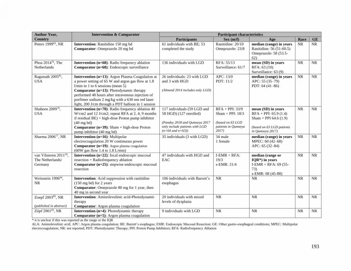
193
Author Year,
Country
Intervention & Comparator Participant characteristics
Participants Sex (m/f) Age Race GE
Peters 199963, NR Intervention: Ranitidine 150 mg bd
Comparator: Omeprazole 20 mg bd
61 individuals with BE; 53
completed the study
Ranitidine: 20/10
Omeprazole: 23/8
median (range) in years
Ranitidine: 56 (51-60.5)
Omeprazole: 58 (53.5-
62)
NR NR
Phoa 201476, The
Netherlands
Intervention (n=68): Radio frequency ablation
Comparator (n=68): Endoscopic surveillance
136 individuals with LGD RFA: 55/13
Surveillance: 61/7
mean (SD) in years
RFA: 63 (10)
Surveillance: 63 (9)
NR NR
Ragunath 200581,
USA
Intervention (n=13): Argon Plasma Coagulation at
a power setting of 65 W and argon gas flow at 1.8
l/min in 1 to 6 sessions (mean 5)
Comparator (n=13): Photodynamic therapy
performed 48 hours after intravenous injection of
porfimer sodium 2 mg/kg with a 630 nm red laser
light, 200 J/cm through a PDT balloon in 1 session
26 individuals: 23 with LGD
and 3 with HGD
(Almond 2014 includes only LGD)
APC: 13/0
PDT: 11/2
median (range) in years
APC: 55 (35–79)
PDT: 64 (41–86)
NR NR
Shaheen 200970,
USA
Intervention (n=78): Radio frequency ablation 40
W/cm2 and 12 J/cm2; repeat RFA at 2, 4, 9 months
if residual BE) + high-dose Proton pump inhibitor
(40 mg bd)
Comparator (n=39): Sham + high-dose Proton
pump inhibitor (40 mg bd)
117 individuals (59 LGD and
58 HGD) (127 enrolled)
(Pandey 2018 and Qumseya 2017
only include patients with LGD
(n=64 and n=63))
RFA + PPI: 33/9
Sham + PPI: 18/3
(based on 63 LGD
patients in Qumseya
2017)
mean (SD) in years
RFA + PPI: 65.9 (1.4)
Sham + PPI 64.6 (1.9)
(based on 63 LGD patients in Qumseya 2017)
NR NR
Sharma 200677, NR Intervention (n=16): Multipolar
electrocoagulation 20 W continuous power
Comparator (n=19): Argon plasma coagulation
(60W gas flow 1.4 to 1.8 L/min)
35 individuals (3 with LGD) 34 male
1 female
median (range) in years
MPEC: 60 (42–68)
APC: 65 (32–84)
NR NR
van Vilsteren 201159,
The Netherlands/
Germany
Intervention (n=22): focal endoscopic mucosal
resection + Radiofrequency ablation
Comparator (n=25): stepwise endoscopic mucosal
resection
47 individuals with HGD and
EAC
f-EMR + RFA:
19/3
s-EMR: 21/4
median (range or
IQR*) in years
f-EMR + RFA: 69 (55-
73)
s-EMR: 68 (45-88)
NR NR
Weinstein 199664,
NR
Intervention: Acid suppression with ranitidine
(150 mg bd) for 2 years
Comparator: Omeprazole 80 mg for 1 year, then
40 mg in second year
106 individuals with Barrett’s
esophagus
NR NR NR NR
Zoepf 200382, NR (published in abstract)
Intervention: Aminolevulinic acid-Photodynamic
therapy
Comparator: Argon plasma coagulation
20 individuals with mixed
levels of dysplasia
NR NR NR NR
Zӧpf 200183, NR Intervention (n=4): Photodynamic therapy
Comparator (n=5): Argon plasma coagulation 9 individuals with LGD NR NR NR NR
* it is unclear if this was reported as the range or the IQR
ALA: Aminolevulinic acid; APC: Argon plasma coagulation; BE: Barrett’s esophagus; EMR: Endoscopic Mucosal Resection; GE: Other gastro-esophageal conditions; MPEC: Multipolar
electrocoagulation; NR: not reported; PDT: Photodynamic Therapy; PPI: Proton Pump Inhibitors; RFA: Radiofrequency Ablation

194
Appendix 10. Evaluation of overlap of studies and concordance of results among reviews
Evidence
Set
Outcome(s) Total
publications†
# index
publications
#
reviews
CCA Concordance
2.1 Reduction in area (%) of BE at 12 months 3 2 2 0.5 Yes
3.1 Progression to cancer at latest possible time point
(up to 2 years)
2 1 2 1 Yes
3.1 Progression to cancer (at 5 years) 2 1 2 1 Yes
3.1 Progression from IM to dysplasia 2 1 2 1 Yes
3.1 Dysplasia eradication 4 2 2 1 Yes, reasonable overlap across reported data.
3.1 Complete eradication of BE over the course of the
study (5 years)
4 2 2 1 Yes
3.1 Reduction in length (cm) of BE at 12 months;
Reduction in area (%) of BE at 12 months; Area of
regression; Evidence of regression
3 1 3 1 Outcomes measured differently across reviews;
Li 2008 and Fayter 2010 are concordant for area
of regression.
5.1 Progression to cancer at five years or latest time
point; Cumulative progression to EAC over follow
up
2 1 2 1 n/a: one review reports on patient subset
5.1 Progression to higher grades of dysplasia;
Progression to high-grade dysplasia (Qumseya
2017); Progression to high-grade dysplasia (per
patient-year) (among those with LGD);
Progression to high-grade dysplasia (Pandey 2018)
3 1 3 1 Outcomes measured differently across reviews
but the two reviews that report data in the same
way are concordant.
5.1 Complete eradication of dysplasia at 12 months;
Complete eradication of dysplasia
2 1 2 1 n/a: one reviews reports on patient subset
6.1 Progression to cancer 2 1 2 1 Yes
6.1 Progression to dysplasia from IM; Progression
from non-dysplastic BE to BE with dysplasia
2 1 2 1 Different data reported for intervention group, led
to discordant results.
7.1 Eradication of high-grade dysplasia 2 1 2 1 Yes
8.1 Progression to high-grade dysplasia 2 1 2 1 Yes
9.2 Histological complete ablation of BE; Treatment
failure (no ablation of BE)
6 2 3 1 Yes, likely but some reporting issues.
10.1 Complete eradication of dysplasia at 12 months
(see notes column of the evidence set 10.1 for this
outcome that comments on 4 month data)
n/a 1 review
10.1 Complete eradication of dysplasia at 12 months 5 2 4 0.5 Concordance not relevant; patient subset in
Almond
10.1 Complete eradication of BE at 12 months;
Complete eradication of IM
5 3 3 0.3333333 Differences in how information reported makes
concordance assessment difficult across all three
reviews, but Rees 2010 and Almond 2014 results
overlap.

195
Evidence
Set
Outcome(s) Total
publications†
# index
publications
#
reviews
CCA Concordance
10.1 Reduction in length (cm) of BE at 12 months; BE
surface reduction; Length of regression (median)
(endoscopic change); Reduction in length
4 3 3 0.1666667 n/a: differences in measurement and reporting
preclude assessment
10.1 Stricture formation; Stricture 6 3 3 0.5 n/a: Almond 2014 focused on patient subset
(LGD), precluding concordance comparison;
unclear if this explains inclusion of one trial only.
Other two reviews reported information
differently.
11.1 Complete eradication of IM; Complete eradication
of IM (end of treatment)
2 1 2 1 Yes (although effect estimates not provided in
one study, results are similar)
11.1 Complete eradication of IM with no recurrence at
follow- up
2 1 2 1 Yes, although effect estimate available for one,
available information similar
11.1 Acute bleeding endoscopically treated; Bleeding 2 1 2 1 Yes
† including double counting
BE: Barrett’s esophagus; CCA: corrected covered area; IM: Intestinal metaplasia; LGD: Low-grade dysplasia


