Embed Size (px)
Citation preview
Please cite this article in press as: Bartos JA, et al. Bundled postconditioning therapies improve hemodynamics and neurologic recoveryafter 17 min of untreated cardiac arrest. Resuscitation (2014), http://dx.doi.org/10.1016/j.resuscitation.2014.10.019
ARTICLE IN PRESSG ModelRESUS 6180 1–7
Resuscitation xxx (2014) xxx–xxx
Contents lists available at ScienceDirect
Resuscitation
j ourna l h o me pa g e : www.elsev ier .com/ locate / resusc i ta t ion
Experimental paper
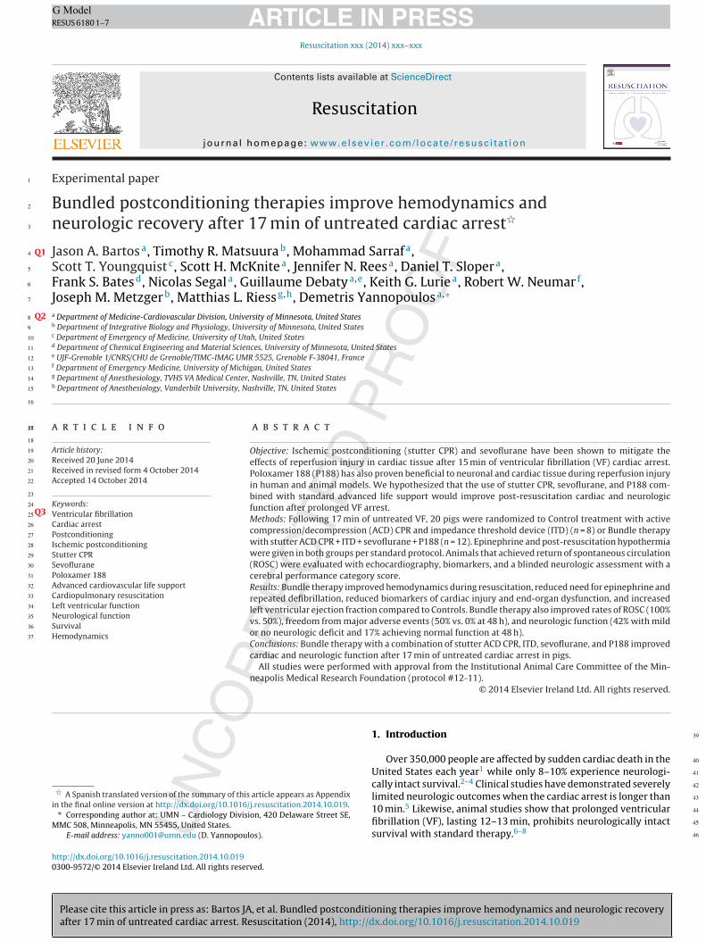
Bundled postconditioning therapies improve hemodynamics andneurologic recovery after 17 min of untreated cardiac arrest!
Jason A. Bartosa, Timothy R. Matsuurab, Mohammad Sarrafa,Q1
Scott T. Youngquist c, Scott H. McKnitea, Jennifer N. Reesa, Daniel T. Slopera,Frank S. Batesd, Nicolas Segala, Guillaume Debatya,e, Keith G. Luriea, Robert W. Neumarf,Joseph M. Metzgerb, Matthias L. Riessg,h, Demetris Yannopoulosa,∗
a Department of Medicine-Cardiovascular Division, University of Minnesota, United StatesQ2b Department of Integrative Biology and Physiology, University of Minnesota, United Statesc Department of Emergency of Medicine, University of Utah, United Statesd Department of Chemical Engineering and Material Sciences, University of Minnesota, United Statese UJF-Grenoble 1/CNRS/CHU de Grenoble/TIMC-IMAG UMR 5525, Grenoble F-38041, Francef Department of Emergency Medicine, University of Michigan, United Statesg Department of Anesthesiology, TVHS VA Medical Center, Nashville, TN, United Statesh Department of Anesthesiology, Vanderbilt University, Nashville, TN, United States
a r t i c l e i n f o
Article history:Received 20 June 2014Received in revised form 4 October 2014Accepted 14 October 2014
Keywords:Ventricular fibrillationQ3Cardiac arrestPostconditioningIschemic postconditioningStutter CPRSevofluranePoloxamer 188Advanced cardiovascular life supportCardiopulmonary resuscitationLeft ventricular functionNeurological functionSurvivalHemodynamics
a b s t r a c t
Objective: Ischemic postconditioning (stutter CPR) and sevoflurane have been shown to mitigate theeffects of reperfusion injury in cardiac tissue after 15 min of ventricular fibrillation (VF) cardiac arrest.Poloxamer 188 (P188) has also proven beneficial to neuronal and cardiac tissue during reperfusion injuryin human and animal models. We hypothesized that the use of stutter CPR, sevoflurane, and P188 com-bined with standard advanced life support would improve post-resuscitation cardiac and neurologicfunction after prolonged VF arrest.Methods: Following 17 min of untreated VF, 20 pigs were randomized to Control treatment with activecompression/decompression (ACD) CPR and impedance threshold device (ITD) (n = 8) or Bundle therapywith stutter ACD CPR + ITD + sevoflurane + P188 (n = 12). Epinephrine and post-resuscitation hypothermiawere given in both groups per standard protocol. Animals that achieved return of spontaneous circulation(ROSC) were evaluated with echocardiography, biomarkers, and a blinded neurologic assessment with acerebral performance category score.Results: Bundle therapy improved hemodynamics during resuscitation, reduced need for epinephrine andrepeated defibrillation, reduced biomarkers of cardiac injury and end-organ dysfunction, and increasedleft ventricular ejection fraction compared to Controls. Bundle therapy also improved rates of ROSC (100%vs. 50%), freedom from major adverse events (50% vs. 0% at 48 h), and neurologic function (42% with mildor no neurologic deficit and 17% achieving normal function at 48 h).Conclusions: Bundle therapy with a combination of stutter ACD CPR, ITD, sevoflurane, and P188 improvedcardiac and neurologic function after 17 min of untreated cardiac arrest in pigs.
All studies were performed with approval from the Institutional Animal Care Committee of the Min-neapolis Medical Research Foundation (protocol #12-11).
© 2014 Elsevier Ireland Ltd. All rights reserved.
! A Spanish translated version of the summary of this article appears as Appendixin the final online version at http://dx.doi.org/10.1016/j.resuscitation.2014.10.019.
∗ Corresponding author at: UMN – Cardiology Division, 420 Delaware Street SE,MMC 508, Minneapolis, MN 55455, United States.
E-mail address: [email protected] (D. Yannopoulos).
1. Introduction
Over 350,000 people are affected by sudden cardiac death in theUnited States each year1 while only 8–10% experience neurologi-cally intact survival.2–4 Clinical studies have demonstrated severelylimited neurologic outcomes when the cardiac arrest is longer than10 min.5 Likewise, animal studies show that prolonged ventricularfibrillation (VF), lasting 12–13 min, prohibits neurologically intactsurvival with standard therapy.6–8
http://dx.doi.org/10.1016/j.resuscitation.2014.10.0190300-9572/© 2014 Elsevier Ireland Ltd. All rights reserved.
1
2
3
4
5
6
7
8
9
10
11
12
13
14
15
16
17
18
19
20
21
22
23
24
25
26
27
28
29
30
31
32
33
34
35
36
37
38
39
40
41
42
43
44
45
46
Please cite this article in press as: Bartos JA, et al. Bundled postconditioning therapies improve hemodynamics and neurologic recoveryafter 17 min of untreated cardiac arrest. Resuscitation (2014), http://dx.doi.org/10.1016/j.resuscitation.2014.10.019
ARTICLE IN PRESSG ModelRESUS 6180 1–7
2 J.A. Bartos et al. / Resuscitation xxx (2014) xxx–xxx
Further study of global ischemia has revealed two types of injurywith distinct mechanisms. The first is related to ischemic damageincurred during the period of arrest. The second is injury induced bytissue reperfusion.9–11 In contrast to ischemic injury which occursprior to the arrival of emergency medical services, reperfusioninjury, the injury caused by re-establishing blood flow, is amenableto medical management as it occurs when therapy begins.
Recent studies in animal models of cardiac arrest providepromising evidence for improved cardiac function and neurologicrecovery when therapies targeting reperfusion injury are used.Ischemic postconditioning, which utilizes structured pauses in CPR,was shown to improve left ventricular ejection fraction (LVEF)and neurologically intact survival in a porcine model of cardiacarrest including 15 min of untreated VF.12 Sevoflurane has beenshown to reduce release of cardiac biomarkers, reduce apoptosis,and improve LVEF; however, no difference in neurologic functionwas reported.13,14 Poloxamer 188 (P188) is a nonionic copolymerthought to adhere to gaps in the cell membrane, thereby blockingpores caused by severe cellular stress.15
While each of these treatments is likely to provide protec-tion against cardiac and neuronal reperfusion injury induced bycardiac arrest, we hypothesized that combining these therapieswould provide a synergistic benefit even in severe injury. Wetherefore combined these therapies in a porcine model of cardiacarrest including 17 min of untreated VF. The tissue damage inducedby this duration of cardiac arrest has generally been consideredirreversible. We assessed hemodynamic parameters, markers ofcardiac injury and function, neurologic recovery, and freedom fromserious adverse events.
2. Materials and methods
All studies were performed with approval from the Institu-tional Animal Care and Use Committee of the Minneapolis MedicalResearch Foundation and the National Research Council’s Guide-lines for the Care and Use of Laboratory Animals. Yorkshire farmpigs weighing 38.6 ± 0.4 kg were used.
2.1. Preparatory phase
The anesthesia, data monitoring and recording, and surgicalpreparation have been described in detail previously.12 The femoralartery and right external jugular vein were cannulated percuta-neously with eight French sheaths to provide access for continuoushemodynamic monitoring and repeated blood tests.
2.2. Hemodynamic monitoring
Surface electrocardiographic tracings were recorded contin-uously. Central aortic and right atrial pressures were recordedcontinuously with micromanometer-tipped catheters (MillarInstruments) placed at the proximal descending thoracic aorta viathe femoral artery and right atrium via the right external jugularvein, respectively. Coronary perfusion pressure (CPP) was calcu-lated as the difference between the diastolic aortic pressure andright atrial pressure during the decompression phase of CPR. Endtidal CO2 (ETCO2), blood oxygen saturation, tidal volume, andminute ventilation were continuously monitored with a respira-tory monitor (CO2SMO Plus, Novametrix Medical Systems). All datawere recorded with a digital recording system (BIOPAC MP150,BIOPAC Systems Inc.).
2.3. Experimental protocol
The animals were allowed to stabilize with oxygen satu-ration >95% and ETCO2 between 35 and 42 mmHg for 5 min.
Isoflurane anesthesia was stopped 5 min prior to VF inductionwith direct intracardiac current delivered via a temporary pac-ing wire (St. Jude Medical). Following 17 min of untreated VF,20 pigs were randomized to Control or Bundle treatment result-ing in 8 pigs in the Control group and 12 pigs in the Bundlegroup.
3. Experimental groups (Fig. 1):
Control therapy: Active compression/decompression (ACD) CPRwas delivered using a pneumatically driven automated piston(Pneumatic Compression Controller, Ambu International) withchest compressions occurring at a rate of 100 per minute. Com-pression depth was maintained at 25% of the anteroposterior chestdiameter. Compression force, rate, and depth were continuouslyrecorded and controlled. An impedance threshold device (ITD;ResQPOD, Advanced Circulatory Systems Inc.) was used in addi-tion to ACD CPR. Asynchronous positive-pressure ventilation wasdelivered during CPR with room air. A tidal volume of 10 mL kg−1
was delivered at 10 breaths min−1. Isoflurane was not restarteduntil return of spontaneous circulation (ROSC) was achieved. Atminute 3 of CPR all animals received 0.015 mg kg−1 of epinephrine.Up to three 275 J biphasic shocks were delivered at minute 4. IfROSC was not achieved, CPR continued with epinephrine adminis-tered every 3 min. Defibrillation was attempted every 2 min untilROSC was achieved or a total of 15 min of CPR was complete. Ifventricular arrhythmias developed after ROSC was achieved, defi-brillation was attempted and amiodarone 40 mg IV was given.Sodium bicarbonate (50 meq) was given to all animals at 5 min ofresuscitation.
Bundle therapy: After 17 min of untreated VF, resuscitationbegan with ischemic postconditioning including stutter ACDCPR + ITD composed of 20 s of compressions followed by a 20 spause. Sevoflurane was delivered during each pause at an end-tidalconcentration of 2.0 vol.% with three positive pressure ventilationsat a rate of 10 breaths min−1. No ventilations were delivered whilecompressions were performed. Once three cycles of stutter ACDCPR + ITD were complete, continuous chest compressions were per-formed similar to the Control group. Continuous ventilation with10 breaths min−1 was initiated 20 s later. P188 (250 mg kg−1) wasdelivered at minute 2 of resuscitation. Epinephrine (0.015 mg kg−1)was given at minute 3 of the resuscitation. Defibrillation wasattempted at minute 4 as discussed above. If ROSC was achieved,P188 (460 mg kg−1) was infused over 4 h.
3.1. Post-ROSC care
The details of post-ROSC care have been described in detailelsewhere.12 Both groups received post-resuscitation therapeutichypothermia to simulate best practice and optimize neurologicrecovery. All animals received 1 L chilled saline (8–10 ◦C) imme-diately post-ROSC followed by surface cooling with ethanol soakedtowels. Target temperature (34 ◦C) was maintained for 4 h with theuse of external cooling (Arctic Sun, Medivance Inc.). Animals wererewarmed at 0.5 ◦C/h to 36 ◦C.
In the event of an adverse event meeting predetermined criteriaincluding status epilepticus, severe cardiopulmonary distress withevidence of agonal breathing, cyanosis, pulmonary edema, or deepcoma at 24 h with the inability to respond to painful stimuli basedon the judgment of the veterinarian blinded to the intervention,animals were euthanized.
3.2. Echocardiographic evaluation
A transthoracic echocardiogram was obtained at baseline andagain at 15 min, 1, 4, 24, and 48 h post-ROSC. Parasternal long
47
48
49
50
51
52
53
54
55
56
57
58
59
60
61
62
63
64
65
66
67
68
69
70
71
72
73
74
75
76
77
78
79
80
81
82
83
84
85
86
87
88
89
90
91
92
93
94
95
96
97
98
99
100
101
102
103
104
105
106
107
108
109
110
111
112
113
114
115
116
117
118
119
120
121
122
123
124
125
126
127
128
129
130
131
132
133
134
135
136
137
138
139
140
141
142
143
144
145
146
147
148
149
150
151
152
153
154
155
156
157
158
159
160
161
162
163
164
Please cite this article in press as: Bartos JA, et al. Bundled postconditioning therapies improve hemodynamics and neurologic recoveryafter 17 min of untreated cardiac arrest. Resuscitation (2014), http://dx.doi.org/10.1016/j.resuscitation.2014.10.019
ARTICLE IN PRESSG ModelRESUS 6180 1–7
J.A. Bartos et al. / Resuscitation xxx (2014) xxx–xxx 3
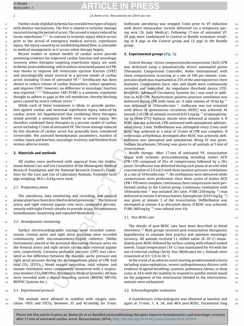
Fig. 1. Bundle therapy and Control protocols with associated representative hemodynamic tracings. Therapy was initiated after 17 min of untreated ventricular fibrillation(VF). (A) Control therapy included active compression decompression CPR, application of an impedance threshold device (ACD CPR + ITD), epinephrine (Epi), and defibrillation(Defib). Return of spontaneous circulation (ROSC) was not achieved after the initial defibrillation in this representative study leading to continuation of the resuscitation. (B)The Bundle therapy protocol included sevoflurane (Sevo), poloxamer 188 (P188), epinephrine (Epi), stutter ACD CPR + ITD, and defibrillation (Defib). ROSC is achieved aftera single defibrillation in this representative study.
and short axis views were obtained and analyzed by clinicalechocardiographers blinded to the intervention as described pre-viously.12
3.3. Cardiac biomarkers, liver function tests, and renal functionassessment
Arterial blood was collected at baseline and from all survivors 4 hpost-ROSC. Cardiac specific troponin I (cTnI) and creatinine phos-phokinase MB (CK-MB) were quantified via a two-site sandwichassay (Stratus CS Acute Care, Siemens). Aspartate aminotrans-ferase, alanine aminotransferase, total bilirubin, and creatininewere measured with standard human assays. Personnel performingthe analyses were blinded to the treatment.
3.4. Neurological assessment
Neurologic function was assessed using a swine-specific cere-bral performance category (CPC) at 24 and 48 h post-ROSC by acertified veterinarian blinded to the treatment. Clinical assessmentof behaviors, reflexes, and coordination are assessed as previ-ously described12 using the following scoring system: 1 = normal,2 = slightly disabled, 3 = moderately disabled but not comatose,4 = coma, 5 = dead.
3.5. Statistical analysis
Values are expressed as means ± standard error of the mean.The primary end points were the incidence of major adverse out-comes at 48 h and the CPC at 24 and 48 h. Secondary endpoints
165
166
167
168
169
170
171
172
173
174
175
176
177
178
179
180
181
182
183
184
185
186
187
188
Please cite this article in press as: Bartos JA, et al. Bundled postconditioning therapies improve hemodynamics and neurologic recoveryafter 17 min of untreated cardiac arrest. Resuscitation (2014), http://dx.doi.org/10.1016/j.resuscitation.2014.10.019
ARTICLE IN PRESSG ModelRESUS 6180 1–7
4 J.A. Bartos et al. / Resuscitation xxx (2014) xxx–xxx
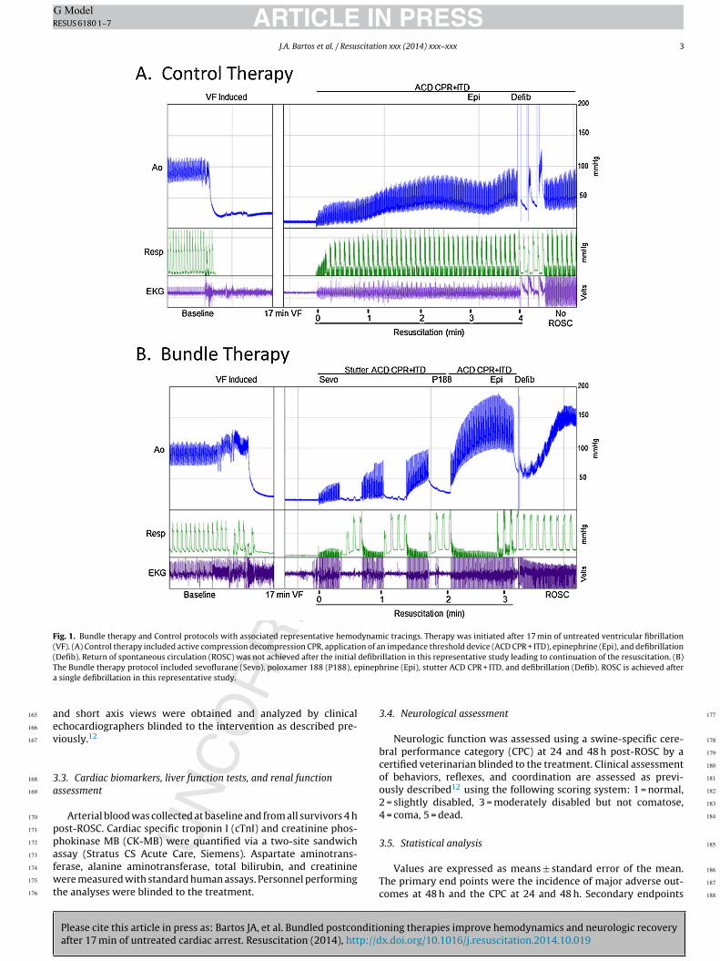
Fig. 2. Hemodynamic monitoring during resuscitation. Bundle (BND; n = 12) andControl (n = 8) therapy are compared. (A) Aortic systolic blood pressure measuredat baseline (BL), throughout the resuscitation, and after return of spontaneous cir-culation (ROSC) was achieved. (B) Aortic diastolic blood pressure measured duringall phases of resuscitation. (C) Coronary perfusion pressure is shown as measuredduring all phases of resuscitation. Error bars represent SEM. Asterisk (*) indicatesstatistical significance (p < 0.05).
included hemodynamics, need for epinephrine and defibrillations,LVEF post-ROSC, and biomarkers. Baseline data, hemodynamicparameters, epinephrine and defibrillation requirements, LVEF,biomarkers, and mean CPC scores were compared using two-tailedunpaired t-tests. Kaplan–Meier survival curves were analyzed viaMantel–Cox test. A p-value of <0.05 was considered statisticallysignificant.
4. Results
All results were obtained after 17 min of untreated VF. Therewere no significant differences in baseline hemodynamic parame-ters between treatment groups (Fig. 2).
4.1. Resuscitation hemodynamics
Systolic and diastolic blood pressures were greater with Bun-dle therapy vs. Controls at minutes 2–4 of CPR (Fig. 2A). Thisdifference did not persist once ROSC was achieved. CPP was sig-nificantly lower with Bundle therapy compared to Control therapyearly in the resuscitation while the increasing aortic pressuresobserved with Bundle therapy translated into higher CPP at minute3 of the resuscitation (Fig. 2B). The duration of CPR was signif-icantly reduced in the Bundle therapy group (5.0 ± 0.04 min vs.11.3 ± 1.5 min, p = 0.0001).
4.2. Hemodynamic and rhythm support
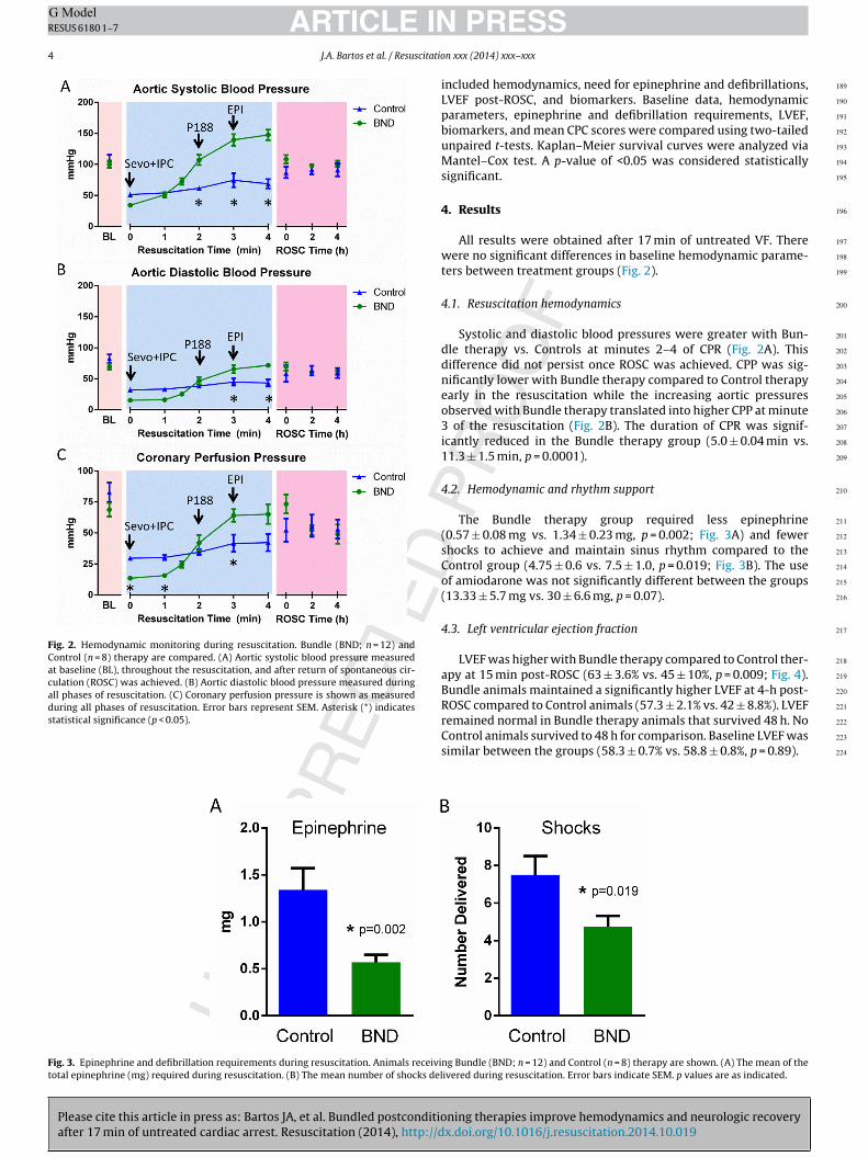
The Bundle therapy group required less epinephrine(0.57 ± 0.08 mg vs. 1.34 ± 0.23 mg, p = 0.002; Fig. 3A) and fewershocks to achieve and maintain sinus rhythm compared to theControl group (4.75 ± 0.6 vs. 7.5 ± 1.0, p = 0.019; Fig. 3B). The useof amiodarone was not significantly different between the groups(13.33 ± 5.7 mg vs. 30 ± 6.6 mg, p = 0.07).
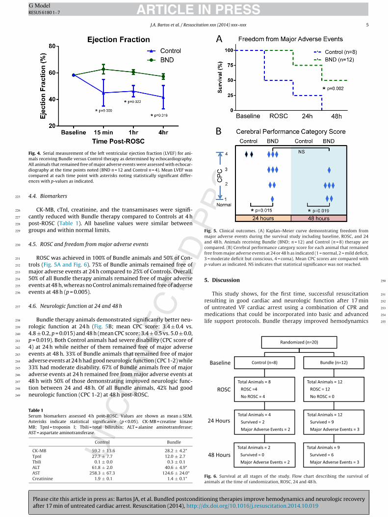
4.3. Left ventricular ejection fraction
LVEF was higher with Bundle therapy compared to Control ther-apy at 15 min post-ROSC (63 ± 3.6% vs. 45 ± 10%, p = 0.009; Fig. 4).Bundle animals maintained a significantly higher LVEF at 4-h post-ROSC compared to Control animals (57.3 ± 2.1% vs. 42 ± 8.8%). LVEFremained normal in Bundle therapy animals that survived 48 h. NoControl animals survived to 48 h for comparison. Baseline LVEF wassimilar between the groups (58.3 ± 0.7% vs. 58.8 ± 0.8%, p = 0.89).
Fig. 3. Epinephrine and defibrillation requirements during resuscitation. Animals receiving Bundle (BND; n = 12) and Control (n = 8) therapy are shown. (A) The mean of thetotal epinephrine (mg) required during resuscitation. (B) The mean number of shocks delivered during resuscitation. Error bars indicate SEM. p values are as indicated.
189
190
191
192
193
194
195
196
197
198
199
200
201
202
203
204
205
206
207
208
209
210
211
212
213
214
215
216
217
218
219
220
221
222
223
224
Please cite this article in press as: Bartos JA, et al. Bundled postconditioning therapies improve hemodynamics and neurologic recoveryafter 17 min of untreated cardiac arrest. Resuscitation (2014), http://dx.doi.org/10.1016/j.resuscitation.2014.10.019
ARTICLE IN PRESSG ModelRESUS 6180 1–7
J.A. Bartos et al. / Resuscitation xxx (2014) xxx–xxx 5
Fig. 4. Serial measurement of the left ventricular ejection fraction (LVEF) for ani-mals receiving Bundle versus Control therapy as determined by echocardiography.All animals that remained free of major adverse events were assessed with echocar-diography at the time points noted (BND n = 12 and Control n = 4). Mean LVEF wascompared at each time point with asterisks noting statistically significant differ-ences with p-values as indicated.
4.4. Biomarkers
CK-MB, cTnI, creatinine, and the transaminases were signifi-cantly reduced with Bundle therapy compared to Controls at 4 hpost-ROSC (Table 1). All baseline values were similar betweengroups and within normal limits.
4.5. ROSC and freedom from major adverse events
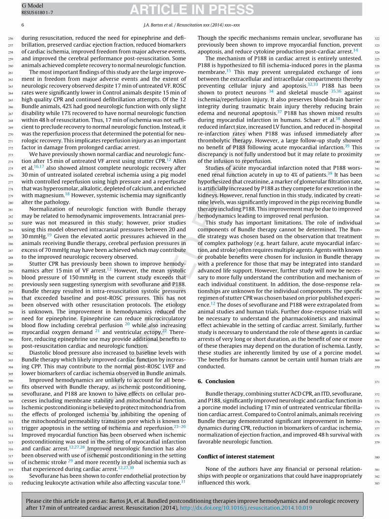
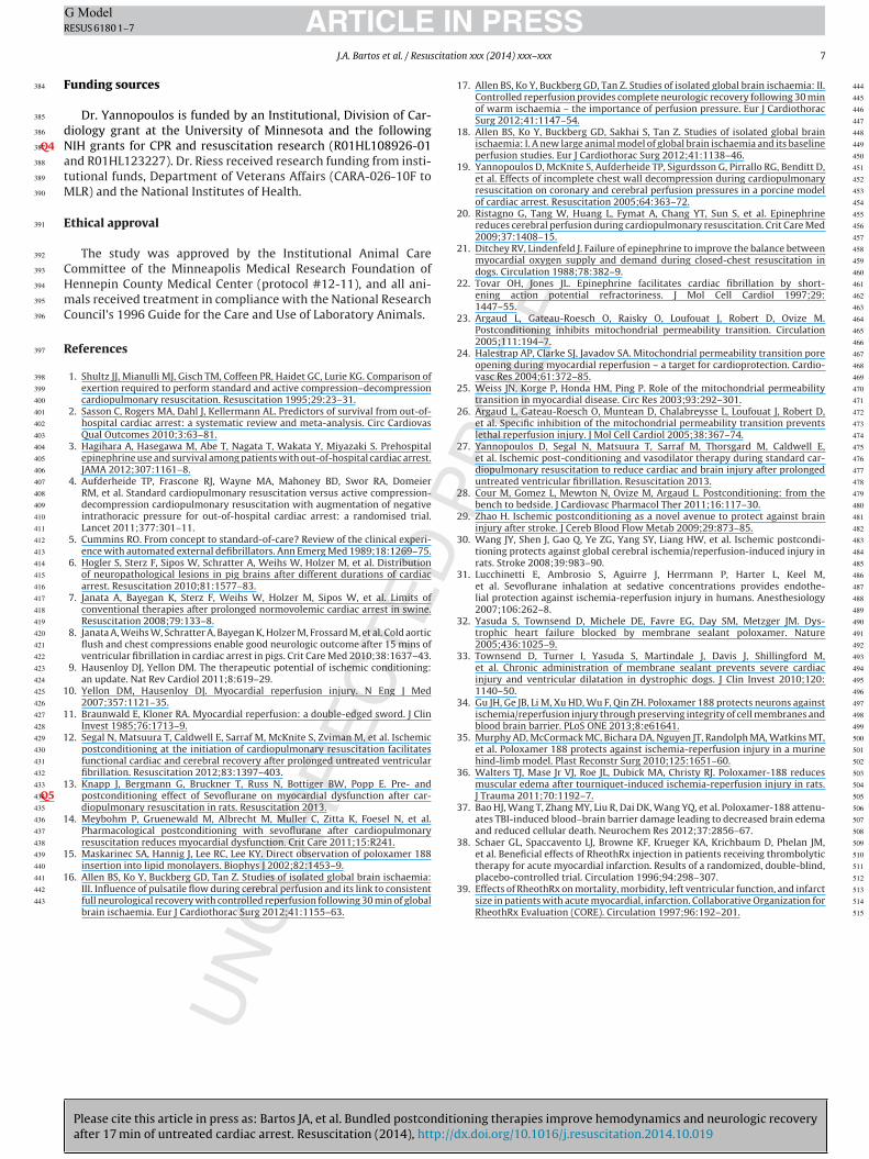
ROSC was achieved in 100% of Bundle animals and 50% of Con-trols (Fig. 5A and Fig. 6). 75% of Bundle animals remained free ofmajor adverse events at 24 h compared to 25% of Controls. Overall,50% of all Bundle therapy animals remained free of major adverseevents at 48 h, whereas no Control animals remained free of adverseevents at 48 h (p = 0.005).
4.6. Neurologic function at 24 and 48 h
Bundle therapy animals demonstrated significantly better neu-rologic function at 24 h (Fig. 5B; mean CPC score: 3.4 ± 0.4 vs.4.8 ± 0.2, p = 0.015) and 48 h (mean CPC score: 3.4 ± 0.5 vs. 5.0 ± 0.0,p = 0.019). Both Control animals had severe disability (CPC score of4) at 24 h while neither of them remained free of major adverseevents at 48 h. 33% of Bundle animals that remained free of majoradverse events at 24 h had good neurologic function (CPC 1-2) while33% had moderate disability. 67% of Bundle animals free of majoradverse events at 24 h remained free from major adverse events at48 h with 50% of those demonstrating improved neurologic func-tion between 24 and 48 h. Of all Bundle animals, 42% had goodneurologic function (CPC 1-2) at 48 h post-ROSC.
Table 1Serum biomarkers assessed 4 h post-ROSC. Values are shown as mean ± SEM.Asterisks indicate statistical significance (p < 0.05). CK-MB = creatine kinaseMB; TpnI = troponin I; Tbili = total bilirubin; ALT = alanine aminotransferase;AST = aspartate aminotransferase.
Control Bundle
CK-MB 59.2 ± 13.6 28.2 ± 4.2*TpnI 27.7 ± 7.7 12.0 ± 2.7Tbili 0.1 ± 0.0 0.3 ± 0.1ALT 61.8 ± 2.0 40.6 ± 4.9*AST 258.3 ± 67.3 124.6 ± 24.0*Creatinine 1.9 ± 0.1 1.4 ± 0.1*
Fig. 5. Clinical outcomes. (A) Kaplan–Meier curve demonstrating freedom frommajor adverse events during the survival study including baseline, ROSC, and 24and 48 h. Animals receiving Bundle (BND; n = 12) and Control (n = 8) therapy arecompared. (B) Cerebral performance category score for each animal that remainedfree from major adverse events at 24 or 48 h as indicated (1 = normal, 2 = mild deficit,3 = moderate deficit but conscious, 4 = coma). Mean CPC scores are compared withp-values as indicated. NS indicates that statistical significance was not reached.
5. Discussion
This study shows, for the first time, successful resuscitationresulting in good cardiac and neurologic function after 17 minof untreated VF cardiac arrest using a combination of CPR andmedications that could be incorporated into basic and advancedlife support protocols. Bundle therapy improved hemodynamics
Fig. 6. Survival at all stages of the study. Flow chart describing the survival ofanimals at the time of randomization, ROSC, 24 and 48 h.
225
226
227
228
229
230
231
232
233
234
235
236
237
238
239
240
241
242
243
244
245
246
247
248
249
250
251
252
253
254
255
Please cite this article in press as: Bartos JA, et al. Bundled postconditioning therapies improve hemodynamics and neurologic recoveryafter 17 min of untreated cardiac arrest. Resuscitation (2014), http://dx.doi.org/10.1016/j.resuscitation.2014.10.019
ARTICLE IN PRESSG ModelRESUS 6180 1–7
6 J.A. Bartos et al. / Resuscitation xxx (2014) xxx–xxx
during resuscitation, reduced the need for epinephrine and defi-brillation, preserved cardiac ejection fraction, reduced biomarkersof cardiac ischemia, improved freedom from major adverse events,and improved the cerebral performance post-resuscitation. Someanimals achieved complete recovery to normal neurologic function.
The most important findings of this study are the large improve-ment in freedom from major adverse events and the extent ofneurologic recovery observed despite 17 min of untreated VF. ROSCrates were significantly lower in Control animals despite 15 min ofhigh quality CPR and continued defibrillation attempts. Of the 12Bundle animals, 42% had good neurologic function with only slightdisability while 17% recovered to have normal neurologic functionwithin 48 h of resuscitation. Thus, 17 min of ischemia was not suffi-cient to preclude recovery to normal neurologic function. Instead, itwas the reperfusion process that determined the potential for neu-rologic recovery. This implicates reperfusion injury as an importantfactor in damage from prolonged cardiac arrest.
We have previously shown normal cardiac and neurologic func-tion after 15 min of untreated VF arrest using stutter CPR.12 Allenet al.16,17 also showed almost complete neurologic recovery after30 min of untreated isolated cerebral ischemia using a pig modelwith controlled reperfusion using high pressure and a reperfusatethat was hyperosmolar, alkalotic, depleted of calcium, and enrichedwith magnesium.18 However, systemic ischemia may significantlyalter the pathology.
Normalization of neurologic function with Bundle therapymay be related to hemodynamic improvements. Intracranial pres-sure was not measured in this study; however, prior studiesusing this model observed intracranial pressures between 20 and30 mmHg.19 Given the elevated aortic pressures achieved in theanimals receiving Bundle therapy, cerebral perfusion pressures inexcess of 70 mmHg may have been achieved which may contributeto the improved neurologic recovery observed.
Stutter CPR has previously been shown to improve hemody-namics after 15 min of VF arrest.12 However, the mean systolicblood pressure of 150 mmHg in the current study exceeds thatpreviously seen suggesting synergism with sevoflurane and P188.Bundle therapy resulted in intra-resuscitation systolic pressuresthat exceeded baseline and post-ROSC pressures. This has notbeen observed with other resuscitation protocols. The etiologyis unknown. The improvement in hemodynamics reduced theneed for epinephrine. Epinephrine can reduce microcirculatoryblood flow including cerebral perfusion 20 while also increasingmyocardial oxygen demand 21 and ventricular ectopy.22 There-fore, reducing epinephrine use may provide additional benefits topost-resuscitation cardiac and neurologic function.
Diastolic blood pressure also increased to baseline levels withBundle therapy which likely improved cardiac function by increas-ing CPP. This may contribute to the normal post-ROSC LVEF andlower biomarkers of cardiac ischemia observed in Bundle animals.
Improved hemodynamics are unlikely to account for all bene-fits observed with Bundle therapy, as ischemic postconditioning,sevoflurane, and P188 are known to have effects on cellular pro-cesses including membrane stability and mitochondrial function.Ischemic postconditioning is believed to protect mitochondria fromthe effects of prolonged ischemia by inhibiting the opening ofthe mitochondrial permeability transition pore which is known totrigger apoptosis in the setting of ischemia and reperfusion.23–26
Improved myocardial function has been observed when ischemicpostconditioning was used in the setting of myocardial infarctionand cardiac arrest.12,27,28 Improved neurologic function has alsobeen observed with use of ischemic postconditioning in the settingof ischemic stroke 29 and more recently in global ischemia such asthat experienced during cardiac arrest.12,27,30
Sevoflurane has been shown to confer endothelial protection byreducing leukocyte activation while also affecting vascular tone.31
Though the specific mechanisms remain unclear, sevoflurane haspreviously been shown to improve myocardial function, preventapoptosis, and reduce cytokine production post-cardiac arrest.14
The mechanism of P188 in cardiac arrest is entirely untested.P188 is hypothesized to fill ischemia-induced pores in the plasmamembrane.15 This may prevent unregulated exchange of ionsbetween the extracellular and intracellular compartments therebypreventing cellular injury and apoptosis.32,33 P188 has beenshown to protect neurons 34 and skeletal muscle 35,36 againstischemia/reperfusion injury. It also preserves blood-brain barrierintegrity during traumatic brain injury thereby reducing brainedema and neuronal apoptosis.37 P188 has shown mixed resultsduring myocardial infarction in humans. Schaer et al.38 showedreduced infarct size, increased LV function, and reduced in-hospitalre-infarction rates when P188 was infused immediately afterthrombolytic therapy. However, a large follow-up study showedno benefit of P188 following acute myocardial infarction.39 Thisdiscrepancy is not fully understood but it may relate to proximityof the infusion to reperfusion.
Studies of acute myocardial infarction noted that P188 wors-ened renal function acutely in up to 4% of patients.39 It has beenhypothesized that creatinine, a marker of glomerular filtration rate,is artificially increased by P188 as they compete for excretion in thekidneys. However, renal function in this study, indicated by creati-nine levels, was significantly improved in the pigs receiving Bundletherapy including P188. This improvement may be due to improvedhemodynamics leading to improved renal perfusion.
This study has important limitations. The role of individualcomponents of Bundle therapy cannot be determined. The Bun-dle strategy was chosen based on the observation that treatmentof complex pathology (e.g. heart failure, acute myocardial infarc-tion, and stroke) often requires multiple agents. Agents with knownor probable benefits were chosen for inclusion in Bundle therapywith a preference for those that may be integrated into standardadvanced life support. However, further study will now be neces-sary to more fully understand the contribution and mechanism ofeach individual constituent. In addition, the dose-response rela-tionships are unknown for the individual components. The specificregimen of stutter CPR was chosen based on prior published experi-ence.12 The doses of sevoflurane and P188 were extrapolated fromanimal studies and human trials. Further dose-response trials willbe necessary to understand the pharmacokinetics and maximaleffect achievable in the setting of cardiac arrest. Similarly, furtherstudy is necessary to understand the role of these agents in cardiacarrests of very long or short duration, as the benefit of one or moreof these therapies may depend on the duration of ischemia. Lastly,these studies are inherently limited by use of a porcine model.The benefits for humans cannot be certain until human trials areconducted.
6. Conclusion
Bundle therapy, combining stutter ACD CPR, an ITD, sevoflurane,and P188, significantly improved neurologic and cardiac function ina porcine model including 17 min of untreated ventricular fibrilla-tion cardiac arrest. Compared to Control animals, animals receivingBundle therapy demonstrated significant improvement in hemo-dynamics during CPR, reduction in biomarkers of cardiac ischemia,normalization of ejection fraction, and improved 48 h survival withfavorable neurologic function.
Conflict of interest statement
None of the authors have any financial or personal relation-ships with people or organizations that could have inappropriatelyinfluenced this work.
256
257
258
259
260
261
262
263
264
265
266
267
268
269
270
271
272
273
274
275
276
277
278
279
280
281
282
283
284
285
286
287
288
289
290
291
292
293
294
295
296
297
298
299
300
301
302
303
304
305
306
307
308
309
310
311
312
313
314
315
316
317
318
319
320
321
322
323
324
325
326
327
328
329
330
331
332
333
334
335
336
337
338
339
340
341
342
343
344
345
346
347
348
349
350
351
352
353
354
355
356
357
358
359
360
361
362
363
364
365
366
367
368
369
370
371
372
373
374
375
376
377
378
379
380
381
382
383
Please cite this article in press as: Bartos JA, et al. Bundled postconditioning therapies improve hemodynamics and neurologic recoveryafter 17 min of untreated cardiac arrest. Resuscitation (2014), http://dx.doi.org/10.1016/j.resuscitation.2014.10.019
ARTICLE IN PRESSG ModelRESUS 6180 1–7
J.A. Bartos et al. / Resuscitation xxx (2014) xxx–xxx 7
Funding sources
Dr. Yannopoulos is funded by an Institutional, Division of Car-diology grant at the University of Minnesota and the followingNIH grants for CPR and resuscitation research (R01HL108926-01Q4and R01HL123227). Dr. Riess received research funding from insti-tutional funds, Department of Veterans Affairs (CARA-026-10F toMLR) and the National Institutes of Health.
Ethical approval
The study was approved by the Institutional Animal CareCommittee of the Minneapolis Medical Research Foundation ofHennepin County Medical Center (protocol #12-11), and all ani-mals received treatment in compliance with the National ResearchCouncil’s 1996 Guide for the Care and Use of Laboratory Animals.
References
1. Shultz JJ, Mianulli MJ, Gisch TM, Coffeen PR, Haidet GC, Lurie KG. Comparison ofexertion required to perform standard and active compression–decompressioncardiopulmonary resuscitation. Resuscitation 1995;29:23–31.
2. Sasson C, Rogers MA, Dahl J, Kellermann AL. Predictors of survival from out-of-hospital cardiac arrest: a systematic review and meta-analysis. Circ CardiovasQual Outcomes 2010;3:63–81.
3. Hagihara A, Hasegawa M, Abe T, Nagata T, Wakata Y, Miyazaki S. Prehospitalepinephrine use and survival among patients with out-of-hospital cardiac arrest.JAMA 2012;307:1161–8.
4. Aufderheide TP, Frascone RJ, Wayne MA, Mahoney BD, Swor RA, DomeierRM, et al. Standard cardiopulmonary resuscitation versus active compression-decompression cardiopulmonary resuscitation with augmentation of negativeintrathoracic pressure for out-of-hospital cardiac arrest: a randomised trial.Lancet 2011;377:301–11.
5. Cummins RO. From concept to standard-of-care? Review of the clinical experi-ence with automated external defibrillators. Ann Emerg Med 1989;18:1269–75.
6. Hogler S, Sterz F, Sipos W, Schratter A, Weihs W, Holzer M, et al. Distributionof neuropathological lesions in pig brains after different durations of cardiacarrest. Resuscitation 2010;81:1577–83.
7. Janata A, Bayegan K, Sterz F, Weihs W, Holzer M, Sipos W, et al. Limits ofconventional therapies after prolonged normovolemic cardiac arrest in swine.Resuscitation 2008;79:133–8.
8. Janata A, Weihs W, Schratter A, Bayegan K, Holzer M, Frossard M, et al. Cold aorticflush and chest compressions enable good neurologic outcome after 15 mins ofventricular fibrillation in cardiac arrest in pigs. Crit Care Med 2010;38:1637–43.
9. Hausenloy DJ, Yellon DM. The therapeutic potential of ischemic conditioning:an update. Nat Rev Cardiol 2011;8:619–29.
10. Yellon DM, Hausenloy DJ. Myocardial reperfusion injury. N Eng J Med2007;357:1121–35.
11. Braunwald E, Kloner RA. Myocardial reperfusion: a double-edged sword. J ClinInvest 1985;76:1713–9.
12. Segal N, Matsuura T, Caldwell E, Sarraf M, McKnite S, Zviman M, et al. Ischemicpostconditioning at the initiation of cardiopulmonary resuscitation facilitatesfunctional cardiac and cerebral recovery after prolonged untreated ventricularfibrillation. Resuscitation 2012;83:1397–403.
13. Knapp J, Bergmann G, Bruckner T, Russ N, Bottiger BW, Popp E. Pre- andpostconditioning effect of Sevoflurane on myocardial dysfunction after car-Q5diopulmonary resuscitation in rats. Resuscitation 2013.
14. Meybohm P, Gruenewald M, Albrecht M, Muller C, Zitta K, Foesel N, et al.Pharmacological postconditioning with sevoflurane after cardiopulmonaryresuscitation reduces myocardial dysfunction. Crit Care 2011;15:R241.
15. Maskarinec SA, Hannig J, Lee RC, Lee KY. Direct observation of poloxamer 188insertion into lipid monolayers. Biophys J 2002;82:1453–9.
16. Allen BS, Ko Y, Buckberg GD, Tan Z. Studies of isolated global brain ischaemia:III. Influence of pulsatile flow during cerebral perfusion and its link to consistentfull neurological recovery with controlled reperfusion following 30 min of globalbrain ischaemia. Eur J Cardiothorac Surg 2012;41:1155–63.
17. Allen BS, Ko Y, Buckberg GD, Tan Z. Studies of isolated global brain ischaemia: II.Controlled reperfusion provides complete neurologic recovery following 30 minof warm ischaemia – the importance of perfusion pressure. Eur J CardiothoracSurg 2012;41:1147–54.
18. Allen BS, Ko Y, Buckberg GD, Sakhai S, Tan Z. Studies of isolated global brainischaemia: I. A new large animal model of global brain ischaemia and its baselineperfusion studies. Eur J Cardiothorac Surg 2012;41:1138–46.
19. Yannopoulos D, McKnite S, Aufderheide TP, Sigurdsson G, Pirrallo RG, Benditt D,et al. Effects of incomplete chest wall decompression during cardiopulmonaryresuscitation on coronary and cerebral perfusion pressures in a porcine modelof cardiac arrest. Resuscitation 2005;64:363–72.
20. Ristagno G, Tang W, Huang L, Fymat A, Chang YT, Sun S, et al. Epinephrinereduces cerebral perfusion during cardiopulmonary resuscitation. Crit Care Med2009;37:1408–15.
21. Ditchey RV, Lindenfeld J. Failure of epinephrine to improve the balance betweenmyocardial oxygen supply and demand during closed-chest resuscitation indogs. Circulation 1988;78:382–9.
22. Tovar OH, Jones JL. Epinephrine facilitates cardiac fibrillation by short-ening action potential refractoriness. J Mol Cell Cardiol 1997;29:1447–55.
23. Argaud L, Gateau-Roesch O, Raisky O, Loufouat J, Robert D, Ovize M.Postconditioning inhibits mitochondrial permeability transition. Circulation2005;111:194–7.
24. Halestrap AP, Clarke SJ, Javadov SA. Mitochondrial permeability transition poreopening during myocardial reperfusion – a target for cardioprotection. Cardio-vasc Res 2004;61:372–85.
25. Weiss JN, Korge P, Honda HM, Ping P. Role of the mitochondrial permeabilitytransition in myocardial disease. Circ Res 2003;93:292–301.
26. Argaud L, Gateau-Roesch O, Muntean D, Chalabreysse L, Loufouat J, Robert D,et al. Specific inhibition of the mitochondrial permeability transition preventslethal reperfusion injury. J Mol Cell Cardiol 2005;38:367–74.
27. Yannopoulos D, Segal N, Matsuura T, Sarraf M, Thorsgard M, Caldwell E,et al. Ischemic post-conditioning and vasodilator therapy during standard car-diopulmonary resuscitation to reduce cardiac and brain injury after prolongeduntreated ventricular fibrillation. Resuscitation 2013.
28. Cour M, Gomez L, Mewton N, Ovize M, Argaud L. Postconditioning: from thebench to bedside. J Cardiovasc Pharmacol Ther 2011;16:117–30.
29. Zhao H. Ischemic postconditioning as a novel avenue to protect against braininjury after stroke. J Cereb Blood Flow Metab 2009;29:873–85.
30. Wang JY, Shen J, Gao Q, Ye ZG, Yang SY, Liang HW, et al. Ischemic postcondi-tioning protects against global cerebral ischemia/reperfusion-induced injury inrats. Stroke 2008;39:983–90.
31. Lucchinetti E, Ambrosio S, Aguirre J, Herrmann P, Harter L, Keel M,et al. Sevoflurane inhalation at sedative concentrations provides endothe-lial protection against ischemia-reperfusion injury in humans. Anesthesiology2007;106:262–8.
32. Yasuda S, Townsend D, Michele DE, Favre EG, Day SM, Metzger JM. Dys-trophic heart failure blocked by membrane sealant poloxamer. Nature2005;436:1025–9.
33. Townsend D, Turner I, Yasuda S, Martindale J, Davis J, Shillingford M,et al. Chronic administration of membrane sealant prevents severe cardiacinjury and ventricular dilatation in dystrophic dogs. J Clin Invest 2010;120:1140–50.
34. Gu JH, Ge JB, Li M, Xu HD, Wu F, Qin ZH. Poloxamer 188 protects neurons againstischemia/reperfusion injury through preserving integrity of cell membranes andblood brain barrier. PLoS ONE 2013;8:e61641.
35. Murphy AD, McCormack MC, Bichara DA, Nguyen JT, Randolph MA, Watkins MT,et al. Poloxamer 188 protects against ischemia-reperfusion injury in a murinehind-limb model. Plast Reconstr Surg 2010;125:1651–60.
36. Walters TJ, Mase Jr VJ, Roe JL, Dubick MA, Christy RJ. Poloxamer-188 reducesmuscular edema after tourniquet-induced ischemia-reperfusion injury in rats.J Trauma 2011;70:1192–7.
37. Bao HJ, Wang T, Zhang MY, Liu R, Dai DK, Wang YQ, et al. Poloxamer-188 attenu-ates TBI-induced blood–brain barrier damage leading to decreased brain edemaand reduced cellular death. Neurochem Res 2012;37:2856–67.
38. Schaer GL, Spaccavento LJ, Browne KF, Krueger KA, Krichbaum D, Phelan JM,et al. Beneficial effects of RheothRx injection in patients receiving thrombolytictherapy for acute myocardial infarction. Results of a randomized, double-blind,placebo-controlled trial. Circulation 1996;94:298–307.
39. Effects of RheothRx on mortality, morbidity, left ventricular function, and infarctsize in patients with acute myocardial, infarction. Collaborative Organization forRheothRx Evaluation (CORE). Circulation 1997;96:192–201.
384
385
386
387
388
389
390
391
392
393
394
395
396
397
398
399
400
401
402
403
404
405
406
407
408
409
410
411
412
413
414
415
416
417
418
419
420
421
422
423
424
425
426
427
428
429
430
431
432
433
434
435
436
437
438
439
440
441
442
443
444
445
446
447
448
449
450
451
452
453
454
455
456
457
458
459
460
461
462
463
464
465
466
467
468
469
470
471
472
473
474
475
476
477
478
479
480
481
482
483
484
485
486
487
488
489
490
491
492
493
494
495
496
497
498
499
500
501
502
503
504
505
506
507
508
509
510
511
512
513
514
515


