Embed Size (px)
Citation preview

Neurologic Presentations of AIDS
Elyse J. Singer, M.D. [Director; Professor of Neurology]a,b, Miguel Valdes Sueiras, M.D.[Assistant Professor of Neurology]c, Deborah Commins, M.D., Ph.D. [Associate Professor ofPathology (Neuropathology)]d, and Andrew Levine, Ph.D. [Assistant Researcher inNeurology]ea)David Geffen School of Medicine at University of California, Los Angeles, CAb)National Neurological AIDS Bank, Los Angeles, CAc)David Geffen School of Medicine at University of California, Los Angeles, CAd)University of Southern California Keck School of Medicine, Los Angeles, CAe)David Geffen School of Medicine at University of California, Los Angeles, CA
KeywordsAIDS; HIV; brain; opportunistic infection; encephalitis
The human immunodeficiency virus (HIV), the cause of acquired immunodeficiency syndrome(AIDS) has infected an estimated 33 million individuals worldwide{1}. HIV is a member ofthe lentivirus genus, part of the Retroviridae (retrovirus) family{2}. HIV is associated withimmunodeficiency, neoplasia and neurological disease.
The development of an identifiable neurological syndrome in an HIV infected person is theculmination of a chain of events, determined by properties of HIV itself, genetic characteristicsof the host, and interactions with the environment (including treatment). HIV-associatedneurological syndromes can be classified as primary HIV neurological disease (in which HIVis both necessary and sufficient to cause the illness), secondary or opportunistic neurologicaldisease (in which HIV interacts with other pathogens, resulting in opportunistic infections((OI)) and tumors), and treatment related neurological disease (such as immune reconstitutioninflammatory syndrome or IRIS).
HIV is neuroinvasive (can enter the central nervous system ((CNS)), neurotrophic (can live inneural tissues), and neurovirulent (causes disease of the nervous system){2}. Presumedmechanisms of CNS invasion include the “Trojan horse” mechanism in which HIV-infectedmonocytes are admitted by the blood-brain barrier and mature into long-lived, persistently
© 2009 Elsevier Inc. All rights reserved.a,bCorresponding author for proof and reprints: Elyse J. Singer, M.D. Department of Neurology David Geffen School of Medicine atUCLA 11645 Wilshire Blvd., Suite 770 Los Angeles, CA 90025 310-473-6823 310-473-7772 (fax) [email protected] Miguel Valdes-Sueiras, M.D. Department of Neurology David Geffen School of Medicine at UCLA 11645 Wilshire Blvd., Suite 770 Los Angeles, CA90025 310-473-5500 310-473-7772 (fax) [email protected]. Deborah Commins, M.D., Ph.D. Department of Pathology University ofSouthern California Keck School of Medicine 2011 Zonal Ave Los Angeles, CA 90089 323-442-1590 310-473-7772 (fax)[email protected] Andrew Levine, Ph.D. Department of Neurology David Geffen School of Medicine at UCLA 11645 Wilshire Blvd.,Suite 770 Los Angeles, CA 90025 310-473-5500 310-473-7772 (fax) [email protected]'s Disclaimer: This is a PDF file of an unedited manuscript that has been accepted for publication. As a service to our customerswe are providing this early version of the manuscript. The manuscript will undergo copyediting, typesetting, and review of the resultingproof before it is published in its final citable form. Please note that during the production process errors may be discovered which couldaffect the content, and all legal disclaimers that apply to the journal pertain.
NIH Public AccessAuthor ManuscriptNeurol Clin. Author manuscript; available in PMC 2011 February 1.
Published in final edited form as:Neurol Clin. 2010 February ; 28(1): 253–275. doi:10.1016/j.ncl.2009.09.018.
NIH
-PA Author Manuscript
NIH
-PA Author Manuscript
NIH
-PA Author Manuscript
infected perivascular macrophages; infection of the choroid plexus; and direct infection ofcapillary endothelial cells, among others. HIV-infected cells include capillary endothelium,microglia, monocytes, macrophages, astrocytes, and choroid plexus {3}. Neurons andoligodendrocytes are rarely, if ever, infected (although this is still under discussion), and“indirect” mechanisms are postulated to account for most damage {4}. There is a burst of viralreplication in primary infection, followed by an aggressive immune response that declines overtime, and by a long period of subclinical infection, followed by recrudescence of disease, anddeath {2}. Persistent infection and inflammation results in blood-brain barrier breakdown,neuronal and axonal injury, neurotoxicity, and clinical symptoms; damage to the immunesystem, particularly cell-mediated immunity, results in vulnerability to OI.
In addition to its importance as a cause of neurological problems, HIV infection of the CNSconstitutes a serious barrier to management and eradication of the virus. The CNS isincompletely permeable to antiretroviral drugs, resulting in subtherapeutic levels of manyantiretrovirals {5}; it is part of a protected reservoir {6} (along with the gut and several otherorgans), where HIV can evade the immune system; and it provides an environment where HIVcan replicate, mutate, and re-infect the circulation. HIV stimulates a persistent inflammatoryresponse that may activate pathways leading to other neurodegenerative diseases {7}.
HIV-1-associated syndromes in primary infectionAcute HIV infection is the period from initial infection to complete seroconversion. During thistime 40-90% of individuals describe physical symptoms, similar to influenza, ormononucleosis. The most common features include a short period of fever, lymphadenopathy,night sweats, headache, and/or rash {8,9}. Early CNS infection is usually asymptomatic, butcerebrospinal fluid (CSF) {10} and imaging studies {11} can detect abnormalities even duringthe “asymptomatic” period that presage later neurological events.
A minority of seroconverters will experience a neurologic event that brings them to medicalattention, such as aseptic meningitis, Bell’s palsy {12,13}, or inflammatory neuropathy.Individuals with symptomatic neurological disease tend to have higher CSF HIV levels thanthose without. Neurological symptoms may occur before an HIV diagnosis is suspected, e.g.,before there are sufficient HIV antibodies to produce a positive HIV enzyme-linkedimmunosorbent antibody (ELISA, also called an HIV enzyme immunoassay). In such cases,a Western Blot or a polymerase chain reaction (PCR) test for HIV may lead to the diagnosis.Early diagnosis of acute HIV infection is important, as these individuals are at high risk totransmit the virus.
The most common neurologic syndrome associated with primary HIV infection is an acuteaseptic (viral) meningitis or meningoencephalitis. The symptoms are similar to other viralmeningitides, with fever, headache, stiff neck, and photophobia. Cerebrospinal fluid (CSF)shows a mild lymphocytic pleocytosis, normal or slightly elevated total protein, and normalglucose {14}. HIV may be detectable by antigen or PCR testing {15}. Most individuals willrecover with supportive care. A few will have recurrent bouts.
Information on the management of HIV aseptic meningitis is limited to case reports. Initiatingtreatment with cART, or changing and intensifying the regimen to include more CNS-penetrating drugs, may suppress the symptoms {16}. Others have recurrent meningitis whenthey stop combined antiretroviral therapy (cART), e.g. during structured treatmentinterruptions {17}.
Singer et al. Page 2
Neurol Clin. Author manuscript; available in PMC 2011 February 1.
NIH
-PA Author Manuscript
NIH
-PA Author Manuscript
NIH
-PA Author Manuscript

HIV Associated Neurocognitive Disorders (HAND)The most common CNS manifestation of HIV is a chronic neurodegenerative conditioncharacterized by cognitive, central motor and behavioral abnormalities. A variety of names(e.g. AIDS Dementia Complex, HIV Associated Dementia, HIV Associated Cognitive MotorComplex) have been applied to this syndrome. Recently, HIV Associated NeurocognitiveDisorder (HAND) {18} has become a widely accepted nosology for classifying individualswith varying levels of HIV-associated neurocognitive deficits. HAND is stratified into 1)asymptomatic neurocognitive impairment (ANI) 2) minor neurocognitive disorder (MND) and3) HIV Associated Dementia (HAD). ANI is characterized a subclinical decline in cognition.MND is characterized as mild decline in cognition in addition to mild everyday functioningimpairment that affects the more difficult activities of daily living). HAD is characterized bysignificant decline in cognition along with a significant degree of functional impairment thataffects routine activities{19}. There is no diagnostic marker or combination of markers forHAND. The diagnosis is made in HIV+ patients with cognitive impairment, after ruling outconfounding conditions (CNS OI, neurosyphilis, substance abuse, delirium, toxic-metabolicdisorders, psychiatric disease, age related dementias).
Although HAND can affect any neuropsychological domain, the most commonly reporteddeficits are in attention/concentration, psychomotor speed, memory and learning, informationprocessing, and executive function, while language and visuospatial abilities are relativelyunaffected {20}. HAND has been characterized as a “subcortical dementia”{21}, in whichdeficits in working memory (e.g., “short-term” memory, the ability to remember informationover a brief period of time) and executive function (e.g., planning, cognitive flexibility, abstractthinking, rule acquisition, initiating appropriate actions and inhibiting inappropriate actions)tend to occur early on. Deficits in delayed recall are more typical of “cortical” dementias suchas Alzheimer’s disease. Like other subcortical dementias, the pattern of episodic memoryimpairment in HAND is consistent with a mixed encoding and retrieval profile {22}, whereasthat of Alzheimer’s Disease consists of rapid forgetting due to inability to encode novelinformation {23}.
HIV can have profound effects on the pyramidal and extrapyramidal motor systems. Mildermanifestations of CNS motor impairment include ataxia, motor slowing, incoordination, andtremor. This may progress to disabling weakness, spasticity, extrapyramidal movementdisorders, and paraparesis {24,25}. Behavioral effects of HAND include apathy, irritability,and psychomotor retardation {26-28}, which can be mistaken for depression. This is difficultto disentangle because of the high rate of major depression and dysthymia in the HIVpopulation, {29}, and because many symptoms queried in depression screening instruments,such as loss of appetite, can be due to HIV. Some AIDS patients develop “manic” symptoms{30}. Again, this must be disentangled from a pre-existing bipolar disorder, or a reaction todrugs. So-called “secondary” or AIDS mania tends to occur in patients with poorly controlleddisease, concurrent cognitive deficits, irritability, aggression, and talkativeness, and havehallucinations and paranoia {31}.
Historically, the onset of HAND was associated with low CD4+ counts{32}, other AIDSsymptoms{33}, elevated CSF viral load {34}, and elevated CSF markers of immune activation(e.g. beta 2 microglobulin and neopterin) {35}. Much of this dates back to the 1980s when noeffective treatment was available and only the most demented patients came to medicalattention {36,37}. At that time, a diagnosis of HAD was considered to be a precursor of death{32}. These aggressive forms of HAND remain prevalent in Third World countries and amongindividuals with late diagnosis, or who have refused or failed cART. However, suchpresentations are uncommon in cART-treated individuals, who manifest a milder, more slowlyprogressing and less lethal “attenuated” form of HAND {38,39}.These mild presentations
Singer et al. Page 3
Neurol Clin. Author manuscript; available in PMC 2011 February 1.
NIH
-PA Author Manuscript
NIH
-PA Author Manuscript
NIH
-PA Author Manuscript
typically occur in persons with partial immune reconstitution, higher CD4 + counts, andsuppressed viral loads, and are less strongly associated with markers of immune activation{40,41} {42} {43}. Due to increased awareness of HAND it is also possible that it is identifiedat an earlier stage.
The traditional neuroimaging approach (in which a diagnosis is made through qualitative imageexamination) is useful in excluding structural or inflammatory processes, such as abscess, ortumor, that may mimic HAND, but is limited as a diagnostic marker. The HAND brain mayappear grossly normal until advanced disease, when atrophy may be noted. White matterchanges may appear, typically in the periventricular region. These must be differentiated fromother diseases that affect myelin such as progressive multifocal leukoencephalopathy (PML),and unrelated processes such as leukoariosis. Currently, there are few ways to distinguishamong these white matter abnormalities except for the pattern and distribution of the lesions,which are not definitive. Advanced, investigative neuroimaging techniques show promise asbiomarkers of HAND {44}. Brain mapping indicates that there may be a unique pattern ofatrophy in HAND that distinguishes it from other dementias. There are reports of ventricularenlargement, atrophy of the caudate, putamen, nucleus accumbens, and other subcorticalregions that may characterize HAND {45,46} {47}. Another technique that may identifyHAND and measure its progression or improvement, is magnetic resonance spectroscopy(MRS){44,45}. MRS is sensitive to the earliest signs of CNS infection. Lentz et al {48},reported on subjects followed from early seroconversion and reported initial decline in n-acetylaspartate (NAA), a marker of neuronal viability, in the frontocortical gray matter. NAAlevels were found to be decreased in the centrum semiovale white matter of chronically HIV-infected subjects, but not in early infection. This study is still ongoing so further follow up willbe needed to map changes over time. Paul et al, {49} reported that impaired neuropsychologicalperformance in HIV is associated with reduced NAA and increased myoinositol (MI, a markerof gliosis) in the basal ganglia and frontal white matter, but not with markers in the parietalregion (Figure 1). Flurodeoxyglucose (FDG) positron emission tomography (PET) studies inHIV are few, but show early hypermetabolism in the basal ganglia {50}, followed byhypometabolism in late HAND {51}. This pattern is unlike Alzheimer’s disease and otherdementias.
Neurological and neuropsychological improvement of HAND after treatment withantiretrovirals was established with studies of zidovudine monotherapy in randomized placebo-controlled trials that demonstrated improved neuropsychological test scores versus placebo{52,53}. Current treatment of HIV is based on the use of cART, that includes two or moreantiretroviral agents to suppress HIV, and increase CD4+ counts. The most common initialcART regimens consist of two nucleoside or nucleotide reverse transcriptase inhibitors(NRTIs) combined with either a non-nucleoside reverse transcriptase inhibitor (NNRTI) or a“boosted” protease inhibitor (PI). Ritonavir (in small doses) is a PI most commonly used as abooster; it enhances other PI so they can be given in lower doses. No placebo–controlled trialsof cART for HAND have been performed, because it would be unethical to refuse treatmentto a symptomatic patient. Based on well-conducted (albeit, not randomized, double blinded,controlled) studies {54}, it appears that cART improves neuropsychological performance{55}. It also appears that cART has reduced the incidence of new cases of severe HAND(HAD), while apparently increasing the number and lifespan of individuals living with milderforms of HAND {38,56}.
The CNS penetration of the various components of a cART regimen may vary widely, andthere is ongoing discussion regarding the importance of the CNS-penetrability of a cARTregimen. Letendre et al., have proposed a CNS-Penetration Effectiveness (CPE) Rank {5} inwhich cART regimens are ranked by summing up various criteria associated with CNSeffectiveness. In a large study, subjects whose regimens scored low on the CPE ranking had
Singer et al. Page 4
Neurol Clin. Author manuscript; available in PMC 2011 February 1.
NIH
-PA Author Manuscript
NIH
-PA Author Manuscript
NIH
-PA Author Manuscript

higher CSF viral loads{5} In a related study, Marra et al.{57}, confirmed the association oflow CPE scores with higher probability of detectable CSF viral load, and poorerneuropsychological performance. An independent group {58} confirmed that higher CPEscores were associated with greater improvements in neuropsychological performance.
In addition to cART there have been attempts to identify non-antiretroviral neuroprotectivedrugs to avert HAND. A review of clinical studies {59} have been performed on memantine(an N-methyl-D-aspartate antagonist drug), selegiline (a monoamine oxidase-B inhibitor),nimodipine (a calcium channel blocker), and non-approved drugs Peptide T, CPI-1189, andothers. None of the drugs studied were effective. Studies of valproic acid (an anticonvulsant)and minocycline (an antibiotic) are in progress.
There are a few studies of psychostimulant drugs to palliate neuropsychiatric symptomsfrequently seen in HAND. These included a randomized, placebo-controlled trial ofmethylphenidate for severe HIV associated fatigue {60}, and a single-blind, placebo-controlled, crossover-design study of methylphenidate for HIV-associated cognitive slowing{61}. Both showed significantly more improvement with methylphenidate than placebo.
HIV-Associated Vacuolar Myelopathy (HIV VM)Vacuolar myelopathy is the most common spinal cord disease in AIDS, found in up to 30% ofAutopsies in the pre-cART era {62}. It is under diagnosed {63} and must be differentiatedfrom other causes of myelopathy such as infection with Human T-cell lymphotropic virus(HTLV) I or II; herpes simplex 1 or 2, varicella zoster, cytomegalovirus, enteroviruses, syphilisand tuberculosis, tumors, and nutritional deficiencies such as B12 {64}. In the past, HIV VMwas more common in patients with opportunistic conditions{63}, but this association has notbe reviewed in the cART era.
The clinical presentation of HIV VM is slowly progressive leg weakness, which may beasymmetric at first, spasticity, dorsal column (vibration, position) sensory loss, ataxic gait, andurinary frequency and urgency{65}. Erectile dysfunction is an early sign in men. Paresthesiasin the legs are common but neuropathic pain rarely rises to the level seen with peripheralneuropathy. There is prominent hyperreflexia, and extensor plantar responses. In advancedcases, patients may become wheelchair-bound and doubly incontinent {65}.The diagnosisshould be questioned if symptoms present in an acute fashion, the arms are affected, there is asensory level, or if there is back pain.
The most important test is a spinal MRI, to rule out abscess or tumor. In many HIV VM cases,the MRI is normal. Some will have high signal hyperintense areas on T2 weighted imaging,primarily in the thoracic region and affecting the posterior columns, that do not enhance withcontrast; these areas correlate to vacuolation on histopathology{66}. Cord atrophy has alsobeen reported{67}. A lumbar puncture is important to exclude treatable infections orcarcinomatous meningitis. Somatosensory evoked potentials are useful to disentangle cases inwhich both HIV VM and sensory neuropathy are present {68}.
The precise pathophysiology of HIV VM is unknown. The distribution of pathology, involvingthe posterior columns and pyramidal tracts, resembles B12 deficiency {69}. The relationshipto productive HIV infection within the cord remains controversial {70-73}. Suspectedmechanisms include defective methylation due to a deficiency of S-adenosylmethionine{74}, triggered by inflammatory products secreted by activated macrophages and microglia{75} {76}.
There is no specific treatment for HIV VM. A pilot, open label study of L-methionine to addressthe suspected abnormality of transmethylation mechanisms in HIV VM did not show benefit
Singer et al. Page 5
Neurol Clin. Author manuscript; available in PMC 2011 February 1.
NIH
-PA Author Manuscript
NIH
-PA Author Manuscript
NIH
-PA Author Manuscript

{77}. There are case reports of improvement with cART{78-80}. However, axonaldegeneration is a late feature of HIV VM {81}, and would not be expected to resolve. Patientswith HIV VM benefit from physical and occupational therapy, baclofen, tizanidine, dantrolene,and intramuscular botulinum toxin to manage spasticity, pain management, and anticholinergicdrugs to improve bladder function{65}.
HIV associated peripheral neuropathiesMany peripheral neuropathic syndromes have been reported in the context of HIV infection.Herein, we will confine our discussion to HIV-associated distal sensory neuropathy, neurotoxicnucleoside neuropathy, and inflammatory demyelinating neuropathy.
HIV associated distal peripheral sensory neuropathy (DSPN) (also called predominantlysensory neuropathy, or distal symmetrical peripheral neuropathy), is the most commonneurological problem in AIDS {82}, with incidences ranging from 19-66%, depending uponthe age, disease stage, and treatment history of the cohort {83}. The risk factors for HIV DSPNare older age, history of alcohol abuse, and advanced HIV disease (e.g., a low nadir CD4+count and high plasma HIV viral load){84,85}, prior use of a neurotoxic antiretroviral drug(e.g., didanosine, stavudine, zalcitabine), and diabetes.
The most universally reported symptoms are paresthesias {86}, that virtually always begin inthe feet, as this is a length-dependent neuropathy. Patients complain of burning, of numbness,of hot or cold sensations, and of episodic electric-shock like sensations. Some complain of asensation that they are walking on sand, or glass. Many cannot bear to wear shoes. Thesymptoms ascend over time, as far as the thighs, and will also involve the hands in a glove-like fashion. Cramps and fasiculations may develop in the extremities. Most patients do notdevelop any motor weakness or muscle wasting until late in their course, and this is limited tothe distal extremities {87}. The most common physical findings are decreased or absent anklejerks, diminished vibratory sensation in the legs, and increased threshold to temperature andpinprick (alternatively, some patients develop hyperesthesia) {87}. Some patients will haveall the physical findings of neuropathy but do not report pain. They usually have anasymptomatic neuropathy. Others will have hyper-reflexia proximally and hypo-reflexiadistally, in which case a mixed myelopathy and neuropathy should be suspected.
The pathogenesis of HIV DSPN is unknown. Related viruses such as feline immunodeficiencyvirus (FIV) also cause neuropathy in cats {88}. FIV (and HIV-1) infects and activatesmacrophages{88} and CD8+lymphocytes {89} in the dorsal root ganglion, and these cells canrelease substances, such as tumor necrosis factor {88}, that are toxic to neurons andoligodendrocytes. The HIV protein gp120 is also neurotoxic, causing hyperesthesia, allodynia,and spinal gliosis{90}. The major neuropathologic features in HIV DSPN include a loss ofunmyelinated axons in the distal regions of sensory nerves, followed by Wallerian degenerationof the distal myelinated fibers. Some degree of demyelination and remyelination and has alsobeen reported{91}.
In most cases, an electromyogram (EMG) and nerve conduction studies (NCS) are notnecessary to diagnose HIV DSPN {92}. If an electrodiagnostic study is performed, it willdemonstrate findings similar to other degenerative, predominantly axonal neuropathies, suchas reduced or absent action potentials. A few patients have apparently normal studies. Theymost likely have a small-fiber neuropathy, and quantitative sensory testing may be helpful ifthere is a reason to document the clinical diagnosis. It is important to search for processes thatcan mimic or exacerbate HIV DSPN, including syphilis, diabetes, B12 or folate deficiency,thyroid disease, hepatitis C virus, and any neurotoxic medication.
Singer et al. Page 6
Neurol Clin. Author manuscript; available in PMC 2011 February 1.
NIH
-PA Author Manuscript
NIH
-PA Author Manuscript
NIH
-PA Author Manuscript

Treatment of DSPN itself is generally frustrating. Studies of nerve growth factor {93} and theexperimental drug prosaptide {94} were unproductive. Randomized, controlled clinical trialsof drugs to control neuropathic pain showed positive results for lamotrigine {95}, for anexperimental a high-concentration capsaicin dermal patch {96}, cannabinoids {97,98} andgabapentin {99}. Treatments that were ineffective included amitriptyline {100,101},mexilitene,{100}, memantine {102}, Peptide T {103}, and acupuncture {101}.
Nucleoside neuropathy in HIV+ patients (also called antiretroviral toxic neuropathy, orneurotoxic neuropathy), has classically been associated with three NRTI drugs: didanosine(ddI), zalcitabine (ddC), and stavudine (d4T). These drugs were used extensively early in theepidemic, and they are still used in resource-limited settings. Other risk factors include another,prior neuropathy, diabetes, alcoholism, poor nutrition, using higher doses of the offendingnucleoside, and use of more than one nucleoside {86,104}. The clinical andelectrophysiological features of neurotoxic neuropathy are very similar to HIV DSPN {105},but usually begin within six months of starting the offending drug, with a peak around 3 months{106}. It has been proposed that mitochondrial toxicity {107}, competitive inhibition of humanmitochondrial DNA polymerase-gamma {108}, downregulation of gene expression for brainderived neurotrophic factor in the dorsal root ganglion{109}, and specific host geneticpolymorphisms {110} may predispose to nucleoside neuropathy. The only specific treatmentis to remove the offending drug; if this is impossible, it should be maintained at the lowestdose. Many patients take up to three weeks after the drug is discontinued to see anyimprovement{111}; reduction in symptoms usually occurs by six weeks, but some may takeup to six months.
Inflammatory Demyelinating Polyneuropathies (IDP)—The true prevalence of thiscomplication is unknown, but it appears to be relatively rare. There are two major types of HIVinflammatory demyelinating polyneuropathies (HIV IDP). Acute IDP is similar to Guillain-Barre syndrome, and often occurs during or near primary infection {112}. Patients develop therapid onset of ascending weakness, areflexia, autonomic instability, and some (usually minor)sensory symptoms{113}, but bowel and bladder function is spared. The disease can progressto involve the muscles of respiration. Unlike non-HIV Guillain Barre, there is usually a mildlymphocytic pleocytosis. Since the advent of cART, a few cases have occurred during immunereconstitution {114}.Electrophysiological studies show patchy distribution of abnormalities,including slow or absent nerve conduction, and abnormal F-waves{115}. Treatment consistsof supportive care, intravenous gamma globulin, plasma exchange, and possibly cART{116-118}, and is based on case reports and extrapolation from the non-HIV literature {119}.
A chronic IDP (CIDP) may occur in late infection and is often associated with a CD4+ countof under 50 cells/mm3 {120}. Unlike acute HIV IDP, this syndrome progresses slowly andmay have a relapsing and remitting nature. It must be differentiated from neuropathies causedby CMV and related viruses. Treatment of HIV CIDP is similar to that of non-HIV relatedCIDP, with the exception for the need to control HIV infection, and is based on case reportson HIV patients and the non-HIV literature.
Progressive Multifocal LeukoencephalopathyProgressive multifocal leukoencephalopathy (PML) is a demyelinating disease of the CNS{121}. It is caused by the John Cunningham virus (JCV), a polyoma virus found world-wide,with a seroprevalence of 70-90% {122,123}. Previously a rare disease, PML became a frequent(up to 5%) complication of AIDS in the 1980s {124}, although the incidence has declined withcART {125}. Recently, PML has received occurred in association with the treatments formultiple sclerosis and rheumatological disorders {126}.
Singer et al. Page 7
Neurol Clin. Author manuscript; available in PMC 2011 February 1.
NIH
-PA Author Manuscript
NIH
-PA Author Manuscript
NIH
-PA Author Manuscript

Most cases of AIDS PML occur during severe immunosuppression (under 100 CD4+cells/mm3) although exceptions occur in about 11% of cases {124}. Most present with the subacuteonset of altered mental status, accompanied by focal symptoms referable to the location of theone or more PML lesions, such as hemiparesis, hemianopsia, ataxia, vertigo, speech disorders,and seizures {125}. Patients usually do not have headaches, fevers, nausea, vomiting, orpapilledema.
A definitive diagnosis is established by biopsy or autopsy. However, a diagnosis of probablePML can be made with a supportive clinical history along with correlative radiological andlaboratory findings. The most common neuroimaging findings {127} are one or more space-occupying white matter lesions that are non-enhancing, hyperintense on T2 and hypointenseon T1, and spare the cortical U-fibers. Having said that, some cases do enhance, a featureassociated with a better prognosis {128}. There is no definitive MRI marker of PML.Cerebrospinal fluid examination (CSF) is invaluable in ruling out other infections, butotherwise is nonspecific, with mild pleocytosis, elevated total protein, and normal glucose. Apositive JCV PCR is considered diagnostic in a case with typical clinical and imaging features;however, the sensitivity and specificity of this test is under discussion, as JCV has been detectedin the CSF of immune suppressed persons without PML {129} and may not be detected incART-treated patients with tissue-diagnosed PML or those with low JCV viral loads {130}.In a severe case such as shown in Figure 2 gross examination at autopsy may show multifocalareas of tan-gay discoloration and softening. The gray-white junction is a favored site. Thelarger lesions may be necrotizing at the center with loss of both myelin and axons andreplacement by confluent lipid-laden macrophages. At the periphery there are scattered virallyinfected oligodendroglial cells with enlarged nuclei that contain deep amphophilic (purple)viral inclusions. PML lesions also contain grossly enlarged, pleomorphic “pseudoneoplastic”astrocytes. If necessary the diagnosis can be confirmed with immunohistochemistry or insitu hybridization for JCV.
There is no specific treatment for PML with or without AIDS. Multiple agents have been triedwithout success including topotecan {131}, cytarabine {132,133}, and cidofovir {134}.However, cART has improved the course of AIDS PML, decreasing the mortality rate,improving the neuroimaging features, improving survival, and decreasing CSF JCV viral load{135,136} {125}. Patients who survive AIDS PML are likely to have serious residualneurocognitive deficits. Levine et al (2008) reported that eight patients with past PML differedas a group from AIDS patients without history of CNS-OI with regards to informationprocessing and motor functioning{137}. Further, while the PML group was less severelyimpaired overall than those with history of AIDS and toxoplasmosis, their deficits ininformation processing and motor functioning were the most severe of all groups examined.
Cytomegalovirus (CMV)Cytomegalovirus (CMV), a member of the Herpesvirus family, can infect the brain, spinalcord, meninges, retina, the dorsal root ganglion of peripheral nerves, and many visceral organs{138}. Approximately 60% of the population show evidence of exposure to CMV {139} butthe prevalence is higher in homosexual men {140}. Cytomegalovirus (CMV) establishes a life-long, latent infection without clinical disease in immunocompetent individuals after initialinfection, and may remain latent for years, or reactivate under conditions such as HIV or otherimmunodeficient states. CMV and HIV are known to transactivate each other in vitro {141}.
Typically, CMV of the nervous system presents in individuals with CD4+ counts under 50CD4+ cells/mm3, CMV viremia, and one or more systemic site(s) of infection {138}.Neurological CMV disease can present as encephalitis, ventriculitis, myelitis,radiculoganglionitis and peripheral polyneuropathy, or various combinations thereof {138,
Singer et al. Page 8
Neurol Clin. Author manuscript; available in PMC 2011 February 1.
NIH
-PA Author Manuscript
NIH
-PA Author Manuscript
NIH
-PA Author Manuscript

142}. Presenting signs and symptoms are extremely variable depending upon the area affected;CMV encephalitis and ventriculitis may present with fever, lethargy, confusion, or coma,seizures, and cranial nerve palsies, ataxia and hemiparesis, or even coma; some patients presentwith dementia, which may or may not be due to a concurrent HIV encephalitis {143,144}.CMV infection of the spinal cord may cause either a transverse myelitis or a myeloradiculitischaracterized by flaccid paraparesis associated with back pain, incontinence, areflexia,paresthesias and sensory loss, and ascending weakness{145,146}.
The CSF CMV PCR is considered the gold standard for identifying and quantifying CNS CMVand for following the response to therapy {147}. The literature also refers to a CSF profile thatconsists of a polymorphonuclear pleocytosis, {148}, but this is not a consistent finding{145}.
Unlike HIV, CMV can directly infect astrocytes, neurons, oligodendroglia, endothelial,ependyma, and meningeal cells {149,150}, and can directly kill, neural cells, e.g., by inducingapoptosis {151,152}. The most common pathological finding is a microglial noduleencephalitis, but other findings include ventriculoencephalitis (a focal or diffuse destructionof the ependymal lining and necrosis of periventricular tissue); focal necrosis; and isolatedcytomegalic cells{153}.(Figure.3)
Randomized, placebo-controlled trial data regarding treatment of CNS CMV is lacking. Basedon data extrapolated from case reports and clinical trials of other organ systems, it isrecommended that treatment be initiated immediately with intravenous ganciclovir, at aninduction dose of 5mg/kg twice daily{54}. Intravenous foscarnet 90 mg/kg twice a day, canbe used in lieu of ganciclovir{54} but has greater renal toxicity. Cidofovir can be used if theseregimens fail {54}. Based on information extrapolated from other CMV infections, patientsshould continue chronic suppressive therapy, until CD4+ cell counts rise above 100 cells/mm3 {154}. Patients who have not started cART should do so, and those with suboptimalcART should have that adjusted {155}.
Cryptococal meningitis (CM)Cryptococcus neoformans is an encapsulated yeast found throughout the world. It is spreadthrough inhalation of spores, which can be found in dust and bird droppings. The initialinfection is usually a self-limited pneumonitis. In most individuals the immune system clearsthe disease, but some the organism remains in a latent state within granulomas {156}, fromwhich it can disseminate to multiple organs, particularly in immunosuppressed patients. InAIDS, the most common presentation is a subacute meningoencephalitis, usually in a patientwith under 100 CD4+ cells/mm3. Cryptococcus has an affinity for the CNS, possibly relatedto its consumption of catecholamines {157}.
Common presenting symptoms of CM include malaise, headache, and fever. As the diseaseprogresses, patients may develop seizures and signs of increased intracranial pressure (nausea,vomiting, visual loss, diplopia, coma){156}. A diagnosis of CM can be made by visualizingthe yeast in CSF, using India ink; or by detecting cryptococcal antigen in the CSF using thelatex agglutination test {54}. If lumbar puncture is contraindicated, a presumptive diagnosiscan be made with a serum antigen test. AIDS patients may not have a CSF cellular pleocytosis,abnormal protein, or low CSF glucose{158}. Neuroimaging may be normal, but abnormalitiessuch as masses (cryptococcomas), dilated perivascular spaces, or pseudocysts, are associatedwith higher blood and CSF antigen titers {159}.
Immediate treatment is essential to prevent loss of brain and loss of life, as this is a lethaldisease and even with optimal treatment, the mortality rate is still 15% {160}. Therecommended initial standard treatment is amphotericin B, at a dose of 0.7 mg/kg daily,combined with flucytosine, at a dose of 100 mg/kg daily in four divided doses, for at least 2
Singer et al. Page 9
Neurol Clin. Author manuscript; available in PMC 2011 February 1.
NIH
-PA Author Manuscript
NIH
-PA Author Manuscript
NIH
-PA Author Manuscript

weeks for those with normal renal function {155}. Primary treatment with fluconazole hasfailed {161}. In addition to antifungal therapy and cART, it is important to manage increasedintracranial pressure, as this may lead to permanent neurologic deficits, blindness, and death{162}. The CSF can be removed by repeated lumbar puncture, or a lumbar drain or shunt maybe necessary{54}. After at least a 2-week period of successful induction therapy, defined assignificant clinical improvement and a negative repeat CSF culture, amphotericin B andflucytosine may be discontinued and follow-up therapy initiated with fluconazole 400 mg daily{155}. This should continue for at least eight weeks. Discontinuation of secondary prophylaxiscan be considered in patients with sterile CSF, clinical improvement, and an increase in CD4+ cell count to at least 200 cells/mm3.
With treatment, most HIV+ individuals will survive CM. Long-term outcomes inneurocognitive functioning have only recently been examined. In an exploratory study, Levineet. al., examined neurocognitive functioning in a cohort of fifteen individuals with history ofAIDS and CM, compared to 61 individuals with AIDS, but without history of CNS disease.Those with a history of CM continued to demonstrate deficits in verbal fluency and motorfunctioning relative to HIV infected controls without CM.
Toxoplasmosis encephalitis (TE)—Toxoplasma gondii is a ubiquitous intracellularprotozoan pathogen of both humans and animals. From 15% to 85% of the world’s adult humanpopulation is infected with Toxoplasma gondii depending on geographical location. Thedefinitive host is the cat, but the parasite can be carried by all mammals. Infection can beacquired transplacentally, or by ingesting contaminated water, undercooked meat, soil, or catfeces. Once in the gut, the parasite disseminates to the brain, muscles, and eyes, and invadescells, where it forms intracellular cysts. Most primary infections are asymptomatic or theremay be flu-like symptoms. The parasite may remain latent for years; cases of AIDS-associatedToxoplasmosis encephalitis (TE) almost always result from reactivation, usually when the CD4+ count has declined below 200 cells/mm3; higher risk is present when the CD4+ is under 50cells/mm3 {155,163}.
Fever, headache, focal neurological deficit, cognitive dysfunction, seizures, and altered mentalstatus are the most common presenting symptoms of TE {164,165}. Because these are highlyinflammatory and necrotic lesions with mass effect, elevated intracranial pressure is often aserious problem. The typical neuroimaging presentation includes multiple (in seventy percentof cases), contrast-enhancing lesions, frequently surrounded by edema {166}. Most lesions aresupratentorial, and located at the gray-white matter junction or in the basal ganglia. MRItypically shows several T2 weighted hyperintense lesions with enhancement on postcontrastT1 images{166}. Some lesions are hemmorhagic.{167}. The most important differentialdiagnosis in AIDS patients is primary central nervous system lymphoma (PCNSL). Someinvestigators advocate the use of thallium-201 brain single-photon emission CT {168} orpositron emission tomography{169} (PET) to differentiate between PCNSL (which has a highrate of uptake) and TE (which does not). However, most physicians still require a tissuediagnosis before treating a patient for PCNSL because of the lack of specificity of thesetechniques. Cerebrospinal fluid (CSF) frequently is not sampled in TE, because the masslesions may make lumbar puncture unsafe. It is useful in excluding other pathogens. Almostall AIDS patients with TE are have toxoplasma immunoglobulin G (IgG) antibodies in blood.While a definitive diagnosis requires brain biopsy, a response to empiric toxoplasmosistreatment is also considered to be diagnostic {54}; failure to respond is an indication for biopsy{155}. The response to anti-parasitic therapy may be confounded, however, if corticosteroidsare required to reduce brain inflammation, intracranial pressure, and prevent herniation.
The treatment of choice for toxoplasmosis encephalitis is a combination of pyrimethamine(200 mg oral loading dose followed by 50 mg by mouth per day, plus sulfadiazine 1 gram by
Singer et al. Page 10
Neurol Clin. Author manuscript; available in PMC 2011 February 1.
NIH
-PA Author Manuscript
NIH
-PA Author Manuscript
NIH
-PA Author Manuscript

mouth, four times a day (to treat the parasite) and leucovorin (folinic acid) 10 mg by mouthper day, to reduce toxicity caused by pyrimethamine {54,155}. An alternative regimen ispyrimethamine 200 mg by mouth loading dose followed by 50 mg by mouth per day, plusclindamycin 600 mg by mouth four times a day plus leucovorin 10 mg per day. Acute therapyshould be continued for at least six weeks, provided that the patient is improving, and longerin cases with extensive disease. Secondary prophylaxis should be continued until the lesionsare resolved, symptoms have improved, cART has raised the CD4+ cell count to at least 200cells/mm3, and viral load is suppressed{170}.
Persistent neuropsychological deficits are evident in many survivors of TE. Examining thelong term neurocognitive outcomes of individuals who survived AIDS CNS-OIs, Levine et al(2008) found that those with past TE performed worse on all but one neuropsychologicaldomain than those with history of other AIDS CNS OI, including PML and CM {137}.
Primary CNS LYMPHOMA (PCNSL) in AIDSPrimary CNS lymphoma (PCNSL) arises in and is confined to, the CNS. It is second mostcommon mass lesion in AIDS. The major risk factor is a CD4+ count under 100 cells/mm3.These tumors are promoted by immunosuppression, chronic antigenic stimulation, andcytokine overproduction. In the setting of immunosuppression, PCNSL is almost alwaysassociated with Epstein-Barr Virus (EBV) {171}, a ubiquitous herpes virus with aseroprevalence of 90%. Most EBV infections are asymptomatic, or present as acutemononucleosis. The EBV can remain latent in B cells, and can immortalize the cell. In AIDS,immune surveillance fails and the immortalized EBV-infected B cells are no longer held incheck {172}. Thus the risk of PCNSL is greatly increased. Prior to cART, PCNSL occurredin 5% of those with neurological symptoms {173,174}. The use of cART has resulted in lowerincidence of PCNSL and improved survival {175,176}.
The presenting symptoms of PCNSL include lethargy, confusion, impaired memory, headache,seizures, or focal weakness. Many patients develop cranial neuropathies and/or ocularinvolvement. Increased intracranial pressure and herniation can result in papilledema, andcoma if untreated.
The usual neuroimaging findings on CT or MRI are one or sometimes multiple, contrast-enhancing lesions surrounded by edema, with mass effect. On MRI, they are hyperintense onT1 imaging and often show a periventricular distribution These lesions typically have a highuptake of radioactive tracers on thallium 201 SPECT {168} or fluorodeoxyglucose PET{169}, as opposed to TE. If it is safe to perform a lumbar puncture, the CSF may be helpful.EBV can be detected and quantified by PCR in the CSF{177} and plasma {178} of thesepatients, and may be a biomarker of PCNSL. However, diagnosis ultimately depends on atissue diagnosis.
AIDS PCNSL are almost always high-grade, diffuse B-cell lymphomas, often ofimmunoblastic subtype. Compared with similar tumors seen in immunocompetent individuals,they are more likely to be multifocal and necrotic. Biopsy can be problematic{179}, especiallya needle biopsy, because of the small sample, the possibility of extensive necrosis of the tumor,and the angiocentric nature. Administration of corticosteroids to reduce cerebral edema priorto biopsy can result in lysis of most neoplastic lymphocytes, resulting in a non-diagnosticbiopsy{179}.
The tumor is treated with cranial irradiation (usual adult dose is fractionated 4000-5000 cGy),and by instituting or optimizing cART. Chemotherapy, if used, typically includes methotrexate,and there are also some positive results using antiviral therapies (e.g., ganciclovir) that decreaseEBV viral load {180}, but there are no large controlled trials that establish optimal therapy.
Singer et al. Page 11
Neurol Clin. Author manuscript; available in PMC 2011 February 1.
NIH
-PA Author Manuscript
NIH
-PA Author Manuscript
NIH
-PA Author Manuscript

Unfortunately, many treatments for AIDS PCNSL can result in residual cognitive impairment,particularly when both whole brain radiation and methotrexate-based chemotherapy are used{181}. The literature on this topic almost exclusively involves non-HIV cases. Harder et al.{182} reported on the neurocognitive status and quality of life among 19 non-HIV patientstreated for PCNSL. All patients were in remission after combined whole-brain radiation andmethotrexate-based chemotherapy. Neurocognitive and quality of life scores were comparedto demographically-matched controls who had systemic malignancies and had undergonechemotherapy or non-brain radiotherapy. The authors found neurocognitive impairment intwelve patients with PCNSL, with four showing severe cognitive deficits. Only two controlsubjects were cognitively impaired according to their criteria. Only 42% of the PCNSL patientsreturned to work, as compared to 81% of controls.
Immune reconstitution inflammatory syndrome (IRIS)The immune reconstitution inflammatory syndromes (IRIS) is a serious problem complicatingthe treatment of AIDS{183}. It refers to a group of syndromes characterized by paradoxicalclinical worsening that usually occurs within the first four to eight weeks after starting cART{155}. The reconstituted immune system generates an inflammatory response, resulting ineither a worsening of a known, underlying infection, or the unmasking of a subclinical, indolentinfection. This exaggerated “dysregulated” inflammatory response is characterized by massiveinfiltration of CD8+ cells. Neuroimaging features include development of, or increase in,contrast enhancement, and unusual patterns of contrast enhancement {184}. Intracranialpressure may rise {185}, requiring the use of corticosteroids. Among the most common CNSinfections reported to be involved in IRIS are HIV encephalitis {186-188}, TE {187,189}, CM{185}, and PML {184} {155}. Risk factors for IRIS include taking cART for the first time,active or subclinical OI, CD4+ counts under 50 cells/mm3, high CD8+ cells, anemia, and arapid decline in HIV viral load {190,191}. There are relatively few biopsy or autopsy studiesof IRIS, in part because most patients survive the syndrome. Some studies have reported bothactive lesions containing the pathogen (HIV-associated multinucleated giant cells, JCV,Toxoplasma parasites, etc.), and “sterile” lesions with inflammatory infiltrates. The treatmentof CNS IRIS with corticosteroids has been advocated and remains controversial, as there areno formal studies, but should be considered if increased intracranial pressure is present.
The continuing evolution of the HIV epidemic has spurred an intense interest into a hithertoneglected area of medicine, neuroinfectious diseases and their consequences. This work hasbroad applications for the study of CNS tumors, dementias, neuropathies, and CNS disease inother immunosuppressed individuals.
AcknowledgmentsThis work was supported by Grants No. NS38841, U01MH083500, and R03DA026099, from the National Institutesof Health
References1. UNAIDS. Report on the global AIDS epidemic 2008. UNAIDS; Geneva: 2007.2. Patrick MK, Johnston JB, Power C. Lentiviral neuropathogenesis: comparative neuroinvasion,
neurotropism, neurovirulence, and host neurosusceptibility. J Virol 2002;76:7923–7931. [PubMed:12133996]
3. Wiley CA, Schrier RD, Nelson JA, et al. Cellular localization of human immunodeficiency virusinfection within the brains of acquired immune deficiency syndrome patients. Proceedings of theNational Academy of Sciences of the United States of America 1986;83:7089–7093. [PubMed:3018755]
Singer et al. Page 12
Neurol Clin. Author manuscript; available in PMC 2011 February 1.
NIH
-PA Author Manuscript
NIH
-PA Author Manuscript
NIH
-PA Author Manuscript

4. Kaul M, Lipton SA. Mechanisms of neuronal injury and death in HIV-1 associated dementia. CurrHIV Res 2006;4:307–318. [PubMed: 16842083]
5. Letendre S, Marquie-Beck J, Capparelli E, et al. Validation of the CNS Penetration-Effectiveness rankfor quantifying antiretroviral penetration into the central nervous system. Arch Neurol 2008;65:65–70. [PubMed: 18195140]
6. Clements JE, Li M, Gama L, et al. The central nervous system is a viral reservoir in simianimmunodeficiency virus--infected macaques on combined antiretroviral therapy: a model for humanimmunodeficiency virus patients on highly active antiretroviral therapy. Journal of neurovirology2005;11:180–189. [PubMed: 16036796]
7. Brew BJ, Crowe SM, Landay A, et al. Neurodegeneration and ageing in the HAART era. JNeuroimmune Pharmacol 2009;4:163–174. [PubMed: 19067177]
8. Huang ST, Lee HC, Liu KH, et al. Acute human immunodeficiency virus infection. Journal ofmicrobiology, immunology, and infection = Wei mian yu gan ran za zhi 2005;38:65–68.
9. Fox R, Eldred LJ, Fuchs EJ, et al. Clinical manifestations of acute infection with humanimmunodeficiency virus in a cohort of gay men. AIDS (London, England) 1987;1:35–38.
10. Resnick L, Berger JR, Shapshak P, et al. Early penetration of the blood-brain-barrier by HIV.Neurology 1988;38:9–14. [PubMed: 3422110]
11. Tarasow E, Wiercinska-Drapalo A, Kubas B, et al. Cerebral MR spectroscopy in neurologicallyasymptomatic HIV-infected patients. Acta Radiol 2003;44:206–212. [PubMed: 12694109]
12. Krasner CG, Cohen SH. Bilateral Bell’s palsy and aseptic meningitis in a patient with acute humanimmunodeficiency virus seroconversion. West J Med 1993;159:604–605. [PubMed: 8279169]
13. Serrano P, Hernandez N, Arroyo JA, et al. Bilateral Bell palsy and acute HIV type 1 infection: reportof 2 cases and review. Clin Infect Dis 2007;44:e57–61. [PubMed: 17304442]
14. Hollander H, Stringari S. Human immunodeficiency virus-associated meningitis. Clinical course andcorrelations. Am J Med 1987;83:813–816. [PubMed: 3674088]
15. Hollander H, Levy JA. Neurologic abnormalities and recovery of human immunodeficiency virusfrom cerebrospinal fluid. Ann Intern Med 1987;106:692–695. [PubMed: 3646001]
16. Wendel KA, McArthur JC. Acute meningoencephalitis in chronic human immunodeficiency virus(HIV) infection: putative central nervous system escape of HIV replication. Clin Infect Dis2003;37:1107–1111. [PubMed: 14523776]
17. Worthington MG, Ross JJ. Aseptic meningitis and acute HIV syndrome after interruption ofantiretroviral therapy: implications for structured treatment interruptions. AIDS (London, England)2003;17:2145–2146.
18. Antinori A, Arendt G, Becker JT, et al. Updated research nosology for HIV-associated neurocognitivedisorders. Neurology 2007;69:1789–1799. [PubMed: 17914061]
19. Woods SP, Moore DJ, Weber E, et al. Cognitive neuropsychology of HIV-associated neurocognitivedisorders. Neuropsychology review 2009;19:152–168. [PubMed: 19462243]
20. Reger M, Welsh R, Razani J, et al. A meta-analysis of the neuropsychological sequelae of HIVinfection. J Int Neuropsychol Soc 2002;8:410–424. [PubMed: 11939699]
21. Tross S, Price RW, Navia B, et al. Neuropsychological characterization of the AIDS dementiacomplex: a preliminary report. AIDS (London, England) 1988;2:81–88.
22. Delis DC, Peavy G, Heaton R, et al. Do patients with HIV-associated minor cognitive/ motor disorderexhibit a ‘subcortical’ memory profile? Evidence using the California verbal learning test.Assessment 1995;2:151–165.
23. Pillon B, Deweer B, Michon A, et al. Are explicit memory disorders of progressive supranuclearpalsy related to damage to striatofrontal circuits? Comparison with Alzheimer’s, Parkinson’s, andHuntington’s diseases. Neurology 1994;44:1264–1270. [PubMed: 8035927]
24. Navia BA, Cho ES, Petito CK, et al. The AIDS dementia complex: II. Neuropathology. Ann Neurol1986;19:525–535. [PubMed: 3014994]
25. Tisch S, Brew B. Parkinsonism in hiv-infected patients on highly active antiretroviral therapy.Neurology 2009;73:401–403. [PubMed: 19652146]
26. Castellon SA, Hinkin CH, Myers HF. Neuropsychiatric disturbance is associated with executivedysfunction in HIV-1 infection. J Int Neuropsychol Soc 2000;6:336–347. [PubMed: 10824505]
Singer et al. Page 13
Neurol Clin. Author manuscript; available in PMC 2011 February 1.
NIH
-PA Author Manuscript
NIH
-PA Author Manuscript
NIH
-PA Author Manuscript

27. Cole MA, Castellon SA, Perkins AC, et al. Relationship between psychiatric status and frontal-subcortical systems in HIV-infected individuals. J Int Neuropsychol Soc 2007;13:549–554.[PubMed: 17445305]
28. Mateos, JL Ayuso; Singh, AN.; Catalan, J. Drug treatment of HIV associated neuropsychiatricsyndromes. Neurologia (Barcelona, Spain) 2000;15:164–171.
29. Bing EG, Burnam MA, Longshore D, et al. Psychiatric disorders and drug use among humanimmunodeficiency virus-infected adults in the United States. Archives of general psychiatry2001;58:721–728. [PubMed: 11483137]
30. Lyketsos CG, Schwartz J, Fishman M, et al. AIDS mania. The Journal of neuropsychiatry and clinicalneurosciences 1997;9:277–279. [PubMed: 9144109]
31. Nakimuli-Mpungu E, Musisi S, Mpungu SK, et al. Primary mania versus HIV-related secondarymania in Uganda. The American journal of psychiatry 2006;163:1349–1354. quiz 1480. [PubMed:16877646]
32. Portegies P, Enting RH, de Gans J, et al. Presentation and course of AIDS dementia complex: 10years of follow-up in Amsterdam, The Netherlands. AIDS (London, England) 1993;7:669–675.
33. McArthur JC, Hoover DR, Bacellar H, et al. Dementia in AIDS patients: incidence and risk factors.Multicenter AIDS Cohort Study. Neurology 1993;43:2245–2252. [PubMed: 8232937]
34. Ellis RJ, Moore DJ, Childers ME, et al. Progression to neuropsychological impairment in humanimmunodeficiency virus infection predicted by elevated cerebrospinal fluid levels of humanimmunodeficiency virus RNA. Arch Neurol 2002;59:923–928. [PubMed: 12056927]
35. Stern Y, McDermott MP, Albert S, et al. Factors associated with incident human immunodeficiencyvirus-dementia. Arch Neurol 2001;58:473–479. [PubMed: 11255452]
36. Navia BA, Jordan BD, Price RW. The AIDS dementia complex: I. Clinical features. Ann Neurol1986;19:517–524. [PubMed: 3729308]
37. Snider WD, Simpson DM, Nielsen S, et al. Neurological complications of acquired immunedeficiency syndrome: analysis of 50 patients. Annals of neurology 1983;14:403–418. [PubMed:6314874]
38. McArthur JC. HIV dementia: an evolving disease. Journal of neuroimmunology 2004;157:3–10.[PubMed: 15579274]
39. Brew BJ. Evidence for a change in AIDS dementia complex in the era of highly active antiretroviraltherapy and the possibility of new forms of AIDS dementia complex. Aids 2004;18(Suppl 1):S75–78. [PubMed: 15075501]
40. Ernst T, Chang L. Effect of aging on brain metabolism in antiretroviral-naive HIV patients. Aids2004;18(Suppl 1):S61–67. [PubMed: 15075499]
41. Dore GJ, Correll PK, Li Y, et al. Changes to AIDS dementia complex in the era of highly activeantiretroviral therapy. AIDS (London, England) 1999;13:1249–1253.
42. Sevigny JJ, Albert SM, McDermott MP, et al. Evaluation of HIV RNA and markers of immuneactivation as predictors of HIV-associated dementia. Neurology 2004;63:2084–2090. [PubMed:15596754]
43. Cysique LA, Brew BJ, Halman M, et al. Undetectable cerebrospinal fluid HIV RNA and beta-2microglobulin do not indicate inactive AIDS dementia complex in highly active antiretroviraltherapy-treated patients. Journal of acquired immune deficiency syndromes (1999) 2005;39:426–429. [PubMed: 16010165]
44. Descamps M, Hyare H, Stebbing J, et al. Magnetic resonance imaging and spectroscopy of the brainin HIV disease. Journal of HIV therapy 2008;13:55–58. [PubMed: 19039299]
45. Paul RH, Ernst T, Brickman AM, et al. Relative sensitivity of magnetic resonance spectroscopy andquantitative magnetic resonance imaging to cognitive function among nondemented individualsinfected with HIV. J Int Neuropsychol Soc 2008;14:725–733. [PubMed: 18764968]
46. Paul RH, Brickman AM, Navia B, et al. Apathy is associated with volume of the nucleus accumbensin patients infected with HIV. J Neuropsychiatry Clin Neurosci 2005;17:167–171. [PubMed:15939969]
47. Chiang MC, Dutton RA, Hayashi KM, et al. 3D pattern of brain atrophy in HIV/AIDS visualizedusing tensor-based morphometry. NeuroImage 2007;34:44–60. [PubMed: 17035049]
Singer et al. Page 14
Neurol Clin. Author manuscript; available in PMC 2011 February 1.
NIH
-PA Author Manuscript
NIH
-PA Author Manuscript
NIH
-PA Author Manuscript

48. Lentz MR, Kim WK, Lee V, et al. Changes in MRS neuronal markers and T cell phenotypes observedduring early HIV infection. Neurology 2009;72:1465–1472. [PubMed: 19398702]
49. Paul RH, Yiannoutsos CT, Miller EN, et al. Proton MRS and neuropsychological correlates in AIDSdementia complex: evidence of subcortical specificity. J Neuropsychiatry Clin Neurosci2007;19:283–292. [PubMed: 17827413]
50. Rottenberg DA, Sidtis JJ, Strother SC, et al. Abnormal cerebral glucose metabolism in HIV-1seropositive subjects with and without dementia. J Nucl Med 1996;37:1133–1141. [PubMed:8965184]
51. Rottenberg DA, Moeller JR, Strother SC, et al. The metabolic pathology of the AIDS dementiacomplex. Ann Neurol 1987;22:700–706. [PubMed: 3501695]
52. Schmitt FA, Bigley JW, McKinnis R, et al. Neuropsychological outcome of zidovudine (AZT)treatment of patients with AIDS and AIDS-related complex. The New England journal of medicine1988;319:1573–1578. [PubMed: 3059187]
53. Sidtis JJ, Gatsonis C, Price RW, et al. Zidovudine treatment of the AIDS dementia complex: resultsof a placebo-controlled trial. AIDS Clinical Trials Group. Annals of neurology 1993;33:343–349.[PubMed: 8489204]
54. Portegies P, Solod L, Cinque P, et al. Guidelines for the diagnosis and management of neurologicalcomplications of HIV infection. Eur J Neurol 2004;11:297–304. [PubMed: 15142222]
55. Sacktor NC, Lyles RH, Skolasky RL, et al. Combination antiretroviral therapy improves psychomotorspeed performance in HIV-seropositive homosexual men. Multicenter AIDS Cohort Study (MACS).Neurology 1999;52:1640–1647. [PubMed: 10331692]
56. Brodt HR, Kamps BS, Gute P, et al. Changing incidence of AIDS-defining illnesses in the era ofantiretroviral combination therapy. AIDS (London, England) 1997;11:1731–1738.
57. Marra CM, Zhao Y, Clifford DB, et al. Impact of combination antiretroviral therapy on cerebrospinalfluid HIV RNA and neurocognitive performance. AIDS (London, England) 2009;23:1359–1366.
58. Tozzi V, Balestra P, Salvatori MF, et al. Changes in cognition during antiretroviral therapy:comparison of 2 different ranking systems to measure antiretroviral drug efficacy on HIV-associatedneurocognitive disorders. Journal of acquired immune deficiency syndromes (1999) 2009;52:56–63.[PubMed: 19731418]
59. Uthman OA, Abdulmalik JO. Adjunctive therapies for AIDS dementia complex. Cochrane DatabaseSyst Rev 2008:CD006496. [PubMed: 18646159]
60. Breitbart W, Rosenfeld B, Kaim M, et al. A randomized, double-blind, placebo-controlled trial ofpsychostimulants for the treatment of fatigue in ambulatory patients with human immunodeficiencyvirus disease. Arch Intern Med 2001;161:411–420. [PubMed: 11176767]
61. Hinkin CH, Castellon SA, Hardy DJ, et al. Methylphenidate improves HIV-1-associated cognitiveslowing. J Neuropsychiatry Clin Neurosci 2001;13:248–254. [PubMed: 11449032]
62. Petito CK. Review of central nervous system pathology in human immunodeficiency virus infection.Annals of neurology 1988;23(Suppl):S54–57. [PubMed: 3279904]
63. Dal Pan GJ, Glass JD, McArthur JC. Clinicopathologic correlations of HIV-1-associated vacuolarmyelopathy: an autopsy-based case-control study. Neurology 1994;44:2159–2164. [PubMed:7969977]
64. Berger JR, Sabet A. Infectious myelopathies. Seminars in neurology 2002;22:133–142. [PubMed:12524558]
65. Di Rocco A, Simpson DM. AIDS-associated vacuolar myelopathy. AIDS patient care and STDs1998;12:457–461. [PubMed: 11361993]
66. Sartoretti-Schefer S, Blattler T, Wichmann W. Spinal MRI in vacuolar myelopathy, and correlationwith histopathological findings. Neuroradiology 1997;39:865–869. [PubMed: 9457712]
67. Chong J, Di Rocco A, Tagliati M, et al. MR findings in AIDS-associated myelopathy. Ajnr1999;20:1412–1416. [PubMed: 10512221]
68. Tagliati M, Di Rocco A, Danisi F, et al. The role of somatosensory evoked potentials in the diagnosisof AIDS-associated myelopathy. Neurology 2000;54:1477–1482. [PubMed: 10751261]
69. Petito CK, Navia BA, Cho ES, et al. Vacuolar myelopathy pathologically resembling subacutecombined degeneration in patients with the acquired immunodeficiency syndrome. The New Englandjournal of medicine 1985;312:874–879. [PubMed: 3974673]
Singer et al. Page 15
Neurol Clin. Author manuscript; available in PMC 2011 February 1.
NIH
-PA Author Manuscript
NIH
-PA Author Manuscript
NIH
-PA Author Manuscript

70. Petito CK, Vecchio D, Chen YT. HIV antigen and DNA in AIDS spinal cords correlate withmacrophage infiltration but not with vacuolar myelopathy. J Neuropathol Exp Neurol 1994;53:86–94. [PubMed: 8301324]
71. Rosenblum M, Scheck AC, Cronin K, et al. Dissociation of AIDS-related vacuolar myelopathy andproductive HIV-1 infection of the spinal cord. Neurology 1989;39:892–896. [PubMed: 2739916]
72. Eilbott DJ, Peress N, Burger H, et al. Human immunodeficiency virus type 1 in spinal cords of acquiredimmunodeficiency syndrome patients with myelopathy: expression and replication in macrophages.Proc Natl Acad Sci U S A 1989;86:3337–3341. [PubMed: 2717618]
73. Geraci A, Di Rocco A, Liu M, et al. AIDS myelopathy is not associated with elevated HIV viral loadin cerebrospinal fluid. Neurology 2000;55:440–442. [PubMed: 10932285]
74. Di Rocco A, Bottiglieri T, Werner P, et al. Abnormal cobalamin-dependent transmethylation in AIDS-associated myelopathy. Neurology 2002;58:730–735. [PubMed: 11889235]
75. Tan SV, Guiloff RJ. Hypothesis on the pathogenesis of vacuolar myelopathy, dementia, and peripheralneuropathy in AIDS. Journal of neurology, neurosurgery, and psychiatry 1998;65:23–28.
76. Tan SV, Guiloff RJ, Henderson DC, et al. AIDS-associated vacuolar myelopathy and tumor necrosisfactor-alpha (TNF alpha). Journal of the neurological sciences 1996;138:134–144. [PubMed:8791251]
77. Di Rocco A, Werner P, Bottiglieri T, et al. Treatment of AIDS-associated myelopathy with L-methionine: a placebo-controlled study. Neurology 2004;63:1270–1275. [PubMed: 15477550]
78. Bizaare M, Dawood H, Moodley A. Vacuolar myelopathy: a case report of functional, clinical, andradiological improvement after highly active antiretroviral therapy. Int J Infect Dis 2008;12:442–444. [PubMed: 18082439]
79. Di Rocco A, Tagliati M. Remission of HIV myelopathy after highly active antiretroviral therapy.Neurology 2000;55:456. [PubMed: 10932295]
80. Staudinger R, Henry K. Remission of HIV myelopathy after highly active antiretroviral therapy.Neurology 2000;54:267–268. [PubMed: 10636172]
81. Rottnek M, Di Rocco A, Laudier D, et al. Axonal damage is a late component of vacuolar myelopathy.Neurology 2002;58:479–481. [PubMed: 11839857]
82. Verma S, Estanislao L, Simpson D. HIV-associated neuropathic pain: epidemiology, pathophysiologyand management. CNS Drugs 2005;19:325–334. [PubMed: 15813646]
83. Letendre SL, Ellis RJ, Everall I, et al. Neurologic complications of HIV disease and their treatment.Top HIV Med 2009;17:46–56. [PubMed: 19401607]
84. Lopez OL, Becker JT, Dew MA, et al. Risk modifiers for peripheral sensory neuropathy in HIVinfection/AIDS. Eur J Neurol 2004;11:97–102. [PubMed: 14748769]
85. Lichtenstein KA, Armon C, Baron A, et al. Modification of the incidence of drug-associatedsymmetrical peripheral neuropathy by host and disease factors in the HIV outpatient study cohort.Clin Infect Dis 2005;40:148–157. [PubMed: 15614705]
86. Simpson DM. Selected peripheral neuropathies associated with human immunodeficiency virusinfection and antiretroviral therapy. Journal of neurovirology 2002;8(Suppl 2):33–41. [PubMed:12491149]
87. Cornblath DR, McArthur JC. Predominantly sensory neuropathy in patients with AIDS and AIDS-related complex. Neurology 1988;38:794–796. [PubMed: 2834669]
88. Kennedy JM, Hoke A, Zhu Y, et al. Peripheral neuropathy in lentivirus infection: evidence ofinflammation and axonal injury. AIDS (London, England) 2004;18:1241–1250.
89. Zhu Y, Antony J, Liu S, et al. CD8+ lymphocyte-mediated injury of dorsal root ganglion neuronsduring lentivirus infection: CD154-dependent cell contact neurotoxicity. J Neurosci 2006;26:3396–3403. [PubMed: 16571746]
90. Herzberg U, Sagen J. Peripheral nerve exposure to HIV viral envelope protein gp120 inducesneuropathic pain and spinal gliosis. Journal of neuroimmunology 2001;116:29–39. [PubMed:11311327]
91. de la Monte SM, Gabuzda DH, Ho DD, et al. Peripheral neuropathy in the acquired immunodeficiencysyndrome. Annals of neurology 1988;23:485–492. [PubMed: 2839106]
Singer et al. Page 16
Neurol Clin. Author manuscript; available in PMC 2011 February 1.
NIH
-PA Author Manuscript
NIH
-PA Author Manuscript
NIH
-PA Author Manuscript

92. Evans SR, Clifford DB, Kitch DW, et al. Simplification of the research diagnosis of HIV-associatedsensory neuropathy. HIV clinical trials 2008;9:434–439. [PubMed: 19203909]
93. McArthur JC, Yiannoutsos C, Simpson DM, et al. A phase II trial of nerve growth factor for sensoryneuropathy associated with HIV infection. AIDS Clinical Trials Group Team 291. Neurology2000;54:1080–1088. [PubMed: 10720278]
94. Evans SR, Simpson DM, Kitch DW, et al. A randomized trial evaluating Prosaptide for HIV-associated sensory neuropathies: use of an electronic diary to record neuropathic pain. PLoS One2007;2:e551. [PubMed: 17653259]
95. Simpson DM, McArthur JC, Olney R, et al. Lamotrigine for HIV-associated painful sensoryneuropathies: a placebo-controlled trial. Neurology 2003;60:1508–1514. [PubMed: 12743240]
96. Simpson DM, Brown S, Tobias J. Controlled trial of high-concentration capsaicin patch for treatmentof painful HIV neuropathy. Neurology 2008;70:2305–2313. [PubMed: 18541884]
97. Abrams DI, Jay CA, Shade SB, et al. Cannabis in painful HIV-associated sensory neuropathy: arandomized placebo-controlled trial. Neurology 2007;68:515–521. [PubMed: 17296917]
98. Ellis RJ, Toperoff W, Vaida F, et al. Smoked medicinal cannabis for neuropathic pain in HIV: arandomized, crossover clinical trial. Neuropsychopharmacology 2009;34:672–680. [PubMed:18688212]
99. Hahn K, Arendt G, Braun JS, et al. A placebo-controlled trial of gabapentin for painful HIV-associatedsensory neuropathies. Journal of neurology 2004;251:1260–1266. [PubMed: 15503108]
100. Kieburtz K, Simpson D, Yiannoutsos C, et al. A randomized trial of amitriptyline and mexiletinefor painful neuropathy in HIV infection. AIDS Clinical Trial Group 242 Protocol Team. Neurology1998;51:1682–1688. [PubMed: 9855523]
101. Shlay JC, Chaloner K, Max MB, et al. Acupuncture and amitriptyline for pain due to HIV-relatedperipheral neuropathy: a randomized controlled trial. Terry Beirn Community Programs for ClinicalResearch on AIDS. Jama 1998;280:1590–1595. [PubMed: 9820261]
102. Schifitto G, Yiannoutsos CT, Simpson DM, et al. A placebo-controlled study of memantine for thetreatment of human immunodeficiency virus-associated sensory neuropathy. Journal ofneurovirology 2006;12:328–331. [PubMed: 16966223]
103. Simpson DM, Dorfman D, Olney RK, et al. Peptide T in the treatment of painful distal neuropathyassociated with AIDS: results of a placebo-controlled trial. The Peptide T Neuropathy Study Group.Neurology 1996;47:1254–1259. [PubMed: 8909439]
104. Moore RD, Wong WM, Keruly JC, et al. Incidence of neuropathy in HIV-infected patients onmonotherapy versus those on combination therapy with didanosine, stavudine and hydroxyurea.AIDS (London, England) 2000;14:273–278.
105. Verma A. Epidemiology and clinical features of HIV-1 associated neuropathies. J Peripher NervSyst 2001;6:8–13. [PubMed: 11293807]
106. Arenas-Pinto A, Bhaskaran K, Dunn D, et al. The risk of developing peripheral neuropathy inducedby nucleoside reverse transcriptase inhibitors decreases over time: evidence from the Delta trial.Antiviral therapy 2008;13:289–295. [PubMed: 18505180]
107. Lee H, Hanes J, Johnson KA. Toxicity of nucleoside analogues used to treat AIDS and the selectivityof the mitochondrial DNA polymerase. Biochemistry 2003;42:14711–14719. [PubMed: 14674745]
108. Peltier AC, Russell JW. Recent advances in drug-induced neuropathies. Current opinion in neurology2002;15:633–638. [PubMed: 12352008]
109. Zhu Y, Antony JM, Martinez JA, et al. Didanosine causes sensory neuropathy in an HIV/AIDSanimal model: impaired mitochondrial and neurotrophic factor gene expression. Brain2007;130:2011–2023. [PubMed: 17616550]
110. Canter JA, Haas DW, Kallianpur AR, et al. The mitochondrial pharmacogenomics of haplogroupT: MTND2*LHON4917G and antiretroviral therapy-associated peripheral neuropathy. Thepharmacogenomics journal 2008;8:71–77. [PubMed: 17684475]
111. Berger AR, Arezzo JC, Schaumburg HH, et al. 2′,3′-dideoxycytidine (ddC) toxic neuropathy: a studyof 52 patients. Neurology 1993;43:358–362. [PubMed: 8382349]
112. Hagberg L, Malmvall BE, Svennerholm L, et al. Guillain-Barre syndrome as an early manifestationof HIV central nervous system infection. Scandinavian journal of infectious diseases 1986;18:591–592. [PubMed: 3468607]
Singer et al. Page 17
Neurol Clin. Author manuscript; available in PMC 2011 February 1.
NIH
-PA Author Manuscript
NIH
-PA Author Manuscript
NIH
-PA Author Manuscript

113. de Castro G, Bastos PG, Martinez R, et al. Episodes of Guillain-Barre syndrome associated with theacute phase of HIV-1 infection and with recurrence of viremia. Arquivos de neuro-psiquiatria2006;64:606–608. [PubMed: 17119803]
114. Teo EC, Azwra A, Jones RL, et al. Guillain--Barre syndrome following immune reconstitution afterantiretroviral therapy for primary HIV infection. Journal of HIV therapy 2007;12:62–63. [PubMed:17962793]
115. McLeod JG. Electrophysiological studies in the Guillain-Barre syndrome. Annals of neurology1981;9(Suppl):20–27. [PubMed: 6261678]
116. Cornblath DR. Treatment of the neuromuscular complications of human immunodeficiency virusinfection. Annals of neurology 1988;23(Suppl):S88–91. [PubMed: 2831807]
117. Bani-Sadr F, Neuville S, Crassard I, et al. Acute Guillain-Barre syndrome during the chronic phaseof HIV infection and dramatic improvement under highly active antiretroviral therapy. AIDS(London, England) 2002;16:1562.
118. Sloan DJ, Nicolson A, Miller AR, et al. Human immunodeficiency virus seroconversion presentingwith acute inflammatory demyelinating polyneuropathy: a case report. J Med Case Reports2008;2:370. [PubMed: 19055816]
119. Gorshtein A, Levy Y. Intravenous immunoglobulin in therapy of peripheral neuropathy. Clinicalreviews in allergy & immunology 2005;29:271–279. [PubMed: 16391402]
120. Wulff EA, Wang AK, Simpson DM. HIV-associated peripheral neuropathy: epidemiology,pathophysiology and treatment. Drugs 2000;59:1251–1260. [PubMed: 10882161]
121. Astrom KE, Mancall EL, Richardson EP Jr. Progressive multifocal leuko-encephalopathy; a hithertounrecognized complication of chronic lymphatic leukaemia and Hodgkin’s disease. Brain1958;81:93–111. [PubMed: 13523006]
122. Padgett BL, Walker DL. Prevalence of antibodies in human sera against JC virus, an isolate from acase of progressive multifocal leukoencephalopathy. The Journal of infectious diseases1973;127:467–470. [PubMed: 4571704]
123. Weber T, Trebst C, Frye S, et al. Analysis of the systemic and intrathecal humoral immune responsein progressive multifocal leukoencephalopathy. The Journal of infectious diseases 1997;176:250–254. [PubMed: 9207375]
124. Berger JR, Pall L, Lanska D, et al. Progressive multifocal leukoencephalopathy in patients with HIVinfection. Journal of neurovirology 1998;4:59–68. [PubMed: 9531012]
125. Engsig FN, Hansen AB, Omland LH, et al. Incidence, clinical presentation, and outcome ofprogressive multifocal leukoencephalopathy in HIV-infected patients during the highly activeantiretroviral therapy era: a nationwide cohort study. The Journal of infectious diseases2009;199:77–83. [PubMed: 19007313]
126. Yousry TA, Major EO, Ryschkewitsch C, et al. Evaluation of patients treated with natalizumab forprogressive multifocal leukoencephalopathy. The New England journal of medicine 2006;354:924–933. [PubMed: 16510746]
127. Whiteman ML, Post MJ, Berger JR, et al. Progressive multifocal leukoencephalopathy in 47 HIV-seropositive patients: neuroimaging with clinical and pathologic correlation. Radiology1993;187:233–240. [PubMed: 8451420]
128. Du Pasquier RA, Koralnik IJ. Inflammatory reaction in progressive multifocal leukoencephalopathy:harmful or beneficial? Journal of neurovirology 2003;9(Suppl 1):25–31. [PubMed: 12709868]
129. Hammarin AL, Bogdanovic G, Svedhem V, et al. Analysis of PCR as a tool for detection of JC virusDNA in cerebrospinal fluid for diagnosis of progressive multifocal leukoencephalopathy. Journalof clinical microbiology 1996;34:2929–2932. [PubMed: 8940424]
130. Wang Y, Kirby JE, Qian Q. Effective use of JC virus PCR for diagnosis of progressive multifocalleukoencephalopathy. Journal of medical microbiology 2009;58:253–255. [PubMed: 19141745]
131. Royal W 3rd, Dupont B, McGuire D, et al. Topotecan in the treatment of acquired immunodeficiencysyndrome-related progressive multifocal leukoencephalopathy. Journal of neurovirology2003;9:411–419. [PubMed: 12775425]
132. Enting RH, Portegies P. Cytarabine and highly active antiretroviral therapy in HIV-relatedprogressive multifocal leukoencephalopathy. Journal of neurology 2000;247:134–138. [PubMed:10751117]
Singer et al. Page 18
Neurol Clin. Author manuscript; available in PMC 2011 February 1.
NIH
-PA Author Manuscript
NIH
-PA Author Manuscript
NIH
-PA Author Manuscript

133. Hall CD, Dafni U, Simpson D, et al. Failure of cytarabine in progressive multifocalleukoencephalopathy associated with human immunodeficiency virus infection. AIDS ClinicalTrials Group 243 Team. The New England journal of medicine 1998;338:1345–1351. [PubMed:9571254]
134. Marra CM, Rajicic N, Barker DE, et al. A pilot study of cidofovir for progressive multifocalleukoencephalopathy in AIDS. AIDS (London, England) 2002;16:1791–1797.
135. Clifford DB, Yiannoutsos C, Glicksman M, et al. HAART improves prognosis in HIV-associatedprogressive multifocal leukoencephalopathy. Neurology 1999;52:623–625. [PubMed: 10025799]
136. Giudici B, Vaz B, Bossolasco S, et al. Highly active antiretroviral therapy and progressive multifocalleukoencephalopathy: effects on cerebrospinal fluid markers of JC virus replication and immuneresponse. Clin Infect Dis 2000;30:95–99. [PubMed: 10619739]
137. Levine AJ, Hinkin CH, Ando K, et al. An exploratory study of long-term neurocognitive outcomesfollowing recovery from opportunistic brain infections in HIV+ adults. J Clin Exp Neuropsychol2008:1–8.
138. Griffiths P. Cytomegalovirus infection of the central nervous system. Herpes 2004;11(Suppl 2):95A–104A.
139. Staras SA, Dollard SC, Radford KW, et al. Seroprevalence of cytomegalovirus infection in theUnited States, 1988-1994. Clin Infect Dis 2006;43:1143–1151. [PubMed: 17029132]
140. Embil JA, Pereira LH, MacNeil JP, et al. Levels of cytomegalovirus seropositivity in homosexualand heterosexual men. Sex Transm Dis 1988;15:85–87. [PubMed: 2840745]
141. Skolnik PR, Kosloff BR, Hirsch MS. Bidirectional interactions between human immunodeficiencyvirus type 1 and cytomegalovirus. J Infect Dis 1988;157:508–514. [PubMed: 2830343]
142. McCutchan JA. Clinical impact of cytomegalovirus infections of the nervous system in patients withAIDS. Clin Infect Dis 1995;21(Suppl 2):S196–201. [PubMed: 8845453]
143. Arribas JR, Storch GA, Clifford DB, et al. Cytomegalovirus encephalitis. Ann Intern Med1996;125:577–587. [PubMed: 8815757]
144. Fiala M, Singer EJ, Graves MC, et al. AIDS dementia complex complicated by cytomegalovirusencephalopathy. J Neurol 1993;240:223–231. [PubMed: 8388434]
145. Miller RF, Fox JD, Thomas P, et al. Acute lumbosacral polyradiculopathy due to cytomegalovirusin advanced HIV disease: CSF findings in 17 patients. Journal of neurology, neurosurgery, andpsychiatry 1996;61:456–460.
146. So YT, Olney RK. Acute lumbosacral polyradiculopathy in acquired immunodeficiency syndrome:experience in 23 patients. Ann Neurol 1994;35:53–58. [PubMed: 8285593]
147. Cinque P, Bossolasco S, Bestetti A, et al. Molecular studies of cerebrospinal fluid in humanimmunodeficiency virus type 1-associated opportunistic central nervous system diseases--anupdate. J Neurovirol 2002;8(Suppl 2):122–128. [PubMed: 12491163]
148. de Gans J, Tiessens G, Portegies P, et al. Predominance of polymorphonuclear leukocytes incerebrospinal fluid of AIDS patients with cytomegalovirus polyradiculomyelitis. J Acquir ImmuneDefic Syndr 1990;3:1155–1158. [PubMed: 2173744]
149. van Den Pol AN, Mocarski E, Saederup N, et al. Cytomegalovirus cell tropism, replication, andgene transfer in brain. J Neurosci 1999;19:10948–10965. [PubMed: 10594076]
150. Wiley CA, Schrier RD, Denaro FJ, et al. Localization of cytomegalovirus proteins and genomeduring fulminant central nervous system infection in an AIDS patient. J Neuropathol Exp Neurol1986;45:127–139. [PubMed: 3005516]
151. Chiou SH, Liu JH, Chen SS, et al. Apoptosis of human retina and retinal pigment cells induced byhuman cytomegalovirus infection. Ophthalmic research 2002;34:77–82. [PubMed: 11914609]
152. Odeberg J, Wolmer N, Falci S, et al. Human cytomegalovirus inhibits neuronal differentiation andinduces apoptosis in human neural precursor cells. J Virol 2006;80:8929–8939. [PubMed:16940505]
153. Morgello S, Cho ES, Nielsen S, et al. Cytomegalovirus encephalitis in patients with acquiredimmunodeficiency syndrome: an autopsy study of 30 cases and a review of the literature. HumPathol 1987;18:289–297. [PubMed: 3028930]
Singer et al. Page 19
Neurol Clin. Author manuscript; available in PMC 2011 February 1.
NIH
-PA Author Manuscript
NIH
-PA Author Manuscript
NIH
-PA Author Manuscript

154. Jabs DA, Bolton SG, Dunn JP, et al. Discontinuing anticytomegalovirus therapy in patients withimmune reconstitution after combination antiretroviral therapy. Am J Ophthalmol 1998;126:817–822. [PubMed: 9860006]
155. Kaplan JE, Benson C, Holmes KH, et al. Guidelines for prevention and treatment of opportunisticinfections in HIV-infected adults and adolescents: recommendations from CDC, the NationalInstitutes of Health, and the HIV Medicine Association of the Infectious Diseases Society ofAmerica. MMWR Recomm Rep 2009;58:1–207. quiz CE201-204.
156. Jarvis JN, Harrison TS. HIV-associated cryptococcal meningitis. Aids 2007;21:2119–2129.[PubMed: 18090038]
157. Polacheck I, Platt Y, Aronovitch J. Catecholamines and virulence of Cryptococcus neoformans.Infect Immun 1990;58:2919–2922. [PubMed: 2117574]
158. Moosa MY, Coovadia YM. Cryptococcal meningitis in Durban, South Africa: a comparison ofclinical features, laboratory findings, and outcome for human immunodeficiency virus (HIV)-positive and HIV-negative patients. Clin Infect Dis 1997;24:131–134. [PubMed: 9114135]
159. Charlier C, Dromer F, Leveque C, et al. Cryptococcal neuroradiological lesions correlate withseverity during cryptococcal meningoencephalitis in HIV-positive patients in the HAART era.PLoS One 2008;3:e1950. [PubMed: 18414656]
160. Lortholary O, Poizat G, Zeller V, et al. Long-term outcome of AIDS-associated cryptococcosis inthe era of combination antiretroviral therapy. Aids 2006;20:2183–2191. [PubMed: 17086058]
161. Larsen RA, Leal MA, Chan LS. Fluconazole compared with amphotericin B plus flucytosine forcryptococcal meningitis in AIDS. A randomized trial. Ann Intern Med 1990;113:183–187.[PubMed: 2197908]
162. Graybill JR, Sobel J, Saag M, et al. Diagnosis and management of increased intracranial pressurein patients with AIDS and cryptococcal meningitis. The NIAID Mycoses Study Group and AIDSCooperative Treatment Groups. Clin Infect Dis 2000;30:47–54. [PubMed: 10619732]
163. Nascimento LV, Stollar F, Tavares LB, et al. Risk factors for toxoplasmic encephalitis in HIV-infected patients: a case-control study in Brazil. Ann Trop Med Parasitol 2001;95:587–593.[PubMed: 11672464]
164. Ho YC, Sun HY, Chen MY, et al. Clinical presentation and outcome of toxoplasmic encephalitis inpatients with human immunodeficiency virus type 1 infection. J Microbiol Immunol Infect2008;41:386–392. [PubMed: 19122919]
165. Luft BJ, Remington JS. Toxoplasmic encephalitis in AIDS. Clin Infect Dis 1992;15:211–222.[PubMed: 1520757]
166. Bousson V, Brunereau L, Meyohas M, et al. Brain imaging in AIDS. J Radiol 1999;80:99–107.[PubMed: 10209706]
167. Bhagavati S, Choi J. Frequent hemorrhagic lesions in cerebral toxoplasmosis in AIDS patients. JNeuroimaging 2009;19:169–173. [PubMed: 19705517]
168. Ruiz A, Ganz WI, Post MJ, et al. Use of thallium-201 brain SPECT to differentiate cerebrallymphoma from toxoplasma encephalitis in AIDS patients. AJNR Am J Neuroradiol 1994;15:1885–1894. [PubMed: 7863938]
169. Pierce MA, Johnson MD, Maciunas RJ, et al. Evaluating contrast-enhancing brain lesions in patientswith AIDS by using positron emission tomography. Ann Intern Med 1995;123:594–598. [PubMed:7677300]
170. Miro JM, Lopez JC, Podzamczer D, et al. Discontinuation of primary and secondary Toxoplasmagondii prophylaxis is safe in HIV-infected patients after immunological restoration with highlyactive antiretroviral therapy: results of an open, randomized, multicenter clinical trial. Clin InfectDis 2006;43:79–89. [PubMed: 16758422]
171. MacMahon EM, Glass JD, Hayward SD, et al. Epstein-Barr virus in AIDS-related primary centralnervous system lymphoma. Lancet 1991;338:969–973. [PubMed: 1681341]
172. Birx DL, Redfield RR, Tosato G. Defective regulation of Epstein-Barr virus infection in patientswith acquired immunodeficiency syndrome (AIDS) or AIDS-related disorders. N Engl J Med1986;314:874–879. [PubMed: 3005862]
173. Petito CK, Cho ES, Lemann W, et al. Neuropathology of acquired immunodeficiency syndrome(AIDS): an autopsy review. J Neuropathol Exp Neurol 1986;45:635–646. [PubMed: 3021914]
Singer et al. Page 20
Neurol Clin. Author manuscript; available in PMC 2011 February 1.
NIH
-PA Author Manuscript
NIH
-PA Author Manuscript
NIH
-PA Author Manuscript
174. Levy RM, Bredesen DE, Rosenblum ML. Neurological manifestations of the acquiredimmunodeficiency syndrome (AIDS): experience at UCSF and review of the literature. J Neurosurg1985;62:475–495. [PubMed: 2983051]
175. Wolf T, Brodt HR, Fichtlscherer S, et al. Changing incidence and prognostic factors of survival inAIDS-related non-Hodgkin’s lymphoma in the era of highly active antiretroviral therapy (HAART).Leuk Lymphoma 2005;46:207–215. [PubMed: 15621803]
176. Skiest DJ, Crosby C. Survival is prolonged by highly active antiretroviral therapy in AIDS patientswith primary central nervous system lymphoma. Aids 2003;17:1787–1793. [PubMed: 12891064]
177. Bossolasco S, Cinque P, Ponzoni M, et al. Epstein-Barr virus DNA load in cerebrospinal fluid andplasma of patients with AIDS-related lymphoma. J Neurovirol 2002;8:432–438. [PubMed:12402169]
178. Fan H, Kim SC, Chima CO, et al. Epstein-Barr viral load as a marker of lymphoma in AIDS patients.J Med Virol 2005;75:59–69. [PubMed: 15543571]
179. Gerstner E, Batchelor T. Primary CNS lymphoma. Expert Rev Anticancer Ther 2007;7:689–700.[PubMed: 17492932]
180. Bossolasco S, Falk KI, Ponzoni M, et al. Ganciclovir is associated with low or undetectable Epstein-Barr virus DNA load in cerebrospinal fluid of patients with HIV-related primary central nervoussystem lymphoma. Clin Infect Dis 2006;42:e21–25. [PubMed: 16421782]
181. Correa DD, DeAngelis LM, Shi W, et al. Cognitive functions in survivors of primary central nervoussystem lymphoma. Neurology 2004;62:548–555. [PubMed: 14981169]
182. Harder H, Holtel H, Bromberg JE, et al. Cognitive status and quality of life after treatment for primaryCNS lymphoma. Neurology 2004;62:544–547. [PubMed: 14981168]
183. Riedel DJ, Pardo CA, McArthur J, et al. Therapy Insight: CNS manifestations of HIV-associatedimmune reconstitution inflammatory syndrome. Nat Clin Pract Neurol 2006;2:557–565. [PubMed:16990829]
184. Vendrely A, Bienvenu B, Gasnault J, et al. Fulminant inflammatory leukoencephalopathy associatedwith HAART-induced immune restoration in AIDS-related progressive multifocalleukoencephalopathy. Acta Neuropathol 2005;109:449–455. [PubMed: 15739098]
185. York J, Bodi I, Reeves I, et al. Raised intracranial pressure complicating cryptococcal meningitis:immune reconstitution inflammatory syndrome or recurrent cryptococcal disease? J Infect2005;51:165–171. [PubMed: 15961162]
186. Miller RF, Isaacson PG, Hall-Craggs M, et al. Cerebral CD8+ lymphocytosis in HIV-1 infectedpatients with immune restoration induced by HAART. Acta Neuropathol 2004;108:17–23.[PubMed: 15085359]
187. Venkataramana A, Pardo CA, McArthur JC, et al. Immune reconstitution inflammatory syndromein the CNS of HIV-infected patients. Neurology 2006;67:383–388. [PubMed: 16894096]
188. Langford TD, Letendre SL, Marcotte TD, et al. Severe, demyelinating leukoencephalopathy in AIDSpatients on antiretroviral therapy. Aids 2002;16:1019–1029. [PubMed: 11953468]
189. Pfeffer G, Prout A, Hooge J, et al. Biopsy-proven immune reconstitution syndrome in a patient withAIDS and cerebral toxoplasmosis. Neurology 2009;73:321–322. [PubMed: 19636053]
190. Shelburne SA, Visnegarwala F, Darcourt J, et al. Incidence and risk factors for immune reconstitutioninflammatory syndrome during highly active antiretroviral therapy. Aids 2005;19:399–406.[PubMed: 15750393]
191. Robertson J, Meier M, Wall J, et al. Immune reconstitution syndrome in HIV: validating a casedefinition and identifying clinical predictors in persons initiating antiretroviral therapy. Clin InfectDis 2006;42:1639–1646. [PubMed: 16652323]
Singer et al. Page 21
Neurol Clin. Author manuscript; available in PMC 2011 February 1.
NIH
-PA Author Manuscript
NIH
-PA Author Manuscript
NIH
-PA Author Manuscript

Figure 1.Marked gliosis of the white matter is a common finding in HIV+ patients at autopsy and mayunderlie changes seen premortem by magnetic resonance spectroscopy (MRS).Immunoperoxidase stain (brown) for the astrocytic marker, glial fibrillary acidic protein(GFAP).
Singer et al. Page 22
Neurol Clin. Author manuscript; available in PMC 2011 February 1.
NIH
-PA Author Manuscript
NIH
-PA Author Manuscript
NIH
-PA Author Manuscript
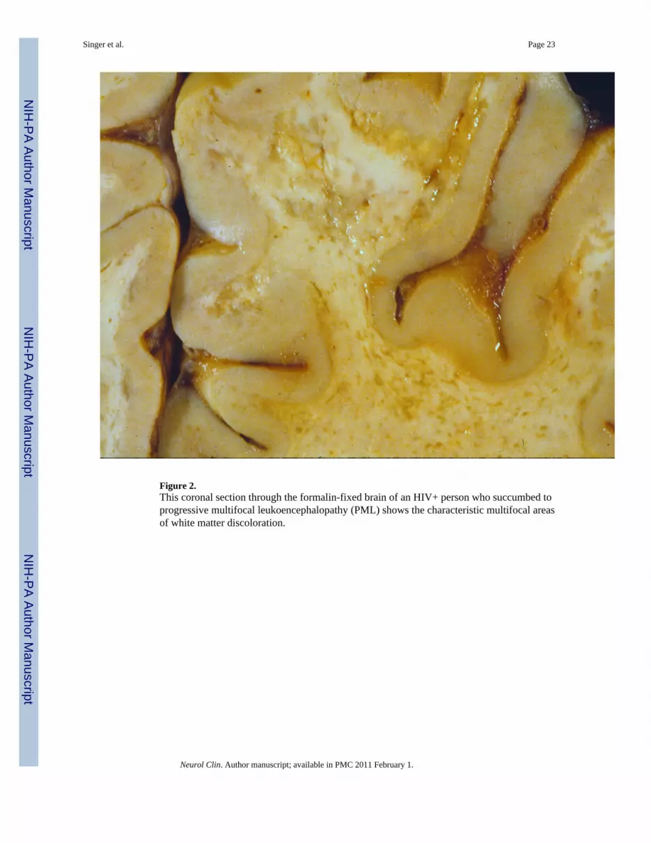
Figure 2.This coronal section through the formalin-fixed brain of an HIV+ person who succumbed toprogressive multifocal leukoencephalopathy (PML) shows the characteristic multifocal areasof white matter discoloration.
Singer et al. Page 23
Neurol Clin. Author manuscript; available in PMC 2011 February 1.
NIH
-PA Author Manuscript
NIH
-PA Author Manuscript
NIH
-PA Author Manuscript

Figure 3.Cytomegalovirus (CMV) infection of cells may result in the morphological changes shownhere ie, cytomegaly and both intranuclear and intracytoplasmic viral inclusions.Immunohistochemistry for CMV (not shown) may detect additional infected cells that appearnormal. Hematoxylin and Eosin stain.
Singer et al. Page 24
Neurol Clin. Author manuscript; available in PMC 2011 February 1.
NIH
-PA Author Manuscript
NIH
-PA Author Manuscript
NIH
-PA Author Manuscript


