Embed Size (px)
Citation preview
Cav1.3 and BK Channels for Timing and RegulatingCell Firing
David Henry Vandael & Andrea Marcantoni &Satyajit Mahapatra & Anton Caro & Peter Ruth &
Annalisa Zuccotti & Marlies Knipper & Emilio Carbone
Received: 1 October 2010 /Accepted: 9 November 2010 /Published online: 20 November 2010# Springer Science+Business Media, LLC 2010
Abstract L-type Ca2+ channels (LTCCs, Cav1) openreadily during membrane depolarization and allow Ca2+ toenter the cell. In this way, LTCCs regulate cell excitabilityand trigger a variety of Ca2+-dependent physiologicalprocesses such as: excitation–contraction coupling inmuscle cells, gene expression, synaptic plasticity, neuronaldifferentiation, hormone secretion, and pacemaker activityin heart, neurons, and endocrine cells. Among the twomajor isoforms of LTCCs expressed in excitable tissues(Cav1.2 and Cav1.3), Cav1.3 appears suitable for supportinga pacemaker current in spontaneously firing cells. It hassteep voltage dependence and low threshold of activationand inactivates slowly. Using Cav1.3
−/− KO mice andmembrane current recording techniques such as the dy-namic and the action potential clamp, it has been possibleto resolve the time course of Cav1.3 pacemaker currentsthat regulate the spontaneous firing of dopaminergicneurons and adrenal chromaffin cells. In several cell types,
Cav1.3 is selectively coupled to BK channels withinmembrane nanodomains and controls both the firingfrequency and the action potential repolarization phase.Here we review the most critical aspects of Cav1.3 channelgating and its coupling to large conductance BK channelsrecently discovered in spontaneously firing neurons andneuroendocrine cells with the aim of furnishing a converg-ing view of the role that these two channel types play in theregulation of cell excitability.
Keywords L-type calcium channels . Pacemakingcurrents . Action potential firing . Neurons .
Chromaffin cells
Introduction
Spontaneous electrical activity in excitable cells is funda-mental to the control of basic physiological functions.Usually, spontaneous firings originate in narrow areas andspread to neighboring cells to drive rhythmic patterns ofactivity such as circadian rhythms in the central nervoussystem, activity-dependent hormone release in neuroendo-crine cells, and heart beating [1, 2]. To undertake theseduties, spontaneously firing cells are equipped with a set ofion channels that turn on at subthreshold membranepotentials and carry sufficient inward current to drive theaction potential (AP) upstroke [3, 4]. In many cells, aspontaneous subthreshold depolarization is associated withNa+ entry through either hyperpolarization-activated cationchannels (HCN) or voltage-gated Na+ channels [5]. Thereis, however, an increasing number of neurons and neuro-endocrine cells in which subthreshold Ca2+ channels areshown to contribute to pacemaking. Besides the lowvoltage-activated T-types that are by definition sub-
David Henry Vandael and Andrea Marcantoni contributed equally tothis work.
D. H. Vandael :A. Marcantoni : S. Mahapatra : E. Carbone (*)Department of Neuroscience, NIS Centre, CNISM,Corso Raffaello 30,10125, Turin, Italye-mail: [email protected]
A. Zuccotti :M. KnipperDepartment of Otorhinolaryngology, THRC,72076, Tübingen, Germany
P. RuthDepartment of Pharmacology and Toxicology, THRC,72076, Tübingen, Germany
A. CaroInstitute of Molecular Physiology & Genetics,83334, Bratislava, Slovakia
Mol Neurobiol (2010) 42:185–198DOI 10.1007/s12035-010-8151-3
Author's personal copy
threshold Ca2+ channels [6, 7], Cav1.3 L-type calciumchannels (LTCCs) are shown to possess all the prerequisitesfor pacemaking excitable cells: low threshold and steepvoltage dependence of activation in addition to slowinactivation kinetics [8–11]. Cav1.3 channels drive thespontaneous firing of dopaminergic neurons in substantianigra pars compacta (SNc) [12, 13] and mid-spiny striatalneurons [14]. They also contribute to the pacemaker currentthat generates the heartbeat at the cardiac sinoatrial node[15] and the spontaneous firing activity of adrenalchromaffin cells [16–18] and cochlear immature inner haircells (IHCs) [19]. Finally, LTCCs contribute to thespontaneous AP firing of suprachiasmatic nucleus neurons[20, 21], locus coeruleus neurons [22], pituitary cells [23],and immortalized hypothalamic neurons [24, 25] andsustain the plateau potentials in spinal cord motoneurons[26]. In these cells, however, the direct involvement ofCav1.3 in pacemaking has not yet been proven.
Here, we will review the features that confer to LTCCs itspacemaker properties in a variety of cells and how, by usingCav1.3
−/− KO mice [8], it has been possible to identifyCav1.3 as the L-type channel responsible for pacemakingspontaneously firing neurons, neuroendocrine, and cardiaccells. We also review how the selective coupling of Cav1.3to BK channels affects the spontaneous activity of a numberof cells with particular emphasis on the adrenal chromaffincells and their implications in adrenal gland function.
L-Type Calcium Channels: Expression and Function
LTCCs are hetero-oligomers consisting of a pore-formingα1 subunit interacting with several accessory subunits (βand α2-δ) to form a functional complex. LTCCs belong tothe large family of voltage-gated Ca2+ channels that arepermeable to Ca2+ and include the N, P/Q, R, and T types[27]. The identification of L-types from the other Ca2+
channel currents (N, P/Q, R, and T) in different cells isfacilitated by the availability of a class of organic moleculesthat possess high affinity and selectivity for LTCCs [27].Among those, the dihydropyridines (DHPs) are availableeither as LTCC blockers (so-called Ca2+ antagonists, e.g.,nifedipine) or LTCC activators (e.g., BayK 8644). Theyallow the unique isolation and characterization of L-typecurrents, although they are unable to distinguish betweendifferent LTCC isoforms.
Presently, LTCCs comprise of four genes that code forthe α1S, α1C, α1D, and α1F subunits, now renamed Cav1.1,Cav1.2, Cav1.3, and Cav1.4, respectively [27]. Of these,Cav1.1 and Cav1.4 exhibit specific expression profiles thatare restricted to the skeletal muscle and the retina, whereasCav1.2 and Cav1.3 are widely expressed throughout thecentral nervous system, sensory and endocrine cells, atrial
myocytes, and cardiac pacemaker cells [11]. In centralneurons and neuroendocrine cells, Cav1.2 and Cav1.3 areoften co-expressed, and the general rule is that Cav1.2expression prevails on Cav1.3 expression in the mouse(80% vs. 20%) [28]. An exception to this rule is thedistribution of Cav1 at the IHCs from the mouse cochlea,chromaffin cells of the adrenal medulla, and SNc neuronswhere Cav1.3 expression takes the overhand [8, 18, 29, 30].
Cav1.2 Versus Cav1.3 in Cell Activity Regulation
LTCCs are commonly defined as high voltage-activated Ca2+
channels since they require strong membrane depolarizationto reach the threshold of activation (−30 mV in physiologicalCa2+) and differ markedly from the low voltage-activated T-type channels that activate, at 20 to 30 mV, more negativepotentials [6, 31, 32]. LTCCs are characterized by relativelyfast activation kinetics, large single-channel conductances,and inactivation gating which is both sensitive to voltage andCa2+ and varies among the different Cav1 subtypes [27].Concerning the neuronal and neuroendocrine LTCCs(Cav1.2 and Cav1.3), a number of studies have shown thatthe two LTCCs possess quite different activation properties,voltage- and Ca2+-dependent inactivation (CDI), and sensi-tivity to DHPs which underlie the different role that the twochannels are likely to play in cell function [9, 33]. Most ofthe currently existing information comes from studies usingheterologously expressed channels, cell preparations with apredominant expression of one of the two channel isoformsand knock-out animal models [9, 18, 30, 33, 34].
The biophysical and pharmacological differences betweenCav1.2 and Cav1.3 can be summarized as follows: (1) Cav1.3activates with steep voltage dependence at voltages signif-icantly more negative than Cav1.2 (up to −20 mV) [9, 18,33]. The lower threshold of activation does not depend onco-expression with the diverse β or α2δ accessory subunitsand is thus an intrinsic property of the pore-forming α1
subunit [33]; (2) Cav1.3 has faster activation but slower andless complete voltage-dependent inactivation as compared toCav1.2 [9, 18]. Inactivation is nearly absent when recordingthe Ca2+ currents of IHCs of the cochlea where 90% of thetotal Ca2+ current is carried by Cav1.3 [8, 29]. This is partlydue to an unusually slow voltage-dependent inactivationbesides an almost complete lack of CDI. The absence of CDIin IHCs is partly due to the high expression of Ca2+-bindingproteins CaBP4 in this tissue [35]. Long-lasting Ca2+
currents persisting for 500 ms at positive potentials are alsoevident in patch-perforated mouse chromaffin cells (MCCs),which express high densities of Cav1.3 channels [18]. (3)Cav1.3 and Cav1.2 have equal Ca2+/Ba2+ permeability ratioand similar high single-channel conductances [27, 36]. (4)Cav1.3 isoforms are less sensitive to DHPs as compared with
186 Mol Neurobiol (2010) 42:185–198
Author's personal copy
Cav1.2, requiring about ten times more DHPs for full blockat normal holding potentials (Vh −70 mV). The IC50 of DHPfor Cav1.3 current block decreases markedly at less negativeholding potentials (Vh −50 mV), reaching comparableIC50 values of Cav1.2 [9, 18]. Saturating concentrations ofDHPs that block Cav1.2 at −50 mV also ensure full blockof Cav1.3. This is important when assaying the role ofCav1.3 during spontaneous AP recordings in firing cellsthat spend most of their time at the interspike potentialaround −50 mV.
Following the above-mentioned properties, it is evidentthat Cav1.3 and Cav1.2 play quite distinct roles in neuronal,endocrine, and muscle cell physiology, depending criticallyon the degree of expression of both isoforms. Cav1.3 is asuitable candidate for supporting pacemaker currents, whileCav1.2 recruited at more positive potentials plays a majorrole in sustaining AP upstrokes. The critical issuescharacterizing Cav1.3 and its functions will be clarified inthe following chapters.
Cav1.3 Expression and Pacemaking
Convincing evidence that Cav1.3 is a suitable channel forpacemaking started to accumulate recently in neuronal,cardiac, and neuroendocrine cells in which Cav1.3 is highlyexpressed. In many cells, LTCCs fulfill the role ofpacemaker channels although a direct involvement ofCav1.3 is still lacking (see Table 1). We will review herethe three most convincing cases in which Cav1.3 has beenproven to control pacemaking.
Cav1.3 in Pacemaking Neostriatal SNc DopaminergicNeurons
In vivo, midbrain dopaminergic (DA) neurons displayintrinsic pacemaker firing, which is thought to set back-ground dopamine levels and receptor signaling at synaptictargets. Pharmacological and molecular studies have shownthat the regular pacemaker activity of DA neuron in theSNc depends on LTCCs: (1) SNc DA neurons expressLTCCs, predominantly Cav1.3 [37] and (2) micromolarconcentrations of DHPs effectively silence neuronal activityin SNc slice preparations [38, 39]. The identification of asub-threshold L-type current dominating the pacemakercurrent came only after using the action potential clamptechnique [13]. The L-type pacemaker current was found torise continuously during the interspike interval to reachmaximal values just before the AP upstroke. P/Q-type Ca2+
and TTX-sensitive Nav1 channels contribute as well topacemaking SNc neurons, but to a minor degree [13].
Convincing evidence for the existence of voltage-gatedCa2+ channels with gating properties similar to Cav1.3 has
been found in SNc slices [40, 41], while direct evidence ofCav1.3 involvement in pacemaking SNc DA neurons andburst firing in striatal medium spiny neurons (MSNs) camefrom works using Cav1.3
−/− KO mice [12, 14]. In MSNs,the absence of Cav1.3 interfered with burst firing. Sponta-neous bursts could be recovered only after adding the DHPCa2+ agonist BayK 8644 [14]. In SNc DA neurons [12],somatodendritic Cav1.3 channels gain importance after thesecond postnatal week. Spontaneous firing in these matureneurons is fully controlled by LTCCs, while in the sameaged neurons of mice lacking Cav1.3 the pacemaking iscontrolled by Nav1 and HCN-gated channels, which are thepacemaker channels during the “juvenile” state. Chan et al.[12] proposed that the switch from a Cav1.3-independent toa Cav1.3-dependent pacemaking mode during aging may beat the basis of the Ca2+-mediated SNc degeneration inParkinson’s disease (PD) [42].
Notice that cells exhibiting a Ca2+-dependent pacemakerare subjected to an increased ATP consumption in order tokeep up with the physiological cytosolic Ca2+ levels. Thiselevated metabolic rate facilitates the accumulation ofmitochondrial DNA damage and reactive oxygen speciesproduction that leads to cellular aging [42]. Interestingly,locus coeruleus and tuberomammillary neurons that fire ina clock like pattern and allow considerable amounts of Ca2+
entry during the pacemaker potential show an increasedvulnerability to aging and cell death [43]. On the contrary,dopaminergic neurons of the ventral tegmental area andneurons of the olfactory bulb that use Na+ for theirpacemaker do not seem to suffer from aging to the samedegree [12, 44]. It is therefore reasonable that Cav1.3channel blockers are expected to reduce the aging processesassociated with Ca2+-dependent pacemaker mechanisms,sparing dopaminergic neurons from Ca2+-dependent celldeath and delaying the clinical appearance and progressionof PD. These findings prompted ongoing clinical trialsbased on the therapeutical use of DHP Cav1.3 blockers forthe treatment of PD in humans [45]. The potential usage ofCav1 blockers in PD has been confirmed by epidemiolog-ical studies showing a decreased risk in humans treated forhypertension with DHPs [46].
Although the above-mentioned findings support the ideaof Cav1.3 as the pacemaker channel in SNc dopaminergicneurons, new evidence suggest the involvement of other ionchannels on the pacemaker mechanism [47]. Low concen-trations of isradipine (5 μM) clearly slow down or block thedendritic spontaneous oscillatory potentials (SOP) whilepreserving the somatic spontaneous firing [47]. Further-more, the SOP frequency does not coincide with thesomatic spiking frequency. Experimental as well assimulation-based studies show that, in order to obtain afull block of spontaneous activity, the simultaneous blockof HCN and Cav1.3 channels is required. Thus, pacemaking
Mol Neurobiol (2010) 42:185–198 187
Author's personal copy
of SNc dopaminergic neurons seems more robust thanexpected and not entirely depending on Cav1.3 but rather toa multichannel system, which includes HCN [47], Cav1.3[12], and Nav1 channels [13].
A confirmation that Cav1.3 plays a role in setting thespontaneous firing of SNc neurons derives from a recentstudy on SNc slices using the dynamic clamp [48]. With thisapproach, native LTCCs are first pharmacologically blockedby high doses of DHPs and then virtually replaced byimposing an ad-hoc simulating Cav1.3 conductance model tothe voltage-clamped neuron (for details on the technique, see[49]). The great advantage of this approach is that it allowsone to identify the key parameters of channel gating thatregulate neuronal firing. Curiously enough, pacemakerrecovery was obtained in the absence of Ca2+ ions (replacedby other conductive ions), indicating that some other featureof Cav1.3 is responsible for its pacemaker properties besidesthe Ca2+-mediated SOP generated at the SNc dendrites [47].Indeed the only parameter that seems critical for Cav1.3 toexert its pacemaking role turns out to be its unique
subthreshold voltage dependence of activation [48]. In fact,simulation to replace the normal pacemaking with a 20-mVmore negative Cav1.3 voltage dependence to reach maximalcurrents at rest (−60 mV) failed to recover the firing. Thissuggests that it is more important how much the pacemakerchannel conductance increases with increasing voltagesrather than how much its maximal value is at the interspikepotential (−50 mV). In conclusion, the somatodendriticCav1.3 is a subthreshold channel that effectively contributesto SNc pacemaking activity.
Cav1.3 Expression in Cardiac Pacemaker Cells
Heart beating in vertebrates is controlled by a number ofchannels that contribute to the diastolic pacemaker current[3, 34]. A major role is played by the HCN-gated channelbut there is also evidence that a sustained inward current(Ist), a T-type channel, an L-type channel and a Na+–Ca2+
exchanger activated by sarcolemmal Ca2+ release duringdiastolic depolarisations contribute to pacemaking sino-
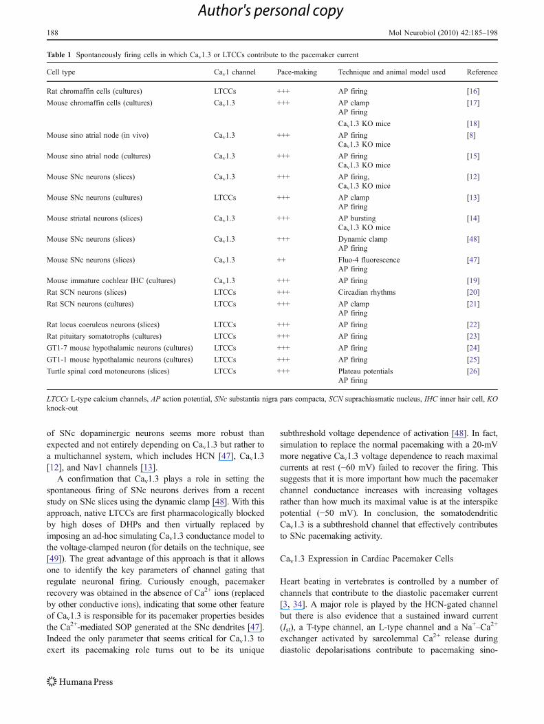
Table 1 Spontaneously firing cells in which Cav1.3 or LTCCs contribute to the pacemaker current
Cell type Cav1 channel Pace-making Technique and animal model used Reference
Rat chromaffin cells (cultures) LTCCs +++ AP firing [16]
Mouse chromaffin cells (cultures) Cav1.3 +++ AP clamp [17]AP firing
Cav1.3 KO mice [18]
Mouse sino atrial node (in vivo) Cav1.3 +++ AP firing [8]Cav1.3 KO mice
Mouse sino atrial node (cultures) Cav1.3 +++ AP firing [15]Cav1.3 KO mice
Mouse SNc neurons (slices) Cav1.3 +++ AP firing, [12]Cav1.3 KO mice
Mouse SNc neurons (cultures) LTCCs +++ AP clamp [13]AP firing
Mouse striatal neurons (slices) Cav1.3 +++ AP bursting [14]Cav1.3 KO mice
Mouse SNc neurons (slices) Cav1.3 +++ Dynamic clamp [48]AP firing
Mouse SNc neurons (slices) Cav1.3 ++ Fluo-4 fluorescence [47]AP firing
Mouse immature cochlear IHC (cultures) Cav1.3 +++ AP firing [19]
Rat SCN neurons (slices) LTCCs +++ Circadian rhythms [20]
Rat SCN neurons (cultures) LTCCs +++ AP clamp [21]AP firing
Rat locus coeruleus neurons (slices) LTCCs +++ AP firing [22]
Rat pituitary somatotrophs (cultures) LTCCs +++ AP firing [23]
GT1-7 mouse hypothalamic neurons (cultures) LTCCs +++ AP firing [24]
GT1-1 mouse hypothalamic neurons (cultures) LTCCs +++ AP firing [25]
Turtle spinal cord motoneurons (slices) LTCCs +++ Plateau potentials [26]AP firing
LTCCs L-type calcium channels, AP action potential, SNc substantia nigra pars compacta, SCN suprachiasmatic nucleus, IHC inner hair cell, KOknock-out
188 Mol Neurobiol (2010) 42:185–198
Author's personal copy
atrial node (SAN) cells [34]. Concerning the role of LTCCsas pacemaker channels, there is evidence that nicardipineinduces bradycardia in anesthetized mice in vivo [50] andthat nifedipine slows down the pacemaker activity ofisolated rabbit SAN cells in vitro [51].
Cav1.3 is expressed in sinoatrial and atrioventricularnodes but not in ventricular cardiac myocytes [15], and itsdeletion in Cav1.3
−/− mice causes bradycardia and arrhyth-mia [8]. Cav1.3
−/− mice display a significant prolongationof the atrioventricular conduction time and suffer fromepisodes of second-degree AV block [8]. This peculiarphenotype of the Cav1.3
−/− mouse persists after the block ofsympathetic and parasympathetic tones and thus is consid-ered to be a direct consequence of an altered SANpacemaker activity [8]. By comparing the amplitude andthe voltage dependence of the Ca2+ currents in WT andCav1.3
−/− SAN cells, it became evident that WT cellsexpressed Ca2+ currents that activated steeply at 22 mVmore negative potentials and were 70% larger than those ofCav1.3
−/− SAN cells [15, 52]. In addition, non-saturatingconcentrations of isradipine blocked more effectively theCa2+ currents of Cav1.3
−/− SAN cells, indicating that theonly LTCCs available in Cav1.3
−/− SAN cells (Cav1.2) aremore sensitive to DHPs. In conclusion, Cav1.3 possessesthe right gating properties for controlling the diastolicpacemaking current in SAN cells [15, 52].
Cav1.3 as Pacemaker Channel of Adrenal Chromaffin Cells
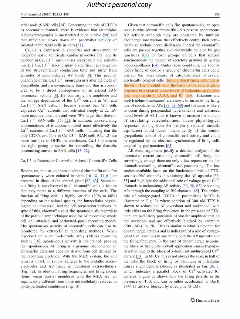
Bovine, rat, mouse, and human adrenal chromaffin cells firespontaneously when cultured in vitro [16–18, 53–61] orprepared in slices of the adrenal gland [62, 63]. Spontane-ous firing is not observed in all chromaffin cells, a featurethat may point to a different function of the cells. Thefraction of firing cells is highly variable (20% to 80%)depending on the animal species, the intracellular physio-logical solution used, and the cell preparation methods. Inspite of this, chromaffin cells fire spontaneously regardlessof the patch–clamp technique used for AP recording: wholecell, cell attached, and perforated–patch recording modes.The spontaneous activity of chromaffin cells can also bemonitored by extracellular recording methods. Whendispersed on a multi-electrode array (MEA) recordingsystem [16], spontaneous activity is maintained, provingthat spontaneous AP firing is a genuine phenomenon ofchromaffin cells and does not derive from cell damage bythe recording electrode. With the MEA system, the cellremains intact. It simply adheres to the metallic micro-electrodes and AP firing is monitored extracellularly(Fig. 1a). In addition, firing frequencies and firing modes(tonic versus bursts) monitored with the MEA are notsignificantly different from those intracellularly recorded inpatch-perforated conditions (Fig. 1b)
Given that chromaffin cells fire spontaneously, an openissue is why adrenal chromaffin cells possess spontaneousAP activity although they are contacted by multiplecholinergic innervations that effectively control their activ-ity by splanchnic nerve discharges. Indeed the chromaffincells are packed together and electrically coupled by gapjunctions [63] to form groups of cells that releasesynchronously the content of secretory granules in nearbyblood capillaries [64]. Under these conditions, the sponta-neous firing of one or a group of chromaffin cells couldwarrant the basal release of catecholamines of severalelectrically coupled cells. Tonic or burst firing activities asshown in Fig. 1 could be at the basis of the adrenal glandresponse to increased blood levels of histamine, acetylcho-line, angiotensin II (ATII), and K+ ions. Histamine andacetylcholine (muscarine) are shown to increase the firingrate of spontaneous APs [57, 59, 60] and the same is likelyto occur during postprandial hyperkaliemia and enhancedblood levels of ATII that is known to increase the amountof circulating catecholamines. These physiologicalresponses coming from the periphery (adrenal medullacapillaries) could occur independently of the centralsympathetic control of chromaffin cell activity and couldbe regulated by the electrical synchronism of firing cellscoupled by gap junctions [63].
All these arguments justify a detailed analysis of thepacemaker current sustaining chromaffin cell firing, butsurprisingly enough there are only a few reports on the ionchannels controlling chromaffin cell pacemaking. The fewstudies available focus on the fundamental role of TTX-sensitive Na+ channels in sustaining the AP upstroke [53,55] and highlight the additional role of voltage-gated Ca2+
channels in maintaining AP activity [53, 58, 62] or shapingAPs through the coupling to BK channels [65]. The criticalrole of voltage-gated LTCCs in pacemaking MCCs isillustrated in Fig. 2a where addition of 300 nM TTX isshown to reduce the AP overshoot and undershoot withlittle effect on the firing frequency. In the presence of TTX,there are oscillatory potentials of smaller amplitude that donot overshoot and are effectively blocked by cadmium(200 μM) (Fig. 2b). This is similar to what is reported fordopaminergic neurons and is indicative of a role of voltage-gated Ca2+ channels in sustaining both the AP upstroke andthe firing frequency. In the case of dopaminergic neurons,the block of firing after cobalt application causes hyperpo-larization due to the block of a dominant subthreshold Ca2+
current [13]. In MCCs, this is not always the case; in half ofthe cells the block of firing by cadmium or nifedipinecauses slight depolarizations, as illustrated in Fig. 2b, c,which indicates a parallel block of Ca2+-activated K+
currents. Figure 2c shows how the firing persists in thepresence of TTX and can be either accelerated by BayK8644 (1 μM) or blocked by nifedipine (3 μM).
Mol Neurobiol (2010) 42:185–198 189
Author's personal copy
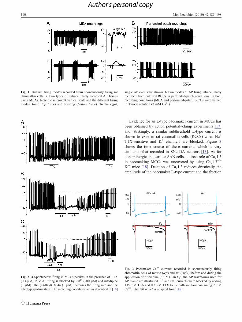
Evidence for an L-type pacemaker current in MCCs hasbeen obtained by action potential–clamp experiments [17]and, strikingly, a similar subthreshold L-type current isshown to exist in rat chromaffin cells (RCCs) when Na+
TTX-sensitive and K+ channels are blocked. Figure 3shows the time course of these currents which is verysimilar to that recorded in SNc DA neurons [13]. As fordopaminergic and cardiac SAN cells, a direct role of Cav1.3in pacemaking MCCs was uncovered by using Cav1.3
−/−
KO mice [18]. Deletion of Cav1.3 reduces drastically theamplitude of the pacemaker L-type current and the fraction
Fig. 2 a Spontaneous firing in MCCs persists in the presence of TTX(0.3 μM). b, c AP firing is blocked by Cd2+ (200 μM) and nifedipine(3 μM). The (±)-BayK 8644 (1 μM) increases the firing rate and theafterhyperpolarization. The recording conditions are as described in [18]
Fig. 1 Distinct firing modes recorded from spontaneously firing ratchromaffin cells. a Two types of extracellularly recorded AP firingsusing MEAs. Note the microvolt vertical scale and the different firingmodes: tonic (top trace) and bursting (bottom trace). To the right,
single AP events are shown. b Two modes of AP firing intracellularlyrecorded from cultured RCCs in perforated-patch conditions. In bothrecording conditions (MEA and perforated-patch), RCCs were bathedin Tyrode solution (2 mM Ca2+)
Fig. 3 Pacemaker Ca2+ currents recorded in spontaneously firingchromaffin cells of mouse (left) and rat (right), before and during theapplication of nifedipine (3 μM). On top, the AP waveforms used forAP clamp are illustrated. K+ and Na+ currents were blocked by adding135 mM TEA and 0.3 μM TTX to the bath solution containing 2 mMCa2+. The left panel is adapted from [18]
190 Mol Neurobiol (2010) 42:185–198
Author's personal copy
of firing MCCs (from 80% to 30%). In conclusion, Cav1.3contributes to the pacemaker current in MCCs (andpossibly in RCCs) and pacemaking is affected by BKchannels that are highly expressed in these cells [18, 65].The peculiarities of Cav1.3-BK channel coupling inpacemaking neurons and chromaffin cells will be discussedbelow.
Cav–BK Crosstalk Affects Cell Firing and Regulates APShape in Neurons and Neuroendocrine Cells
Paradoxically, Cav1.3−/− MCCs have more depolarized
interspike (resting) potentials and exhibit prolonged plateaudepolarizations in response to BayK 8644: two propertiesthat cannot be explained by simply silencing Cav1.3. Thesefindings have a rationale if assuming that Cav1.3 is effectivelycoupled to Ca2+-activated BK channels, responsible for fastAP repolarization.
The large conductance BK channels need a concertedbinding of Ca2+ to the so-called Ca2+ bowl and strongmembrane depolarizations for opening the pore [66, 67].Increasing the cytoplasmic free Ca2+ concentration fromnanomolar to micromolar levels shifts the voltage-dependentactivation curve of BK currents from very positive tonegative resting potentials (from +100 to −50 mV) [68]. Toactivate during physiological depolarization, BK channelsneed an intracellular Ca2+ concentration of at least 10 μM.Considering the diverse and tightly regulated intracellularCa2+ buffering systems, such high concentrations occur onlywithin “Ca2+ nanodomains” in the close vicinity of Ca2+
sources, mainly near Cav channels [67, 69]. Experimentswith the Ca2+ chelators BAPTA and EGTA prove that BKchannels must be within nanometer distances from their Ca2+
source [69]. In fact, BK channels are often functionallycoupled to a voltage-gated Ca channel such as L-type [18,29, 65], P/Q-type [70], or N-type [71] in different cells.Proteomic approaches that combined affinity purificationwith mass spectrometry showed that BK channels in themammalian brain may exist in high molecular weightcomplexes (~1.6 MDa) [72]. Among the proteins co-immunoprecipitated with BK, there are Cav1.2; Cav2.1(P/Q), or Cav2.2 (N) [72]. Heterologous expression studiessupport the idea that the repolarizing responses of Cav–BKcomplexes are distinctly shaped by their Cav subunits [72].
Below we will focus on two examples in whichLTCCs are functionally coupled to BK channels toregulate AP shape and firing frequency. They are onlypart of the many existing examples of Cav–BK crosstalk(for reviews, see [67]) but are representative for theheterogeneous way of how coupling occurring in amembrane nanodomain may contribute to the fine-tuningof different cells’ excitability.
BK and LTCCs Control Circadian Rhythmsin Suprachiasmatic Nucleus Neurons
Neurons from the suprachiasmatic nucleus (SCN) transmitcircadian output to other brain regions by modulating theirdiurnal spontaneous oscillatory firing patterns. It is well knownthat these neurons fire faster during daytime and slower at night[73]. When TTX is applied to fast-firing neurons duringdaytime, the resting membrane potential is depolarized butspontaneous oscillations are preserved. These subthresholdoscillations are completely blocked by cadmium or nifedipineand are insensitive to T-, P/Q-, and N-type channel blockers.Current–voltage relationships indicate that the L-type currentis up-regulated during daytime, giving rise to elevated totalCa2+ currents as compared to neurons recorded duringnighttime [20]. The sustained DHP-sensitive Ca2+ currentsduring daytime activates at very negative membrane potentialsand thus is likely to be carried by Cav1.3 LTCCs. Interest-ingly, SCN neurons that express the Per1 clock genedepolarize in the afternoon and go into a hyperpolarized stateduring nighttime. The depolarized state in the afternoon isaccompanied by a rise of the input resistance due to theclosure of BK and SK channels. Indeed the firing cellsrecorded in the morning could be brought into a depolarizedstate as observed in the afternoon by applying either TEA(30 mM) or nimodipine (2 μM), and the depolarization couldbe mimicked by the co-application of apamine and iberiotoxin(IbTX) [74]. Thus, it seems that LTCCs can effectivelyregulate SCN pacemaking by partly activating BK currents.Fast-firing neurons, in fact, are characterized by afterhyperpo-larizations (AHPs) of shorter duration typically driven by BKchannels [75]. A tight coupling between pacemaking L-typecurrents and BK channels is also evident in AP clamprecordings of isolated SCN neurons [21]. Interestingly, Cav1.3KO mice are shown to have a normal day–night cycle asmeasured by spontaneous homecage activities [76]. Thismight indicate a major contribution by Cav1.2 to activateBK/SK or that some compensatory mechanism to the day–night pacemaker has taken place.
BK and Cav1.3 Control Chromaffin Cell Firing
In RCCs, charybdotoxin (ChTX) significantly broadensthe AP half-width, suggesting that BK channels controlthe fast AP repolarization phase [77]. RCCs and MCCsexpress two different BK channel subtypes that can bedistinguished according to their inactivation kinetics: afast-inactivating and a slowly inactivating subtype [77].The fast-inactivating BK channel is typically expressed inchromaffin cells and is specifically involved in tonic cellfiring. The slowly inactivating BK channel has gatingproperties similar to the central neurons and smoothmuscle BK channels and gives rise to phasic firings [77].
Mol Neurobiol (2010) 42:185–198 191
Author's personal copy
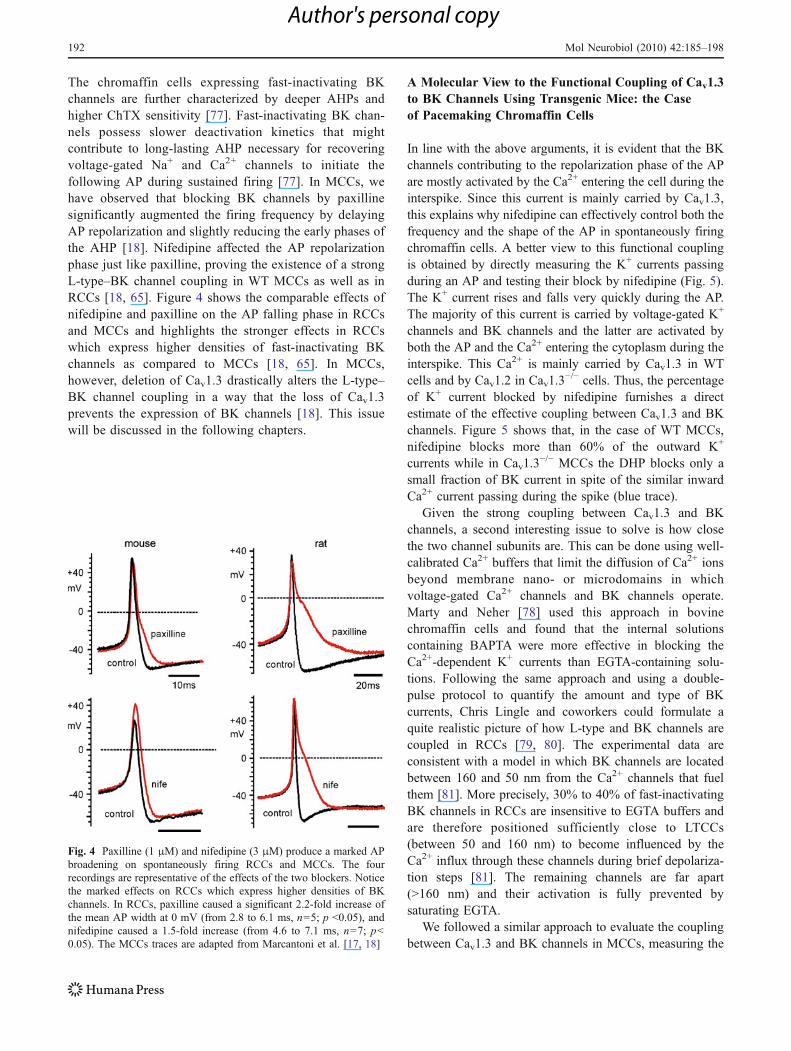
The chromaffin cells expressing fast-inactivating BKchannels are further characterized by deeper AHPs andhigher ChTX sensitivity [77]. Fast-inactivating BK chan-nels possess slower deactivation kinetics that mightcontribute to long-lasting AHP necessary for recoveringvoltage-gated Na+ and Ca2+ channels to initiate thefollowing AP during sustained firing [77]. In MCCs, wehave observed that blocking BK channels by paxillinesignificantly augmented the firing frequency by delayingAP repolarization and slightly reducing the early phases ofthe AHP [18]. Nifedipine affected the AP repolarizationphase just like paxilline, proving the existence of a strongL-type–BK channel coupling in WT MCCs as well as inRCCs [18, 65]. Figure 4 shows the comparable effects ofnifedipine and paxilline on the AP falling phase in RCCsand MCCs and highlights the stronger effects in RCCswhich express higher densities of fast-inactivating BKchannels as compared to MCCs [18, 65]. In MCCs,however, deletion of Cav1.3 drastically alters the L-type–BK channel coupling in a way that the loss of Cav1.3prevents the expression of BK channels [18]. This issuewill be discussed in the following chapters.
A Molecular View to the Functional Coupling of Cav1.3to BK Channels Using Transgenic Mice: the Caseof Pacemaking Chromaffin Cells
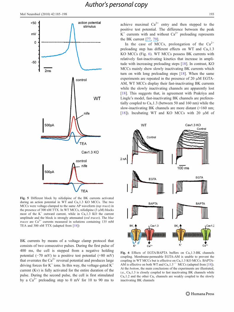
In line with the above arguments, it is evident that the BKchannels contributing to the repolarization phase of the APare mostly activated by the Ca2+ entering the cell during theinterspike. Since this current is mainly carried by Cav1.3,this explains why nifedipine can effectively control both thefrequency and the shape of the AP in spontaneously firingchromaffin cells. A better view to this functional couplingis obtained by directly measuring the K+ currents passingduring an AP and testing their block by nifedipine (Fig. 5).The K+ current rises and falls very quickly during the AP.The majority of this current is carried by voltage-gated K+
channels and BK channels and the latter are activated byboth the AP and the Ca2+ entering the cytoplasm during theinterspike. This Ca2+ is mainly carried by Cav1.3 in WTcells and by Cav1.2 in Cav1.3
−/− cells. Thus, the percentageof K+ current blocked by nifedipine furnishes a directestimate of the effective coupling between Cav1.3 and BKchannels. Figure 5 shows that, in the case of WT MCCs,nifedipine blocks more than 60% of the outward K+
currents while in Cav1.3−/− MCCs the DHP blocks only a
small fraction of BK current in spite of the similar inwardCa2+ current passing during the spike (blue trace).
Given the strong coupling between Cav1.3 and BKchannels, a second interesting issue to solve is how closethe two channel subunits are. This can be done using well-calibrated Ca2+ buffers that limit the diffusion of Ca2+ ionsbeyond membrane nano- or microdomains in whichvoltage-gated Ca2+ channels and BK channels operate.Marty and Neher [78] used this approach in bovinechromaffin cells and found that the internal solutionscontaining BAPTA were more effective in blocking theCa2+-dependent K+ currents than EGTA-containing solu-tions. Following the same approach and using a double-pulse protocol to quantify the amount and type of BKcurrents, Chris Lingle and coworkers could formulate aquite realistic picture of how L-type and BK channels arecoupled in RCCs [79, 80]. The experimental data areconsistent with a model in which BK channels are locatedbetween 160 and 50 nm from the Ca2+ channels that fuelthem [81]. More precisely, 30% to 40% of fast-inactivatingBK channels in RCCs are insensitive to EGTA buffers andare therefore positioned sufficiently close to LTCCs(between 50 and 160 nm) to become influenced by theCa2+ influx through these channels during brief depolariza-tion steps [81]. The remaining channels are far apart(>160 nm) and their activation is fully prevented bysaturating EGTA.
We followed a similar approach to evaluate the couplingbetween Cav1.3 and BK channels in MCCs, measuring the
Fig. 4 Paxilline (1 μM) and nifedipine (3 μM) produce a marked APbroadening on spontaneously firing RCCs and MCCs. The fourrecordings are representative of the effects of the two blockers. Noticethe marked effects on RCCs which express higher densities of BKchannels. In RCCs, paxilline caused a significant 2.2-fold increase ofthe mean AP width at 0 mV (from 2.8 to 6.1 ms, n=5; p <0.05), andnifedipine caused a 1.5-fold increase (from 4.6 to 7.1 ms, n=7; p<0.05). The MCCs traces are adapted from Marcantoni et al. [17, 18]
192 Mol Neurobiol (2010) 42:185–198
Author's personal copy
BK currents by means of a voltage clamp protocol thatconsists of two consecutive pulses. During the first pulse of400 ms, the cell is stepped from a negative holdingpotential (−70 mV) to a positive test potential (+80 mV)that overrates the Ca2+ reversal potential and produces largedriving forces for K+ ions. In this way, the voltage-gated K+
current (Kv) is fully activated for the entire duration of thepulse. During the second pulse, the cell is first stimulatedby a Ca2+ preloading step to 0 mV for 10 to 90 ms to
achieve maximal Ca2+ entry and then stepped to thepositive test potential. The difference between the peakK+ currents with and without Ca2+ preloading representsthe BK current [77, 79].
In the case of MCCs, prolongation of the Ca2+
preloading step has different effects on WT and Cav1.3KO MCCs (Fig. 6). WT MCCs possess BK currents withrelatively fast-inactivating kinetics that increase in ampli-tude with increasing preloading steps [18]. In contrast, KOMCCs mainly show slowly inactivating BK currents whichturn on with long preloading steps [18]. When the sameexperiments are repeated in the presence of 20 μM EGTA-AM, WT MCCs display their fast-inactivating BK currentswhile the slowly inactivating channels are apparently lost[18]. This suggests that, in agreement with Prakriya andLingle’s model, fast-inactivating BK channels are preferen-tially coupled to Cav1.3 (between 50 and 160 nm) while theslow-inactivating BK channels are more distant (>160 nm;[18]). Incubating WT and KO MCCs with 20 μM of
Fig. 5 Different block by nifedipine of the BK currents activatedduring an action potential in WT and Cav1.3 KO MCCs. The twoMCCs were voltage-clamped to the same AP waveform (top trace) inthe presence of 300 nM TTX. In WT MCCs, nifedipine (3 μM) blocksmost of the K+ outward current, while in Cav1.3 KO the currentamplitude and the block is strongly attenuated (red traces). The bluetraces are Ca2+ currents measured in solutions containing 135 mMTEA and 300 nM TTX (adapted from [18])
Fig. 6 Effects of EGTA/BAPTA buffers on Cav1.3-BK channelscoupling. Membrane-permeable EGTA-AM is unable to prevent thecoupling in WT MCCs but is effective on Cav1.3 KO MCCs. BAPTA-AM is effective on both WT and Cav1.3
−/− MCCs (adapted from [18]).At the bottom, the main conclusions of the experiments are illustrated,i.e., Cav1.3 is closely coupled to fast inactivating BK channels whileCav1.2 and the other Cav channels are weakly coupled to the slowlyinactivating BK channels
Mol Neurobiol (2010) 42:185–198 193
Author's personal copy
BAPTA-AM, the BK currents are functionally abolished,indicating that the fast-inactivating BK channels are notphysically coupled to Cav1.3 [18] (see Fig. 6, bottom).
BK−/− and Cav1.3−/− KO Mouse Chromaffin Cells
Reveal Similar Firing Responses to BayK 8644
The loss of Cav1.3 coupling to BK channels is particularlyevident when the DHP Ca2+ agonist BayK 8644 is appliedto spontaneously firing MCCs. BayK 8644 prolongs theopen state of the LTCCs [82] and, in normal conditions,typically accelerates the pacemaking frequency, producingslight hyperpolarizations and increased AHPs [17, 18].When BayK 8644 is applied to spontaneously firingCav1.3
−/− MCCs, in the majority of cases the cells respondwith a long-lasting membrane depolarization (Fig. 7,middle). In our view, this paradoxical response can beexplained by assuming that Cav1.3
−/− MCCs are lackingboth Cav1.3 and a considerable fraction of BK channels.The Ca2+ fluxes passing through the BayK-modified Cav1.2channels are sufficient to preserve the firing, but the lack offast-inactivating BK channels does not allow fast andcomplete AP repolarizations. Without an effective repolar-ization, the cell accumulates Ca2+, depolarizes, and staysdepolarized until Ca2+ is removed by Ca2+ pumps or Na+/Ca2+ exchangers (see [83] for a review).
A prediction to this hypothesis is that MCCs lacking BKchannels should respond to BayK 8644 similarly toCav1.3
−/− MCCs. Indeed MCCs lacking BK channels show
remarkable changes of the AP shape and exhibit somesimilarities to the spontaneous firing of Cav1.3
−/− MCCs.Paradoxically, BK−/− MCCs have a reduced frequency offiring that is apparently in contrast to the potentiatingeffects induced by paxilline in WT MCCs. However, this issimilar to what occurs in Purkinje cells from BK−/− mice,which have lower AP activity than WT cells while IbTXincreases the firing frequency [84]. Regarding BK−/−
MCCs, most striking is the broadening of the AP half-width and the significant prolongation of the interspikeinterval, a feature that confirms not only the important roleof BK channels in MCCs AP repolarization but also its roleon cell firing. There is a significant reduction of the AHPamplitude and we found a slightly depolarized restingmembrane potential in BK−/− MCCs. These changes aresimilar to those of Cav1.3
−/− MCCs with the maindifference that the number of silent cells does not increasein BK−/− MCCs as it does in Cav1.3
−/−, suggesting that BKchannels are potent modulators but are not conditional asCav1.3 in pacemaking chromaffin cells.
As expected, BK−/− and Cav1.3−/− MCCs exhibit similar
responses to BayK 8644 (Fig. 7, bottom). A fraction of theCav1.3
−/− and BK−/− MCCs responds to BayK 8644 with asustained membrane depolarization (Fig. 7, bottom), sug-gesting that BK channels are critical to counterbalance theCav1.3-mediated AP depolarization. In a number of MCCs,the absence of Cav1.3 induces a loss of BK channels,proving the tight coupling between these two channels andan existing adaptive phenomenon regulating their simulta-neous expression or membrane incorporation.
Fig. 7 Paradoxical effects of the (±)-BayK 8644 (1 μM) onspontaneous firing MCCs lacking either Cav1.3 or BK channels. Eachpanel shows typical spontaneous firings (top), single AP waveforms(middle), and responses to BayK 8644 (bottom). In WT MCCs(bottom left), the (±) racemic mixture BayK 8644 causes increased
firing frequency and enhanced AHP. In Cav1.3−/− and BK−/− MCCs
(bottom middle and right), BayK 8644 causes prolonged plateaudepolarizations due to the lack of the K+ channels controlling most ofthe AP repolarization: BK and the Cav channel that opens BK(Cav1.3). For details on BK−/− KO mice, see [84]
194 Mol Neurobiol (2010) 42:185–198
Author's personal copy
A Molecular Approach to the Cav1.3-BK ChannelCoupling: Deletion of Cav1.3 Prevents BK ChannelExpression
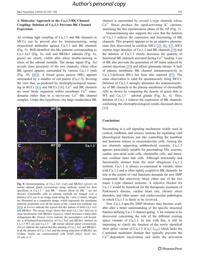
An existing tight coupling of Cav1.3 and BK channels inMCCs can be proved also by immunostaining, usingmonoclonal antibodies against Cav1.3 and BK channels(Fig. 8). Well-identified dot-like patterns corresponding toCav1.3α1 (Fig. 8a, red) and BKSlo1 subunits (Fig. 8a,green) are clearly visible after direct double-staining inslices of the adrenal medulla. The merge signal (Fig. 8a)reveals close proximity of the two channels, where oftenBK (green) appears surrounded by various Cav1.3 (red)(Fig. 8b; [85]). A broad green puncta (BK) appearssurrounded by a number of red puncta (Cav1.3), favoringthe view that, as predicted by electrophysiological reason-ing in RCCs [81] and MCCs [18], Ca2+ and BK channelsare most likely organized within membrane Ca2+ nano-domains rather than in a one-to-one Ca2+–BK channelcomplex. Under this hypothesis, one large conductance BK
channel is surrounded by several L-type channels whoseCa2+ fluxes produce the rapid-activating K+ currents,sustaining the fast repolarization phase of the AP (Fig. 5).
Immunostaining also supports the view that the deletionof Cav1.3 reduces the expression and functioning of BKchannels. This property appears to be an adaptive phenom-enon first discovered in cochlear IHCs [29, 86, 87]. IHCsexpress large densities of Cav1.3 and BK channels [19] andthe deletion of Cav1.3 clearly decreases the quantity offunctional BK channels activated during Ca2+ loading. Lossof BK also prevents the generation of AP trains induced bycurrent injections [29] and affects glutamate release. A lackof plasma membrane BK channel immunoreactivity inCav1.3-deficient IHCs has been also reported [87]. Thesame observation is valid for spontaneously firing MCCs.Deletion of Cav1.3 strongly attenuates the immunoreactiv-ity of BK channels in the plasma membrane of chromaffincells as shown by comparing the density of green dots inWT and Cav1.3
−/− adrenal glands (Fig. 8c, d). Thus,deletion of Cav1.3 reduces the expression of BK channels,confirming the electrophysiological results discussed above[18].
Conclusions
Pacemaking is a cell signaling mechanism widely used incortical, midbrain, and sensory neurons for regulating vitalphysiological functions just like controlling the heartbeatand hormone release in neuroendocrine cells. Among theion channels supporting subthreshold currents, Cav1.3appears particularly suitable for pacemaking SNc neurons,cardiac sino-atrial node cells, chromaffin cells, and imma-ture cochlear inner hair cells. Although structurally andfunctionally distinct from the most ubiquitous Cav1.2isoform, Cav1.3 is always co-expressed at variable ratioswith Cav1.2 and is often tightly coupled to BK channels. Itsrole in the control of vital functions demands for new DHPcompounds that selectively block either one of the twomajor L-type channel isoforms. A selective blocker forCav1.3 would be beneficial for the therapeutic treatment ofParkinson’s disease, cardiac heart rate, chronic stressdisorders, and other neuro- and cardiovascular pathologiesin which Cav1.3 is likely to be involved.
New Cav1.3-specific DHP blockers may become avail-able after a better understanding of the specific structuralfeatures defining Cav1.3 channel gating. A lot remains to bediscovered concerning the role of the different existingsplice variants of Cav1.3. In line with this, it will beinteresting to clarify the function of the newly identifiedshort splice variant of Cav1.3 (Cav1.342A) which lacks theC-terminal modulator domain that typically prevents theCa2+-dependent inactivation and shifts the activation
Fig. 8 Immunostaining of Cav1.3α1 (red) and BKSlo1 (green) onmouse adrenal gland cryosections using antibody tested for theirspecificity in Cav1.3
−/− and BK−/− tissues (those on BK−/− are notshown). Chromaffin cells in adrenal medulla are imaged over adistance of 8 μm in an image stack along the z-axis (z-stack). Imagesare illustrated as a composite image, which represents the maximumintensity projection over all the layers of the z-stack (for methods, see[85]). a Arrows indicate the typical dot-like pattern of both Cav1.3α1and BKSlo1. The merge image shows that most Cav1.3α1 dots are inclose localization with BKSlo1 (square), which becomes evident afterenlargement (b). Dotted circles indicate the presumptive cell bound-ary. c, d Immunohistochemistry of Cav1.3α1 and BKSlo1 channel onCav1.3 WT (c) and Cav1.3
−/− (d) mouse adrenal gland cryosections.Arrows indicate the typical dot-like staining of Cav1.3α1 and BKSlo1.In d, the absence of Cav1.3α1 and the strong reduction of BKSlo1 areevident. Nuclei are counterstained with DAPI (blue). Scale bar,10 μm; n=3
Mol Neurobiol (2010) 42:185–198 195
Author's personal copy
voltage to more positive potentials [88]. It could possiblybe that the short form (Cav1.342A) is in charge of startingthe cellular pacemaking and the long form (Cav1.342) tosustain it (Zuccotti et al., submitted for publication). Splicevariant-specific drugs might create new opportunities totarget precise problems without possible harmful sideeffects.
Acknowledgements We wish to thank Prof. Valentina Carabelli andDr. Daniela Gavello for the helpful discussions, Dr. Claudio Franchinofor the technical assistance, and Prof. Joerg Striessnig for supplyingthe Cav1.3
−/− KO mouse. This work was supported by the Marie CurieResearch Training Network “CavNET” Contract MRTN-CT-2006-035367, the Italian M.I.U.R. Grant PRIN 2007SYRBBH_001 to EC,and the San Paolo Company Grant 2008.2191 to AM.
References
1. Welsh DK, Logothetis DE, Meister M, Reppert SM (1995)Individual neurons dissociated from rat suprachiasmatic nucleusexpress independently phased circadian firing rhythms. Neuron14:697–706
2. Schultz W (2002) Getting formal with dopamine and reward.Neuron 36:241–263
3. Noble D (1984) The surprising heart: a review of recent progressin cardiac electrophysiology. J Physiol 353:1–50
4. Bevan MD, Wilson CJ (1999) Mechanisms underlying spontane-ous oscillation and rhythmic firing in rat subthalamic neurons. JNeurosci 19:7617–7628
5. Bean BP (2007) The action potential in mammalian centralneurons. Nature Rev 8:451–465
6. Pérez-Reyes E (2003) Molecular physiology of low-voltage-activated T-type calcium channels. Physiol Rev 83:117–161
7. Carbone E, Marcantoni A, Giancippoli A, Guido D, Carabelli V(2006) T-type channels-secretion coupling: evidence for a fastlow-threshold exocytosis. Pflügers Archiv 453:373–383
8. Platzer J, Engel J, Schrott-fischer A, Stephan K, Bova S, Chen H,Zheng H, Striessnig J (2000) Congenital deafness and sinoatrialnode dysfunction in mice lacking class D L-type Ca2+ channels.Cell 102:89–97
9. Koschak A, Reimer D, Huber I, Grabner M, Glossmann H, EnglelJ, Striessnig J (2001) Alpha 1D (Cav1.3) subunits can form L-typeCa2+ channels activating at negative voltages. J Biol Chem276:22100–22106
10. Lipscombe D, Helton TD, Xu W (2004) L-type calcium channels:the low down. J Neurophysiol 92:2633–2641
11. Striessnig J, Koschak A (2008) Exploring the function andpharmacotherapeutic potential of voltage-gated Ca2+ channelswith gene knockout models. Channels (Austin) 2:233–251
12. Chan CS, Guzman JN, LLijic E, Mercer JN, RickC TT, MeredithGE, Surmeier DJ (2007) Rejuvenation protects neurons in mousemodels of Parkinson’s disease. Nature 447:1081–1090
13. Puopolo M, Raviola E, Bean BP (2007) Roles of subthresholdcalcium current and sodium current in spontaneous firing ofmouse midbrain dopamine neurons. J Neurosci 27:645–656
14. Olson PA, Tkatch T, Hernandez-Lopez US, Ilijic E, MugnainiE, Zhang H, Bezprozvanny I, Surmeier DJ (2005) G-proteincoupled receptor modulation of striatal Cav1.3 L-type Ca2+
channels is dependent on a shank-binding domain. J Neurosci25:1050–1062
15. Mangoni ME, Couette B, Bourinet E, Platzer J, Reimer D,Striessnig J, Nargeot J (2003) Functional role of L-type Cav1.3
Ca2+ channels in cardiac pacemaker activity. Proc Natl Acad SciUSA 100:5543–5548
16. Marcantoni A, Baldelli P, Hernandez-Guijo J-M, Comunanza V,Carabelli V, Carbone E (2007) L-type calcium channels in adrenalchromaffin cells: role in pace-making and secretion. Cell Calcium42:397–408
17. Marcantoni A, Carabelli V, Vandael DH, Comunanza V, CarboneE (2009) PDE type-4 inhibition increases L-type Ca2+ currents,action potential firing and quantal size of exocytosis in mousechromaffin cells. Pflügers Archiv 457:1093–1110
18. Marcantoni A, Vandael DHF, Mahapatra S, Carabelli V, Sinneger-Brauns MJ, Striessnig J, Carbone E (2010) Loss of Cav1.3channels reveals the critical role of L-type and BK channelcoupling in pacemaking mouse adrenal chromaffin cells. JNeurosci 30:491–504
19. Marcotti W, Johnson SL, Rüsch A, Kros CJ (2003) Sodium andcalcium currents shape action potentials in immature mouse innerhair cells. J Physiol 552:743–761
20. Pennartz CMA, de Jeu MTG, Bos NPA, Schaap J, GeurtsenAMS (2002) Diurnal modulation of pacemaker potentials andcalcium current in the mammalian circadian clock. Nature416:286–290
21. Jackson AC, Yao GL, Bean BP (2004) Mechanism of spontaneousfiring in dorsomedial suprachiasmatic nucleus neurons. J Neurosci24:7985–7998
22. Filosa JA, Putnam RW (2003) Multiple targets of chemosensitivesignaling in locus coeruleus neurons: role of K+ and Ca2+
channels. Am J Physiol Cell Physiol 284:145–15523. Tsaneva-Atanasova K, Sherman A, van Goor F, Stojilkovic SS
(2007) Mechanism of spontaneous and receptor-controlled elec-trical activity in pituitary somatotrophs: experiments and theory. JNeurophysiol 98:131–144
24. Van Goor F, Krsmanovic LK, Catt KJ, Stojilkovi SS (1999)Control of action potential-driven calcium influx in GT1 neuronsby the activation status of sodium and calcium channels. MolecEndocrinol 13:587–603
25. Costantin JL, Charles AC (1999) Spontaneous action potentialsinitiate rhythmic intercellular calcium waves in immortalizedhypothalamic (GT1–1) neurons. J Neurophysiol 82:429–435
26. Hounsgaard J, Kiehn O (1989) Serotonin-induced bistability ofturtle motoneurones caused by a nifedipine-sensitive calciumplateau potential. J Physiol 414:265–282
27. Catterall WA, Perez-Reyez E, Snutch TP, Striessnig J (2005)Nomenclature and structure–function relationships of voltage-gated calcium channels. Pharmacol Rev 57:411–425
28. Calin-Jageman I, Lee A (2008) Ca(v)1 L-type Ca2+ channelsignaling complexes in neurons. J Neurochem 105:573–583
29. Brandt A, Striessnig J, Moser T (2003) Cav1.3 channels areessential for development and presynaptic activity of cochlearinner hair cells. J Neurosci 23:10832–10840
30. Baldelli P, Hernández-Guijo JM, Carabelli V, Novara M, CesettiT, Andrés-Mateos E, Montiel C, Carbone E (2004) Direct andremote modulation of L-channel in chromaffin cells: distinctactions on α1C and α1D subunits? Mol Neurobiol 29:73–96
31. Carbone E, Lux HD (1984) A low-voltage-activated, fullyinactivating calcium channel in vertebrate sensory neurons.Nature 310:501–502
32. De Waard M, Gurnett CA, Campbell KP (1996) Structural andfunctional diversity of voltage activated calcium channels. IonChannels 4:41–87
33. Xu W, Lipscombe D (2001) Neuronal Cav1.3 alpha1 L-typechannels activate at relatively hyperpolarized membrane potentialsand are incompletely inhibited by dihydropyridines. J Neurosci21:5944–5951
34. Mangoni ME, Nargeot J (2008) Genesis and regulation of theheart automaticity. Physiol Rev 88:919–982
196 Mol Neurobiol (2010) 42:185–198
Author's personal copy
35. Yang PS, Alseikhan BA, Hiel H, Grant L, Mori MX, Yang W,Fuchs PA, Yue DT (2006) Switching of Ca2+-dependent inactiva-tion of Cav1.3 channels by calcium binding proteins of auditoryhair cells. J Neurosci 26:10677–10689
36. Chahine M, Qu Y, Mancarella S, Boutjdir M (2008) Protein kinaseC activation inhibits α1D L-type calcium channel: a single-channel analysis. Pflügers-Archiv 455:913–919
37. Takada M, Kang Y, Imanishi M (2001) Immunohistochemicallocalization of voltage-gated calcium channels in substantia nigradopamine neurons. Europ J Neurosci 13:757–762
38. Nedergaard S, Flatman JA, Engberg I (1993) Nifedipine- andomegaconotoxin-sensitive Ca2+ conductances in guinea-pig sub-stantia nigra pars compacta neurons. J Physiol 466:727–747
39. Mercuri NB, Bonci A, Calabresi P, Stratta F, Stefani A,Bernardi G (1994) Effects of dihydropyridine calcium antago-nists on rat midbrain dopaminergic neurones. Br J Pharmacol113:831–838
40. Kang Y, Kitai ST (1993) Calcium spike underlying rhythmicfiring in dopaminergic neurons of the rat substantia nigra.Neurosci Res 18:195–207
41. Wilson CJ, Callaway JC (2000) Coupled oscillator model of thedopaminergic neuron of the substantia nigra. J Neurophysiol83:3084–3100
42. Surmeier DJ, Guzman JN, Sanchez-Padilla J (2010) Calcium,cellular aging, and selective neuronal vulnerability in Parkinson’sdisease. Cell Calcium 47:175–182
43. German DC, Manaye KF, White CL 3rd, Woodward DJ, McIntireDD, Smith WK, Kalaria RN, Mann DM (1992) Disease-specificpatterns of locus coeruleus cell loss. Ann Neurol 32:667–676
44. Puopolo M, Bean BP, Raviola E (2005) Spontaneous activity ofisolated dopaminergic periglomerular cells of the main olfactorybulb. J Neurophysiol 94:3618–3627
45. Chan CS, Gertler TS, Surmeier DJ (2010) A molecular basis forthe increased vulnerability of substantia nigra dopamine neuronsin aging and Parkinson’s disease. Mov Disord 25:S63–S70
46. Becker C, Jick SS, Meier CR (2008) Use of antihypertensives andthe risk of Parkinson disease. Neurology 70:1438–1444
47. Guzman JM, Sanchez-Padilla J, Chan CS, Surmeier DJ (2009)Robust pacemaking in substantia nigra dopaminergic neurons. JNeurosci 29:11011–11019
48. Putzier I, Kullmann PHM, Horn JP, Levitan ES (2009) Cav1.3channel voltage dependence, not Ca2+ selectivity, drives pace-maker activity and amplifies bursts in nigral dopamine neurons. JNeurosci 29:15414–15419
49. Prinz AA, Abbott LF, Marder E (2004) The dynamic clamp comesof age. Trends Neurosci 27:218–224
50. Lande G, Demolombe S, Bammert A, Moorman AF, CharpentierF, Escande D (2001) Transgenic mice overexpressing humanKvLQT1 dominant negative isoform. Part II: pharmacologicalprofile. Cardiovasc Res 50:328–334
51. Kodama I, Nikmaram MR, Boyett MR, Suzuki R, Honjo H, OwenJM (1997) Regional differences in the role of the Ca2+ and Na+
currents in pacemaker activity in the sinoatrial node. Am J Physiol272:H2793–H2806
52. Mangoni ME, Laurine CB, Margera L, Bourinet E, Striessnig J,Nargeot J (2006) Voltage-dependent calcium channels and cardiacpacemaker activity: from ionic currents to genes. Progr BiophysMolec Biol 90:38–63
53. Brandt B, Hagiwara S, Kidokoro Y, Miyazaki S (1976) Actionpotentials in the rat chromaffin cell and effects of acetylcholine. JPhysiol 263:417–439
54. Biales B, Dichter M, Tischler A (1976) Electrical excitability ofcultured adrenal chromaffin cells. J Physiol 262:743–753
55. Fenwick EM, Marty A, Neher E (1982) A patch–clamp study ofbovine chromaffin cells and of their sensitivity to acetylcholine. JPhysiol 331:577–597
56. Kubo Y, Kidokoro Y (1989) Potassium currents induced bymuscarinic receptor activation in the rat adrenal chromaffin cell.Biomed Res 10:71–81
57. Akaike A, Mine Y, Sasa M, Takaori S (1990) Voltage and currentclamp studies of muscarinic and nicotinic excitation of the ratadrenal chromaffin cells. J Pharm Exp Therap 255:333–339
58. Hollins B, Ikeda SR (1996) Inward currents underlying actionpotentials in rat adrenal chromaffin cells. J Neurophysiol76:1195–1211
59. Wallace DJ, Chen C, Marley PD (2002) Histamine promotesexcitability in bovine adrenal chromaffin cells by inhibiting an M-current. J Physiol 540:921–939
60. Gullo F, Ales E, Rosati B, Lecchi M, Masi A, Guasti L, Cano-Abad MF, Arcangeli A, Lopez MG, Wanke E (2003) ERG K+
channel blockade enhances firing and epinephrine secretion in ratchromaffin cells: the missing link to LQT2-related sudden death?FASEB J 17:330–332
61. De Diego AMG (2010) Electrophysiological and morphologicalfeatures underlying neurotransmission efficacy at the splanchnicnerve–chromaffin cell synapse of bovine adrenal medulla. Am JPhysiol Cell Physiol 298:397–405
62. Nassar-Gentina V, Pollard HB, Rojas E (1988) Electrical activityin chromaffin cells of intact mouse adrenal gland. Am J Physiol254:C675–C683
63. Colomer C, Lafont C, Guerineau NC (2008) Stress-inducedintercellular communication remodeling in the rat adrenal medulla.Ann NYAcad Sci 1148:106–111
64. Coupland RE (1965) Electron microscopic observations on thestructure of the rat adrenal medulla. I. The ultrastructure andorganization of chromaffin cells in the normal adrenal medulla. JAnat 99:231–254
65. Prakriya M, Lingle CJ (1999) BK channel activation by briefdepolarizations requires Ca2+ influx through L- and Q-type Ca2+
channels in rat chromaffin cells. J Neurophysiol 81:2267–227866. Dai S, Hall DD, Hell JW (2007) Supramolecular assemblies and
localized regulation of voltage-gated ion channels. Physiol Rev89:411–452
67. Fakler B, Adelman JP (2008) Control of KCa channels by calciumnano/microdomains. Neuron 59:873–881
68. Orio P, Rojas P, Ferreira G, Latorre R (2002) New disguises for anold channel: maxiK channel β-subunits. News Physiol Sci17:156–161
69. Augustine GJ, Santamaria F, Tanaka K (2003) Local calciumsignaling in neurons. Neuron 40:331–246
70. Edgerton JR, Reinhart PH (2002) Distinct contributions of smalland large conductance Ca2+-activated K+ channels to rat Purkinjeneuron function. J Physiol 548:53–69
71. Marrion NV, Tavalin SJ (1998) Selective activation of Ca2+-activated K+channels by co-localized Ca2+ channels in hippocam-pal neurons. Nature 395:900–905
72. Berkefeld H, Sailer CC, Bidl W, Rohde V, Thumfart JO, Eble S,Klugbauer N, Reisinger E, Bischofberger J, Oliver D, Knaus HG,Schulte U, Fakler B (2006) BKCa–Cav channel complexes mediaterapid and localized Ca2+-activated K+ signaling. Science 314:615–620
73. Reppert SM, Weaver DR (2001) Molecular analysis of mamma-lian circadian rhythms. Annu Rev Physiol 63:647–667
74. Belle MD, Diekman CO, Forger DB, Piggins HD (2009) Dailyelectrical silencing in the mammalian circadian clock. Science326:281–284
75. Cloues RK, Sather WA (2003) Afterhyperpolarization regulatesfiring rate in neurons of the suprachiasmatic nucleus. J Neurosci23:1593–1604
76. Busquet P, Nguyena NK, Schmid E, Tanimoto N, Seeliger MW,Ben-Yosef T, Mizuno F, Akopian A, Striessnig J, Singewald N(2010) Cav1.3 L-type Ca2+ channels modulate depression-like
Mol Neurobiol (2010) 42:185–198 197
Author's personal copy
behaviour in mice independent of deaf phenotype. Int J Neuro-psychopharm 13:499–513
77. Solaro CR, Prakriya M, Ding JP, Lingle CP (1995) Inactivatingand noninactivating Ca2+- and voltage-dependent K+ current in ratadrenal chromaffin cells. J Neurosci 15:6110–6123
78. Marty A, Neher E (1985) Potassium channels in cultured bovineadrenal chromaffin cells. J Physiol 367:117–141
79. Herrington J, Solaro CR, Neely A, Lingle CJ (1995) The suppressionof Ca2+- and voltage-dependent K+ current during mAChRactivation in rat adrenal chromaffin cells. J Physiol 452:297–318
80. Prakriya M, Solaro CR, Lingle CJ (1996) [Ca2+]i elevationsdetected by BK channels during Ca2+ influx and muscarine-mediated release of Ca2+ from intracellular stores in rat chromaf-fin cells. J Neurosci 16:4344–4359
81. Prakriya M, Lingle CJ (2000) Activation of BK channels in ratchromaffin cells requires summation of Ca2+ influx from multipleCa2+ channels. J Neurophysiol 84:1123–1135
82. Hess P, Lansman JB, Tsien RW (1984) Different modes of Cachannel gating behaviour favoured by dihydropyridine Caagonists and antagonists. Nature 311:538–544
83. Garcia AG, Garcia de Diego AM, Gandia L, Borges R, Garcia-Sancho J (2006) Calcium signaling and exocytosis in adrenalchromaffin cells. Physiol Rev 86:1093–1131
84. Sausbier M, Hu H, Arntz C, Feil S, Kamm S, Adelsberger H,Sausbier U, Sailer CA, Feil R, Hofmann F, Korth M, Shipston MJ,Knaus HG, Wolfer DP, Pedroarena CM, Storm JF, Ruth P (2004)Cerebellar ataxia and Purkinje cell dysfunction caused by Ca2+-activated K+ channel deficiency. Proc Natl Acad Sci USA101:9474–9478
85. Zampini V, Johnson SL, Franz C, Lawrence ND, Münkner S,Engel J, Knipper M, Magistretti J, Masetto S, Marcotti W (2010)Elementary properties of Cav1.3 Ca2+ channels expressed inmouse cochlear inner hair cells. J Physiol 588:187–199
86. Engel J, Braig C, Ruttiger L, Kuhn S, Zimmerman U, Blin N,Sausbier M, Kalbacher M, Munkner S, Rohbock K, Ruth P,Winter H, Knipper M (2006) Two classes of outer hair cells alongthe tonotopic axis of the cochlea. Neuroscience 143:837–849
87. Nemzou RMN, Bulankina AV, Khimich D, Giese A, Moser T (2006)Synaptic organization in cochlear inner hair cells deficient for theCav1.3 (α1D) subunit of L-type Ca2+ channels. Neuroscience141:1849–1860
88. Singh A, Gebhart M, Fritsch R, Sinnegger-Brauns MJ, PoggianiC, Hoda JC, Engel J, Romanin C, Striessnig J, Koschak A (2008)Modulation of voltage- and Ca2+-dependent gating of Cav1.3 L-type calcium channels by alternative splicing of a C-terminalregulatory domain. J Biol Chem 283:20733–20744
198 Mol Neurobiol (2010) 42:185–198
Author's personal copy


