Embed Size (px)
Citation preview
UNIVERSIDAD COMPLUTENSE DE MADRID FACULTAD DE ODONTOLOGÍA
© Eduardo Montero Solís, 2021
TESIS DOCTORAL Cardiometabolic risk factors and periodontitis: association
and preventive and therapeutic implications
MEMORIA PARA OPTAR AL GRADO DE DOCTOR
PRESENTADA POR
Eduardo Montero Solís
DIRECTORES
David Herrera González Mariano Sanz Alonso
Madrid
UNIVERSIDAD COMPLUTENSE DE MADRID FACULTAD DE ODONTOLOGÍA
Departamento de Especialidades Clínicas Odontológicas
TESIS DOCTORAL
Cardiometabolic risk factors and periodontitis: Association and preventive and therapeutic implications
MEMORIA PARA OPTAR AL GRADO DE DOCTOR PRESENTADA POR
Eduardo Montero Solís
Directores
David Herrera González Mariano Sanz Alonso
Madrid, 2020
© Eduardo Montero Solís, 2020
1
UNIVERSIDAD COMPLUTENSE DE MADRID
FACULTAD DE ODONTOLOGÍA Departamento de Especialidades Clínicas Odontológicas
Cardiometabolic risk factors and periodontitis: Association and preventive and therapeutic implications
Eduardo Montero Solís TESIS DOCTORAL
Directores
Prof. Dr. David Herrera González Prof. Dr. Mariano Sanz Alonso
Madrid, 2020

2


1
A mi familia, con todo mi cariño
2
AGRADECIMIENTOS Me gustaría tener unas palabras de agradecimiento para aquellos que me han
acompañado durante estos años, y que por tanto, han contribuido directamente o
indirectamente a la elaboración de esta tesis doctoral. Me siento muy afortunado de
poder contar con tantas personas que me han brindado su apoyo, confianza, cariño y
consejo durante todo este tiempo.
En primer lugar, querría agradecerles a mis co-directores y mentores en la profesión, los
profesores David Herrera González y Mariano Sanz Alonso, todas las oportunidades que
me han dado para poder hacer aquello con lo que disfruto.
Al Prof. David Herrera, que ha tenido y tiene una influencia en mi como muy pocas
personas. Muchas gracias por todas tus enseñanzas profesionales y personales, así como
por haberme transmitido la pasión por la periodoncia y la investigación, y sobre todo,
por el rigor y el trabajo bien hecho. Nunca podré estarte lo suficientemente agradecido.
Es un verdadero placer trabajar contigo, placer del que espero poder disfrutar toda la
vida.
Al Prof. Mariano Sanz, por su infinita dedicación a la profesión, y sobre todo, a sus
alumnos. Muchas gracias por enseñarme tantas cosas predicando siempre desde el
ejemplo, gracias por confiar en mí, y muchas gracias por haber creado esta familia
periodontal, que hace que me sienta como un auténtico privilegiado en el ambiente
laboral.
Thanks to the Prof. Thomas E. Van Dyke who allowed me to do a research internship at
the Forsyth Institute to learn how to work with diabetic mice. Thank you for always
having time for me despite your many responsibilities and obligations. Thanks to Alp
Kantarci for his generosity and for getting me involved in his research projects. Thanks
to my friends Corneliu, Vanessa and Marcelo, that made the hours in the lab so much
enjoyable.
3
Muchas gracias al Dr. Antonio Zapatero y al Dr. Jorge Marrero por aportar los pacientes
del Servicio de Medicina Interna del Hospital de Fuenlabrada y por encargarse de
gestionar las solicitudes de analíticas. Y sobre todo, gracias por vuestro trabajo velando
por la salud de todos los madrileños, y de los míos en particular.
Muchas gracias al Dr. Miguel Carasol por ofrecerme la base de datos WORALTH para
poder realizar algunas de las publicaciones de la presente tesis, por las oportunidades
brindadas desde los grupos de trabajo de la Sociedad Española de Periodoncia, y por
hacerlo siempre todo con mucho cariño y buen humor.
Gracias a todo el equipo MetS por su disposición e implicación: Merche, Honorato,
María, Leire y Ana O´Connor. Sin vosotros no habría sido posible realizar este trabajo.
Gracias a la Sociedad Española de Periodoncia (SEPA) por concederme la beca de
investigación para el estudio DiabetRisk, y por conseguir la financiación para el mismo.
Especialmente muchas gracias a la Dra. Paula Matesanz, como co-autora de la
investigación, como compañera de mañanas en el Master, y como amiga que siempre
tiene tiempo para los demás.
El trabajo en el Master de Periodoncia de la UCM sería mucho menos estimulante si no
lo compartiera con mi gran amigo el Dr. Ignacio Sanz Sánchez. Gracias por brindarme
tantas oportunidades, por tu generosidad, y por ser el mejor compañero que se puede
tener.
El Master de Periodoncia de la Universidad Complutense de Madrid (UCM) es como una
segunda familia para mi, donde he pasado y paso muchas horas de aprendizaje, y por
ello, no quiero olvidarme de aquellos que fueron mis profesores y ahora son mis amigos:
Alberto Ortiz Vigón, Fabio Vignoletti, Dino Calzavara, Ignacio Sanz Sánchez, Ignacio Sanz
Martín, María Rioboó, Nicola Discepoli y Ana Carrillo. Debo de hacer una mención
especial a la Dra. Berta Legido, que me enseñó a raspar, al Dr. Ion Zabalegui, en quien
siempre pienso cuando estoy en clínica (“¿que es lo que haría Ion?), a los Drs. José Luis
Fernández, Rafael Naranjo y Josune Antía, que me apasionaron por la cirugía plástica
4
periodontal, al Dr. Federico Herrero, que me enseño a poner implantes con cabeza, y
sobre todo, a la Dra. Margarita Iniesta, con quien me inicié en la investigación y que
siempre tiene una disposición total y una sonrisa de oreja a oreja.
A mis compañeros del Master, especialmente a Javi Sanz y a mis “niñas”, Estefanía,
María, Nerea, Carmen y Merche; que siempre hicieron que me sintiera como una más.
Es importante resaltar y agradecer la ayuda económica prestada por la UCM, que me
concedió una beca de formación del personal investigador (FPI) durante 4 años para la
realización de esta tesis. Del mismo modo agradecer a Colgate y a Sunstar que
financiaran el ensayo clínico en pacientes con síndrome metabólico, y el DiabetRisk,
respectivamente.
A mis amigos “no dentistas” o “no periodoncistas”, pues son aquellos que mejor me
conocen y me valoran por méritos no profesionales. Me siento muy afortunado de
teneros y siento no haber podido pasar tanto tiempo con vosotros en los últimos años.
Quiero dedicar mis últimas palabras de agradecimiento a mi familia, que aunque
desgraciadamente no le he podido dedicar tanto tiempo como debiera, son lo más
importante de mi vida. A mi hermana Alicia, con quien sé que siempre puedo contar y
que junto con Carlos, me disculpan mis chorradas sobre la importancia del mm de encía
y otras muchas tonterías más. A mis sobrinos, Jorge y Carlos, que me dan la vida cada
vez que estoy con ellos. A Ana, el amor de mi vida. Sin su apoyo, todo habría sido mucho
más difícil. Por ser capaz de “leerme” y comprenderme. Por ser mi amante y mi mejor
amiga. Por ser la futura madre de nuestro hijo y hacerme tan feliz. Por todo lo bonito
que nos queda por vivir. A mis padres, Alfonso y Alicia, por ser los mejores padres que
uno puede tener. Por estar siempre a mi lado, dejándome escoger mi camino, pero
sabiendo que les tengo a los lados si me tropiezo. Por haberme enseñado a buscar
siempre algo más y no conformarme. Por haber vivido por y para sus hijos.
Esta tesis se la dedico a ellos.
5
PREFACE
This PhD thesis has been developed thanks to the award of a scholarship for the Training
of Research Personnel (FPI) of the Complutense University of Madrid (UCM) of 4 years
duration, which included a three-month stay in the Department of Clinical and
Translational Research, Forsyth Institute (Cambridge, Massachusetts, United States of
America), under the direction of Prof. Thomas E. Van Dyke. This doctoral thesis is based
on the following five articles:
Study #1. Montero E, Carasol M, Fernández-Meseguer A, Calvo-Bonacho E, García-
Margallo MT, Sanz M, Herrera D. (2019) Prediabetes and diabetes prevalence in the
Workers´Oral Health Study. Clinical Oral Investigations 23 (12): 4233-4241
Study #2. Montero E, Molina A, Carasol M, Fernández-Meseguer A, Calvo-Bonacho E,
García-Margallo MT, Sanz M, Herrera D. (2020) The association between metabolic
syndrome and periodontitis in Spain: Results from the WORALTH (Workers´ORAL
healTH) Study. Journal of Clinical Periodontology 00:1-13
Study #3. Montero E, López M, Vidal H, Martínez M, Virto L, Marrero J, Herrera D,
Zapatero A, Sanz M. (2020) Impact of periodontal therapy on systemic markers of
inflammation in patients with metabolic syndrome: A randomized clinical trial. Diabetes,
Obesity and Metabolism 22 (11): 2120-2132. DOI: 10.1111/jcpe.13353
Study #4. Montero E, Herrera D, Sanz M, Dhir S, Van Dyke TE, Sima C. (2019)
Development and validation of a predictive model for periodontitis using NHANES 2011-
2012 data. Journal of Clinical Periodontology 46 (4): 420-429
Study #5. Montero E, Matesanz P, Nobili A, Herrera-Pombo JL, Sanz M, Guerrero A,
Bujaldón A, Herrera D, on behalf of the SEPA Research Network of Dental Clinics. (2020)
Screening of undiagnosed hyperglycemia in the dental setting: the DiabetRisk study.
Journal of Clinical Periodontology. Accepted for publication.
6
INDEX
I. Abstract 8
Resumen 11
II. Introduction 14
1. Abnormal glucose regulation 16
2. Metabolic syndrome 31
III. Justification 40
IV. Hypothesis 41
V. Aims 42
VI. Material and methods. Results 44
Study #1. Montero E, Carasol M, Fernández-Meseguer A, Calvo-
Bonacho E, García-Margallo MT, Sanz M, Herrera D. (2019)
Prediabetes and diabetes prevalence in the Workers´Oral Health
Study. Clinical Oral Investigations 23 (12): 4233-4241
Study #2. Montero E, Molina A, Carasol M, Fernández-Meseguer
A, Calvo-Bonacho E, García-Margallo MT, Sanz M, Herrera D.
(2020) The association between metabolic syndrome and
periodontitis in Spain: Results from the WORALTH
(Workers´ORAL healTH) Study. Journal of Clinical Periodontology
00:1-13.
Study #3. Montero E, López M, Vidal H, Martínez M, Virto L,
Marrero J, Herrera D, Zapatero A, Sanz M. (2020) Impact of
periodontal therapy on systemic markers of inflammation in
patients with metabolic syndrome: A randomized clinical trial.
Diabetes, Obesity and Metabolism 22 (11): 2120-2132. DOI:
10.1111/jcpe.13353
7
Study #4. Montero E, Herrera D, Sanz M, Dhir S, Van Dyke TE,
Sima C. (2019) Development and validation of a predictive model
for periodontitis using NHANES 2011-2012 data. Journal of
Clinical Periodontology 46 (4): 420-429
Study #5. Montero E, Matesanz P, Nobili A, Herrera-Pombo JL,
Sanz M, Guerrero A, Bujaldón A, Herrera D, on behalf of the SEPA
Research Network of Dental Clinics. (2020) Screening of
undiagnosed hyperglycemia in the dental setting: the DiabetRisk
study. Journal of Clinical Periodontology. (accepted for
publication).
VII. Discussion 124
VIII. Conclusions 142 IX. References 143
8
I. ABSTRACT
Background: Periodontitis has been linked to several systemic diseases, most notably
diabetes, for which a clear two-way association has been established. However, whether
periodontitis is associated with other metabolic conditions, such as metabolic syndrome
(MetS), or with other pre-diabetic hyperglycemic states, such as prediabetes, remains
questionable. There is scarce evidence from intervention studies to elucidate if
periodontal treatment could reduce the cardiovascular risk in patients with MetS.
Furthermore, given the relationship between periodontitis and glycemic control, early
detection of both conditions could have a positive impact on their prevention and
management.
Objectives: The general objective was to evaluate the association between periodontitis
and DM and MetS, and in light of this association, to evaluate the positive global
synergistic effects of preventive and/or therapeutic strategies aimed at their early
diagnosis or management of these diseases. The specific objectives were: (i) to study
the association between periodontitis, hyperglycemia (prediabetes and diabetes
mellitus) and MetS in a representative sample of the Spanish employed population
(Studies #1 and #2); (ii) to determine whether the treatment of periodontitis in patients
with MetS could reduce their cardiometabolic risk (Study #3); to develop and validate a
predictive model for moderate-to-severe periodontitis using a combination of cardio-
metabolic and socio-demographic variables (Study #4); to evaluate the efficacy of
different protocols for the detection of undiagnosed diabetes or prediabetes in a
network of dental clinics (Study #5).
Methods and Results.
Studies #1 and #2. WORALTH (Workers’ ORAL healTH) Study is a cross-sectional survey,
conducted on a representative sample of the Spanish employed population, including
5,154 participants. An oral examination following the World Health Organization (WHO)
criteria, evaluated the periodontal status using the Community Periodontal Index (CPI)
and Clinical Attachment Levels (CAL). Logistic regression analysis with adjustment for
potential confounders was used to evaluate the association between periodontitis and
prediabetes, diabetes mellitus and MetS. Prediabetes was not associated with CPI or
CAL in fully adjusted multivariate logistic regressions models. Diabetes was significantly
9
associated with subjects having a CPI=4 after adjustment for potential confounders
[odds ratio (OR) = 1.9, 95% confidence interval (CI) 1.1; 3.1]. Participants presenting a
CPI=4 were more likely to have MetS than subjects with CPI<4 (OR=1.41; 95% CI [1.10;
1.81]; p<0.001).
Study #3. In a parallel-arm, double-blind, randomized controlled clinical trial, 63 patients
with MetS and severe periodontitis were randomly assigned to receive either intensive
periodontal treatment (IPT; scaling and root planing plus azithromycin 500 mg every day
for 3 days) or minimal periodontal treatment (MPT; supragingival professional
mechanical plaque removal plus a placebo). The primary outcome was the impact of the
tested interventions on high-sensitivity C-reactive protein (hs-CRP) serum levels at 6
months. Adjusting for baseline hs-CRP, sex, age, smoking status and body mass index,
hs-CRP at 6 months was 1.2 mg/L (95% CI [0.4; 2.0]; p=0.004) lower in the IPT group than
in the MPT group.
Study #4. A subset of 3017 subjects aged >30 years, with >14 teeth present and having
received a periodontal examination in addition to data collected on cardio-metabolic
risk measures (smoking habit, body mass index [BMI], blood pressure, total cholesterol
and glycated hemoglobin [HbA1c]) were used for model development by multivariable
logistic regression. A final predictive model including age, gender, ethnicity, HbA1c and
smoking habit as variables had 70.0% sensitivity and 67.6% specificity in detecting
moderate-to-severe periodontitis in USA adults.
Study #5. A total of 1143 subjects were included in the study. Participants filled a
questionnaire considering diabetes risk factors (FINDRISC) and received a periodontal
screening examination. Subjects with a slightly elevated score according to the FINDRISC
(≥7), received a point-of-care HbA1c and were eventually referred to their physician for
confirmatory diagnosis. Receiver Operating Characteristic (ROC) curves were used to
assess the performance of various predictive models with confirmed hyperglycaemia as
outcome. From this population, 97 (8.5%) were finally diagnosed of diabetes (n=28;
2.5%) or prediabetes (n=69; 6.0%). When only including the results from the FINDRISC
questionnaire, the model reported an area under the curve (AUC) of 0.866 (95% CI
[0.833; 0.900]). This model significantly improved when the point-of-care HbA1c was
added (AUC= 0.961; 95% CI [0.941; 0.980]; p<0.001).

10
Conclusions: Severe periodontitis is associated with diabetes mellitus and metabolic
syndrome in the Spanish employed population. This association is relevant since
periodontal therapy reduces the cardiovascular risk in patients with metabolic
syndrome and severe periodontitis. Screening of periodontitis in primary care centers
and screening of hyperglycaemia in the dental office seem to be feasible and effective
in the identification of undiagnosed individuals.
11
RESUMEN Introducción: La periodontitis se ha relacionado con diversas enfermedades sistémicas,
entre las que destaca la diabetes mellitus (DM), para la que se ha establecido una clara
relación bidireccional. Sin embargo, sigue siendo cuestionable si la periodontitis se
asocia con otras afecciones metabólicas, como el síndrome metabólico (MetS), o con
otros estados hiperglucémicos, como la prediabetes. Del mismo modo, existe escasa
evidencia derivada de estudios de intervención que establezca si el tratamiento
periodontal podría reducir el riesgo cardiovascular en pacientes con MetS. Además,
dada la relación entre periodontitis y control glucémico, la detección precoz de ambas
afecciones podría tener un impacto positivo en su prevención y manejo.
Objetivos: El objetivo general fue evaluar la asociación entre periodontitis y DM y MetS,
y a la luz de esta asociación, evaluar los efectos sinérgicos globales positivos de las
estrategias preventivas y/o terapéuticas dirigidas al diagnóstico precoz o al tratamiento
de estas enfermedades. Los objetivos específicos fueron: (i) estudiar la asociación entre
periodontitis, hiperglucemia (prediabetes y diabetes mellitus) y MetS en una muestra
representativa de la población laboral española (Estudios # 1 y # 2); (ii) determinar si el
tratamiento de la periodontitis en pacientes con MetS podría reducir su riesgo
cardiometabólico (Estudio # 3); desarrollar y validar un modelo predictivo para
periodontitis moderada o avanzada usando una combinación de variables
cardiometabólicas y sociodemográficas (Estudio # 4); evaluar la eficacia de diferentes
protocolos para la detección de diabetes o prediabetes no diagnosticada en una red de
clínicas dentales (Estudio # 5).
Métodos y resultados.
Estudios # 1 y # 2. El estudio WORALTH (salud bucal de los trabajadores) es una encuesta
transversal, realizada en una muestra representativa de la población laboral española,
que incluyó 5.154 participantes. Un examen oral siguiendo los criterios de la
Organización Mundial de la Salud (OMS) evaluó el estado periodontal utilizando el Índice
Periodontal Comunitario (IPC) y los Niveles de Inserción Clínica (NIC). Se utilizó un
análisis de regresión logística con ajuste para posibles factores de confusión para evaluar
la asociación entre periodontitis y prediabetes, diabetes mellitus y MetS. La prediabetes
no se asoció significativamente con el IPC o el NIC en modelos ajustados de regresión
12
logística. La diabetes se asoció significativamente con aquellos sujetos que tenían un
IPC= 4 después del ajuste para posibles factores de confusión [odds ratio (OR) = 1,9,
intervalo de confianza (IC) del 95% 1,1; 3.1]. Los participantes que presentaban un CPI =
4 tenían más probabilidades de tener MetS que los sujetos con CPI <4 (OR = 1,41; IC del
95% [1,10; 1,81]; p <0,001).
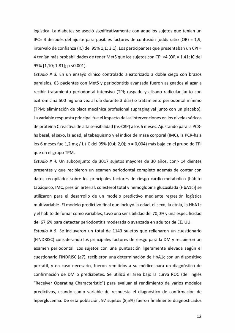
Estudio # 3. En un ensayo clínico controlado aleatorizado a doble ciego con brazos
paralelos, 63 pacientes con MetS y periodontitis avanzada fueron asignados al azar a
recibir tratamiento periodontal intensivo (TPI; raspado y alisado radicular junto con
azitromicina 500 mg una vez al día durante 3 días) o tratamiento periodontal mínimo
(TPM; eliminación de placa mecánica profesional supragingival junto con un placebo).
La variable respuesta principal fue el impacto de las intervenciones en los niveles séricos
de proteína C reactiva de alta sensibilidad (hs-CRP) a los 6 meses. Ajustando para la PCR-
hs basal, el sexo, la edad, el tabaquismo y el índice de masa corporal (IMC), la PCR-hs a
los 6 meses fue 1,2 mg / L (IC del 95% [0,4; 2,0]; p = 0,004) más baja en el grupo de TPI
que en el grupo TPM.
Estudio # 4. Un subconjunto de 3017 sujetos mayores de 30 años, con> 14 dientes
presentes y que recibieron un examen periodontal completo además de contar con
datos recopilados sobre los principales factores de riesgo cardio-metabólico [hábito
tabáquico, IMC, presión arterial, colesterol total y hemoglobina glucosilada (HbA1c)] se
utilizaron para el desarrollo de un modelo predictivo mediante regresión logística
multivariable. El modelo predictivo final que incluyó la edad, el sexo, la etnia, la HbA1c
y el hábito de fumar como variables, tuvo una sensibilidad del 70,0% y una especificidad
del 67,6% para detectar periodontitis moderada o avanzada en adultos de EE. UU.
Estudio # 5. Se incluyeron un total de 1143 sujetos que rellenaron un cuestionario
(FINDRISC) considerando los principales factores de riesgo para la DM y recibieron un
examen periodontal. Los sujetos con una puntuación ligeramente elevada según el
cuestionario FINDRISC (≥7), recibieron una determinación de HbA1c con un dispositivo
portátil, y en caso necesario, fueron remitidos a su médico para un diagnóstico de
confirmación de DM o prediabetes. Se utilizó el área bajo la curva ROC (del inglés
“Receiver Operating Characteristic”) para evaluar el rendimiento de varios modelos
predictivos, usando como variable de respuesta el diagnóstico de confirmación de
hiperglucemia. De esta población, 97 sujetos (8,5%) fueron finalmente diagnosticados
13
de DM (n = 28; 2,5%) o prediabetes (n = 69; 6,0%). Cuando solo se incluyeron los
resultados del cuestionario FINDRISC, el modelo presentó un área bajo la curva (AUC)
de 0,866 (IC del 95% [0,833; 0,900]). Este modelo mejoró significativamente cuando se
añadió la determinación de la HbA1c mediante un dispositivo portátil (AUC = 0,961; IC
del 95% [0,941; 0,980]; p <0,001).
Conclusiones: La periodontitis avanzada se encuentra asociada significativamente al
padecimiento de DM y MetS en la población laboral española. Esta asociación es
relevante ya que la terapia periodontal reduce el riesgo cardiovascular en pacientes con
MetS y periodontitis avanzada. El cribado de la periodontitis en los centros de atención
primaria y el cribado de la hiperglucemia en el consultorio odontológico parecen ser
viables y eficaces en la identificación de individuos no diagnosticados anteriormente de
estas patologías.
14
II. INTRODUCTION Periodontitis is a chronic inflammatory disease associated with oral biofilm dysbiosis
and an unresolved inflammation leading to destruction of the tooth supporting
structures (Papapanou et al., 2018). Severe periodontitis is estimated to affect 796
million people worldwide (G. B. D. Oral Disorders Collaborators et al., 2020), which
implies an enormous public health challenge as periodontitis leads to a significant
deterioration of oral health-related quality of life (OHrQL) (Buset et al., 2016; Cunha-
Cruz, Hujoel, & Kressin, 2007; Gerritsen, Allen, Witter, Bronkhorst, & Creugers, 2010;
Graziani & Tsakos, 2020) and a heavy economic burden on health care systems (Listl,
Galloway, Mossey, & Marcenes, 2015; Tonetti, Jepsen, Jin, & Otomo-Corgel, 2017).
Current concepts on the aetiology and pathogenesis of periodontitis include not only
the activation of immunoinflammatory mechanisms by the subgingival microbiota, but
also several behavioural, genetic and systemic factors that may influence the clinical
expression of the disease (Kornman, 2008). Among the medical conditions that may
impact periodontal health, diabetes mellitus (DM) is the only recognized systemic risk
factor formally included in the 2017 classification system of periodontitis (Tonetti,
Greenwell, & Kornman, 2018). However, other metabolic disorders, such as obesity or
the metabolic syndrome, have been proposed to be associated with the onset and/or
progression of periodontitis (Gorman, Kaye, Nunn, & Garcia, 2012; I. Morita et al., 2011;
T. Morita et al., 2010), even though more longitudinal studies in different populations
are needed before a causal relationship with periodontitis can be clearly established.
In the last 20 years, increasing evidence has shown that periodontitis may also impact
systemic health, giving birth to the term Periodontal Medicine (Beck, Papapanou, Philips,
& Offenbacher, 2019; Genco & Sanz, 2020). Several mechanisms have been
demonstrated to be implicated in these associations, including the spread of the
infection from the subgingival environment to the circulatory system and from there to
distant tissues and organs (bacteraemia) (Reyes, Herrera, Kozarov, Roldan, & Progulske-
Fox, 2013), the release of local inflammatory mediators that secondarily influence
systemic inflammation (Amar et al., 2003), the activation of adaptive immunity, and
15
combinations of all these potential mechanisms (Van Dyke & van Winkelhoff, 2013).
To date, periodontitis has been potentially associated with 57 different systemic
conditions (Monsarrat et al., 2016), with DM being the one presenting most robust
evidence of a bidirectional association (Sanz et al., 2018; Taylor, 2001). Epidemiological
evidence has demonstrated that poor glycaemic control correlates with higher
prevalence, severity, and progression rate of periodontitis, when compared to normo-
glycemic individuals (Borgnakke, Ylostalo, Taylor, & Genco, 2013a; Graziani, Gennai,
Solini, & Petrini, 2018). Conversely, periodontitis has been associated with higher
incidence of type 2 DM (Demmer, Jacobs, & Desvarieux, 2008; Saito et al., 2004), and
significant improvements in glycemic control (measured by the percentage of glycated
haemoglobin, HbA1c) have been reported after periodontal therapy (D´Aiuto et al.,
2018; S. Engebretson & Kocher, 2013). There is a surprising lack of information in Spain
on the prevalence of periodontitis in subjects with normal and abnormal glucose
regulation (prediabetes and DM).
Frequently associated with both diabetes and periodontitis are obesity and metabolic
syndrome (MetS), consisting on a cluster of metabolic abnormalities, including
increased blood pressure, elevated plasma glucose, excess body fat around the waist
and abdominal area, and altered cholesterol levels (Alberti et al., 2009). Numerous
studies have reported a positive association between body mass index (BMI) ≥ 25 and
periodontitis, although the magnitude of this association has varied in different
populations (J. Suvan, D'Aiuto, Moles, Petrie, & Donos, 2011; J. E. Suvan et al., 2015).
Similarly, in the third National Health and Nutrition Examination Survey (NHANES),
individuals ≥ 45 years of age suffering from severe periodontitis were 2.3 times [95%
confidence interval (CI): 1.13; 4.47] more likely to present with MetS when compared
with unaffected individuals (D'Aiuto et al., 2008). Chronic systemic inflammation,
diagnosed by high sensitivity C-reactive protein (hs-CRP) measurements and white
blood cell counts, seems to be the main link among these associations (Demmer et al.,
2013; Genco & Van Dyke, 2010). This chronic state of systemic inflammation seems to
be a common pathophysiological pathway underlying the association between these
conditions with a higher risk for cardiovascular diseases (D'Aiuto et al., 2010), in
16
particular for periodontitis and metabolic syndrome. There is, however, a lack of
knowledge on the impact of periodontal therapy in patients affected by MetS and it
remains questionable whether periodontal treatment may decrease systemic
inflammation in these patients, and therefore, reduce their cardiovascular risk.
1. ABNORMAL GLUCOSE REGULATION (DIABETES MELLITUS AND PREDIABETES)
Diabetes mellitus represents a group of metabolic diseases characterized by
hyperglycemia resulting from defects in insulin secretion, insulin action, or both
(American Diabetes Association, 2020). More than 400 million people in the world suffer
from DM, which corresponds to a global age-standardized prevalence over 8% in the
adult population, with more than 3 million people dying annually as a consequence of
their hyperglycemia (Organization, 2016). The prevalence is expected to rise up to 10.8%
(700 million people) by 2045 (Saeedi et al., 2019).
Long-term elevated blood glucose levels are associated with damage and dysfunction of
different organs, including the eyes (retinopathies), kidneys (nephropathies eventually
leading to renal failure), nerves (peripheral neuropathies) or other macrovascular and
microvascular complications (Forbes & Cooper, 2013). Specifically, patients with DM
present an increased incidence of atherosclerotic cardiovascular and cerebrovascular
disease (Forbes & Cooper, 2013).
The development of DM, a multifactorial disease, may be the consequence of a
combination of different pathogenic processes, including the destruction of pancreatic
β-cells, with subsequent insulin deficiency, or abnormalities resulting in resistance to
insulin action. These situations usually coexist in the same patients, frequently causing
difficulties in determining which is the primary cause of hyperglycemia.
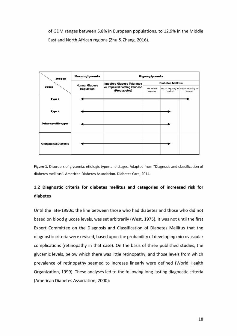
1.1 Classification of Diabetes Mellitus (Figure 1)
The different clinical scenarios considered by the American Diabetes Association (ADA)
(American Diabetes Association, 2020) are:
17
• Type 1 diabetes, resulting from β-cell destruction, usually leading to absolute
insulin deficiency. This form accounts for only 5-10% of those with DM, and it
was previously named insulin-dependent diabetes or juvenile-onset diabetes.
Although it normally presents during childhood or adolescence, it can appear in
some adults that retained residual β-cell function sufficient to prevent
ketoacidosis for many years. There are even some forms of type 1 diabetes with
no evidence of autoimmunity and, therefore, named idiopathic diabetes.
• Type 2 diabetes, results from a progressive insulin secretory defect against a
background of insulin resistance. This form of diabetes accounts for ≈90-95% of
those with DM. It was previously named non-insulin dependent diabetes or
adult-onset diabetes, as it encompasses individuals who usually have relative
(rather than absolute) insulin deficiency. Most of the patients with type 2 DM
are obese or present an increased percentage of abdominal body fat (Bjorntorp,
1991). This form of DM may be present for a long period of time without any
clinical symptom, remaining undiagnosed although these patients are at risk of
developing diabetes-related complications. Although insulin resistance may
improve with weight reduction and/or pharmacological treatment, it is seldomly
restored to normal. Family studies have shown a strong heritability (50-60%) for
type 2 DM, although the genetics of this form of DM are complex and are not
fully defined (Almgren et al., 2011).
• Other specific types of DM include: genetic defects in β-cell or insulin function,
pancreatic diseases such as cystic fibrosis, drug-induced DM (e.g., after organ
transplantation).
• Gestational diabetes mellitus (GDM). For a long time, GDM was defined as any
glucose intolerance initially recognized during pregnancy. However, the
definition persisted even if the case did not resolve post-partum and did not
exclude the possibility that unrecognized glucose intolerance may have begun
before pregnancy. For this reason, the Association of the Diabetes and
Pregnancy Study Groups (IADPSG) recommend that high-risk women (e.g. obese)
found to have diabetes at the initial prenatal visit, should receive a diagnosis of
overt, and not gestational, diabetes (International Association of Diabetes in
Pregnancy Study Group Working Group on Outcome et al., 2015). The prevalence
18
of GDM ranges between 5.8% in European populations, to 12.9% in the Middle
East and North African regions (Zhu & Zhang, 2016).
Figure 1. Disorders of glycemia: etiologic types and stages. Adapted from “Diagnosis and classification of
diabetes mellitus”. American Diabetes Association. Diabetes Care, 2014.
1.2 Diagnostic criteria for diabetes mellitus and categories of increased risk for
diabetes
Until the late-1990s, the line between those who had diabetes and those who did not
based on blood glucose levels, was set arbitrarily (West, 1975). It was not until the first
Expert Committee on the Diagnosis and Classification of Diabetes Mellitus that the
diagnostic criteria were revised, based upon the probability of developing microvascular
complications (retinopathy in that case). On the basis of three published studies, the
glycemic levels, below which there was little retinopathy, and those levels from which
prevalence of retinopathy seemed to increase linearly were defined (World Health
Organization, 1999). These analyses led to the following long-lasting diagnostic criteria
(American Diabetes Association, 2000):
19
• Two-hour plasma glucose ≥200 mg/dL (11.1 mmol/L) during an oral glucose
tolerance test (OGTT). The test should be performed using a glucose load
containing the equivalent of 75 g anhydrous glucose dissolved in water.
• Fasting Plasma Glucose (FPG) ≥126 mg/dL (7.0 mmol/L). Fasting is defined as no
caloric intake for at least 8 hours.
In 2013, the ADA introduced a significant change in the diagnosis of diabetes by
proposing a hemoglobin A1c (HbA1c) level ≥ 6.5% [following venipuncture and
laboratory analyses by a certified National Glycohemoglobin Standardization Program
(NGSP) high-performance liquid chromatography (HLPC)] as a criterion for the diagnosis
of DM (American Diabetes, 2013). HbA1c is a widely used biomarker to evaluate
management of glycemic control, as it reflects the average blood glucose levels over a
2- to 3- month period (Sherwani, Khan, Ekhzaimy, Masood, & Sakharkar, 2016). Apart
from the advantage of being already familiar to clinicians as a marker of glycemic control
through an extended period of time, the use of HbA1c does not require for fasting,
making it more convenient in certain circumstances. However, the cost to evaluate this
parameter is higher, and its analyses may be limited in some regions of the developing
world and in certain hemoglobinopathies (e.g. anemias from hemolysis or iron
deficiency) or congenital ethnic variants may affect the results (Sherwani et al., 2016).
Additionally, patients presenting with severe hyperglycemia symptoms/crisis could be
diagnosed by a casual plasma glucose ≥200 mg/dL (11.1 mmol/L).
It is important to understand that there is no 100% concordance between FPG, OGTT
and HbA1c as diagnostic criteria for DM, with controversy regarding which biomarker
presents a higher sensitivity (Carson, Reynolds, Fonseca, & Muntner, 2010; Ho-Pham,
Nguyen, Tran, & Nguyen, 2017). Furthermore, it is also possible that these biomarkers
identify different groups of diabetic subjects and physiological processes. Particularly, it
seems that HA1c is not sufficiently sensitive to identify those subjects with early
diabetes/prediabetes (Cowie et al., 2010; Fajans, Herman, & Oral, 2011).
Since 2003, a group of people have been recognized that do not meet the diagnostic
criteria for DM but present blood glucose levels higher than normal (Genuth et al.,
20
2003). This condition is named prediabetes, and it is defined either as impaired fasting
glucose (IFG: FPG levels 100 mg/dL to 125 mg/dL) or impaired glucose tolerance (IGT:
OGTT values of 140 mg/dL to 199 mg/dL). The corresponding levels to identify subjects
with prediabetes by means of HbA1c are 5.7%-6.4%. IFG mainly represents hepatic
insulin sensitivity, while IGT primarily reflects pancreatic β-cell function (Faerch, Borch-
Johnsen, Holst, & Vaag, 2009). The Homeostatic Model Assessment-Insulin Resistance
(HOMA-IR) is a model to score insulin resistance, while the Homeostatic Model
Assessment-β (HOMA-β) scores β-cell function (Matthews et al., 1985).
It has been demonstrated that having prediabetes is a risk factor for developing DM,
with a 5-year cumulative incidence ranging from 12% to 25%, which is 3- to 8-fold higher
than the incidence for the whole population (Sato et al., 2009; Shimazaki, Kadowaki,
Ohyama, Ohe, & Kubota, 2007). Furthermore, prediabetes states have been
independently associated to cardiovascular, renal and neurological complications
(Fonseca, 2009; Shaye, Amir, Shlomo, & Yechezkel, 2012; Tabak, Herder, Rathmann,
Brunner, & Kivimaki, 2012). As long as prediabetes is a reversible condition, individuals
with prediabetes should be informed about their increased risk for DM and advised
about preventive strategies such as diet modifications or physical exercise.
Furthermore, early identification and intervention have been successful in delaying
and/or preventing the progression to DM (Diabetes Prevention Program Research
Group, 2015).
1.3 Pathogenesis and complications of Diabetes Mellitus
As a consequence of elevated levels of blood glucose, a series of biochemical events
occur, including:
i. the increased transformation to sorbitol by the enzyme aldose reductase;
ii. an increased production of diacylglycerol, leading to the activation of protein kinase
C;
iii. the formation of non-enzymatic glycation and oxidation products, including HbA1c
and the advanced glycation end-products (AGEs).
21
The first two mechanisms have been proposed to be linked to the development of
retinopathies, neuropathies and nephropathies, while the formation of AGEs may alter
normal cell function of different cell types, including endothelial cells, mononuclear
phagocytes, smooth muscle cells or fibroblasts (Lalla, Lamster, Drury, Fu, & Schmidt,
2000).
Endothelial cell and smooth muscle cells interactions with AGEs may result in the
development of a series of events leading to vascular lesions in DM patients (Wautier et
al., 1996), while the activation of the receptor for AGEs (RAGE) in inflammatory cells has
been linked to the pathogenesis of atherosclerosis and altered responses to infection
(Ross, 1999). Lastly, the AGE-RAGE interaction in fibroblasts is an important contributor
to the impaired formation and remodeling of the connective tissues observed in DM
patients (Ramamurthy & Golub, 1983). All these covert changes may, with time,
contribute to the micro- and macrovascular complications of DM. The microvascular
complications include nephropathy, retinopathy and neuropathy, while the
macrovascular complications comprise cardiovascular diseases resulting into coronary
diseases and stroke.
1.3.1 Nephropathy
Diabetes nephropathy is clinically characterized by the development of proteinuria
(including albumin as the major component) and, if blood glucose is not properly
controlled, subsequent reduced glomerular filtration rate. It is frequently associated
with hypertension and it is a major risk factor for macrovascular complications
(Matsushita et al., 2010).
1.3.2 Retinopathy
Diabetes retinopathy is the leading cause of blindness in adults. It comprises a series of
lesions, including alterations in vascular permeability, microaneurysms and excessive
formation of new blood vessels (neovascularization). Initially, changes in the
permeability of the blood vessels lead to the accumulation of fluid within the retina, and
in late stages, neovascularization and formation of maculae lead to visual impairment
and eventually, in retinal detachment (Frank, 2004).
22
1.3.3 Neuropathy
Diabetic neuropathy affects both the somatic and autonomic peripheral nervous
system. The involvement of the somatic nervous system leads to fiber deterioration and
progressive loss of sensory perception, while the influence on the autonomic nervous
system hampers the ability to maintain vascular tone and an optimum blood flow to the
extremities and different organs such as the heart, the brain or the entire
gastrointestinal tract (Selvarajah et al., 2006; Wessels et al., 2006). Nowadays, there is
no approved therapy for the treatment of diabetic neuropathy.
1.4 Prevention of Diabetes Mellitus
Different approaches have been proposed to prevent the incidence of type 2 DM.
Strategically, these initiatives may be separated into those aimed to slow down the
progression of chronic hyperglycemia among patients with pre-existing IFG or IGT, and
those with the objective of early detection of those subjects at risk of developing
diabetes, or those who remain undiagnosed.
The Diabetes Prevention Program (DPP) trial was a landmark study demonstrating that
lifestyle interventions, mostly focused on weight loss through diet and physical exercise,
are more effective than Metformin to reduce the risk of incident diabetes (Knowler et
al., 2002; Perreault et al., 2012). The results of this study, together with its replications
in primary care settings, have established lifestyle changes as the cornerstone of
diabetes prevention and management (Ackermann, 2013; DeJoy, Padilla, Wilson,
Vandenberg, & Davis, 2013).
At the same time, and since up to 37% of the newly diagnosed cases of type 2 DM have
already, at least, one diabetes complication (e.g. retinopathy), early diagnosis has
become an essential target (Kohner et al., 1998). However, massive screening,
measuring either fasting or postprandial plasma glucose is costly and probably
inefficient due to the relatively low prevalence of undiagnosed diabetes in the general
population. Therefore, selective screening of high-risk subjects identified through self-
reported questionnaires, family background and anthropometric measures (e.g. BMI,
waist circumference, etc.) has emerged as a cost-effective approach (Lindstrom &
23
Tuomilehto, 2003). Using this methodology, it has been postulated that the dental
practice may offer a valuable opportunity to identify subjects at risk of suffering
previously undiagnosed hyperglycemia.
1.5 Management of Diabetes Mellitus
The general management of diabetes patients includes four relevant aspects, described
below (Nyenwe, Jerkins, Umpierrez, & Kitabchi, 2011):
1.5.1 Education
Patients with abnormal glucose regulation (diabetes or prediabetes) should receive
information regarding the disease process, diet and physical exercise advice,
instructions in proper blood glucose monitoring and medication intake, and knowledge
of acute and chronic complications (Nyenwe et al., 2011).
1.5.2 Medical nutrition therapy and exercise advice
Nutrition is a crucial component of a healthy lifestyle. Particularly, in DM patients it has
been shown that dietary measures aiming for weight loss through reduction of saturated
fats and provision of fiber, whole grains, fruits and vegetables, led to significant
improvements in glycemic control and cardiovascular risk factors (i.e. blood pressure
and lipid profiles) (Look et al., 2007). Sedentary lifestyle has been identified as a risk
factor for DM. A systematic review with meta-analyses published almost 20 years ago
reported that a structured moderate exercise program (50 minutes-three times a week)
resulted in a 0.7% reduction in HbA1c levels within 8 weeks (Boule, Haddad, Kenny,
Wells, & Sigal, 2001).
1.5.3 Monitoring of glycemic control
Landmark clinical trials such as the United Kingdom (UK) Prospective Diabetes Study
have demonstrated that self-monitoring blood glucose (SMBG) is effective on the
reduction of the incidence of DM-associated complications, showing that it is an
important component of DM management (Schnell et al., 2013). The advantages of
SMBG include the detection of asymptomatic hypoglycemia and hyperglycemic
24
excursions, which are risk factors for cardiovascular events (Nyenwe et al., 2011).
However, it is controversial whether SMBG is effective in type 2 DM patients not treated
with insulin (O'Kane, Bunting, Copeland, Coates, & group, 2008).
1.5.4 Drug therapy for glycemic control
A plethora of hypoglycemic agents, including different insulin preparations, are
employed in the clinical management hyperglycemia:
Metformin: is considered the first-line agent in the treatment of type 2 DM and in its
prevention from prediabetes. It improves peripheral glucose utilization by increasing
insulin receptor tyrosine kinase activity (Goodarzi & Bryer-Ash, 2005). It also reduces
hepatic gluconeogenesis. It can reduce HbA1c in a range from 0.8% up to 2.0%.
Consequently, it has proven to be effective in the reduction of cardiovascular events and
to diminish the risk of progression from prediabetes to DM by 31% (Knowler et al., 2002;
UK Prospective Diabetes Study (UKPDS) Group, 1998).
Insulin secretagogues: stimulate insulin secretion by the β-cells through interaction with
the sulfonylurea receptor, commonly named sulfonylureas for this reason. They are
considered a second line of treatment for DM in most patients, mainly due to their side
effects (i.e. hypoglycemia and weight gain) (Holstein, Plaschke, & Egberts, 2001).
Glinides (e.g., repaglinide) bind to a different part of the sulfonylurea receptor, making
them less potent than sulfonylureas but also with less side effects, making them ideal
for the combination with metformin (Gerich, Raskin, Jean-Louis, Purkayastha, & Baron,
2005).
Incretins: These are hormones [glucagon-like peptide 1 (GLP-1) and glucose-dependent
insulinotropic peptide (GIP)] that modulate secretions from pancreatic islet cells, but
they can also be administered subcutaneously. They are rapidly inactivated by the
enzyme dipeptidyl peptidase IV and, for this reason, they are commonly administered
together with dipeptidyl peptidase IV inhibitors (sitagliptin and saxagliptin) (Drucker &
Nauck, 2006).
25
Insulin therapy: Chronic hyperglycemia progression leads to a vicious cycle that further
compromise β-cell capacity to secrete insulin, even after administration of hypoglycemic
agents. For this reason, most patients with type 2 DM ultimately require insulin therapy
to achieve an adequate glycemic control. Human insulin or insulin analogs can be used
with this purpose, with the synthetic compounds presenting faster and long-lasting
effects, and producing less hypoglycemia and weight gain (Investigators et al., 2006; J.
Klein et al., 2004).
Other drugs: Thiazolidinediones (rosiglitazone and pioglitazone) and α-glucosidase
inhibitors (acarbose and miglitol) are also regularly prescribed for their ability to
ameliorate postprandial hyperglycemia. However, their regular use is limited, mainly
due to the controversial side effects of fluid retention (thiazolidinediones) or
gastrointestinal effects (α-glucosidase inhibitors).
1.6 Links between periodontitis and abnormal glucose regulation
Different reviews and consensus documents published in recent years have clearly
indicated the bidirectional association between diabetes and periodontitis (Chapple &
Genco, 2013; Genco & Borgnakke, 2020; Genco, Graziani, & Hasturk, 2020; Lalla &
Papapanou, 2011; Preshaw et al., 2012; Sanz et al., 2018). Numerous studies have
shown that DM (both type 1 and type 2) is a risk factor for periodontitis, increasing the
risk approximately three times when compared with non-diabetes subjects, particularly
in patients with diabetes and poor glycemic control (Ide, Hoshuyama, Wilson, Takahashi,
& Higashi, 2011a; Nelson et al., 1990; Seppala, Seppala, & Ainamo, 1993; Taylor, Burt,
Becker, Genco, Shlossman, et al., 1998). Furthermore, periodontitis negatively affects
glycemic control in patients with diabetes, and contributes to the development of
complications (Demmer et al., 2010; Shultis et al., 2007).
1.6.1 Mechanistic links underlying the association between periodontitis and diabetes
As previously explained, immune-dysregulation and systemic inflammation are central
to the pathogenesis of type 2 DM. The main biological processes and mechanisms that
underpin the association between diabetes and periodontitis and vice versa are:
26
Microbiota: whether patients with periodontitis and DM have a characteristic and
distinctive subgingival microbial profile has been a matter of debate for the last 30 years.
While early studies observed a significant effect of chronic hyperglycemia in the
composition of the periodontal microbiota (Zambon et al., 1988), subsequent reports
failed to find a specific bacterial profile among patients with DM (Novaes, Gonzalez
Gutierrez, Grisi, & Novaes, 1997; Thorstensson, Dahlen, & Hugoson, 1995). However,
the advent of 16s rRNA sequencing methods have offered the possibility to observe the
entire microbiome, identifying significant differences in the prevalence or abundance of
certain bacteria at several taxonomic levels, identifying certain genus being more
prevalent in patients with healthy gums and diabetes (genus of Neisseria), or in subjects
with both periodontitis and diabetes (Tannerella forsythia), when compared with their
non-diabetes counterparts (Casarin et al., 2013; M. Zhou et al., 2013). Furthermore,
antibody titers against periodontal pathogens have shown a correlation with HbA1c
levels (glycemic control) (Merchant et al., 2014). Even though most of these studies have
reported an association between poor glycemic control and the periodontal microbiota,
there is a need for longitudinal studies assessing the role of these specific microbial
profiles in the causal pathway of diabetes.
Host response: Inflammation is a central feature of both diabetes and periodontal
diseases, and inflammatory processes are upregulated in the periodontal tissues in
patients with diabetes. An altered immune cell response in DM patients has been
observed, particularly in neutrophil function. A hyperinflammatory phenotype,
releasing increase levels of cytokines, has been characterized after exposure to
lipopolysaccharide (LPS), leading to elevated levels of interleukin (IL)-1β and IL-6, that
may be responsible for both the periodontal destruction in DM subjects and the
contribution of periodontitis to chronic hyperglycemia (Salvi et al., 1997).
Oxidative stress: Both periodontitis and DM have demonstrated to induce oxidative
stress locally and systemically (Allen, Matthews, DJ, Griffiths, & Chapple, 2011; Chapple
& Matthews, 2007; Evans, Goldfine, Maddux, & Grodsky, 2003). Presence of hyper-
active/reactive neutrophils in periodontitis patients generate increased levels of
reactive oxygen species (ROS) in response to the dental biofilm, resulting into collateral
27
host-tissue damage. Furthermore, reduced antioxidant capacity has been previously
reported in periodontitis patients (Chapple, 1997). As long as type 2 DM patients with
periodontitis have increased plasma biomarkers of oxidative stress (Allen et al., 2011),
it may be the case that ROS production by hyper-active/reactive neutrophils in
periodontitis could overcome antioxidant defenses, contributing to β-cell dysfunction.
AGEs/RAGEs axis: The binding of AGEs to different cell types in the periodontium may
hamper their normal function by leading to pro-inflammatory and pro-oxidant effects
(Vlassara & Uribarri, 2014). As an example, it has been proposed that the fibroblasts of
DM-affected individuals may lead to abnormal collagen metabolism by reducing its
production in the periodontal tissues, eventually contributing to the initiation and
progressive destruction of the periodontal support (Claudino et al., 2007). The potential
role of AGEs as a mechanistic link between DM and periodontitis have been proved, as
serum levels of AGEs demonstrated to be associated with the severity of periodontitis
in type 2 DM patients (Takeda et al., 2006).
Bone homeostasis: Several studies have focused on the impact of the most important
determinant for bone resorption in DM, the ratio between the receptor activator of
nuclear factor κ B ligand (RANKL)/osteoprotegerin (OPG). The soluble RANKL expression
and RANKL/OPG ratio has been reported as higher in patients with poorly controlled
diabetes when compared with well controlled subjects (Ribeiro et al., 2011; Santos et
al., 2010). This finding may explain the increased alveolar bone loss observed among DM
patients.
1.6.2 Chronic hyperglycemia as a risk factor for periodontitis
There is plenty of literature stating that type 1 and type 2 DM patients, as well as women
with gestational diabetes, suffer more frequently from periodontitis and present with
more severe forms of the disease (Chavarry, Vettore, Sansone, & Sheiham, 2009;
Cianciola, Park, Bruck, Mosovich, & Genco, 1982; Novak, Taylor, Dawson, Ferguson, &
Novak, 2006; Thorstensson & Hugoson, 1993). The strongest evidence is derived from
the longitudinal studies carried out in Pima Indians, where it was proven for the first
time that DM patients presented a 2.6 times greater risk to suffer periodontitis than
28
those with normal glucose levels (Nelson et al., 1990). Lately, this finding has been
confirmed in other populations with different geographic and ethnic origins, such as the
participants in the Study of Health in Pomerania, in Germany (Demmer, Holtfreter, et
al., 2012). A particularly higher risk for progression of periodontitis has been observed
in those DM subjects with poor glycemic control (Guzman, Karima, Wang, & Van Dyke,
2003; Taylor, Burt, Becker, Genco, & Shlossman, 1998). Indeed, it is abnormal glucose
regulation, and not necessarily the diagnosis of DM, that has been correlated with the
severity of periodontitis (Saito et al., 2004).
1.6.3 Impact of periodontitis on diabetes
Systematic reviews, with or without meta-analyses have reported consistently a worst
glycemic control (either measured as FPG, HBA1c or through OGTT) in patients without
overt diabetes when comparing subjects with periodontitis versus non-periodontitis
(Borgnakke, Ylostalo, Taylor, & Genco, 2013b; Graziani et al., 2018). However, it is
questionable whether there is a true independent association between periodontitis
and prediabetes (either measured as IFG, IGT or HbA1c 5.7%-6.4%), or if this association
applies only for specific subgroups (e.g. obese or overweight individuals) or geographical
areas (Kowall et al., 2015; Rao Deepika & Saxena, 2013).
The longitudinal studies performed in the Gila River Indian community have been
important to determine the effect of periodontitis not just on the deterioration of
glycemic control, but also on the incidence of diabetes complications in subjects with
overt DM. In these studies, periodontitis was associated with a significantly higher
incidence of diabetic nephropathy and future ischemic heart disease (Saremi et al.,
2005). Subjects with diabetes and severe periodontitis had 3.2 times the risk of death
from a cardiovascular or renal event, when compared with diabetes subjects with
no/mild/moderate periodontitis. More recent studies have also observed associations
between periodontitis and incident retinopathy or even neuropathic foot ulceration
(Abrao, Chagas, & Schmid, 2010; Noma et al., 2004). Therefore, it seems clear that
periodontitis contributes to an increased risk of development of diabetes complications
(Borgnakke et al., 2013b; Graziani et al., 2018).
29
It is currently a matter of debate whether periodontitis patients with no manifest DM
have a greater risk of developing type 2 DM, than those with a better periodontal status.
In other words, if periodontitis is a risk factor for the onset of DM. A series of studies,
most of them performed in Asian populations (Japan and Taiwan), have reported that
subjects with poor periodontal health may have an increased risk for developing DM
(Demmer et al., 2010; Demmer et al., 2008; Ide, Hoshuyama, Wilson, Takahashi, &
Higashi, 2011b; I. Morita et al., 2012; Saito et al., 2004) (Chiu et al., 2015; Lin et al., 2014).
However, some of these studies are retrospective and/or periodontitis was diagnosed
by indirect evidence (partial-mouth examinations, surrogate measures such as tooth
loss or simplified index scores), what may overestimate the association between the
periodontal disease and incident diabetes. Furthermore, some of them failed to prove
any association after adjusting for potential confounders. Therefore, there is a need for
more studies with accurate measures for both periodontal status and DM (i.e.
laboratory data) to determine if periodontitis may have an effect on hyperglycemia in
otherwise healthy individuals.
1.6.4 Effect of periodontal treatment on diabetes outcomes
Periodontal treatment reduces inflammation and reduces circulating cytokines among
individuals with diabetes (Artese et al., 2015) and may thus decrease hyperglycaemia in
these subjects. Numerous randomized clinical trials (RCTs) have been performed since
1998 evaluating the effect of periodontal therapy (consisting in most of the cases of
scaling and root planning (SRP) with or without systemic antimicrobials) on HbA1c levels
in DM patients. In parallel to the publication of these trials, a series of systematic
reviews, with or without meta-analyses, have also been published.
In 2012, a systematic review and meta-analyses on the effect of periodontal treatment
on HbA1c levels was presented and discussed at the joint workshop of the European
Federation of Periodontology (EFP) and American Academy of Periodontology on
“Periodontitis and systemic diseases”. That systematic review reported a mean HbA1c
reduction of 0.36% after periodontal therapy (S. Engebretson & Kocher, 2013).
However, it was highlighted by the authors that the conclusions should be cautiously
considered, due to the small sample size and the high risk of bias of some of the included
30
studies. To overcome these limitations, a multicenter RCT (Diabetes and Periodontal
Therapy Trial, DPTT) was performed in five academic medical centers in the United
States of America (USA), including 514 subjects (S. P. Engebretson et al., 2013).
However, the DPTT was unable to demonstrate any beneficial effect of periodontal
therapy for the reduction of HbA1c levels in patients with DM, and created a great
controversy among the scientific community, mainly due to important deficiencies
associated with the study design and implementation that could hamper the
interpretation of the results (Borgnakke et al., 2014; Chapple, Borgnakke, & Genco,
2014; Merchant, 2014; Vergnes, 2014). First, it seems clear that in order to observe any
clinically meaningful improvement in the glycemic control, periodontal treatment would
need to reach a minimum standard of care as pre-requisite. Unfortunately, bleeding on
probing (BOP) and plaque indices were 41.6% and 72.1% in the test group at the 6
months examination. Secondly, it was difficult to expect any reduction on HbA1c levels
as long as they were close to the goal for proper glycemic control at baseline, with
approximately 60% of the patients in the study presenting HbA1c levels below 8.0%. A
third relevant problem was the mean body mass index (BMI) of the participants, which
was approximately 34 kg/m2, most being obese (BMI≥30 kg/m2). A recent systematic
review concluded that there were significant differences in the metabolic response after
periodontal therapy when comparing obese and normal-weight patients (Papageorgiou,
Reichert, Jager, & Deschner, 2015), which would have masked the anti-inflammatory
effect of periodontal treatment. Thus, it is possible that in the DPTT most of the subjects
were resistant to the reduction of systemic inflammation by the periodontal treatment
due to the overwhelming influence of obesity.
Several studies have been published since then, and all of them, in agreement with the
9 systematic reviews published during 2013- 2017, have reported reductions in HbA1c
at 3-4 months after therapy ranging from 0.27% to 1.03% (Madianos & Koromantzos,
2018). A recent prospective cohort study, including more than 120.000 subjects, with
DM and periodontitis treated in the Veteran Administration (VAs) medical centres in the
USA, reported that periodontal treatment reduced HbA1c by 0.02% and 0.074% after
initial treatment and after an average of 1.7 years of supportive periodontal therapy,
respectively (Merchant et al., 2016). Particularly relevant has been a RCT performed in
31
the United Kingdom and published in The Lancet Diabetes & Endocrinology in 2018,
including 264 patients (D´Aiuto et al., 2018). Noteworthy, in this investigation, patients
in the intensive periodontal therapy group received not only non-surgical subgingival
instrumentation but, depending on their needs at the re-evaluation, periodontal surgical
therapy to assure optimum subgingival biofilm control. This entire periodontal therapy
provided, was considered the standard of specialist care for severe forms of
periodontitis, and led in this study to an effective resolution of periodontal inflammation
that correlated with an 0.6% reduction in HbA1c levels. Similar observations have been
reported in a study using electronic medical records and dental insurance data (n=5103
patients), where those subjects receiving periodontal surgery had 0.25-0.36% lower
levels of HbA1c (Spangler et al., 2010).
Therefore, considering all this available evidence, the effects of periodontal treatment
on HbA1c points to an improvement of glycaemic control in diabetes patients. The
reductions in HbA1c may be clinically significant, as a reduction of 0.2% is associated
with a reduction in all-cause mortality of approximately 10% (Khaw et al., 2004), and a
reduction of 0.4% is comparable with the one achieved by a secondary-line
hypoglycaemic drug (Cavaiola & Pettus, 2000). Furthermore, cost-effectiveness analyses
through a life-time have proven that providing periodontal treatment to patients with
DM would provide a cost saving, as the reduction in microvascular complications (20.5%
for nephropathy, 17.7% for neuropathy and 19.2% for retinopathy) corresponds to a
total of net savings close to 6,000 United States dollars (USD) (S. E. Choi, Sima, & Pandya,
2020).
2. METABOLIC SYNDROME
Metabolic syndrome (MetS) is a cluster of metabolic abnormalities that constitute a
multiplex risk factor for cardiovascular diseases (Després & Lemieux, 2006). Metabolic
syndrome includes abnormal glucose regulation (prediabetes or DM), hypertension,
dyslipidaemia and central obesity. The rationale for grouping these conditions is based
on the fact that: a) they occur together more frequently than may be expected by
chance, and b) together they are associated with an increased risk of cardiovascular
disease (Grundy, 2008; Isomaa et al., 2001; Lakka et al., 2002). Although the pathogenic
32
mechanisms are not completely elucidated, abdominal obesity leading to insulin
resistance seems to be the common factor linking the different components (Carr et al.,
2004; Reaven, 1988).
Due to the global obesity epidemic, the prevalence of MetS has increased in the last
decade, concomitant with an increased risk for diabetes and cardiovascular disease
(Grundy, 2008; Zimmet, Alberti, & Shaw, 2001). This increase has been especially
pronounced in developing countries. In Spain, the most recent population study
reported a prevalence of obesity in 31.2% in adults aged 35-74 years (Martinez-Larrad
et al., 2016), which is similar in range to recent European and USA estimates (Aguilar,
Bhuket, Torres, Liu, & Wong, 2015; Han & Lean, 2016). The most important
determinants in the prevalence of MetS are age and ethnicity, with Hispanics showing
the highest rate (Aguilar et al., 2015).
The high prevalence of MetS is relevant from a public health perspective, as people with
MetS are three times more likely to have a heart attack or a stroke and have a five times
greater risk of developing type 2 DM than people without (Stern, Williams, Gonzalez-
Villalpando, Hunt, & Haffner, 2004). Noteworthy, is that as the number of positive
components of MetS increase to four or more, the cardiovascular risk increases by six
times and the risk of DM ris 35 times (B. E. Klein, Klein, & Lee, 2002). Although there is
still some controversy about the practical utility of MetS as a diagnostic or treatment
tool, it is considered extremely useful as a research, epidemiological and educational
tool (Simmons et al., 2010). Furthermore, when certain specific markers of metabolic
alterations (e.g. fasting insulin, apolipoprotein B) are elevated, cardiovascular disease
risk increases beyond that reported by traditional absolute risk tools (Lamarche et al.,
1998).
2.1 Metabolic syndrome definitions
Several organizations such as the World Health Organization (WHO), the National
Cholesterol Education Program – Third Adult Treatment Panel (NCEP ATP III), the
International Diabetes Federation (IDF), the American Heart Association/National Heart,
Lung, and Blood Institute (AHA/NHLBI) or the European Group for the Study of Insulin
33
Resistance (EGIR) have proposed different criteria for the diagnosis of MetS (Alberti et
al., 2009). The differences depend upon whether insulin resistance or abdominal obesity
are mandatory requirements, or whether the cut-offs for the diagnosis of central obesity
were based on waist circumference in different ethnic groups.
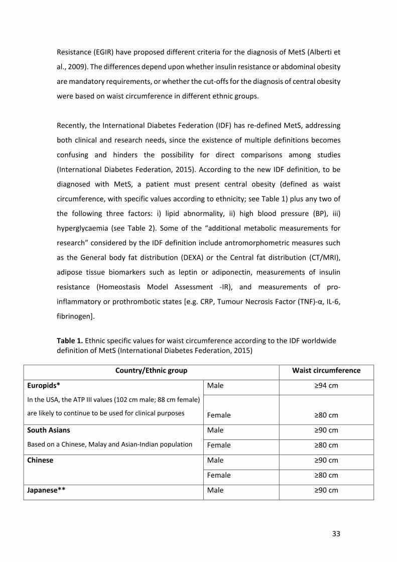
Recently, the International Diabetes Federation (IDF) has re-defined MetS, addressing
both clinical and research needs, since the existence of multiple definitions becomes
confusing and hinders the possibility for direct comparisons among studies
(International Diabetes Federation, 2015). According to the new IDF definition, to be
diagnosed with MetS, a patient must present central obesity (defined as waist
circumference, with specific values according to ethnicity; see Table 1) plus any two of
the following three factors: i) lipid abnormality, ii) high blood pressure (BP), iii)
hyperglycaemia (see Table 2). Some of the “additional metabolic measurements for
research” considered by the IDF definition include antromorphometric measures such
as the General body fat distribution (DEXA) or the Central fat distribution (CT/MRI),
adipose tissue biomarkers such as leptin or adiponectin, measurements of insulin
resistance (Homeostasis Model Assessment -IR), and measurements of pro-
inflammatory or prothrombotic states [e.g. CRP, Tumour Necrosis Factor (TNF)-α, IL-6,
fibrinogen].
Table 1. Ethnic specific values for waist circumference according to the IDF worldwide definition of MetS (International Diabetes Federation, 2015)
Country/Ethnic group Waist circumference
Europids*
In the USA, the ATP III values (102 cm male; 88 cm female)
are likely to continue to be used for clinical purposes
Male ≥94 cm
Female
≥80 cm
South Asians
Based on a Chinese, Malay and Asian-Indian population
Male ≥90 cm
Female ≥80 cm
Chinese Male ≥90 cm
Female ≥80 cm
Japanese** Male ≥90 cm
34
Female ≥80 cm
Ethnic South and Central Americans Use South Asian recommendations until more
specific data are available
Sub-Saharan Africans Use European data until more specific data are
available
Eastern Mediterranean and Middle East (Arab)
populations
Use European data until more specific data are
available
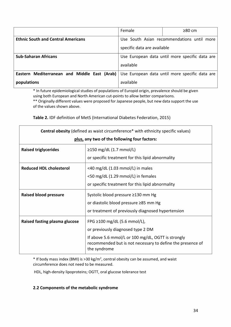
* In future epidemiological studies of populations of Europid origin, prevalence should be given using both European and North American cut-points to allow better comparisons. ** Originally different values were proposed for Japanese people, but new data support the use of the values shown above. Table 2. IDF definition of MetS (International Diabetes Federation, 2015)
Central obesity (defined as waist circumference* with ethnicity specific values)
plus, any two of the following four factors:
Raised triglycerides ≥150 mg/dL (1.7 mmol/L)
or specific treatment for this lipid abnormality
Reduced HDL cholesterol <40 mg/dL (1.03 mmol/L) in males
<50 mg/dL (1.29 mmol/L) in females
or specific treatment for this lipid abnormality
Raised blood pressure Systolic blood pressure ≥130 mm Hg
or diastolic blood pressure ≥85 mm Hg
or treatment of previously diagnosed hypertension
Raised fasting plasma glucose FPG ≥100 mg/dL (5.6 mmol/L),
or previously diagnosed type 2 DM
If above 5.6 mmol/L or 100 mg/dL, OGTT is strongly recommended but is not necessary to define the presence of the syndrome
* If body mass index (BMI) is >30 kg/m2, central obesity can be assumed, and waist circumference does not need to be measured.
HDL, high-density lipoproteins; OGTT, oral glucose tolerance test
2.2 Components of the metabolic syndrome
35
2.2.1 Central obesity
Obesity is considered the most important “driving force” of the MetS. It is important to
note that visceral fat, and not subcutaneous fat, is what increases the risk of metabolic
and cardiovascular diseases (Sironi et al., 2012). Imaging techniques, such as magnetic
resonance or computed tomography are therefore, the most effective way to assess
adiposity (Health, 1998). However, the accessibility and costs of these techniques
hamper their regular use, and anthropometric measures are the ones regularly used. As
BMI cannot discriminate into lean mass and fat mass, waist circumference has been
included as a diagnostic item of metabolic syndrome, although it does not consider
height and may underestimate or overestimate obesity in short and tall individuals,
respectively (Ashwell, Gunn, & Gibson, 2012). Other novel anthropometric indices have
been developed, such as body shape index (ABSI) (Krakauer & Krakauer, 2012), body
roundness index (BRI) or body adiposity index (BAI) (Bergman et al., 2011), but they have
not been validated or extensively used.
Visceral tissue is not only a compartment designed to store lipids, but also acts as an
endocrine organ, releasing pro-inflammatory cytokines (e.g. IL-6, TNF-α). The increase
in visceral adipose tissue leads to the release of free fatty acids reaching the liver
through the splanchnic circulation and affecting liver metabolism via glucose production
and secretion of prothrombotic agents such as plasminogen activator inhibitor 1 (PAI 1)
and fibrinogen (Aubert et al., 2003). Furthermore, hypertrophied intra-abdominal
adipocytes are resistant to the antilipolytic effect of insulin (Mittelman, Van Citters,
Kirkman, & Bergman, 2002). All these mechanisms lead to increased levels of CRP, a
biomarker identified as an important predictor of cardiovascular events (Tsimikas,
Willerson, & Ridker, 2006).
Interestingly, a subset of obese individuals seems to be protected against metabolic
complications and metabolic abnormalities can also be present in non-obese individuals.
In fact, approximately 30% of obese subjects in the USA are metabolically healthy
(normally considered those with fewer than two cardiometabolic abnormalities), and it
remains questionable whether the same approach to treatment based on diet
modification and physical exercise can be beneficial, or on the contrary, detrimental, for
these subjects.
36
2.2.2 Insulin resistance and glucose intolerance
Insulin resistance is considered the central mechanism for the development of the MetS.
The alterations in insulin normal function include failures in the inhibition of glucose
production by the liver and kidney, as well as in the regulation of glucose uptake by
different tissues. Initially, when insulin resistance is developed, to ensure proper
glycemic control, the body compensates by increasing insulin secretion, and decreasing
its clearance. However, with time this mechanism progressively fails, which results in
impairment of insulin secretion and, consequently results in alterations in fasting
glucose and glucose tolerance.
Insulin action is largely considered as “glucocentric”, since this hormone also plays a
crucial role in the inhibition of lipolysis in adipose tissue, what leads to an increase in
the production of fatty acids that further inhibits the effect of insulin (Jensen, Caruso,
Heiling, & Miles, 1989). This mechanism is the one proposed to associate the two main
characteristics of MetS: central obesity and insulin resistance.
2.2.3 Dyslipidemia
Dyslipidemia may be a consequence of insulin resistance, since there is an increase in
the synthesis of triglycerides in the liver as a consequence of the free fatty acid flux to
the liver. Furthermore, in the development of hypertriglyceridemia, a decrease in high
density lipoprotein (HDL) cholesterol may occur as a consequence of the decrease in the
action of cholesterol ester transfer protein.
2.2.4 Hypertension-High Blood Pressure
Increased blood pressure is another consequence of insulin resistance. This effect has
been explained by the vasodilator effect of insulin, which influences sodium resorption
in the kidney. In situations of insulin resistance, this vasodilatory effect is lost, while the
effect on sodium resorption is preserved, which leads to an increase in blood pressure
(Tooke & Hannemann, 2000).
2.5 A bidirectional relationship between periodontitis and metabolic syndrome?
37
In the recent Workshop on the Classification of Periodontal and Peri-implant Diseases
and Conditions, obesity and DM were considered as two of the systemic diseases that
may affect the periodontal tissues (Albandar, Susin, & Hughes, 2018; Jepsen et al.,
2018). Unfortunately, MetS was not evaluated as a distinct entity for its plausible
association with periodontitis, even if they present several common inflammatory
pathways and several epidemiological studies have reported an association between
these two entities.
2.5.1 Mechanistic links underlying the association between periodontitis and metabolic
syndrome
The mechanisms that link periodontitis and hyperglycemia, serving as the basis for a
two-way relationship between periodontitis and DM, have been previously explained.
Besides that, periodontitis has been shown to share a series of pathogenic mechanisms
with the rest of MetS components: central obesity, dyslipidemia and hypertension.
Inflammation seems to exert a pivotal role in the association between periodontitis and
obesity. Adipose tissue is an important source of several inflammatory mediators
produced by adipocytes. Among these mediators, most of them present pro-
inflammatory actions (e.g. visfatin, leptin and resistin), while just a few, like adiponectin,
present anti-inflammatory characteristics (Adamczak & Wiecek, 2013). Obesity is
characterized by a reduction in the levels of adiponectin and, conversely, by an
increased production of visfatin, leptin and resistin. These biomarkers of inflammation
have been isolated in obese subjects not only in serum, but also in gingival crevicular
fluid. Moreover, there is evidence that gingival crevicular fluid levels of adipokines and
TNF-α are elevated in obese subjects with periodontitis when compared to periodontitis
subjects with normal weight (Zuza et al., 2011). These cytokines present a catabolic
action, characterized by the recruitment of immunoinflammatory cells that release
proteases resulting into bone resorption (Zimmermann, Bastos, Dias Goncalves,
Chambrone, & Duarte, 2013). Therefore, it seems clear that the systemic inflammation
induced by obesity may enhance periodontal inflammation in periodontitis subjects,
which may result into the destruction of tooth-supporting tissues.
38
Additionally, periodontitis is a source of pro-inflammatory cytokines and reactive
oxygen species (ROS) not only locally, but also systemically (Baltacioglu et al., 2014;
Gumus, Nizam, Lappin, & Buduneli, 2014). Periodontitis-associated systemic
inflammation can promote insulin resistance, and the resulting hyperinsulinemia may
promote obesity by its effects on glucose uptake and fat storage (Demmer, Squillaro, et
al., 2012).
However, the association between periodontitis and MetS does not seem to rely solely
upon the link with obesity, as periodontitis has proven to be associated with
dyslipidemia and hypertension also (Muñoz Aguilera et al., 2020; Nepomuceno et al.,
2017). LPS of periodontal pathogens has been demonstrated to be able to increase the
lipolytic activity, leading to an up regulation of triglycerides production (Nakarai et al.,
2011), and periodontitis has been linked with endothelial dysfunction (Tonetti et al.,
2007). However, for these entities the mechanisms are not completely elucidated.
2.5.2 Epidemiological evidence of the association between metabolic syndrome and
periodontitis
A recent systematic review with meta-analyses including 20 articles, reported an odds
ratio (OR) of 1.71 (95% CI [1.42;2.03]) for the cross-sectional association between
periodontitis and metabolic syndrome (Nibali et al., 2013). However, still the association
between MetS and periodontitis remains controversial, since the diagnostic criteria
used to define the cases of MetS and periodontitis vary between studies, and some
studies have only been able to find associations in specific groups, such as women,
diabetes patients or in specific age groups (D'Aiuto et al., 2008; Furuta et al., 2013; Sora
et al., 2013; Y. K. Tu, D'Aiuto, Lin, Chen, & Chien, 2013), while others have observed no
association between MetS and periodontitis (Benguigui et al., 2010; LaMonte et al.,
2014; Zuk, Quinonez, Lebenbaum, & Rosella, 2017). Furthermore, just three longitudinal
studies on the potential causative relationship between periodontitis and MetS have
been published, all of them performed in Japan (I. Morita et al., 2011; T. Morita et al.,
2010; Tanaka et al., 2018). Two of them reported significantly higher odds to develop
MetS among patients with periodontitis after 4-5 years of follow-up (ORs ranging from
1.4 to 1.6) (T. Morita et al., 2010; Tanaka et al., 2018); while the other longitudinal study
39
determined that BMI at baseline predicted the 5-year incidence of periodontitis, with a
particularly high OR for obese women (OR=3.2)(I. Morita et al., 2011).
2.5.3 Effect of periodontal treatment on outcomes related to metabolic syndrome
Many studies have evaluated the effect of periodontal therapy in obese patients, with
conflicting results in regard to its efficacy. While some studies have identified a poorer
response to periodontal treatment in obese subjects, others did not observe any
difference in outcomes when comparing patients with different BMI categories (Gerber
et al., 2016; Papageorgiou et al., 2015). Even if a few trials have shown a reduction in
the levels of plasma-reactive oxygen metabolites or adipokines levels in saliva after SRP
(Akram et al., 2017; Suresh et al., 2018), further research is needed to determine if
periodontal treatment may impact somehow on the metabolic alterations that
characterize obesity.
Unlike with obesity, only a few studies have evaluated the impact of periodontal therapy
on MetS patients, and all of them have shown a positive effect of periodontal treatment
on MetS outcomes [e.g. reductions in CRP and triglycerides, increments of high density
lipoprotein (HDL) cholesterol], independently of the treatment modality
(supra/subgingival scaling with/without systemic antimicrobials) (Aneesha Acharya,
Bhavsar, Jadav, & Parikh, 2010; López et al., 2012; Torumtay et al., 2016). However,
there is a high variability in the reported outcomes from these studies, probably due to
an unappropriated inclusion of subjects with a high risk for cardiovascular disease, or a
lack of proper evaluation of the treatment effect on the etiological infectious burden.
40
III. JUSTIFICATION
Periodontitis is one of the most prevalent diseases of humankind, with relevant
morbidity and associated costs, and with a relevant impact in the quality of life and in
overall health, including the association with other chronic non-communicable diseases
(NCDs). Among these NCDs, one of the most relevant is the association with DM, due to
its bidirectional nature. However, despite the demonstrated association between
periodontitis and DM, there are no national reports on the prevalence of periodontitis
in subjects with/without normal glucose regulation. Furthermore, the possible
association between periodontitis and pre-diabetes or pre-diabetic states has not yet
been elucidated.
The potential epidemiological association between other cardiometabolic risk factors
(such as obesity or MetS) and periodontitis remains controversial. Similarly, it is
unknown which of the individual components of MetS may present the strongest
association with periodontitis, or whether this potential association could be
independent of the BMI. It would be also interesting to clarify whether non-surgical
periodontal therapy can affect the systemic inflammatory status of MetS patients,
leading to a reduction in cardiovascular risk in these patients.
Finally, due to the clear relationship between the periodontal condition and overall
health, it would be relevant from a public health perspective to reinforce preventive
strategies that could be applied in primary care settings for the screening of
periodontitis, or in the dental office for the screening of undiagnosed DM or
prediabetes, highlighting the collaboration between medical and dental professionals
for the management of people at high risk of oral and metabolic conditions, favouring
early diagnosis and preventive approaches.
41
IV. HYPOTHESIS
The general hypothesis of the present work is that there is a bi-directional association
between periodontitis and DM and MetS, and in light of this association, preventive
and/or therapeutic strategies, aimed at their early diagnosis or management of these
diseases, could have positive global synergistic effects, for both individual patients and
public health.
Specifically, the following specific hypothesis are proposed:
1. Periodontitis and hyperglycaemia (DM and prediabetes) are significantly
associated in the Spanish employed population. Similarly, the presence of a
worse periodontal status would be associated with MetS and other indicators of
metabolic health, regardless of BMI.
2. Periodontal treatment consisting of scaling and root planning with an adjunctive
systemic antimicrobial, would impact the cardiovascular risk profile of patients
with MetS and severe periodontitis.
3. A predictive model, combining socio-demographic variables and cardiometabolic
risk factors, would be a useful screening tool to identify moderate-to-severe
forms of periodontitis.
4. Including a basic periodontal examination as part of a screening protocol for the
detection of undiagnosed hyperglycaemia in the dental setting would improve
the performance of a patient-reported questionnaire and the use of a point-of-
care HbA1c.
42
V. AIMS
The general objective was to evaluate the bi-directional association between
periodontitis and DM and MetS, and in light of this association, to evaluate the positive
global synergistic effects of preventive and/or therapeutic strategies aimed at their early
diagnosis or management of these diseases.
The specific objectives of the present work were:
1. To study the association between periodontitis and hyperglycemia (diabetes
mellitus and prediabetes) in a representative sample of the Spanish employed
population (Study #1).
2. To evaluate the association between the periodontal status and the presence of
metabolic syndrome (MetS) in a representative sample of the Spanish employed
population, and to study the prevalence of periodontitis among metabolically
healthy obese (MHO) and metabolically non-healthy non-obese (MNHNO)
subjects (Study #2).
3. To determine, in a randomized clinical trial, whether the treatment of
periodontitis in patients with MetS can reduce the cardiometabolic risk,
measured by means of (i) markers of systemic inflammation and prothrombotic
states (hs-CRP, α-1 antitrypsin and fibrinogen levels), (ii) proinflammatory
cytokine profiles, and (iii) markers of carbohydrate and lipid metabolism (Study
#3).
4. To develop and to validate a predictive model for moderate-to-severe
periodontitis, using a combination of cardio-metabolic and socio-demographic
variables from the National Health and Nutrition Examination Survey 2011– 2012
dataset (Study #4).

43
5. To evaluate the efficacy of different protocols for the screening of undiagnosed
diabetes or prediabetes in a network of dental clinics (Study #5).
44
VI. MATERIAL AND METHODS. RESULTS
The detailed description of the Material and Methods, as well as the Results of the
scientific articles included in the present work have been published in five independent
publications with the following references:
Study #1. Montero E, Carasol M, Fernández-Meseguer A, Calvo-Bonacho E, García-
Margallo MT, Sanz M, Herrera D. (2019) Prediabetes and diabetes prevalence in the
Workers´Oral Health Study. Clinical Oral Investigations 23 (12): 4233-4241
Study #2. Montero E, Molina A, Carasol M, Fernández-Meseguer A, Calvo-Bonacho E,
García-Margallo MT, Sanz M, Herrera D. (2020) The association between metabolic
syndrome and periodontitis in Spain: Results from the WORALTH (Workers´ORAL
healTH) Study. Journal of Clinical Periodontology (accepted for publication)
Study #3. Montero E, López M, Vidal H, Martínez M, Virto L, Marrero J, Herrera D,
Zapatero A, Sanz M. (2020) Impact of periodontal therapy on systemic markers of
inflammation in patients with metabolic syndrome: A randomized clinical trial. Diabetes,
Obesity and Metabolism. DOI: 10.1111/jcpe.13353
Study #4. Montero E, Herrera D, Sanz M, Dhir S, Van Dyke TE, Sima C. (2019)
Development and validation of a predictive model for periodontitis using NHANES 2011-
2012 data. Journal of Clinical Periodontology 46 (4): 420-429
Study #5. Montero E, Matesanz P, Nobili A, Herrera-Pombo JL, Sanz M, Guerrero A,
Bujaldón A, Herrera D, on behalf of the SEPA Research Network of Dental Clinics. (2020)
Screening of undiagnosed hyperglycemia in the dental setting: the DiabetRisk study.
Journal of Clinical Periodontology (accepted for publication)
45
STUDY #1:
Montero E, Carasol M, Fernández-Meseguer A, Calvo-Bonacho E, García-Margallo MT,
Sanz M, Herrera D. (2019) Prediabetes and diabetes prevalence in the Workers´Oral
Health Study. Clinical Oral Investigations 23 (12): 4233-4241
Prediabetes and diabetes prevalence in the Workers´Oral Health Study
Objective: To examine the association between periodontitis, diabetes, and
prediabetes, assessed by fasting plasma glucose (FPG).
Materials and methods: Workers’ Oral Health Study is a cross-sectional survey
conducted on a representative sample of the Spanish employed population including
5154 participants (59.5% men, aged 16–65). Examination of periodontal status assessed
Community Periodontal Index (CPI) and clinical attachment levels (CAL). Biochemical
determinations included fasting plasma glucose (FPG), triglycerides, and total
cholesterol. Logistic regression analysis with adjustment for potential confounders was
used to evaluate the association between periodontitis and abnormal glucose
regulation.
Results: Ninety-five participants (2.2%) of the study population had diabetes, while 373
(8.8%) presented prediabetes. Prediabetes was not associated with CPI or CAL in fully
adjusted multivariate logistic regressions models. Diabetes was significantly associated
with subjects having a CPI 4 after adjustment for potential confounders (odds ratio OR
= 1.9, 95% confidence interval (CI) 1.1–3.1). This association was stronger in subjects <
45 years (OR = 4.0, 95% CI 1.2–12.7).
Conclusion: Periodontitis was associated with diabetes mellitus, but not with
prediabetes, in a representative sample of the Spanish employed population. The
association was stronger for younger subjects, which emphasizes the need for early
detection of diabetes in younger patients affected by periodontitis, particularly because
periodontal therapy may help to improve glycemic control.
46
47
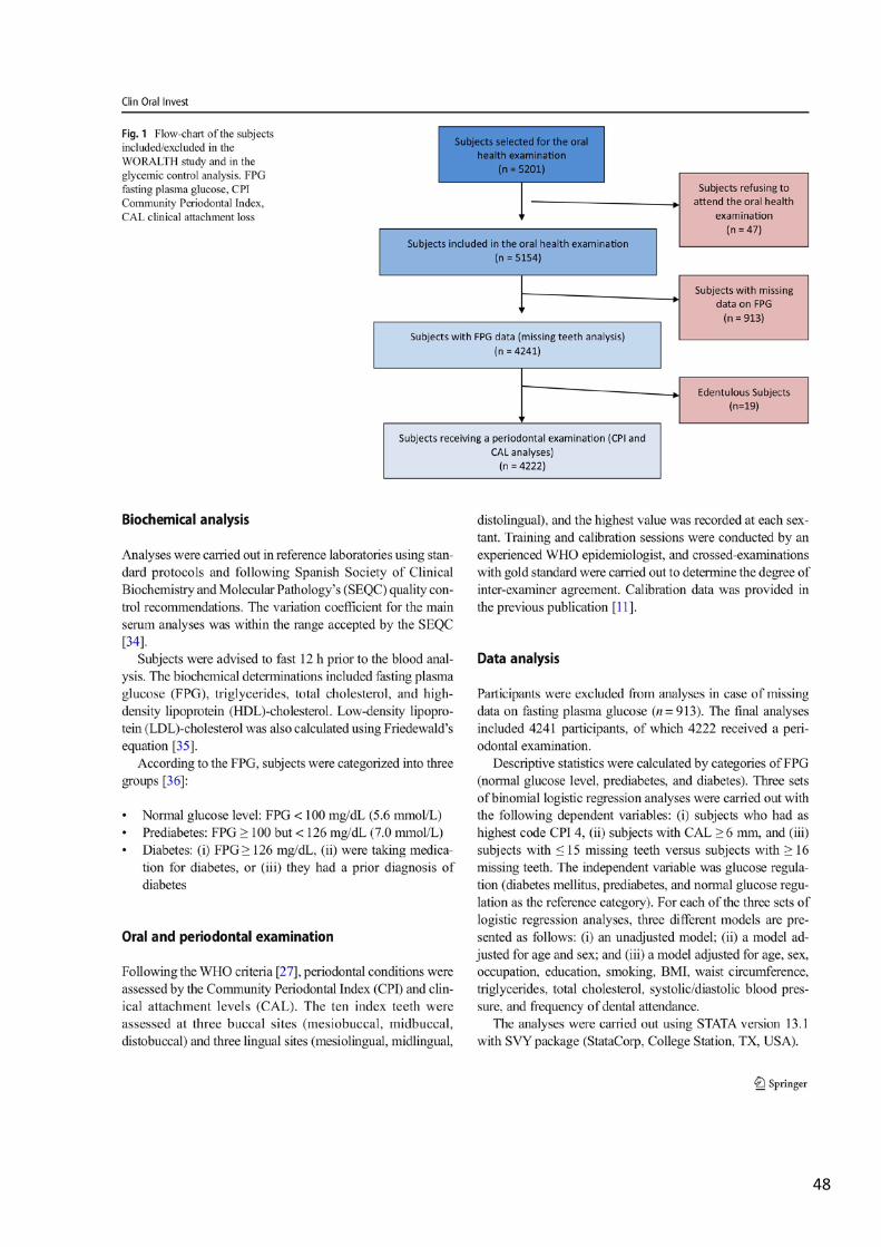
48
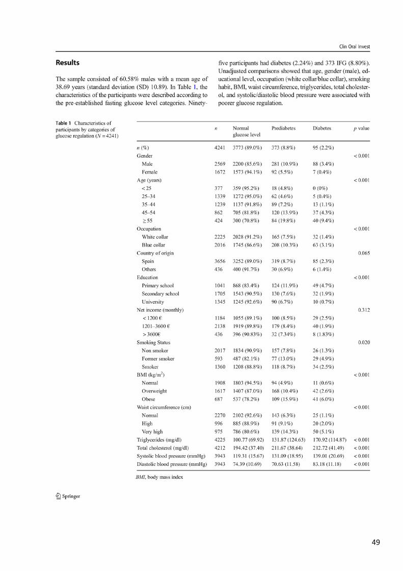
49
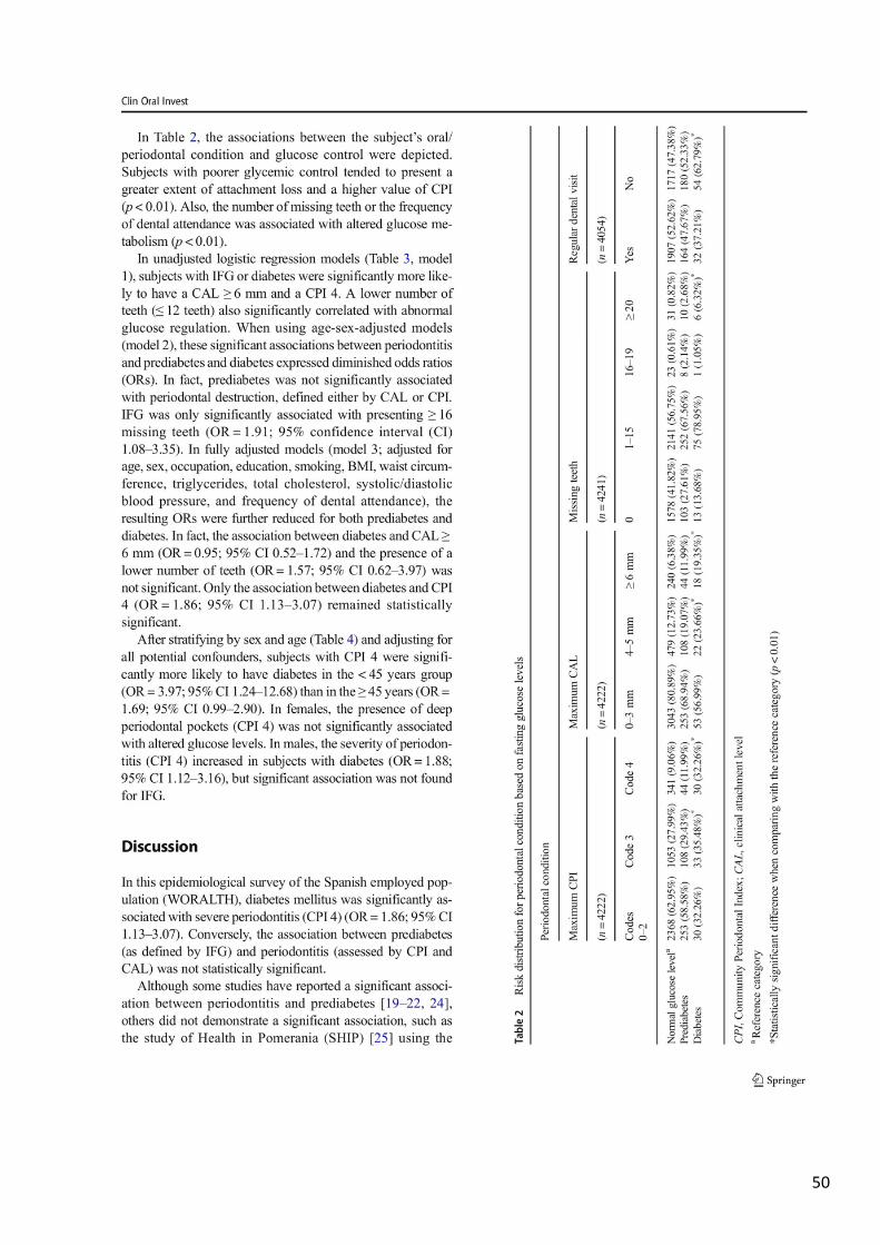
50
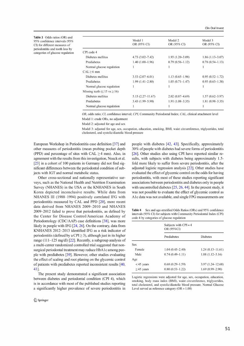
51
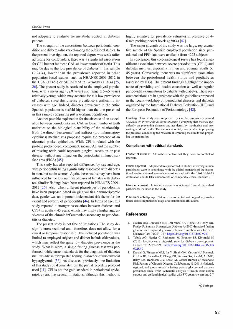
52
53
54
55
STUDY #2 Montero E, Molina A, Carasol M, Fernández-Meseguer A, Calvo-Bonacho E, García-
Margallo MT, Sanz M, Herrera D. (2020) The association between metabolic syndrome
and periodontitis in Spain: Results from the WORALTH (Workers´ORAL healTH) Study.
Journal of Clinical Periodontology (accepted for publication)
The association between metabolic syndrome and periodontitis in Spain: Results from
the WORALTH (Workers´ORAL healTH) Study
Introduction: Evidence of an association between periodontitis and MetS (metabolic
syndrome) remains controversial. The objective of this study is to evaluate the
association between periodontitis and MetS in a cross-sectional population survey.
Material and Methods: WORALTH (Workers’ ORAL healTH) Study is a cross-sectional
survey, conducted on a representative sample of the Spanish employed population,
including 5,154 participants. An oral examination following the World Health
Organization (WHO) criteria, evaluated the periodontal status using the Community
Periodontal Index (CPI) and Clinical Attachment Levels (CAL). Logistic regression analysis
with adjustment for potential confounders was used to evaluate the association
between periodontitis and MetS, and its individual components.
Results: Participants presenting a CPI=4 were more likely to have MetS than subjects
with CPI<4 [odds ratio, OR=1.41; 95% confidence interval (CI) 1.10-1.81; p<0.001]. High
blood pressure was the component with stronger association with periodontal status
(OR=1.94 for CAL≥ 6 mm; 95% CI 1.49-2.53; p<0.001). After stratifying for sex, the
association was higher in women (OR=2.20 for CPI=4; 95% CI 1.31-3.62; p<0.001). Non-
metabolically healthy subjects, obese or not, presented a worse periodontal condition.
Conclusion: Severe periodontitis (CPI=4) was associated with MetS in a representative
sample of the Spanish employed population. This association seems to be independent
of body mass index and other potential confounders.
56
57
58
59
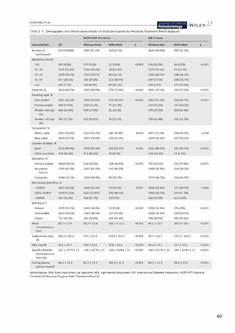
60
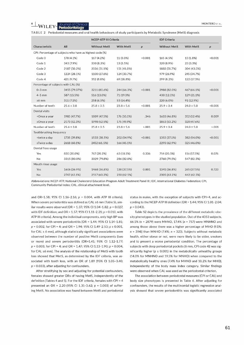
61
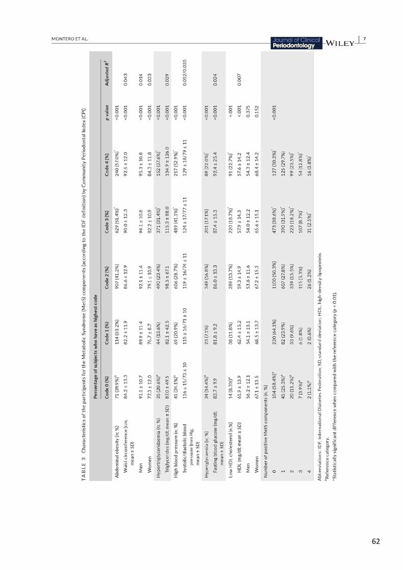
62
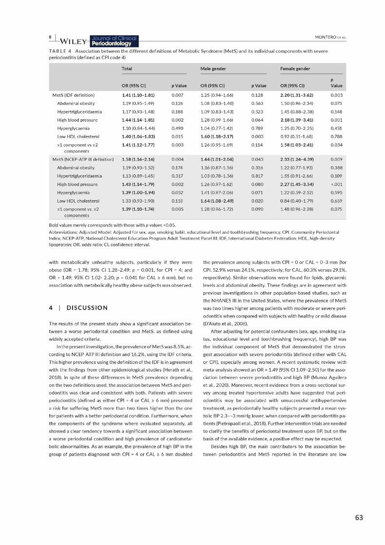
63
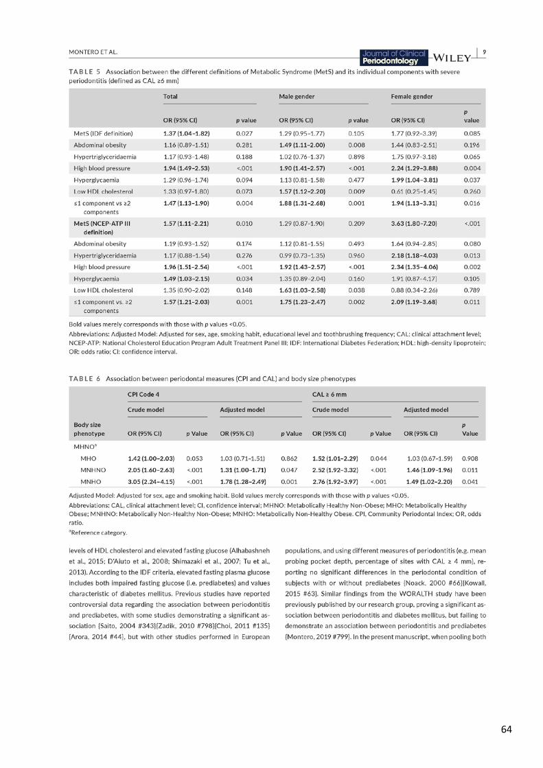
64
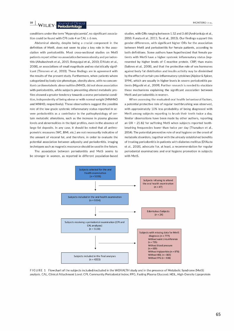
65
66
67
68
69
STUDY #3
Montero E, López M, Vidal H, Martínez M, Virto L, Marrero J, Herrera D, Zapatero A, Sanz
M. (2020) Impact of periodontal therapy on systemic markers of inflammation in
patients with metabolic syndrome: A randomized clinical trial. Diabetes, Obesity and
Metabolism. DOI: 10.1111/jcpe.13353
Impact of periodontal therapy on systemic markers of inflammation in patients with
metabolic syndrome: A randomized clinical trial
Aim: To determine the impact of periodontal treatment on systemic markers of
inflammation in patients with metabolic syndrome (MetS) and periodontitis.
Materials and Methods: In this parallel-arm, double-blind, randomized controlled
clinical trial, 63 patients with MetS and severe periodontitis were randomly assigned to
receive either intensive periodontal treatment (IPT; scaling and root planing plus
azithromycin 500 mg every day for 3 days) or minimal periodontal treatment (MPT;
supragingival professional mechanical plaque removal plus a placebo). The primary
outcome was the impact of the tested interventions on high-sensitivity C-reactive
protein (hs-CRP) serum levels at 6 months. As secondary outcomes, differences in the
levels of cytokines, markers of prothrombotic states, carbohydrate and lipid
metabolism, as well as blood pressure, were measured at 3 and 6 months after therapy.
Results: The intention-to-treat population consisted of 63 subjects randomly assigned
to either the MPT (n = 31) or the IPT (n = 32) group. At baseline, mean hs- CRP was 3.9
mg/L (standard deviation [SD] = 2.9) and 3.9 mg/L (SD = 3.4), respectively, and no
significant differences in cardiometabolic risk profiles were detected between the
groups. Adjusting for baseline hs-CRP, sex, age, smoking status and body mass index, hs-
CRP at 6 months was 1.2 mg/L (95% CI 0.4; 2.0; P = .004) lower in the IPT group than in
the MPT group. In the secondary outcomes, significant reductions in IL-1β, TNF-α, HbA1c
and blood pressure were observed in the IPT group at 3 months compared with the MPT
group.

70
Conclusion: Effective periodontal treatment significantly reduced hs-CRP after 6 months
in patients with MetS and severe periodontitis. Periodontal therapy might be useful to
reduce cardiovascular risk in these patients.
71
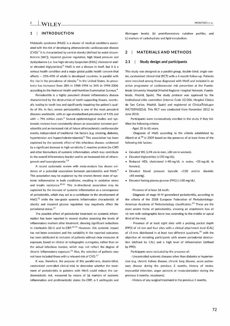
72
73
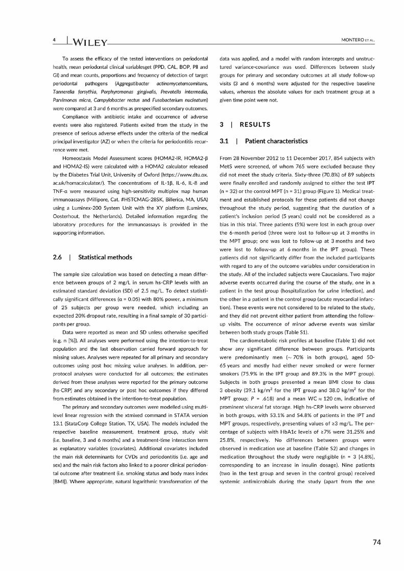
74
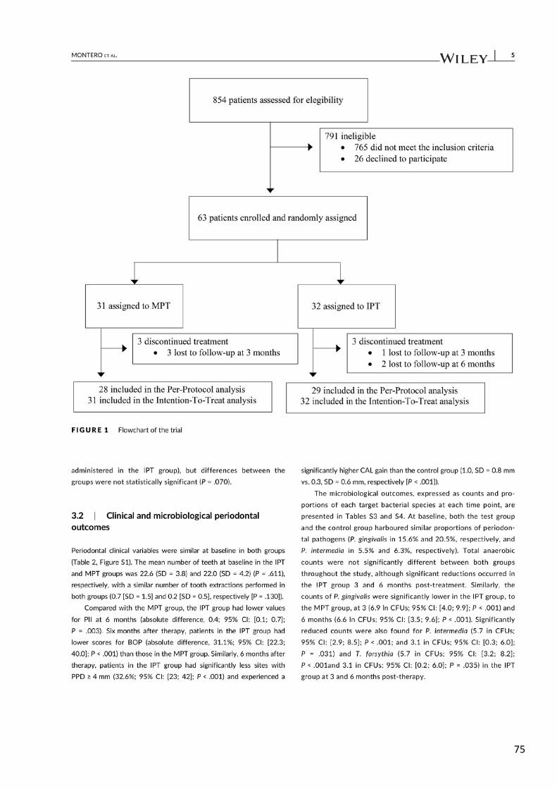
75
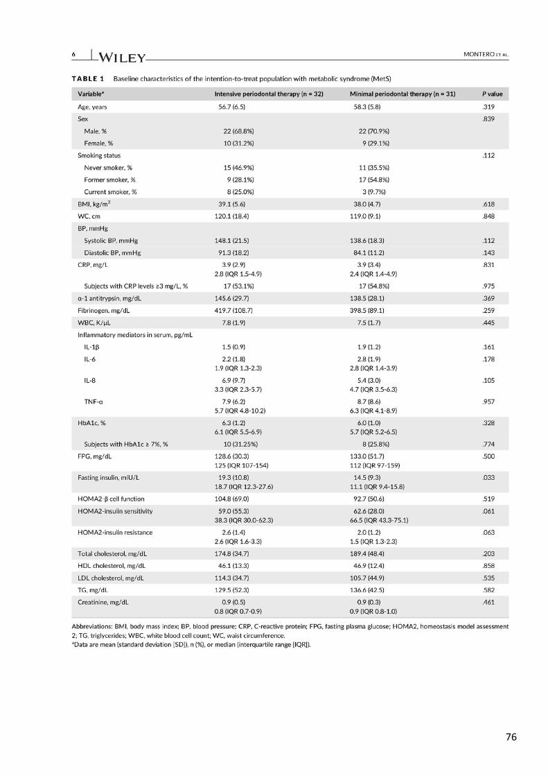
76
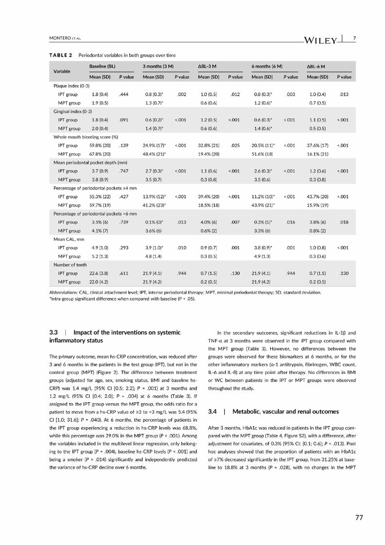
77
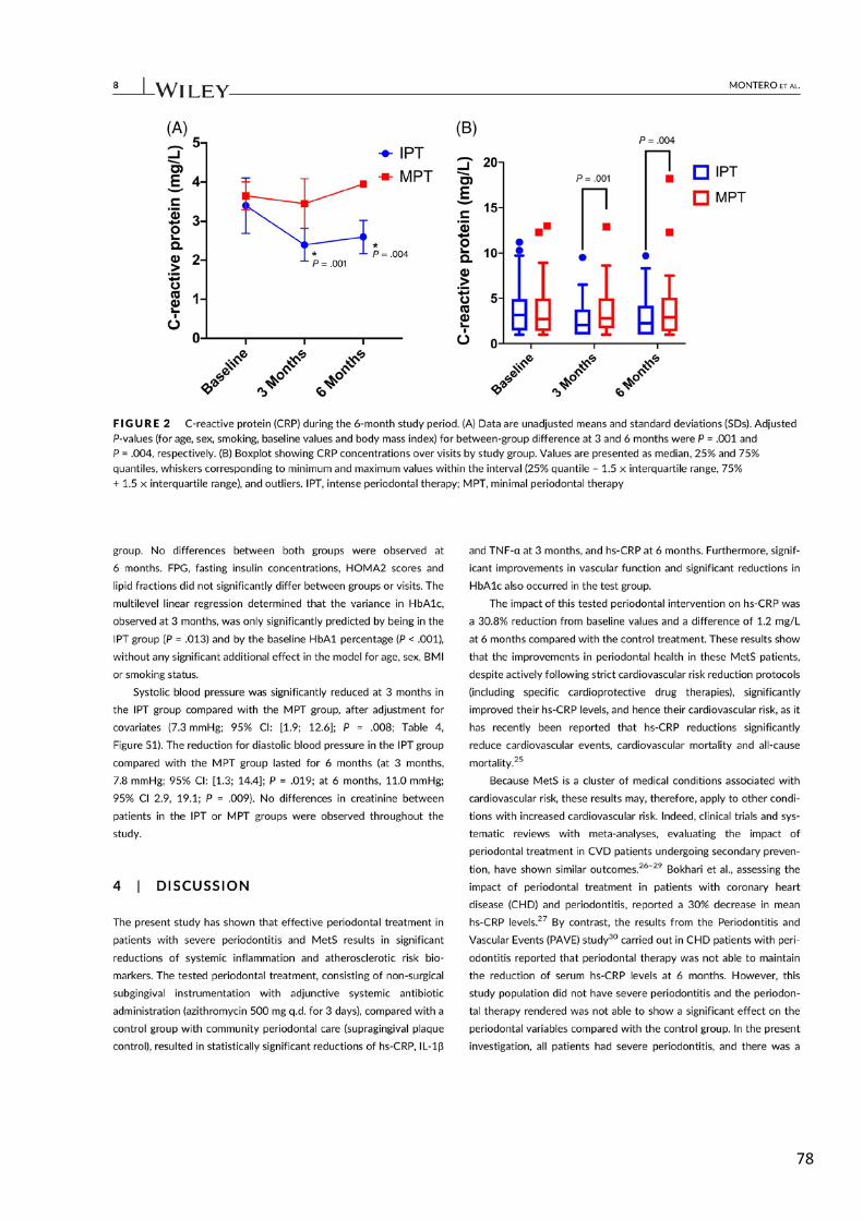
78
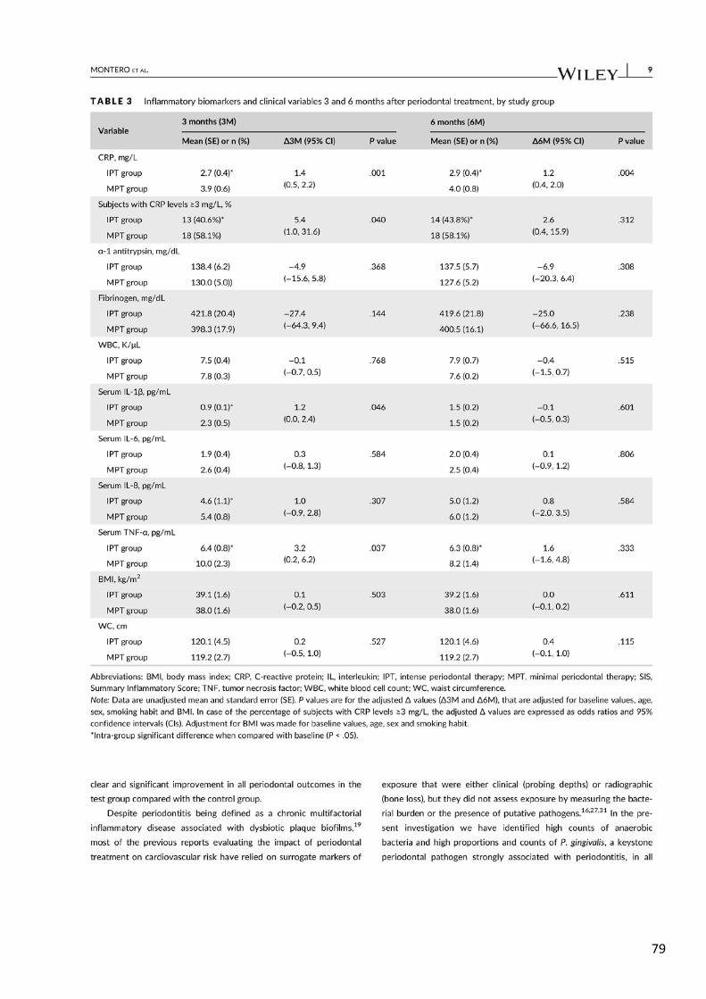
79
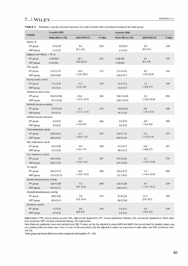
80
81
82

83
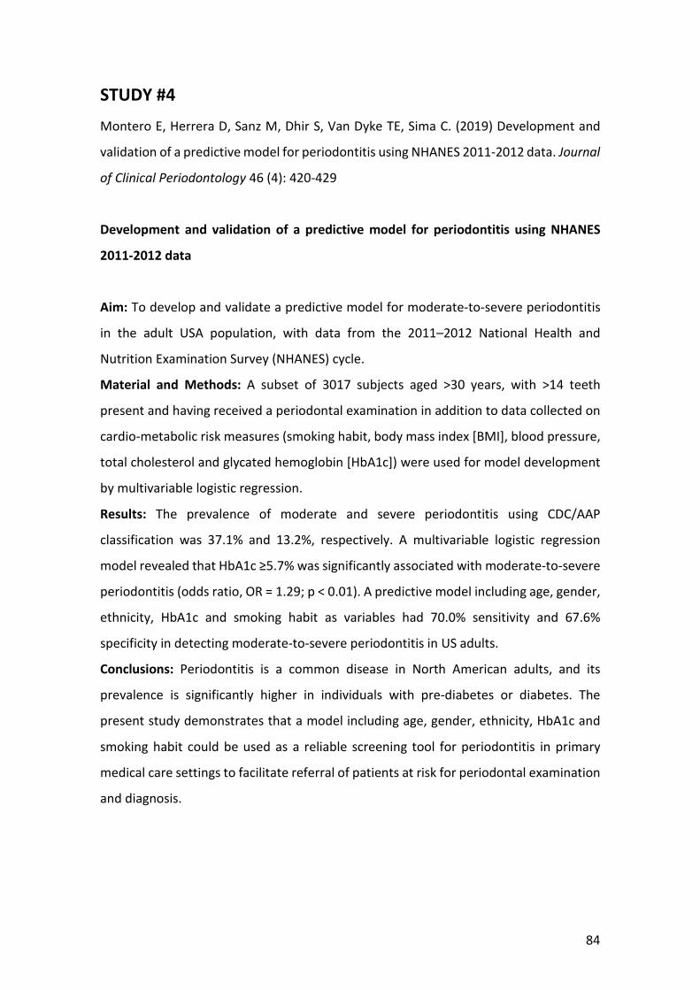
84
STUDY #4
Montero E, Herrera D, Sanz M, Dhir S, Van Dyke TE, Sima C. (2019) Development and
validation of a predictive model for periodontitis using NHANES 2011-2012 data. Journal
of Clinical Periodontology 46 (4): 420-429
Development and validation of a predictive model for periodontitis using NHANES
2011-2012 data
Aim: To develop and validate a predictive model for moderate-to-severe periodontitis
in the adult USA population, with data from the 2011–2012 National Health and
Nutrition Examination Survey (NHANES) cycle.
Material and Methods: A subset of 3017 subjects aged >30 years, with >14 teeth
present and having received a periodontal examination in addition to data collected on
cardio-metabolic risk measures (smoking habit, body mass index [BMI], blood pressure,
total cholesterol and glycated hemoglobin [HbA1c]) were used for model development
by multivariable logistic regression.
Results: The prevalence of moderate and severe periodontitis using CDC/AAP
classification was 37.1% and 13.2%, respectively. A multivariable logistic regression
model revealed that HbA1c ≥5.7% was significantly associated with moderate-to-severe
periodontitis (odds ratio, OR = 1.29; p < 0.01). A predictive model including age, gender,
ethnicity, HbA1c and smoking habit as variables had 70.0% sensitivity and 67.6%
specificity in detecting moderate-to-severe periodontitis in US adults.
Conclusions: Periodontitis is a common disease in North American adults, and its
prevalence is significantly higher in individuals with pre-diabetes or diabetes. The
present study demonstrates that a model including age, gender, ethnicity, HbA1c and
smoking habit could be used as a reliable screening tool for periodontitis in primary
medical care settings to facilitate referral of patients at risk for periodontal examination
and diagnosis.
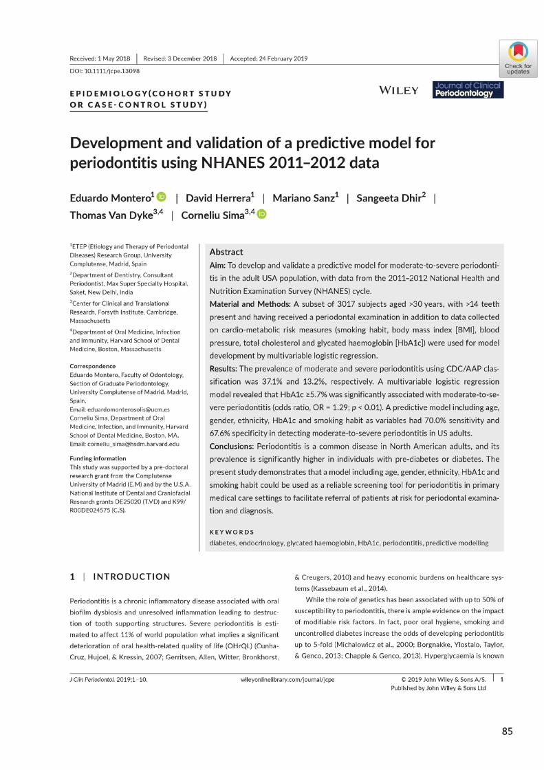
85
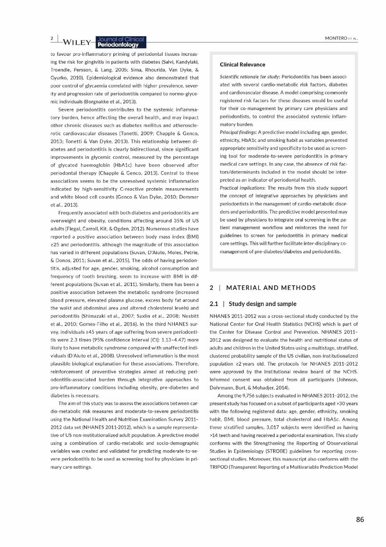
86
87
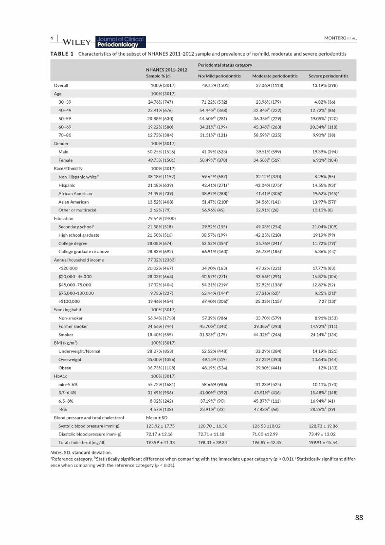
88
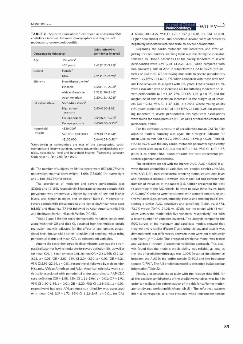
89
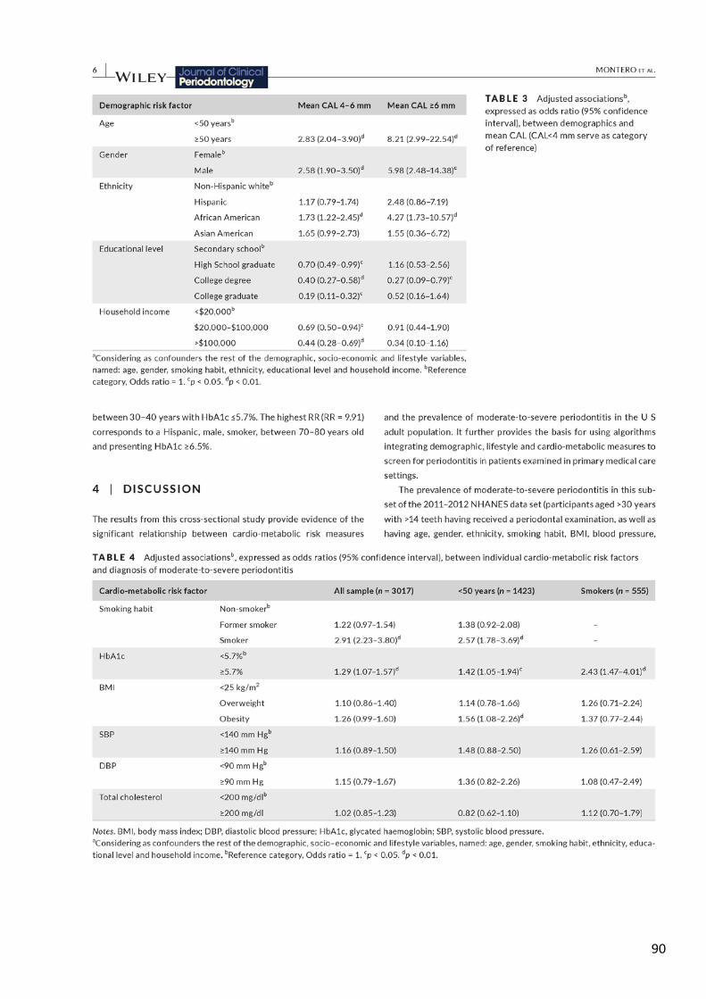
90
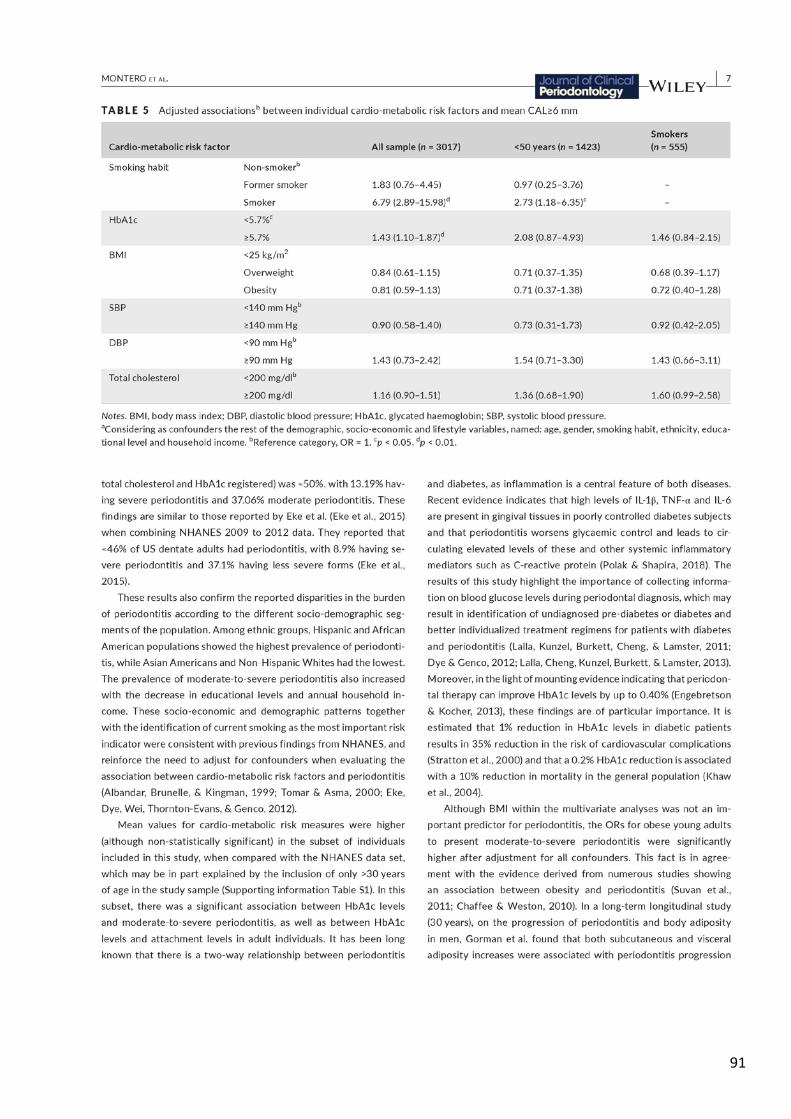
91
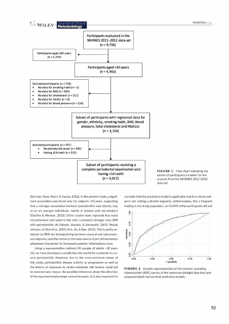
92
93
94
95
STUDY #5
Montero E, Matesanz P, Nobili A, Herrera-Pombo JL, Sanz M, Guerrero A, Bujaldón A,
Herrera D, on behalf of the SEPA Research Network of Dental Clinics. (2020) Screening
of undiagnosed hyperglycemia in the dental setting: the DiabetRisk study. Journal of
Clinical Periodontology (accepted for publication)
Screening of undiagnosed hyperglycemia in the dental setting: the DiabetRisk study.
Aim: To evaluate the efficacy of different screening protocols for undiagnosed
hyperglycaemia in a Research Network of Dental Clinics coordinated by the Spanish
Society of Periodontology (SEPA).
Material and Methods: A total of 1143 subjects were included in the study. Participants
filled a questionnaire considering diabetes risk factors (FINDRISC) and received a
periodontal screening examination. Subjects with a slightly elevated score according to
the FINDRISC (≥7), received a point-of-care HbA1c and were eventually referred to their
physician for confirmatory diagnosis. Receiver Operating Characteristic (ROC) curves
were used to assess the performance of various predictive models with confirmed
hyperglycaemia as outcome.
Results: From this population, 97 (8.5%) were finally diagnosed of diabetes (n=28; 2.5%)
or prediabetes (n=69; 6.0%). When only including the results from the FINDRISC
questionnaire, the model reported an area under the curve (AUC) of 0.866 (95%
confidence interval - CI [0.833; 0.900]). This model significantly improved when the
point-of-care HbA1c was added (AUC= 0.961; 95% CI [0.941; 0.980]; p<0.001).
Conclusions: The tested protocol, combining the FINDRISC questionnaire and a point-
of-care HbA1c, showed to be feasible when carried out in a dental clinic setting and was
efficient to identify subjects with undiagnosed diabetes or prediabetes.
96
97
98
99
100
101
102
103
104
105
106
107
108
109
110
111
112
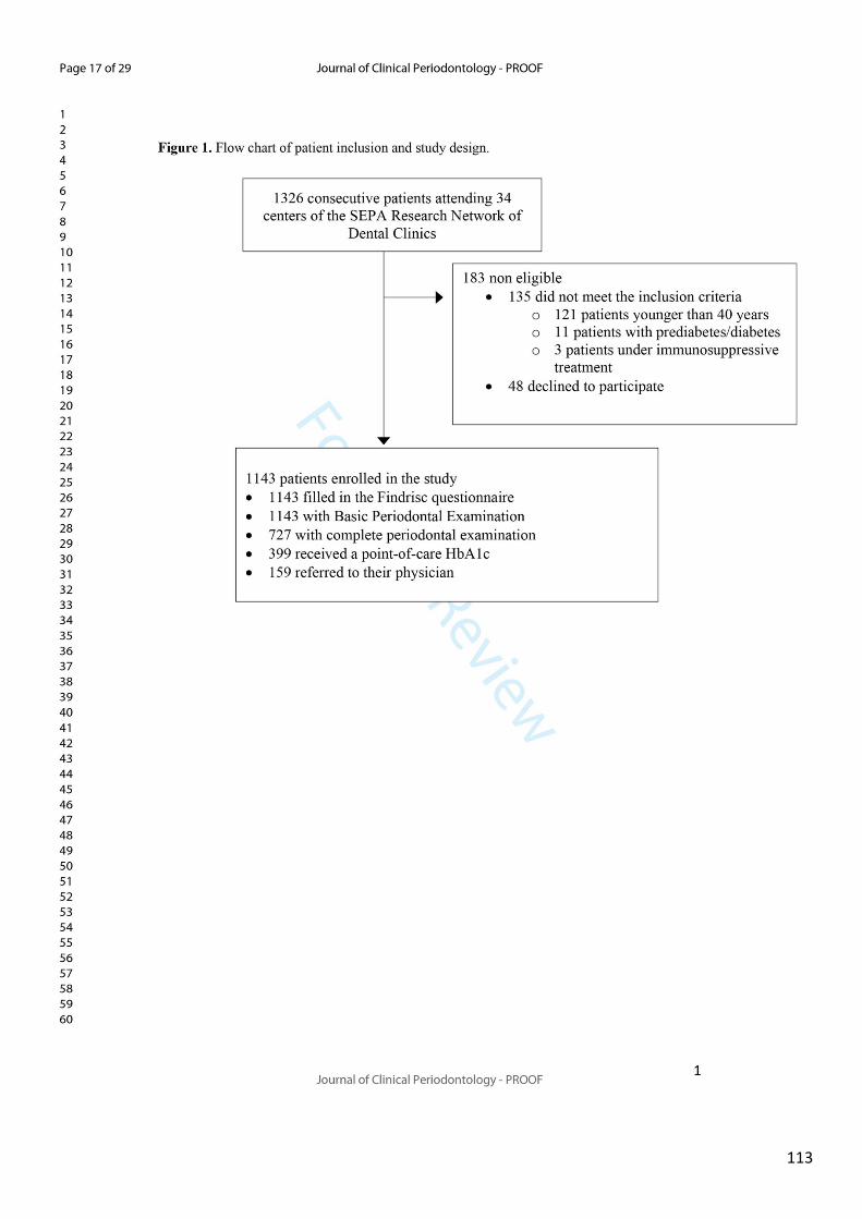
113
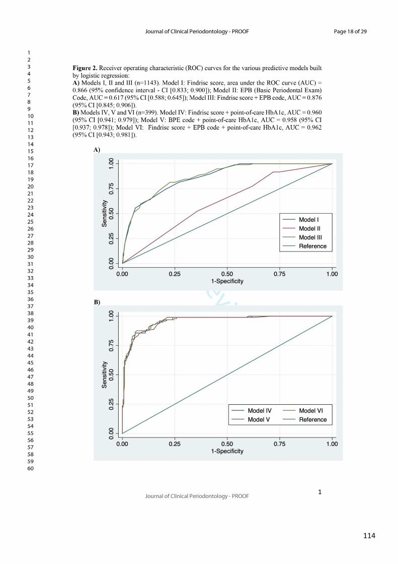
114
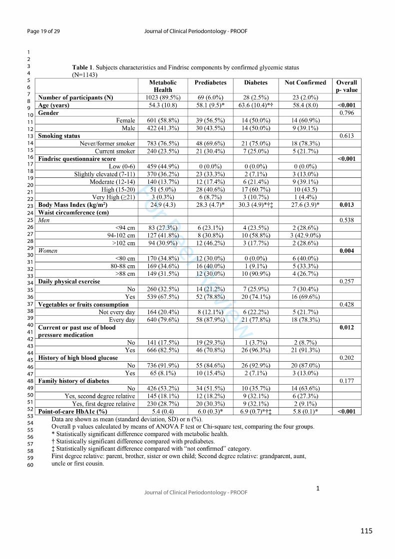
115
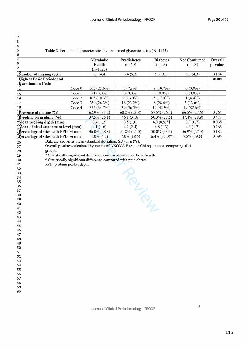
116
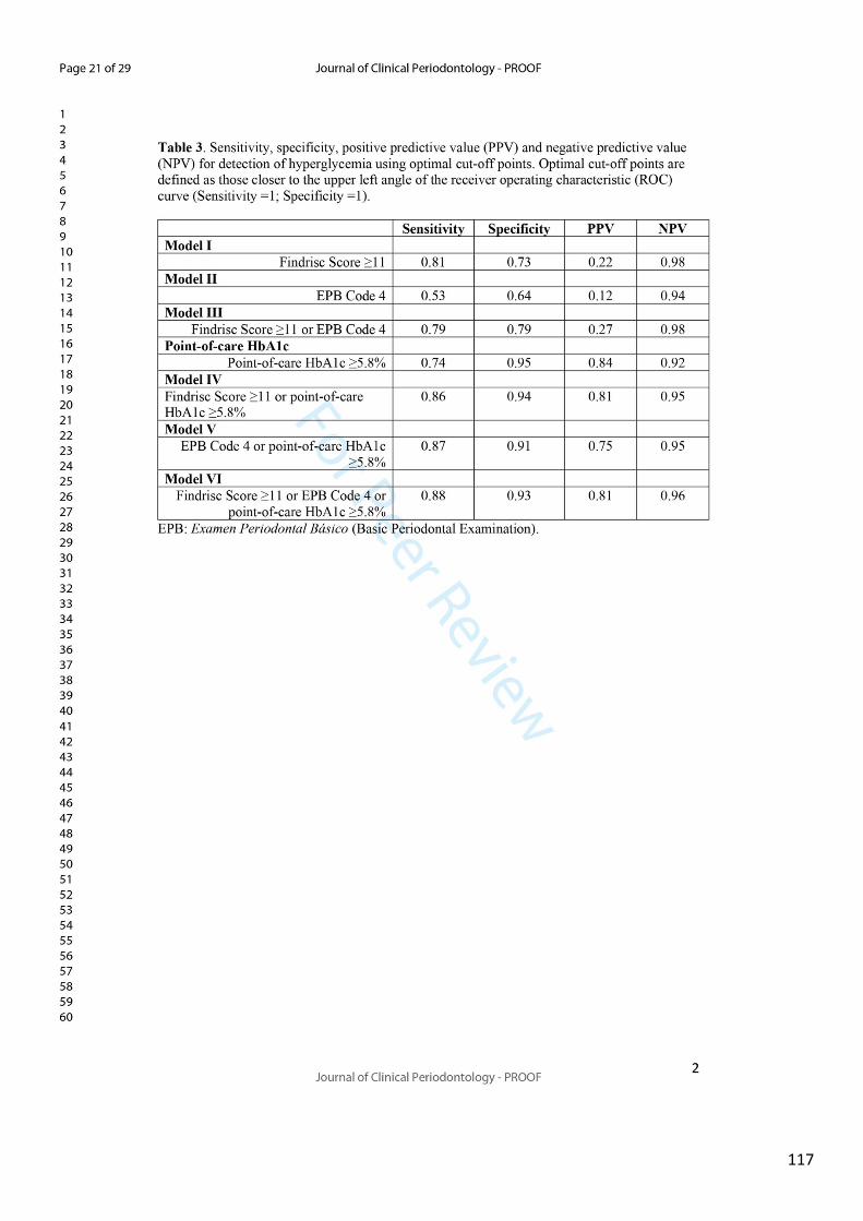
117
118
119
120
121
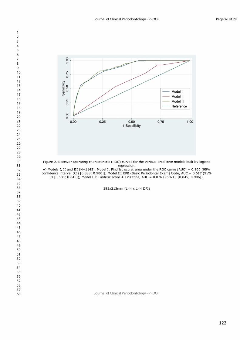
122
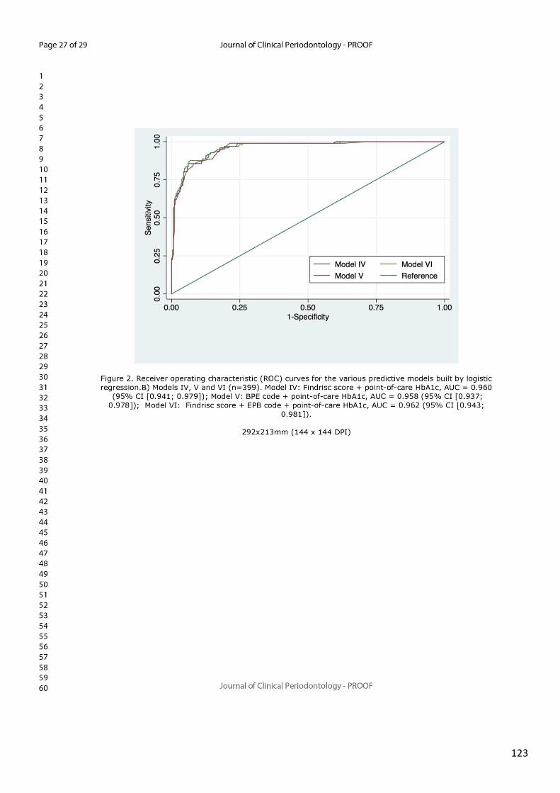
123
124
VII. DISCUSSION The global objective of the present doctoral thesis was to evaluate the association
between periodontitis and two metabolic conditions (hyperglycaemia and MetS), and
to determine whether periodontal therapy or screening tools, both for periodontitis or
hyperglycaemia, might be useful in the simultaneous management and prevention of
periodontitis and metabolic diseases.
Specifically, an oral epidemiological survey on a representative sample of the Spanish
employed population (WORALTH Study) was used to evaluate the association between
periodontal status, as determined by the Community Periodontal Index (CPI) and Clinical
Attachment Level (CAL), and i) hyperglycaemia (either prediabetes or DM) or ii) MetS
(according to IDF and NCEP-ATP III criteria). After adjusting for potential confounders
(i.e. age, gender, occupation, education, smoking, BMI, waist circumference,
triglycerides, total cholesterol and systolic/diastolic blood pressure), DM was found to
be significantly associated with severe periodontitis, as defined by CPI code 4 (OR=1.86;
95% CI 1.13-3.07); however, prediabetes was not significantly associated with
periodontal status (Study #1; (Montero, Carasol, et al., 2019)). In the case of MetS, and
regardless of the definition, it was found to be significantly associated with both CPI
code 4 (OR=1.41; 95% CI 1.10-1.81 for the IDF definition) and CAL ≥6 mm (OR=1.37; 95%
1.04-1.82 for the IDF definition), with hypertension being the component with a
strongest association with periodontal status, and the association being stronger among
women (Study #2; Montero et al. 2020. Accepted for publication). The results of these
two epidemiological studies clearly demonstrate that there is an association between
periodontitis and certain metabolic disorders in the Spanish employed population,
which makes the case for the promotion of oral health education and the performance
of regular periodontal check-ups in subjects with DM or MetS.
To evaluate the effect of periodontal treatment on systemic inflammation in patients
with MetS, a RCT was carried out. The hypothesis was that effective treatment to reduce
periodontal inflammation may lead to a reduction in cardiovascular risk in these
patients. The results confirmed this hypothesis, as the IPT group experienced a
125
statistically significant reduction in hs-CRP after 6 months (adjusted difference between
treatment groups was 1.2 mg/L; p=0.004), together with a significant improvement in
the vascular function (as determined by blood pressure measurements) (Study #3;
(Montero et al., 2020)). These findings support the evidence that effective periodontal
therapy has a positive short-term systemic effect in patients with MetS, although studies
with a longer follow-up times are needed to confirm whether these benefits could be
sustained over time, and whether they lead to reductions in cardiovascular morbidity
and mortality.
The last part of the present work investigates the potential benefits of carrying out
integrative approaches by physicians and periodontists in the management of metabolic
disorders and periodontitis. With that objective in mind, a predictive model to screen
for moderate-to-severe periodontitis was established using a combination of cardio-
metabolic and socio-demographic variables from NHANES 2011-2012 data. The
proposed model comprising just five variables, commonly registered in medical care
settings (i.e. age, gender, ethnicity, smoking habit and HbA1c) presented a sensitivity of
70% and a specificity of 68% to identify patients with moderate-to-severe periodontitis,
making it a good candidate to be regularly used by physicians in primary care settings
(Study #4; (Montero, Herrera, et al., 2019)). However, further validation of the model
in other, if possible, prospective cohorts, is mandatory before it can be included in
formal clinical guidelines.
Analogous to the screening for periodontitis by the medical community, the dental clinic
has been proposed as an ideal setting for the detection of previously unknown diabetes
or prediabetes. With that objective in mind, a research project, developed as a
collaboration between the Spanish Society of Periodontology (SEPA) and the Spanish
Society of Diabetes (SED) through the establishment of a joint Working Group, resulted
into a research protocol within the Research Network of Dental Clinics coordinated by
SEPA. The protocol aimed to evaluate different screening protocols for risk assessment
in the detection of undiagnosed hyperglycaemia in a network of dental clinics (including
both dental schools and private practices). The results demonstrated that the dental
setting may be a suitable place for the identification of patients with previously
126
unknown hyperglycaemia, as 8.5% of the included subjects were diagnosed either with
diabetes or prediabetes. However, the periodontal assessment did not seem to provide
any further effectiveness over the use of an already validated self-reported
questionnaire (FINDRISC and a point-of-care HbA1c test, that presented a sensitivity of
86% and a specificity of 94% for the detection of hyperglycaemia (Study #5, Montero et
al. 2020. Accepted for publication). The results of Study #5 support the
recommendations by the International Diabetes Federation (IDF) and the European
Federation of Periodontology (EFP) regarding the dentist´s role in the screening for
undiagnosed hyperglycaemia (Sanz et al., 2018), and the proposed model seems to be
easy to implement in the dental clinic.
Epidemiological association between periodontal status and cardiometabolic risk
factors
The cross-sectional association between type 2 DM and periodontitis has been already
substantiated. The studies performed on the Pima Indians of Arizona, the population
with the highest reported prevalence of DM in the world (40-50% in adults over 35 years
of age) (Knowler, Bennett, Hamman, & Miller, 1978), clearly demonstrated a strong
correlation between DM and the prevalence and severity of periodontitis,
independently of already known risk factors/determinants such as age or oral hygiene
(Emrich, Shlossman, & Genco, 1991). Subsequently, similar findings were reported in
other populations, such as subjects with type 1 DM or Gullah- African Americans
(Fernandes et al., 2009; Hodge et al., 2012). Nevertheless, the definitive causative two-
way relationship between DM and periodontitis emanates from longitudinal trials
establishing that: i) DM and poor glycaemic control correlates with incident
periodontitis and more severe progression of the disease (Bandyopadhyay, Marlow,
Fernandes, & Leite, 2010; Nelson et al., 1990); and ii) periodontitis affects glycaemic
control in diabetic (Chang et al., 2017; Taylor et al., 1996) and not diabetic subjects
(Graziani et al., 2018).
However, the potential association between periodontitis and prediabetes is less clear.
This association could be of particular interest, since prediabetes is, in itself, a risk factor
for the development of cardiovascular, renal and neurologic complications, but at the
127
same time is a reversible condition (Fonseca, 2009; Shaye et al., 2012; Tabak et al.,
2012). If periodontitis and prediabetes are significantly associated, it could be
hypothesized that periodontal treatment may reduce progression to hyperglycaemia in
these patients. Indeed, a secondary analysis of a multicentre RCT (ABPARO,
“Antiobiotika und Periodontitis) observed a decrease in the number of prediabetes
patients to approximately 50%, after 27.5 months, when compared to baseline (Kocher
et al., 2018). Unfortunately, since the main objective of the trial was not to evaluate the
impact of treatment on HbA1c, no randomization across groups stratified by baseline
HbA1c was performed. Therefore, the possibility that changes in HbA1 were attributable
to regression to the mean, limits conclusions from this post-hoc analysis to those
associated with hypothesis generation.
In the WORALTH study, DM was significantly associated with severe periodontitis, as
defined by CPI code 4 (OR=1.86; 95% CI 1.13-3.07). Conversely, no association could be
found between periodontitis (assessed by CPI or CAL) and prediabetes (as defined by
IFG) among the 4,222 subjects receiving a periodontal examination after adjusting for
confounders (Study #1; (Montero, Carasol, et al., 2019)). Results of this investigation
are in agreement with other studies using different periodontal indices or periodontitis
case definitions. Two studies performed in Germany failed to find any association
between different measures of periodontitis (mean probing depth, percentage of sites
with CAL≥ 4 mm) and prediabetes (Kowall et al., 2015; Noack et al., 2000). Similarly, data
derived from NHANES 2019-2012 do not support the proposal that periodontitis, as
defined by the Centres for Disease Control/American Academy of Periodontology
(CDC/AAP) case definitions, was more likely in people with IFG (Arora et al., 2014; Eke
et al., 2016). Noteworthy, significant associations between periodontitis and
prediabetes were observed only when either periodontal outcomes (mostly CAL) or IFG
were considered as continuous variables, with subjects in the upper quintile category of
CAL or those with IFG=111-125 mg/dL presenting an association between both diseases
(Y. H. Choi et al., 2011; Hong, Noh, & Kim, 2016). Thus, it seems that is the level of
hyperglycaemia, rather than the diagnosis of prediabetes/DM, is the likely determining
factor in the association of periodontitis and abnormal glucose regulation. It is important
to understand that the cut-off values used in the diagnosis of DM are based on the risk
128
of developing diabetes-related complications (particularly, retinopathies).
Unfortunately, little attention has been paid to periodontitis, despite being related to
insulin resistance, so it is possible that periodontitis could predict the incidence of DM.
Early insulin resistance and β-cells function, as assessed by HOMA-IR and HOMA-IS have
demonstrated to be associated with the development of periodontal pockets and
periodontal breakdown, suggesting that early hyperglycemia, before becoming overt
DM, may influence the pathogenesis of periodontitis (Allen et al., 2011; P. Timonen et
al., 2013). Particularly interesting is a community-based cohort study of 5,885 Taiwanese
that explored the temporal sequence of the bidirectional relationship between
periodontitis and hyperglycaemia (Chiu et al., 2015). In that study, it was reported that
incident cases of periodontitis (as defined as CPI≥ 3) presented a 33% increase risk for
incident hyperglycaemia (prediabetes and DM), while at the same time prediabetes and
DM led to a significantly higher risk for periodontitis [hazard ratio (HR)=1.25 for
prediabetes and HR=1.95 for DM].
In this study, results were heavily influenced by the low prevalence of DM in the sample
(2.24%), which is lower than the prevalence reported in other population-based studies,
such as NHANES 2009-2012 in the USA (12.6%) or SHIP-Trend in Germany (11.8%) (Eke
et al., 2016; Kowall et al., 2015). The WORALTH study was restricted to the employed
population (range 16-65 years), which may account for this low prevalence of DM, since
this disease prevalence significantly increases with age. The low number of diabetes
cases observed in the WORALTH study may be related with the reported differences by
sex and age, with periodontitis being significantly associated with DM in men, but not in
women (there were just 7 females with DM). In terms of age, this study reported a
stronger association between DM and CPI =4 in adults <45 years, similarly to what it has
been reported for the association between atherosclerotic cardiovascular disease and
periodontitis (Dietrich, Sharma, Walter, Weston, & Beck, 2013), suggesting a possible
higher aggressiveness of the chronic inflammation secondary to periodontitis or
diabetes in young individuals.
129
In conclusion, even if no specific association was observed in the WORALTH study
between periodontitis and prediabetes, the clear association observed for DM, together
with the overwhelming scientific literature, support a linear bidirectional relationship
between insulin resistance/hyperglycaemia and periodontitis. However, the glycemic
control threshold over which periodontitis risk can be increased is unknown, and the
same is true for the severity of periodontitis that contributes to hyperglycaemia.
Longitudinal studies using different measures for abnormal glucose regulation (HOMA-
IR, HOMA-β, fasting plasma glucose, oral glucose tolerance) and measures to adequately
define the prevalence and extent of periodontitis (CAL and probing depth at site-, tooth-
and patient-level according to specific thresholds) are needed in order to clarify the
relationship between these conditions.
The WORALTH Study was also used to investigate the association between periodontitis
and MetS (Study #2, Montero et al. 2020. Accepted for publication). Previous studies,
including a systematic review with meta-analysis, have found significant associations
between periodontitis and MetS (Nibali et al., 2013). However, this association still
remains controversial, since the diagnostic criteria used to define the case of MetS and
the case definition of periodontitis vary between studies, and some studies were only
capable of finding associations in specific groups, such as women, diabetic patients or in
specific age groups (D'Aiuto et al., 2008; Furuta et al., 2013; Sora et al., 2013; Y. K. Tu et
al., 2013), while others observed no association between MetS and periodontitis at all
(Benguigui et al., 2010; LaMonte et al., 2014; Zuk et al., 2017). Hence, there was a need
of further research to clarify these inconsistencies and heterogeneous results among
distinct groups, and to evaluate the association between the individual components of
MetS and periodontitis, especially in Western populations, since published results on
those populations are particularly conflicting.
Our study, included 4,353 patients in the final analyses, and it was reported that severe
periodontitis (either defined as CPI code 4, or CAL ≥ 6 mm), was consistently associated
with MetS, independently of its definition. Specifically, ORs for the association between
130
CPI code 4 and MetS were 1.41 and 1.58 for the IDF and NCEP-ATP III definitions,
respectively; while the associations between CAL ≥ 6 mm and MetS were 1.37 and 1.57.
Furthermore, despite the fact that not all the components of the syndrome presented a
significant association with severe periodontitis in fully adjusted models, there was a
tendency towards a linear relationship between a poorer periodontal condition and a
higher prevalence of cardiometabolic abnormalities. As an example, the prevalence of
hypertension in patients with severe periodontitis (CPI code 4 or CAL≥ 6 mm) was double
than the prevalence among subjects with CPI code 0 or CAL= 0-3 mm, and similar
observations were found for abdominal obesity (defined by waist circumference),
hyperglycaemia or dyslipidaemia.
In agreement with previous reports (D'Aiuto et al., 2008; Furuta et al., 2013; Y.-K. Tu,
D'Aiuto, Lin, Chen, & Chien, 2013; Y. K. Tu et al., 2013), in our cross-sectional
study, the association between periodontitis and MetS was stronger in women. A
significant association between severe periodontitis and MetS among males was just
observed for the NCEP-ATP III definition and CPI code 4 (OR=1.44; 95% CI 1.01-2.04).
These findings may have different explanations. It is expected that periodontal
infections may have a weaker association than other major environmental risk factors
with MetS (e.g. tobacco smoking). The fact that men tend to have a less healthy lifestyle
may “mask” the contribution of periodontitis to MetS, explaining the smaller ORs.
Another possible explanation is that, among patients with MetS, females present higher
levels of CRP than men (Saltevo, Vanhala, Kautiainen, Kumpusalo, & Laakso, 2008),
suggesting that the higher levels of systemic inflammation could explain the stronger
association with periodontitis. Lastly, sex hormones may play an important role in the
pathogenesis of both diseases: while inflammation decreases oestradiol production and
thus, reduce the protective effect of oestrogens on body fat distribution and insulin
action (Alpizar & Spicer, 1994), oestrogens alter the host response to the subgingival
biofilm (Mealey & Moritz, 2003), contributing to periodontal diseases in females.
Therefore, gender differences may influence preventive and therapeutic measures for
patients with periodontitis and MetS.
131
Among the individual components of MetS, hypertension presented with the strongest
association with periodontal outcomes (OR= 1.94; 95% CI 1.49-2.53; for the association
between CAL≥ 6 mm and MetS according to the IDF definition). A recent systematic
review with meta-analysis showed an OR=1.49 (95% CI 1.09-2.50) for the association
between severe periodontitis and high BP (Muñoz Aguilera et al., 2020). Moreover, in
that systematic review, a reduction in BP following periodontal treatment was observed,
with reductions in systolic blood pressure (SBP) ranging from 3 to 12.5 mmHg and in
diastolic blood pressure (DBP) from 0 to 10 mmHg. These results are in agreement with
the ones reported in the third study of the present work (Study #3; (Montero et al.,
2020), since reductions of approximately 7 mmHg in SBP and DBP were observed in
MetS patients with severe periodontitis after non-surgical periodontal therapy.
Surprisingly, in Study #2, no association could be found between abdominal obesity, as
determined by waist circumference and periodontitis. Whilst several cross-sectional
studies on MetS patients report either no association between obesity and periodontitis
(Alhabashneh, Khader, Herra, Asa'ad, & Assad, 2015; Benguigui et al., 2010;
D'Aiuto et al., 2008), or associations of small magnitude and not statistically significant
(P Timonen et al., 2010), a recent meta-review including evidence from 14 systematic
reviews suggested a positive association between obesity and periodontitis onset,
progression, and response to periodontal treatment (Jean E Suvan, Finer, &
D'Aiuto, 2018). Notably, in most of the studies included in the published
systematic reviews on the association between obesity and periodontitis (Chaffee &
Weston, 2010; Keller, Rohde, Raymond, & Heitmann, 2015; J. Suvan et al., 2011), data
on glycemic status, lipid profiles or BP were not evaluated or not considered as potential
confounders in the association. However, most of the studies specifically including MetS
patients, elaborated multivariable regression models considering the majority of
potential confounders. Therefore, it is possible that the strongest association with other
individual components of the MetS (e.g., hyperglycemia, hypertension) leads to smaller
or even non-significant ORs between periodontitis and obesity in this subset of patients.
Indeed, when patients in the WORALTH Study were categorized by body size phenotype,
obesity alone, with no concomitant cardiometabolic abnormalities (metabolically
healthy obese, MHO), did not show association with periodontitis, while subjects
132
presenting altered metabolic profiles showed a greater tendency towards a worse
periodontal condition, independently of being obese or with normal weight. These
observations suggest the possible role of the low-grade systemic inflammatory status
reported in severe periodontitis as a contributor in the pathophysiology of certain
metabolic alterations, such as the increase in plasma glucose levels or abnormalities in
the lipid profiles, even in the absence of large fat deposits. In any case, it should be noted
that all anthropometric measures (WC, BMI, etc.) are not necessarily indicative of the
amount of visceral fat, and therefore, in order to evaluate the potential association
between adiposity and periodontitis, imaging techniques, such as magnetic resonance,
should be used in the future.
Several limitations of the WORALTH Study should be acknowledged. Firstly, the cross-
sectional nature of the study does not allow to ascertain any causal relationship.
Secondly, the population was limited to employed subjects, not including older adults
or children or adolescents, which could explain the relatively low prevalence of
hyperglycaemia and MetS observed in this sample. Lastly, the most important
limitations are related to the periodontal and medical examinations. On one hand, the
periodontal exam was based on the WHO recommendations, consisting on a partial
mouth recording that may lead to underestimation of the disease presence, severity and
extent (Kingman, Susin, & Albandar, 2008; Susin, Kingman, & Albandar, 2005). What is
more, CPI is not a diagnostic tool, but only a screening tool, same as other indices
developed to evaluate periodontal treatment needs (e.g. Basic Periodontal Examination
(Tugnait, Clerehugh, & Hirschmann, 2004)), and therefore, may hamper the accuracy of
periodontal diagnosis, as it is based solely on the pocket component. However, a strict
measure of attachment loss (i.e. CAL) was the other periodontal outcome variable
registered in this survey. On the other hand, hyperglycaemia was assessed by fasting
glucose only. Other parameters such as HOMA-IR, HOMA-β, oral glucose tolerance tests
or HbA1c could have provided deeper information on insulin resistance, β-cell function
and natural history of hyperglycemia in these patients, including long-term metabolic
control. Similarly, anthropometric measures for the evaluation of central obesity could
have been complemented with imaging techniques such as magnetic resonance or
computed tomography, in order to properly evaluate adiposity and visceral fat.
133
Impact of periodontal therapy on systemic markers of inflammation in patients with
MetS
The second part of the present doctoral thesis has evaluated, through a parallel-arm,
double-blind, RCT, the impact of non-surgical (steps 1 and 2) periodontal treatment on
systemic markers of inflammation in patients with MetS and periodontitis (Study 3;
(Montero et al., 2020). In this study, patients were randomly assigned to receive
intensive periodontal treatment (IPT; consisting on scaling and root planing plus
azithromycin 500 mg every day for 3 days) or minimal periodontal treatment (MPT;
consisting on supragingival professional mechanical plaque removal plus a placebo), the
outcomes assessed at baseline and 3 and 6 months after treatment were: markers of
systemic inflammation and prothrombotic states (i.e. hs-CRP, α-1 antitrypsin and
fibrinogen), pro-inflammatory cytokines (i.e. IL-1β, IL-6, IL-8 and TNF-α), indicators od
carbohydrate and lipid metabolism, as well as BP. The results of the trial have shown
that effective periodontal therapy in patients with severe periodontitis and MetS results
in significant reductions of several cardiovascular risk biomarkers, such as hs-CRP, IL-1β,
TNF-α, HbA1c and improved vascular function. Subjects in the IPT group experienced a
30.8% reduction in hs-CRP from baseline, resulting in an adjusted difference of 1.2 mg/L
at 6 months, when compared to the MPT group. These findings support the contribution
of periodontitis to systemic inflammation (Paraskevas, Huizinga, & Loos, 2008),
particularly in patients with MetS, a condition identified as an important risk factor for
cardiovascular disease (Mottillo et al., 2010).
One of the main characteristics of the study is related to the severity of both periodontal
and systemic involvement of the enrolled subjects. More than 50% of the patients
presented hs-CRP levels ≥3 mg/L at baseline, more than 25% presented HbA1c ≥ 7%, and
mean BMI was close to class 3 obesity (≈38.0 kg/m2). Despite being under specific
cardioprotective drug therapies, such as calcium channel blockers, Angiotensin
Converting Enzyme (ACE) inhibitors or statins, the patients included presented high
levels of hs-CRP and an increased cardiovascular risk. Additionally, these patients
presented with particularly severe forms of periodontitis (stages III/IV), with a strong
infectious-inflammatory component, as represented by high percentages of periodontal
134
inflammation (bleeding on probing, BOP ≈60% in both groups) and presence of deep
pockets (≈60% of sites with probing pocket depth, PPD ≥4 mm). Our results are of
particular interest, since they provide the evidence that improving periodontal health
may offer an important therapeutic opportunity to reduce the cardiovascular risk in
patients with severe periodontitis and presenting difficulties in achieving cardiovascular
stability. Importantly, the magnitude of the effect in the reduction of hs-CRP is within
the range of extensively evaluated LDL-lowering therapies (Kinlay, 2007; Ridker et al.,
2005).
A strength of the study was the statistically significant improvement in clinical and
microbiological outcomes observed in the IPT, when compared with the MPT. One of
the main criticisms of the results of two multicentre RCTs, that failed to report an any
effect of non-surgical periodontal therapy on hs-CRP and HbA1c, in patients with recent
cardiovascular events and diabetes, respectively, was that periodontal therapy did not
achieve accepted standards of success in terms of residual periodontal inflammation
(BOP) and dental biofilm (plaque) control (S. P. Engebretson et al., 2013; Offenbacher et
al., 2009). In our study, there was a 43.7% reduction in the number of periodontal
pockets ≥ 4 mm and a 37.6% reduction in the BOP score in the IPT group, while in PAVE
(Offenbacher et al., 2009) the reduction in the extent of sites with PD ≥4 mm was 6.6%,
and BOP was 38.3% at 6 months. At the end of the follow-up, in the PAVE trial, many of
the test patients still had sufficient gingival inflammation to meet the periodontal entry
criteria to be enrolled in the study for periodontal treatment.
Furthermore, in our study the tested intervention significantly reduced both the total
counts of anaerobic bacteria and P. gingivalis. Although previous cross-sectional studies
have shown an independent significant association between the presence of a
significant subgingival bacterial burden and levels of periodontal pathogens, with
surrogate measurements of atherosclerotic risk, such as intima-media thickness or hs-
CRP (Desvarieux et al., 2005), this is the first RCT to show that effective periodontal
treatment significantly reduced these microbiological exposure measurements and that
this microbiological impact was associated with significant reductions in hs-CRP.
135
The impact of periodontal therapy was not limited to hs-CRP reductions, since several
inflammatory cytokines, such as IL-1β or TNF-α, showed significant reductions at 3
months but not at the end of the follow-up. These results are somewhat in agreement
with a recent RCT in periodontitis patients with type 2 DM, where significant differences
between groups were noted in TNF-α after treatment (D´Aiuto et al., 2018). This finding
may be meaningful, as both IL-1β and TNF-α have a pro-atherogenic effect and they
have been established as significant and independent predictors of cardiovascular
events and overall mortality (Tuomisto, Jousilahti, Sundvall, Pajunen, & Salomaa, 2006).
However, there is conflicting evidence on the effect of periodontal therapy on these
cytokines (D´Aiuto, Orlandi, & Gunsolley, 2013), and studying the effect of periodontal
therapy on isolated inflammatory mediators does not necessarily provide an additional
understanding on the links between periodontitis and systemic inflammation
(Gaudilliere et al., 2019).
Additional statistically significant improvements in secondary outcomes, such as HbA1c
and BP, were observed in this study. Periodontal inflammation has been identified as a
driver to both insulin resistance and endothelial dysfunction, two of the most important
risk factors in the pathogenesis of atherosclerotic CVD (Demmer et al., 2010; Teeuw et
al., 2014; Tonetti et al., 2007). In our trial, the IPT group led a to a significant 0.3%
reduction in HbA1c at 3 months, with differences between groups not reaching
statistical significance at 6 months. Our results are in agreement with a 2015 Cochrane
systematic review, concluding that the improvement in glucose control after
periodontal therapy disappears after 3 months (Simpson et al., 2015). On the contrary,
at least three other trials have reported positive effects of periodontal therapy on
glycemic control in the long term (Chen et al., 2012; D´Aiuto et al., 2018; Koromantzos
et al., 2011). The absence of a long-lasting effect in our study might be explained by the
fact that, while aforementioned trials performed repeated subgingival
instrumentations, or at least supragingival prophylaxis, every 3 months, no further
periodontal therapy was provided in our study. This finding reinforces the concept that
supportive periodontal care should be performed at regular intervals in order to
decrease the intraoral bacterial burden and its systemic impact.
136
Recent evidence suggests that periodontitis may be associated with hypertension and
that periodontal treatment could reduce arterial BP (Muñoz Aguilera et al., 2020; Vidal,
Cordovil, Figueredo, & Fischer, 2013; Q.-B. Zhou et al., 2017). As occurs with hs-CRP
levels, despite available treatments, hypertension remains poorly controlled in over half
of hypertensive adults (Joffres et al., 2013). In our study, we observed an adjusted
statistically significant difference of systolic BP of 7.8 mmHg, and of diastolic BP of 7.3
mmHg, at 3 months after IPT. The difference in diastolic BP persisted up to 6 months
(11.0 mmHg). A 10 mmHg reduction in systolic BP, or a 5 mmHg reduction in diastolic
BP, have been quantified to account for a 25-30% reduction of cardiovascular events
(Law, Morris, & Wald, 2009), indicating that periodontal therapy may offer a novel
alternative to pharmacological therapy to help in the management of hypertension.
This clinical trial, however, presents several limitations. One limitation is that our
findings could be partly attributed to the effect of azithromycin by itself. Indeed, results
from the ACADEMIC (Azithromycin in Coronary Artery Disease: Elimination of
Myocardial Infection with Chlamydia) study showed that this systemic antimicrobial
could lead to a significant reduction in a global inflammatory score comprised by hs-
CRP, IL-1β, IL-6 and TNF-α, at 6 months (Anderson et al., 1999). However, the treatment
protocol in the ACADEMIC study consisted on azithromycin 500 mg/d for 3 days followed
by 500 mg/weekly for 3 months, so administration was repeated for a longer period of
time. In addition, no specific effect was observed for hs-CRP at 3 or 6 months- when it
was considered alone, and not as part of the global inflammatory score. Nevertheless,
the periodontitis cases included in our trial are routinely treated in everyday practice by
a combination of both mechanical and antimicrobial therapy due to the severity of the
infectious-inflammatory component (Herrera, Alonso, Leon, Roldan, & Sanz, 2008), so
the IPT group consisted of the usual specialist care for these forms of disease. Another
limitation is that no periodontal supportive care was provided in this study. Probably,
the short-term follow-up (6 months), prevented the observation of certain recurrence
of the periodontal infection, that may hamper the lower state of systemic inflammation
obtained after therapy. Future studies should address the efficacy of the entire course
of periodontal therapy (non-surgical or steps 1 and 2, surgical or step 3 -if needed- and
supportive periodontal care) on patients with high cardiovascular risk in the long term.
137
Lastly, the selection of the main outcome variable (hs-CRP) may present several
shortcomings: although it is the inflammatory biomarker most frequently reported for
use in screening and cardiovascular risk reclassification, it is questionable whether its
incremental predictive value in risk prediction over the classical Framingham risk factors
is clinically meaningful, or useful to tailor statin therapy (Yousuf et al., 2013). Further
investigations on the effect of periodontal therapy on the reduction of CVD risk should
also focus on other surrogate measures of subclinical atherosclerosis, like the coronary
artery calcium (CAC) or, when possible, in true endpoints such as major cardiovascular
events.
Integrative approaches for the screening of periodontitis and hyperglycemia
The third part of the present doctoral thesis focussed on screening strategies that could
be applied in primary health care centers or in the dental office, in light of the
association between periodontitis, diabetes and cardiovascular diseases. Therefore, a
first attempt was made to investigate the associations between well-established cardio-
metabolic risk measures (i.e. age, gender, smoking habit, BMI, blood pressure,
cholesterol and HbA1c) and moderate-to-severe periodontitis using the NHANES 2011-
2012 dataset, and subsequently, to develop and validate a predictive model for
moderate-to-severe periodontitis using a combination of cardio-metabolic and socio-
demographic variables (Study #4; (Montero, Herrera, et al., 2019)).
After excluding those participants aged ≤30 years, with ≤14 teeth or without periodontal
examination or registered data for age, gender, ethnicity, smoking habit, BMI, BP, total
cholesterol or HbA1c, 3,017 subjects were finally included in the analyses. Prevalence of
moderate and severe periodontitis were 37% and 13%, respectively, which are similar
estimates to those reported by Eke et al. when combining NHANES 2019 to 2012 data
(Eke et al., 2015). In agreement with previous findings from NHANES, smoking habit,
followed by age, gender and HbA1c were the strongest indicators for moderate-to-
severe periodontitis (according to the AAP-CDC case definition) and to mean CAL as
continuous measure (Albandar, Brunelle, & Kingman, 1999; Eke, Dye, Wei, Thornton-
Evans, & Genco, 2012; Tomar & Asma, 2000).
138
Similarly to the results from Study #2, BMI was not a significant predictor for
periodontitis after adjusting for confounders. However, in Study #4, a statistically
significant association was observed for obese subjects <50 years, suggesting that the
association between obesity and periodontitis may be stronger in younger individuals,
which is in agreement with a previous systematic review with meta-analysis (Chaffee &
Weston, 2010). As previously stated, BMI and other anthropometric measures do not
allow any distinction between visceral and subcutaneous adiposity, former being the
main source of pro-inflammatory adipokines (Fox et al., 2007), and for this reason, these
associations should be interpreted with caution.
Finally, a simplified predictive model for moderate-to-severe periodontitis with five
variables (age, gender, ethnicity, HbA1c and smoking habit) was proposed and validated
by means of a bootstrap validation approach. The model presented an Area Under the
Curve (AUC) of 0.773, with sensitivity and specificity values of 70.0% and 67.6%,
respectively. This model, together with the prognostic index table presenting the
relative risks (RRs) for all possible combinations of the predictive variables, could be
useful as a screening tool for moderate-to-severe periodontitis by healthcare
professionals, for example, in primary healthcare centres. Future research should focus
on the potential of this algorithm to be included in electronic health records to easily
help physicians to identify subjects at risk of suffering moderate-to-severe periodontitis.
Unfortunately, the predictive model is only applicable to the USA population, and before
extrapolating it to countries with different demographics and health-care systems, it
would need validation in other, if possible, prospective cohort studies. Furthermore, the
model was built with the NHANES 2011-2012 dataset, and probably, it would need to be
updated with more recent available data (the last NHANES survey with a full-mouth
periodontal examination corresponds to 2013-2014), even if no significant changes in
prevalence or risk indicators for periodontitis are expected (Eke, Borgnakke, & Genco,
2020).
Thus, the proposed model should be considered as a proof of principle that highlights
the bidirectional relationship between periodontitis and cardiometabolic health,
demonstrating how different integrative measures may be applied not only in the dental
139
setting, but also in other environments to allow for early identification, and eventually,
treatment, that may be helpful in the co-management of periodontal diseases and
hyperglycaemia (i.e. prediabetes and DM).
It has been proposed that the dental setting may also be a suitable environment for
diabetes screening (Lalla, Cheng, Kunzel, Burkett, & Lamster, 2013; Lalla, Kunzel,
Burkett, Cheng, & Lamster, 2011). With that objective in mind, a field trial (DiabetRisk
Study) was designed as an initiative of the joint Working Group “Diabetes and
Periodontal Diseases” of the Spanish Society of Diabetes (SED) and the Spanish Society
of Periodontology (SEPA) (Study #5, Montero et al. 2020. Accepted for publication),
with the aim to evaluate the efficacy of different protocols for risk assessment in the
detection of undiagnosed diabetes or prediabetes in a network of dental clinics (SEPA
Research Network of Dental Clinics). In this study, consecutive patients attending the
clinics (41 centers) were asked to fill a self-reported questionnaire (FINDRISC; (Lindstrom
& Tuomilehto, 2003)) including measurements of BMI and waist circumference. In case
the FINDRISC score was ≥7 (indicative of slightly elevated DM risk), participants received
a point-of-care HbA1c analysed with a portable device, and those subjects with HbA1c
values ≥ 5.7% were prompted to visit their physician for confirmation diagnosis.
Additionally, all patients received a periodontal screening examination (EPB, from the
Spanish “Examen Periodontal Básico”) based on the Basic Periodontal Exam (Tugnait et
al., 2004), and those subjects with a code 3, 4 or *, a complete periodontal examination.
Therefore, 1,143 individuals older than 40 years were included, of whom 97 subjects
(8.5%) received a confirmatory diagnosis of diabetes (2.5%) or prediabetes (6.0%). There
was a significant overrepresentation of subjects with EPB code 4 (one site with PPD≥ 6
mm) in the prediabetes and diabetes groups, confirming the assumption that
periodontal status could be one of the first signs of metabolic dysregulation.
However, when the different models were assessed, the highest EPB code alone
presented a low discriminating capacity (AUC=0.62), similarly to other reports using
different measures of the periodontal status (A. Acharya et al., 2018; Holm et al., 2016;
Lalla et al., 2013). This low predictive capacity of periodontal parameters reflects that
140
they should not be used as sole predictors, since it is generally considered that models
with AUC<0.75 have no real clinical usefulness for the screening of undiagnosed
hyperglycaemia (Fan, Upadhye, & Worster, 2006). Nevertheless, when the EPB was
added to the FINDRISC questionnaire data, the performance of the FINDRISC score
significantly improved (AUC=0.88). As expected, adding the point-of-care HbA1c
significantly improved the screening capacity (AUC≈0.96). Although HPLC is still
considered the reference method, several HbA1c point-of-care devices are available in
the market, being less time-consuming (not requiring venous blood collection) and
providing adequate screening capacity without the need for strict laboratory validation
(Cagliero, Levina, & Nathan, 1999). In the case of the DiabetRisk study, we used the
A1CNow+ device (Bayer Healthcare, Leverkusen, Germany), which has shown
validation data that correlates with the HPLC reference results (Jiang et al., 2014).
Considerable variation exist in the published protocols to assess patients at risk of
presenting DM in the dental setting, although most of the protocols include the
application of validated risk-assessment tools combined to or not, with chairside testing
of HbA1c (Harase et al., 2015; Holm et al., 2016; Lalla et al., 2013; Lalla et al., 2011). The
FINDRISC questionnaire is one of the most widely used tools to evaluate the risk to
develop type 2 diabetes mellitus (Lindstrom & Tuomilehto, 2003; Salinero-Fort et al.,
2016; Stiglic, Fijacko, Stozer, Sheikh, & Pajnkihar, 2016), and its use has shown a good
screening capacity (AUC=0.87) to discriminate between health and
prediabetes/diabetes states, with an optimal cut-off established at a score ≥11 (Fan et
al., 2006). The selection of FINDRISC for our protocol is, therefore, justified, since it is
considered as the first step in the identification of previously unknown hyperglycaemia
(Schwarz, Li, Lindstrom, & Tuomilehto, 2009), at least in Caucasian populations, which
may be reasonable, considering that it is based on the most important established risk
factors for this condition (e.g. age, BMI, family history). There are other risk-assessment
tools, such as the Leicester Risk Assessment score, that have been validated in UK multi-
ethnic populations, but that have not been previously used in Mediterranean countries
(Gray et al., 2010).
141
The DiabetRisk Study proposes an easy protocol using minimally invasive and cost-
effective methods to screen for undiagnosed hyperglycaemia in the dental setting. The
potential benefits of early diagnosis and, eventually, treatment of hyperglycaemia,
include the prevention of micro- and macro-vascular complications (Herman et al.,
2015). Furthermore, considering the national estimates of undiagnosed DM in Spain
(≈6.0%, corresponding to ≈ 2,300,000 people), the proposed protocol may help to
identify approximately ≈40% of these subjects. Nonetheless, further research is needed
to clarify the acceptance of this protocol for patients, dentists and physicians, as well as
to estimate the potential financial savings of this approach.
The public health impact of these measures (screening of periodontitis in primary
healthcare centres and screening of hyperglycaemia in the dental office) needs to be
evaluated in future studies, but it seems reasonable to contend that, considering that
both periodontitis and hyperglycaemia are highly prevalent and considering the
periodontal-systemic inflammatory connection, oral health professionals could play a
role in identifying hyperglycaemia and, conversely, physicians should be aware of
periodontal diseases and their systemic implications. Synergies among healthcare
providers may encourage: i) preventive programs for both periodontal diseases and
hyperglycaemia; ii) early diagnosis of these conditions; and iii) treatment opportunities,
with potentially better adherence by patients, leading to more desirable health and
financial outcomes.
Future research considerations
It has been recognized since long time ago that DM presents a bidirectional association
with periodontitis, and a large body of recent evidence from epidemiological studies
supports also the association between periodontitis and metabolic syndrome. However,
the glycemic control threshold over which periodontitis risk can be increased is unclear,
and the same occurs for the severity and/or extent of periodontitis affecting glycemic
control. Future epidemiological studies should aim to evaluate the association of
different measures of abnormal glucose regulation and insulin resistance (e.g. HOMA-
IR, HOMA-β, fasting plasma glucose, oral glucose tolerance) and periodontal data,
including the amount and extent of attachment loss, and measures quantifying the
142
wound area (e.g. probing depth, bleeding on probing, etc.). Furthermore, large
randomized controlled intervention trials are required to evaluate if periodontal
treatment could have an impact on the prevention of diabetes complications,
cardiovascular events and mortality in high risk populations, such as individuals with
metabolic syndrome.
Lastly, there is increasing evidence related to the potential role of dental offices to
identify subjects with undiagnosed DM, and similarly, physicians could use different
screening tools to evaluate the periodontal status of their patients in light of the
systemic consequences of periodontitis. In order to determine the most suitable
protocols and interventions, feasibility studies including technical, economic, legal and
scheduling considerations are required.
143
VIII. CONCLUSIONS
The present work demonstrates a significant association between certain
cardiometabolic factors, such as diabetes mellitus or metabolic syndrome, and
periodontitis. Furthermore, this association has additional implications from a public
health perspective, since periodontal treatment leads to a reduction in surrogate
measures of cardiovascular risk in these patients, highlighting the importance of early
diagnosis and management of these pathologies. With that purpose in mind, different
screening protocols seem to be effective in the identification of previously undiagnosed
diabetes mellitus or periodontitis. Specifically:
Severe periodontitis is associated with diabetes mellitus and metabolic syndrome in the
Spanish employed population, and this association is independent of body mass index
and other confounders such as age, gender or toothbrushing frequency.
Periodontal therapy reduces certain measures of cardiovascular risk in patients with
metabolic syndrome and severe periodontitis by significantly reducing hs-CRP levels,
pro-inflammatory mediators, blood pressure and HbA1c levels.
Personalized integrative approaches by physicians and dentists for the co-management
of metabolic disorders and periodontitis may be feasible. A predictive model using a
combination of metabolic (HbA1c), demographic (age, gender and ethnicity) and
lifestyle variables (smoking habit), may be applicable in medical care settings to screen
for moderate-to-severe periodontitis.
Similarly, a protocol combining a validated questionnaire (FINDRISC) with a chairside
HbA1c shows potential for the identification of undiagnosed hyperglycaemia.
144
REFERENCES
Abrao, L., Chagas, J. K., & Schmid, H. (2010). Periodontal disease and risk for neuropathic foot ulceration in type 2 diabetes. Diabetes Res Clin Pract, 90(1), 34-39. doi:10.1016/j.diabres.2010.06.014
Acharya, A., Bhavsar, N., Jadav, B., & Parikh, H. (2010). Cardioprotective effect of periodontal therapy in metabolic syndrome: a pilot study in Indian subjects. Metabolic syndrome and related disorders, 8(4), 335-341. doi:10.1089/met.2010.0002
Acharya, A., Cheng, B., Koralkar, R., Olson, B., Lamster, I. B., Kunzel, C., & Lalla, E. (2018). Screening for Diabetes Risk Using Integrated Dental and Medical Electronic Health Record Data. JDR Clin Trans Res, 3(2), 188-194. doi:10.1177/2380084418759496
Ackermann, R. T. (2013). Working with the YMCA to implement the Diabetes Prevention Program. Am J Prev Med, 44(4 Suppl 4), S352-356. doi:10.1016/j.amepre.2012.12.010
Adamczak, M., & Wiecek, A. (2013). The adipose tissue as an endocrine organ. Semin Nephrol, 33(1), 2-13. doi:10.1016/j.semnephrol.2012.12.008
Aguilar, M., Bhuket, T., Torres, S., Liu, B., & Wong, R. J. (2015). Prevalence of the metabolic syndrome in the United States, 2003-2012. JAMA, 313(19), 1973-1974. doi:10.1001/jama.2015.4260
Akram, Z., Baharuddin, N. A., Vaithilingam, R. D., Rahim, Z. H., Chinna, K., Krishna, V. G., Saub, R., & Safii, S. H. (2017). Effect of nonsurgical periodontal treatment on clinical periodontal variables and salivary resistin levels in obese Asians. J Oral Sci, 59(1), 93-102. doi:10.2334/josnusd.16-0127
Albandar, J. M., Brunelle, J. A., & Kingman, A. (1999). Destructive periodontal disease in adults 30 years of age and older in the United States, 1988-1994. J Periodontol, 70(1), 13-29. doi:10.1902/jop.1999.70.1.13
Albandar, J. M., Susin, C., & Hughes, F. J. (2018). Manifestations of systemic diseases and conditions that affect the periodontal attachment apparatus: Case definitions and diagnostic considerations. J Clin Periodontol, 45 Suppl 20, S171-S189. doi:10.1111/jcpe.12947
Alberti, K. G. M. M., Eckel, R. H., Grundy, S. M., Zimmet, P. Z., Cleeman, J. I., Donato, K. A., Fruchart, J.-C., James, W. P. T., Loria, C. M., Smith, S. C., Prevention, I. D. F. T. F. o. E. a., Hational Heart, L., and Blood Institute, Association, A. H., Federation, W. H., Society, I. A., & Obesity, I. A. f. t. S. o. (2009). Harmonizing the metabolic syndrome: a joint interim statement of the International Diabetes Federation Task Force on Epidemiology and Prevention; National Heart, Lung, and Blood Institute; American Heart Association; World Heart Federation; International Atherosclerosis Society; and International Association for the Study of Obesity. Paper presented at the Circulation. Consensus Development Conference retrieved from http://circ.ahajournals.org/cgi/doi/10.1161/CIRCULATIONAHA.109.192644
Alhabashneh, R., Khader, Y., Herra, Z., Asa'ad, F., & Assad, F. (2015). The association between periodontal disease and metabolic syndrome among
145
outpatients with diabetes in Jordan. Journal of diabetes and metabolic disorders, 14(1), 67. doi:10.1186/s40200-015-0192-8
Allen, E. M., Matthews, J. B., DJ, O. H., Griffiths, H. R., & Chapple, I. L. (2011). Oxidative and inflammatory status in Type 2 diabetes patients with periodontitis. J Clin Periodontol, 38(10), 894-901. doi:10.1111/j.1600-051X.2011.01764.x
Almgren, P., Lehtovirta, M., Isomaa, B., Sarelin, L., Taskinen, M. R., Lyssenko, V., Tuomi, T., Groop, L., & Botnia Study, G. (2011). Heritability and familiality of type 2 diabetes and related quantitative traits in the Botnia Study. Diabetologia, 54(11), 2811-2819. doi:10.1007/s00125-011-2267-5
Alpizar, E., & Spicer, L. J. (1994). Effects of interleukin-6 on proliferation and follicle-stimulating hormone-induced estradiol production by bovine granulosa cells in vitro: dependence on size of follicle. Biol Reprod, 50(1), 38-43. doi:10.1095/biolreprod50.1.38
Amar, S., Gokce, N., Morgan, S., Loukideli, M., Van Dyke, T. E., & Vita, J. A. (2003). Periodontal disease is associated with brachial artery endothelial dysfunction and systemic inflammation. Arteriosclerosis, thrombosis, and vascular biology, 23(7), 1245-1249. doi:10.1161/01.ATV.0000078603.90302.4A
American Diabetes, A. (2013). Diagnosis and classification of diabetes mellitus. Diabetes care, 36 Suppl 1, S67-74. doi:10.2337/dc13-S067
American Diabetes Association. (2000). Screening for type 2 diabetes. Diabetes care, 23 Suppl 1, S20-23.
American Diabetes Association. (2020). 2. Classification and Diagnosis of Diabetes: Standards of Medical Care in Diabetes-2020. Diabetes care, 43(Suppl 1), S14-S31. doi:10.2337/dc20-S002
Anderson, J. L., Muhlestein, J. B., Carlquist, J., Allen, A., Trehan, S., Nielson, C., Hall, S., Brady, J., Egger, M., Horne, B., & Lim, T. (1999). Randomized secondary prevention trial of azithromycin in patients with coronary artery disease and serological evidence for Chlamydia pneumoniae infection: The Azithromycin in Coronary Artery Disease: Elimination of Myocardial Infection with Chlamydia (ACADEMIC) study. Circulation, 99(12), 1540-1547. doi:10.1161/01.cir.99.12.1540
Arora, N., Papapanou, P. N., Rosenbaum, M., Jacobs, D. R., Jr., Desvarieux, M., & Demmer, R. T. (2014). Periodontal infection, impaired fasting glucose and impaired glucose tolerance: results from the Continuous National Health and Nutrition Examination Survey 2009-2010. J Clin Periodontol, 41(7), 643-652. doi:10.1111/jcpe.12258
Artese, H. P., Foz, A. M., Rabelo Mde, S., Gomes, G. H., Orlandi, M., Suvan, J., D'Aiuto, F., & Romito, G. A. (2015). Periodontal therapy and systemic inflammation in type 2 diabetes mellitus: a meta-analysis. PLoS One, 10(5), e0128344. doi:10.1371/journal.pone.0128344
Ashwell, M., Gunn, P., & Gibson, S. (2012). Waist-to-height ratio is a better screening tool than waist circumference and BMI for adult cardiometabolic risk factors: systematic review and meta-analysis. Obes Rev, 13(3), 275-286. doi:10.1111/j.1467-789X.2011.00952.x
Aubert, H., Frere, C., Aillaud, M. F., Morange, P. E., Juhan-Vague, I., & Alessi, M. C. (2003). Weak and non-independent association between plasma TAFI antigen
146
levels and the insulin resistance syndrome. Journal of thrombosis and haemostasis : JTH, 1(4), 791-797. doi:10.1046/j.1538-7836.2003.00147.x
Baltacioglu, E., Yuva, P., Aydin, G., Alver, A., Kahraman, C., Karabulut, E., & Akalin, F. A. (2014). Lipid peroxidation levels and total oxidant/antioxidant status in serum and saliva from patients with chronic and aggressive periodontitis. Oxidative stress index: a new biomarker for periodontal disease? J Periodontol, 85(10), 1432-1441. doi:10.1902/jop.2014.130654
Bandyopadhyay, D., Marlow, N. M., Fernandes, J. K., & Leite, R. S. (2010). Periodontal disease progression and glycaemic control among Gullah African Americans with type-2 diabetes. J Clin Periodontol, 37(6), 501-509. doi:10.1111/j.1600-051X.2010.01564.x
Beck, J. D., Papapanou, P. N., Philips, K. H., & Offenbacher, S. (2019). Periodontal Medicine: 100 Years of Progress. J Dent Res, 98(10), 1053-1062. doi:10.1177/0022034519846113
Benguigui, C., Bongard, V., Ruidavets, J.-B., Chamontin, B., Sixou, M., Ferrières, J., & Amar, J. (2010). Metabolic syndrome, insulin resistance, and periodontitis: a cross-sectional study in a middle-aged French population. Journal of clinical periodontology, 37(7), 601-608. doi:10.1111/j.1600-051X.2010.01571.x
Bergman, R. N., Stefanovski, D., Buchanan, T. A., Sumner, A. E., Reynolds, J. C., Sebring, N. G., Xiang, A. H., & Watanabe, R. M. (2011). A better index of body adiposity. Obesity (Silver Spring), 19(5), 1083-1089. doi:10.1038/oby.2011.38
Bjorntorp, P. (1991). Metabolic implications of body fat distribution. Diabetes care, 14(12), 1132-1143. doi:10.2337/diacare.14.12.1132
Borgnakke, W. S., Chapple, I. L., Genco, R. J., Armitage, G., Bartold, P. M., D'Aiuto, F., Eke, P. I., Giannobile, W. V., Kocher, T., Kornman, K. S., Lang, N. P., Madianos, P. N., Murakami, S., Nishimura, F., Offenbacher, S., Preshaw, P. M., Rahman, A. U., Sanz, M., Slots, J., Tonetti, M. S., & Van Dyke, T. E. (2014). The multi-center randomized controlled trial (RCT) published by the journal of the American Medical Association (JAMA) on the effect of periodontal therapy on glycated hemoglobin (HbA1c) has fundamental problems. J Evid Based Dent Pract, 14(3), 127-132. doi:10.1016/j.jebdp.2014.04.017
Borgnakke, W. S., Ylostalo, P. V., Taylor, G. W., & Genco, R. J. (2013a). Effect of periodontal disease on diabetes: systematic review of epidemiologic observational evidence. Journal of clinical periodontology, 40 Suppl 14, S135-152. doi:10.1111/jcpe.12080
Borgnakke, W. S., Ylostalo, P. V., Taylor, G. W., & Genco, R. J. (2013b). Effect of periodontal disease on diabetes: systematic review of epidemiologic observational evidence. J Periodontol, 84(4 Suppl), S135-152. doi:10.1902/jop.2013.1340013
Boule, N. G., Haddad, E., Kenny, G. P., Wells, G. A., & Sigal, R. J. (2001). Effects of exercise on glycemic control and body mass in type 2 diabetes mellitus: a meta-analysis of controlled clinical trials. JAMA, 286(10), 1218-1227. doi:10.1001/jama.286.10.1218
Buset, S. L., Walter, C., Friedmann, A., Weiger, R., Borgnakke, W. S., & Zitzmann, N. U. (2016). Are periodontal diseases really silent? A systematic review of their effect on quality of life. J Clin Periodontol, 43(4), 333-344. doi:10.1111/jcpe.12517
147
Cagliero, E., Levina, E. V., & Nathan, D. M. (1999). Immediate feedback of HbA1c levels improves glycemic control in type 1 and insulin-treated type 2 diabetic patients. Diabetes care, 22(11), 1785-1789. doi:10.2337/diacare.22.11.1785
Carr, D. B., Utzschneider, K. M., Hull, R. L., Kodama, K., Retzlaff, B. M., Brunzell, J. D., Shofer, J. B., Fish, B. E., Knopp, R. H., & Kahn, S. E. (2004). Intra-abdominal fat is a major determinant of the National Cholesterol Education Program Adult Treatment Panel III criteria for the metabolic syndrome. Diabetes, 53(8), 2087-2094. doi:10.2337/diabetes.53.8.2087
Carson, A. P., Reynolds, K., Fonseca, V. A., & Muntner, P. (2010). Comparison of A1C and fasting glucose criteria to diagnose diabetes among U.S. adults. Diabetes care, 33(1), 95-97. doi:10.2337/dc09-1227
Casarin, R. C., Barbagallo, A., Meulman, T., Santos, V. R., Sallum, E. A., Nociti, F. H., Duarte, P. M., Casati, M. Z., & Goncalves, R. B. (2013). Subgingival biodiversity in subjects with uncontrolled type-2 diabetes and chronic periodontitis. J Periodontal Res, 48(1), 30-36. doi:10.1111/j.1600-0765.2012.01498.x
Cavaiola, T. S., & Pettus, J. H. (2000). Management Of Type 2 Diabetes: Selecting Amongst Available Pharmacological Agents. In K. R. Feingold, B. Anawalt, A. Boyce, G. Chrousos, W. W. de Herder, K. Dungan, A. Grossman, J. M. Hershman, H. J. Hofland, G. Kaltsas, C. Koch, P. Kopp, M. Korbonits, R. McLachlan, J. E. Morley, M. New, J. Purnell, F. Singer, C. A. Stratakis, D. L. Trence, & D. P. Wilson (Eds.), Endotext. South Dartmouth (MA).
Chaffee, B. W., & Weston, S. J. (2010). Association between chronic periodontal disease and obesity: a systematic review and meta-analysis. J Periodontol, 81(12), 1708-1724. doi:10.1902/jop.2010.100321
Chang, J. F., Yeh, J. C., Chiu, Y. L., Liou, J. C., Hsiung, J. R., & Tung, T. H. (2017). Periodontal Pocket Depth, Hyperglycemia, and Progression of Chronic Kidney Disease: A Population-Based Longitudinal Study. Am J Med, 130(1), 61-69 e61. doi:10.1016/j.amjmed.2016.08.024
Chapple, I. L. (1997). Reactive oxygen species and antioxidants in inflammatory diseases. J Clin Periodontol, 24(5), 287-296. doi:10.1111/j.1600-051x.1997.tb00760.x
Chapple, I. L., Borgnakke, W. S., & Genco, R. J. (2014). Hemoglobin A1c levels among patients with diabetes receiving nonsurgical periodontal treatment. JAMA, 311(18), 1919-1920. doi:10.1001/jama.2014.2228
Chapple, I. L., & Genco, R. (2013). Diabetes and periodontal diseases: consensus report of the Joint EFP/AAP Workshop on Periodontitis and Systemic Diseases. Journal of clinical periodontology, 40 Suppl 14, S106-112. doi:10.1111/jcpe.12077
Chapple, I. L., & Matthews, J. B. (2007). The role of reactive oxygen and antioxidant species in periodontal tissue destruction. Periodontol 2000, 43, 160-232. doi:10.1111/j.1600-0757.2006.00178.x
Chavarry, N. G., Vettore, M. V., Sansone, C., & Sheiham, A. (2009). The relationship between diabetes mellitus and destructive periodontal disease: a meta-analysis. Oral health & preventive dentistry, 7(2), 107-127.
Chen, L., Luo, G., Xuan, D., Wei, B., Liu, F., Li, J., & Zhang, J. (2012). Effects of non-surgical periodontal treatment on clinical response, serum inflammatory parameters, and metabolic control in patients with type 2 diabetes: a
148
randomized study. Journal of Periodontology, 83(4), 435-443. doi:10.1902/jop.2011.110327
Chiu, S. Y., Lai, H., Yen, A. M., Fann, J. C., Chen, L. S., & Chen, H. H. (2015). Temporal sequence of the bidirectional relationship between hyperglycemia and periodontal disease: a community-based study of 5,885 Taiwanese aged 35-44 years (KCIS No. 32). Acta Diabetol, 52(1), 123-131. doi:10.1007/s00592-014-0612-0
Choi, S. E., Sima, C., & Pandya, A. (2020). Impact of Treating Oral Disease on Preventing Vascular Diseases: A Model-Based Cost-effectiveness Analysis of Periodontal Treatment Among Patients With Type 2 Diabetes. Diabetes care, 43(3), 563-571. doi:10.2337/dc19-1201
Choi, Y. H., McKeown, R. E., Mayer-Davis, E. J., Liese, A. D., Song, K. B., & Merchant, A. T. (2011). Association between periodontitis and impaired fasting glucose and diabetes. Diabetes care, 34(2), 381-386. doi:10.2337/dc10-1354
Cianciola, L. J., Park, B. H., Bruck, E., Mosovich, L., & Genco, R. J. (1982). Prevalence of periodontal disease in insulin-dependent diabetes mellitus (juvenile diabetes). Journal of the American Dental Association, 104(5), 653-660.
Claudino, M., Ceolin, D. S., Alberti, S., Cestari, T. M., Spadella, C. T., Rubira-Bullen, I. R., Garlet, G. P., & de Assis, G. F. (2007). Alloxan-induced diabetes triggers the development of periodontal disease in rats. PLoS One, 2(12), e1320. doi:10.1371/journal.pone.0001320
Cowie, C. C., Rust, K. F., Byrd-Holt, D. D., Gregg, E. W., Ford, E. S., Geiss, L. S., Bainbridge, K. E., & Fradkin, J. E. (2010). Prevalence of diabetes and high risk for diabetes using A1C criteria in the U.S. population in 1988-2006. Diabetes care, 33(3), 562-568. doi:10.2337/dc09-1524
Cunha-Cruz, J., Hujoel, P. P., & Kressin, N. R. (2007). Oral health-related quality of life of periodontal patients. J Periodontal Res, 42(2), 169-176. doi:10.1111/j.1600-0765.2006.00930.x
D'Aiuto, F., Nibali, L., Parkar, M., Patel, K., Suvan, J., & Donos, N. (2010). Oxidative stress, systemic inflammation, and severe periodontitis. J Dent Res, 89(11), 1241-1246. doi:10.1177/0022034510375830
D'Aiuto, F., Sabbah, W., Netuveli, G., Donos, N., Hingorani, A. D., Deanfield, J., & Tsakos, G. (2008). Association of the metabolic syndrome with severe periodontitis in a large U.S. population-based survey. J Clin Endocrinol Metab, 93(10), 3989-3994. doi:10.1210/jc.2007-2522
D´Aiuto, F., Gkranias, N., Bhowruth, D., Khan, T., Orlandi, M., Suvan, J., Masi, S., Tsakos, G., Hurel, S., Hingorani, A. D., Donos, N., Deanfield, J. E., & Group, T. (2018). Systemic effects of periodontitis treatment in patients with type 2 diabetes: a 12 month, single-centre, investigator-masked, randomised trial. The lancet. Diabetes & endocrinology, 6(12), 954-965. doi:10.1016/S2213-8587(18)30038-X
D´Aiuto, F., Orlandi, M., & Gunsolley, J. C. (2013). Evidence that periodontal treatment improves biomarkers and CVD outcomes. Journal of clinical periodontology, 40 Suppl 14, S85-105. doi:10.1111/jcpe.12061
DeJoy, D. M., Padilla, H. M., Wilson, M. G., Vandenberg, R. J., & Davis, M. A. (2013). Worksite translation of the Diabetes Prevention Program: formative research
149
and pilot study results from FUEL Your Life. Health Promot Pract, 14(4), 506-513. doi:10.1177/1524839912461014
Demmer, R. T., Desvarieux, M., Holtfreter, B., Jacobs, D. R., Jr., Wallaschofski, H., Nauck, M., Volzke, H., & Kocher, T. (2010). Periodontal status and A1C change: longitudinal results from the study of health in Pomerania (SHIP). Diabetes care, 33(5), 1037-1043. doi:10.2337/dc09-1778
Demmer, R. T., Holtfreter, B., Desvarieux, M., Jacobs, D. R., Jr., Kerner, W., Nauck, M., Volzke, H., & Kocher, T. (2012). The influence of type 1 and type 2 diabetes on periodontal disease progression: prospective results from the Study of Health in Pomerania (SHIP). Diabetes care, 35(10), 2036-2042. doi:10.2337/dc11-2453
Demmer, R. T., Jacobs, D. R., Jr., & Desvarieux, M. (2008). Periodontal disease and incident type 2 diabetes: results from the First National Health and Nutrition Examination Survey and its epidemiologic follow-up study. Diabetes care, 31(7), 1373-1379. doi:10.2337/dc08-0026
Demmer, R. T., Squillaro, A., Papapanou, P. N., Rosenbaum, M., Friedewald, W. T., Jacobs, D. R., Jr., & Desvarieux, M. (2012). Periodontal infection, systemic inflammation, and insulin resistance: results from the continuous National Health and Nutrition Examination Survey (NHANES) 1999-2004. Diabetes care, 35(11), 2235-2242. doi:10.2337/dc12-0072
Demmer, R. T., Trinquart, L., Zuk, A., Fu, B. C., Blomkvist, J., Michalowicz, B. S., Ravaud, P., & Desvarieux, M. (2013). The influence of anti-infective periodontal treatment on C-reactive protein: a systematic review and meta-analysis of randomized controlled trials. PLoS One, 8(10), e77441. doi:10.1371/journal.pone.0077441
Després, J.-P., & Lemieux, I. (2006). Abdominal obesity and metabolic syndrome. Nature, 444(7121), 881-887. doi:10.1038/nature05488
Desvarieux, M., Demmer, R. T., Rundek, T., Boden-Albala, B., Jacobs, D. R., Sacco, R. L., & Papapanou, P. N. (2005). Periodontal microbiota and carotid intima-media thickness: the Oral Infections and Vascular Disease Epidemiology Study (INVEST). Circulation, 111(5), 576-582. doi:10.1161/01.CIR.0000154582.37101.15
Diabetes Prevention Program Research Group. (2015). Long-term effects of lifestyle intervention or metformin on diabetes development and microvascular complications over 15-year follow-up: the Diabetes Prevention Program Outcomes Study. Lancet Diabetes Endocrinol, 3(11), 866-875. doi:10.1016/S2213-8587(15)00291-0
Dietrich, T., Sharma, P., Walter, C., Weston, P., & Beck, J. (2013). The epidemiological evidence behind the association between periodontitis and incident atherosclerotic cardiovascular disease. J Clin Periodontol, 40 Suppl 14, S70-84. doi:10.1111/jcpe.12062
Drucker, D. J., & Nauck, M. A. (2006). The incretin system: glucagon-like peptide-1 receptor agonists and dipeptidyl peptidase-4 inhibitors in type 2 diabetes. Lancet, 368(9548), 1696-1705. doi:10.1016/S0140-6736(06)69705-5
Eke, P. I., Borgnakke, W. S., & Genco, R. J. (2020). Recent epidemiologic trends in periodontitis in the USA. Periodontol 2000, 82(1), 257-267. doi:10.1111/prd.12323
150
Eke, P. I., Dye, B. A., Wei, L., Slade, G. D., Thornton-Evans, G. O., Borgnakke, W. S., Taylor, G. W., Page, R. C., Beck, J. D., & Genco, R. J. (2015). Update on Prevalence of Periodontitis in Adults in the United States: NHANES 2009 to 2012. J Periodontol, 86(5), 611-622. doi:10.1902/jop.2015.140520
Eke, P. I., Dye, B. A., Wei, L., Thornton-Evans, G. O., & Genco, R. J. (2012). Prevalence of periodontitis in adults in the United States: 2009 and 2010. Journal of dental research, 91(10), 914-920. doi:10.1177/0022034512457373
Eke, P. I., Wei, L., Thornton-Evans, G. O., Borrell, L. N., Borgnakke, W. S., Dye, B., & Genco, R. J. (2016). Risk Indicators for Periodontitis in US Adults: NHANES 2009 to 2012. J Periodontol, 87(10), 1174-1185. doi:10.1902/jop.2016.160013
Emrich, L. J., Shlossman, M., & Genco, R. J. (1991). Periodontal disease in non-insulin-dependent diabetes mellitus. Journal of Periodontology, 62(2), 123-131.
Engebretson, S., & Kocher, T. (2013). Evidence that periodontal treatment improves diabetes outcomes: a systematic review and meta-analysis. Journal of clinical periodontology, 40 Suppl 14, S153-163. doi:10.1111/jcpe.12084
Engebretson, S. P., Hyman, L. G., Michalowicz, B. S., Schoenfeld, E. R., Gelato, M. C., Hou, W., Seaquist, E. R., Reddy, M. S., Lewis, C. E., Oates, T. W., Tripathy, D., Katancik, J. A., Orlander, P. R., Paquette, D. W., Hanson, N. Q., & Tsai, M. Y. (2013). The effect of nonsurgical periodontal therapy on hemoglobin A1c levels in persons with type 2 diabetes and chronic periodontitis: a randomized clinical trial. JAMA, 310(23), 2523-2532. doi:10.1001/jama.2013.282431
Evans, J. L., Goldfine, I. D., Maddux, B. A., & Grodsky, G. M. (2003). Are oxidative stress-activated signaling pathways mediators of insulin resistance and beta-cell dysfunction? Diabetes, 52(1), 1-8. doi:10.2337/diabetes.52.1.1
Faerch, K., Borch-Johnsen, K., Holst, J. J., & Vaag, A. (2009). Pathophysiology and aetiology of impaired fasting glycaemia and impaired glucose tolerance: does it matter for prevention and treatment of type 2 diabetes? Diabetologia, 52(9), 1714-1723. doi:10.1007/s00125-009-1443-3
Fajans, S. S., Herman, W. H., & Oral, E. A. (2011). Insufficient sensitivity of hemoglobin A((1)C) determination in diagnosis or screening of early diabetic states. Metabolism, 60(1), 86-91. doi:10.1016/j.metabol.2010.06.017
Fan, J., Upadhye, S., & Worster, A. (2006). Understanding receiver operating characteristic (ROC) curves. CJEM, 8(1), 19-20. doi:10.1017/s1481803500013336
Fernandes, J. K., Wiegand, R. E., Salinas, C. F., Grossi, S. G., Sanders, J. J., Lopes-Virella, M. F., & Slate, E. H. (2009). Periodontal disease status in gullah african americans with type 2 diabetes living in South Carolina. J Periodontol, 80(7), 1062-1068. doi:10.1902/jop.2009.080486
Fonseca, V. A. (2009). Defining and characterizing the progression of type 2 diabetes. Diabetes care, 32 Suppl 2, S151-156. doi:10.2337/dc09-S301
Forbes, J. M., & Cooper, M. E. (2013). Mechanisms of diabetic complications. Physiol Rev, 93(1), 137-188. doi:10.1152/physrev.00045.2011
Fox, C. S., Massaro, J. M., Hoffmann, U., Pou, K. M., Maurovich-Horvat, P., Liu, C. Y., Vasan, R. S., Murabito, J. M., Meigs, J. B., Cupples, L. A., D'Agostino, R. B., Sr., & O'Donnell, C. J. (2007). Abdominal visceral and subcutaneous adipose tissue compartments: association with metabolic risk factors in the Framingham
151
Heart Study. Circulation, 116(1), 39-48. doi:10.1161/CIRCULATIONAHA.106.675355
Frank, R. N. (2004). Diabetic retinopathy. N Engl J Med, 350(1), 48-58. doi:10.1056/NEJMra021678
Furuta, M., Shimazaki, Y., Takeshita, T., Shibata, Y., Akifusa, S., Eshima, N., Kiyohara, Y., Ninomiya, T., Hirakawa, Y., Mukai, N., Nagata, M., & Yamashita, Y. (2013). Gender differences in the association between metabolic syndrome and periodontal disease: the Hisayama Study. J Clin Periodontol, 40(8), 743-752. doi:10.1111/jcpe.12119
G. B. D. Oral Disorders Collaborators, Bernabe, E., Marcenes, W., Hernandez, C. R., Bailey, J., Abreu, L. G., Alipour, V., Amini, S., Arabloo, J., Arefi, Z., Arora, A., Ayanore, M. A., Barnighausen, T. W., Bijani, A., Cho, D. Y., Chu, D. T., Crowe, C. S., Demoz, G. T., Demsie, D. G., Dibaji Forooshani, Z. S., Du, M., El Tantawi, M., Fischer, F., Folayan, M. O., Futran, N. D., Geramo, Y. C. D., Haj-Mirzaian, A., Hariyani, N., Hasanzadeh, A., Hassanipour, S., Hay, S. I., Hole, M. K., Hostiuc, S., Ilic, M. D., James, S. L., Kalhor, R., Kemmer, L., Keramati, M., Khader, Y. S., Kisa, S., Kisa, A., Koyanagi, A., Lalloo, R., Le Nguyen, Q., London, S. D., Manohar, N. D., Massenburg, B. B., Mathur, M. R., Meles, H. G., Mestrovic, T., Mohammadian-Hafshejani, A., Mohammadpourhodki, R., Mokdad, A. H., Morrison, S. D., Nazari, J., Nguyen, T. H., Nguyen, C. T., Nixon, M. R., Olagunju, T. O., Pakshir, K., Pathak, M., Rabiee, N., Rafiei, A., Ramezanzadeh, K., Rios-Blancas, M. J., Roro, E. M., Sabour, S., Samy, A. M., Sawhney, M., Schwendicke, F., Shaahmadi, F., Shaikh, M. A., Stein, C., Tovani-Palone, M. R., Tran, B. X., Unnikrishnan, B., Vu, G. T., Vukovic, A., Warouw, T. S. S., Zaidi, Z., Zhang, Z. J., & Kassebaum, N. J. (2020). Global, Regional, and National Levels and Trends in Burden of Oral Conditions from 1990 to 2017: A Systematic Analysis for the Global Burden of Disease 2017 Study. J Dent Res, 99(4), 362-373. doi:10.1177/0022034520908533
Gaudilliere, D. K., Culos, A., Djebali, K., Tsai, A. S., Ganio, E. A., Choi, W. M., Han, X., Maghaireh, A., Choisy, B., Baca, Q., Einhaus, J. F., Hedou, J. J., Bertrand, B., Ando, K., Fallahzadeh, R., Ghaemi, M. S., Okada, R., Stanley, N., Tanada, A., Tingle, M., Alpagot, T., Helms, J. A., Angst, M. S., Aghaeepour, N., & Gaudilliere, B. (2019). Systemic Immunologic Consequences of Chronic Periodontitis. J Dent Res, 98(9), 985-993. doi:10.1177/0022034519857714
Genco, R. J., & Borgnakke, W. S. (2020). Diabetes as a potential risk for periodontitis: association studies. Periodontol 2000, 83(1), 40-45. doi:10.1111/prd.12270
Genco, R. J., Graziani, F., & Hasturk, H. (2020). Effects of periodontal disease on glycemic control, complications, and incidence of diabetes mellitus. Periodontol 2000, 83(1), 59-65. doi:10.1111/prd.12271
Genco, R. J., & Sanz, M. (2020). Clinical and public health implications of periodontal and systemic diseases: An overview. Periodontol 2000, 83(1), 7-13. doi:10.1111/prd.12344
Genco, R. J., & Van Dyke, T. E. (2010). Prevention: Reducing the risk of CVD in patients with periodontitis. Nat Rev Cardiol, 7(9), 479-480. doi:10.1038/nrcardio.2010.120
Genuth, S., Alberti, K. G., Bennett, P., Buse, J., Defronzo, R., Kahn, R., Kitzmiller, J., Knowler, W. C., Lebovitz, H., Lernmark, A., Nathan, D., Palmer, J., Rizza, R.,
152
Saudek, C., Shaw, J., Steffes, M., Stern, M., Tuomilehto, J., & Zimmet, P. (2003). Follow-up report on the diagnosis of diabetes mellitus. Diabetes care, 26(11), 3160-3167.
Gerber, F. A., Sahrmann, P., Schmidlin, O. A., Heumann, C., Beer, J. H., & Schmidlin, P. R. (2016). Influence of obesity on the outcome of non-surgical periodontal therapy - a systematic review. BMC Oral Health, 16(1), 90. doi:10.1186/s12903-016-0272-2
Gerich, J., Raskin, P., Jean-Louis, L., Purkayastha, D., & Baron, M. A. (2005). PRESERVE-beta: two-year efficacy and safety of initial combination therapy with nateglinide or glyburide plus metformin. Diabetes care, 28(9), 2093-2099. doi:10.2337/diacare.28.9.2093
Gerritsen, A. E., Allen, P. F., Witter, D. J., Bronkhorst, E. M., & Creugers, N. H. (2010). Tooth loss and oral health-related quality of life: a systematic review and meta-analysis. Health Qual Life Outcomes, 8, 126. doi:10.1186/1477-7525-8-126
Goodarzi, M. O., & Bryer-Ash, M. (2005). Metformin revisited: re-evaluation of its properties and role in the pharmacopoeia of modern antidiabetic agents. Diabetes Obes Metab, 7(6), 654-665. doi:10.1111/j.1463-1326.2004.00448.x
Gorman, A., Kaye, E. K., Nunn, M., & Garcia, R. I. (2012). Changes in body weight and adiposity predict periodontitis progression in men. J Dent Res, 91(10), 921-926. doi:10.1177/0022034512457372
Gray, L. J., Taub, N. A., Khunti, K., Gardiner, E., Hiles, S., Webb, D. R., Srinivasan, B. T., & Davies, M. J. (2010). The Leicester Risk Assessment score for detecting undiagnosed Type 2 diabetes and impaired glucose regulation for use in a multiethnic UK setting. Diabetic medicine : a journal of the British Diabetic Association, 27(8), 887-895. doi:10.1111/j.1464-5491.2010.03037.x
Graziani, F., Gennai, S., Solini, A., & Petrini, M. (2018). A systematic review and meta-analysis of epidemiologic observational evidence on the effect of periodontitis on diabetes An update of the EFP-AAP review. J Clin Periodontol, 45(2), 167-187. doi:10.1111/jcpe.12837
Graziani, F., & Tsakos, G. (2020). Patient-based outcomes and quality of life. Periodontol 2000, 83(1), 277-294. doi:10.1111/prd.12305
Grundy, S. M. (2008). Metabolic syndrome pandemic. Arteriosclerosis, thrombosis, and vascular biology, 28(4), 629-636. doi:10.1161/ATVBAHA.107.151092
Gumus, P., Nizam, N., Lappin, D. F., & Buduneli, N. (2014). Saliva and serum levels of B-cell activating factors and tumor necrosis factor-alpha in patients with periodontitis. J Periodontol, 85(2), 270-280. doi:10.1902/jop.2013.130117
Guzman, S., Karima, M., Wang, H. Y., & Van Dyke, T. E. (2003). Association between interleukin-1 genotype and periodontal disease in a diabetic population. Journal of Periodontology, 74(8), 1183-1190. doi:10.1902/jop.2003.74.8.1183
Han, T. S., & Lean, M. E. (2016). A clinical perspective of obesity, metabolic syndrome and cardiovascular disease. JRSM Cardiovasc Dis, 5, 2048004016633371. doi:10.1177/2048004016633371
Harase, T., Nishida, W., Hamakawa, T., Hino, S., Shigematsu, K., Kobayashi, S., Sako, H., Ito, S., Murakami, H., Nishida, K., Inoue, H., Fujisawa, M., Yoshizu, H., Kawamura, R., Takata, Y., Onuma, H., Shimizu, K., Hamakawa, H., & Osawa, H. (2015). Clinical implication of blood glucose monitoring in general dental
153
offices: the Ehime Dental Diabetes Study. BMJ Open Diabetes Res Care, 3(1), e000151. doi:10.1136/bmjdrc-2015-000151
Health, N. I. o. (1998). Clinical Guidelines on the Identification, Evaluation, and Treatment of Overweight and Obesity in Adults--The Evidence Report. National Institutes of Health. Obes Res, 6 Suppl 2, 51S-209S.
Herman, W. H., Ye, W., Griffin, S. J., Simmons, R. K., Davies, M. J., Khunti, K., Rutten, G. E., Sandbaek, A., Lauritzen, T., Borch-Johnsen, K., Brown, M. B., & Wareham, N. J. (2015). Early Detection and Treatment of Type 2 Diabetes Reduce Cardiovascular Morbidity and Mortality: A Simulation of the Results of the Anglo-Danish-Dutch Study of Intensive Treatment in People With Screen-Detected Diabetes in Primary Care (ADDITION-Europe). Diabetes care, 38(8), 1449-1455. doi:10.2337/dc14-2459
Herrera, D., Alonso, B., Leon, R., Roldan, S., & Sanz, M. (2008). Antimicrobial therapy in periodontitis: the use of systemic antimicrobials against the subgingival biofilm. Journal of clinical periodontology, 35(8 Suppl), 45-66. doi:10.1111/j.1600-051X.2008.01260.x
Ho-Pham, L. T., Nguyen, U. D. T., Tran, T. X., & Nguyen, T. V. (2017). Discordance in the diagnosis of diabetes: Comparison between HbA1c and fasting plasma glucose. PLoS One, 12(8), e0182192. doi:10.1371/journal.pone.0182192
Hodge, P. J., Robertson, D., Paterson, K., Smith, G. L., Creanor, S., & Sherriff, A. (2012). Periodontitis in non-smoking type 1 diabetic adults: a cross-sectional study. J Clin Periodontol, 39(1), 20-29. doi:10.1111/j.1600-051X.2011.01791.x
Holm, N. C., Belstrom, D., Ostergaard, J. A., Schou, S., Holmstrup, P., & Grauballe, M. B. (2016). Identification of Individuals With Undiagnosed Diabetes and Pre-Diabetes in a Danish Cohort Attending Dental Treatment. J Periodontol, 87(4), 395-402. doi:10.1902/jop.2016.150266
Holstein, A., Plaschke, A., & Egberts, E. H. (2001). Lower incidence of severe hypoglycaemia in patients with type 2 diabetes treated with glimepiride versus glibenclamide. Diabetes/metabolism research and reviews, 17(6), 467-473. doi:10.1002/dmrr.235
Hong, J. W., Noh, J. H., & Kim, D. J. (2016). The Prevalence and Associated Factors of Periodontitis According to Fasting Plasma Glucose in the Korean Adults: The 2012-2013 Korea National Health and Nutrition Examination Survey. Medicine (Baltimore), 95(14), e3226. doi:10.1097/MD.0000000000003226
Ide, R., Hoshuyama, T., Wilson, D., Takahashi, K., & Higashi, T. (2011a). Periodontal disease and incident diabetes: a seven-year study. J Dent Res, 90(1), 41-46. doi:10.1177/0022034510381902
Ide, R., Hoshuyama, T., Wilson, D., Takahashi, K., & Higashi, T. (2011b). Periodontal disease and incident diabetes: a seven-year study. Journal of dental research, 90(1), 41-46. doi:10.1177/0022034510381902
International Association of Diabetes in Pregnancy Study Group Working Group on Outcome, D., Feig, D. S., Corcoy, R., Jensen, D. M., Kautzky-Willer, A., Nolan, C. J., Oats, J. J., Sacks, D. A., Caimari, F., & McIntyre, H. D. (2015). Diabetes in pregnancy outcomes: a systematic review and proposed codification of definitions. Diabetes/metabolism research and reviews, 31(7), 680-690. doi:10.1002/dmrr.2640
154
International Diabetes Federation. (2015). The IDF consensus worldwide definition of the metabolic syndrome. Retrieved from Available from https://www.idf.org/e-library/consensus-statements/60-idfconsensus-worldwide-definitionof-the-metabolic-syndrome.html:
Investigators, D. T., Gerstein, H. C., Yusuf, S., Bosch, J., Pogue, J., Sheridan, P., Dinccag, N., Hanefeld, M., Hoogwerf, B., Laakso, M., Mohan, V., Shaw, J., Zinman, B., & Holman, R. R. (2006). Effect of rosiglitazone on the frequency of diabetes in patients with impaired glucose tolerance or impaired fasting glucose: a randomised controlled trial. Lancet, 368(9541), 1096-1105. doi:10.1016/S0140-6736(06)69420-8
Isomaa, B., Almgren, P., Tuomi, T., Forsen, B., Lahti, K., Nissen, M., Taskinen, M. R., & Groop, L. (2001). Cardiovascular morbidity and mortality associated with the metabolic syndrome. Diabetes care, 24(4), 683-689. doi:10.2337/diacare.24.4.683
Jensen, M. D., Caruso, M., Heiling, V., & Miles, J. M. (1989). Insulin regulation of lipolysis in nondiabetic and IDDM subjects. Diabetes, 38(12), 1595-1601. doi:10.2337/diab.38.12.1595
Jepsen, S., Caton, J. G., Albandar, J. M., Bissada, N. F., Bouchard, P., Cortellini, P., Demirel, K., de Sanctis, M., Ercoli, C., Fan, J., Geurs, N. C., Hughes, F. J., Jin, L., Kantarci, A., Lalla, E., Madianos, P. N., Matthews, D., McGuire, M. K., Mills, M. P., Preshaw, P. M., Reynolds, M. A., Sculean, A., Susin, C., West, N. X., & Yamazaki, K. (2018). Periodontal manifestations of systemic diseases and developmental and acquired conditions: Consensus report of workgroup 3 of the 2017 World Workshop on the Classification of Periodontal and Peri-Implant Diseases and Conditions. J Clin Periodontol, 45 Suppl 20, S219-S229. doi:10.1111/jcpe.12951
Jiang, F., Hou, X., Lu, J., Zhou, J., Lu, F., Kan, K., Tang, J., Bao, Y., & Jia, W. (2014). Assessment of the performance of A1CNow(+) and development of an error grid analysis graph for comparative hemoglobin A1c measurements. Diabetes Technol Ther, 16(6), 363-369. doi:10.1089/dia.2013.0289
Joffres, M., Falaschetti, E., Gillespie, C., Robitaille, C., Loustalot, F., Poulter, N., McAlister, F. A., Johansen, H., Baclic, O., & Campbell, N. (2013). Hypertension prevalence, awareness, treatment and control in national surveys from England, the USA and Canada, and correlation with stroke and ischaemic heart disease mortality: a cross-sectional study. BMJ Open, 3(8), e003423. doi:10.1136/bmjopen-2013-003423
Keller, A., Rohde, J. F., Raymond, K., & Heitmann, B. L. (2015). Association between periodontal disease and overweight and obesity: a systematic review. J Periodontol, 86(6), 766-776. doi:10.1902/jop.2015.140589
Khaw, K. T., Wareham, N., Bingham, S., Luben, R., Welch, A., & Day, N. (2004). Association of hemoglobin A1c with cardiovascular disease and mortality in adults: the European prospective investigation into cancer in Norfolk. Ann Intern Med, 141(6), 413-420.
Kingman, A., Susin, C., & Albandar, J. M. (2008). Effect of partial recording protocols on severity estimates of periodontal disease. J Clin Periodontol, 35(8), 659-667. doi:10.1111/j.1600-051X.2008.01243.x
155
Kinlay, S. (2007). Low-density lipoprotein-dependent and -independent effects of cholesterol-lowering therapies on C-reactive protein: a meta-analysis. J Am Coll Cardiol, 49(20), 2003-2009. doi:10.1016/j.jacc.2007.01.083
Klein, B. E., Klein, R., & Lee, K. E. (2002). Components of the metabolic syndrome and risk of cardiovascular disease and diabetes in Beaver Dam. Diabetes care, 25(10), 1790-1794. doi:10.2337/diacare.25.10.1790
Klein, J., Westphal, S., Kraus, D., Meier, B., Perwitz, N., Ott, V., Fasshauer, M., & Klein, H. H. (2004). Metformin inhibits leptin secretion via a mitogen-activated protein kinase signalling pathway in brown adipocytes. J Endocrinol, 183(2), 299-307. doi:10.1677/joe.1.05646
Knowler, W. C., Barrett-Connor, E., Fowler, S. E., Hamman, R. F., Lachin, J. M., Walker, E. A., Nathan, D. M., & Diabetes Prevention Program Research, G. (2002). Reduction in the incidence of type 2 diabetes with lifestyle intervention or metformin. N Engl J Med, 346(6), 393-403. doi:10.1056/NEJMoa012512
Knowler, W. C., Bennett, P. H., Hamman, R. F., & Miller, M. (1978). Diabetes incidence and prevalence in Pima Indians: a 19-fold greater incidence than in Rochester, Minnesota. Am J Epidemiol, 108(6), 497-505. doi:10.1093/oxfordjournals.aje.a112648
Kocher, T., Holtfreter, B., Petersmann, A., Eickholz, P., Hoffmann, T., Kaner, D., Kim, T. S., Meyle, J., Schlagenhauf, U., Doering, S., Gravemeier, M., Prior, K., Rathmann, W., Harks, I., Ehmke, B., & Koch, R. (2018). Effect of Periodontal Treatment on HbA1c among Patients with Prediabetes. J Dent Res, 22034518804185. doi:10.1177/0022034518804185
Kohner, E. M., Aldington, S. J., Stratton, I. M., Manley, S. E., Holman, R. R., Matthews, D. R., & Turner, R. C. (1998). United Kingdom Prospective Diabetes Study, 30: diabetic retinopathy at diagnosis of non-insulin-dependent diabetes mellitus and associated risk factors. Arch Ophthalmol, 116(3), 297-303. doi:10.1001/archopht.116.3.297
Kornman, K. S. (2008). Mapping the pathogenesis of periodontitis: a new look. J Periodontol, 79(8 Suppl), 1560-1568. doi:10.1902/jop.2008.080213
Koromantzos, P. A., Makrilakis, K., Dereka, X., Katsilambros, N., Vrotsos, I. A., & Madianos, P. N. (2011). A randomized, controlled trial on the effect of non-surgical periodontal therapy in patients with type 2 diabetes. Part I: effect on periodontal status and glycaemic control. Journal of clinical periodontology, 38(2), 142-147. doi:10.1111/j.1600-051X.2010.01652.x
Kowall, B., Holtfreter, B., Volzke, H., Schipf, S., Mundt, T., Rathmann, W., & Kocher, T. (2015). Pre-diabetes and well-controlled diabetes are not associated with periodontal disease: the SHIP Trend Study. J Clin Periodontol, 42(5), 422-430. doi:10.1111/jcpe.12391
Krakauer, N. Y., & Krakauer, J. C. (2012). A new body shape index predicts mortality hazard independently of body mass index. PLoS One, 7(7), e39504. doi:10.1371/journal.pone.0039504
Lakka, H. M., Laaksonen, D. E., Lakka, T. A., Niskanen, L. K., Kumpusalo, E., Tuomilehto, J., & Salonen, J. T. (2002). The metabolic syndrome and total and cardiovascular disease mortality in middle-aged men. JAMA, 288(21), 2709-2716. doi:10.1001/jama.288.21.2709
156
Lalla, E., Cheng, B., Kunzel, C., Burkett, S., & Lamster, I. B. (2013). Dental findings and identification of undiagnosed hyperglycemia. J Dent Res, 92(10), 888-892. doi:10.1177/0022034513502791
Lalla, E., Kunzel, C., Burkett, S., Cheng, B., & Lamster, I. B. (2011). Identification of unrecognized diabetes and pre-diabetes in a dental setting. Journal of dental research, 90(7), 855-860. doi:10.1177/0022034511407069
Lalla, E., Lamster, I. B., Drury, S., Fu, C., & Schmidt, A. M. (2000). Hyperglycemia, glycoxidation and receptor for advanced glycation endproducts: potential mechanisms underlying diabetic complications, including diabetes-associated periodontitis. Periodontology 2000, 23, 50-62.
Lalla, E., & Papapanou, P. N. (2011). Diabetes mellitus and periodontitis: a tale of two common interrelated diseases. Nature reviews. Endocrinology, 7(12), 738-748. doi:10.1038/nrendo.2011.106
Lamarche, B., Tchernof, A., Mauriege, P., Cantin, B., Dagenais, G. R., Lupien, P. J., & Despres, J. P. (1998). Fasting insulin and apolipoprotein B levels and low-density lipoprotein particle size as risk factors for ischemic heart disease. JAMA, 279(24), 1955-1961. doi:10.1001/jama.279.24.1955
LaMonte, M. J., Williams, A. M., Genco, R. J., Andrews, C. A., Hovey, K. M., Millen, A. E., Browne, R. W., Trevisan, M., & Wactawski-Wende, J. (2014). Association between metabolic syndrome and periodontal disease measures in postmenopausal women: the Buffalo OsteoPerio study. J Periodontol, 85(11), 1489-1501. doi:10.1902/jop.2014.140185
Law, M. R., Morris, J. K., & Wald, N. J. (2009). Use of blood pressure lowering drugs in the prevention of cardiovascular disease: meta-analysis of 147 randomised trials in the context of expectations from prospective epidemiological studies. BMJ, 338, b1665. doi:10.1136/bmj.b1665
Lin, S. Y., Lin, C. L., Liu, J. H., Wang, I. K., Hsu, W. H., Chen, C. J., Ting, I. W., Wu, I. T., Sung, F. C., Huang, C. C., & Chang, Y. J. (2014). Association between periodontitis needing surgical treatment and subsequent diabetes risk: a population-based cohort study. J Periodontol, 85(6), 779-786. doi:10.1902/jop.2013.130357
Lindstrom, J., & Tuomilehto, J. (2003). The diabetes risk score: a practical tool to predict type 2 diabetes risk. Diabetes care, 26(3), 725-731.
Listl, S., Galloway, J., Mossey, P. A., & Marcenes, W. (2015). Global Economic Impact of Dental Diseases. J Dent Res, 94(10), 1355-1361. doi:10.1177/0022034515602879
Look, A. R. G., Pi-Sunyer, X., Blackburn, G., Brancati, F. L., Bray, G. A., Bright, R., Clark, J. M., Curtis, J. M., Espeland, M. A., Foreyt, J. P., Graves, K., Haffner, S. M., Harrison, B., Hill, J. O., Horton, E. S., Jakicic, J., Jeffery, R. W., Johnson, K. C., Kahn, S., Kelley, D. E., Kitabchi, A. E., Knowler, W. C., Lewis, C. E., Maschak-Carey, B. J., Montgomery, B., Nathan, D. M., Patricio, J., Peters, A., Redmon, J. B., Reeves, R. S., Ryan, D. H., Safford, M., Van Dorsten, B., Wadden, T. A., Wagenknecht, L., Wesche-Thobaben, J., Wing, R. R., & Yanovski, S. Z. (2007). Reduction in weight and cardiovascular disease risk factors in individuals with type 2 diabetes: one-year results of the look AHEAD trial. Diabetes care, 30(6), 1374-1383. doi:10.2337/dc07-0048
157
López, N. J., Quintero, A., Casanova, P. A., Ibieta, C. I., Baelum, V., & López, R. (2012). Effects of periodontal therapy on systemic markers of inflammation in patients with metabolic syndrome: a controlled clinical trial. Journal of Periodontology, 83(3), 267-278. doi:10.1902/jop.2011.110227
Madianos, P. N., & Koromantzos, P. A. (2018). An update of the evidence on the potential impact of periodontal therapy on diabetes outcomes. J Clin Periodontol, 45(2), 188-195. doi:10.1111/jcpe.12836
Martinez-Larrad, M. T., Corbaton-Anchuelo, A., Fernandez-Perez, C., Lazcano-Redondo, Y., Escobar-Jimenez, F., & Serrano-Rios, M. (2016). Metabolic syndrome, glucose tolerance categories and the cardiovascular risk in Spanish population. Diabetes Res Clin Pract, 114, 23-31. doi:10.1016/j.diabres.2016.02.003
Matsushita, K., van der Velde, M., Astor, B. C., Woodward, M., Levey, A. S., de Jong, P. E., Coresh, J., & Gansevoort, R. T. (2010). Association of estimated glomerular filtration rate and albuminuria with all-cause and cardiovascular mortality in general population cohorts: a collaborative meta-analysis. Lancet, 375(9731), 2073-2081. doi:10.1016/S0140-6736(10)60674-5
Matthews, D. R., Hosker, J. P., Rudenski, A. S., Naylor, B. A., Treacher, D. F., & Turner, R. C. (1985). Homeostasis model assessment: insulin resistance and beta-cell function from fasting plasma glucose and insulin concentrations in man. Diabetologia, 28(7), 412-419. doi:10.1007/BF00280883
Mealey, B. L., & Moritz, A. J. (2003). Hormonal influences: effects of diabetes mellitus and endogenous female sex steroid hormones on the periodontium. Periodontol 2000, 32, 59-81. doi:10.1046/j.0906-6713.2002.03206.x
Merchant, A. T. (2014). Hemoglobin A1c levels among patients with diabetes receiving nonsurgical periodontal treatment. JAMA, 311(18), 1919. doi:10.1001/jama.2014.2222
Merchant, A. T., Georgantopoulos, P., Howe, C. J., Virani, S. S., Morales, D. A., & Haddock, K. S. (2016). Effect of Long-Term Periodontal Care on Hemoglobin A1c in Type 2 Diabetes. J Dent Res, 95(4), 408-415. doi:10.1177/0022034515622197
Merchant, A. T., Shrestha, D., Chaisson, C., Choi, Y. H., Hazlett, L. J., & Zhang, J. (2014). Association between Serum Antibodies to Oral Microorganisms and Hyperglycemia in Adults. J Dent Res, 93(8), 752-759. doi:10.1177/0022034514538451
Mittelman, S. D., Van Citters, G. W., Kirkman, E. L., & Bergman, R. N. (2002). Extreme insulin resistance of the central adipose depot in vivo. Diabetes, 51(3), 755-761. doi:10.2337/diabetes.51.3.755
Monsarrat, P., Blaizot, A., Kemoun, P., Ravaud, P., Nabet, C., Sixou, M., & Vergnes, J. N. (2016). Clinical research activity in periodontal medicine: a systematic mapping of trial registers. J Clin Periodontol, 43(5), 390-400. doi:10.1111/jcpe.12534
Montero, E., Carasol, M., Fernandez-Meseguer, A., Calvo-Bonacho, E., Garcia-Margallo, M. T., Sanz, M., & Herrera, D. (2019). Prediabetes and diabetes prevalence in the Workers' Oral Health Study. Clinical oral investigations, 23(12), 4233-4241. doi:10.1007/s00784-019-02875-3
Montero, E., Herrera, D., Sanz, M., Dhir, S., Van Dyke, T., & Sima, C. (2019). Development and validation of a predictive model for periodontitis using NHANES 2011-2012 data. J Clin Periodontol, 46(4), 420-429. doi:10.1111/jcpe.13098
158
Montero, E., Lopez, M., Vidal, H., Martinez, M., Virto, L., Marrero, J., Herrera, D., Zapatero, A., & Sanz, M. (2020). Impact of periodontal therapy on systemic markers of inflammation in patients with metabolic syndrome: A randomized clinical trial. Diabetes Obes Metab. doi:10.1111/dom.14131
Morita, I., Inagaki, K., Nakamura, F., Noguchi, T., Matsubara, T., Yoshii, S., Nakagaki, H., Mizuno, K., Sheiham, A., & Sabbah, W. (2012). Relationship between periodontal status and levels of glycated hemoglobin. Journal of dental research, 91(2), 161-166. doi:10.1177/0022034511431583
Morita, I., Okamoto, Y., Yoshii, S., Nakagaki, H., Mizuno, K., Sheiham, A., & Sabbah, W. (2011). Five-year incidence of periodontal disease is related to body mass index. J Dent Res, 90(2), 199-202. doi:10.1177/0022034510382548
Morita, T., Yamazaki, Y., Mita, A., Takada, K., Seto, M., Nishinoue, N., Sasaki, Y., Motohashi, M., & Maeno, M. (2010). A cohort study on the association between periodontal disease and the development of metabolic syndrome. Journal of Periodontology, 81(4), 512-519. doi:10.1902/jop.2010.090594
Mottillo, S., Filion, K. B., Genest, J., Joseph, L., Pilote, L., Poirier, P., Rinfret, S., Schiffrin, E. L., & Eisenberg, M. J. (2010). The metabolic syndrome and cardiovascular risk a systematic review and meta-analysis. J Am Coll Cardiol, 56(14), 1113-1132. doi:10.1016/j.jacc.2010.05.034
Muñoz Aguilera, E., Suvan, J., Buti, J., Czesnikiewicz-Guzik, M., Barbosa Ribeiro, A., Orlandi, M., Guzik, T. J., Hingorani, A. D., Nart, J., & D'Aiuto, F. (2020). Periodontitis is associated with hypertension: a systematic review and meta-analysis. Cardiovascular research, 116(1), 28-39. doi:10.1093/cvr/cvz201
Nakarai, H., Yamashita, A., Takagi, M., Adachi, M., Sugiyama, M., Noda, H., Katano, M., Yamakawa, R., Nakayama, K., Takumiya, H., Nakai, Y., Taniguchi, A., & Nishimura, F. (2011). Periodontal disease and hypertriglyceridemia in Japanese subjects: potential association with enhanced lipolysis. Metabolism, 60(6), 823-829. doi:10.1016/j.metabol.2010.07.034
Nelson, R. G., Shlossman, M., Budding, L. M., Pettitt, D. J., Saad, M. F., Genco, R. J., & Knowler, W. C. (1990). Periodontal disease and NIDDM in Pima Indians. Diabetes care, 13(8), 836-840.
Nepomuceno, R., Pigossi, S. C., Finoti, L. S., Orrico, S. R. P., Cirelli, J. A., Barros, S. P., Offenbacher, S., & Scarel-Caminaga, R. M. (2017). Serum lipid levels in patients with periodontal disease: A meta-analysis and meta-regression. J Clin Periodontol, 44(12), 1192-1207. doi:10.1111/jcpe.12792
Nibali, L., Tatarakis, N., Needleman, I., Tu, Y. K., D'Aiuto, F., Rizzo, M., & Donos, N. (2013). Clinical review: Association between metabolic syndrome and periodontitis: a systematic review and meta-analysis. J Clin Endocrinol Metab, 98(3), 913-920. doi:10.1210/jc.2012-3552
Noack, B., Jachmann, I., Roscher, S., Sieber, L., Kopprasch, S., Luck, C., Hanefeld, M., & Hoffmann, T. (2000). Metabolic diseases and their possible link to risk indicators of periodontitis. J Periodontol, 71(6), 898-903. doi:10.1902/jop.2000.71.6.898
Noma, H., Sakamoto, I., Mochizuki, H., Tsukamoto, H., Minamoto, A., Funatsu, H., Yamashita, H., Nakamura, S., Kiriyama, K., Kurihara, H., & Mishima, H. K. (2004). Relationship between periodontal disease and diabetic retinopathy. Diabetes care, 27(2), 615.
159
Novaes, A. B., Jr., Gonzalez Gutierrez, F., Grisi, M. F., & Novaes, A. B. (1997). Periodontal disease progression in type II non-insulin-dependent diabetes mellitus patients (NIDDM). Part II--Microbiological analysis using the BANA test. Braz Dent J, 8(1), 27-33.
Novak, K. F., Taylor, G. W., Dawson, D. R., Ferguson, J. E., 2nd, & Novak, M. J. (2006). Periodontitis and gestational diabetes mellitus: exploring the link in NHANES III. Journal of public health dentistry, 66(3), 163-168. doi:10.1111/j.1752-7325.2006.tb02574.x
Nyenwe, E. A., Jerkins, T. W., Umpierrez, G. E., & Kitabchi, A. E. (2011). Management of type 2 diabetes: evolving strategies for the treatment of patients with type 2 diabetes. Metabolism, 60(1), 1-23. doi:10.1016/j.metabol.2010.09.010
O'Kane, M. J., Bunting, B., Copeland, M., Coates, V. E., & group, E. s. (2008). Efficacy of self monitoring of blood glucose in patients with newly diagnosed type 2 diabetes (ESMON study): randomised controlled trial. BMJ, 336(7654), 1174-1177. doi:10.1136/bmj.39534.571644.BE
Offenbacher, S., Beck, J. D., Moss, K., Mendoza, L., Paquette, D. W., Barrow, D. A., Couper, D. J., Stewart, D. D., Falkner, K. L., Graham, S. P., Grossi, S., Gunsolley, J. C., Madden, T., Maupome, G., Trevisan, M., Van Dyke, T. E., & Genco, R. J. (2009). Results from the Periodontitis and Vascular Events (PAVE) Study: a pilot multicentered, randomized, controlled trial to study effects of periodontal therapy in a secondary prevention model of cardiovascular disease. J Periodontol, 80(2), 190-201. doi:10.1902/jop.2009.080007
Organization, W. H. (2016). Global Report on Diabetes 2016. Papageorgiou, S. N., Reichert, C., Jager, A., & Deschner, J. (2015). Effect of
overweight/obesity on response to periodontal treatment: systematic review and a meta-analysis. J Clin Periodontol, 42(3), 247-261. doi:10.1111/jcpe.12365
Papapanou, P. N., Sanz, M., Buduneli, N., Dietrich, T., Feres, M., Fine, D. H., Flemmig, T. F., Garcia, R., Giannobile, W. V., Graziani, F., Greenwell, H., Herrera, D., Kao, R. T., Kebschull, M., Kinane, D. F., Kirkwood, K. L., Kocher, T., Kornman, K. S., Kumar, P. S., Loos, B. G., Machtei, E., Meng, H., Mombelli, A., Needleman, I., Offenbacher, S., Seymour, G. J., Teles, R., & Tonetti, M. S. (2018). Periodontitis: Consensus report of workgroup 2 of the 2017 World Workshop on the Classification of Periodontal and Peri-Implant Diseases and Conditions. J Clin Periodontol, 45 Suppl 20, S162-S170. doi:10.1111/jcpe.12946
Paraskevas, S., Huizinga, J. D., & Loos, B. G. (2008). A systematic review and meta-analyses on C-reactive protein in relation to periodontitis. J Clin Periodontol, 35(4), 277-290. doi:10.1111/j.1600-051X.2007.01173.x
Perreault, L., Pan, Q., Mather, K. J., Watson, K. E., Hamman, R. F., Kahn, S. E., & Diabetes Prevention Program Research, G. (2012). Effect of regression from prediabetes to normal glucose regulation on long-term reduction in diabetes risk: results from the Diabetes Prevention Program Outcomes Study. Lancet, 379(9833), 2243-2251. doi:10.1016/S0140-6736(12)60525-X
Preshaw, P. M., Alba, A. L., Herrera, D., Jepsen, S., Konstantinidis, A., Makrilakis, K., & Taylor, R. (2012). Periodontitis and diabetes: a two-way relationship. Diabetologia, 55(1), 21-31. doi:10.1007/s00125-011-2342-y
160
Ramamurthy, N. S., & Golub, L. M. (1983). Diabetes increases collagenase activity in extracts of rat gingiva and skin. J Periodontal Res, 18(1), 23-30. doi:10.1111/j.1600-0765.1983.tb00331.x
Rao Deepika, P. C., & Saxena, R. M. (2013). Comparison of glycosylated hemoglobin levels in severe periodontitis patients and healthy controls: a study in an Indian population. Quintessence Int, 44(4), 319-325. doi:10.3290/j.qi.a29144
Reaven, G. M. (1988). Banting lecture 1988. Role of insulin resistance in human disease. Diabetes, 37(12), 1595-1607. doi:10.2337/diab.37.12.1595
Reyes, L., Herrera, D., Kozarov, E., Roldan, S., & Progulske-Fox, A. (2013). Periodontal bacterial invasion and infection: contribution to atherosclerotic pathology. Journal of clinical periodontology, 40 Suppl 14, S30-50. doi:10.1111/jcpe.12079
Ribeiro, F. V., de Mendonca, A. C., Santos, V. R., Bastos, M. F., Figueiredo, L. C., & Duarte, P. M. (2011). Cytokines and bone-related factors in systemically healthy patients with chronic periodontitis and patients with type 2 diabetes and chronic periodontitis. J Periodontol, 82(8), 1187-1196. doi:10.1902/jop.2011.100643
Ridker, P. M., Cannon, C. P., Morrow, D., Rifai, N., Rose, L. M., McCabe, C. H., Pfeffer, M. A., Braunwald, E., Pravastatin or Atorvastatin, E., & Infection Therapy-Thrombolysis in Myocardial Infarction, I. (2005). C-reactive protein levels and outcomes after statin therapy. N Engl J Med, 352(1), 20-28. doi:10.1056/NEJMoa042378
Ross, R. (1999). Atherosclerosis is an inflammatory disease. Am Heart J, 138(5 Pt 2), S419-420. doi:10.1016/s0002-8703(99)70266-8
Saeedi, P., Petersohn, I., Salpea, P., Malanda, B., Karuranga, S., Unwin, N., Colagiuri, S., Guariguata, L., Motala, A. A., Ogurtsova, K., Shaw, J. E., Bright, D., Williams, R., & Committee, I. D. F. D. A. (2019). Global and regional diabetes prevalence estimates for 2019 and projections for 2030 and 2045: Results from the International Diabetes Federation Diabetes Atlas, 9(th) edition. Diabetes Res Clin Pract, 157, 107843. doi:10.1016/j.diabres.2019.107843
Saito, T., Shimazaki, Y., Kiyohara, Y., Kato, I., Kubo, M., Iida, M., & Koga, T. (2004). The severity of periodontal disease is associated with the development of glucose intolerance in non-diabetics: the Hisayama study. Journal of dental research, 83(6), 485-490.
Salinero-Fort, M. A., Burgos-Lunar, C., Lahoz, C., Mostaza, J. M., Abanades-Herranz, J. C., Laguna-Cuesta, F., Estirado-de Cabo, E., Garcia-Iglesias, F., Gonzalez-Alegre, T., Fernandez-Puntero, B., Montesano-Sanchez, L., Vicent-Lopez, D., Cornejo-Del Rio, V., Fernandez-Garcia, P. J., Sanchez-Arroyo, V., Sabin-Rodriguez, C., Lopez-Lopez, S., Patron-Barandio, P., Gomez-Campelo, P., & Group, S.-. (2016). Performance of the Finnish Diabetes Risk Score and a Simplified Finnish Diabetes Risk Score in a Community-Based, Cross-Sectional Programme for Screening of Undiagnosed Type 2 Diabetes Mellitus and Dysglycaemia in Madrid, Spain: The SPREDIA-2 Study. PLoS One, 11(7), e0158489. doi:10.1371/journal.pone.0158489
Saltevo, J., Vanhala, M., Kautiainen, H., Kumpusalo, E., & Laakso, M. (2008). Gender differences in C-reactive protein, interleukin-1 receptor antagonist and adiponectin levels in the metabolic syndrome: a population-based study.
161
Diabetic medicine : a journal of the British Diabetic Association, 25(6), 747-750. doi:10.1111/j.1464-5491.2008.02440.x
Salvi, G. E., Collins, J. G., Yalda, B., Arnold, R. R., Lang, N. P., & Offenbacher, S. (1997). Monocytic TNF alpha secretion patterns in IDDM patients with periodontal diseases. Journal of clinical periodontology, 24(1), 8-16.
Santos, V. R., Lima, J. A., Goncalves, T. E., Bastos, M. F., Figueiredo, L. C., Shibli, J. A., & Duarte, P. M. (2010). Receptor activator of nuclear factor-kappa B ligand/osteoprotegerin ratio in sites of chronic periodontitis of subjects with poorly and well-controlled type 2 diabetes. J Periodontol, 81(10), 1455-1465. doi:10.1902/jop.2010.100125
Sanz, M., Ceriello, A., Buysschaert, M., Chapple, I., Demmer, R. T., Graziani, F., Herrera, D., Jepsen, S., Lione, L., Madianos, P., Mathur, M., Montanya, E., Shapira, L., Tonetti, M., & Vegh, D. (2018). Scientific evidence on the links between periodontal diseases and diabetes: Consensus report and guidelines of the joint workshop on periodontal diseases and diabetes by the International Diabetes Federation and the European Federation of Periodontology. J Clin Periodontol, 45(2), 138-149. doi:10.1111/jcpe.12808
Saremi, A., Nelson, R. G., Tulloch-Reid, M., Hanson, R. L., Sievers, M. L., Taylor, G. W., Shlossman, M., Bennett, P. H., Genco, R., & Knowler, W. C. (2005). Periodontal disease and mortality in type 2 diabetes. Diabetes care, 28(1), 27-32.
Sato, K. K., Hayashi, T., Harita, N., Yoneda, T., Nakamura, Y., Endo, G., & Kambe, H. (2009). Combined measurement of fasting plasma glucose and A1C is effective for the prediction of type 2 diabetes: the Kansai Healthcare Study. Diabetes care, 32(4), 644-646. doi:10.2337/dc08-1631
Schnell, O., Hinzmann, R., Kulzer, B., Freckmann, G., Erbach, M., Lodwig, V., & Heinemann, L. (2013). Assessing the analytical performance of systems for self-monitoring of blood glucose: concepts of performance evaluation and definition of metrological key terms. J Diabetes Sci Technol, 7(6), 1585-1594. doi:10.1177/193229681300700619
Schwarz, P. E., Li, J., Lindstrom, J., & Tuomilehto, J. (2009). Tools for predicting the risk of type 2 diabetes in daily practice. Horm Metab Res, 41(2), 86-97. doi:10.1055/s-0028-1087203
Selvarajah, D., Wilkinson, I. D., Emery, C. J., Harris, N. D., Shaw, P. J., Witte, D. R., Griffiths, P. D., & Tesfaye, S. (2006). Early involvement of the spinal cord in diabetic peripheral neuropathy. Diabetes care, 29(12), 2664-2669. doi:10.2337/dc06-0650
Seppala, B., Seppala, M., & Ainamo, J. (1993). A longitudinal study on insulin-dependent diabetes mellitus and periodontal disease. J Clin Periodontol, 20(3), 161-165.
Shaye, K., Amir, T., Shlomo, S., & Yechezkel, S. (2012). Fasting glucose levels within the high normal range predict cardiovascular outcome. Am Heart J, 164(1), 111-116. doi:10.1016/j.ahj.2012.03.023
Sherwani, S. I., Khan, H. A., Ekhzaimy, A., Masood, A., & Sakharkar, M. K. (2016). Significance of HbA1c Test in Diagnosis and Prognosis of Diabetic Patients. Biomark Insights, 11, 95-104. doi:10.4137/BMI.S38440
Shimazaki, T., Kadowaki, T., Ohyama, Y., Ohe, K., & Kubota, K. (2007). Hemoglobin A1c (HbA1c) predicts future drug treatment for diabetes mellitus: a follow-up study
162
using routine clinical data in a Japanese university hospital. Transl Res, 149(4), 196-204. doi:10.1016/j.trsl.2006.09.008
Shultis, W. A., Weil, E. J., Looker, H. C., Curtis, J. M., Shlossman, M., Genco, R. J., Knowler, W. C., & Nelson, R. G. (2007). Effect of periodontitis on overt nephropathy and end-stage renal disease in type 2 diabetes. Diabetes care, 30(2), 306-311. doi:10.2337/dc06-1184
Simmons, R. K., Alberti, K. G. M. M., Gale, E. A. M., Colagiuri, S., Tuomilehto, J., Qiao, Q., Ramachandran, A., Tajima, N., Brajkovich Mirchov, I., Ben-Nakhi, A., Reaven, G., Hama Sambo, B., Mendis, S., & Roglic, G. (2010). The metabolic syndrome: useful concept or clinical tool? Report of a WHO Expert Consultation. Diabetologia, 53(4), 600-605. doi:10.1007/s00125-009-1620-4
Simpson, T. C., Weldon, J. C., Worthington, H. V., Needleman, I., Wild, S. H., Moles, D. R., Stevenson, B., Furness, S., & Iheozor-Ejiofor, Z. (2015). Treatment of periodontal disease for glycaemic control in people with diabetes mellitus. Cochrane database of systematic reviews(11), CD004714. doi:10.1002/14651858.CD004714.pub3
Sironi, A. M., Petz, R., De Marchi, D., Buzzigoli, E., Ciociaro, D., Positano, V., Lombardi, M., Ferrannini, E., & Gastaldelli, A. (2012). Impact of increased visceral and cardiac fat on cardiometabolic risk and disease. Diabetic medicine : a journal of the British Diabetic Association, 29(5), 622-627. doi:10.1111/j.1464-5491.2011.03503.x
Sora, N. D., Marlow, N. M., Bandyopadhyay, D., Leite, R. S., Slate, E. H., & Fernandes, J. K. (2013). Metabolic syndrome and periodontitis in Gullah African Americans with type 2 diabetes mellitus. J Clin Periodontol, 40(6), 599-606. doi:10.1111/jcpe.12104
Spangler, L., Reid, R. J., Inge, R., Newton, K. M., Hujoel, P., Chaudhari, M., Genco, R. J., & Barlow, W. E. (2010). Cross-sectional study of periodontal care and Glycosylated Hemoglobin in an insured population. Diabetes care, 33(8), 1753-1758. doi:10.2337/dc09-1412
Stern, M. P., Williams, K., Gonzalez-Villalpando, C., Hunt, K. J., & Haffner, S. M. (2004). Does the metabolic syndrome improve identification of individuals at risk of type 2 diabetes and/or cardiovascular disease? Diabetes care, 27(11), 2676-2681. doi:10.2337/diacare.27.11.2676
Stiglic, G., Fijacko, N., Stozer, A., Sheikh, A., & Pajnkihar, M. (2016). Validation of the Finnish Diabetes Risk Score (FINDRISC) questionnaire for undiagnosed type 2 diabetes screening in the Slovenian working population. Diabetes Res Clin Pract, 120, 194-197. doi:10.1016/j.diabres.2016.08.010
Suresh, S., Mahendra, J., Singh, G., Pradeep Kumar, A. R., Thilagar, S., & Rao, N. (2018). Effect of nonsurgical periodontal therapy on plasma-reactive oxygen metabolite and gingival crevicular fluid resistin and serum resistin levels in obese and normal weight individuals with chronic periodontitis. J Indian Soc Periodontol, 22(4), 310-316. doi:10.4103/jisp.jisp_108_18
Susin, C., Kingman, A., & Albandar, J. M. (2005). Effect of partial recording protocols on estimates of prevalence of periodontal disease. J Periodontol, 76(2), 262-267. doi:10.1902/jop.2005.76.2.262
163
Suvan, J., D'Aiuto, F., Moles, D. R., Petrie, A., & Donos, N. (2011). Association between overweight/obesity and periodontitis in adults. A systematic review. Obes Rev, 12(5), e381-404. doi:10.1111/j.1467-789X.2010.00808.x
Suvan, J. E., Finer, N., & D'Aiuto, F. (2018). Periodontal complications with obesity. Periodontology 2000, 78(1), 98-128. doi:10.1111/prd.12239
Suvan, J. E., Petrie, A., Nibali, L., Darbar, U., Rakmanee, T., Donos, N., & D'Aiuto, F. (2015). Association between overweight/obesity and increased risk of periodontitis. J Clin Periodontol. doi:10.1111/jcpe.12421
Tabak, A. G., Herder, C., Rathmann, W., Brunner, E. J., & Kivimaki, M. (2012). Prediabetes: a high-risk state for diabetes development. Lancet, 379(9833), 2279-2290. doi:10.1016/S0140-6736(12)60283-9
Takeda, M., Ojima, M., Yoshioka, H., Inaba, H., Kogo, M., Shizukuishi, S., Nomura, M., & Amano, A. (2006). Relationship of serum advanced glycation end products with deterioration of periodontitis in type 2 diabetes patients. J Periodontol, 77(1), 15-20. doi:10.1902/jop.2006.77.1.15
Tanaka, A., Takeuchi, K., Furuta, M., Takeshita, T., Suma, S., Shinagawa, T., Shimazaki, Y., & Yamashita, Y. (2018). Relationship of toothbrushing to metabolic syndrome in middle-aged adults. J Clin Periodontol, 45(5), 538-547. doi:10.1111/jcpe.12876
Taylor, G. W. (2001). Bidirectional interrelationships between diabetes and periodontal diseases: an epidemiologic perspective. Annals of periodontology / the American Academy of Periodontology, 6(1), 99-112. doi:10.1902/annals.2001.6.1.99
Taylor, G. W., Burt, B. A., Becker, M. P., Genco, R. J., & Shlossman, M. (1998). Glycemic control and alveolar bone loss progression in type 2 diabetes. Annals of periodontology / the American Academy of Periodontology, 3(1), 30-39.
Taylor, G. W., Burt, B. A., Becker, M. P., Genco, R. J., Shlossman, M., Knowler, W. C., & Pettitt, D. J. (1996). Severe periodontitis and risk for poor glycemic control in patients with non-insulin-dependent diabetes mellitus. Journal of Periodontology, 67(10 Suppl), 1085-1093.
Taylor, G. W., Burt, B. A., Becker, M. P., Genco, R. J., Shlossman, M., Knowler, W. C., & Pettitt, D. J. (1998). Non-insulin dependent diabetes mellitus and alveolar bone loss progression over 2 years. Journal of Periodontology, 69(1), 76-83.
Teeuw, W. J., Slot, D. E., Susanto, H., Gerdes, V. E. A., Abbas, F., D'Aiuto, F., Kastelein, J. J. P., & Loos, B. G. (2014). Treatment of periodontitis improves the atherosclerotic profile: a systematic review and meta-analysis. Journal of clinical periodontology, 41(1), 70-79. doi:10.1111/jcpe.12171
Thorstensson, H., Dahlen, G., & Hugoson, A. (1995). Some suspected periodontopathogens and serum antibody response in adult long-duration insulin-dependent diabetics. Journal of clinical periodontology, 22(6), 449-458.
Thorstensson, H., & Hugoson, A. (1993). Periodontal disease experience in adult long-duration insulin-dependent diabetics. Journal of clinical periodontology, 20(5), 352-358.
Timonen, P., Niskanen, M., Suominen-Taipale, L., Jula, A., Knuuttila, M., & Ylöstalo, P. (2010). Metabolic syndrome, periodontal infection, and dental caries. Journal of dental research, 89(10), 1068-1073. doi:10.1177/0022034510376542
164
Timonen, P., Saxlin, T., Knuuttila, M., Suominen, A. L., Jula, A., Tervonen, T., & Ylostalo, P. (2013). Role of insulin sensitivity and beta cell function in the development of periodontal disease in adults without diabetes. J Clin Periodontol, 40(12), 1079-1086. doi:10.1111/jcpe.12162
Tomar, S. L., & Asma, S. (2000). Smoking-attributable periodontitis in the United States: findings from NHANES III. National Health and Nutrition Examination Survey. J Periodontol, 71(5), 743-751. doi:10.1902/jop.2000.71.5.743
Tonetti, M. S., D'Aiuto, F., Nibali, L., Donald, A., Storry, C., Parkar, M., Suvan, J., Hingorani, A. D., Vallance, P., & Deanfield, J. (2007). Treatment of periodontitis and endothelial function. N Engl J Med, 356(9), 911-920. doi:10.1056/NEJMoa063186
Tonetti, M. S., Greenwell, H., & Kornman, K. S. (2018). Staging and grading of periodontitis: Framework and proposal of a new classification and case definition. J Clin Periodontol, 45 Suppl 20, S149-S161. doi:10.1111/jcpe.12945
Tonetti, M. S., Jepsen, S., Jin, L., & Otomo-Corgel, J. (2017). Impact of the global burden of periodontal diseases on health, nutrition and wellbeing of mankind: A call for global action. J Clin Periodontol, 44(5), 456-462. doi:10.1111/jcpe.12732
Tooke, J. E., & Hannemann, M. M. (2000). Adverse endothelial function and the insulin resistance syndrome. J Intern Med, 247(4), 425-431. doi:10.1046/j.1365-2796.2000.00671.x
Torumtay, G., Kırzıoğlu, F. Y., Öztürk Tonguç, M., Kale, B., Calapoğlu, M., & Orhan, H. (2016). Effects of periodontal treatment on inflammation and oxidative stress markers in patients with metabolic syndrome. Journal of periodontal research, 51(4), 489-498. doi:10.1111/jre.12328
Tsimikas, S., Willerson, J. T., & Ridker, P. M. (2006). C-reactive protein and other emerging blood biomarkers to optimize risk stratification of vulnerable patients. J Am Coll Cardiol, 47(8 Suppl), C19-31. doi:10.1016/j.jacc.2005.10.066
Tu, Y.-K., D'Aiuto, F., Lin, H.-J., Chen, Y.-W., & Chien, K.-L. (2013). Relationship between metabolic syndrome and diagnoses of periodontal diseases among participants in a large Taiwanese cohort. Journal of clinical periodontology, 40(11), 994-1000. doi:10.1111/jcpe.12157
Tu, Y. K., D'Aiuto, F., Lin, H. J., Chen, Y. W., & Chien, K. L. (2013). Relationship between metabolic syndrome and diagnoses of periodontal diseases among participants in a large Taiwanese cohort. J Clin Periodontol, 40(11), 994-1000. doi:10.1111/jcpe.12157
Tugnait, A., Clerehugh, V., & Hirschmann, P. N. (2004). Use of the basic periodontal examination and radiographs in the assessment of periodontal diseases in general dental practice. Journal of dentistry, 32(1), 17-25. doi:10.1016/s0300-5712(03)00126-x
Tuomisto, K., Jousilahti, P., Sundvall, J., Pajunen, P., & Salomaa, V. (2006). C-reactive protein, interleukin-6 and tumor necrosis factor alpha as predictors of incident coronary and cardiovascular events and total mortality. A population-based, prospective study. Thromb Haemost, 95(3), 511-518. doi:10.1160/TH05-08-0571
165
UK Prospective Diabetes Study (UKPDS) Group. (1998). Effect of intensive blood-glucose control with metformin on complications in overweight patients with type 2 diabetes (UKPDS 34). . Lancet, 352(9131), 854-865.
Van Dyke, T. E., & van Winkelhoff, A. J. (2013). Infection and inflammatory mechanisms. J Clin Periodontol, 40 Suppl 14, S1-7. doi:10.1111/jcpe.12088
Vergnes, J. N. (2014). Hemoglobin A1c levels among patients with diabetes receiving nonsurgical periodontal treatment. JAMA, 311(18), 1920-1921. doi:10.1001/jama.2014.2225
Vidal, F., Cordovil, I., Figueredo, C. M., & Fischer, R. G. (2013). Non-surgical periodontal treatment reduces cardiovascular risk in refractory hypertensive patients: a pilot study. J Clin Periodontol, 40(7), 681-687. doi:10.1111/jcpe.12110
Vlassara, H., & Uribarri, J. (2014). Advanced glycation end products (AGE) and diabetes: cause, effect, or both? Curr Diab Rep, 14(1), 453. doi:10.1007/s11892-013-0453-1
Wautier, J. L., Zoukourian, C., Chappey, O., Wautier, M. P., Guillausseau, P. J., Cao, R., Hori, O., Stern, D., & Schmidt, A. M. (1996). Receptor-mediated endothelial cell dysfunction in diabetic vasculopathy. Soluble receptor for advanced glycation end products blocks hyperpermeability in diabetic rats. The Journal of clinical investigation, 97(1), 238-243. doi:10.1172/JCI118397
Wessels, A. M., Rombouts, S. A., Simsek, S., Kuijer, J. P., Kostense, P. J., Barkhof, F., Scheltens, P., Snoek, F. J., & Heine, R. J. (2006). Microvascular disease in type 1 diabetes alters brain activation: a functional magnetic resonance imaging study. Diabetes, 55(2), 334-340. doi:10.2337/diabetes.55.02.06.db05-0680
West, K. M. (1975). Substantial differences in the diagnostic criteria used by diabetes experts. Diabetes, 24(7), 641-644. doi:10.2337/diab.24.7.641
World Health Organization. (1999). Definition, diagnosis, and classification of diabetes mellitus and its complications: report of a WHO consultation. Geneva, World Health Organization.
Yousuf, O., Mohanty, B. D., Martin, S. S., Joshi, P. H., Blaha, M. J., Nasir, K., Blumenthal, R. S., & Budoff, M. J. (2013). High-sensitivity C-reactive protein and cardiovascular disease: a resolute belief or an elusive link? J Am Coll Cardiol, 62(5), 397-408. doi:10.1016/j.jacc.2013.05.016
Zambon, J. J., Reynolds, H., Fisher, J. G., Shlossman, M., Dunford, R., & Genco, R. J. (1988). Microbiological and immunological studies of adult periodontitis in patients with noninsulin-dependent diabetes mellitus. J Periodontol, 59(1), 23-31. doi:10.1902/jop.1988.59.1.23
Zhou, M., Rong, R., Munro, D., Zhu, C., Gao, X., Zhang, Q., & Dong, Q. (2013). Investigation of the effect of type 2 diabetes mellitus on subgingival plaque microbiota by high-throughput 16S rDNA pyrosequencing. PLoS One, 8(4), e61516. doi:10.1371/journal.pone.0061516
Zhou, Q.-B., Xia, W.-H., Ren, J., Yu, B.-B., Tong, X.-Z., Chen, Y.-B., Chen, S., Feng, L., Dai, J., Tao, J., & Yang, J.-Y. (2017). Effect of Intensive Periodontal Therapy on Blood Pressure and Endothelial Microparticles in Patients With Prehypertension and Periodontitis: A Randomized Controlled Trial. Journal of Periodontology, 88(8), 711-722. doi:10.1902/jop.2017.160447
166
Zhu, Y., & Zhang, C. (2016). Prevalence of Gestational Diabetes and Risk of Progression to Type 2 Diabetes: a Global Perspective. Curr Diab Rep, 16(1), 7. doi:10.1007/s11892-015-0699-x
Zimmermann, G. S., Bastos, M. F., Dias Goncalves, T. E., Chambrone, L., & Duarte, P. M. (2013). Local and circulating levels of adipocytokines in obese and normal weight individuals with chronic periodontitis. J Periodontol, 84(5), 624-633. doi:10.1902/jop.2012.120254
Zimmet, P., Alberti, K. G., & Shaw, J. (2001). Global and societal implications of the diabetes epidemic. Nature, 414(6865), 782-787. doi:10.1038/414782a
Zuk, A., Quinonez, C., Lebenbaum, M., & Rosella, L. C. (2017). The association between undiagnosed glycaemic abnormalities and cardiometabolic risk factors with periodontitis: results from 2007-2009 Canadian Health Measures Survey. J Clin Periodontol, 44(2), 132-141. doi:10.1111/jcpe.12684
Zuza, E. P., Barroso, E. M., Carrareto, A. L., Pires, J. R., Carlos, I. Z., Theodoro, L. H., & Toledo, B. E. (2011). The role of obesity as a modifying factor in patients undergoing non-surgical periodontal therapy. J Periodontol, 82(5), 676-682. doi:10.1902/jop.2010.100545


