Embed Size (px)
Citation preview

Al-hebshi et al. BMC Oral Health 2014, 14:13http://www.biomedcentral.com/1472-6831/14/13
RESEARCH ARTICLE Open Access
Subgingival periodontal pathogens associatedwith chronic periodontitis in YemenisNezar N Al-hebshi1,2*, Hussein M Shuga-Aldin3, Ali K Al-Sharabi3 and Ibrahim Ghandour4
Abstract
Background: Subgingival microbial profile associated with periodontitis has been reported to significantly differ bygeographical location. The purpose of this study was to assess the association between a panel of putativeperiodontal bacterial pathogens and chronic periodontitis among Yemenis.
Methods: Subgingival DNA samples were obtained from diseased and healthy sites of 20 non-smoking, moderateto severe chronic periodontitis subjects. Absolute counts (bacterial DNA copies per sample) and relative counts(% total bacteria) of seven periopathogenic species/genera representative of the red and orange complexes weredetermined using Taqman q-PCR assays.
Results: The q-PCR assays showed excellent linearity (R2 > 0.99) and a sensitivity of 100 copies/sample. The detectionrate was 100% for all tested species/genera except for P. gingivalis and A. actinomycetemcomitans that were detected at97.5% and 67.5%, respectively. The median log absolute counts were in the range of 2.41-6.53 copies per samplewhile median relative counts were in the range of 0.001-0.77%, both being highest for fusobacteria and lowestfor A. actinomycetemcomitans. Significant interspecies correlations were observed. Adjusting for multiple comparisons(P≤0.0063), only T. forsythia, T. denticola and P. micra maintained significant association with periodontal destruction.The latter species, however, showed the strongest association and was found in higher proportions at the periodontitissites across all subjects (3.39 median fold increase). No significant differences were observed for P. gingivalis.
Conclusions: P. micra rather than P. gingivalis appears as a keystone pathogen in this Yemeni Sample. However,these findings need to be validated in a larger-scale study before they can be claimed to represent ethnic variations inpathogens’ association with periodontitis.
Keywords: Microbiology, Pathogens, Real-time PCR, Parvimonas micra, Periodontitis
BackgroundPeriodontitis represents a range of clinical entities thatare characterized by immunological destruction of thetooth supporting structures in response to chronic chal-lenge by specific bacteria in subgingival biofilm [1]. Thelast three decades or so witnessed an explosion in ourunderstanding of the microbiology of periodontitis. Earl-ier studies employing cultivation-based techniques re-covered about 250 bacterial species (at > 1% relativeabundance) from plaque samples [2]. With the extensiveapplication of molecular techniques over the last decade,
* Correspondence: [email protected] of Preventive Dentistry, Faculty of Dentistry, Jazan University,P.O. box: 114, Jazan, Saudi Arabia2Molecular Research Laboratory, Faculty of Medical Sciences, University ofScience and Technology, Sana’a, YemenFull list of author information is available at the end of the article
© 2014 Al-hebshi et al.; licensee BioMed CentCommons Attribution License (http://creativecreproduction in any medium, provided the orDedication waiver (http://creativecommons.orunless otherwise stated.
double this number of novel species has been identified[3-5] bringing the richness of the subgingival microbiotato more than 700 bacterial species, about 50% of whichare uncultivable.While the majority of subgingival microbiota is considered
commensal, several species have been implicated as peri-odontal pathogens. Porphyromonas gingivalis, Tannerellaforsythia, and Treponema denticola (the so called redcomplex) have so far shown the strongest associationwith chronic periodontitis [6]. Other putative pathogensinclude Fusobacterium spp., Prevotella spp., Campylobacterrectus, Eubacterium nodatom, and Parvimonas micra (pre-viously Peptostreptococcus micros) [6]. Uncultivable phylo-types such as Synergistetes, TM7 and Treponema taxaare also believed to play a pathogenic role in chronicperiodontitis [3,4,7]. In fact, it is believed that periodontal
ral Ltd. This is an Open Access article distributed under the terms of the Creativeommons.org/licenses/by/2.0), which permits unrestricted use, distribution, andiginal work is properly credited. The Creative Commons Public Domaing/publicdomain/zero/1.0/) applies to the data made available in this article,

Al-hebshi et al. BMC Oral Health 2014, 14:13 Page 2 of 8http://www.biomedcentral.com/1472-6831/14/13
destruction is triggered by a bacterial consortium ratherthan a single pathogen [1].A number of molecular techniques have been employed
for detection and quantification of periodontal pathogensin plaque samples including DNA-DNA hybridization,conventional and real time PCR, and 16S rRNA clone se-quencing [8]. Of these, real-time PCR is the most sensitiveallowing detection of as low as 1.6 cells per reaction[9,10]. It also makes it possible to normalize target DNAcounts to total bacterial counts in the sample (relativequantification), thus adjusting for variations in samplingand making comparisons between samples more reliable[11,12]. Surprisingly, real-time PCR has not been as widelyused in the study of microbiology of periodontitis as maybe expected.Subgingival microbial profile associated with periodon-
titis have been reported to significantly differ by geo-graphical location independent of other factors knownto modify subgingival microbial composition [8,13]. Itbecomes prudent, therefore, that obtaining more infor-mation about the global distribution of periodontal path-ogens and patterns of their association with disease canimprove our understanding of the differences in the rolethey play in periodontitis in different populations. In theabsence of data on this from the Middle East, the ob-jective of the current study was to assess the associationof seven putative periodontal pathogens with chronicperiodontitis in a Yemeni population using quantitativePCR assays.
MethodsStudy subjects and clinical examinationTwenty subjects, 30–50 years old, with moderate to se-vere chronic periodontitis (having at least 1 site perquadrant with pocket depth ≥ 5 mm and attachmentloss > 3 mm), were recruited from among patients at-tending dental clinics at Al-thawra hospital, Sana’a,Yemen. Subjects presenting with less than 20 teeth ordiagnosed with aggressive periodontitis (those with typicalfirst molar/central incisor presentation) were excluded.Other exclusion criteria included history of smoking, peri-odontal treatment or antibiotic/oral antiseptic use in theprevious 6 months, pregnancy or breast feeding, and anysystemic disease or medication intake known to modifyperiodontal inflammation.The community periodontal index [14] was used to
screen periodontal status by a single, well-trained andprecaliberated examiner (Shuga-aldin HM). In eligiblesubjects, pocket depth (PD) for the deepest pocket ineach quadrant in millimeters was established using aWilliams probe. The plaque index [15], was measuredon the labial/buccal and lingual/palatal surfaces of indexteeth. The clinical characteristics of the study group areshown in Table 1.
The study was carried in compliance with the Helsinkideclaration. It was approved by the Medical and HealthStudies Board, Graduate College, Khartoum University.Informed consent was obtained from all subjects.
Sampling and DNA extractionFor each subject, one pooled subgingival sample fromthe deepest pocket in each quadrant (PD ≥ 5 mm) andanother from 4 healthy sites (PD ≤ 3 mm; no attachmentloss) were obtained, using sterile paper points. Supragin-gival plaque was removed prior to sampling using sterilecotton pellets. The samples (40 in total) were stored inlow EDTA TE buffer (Invitrogen, USA) at −80°C untilprocessing.At the time of DNA extraction, samples were centrifuged
at 15,000 g for 1 minute and the pellet was resuspendedin 180 μl lysozyme digestion buffer (25 mM Tris–HCl,pH 8.0, 2.5 mM EDTA, 1% Triton X-100) containing20 mg/ml lysozyme, and incubated at 37°C overnight.The digest was then subject to DNA extraction usingthe Purelink Genomic DNA extraction kit (Invitrogen,USA); DNA was eluted in 100 μl of the supplied bufferand stored at 4°C for subsequent analysis.
Quantitative PCR assaysTotal bacteria, Fusobacterium spp., Prevotella spp.,Aggregatibacter actinomycetemcomitans (previously Acti-nobacillus actinomycetemcomitans), Parvimonas micra(previously Micromonas micra or Peptostreptococcusmicros), Porphyromonas gingivalis, Tannerella forsythia,and Treponema denticola were detected and quantifiedin the DNA extracts using Taqman real-time PCR tech-nology [16]. Sequences of probes and primers used inthe study are shown in Table 2. They were supplied byPrimerdesign, UK, as optimized and ready to use kitswhich also included plasmid-based positive control(amplicon sequence inserted). The latter was seriallydiluted to construct standard curves for absolute quan-tification of the test species, and to assess efficiency,linearity and sensitivity of the assays.To check for specificity, primers’ sequences were first
blasted against eubacterial sequences database at theNational Center for Biotechnology Information (NCBI;http://www.ncbi.nlm.nih.gov/tools/primer-blast/index.cgi?LINK_LOC=BlastHome). Then, each set was tested in aSYBR Green real-time PCR assay against a pooled sub-gingival DNA sample from 5 periodontitis patients,followed by disassociation curve analysis. A primer setwas judged as being specific if it resulted in a singledisassociation peak that is identical to the positivestandard peak.Each reaction comprised of 10 μl mastermix with ROX
(Primerdesign, UK), 1 μl primers/probe mix, 5 μl templateDNA (or positive standard), and 4 μl PCR-grade water; all

Table 1 Clinical characteristics of the study group
Gender (M/F %) 60/40
Age, median (interquartile range) 40 (30–45)
Plaque index, median (interquartile range) 1.5 (1.25-1.65)
Pocket depth at sampled sites, median (interquartile range) 5.5 (5.00-6.75)
Al-hebshi et al. BMC Oral Health 2014, 14:13 Page 3 of 8http://www.biomedcentral.com/1472-6831/14/13
runs were carried out on an ABI 7000 real-time PCRplatform (Applied Biosystems, USA) using the followingprogram: initial enzyme activation at 95°C for 10 minfollowed by 40 cycles of denaturation at 95°C for 15 sec-onds and annealing/extension at 60°C for 1 min. Datawere acquired through the FAM channel.Absolute counts, in copies/reaction, were calculated
using the standard curves; these were then converted intocopies/sample by multiplying by 20 (since 5 μl of the ex-tract was included in the reaction). Relative counts of thetest species/genera were then calculated as % total bacteria.
Statistical analysisExamining clinical and microbiological data with theKolmogorov-Smirnov statistic revealed non-normal
Table 2 Sequences of primers and probes used in the quantit
Test species Sequences 5′-3′
A. actinomycetemcomitans F-primer: GGRAGAATGGATGGCGATAT
R-primer: ATCAGAATGAACATAACCTATACCA
Probe: FAM- ATGAACGCAATTCAGCCCAGA A
P. micra F-primer: TGAGCAACCTACCTTACACAG
R-primer: GCCCTTCTTACACCGATAAATC
Probe: FAM- ACCGCATGAGACCACAGAA TC
P. gingivalis F-primer: ACGAATCAAAGGTGGCTAAGTT
R-primer: TTAGTCGCATTTTCGGCTGAT
Probe: FAM- CCTGCTGTTCTCCATTATAAAC C
T. forsythia F-primer: GATAGGCTTAACACATGCAAGTC
R-primer: GTTGCGGGCAGGTTACATAC
Probe: FAM- TTACTCACCCGTGCGCCGGTCG
T. denticola F-primer: GGGCGGCTTGAAATAATRATG
R-primer: CTCCCTTACCGTTCGACTTG
Probe: FAM- CAGCGTTCGTTCTGAGCCA GGA
Total bacteria F-primer: AAACTCAAAGGAATTGACGGGG
R-primer: TTGCGCTCGTTGCGGGACT
Probe: FAM-CTGTCGTCAGCTCGTGTCGTGA-B
Fusobacterium spp.* F-primer: CGCAGAAGGTGAAAGTCCTGTAT
R-primer: TGGTCCTCACTGATTCACACAGA
Probe: FAM- CTTTGCTCCCAAGTAACATG GA
Prevotella spp.* F-primer: ACCAGCCAAGTAGCGTGCA
R-primer: TGGACCTTCCGTATTACCGC
Probe: FAM- AATAAGGACCGGCTAATTCC GT
*Species coverage is provided in the original reports.
distribution. Consequently, they were summarized asmedians and interquartile ranges (IQR). Significance of dif-ferences between healthy and periodontitis sites in termsof absolute (log-transformed) and relative counts weresought using the Wilcoxon-signed rank test. Bonferroni’scorrection for multiple comparison was applied so anadjusted p-value of 0.0063 was used to describe signifi-cant difference. All tests were performed using SPSS 17(SPSS Inc., Chicago, IL, USA).
ResultsThe quantitative PCR assaysAll eight real-time PCR assays showed excellent linearity(R2 ≥ 0.99) over a dynamic range of 5-106 copies/reaction(Figure 1), achieving a sensitivity of 100 copies/sample(given DNA extraction was 100% efficient). All primer setsproduced single disassociation peaks in the SYBR Greenassays that corresponded to the standard peaks (Figure 2).
General microbiological findingsAll tested species/genera were detected in 100% of thesamples except P. gingivalis and A. actinomycetemcomitans,
ative PCR assays
Target gene Product size Ref
hgpA 81 bp This study
CTG-BHQ
16S rRNA 112 bp [17]
GCA-BHQ
fimA 85 bp [17]
ATTACGG -BHQ
16S rRNA 99 bp [17]
-BHQ
16S rRNA 92 bp [17]
TCA-BHQ
16S rRNA 205 bp [17]
HQ
23S r RNA 101 bp [18]
ACACGA-BHQ
16S rRNA 153 bp [19]
GCCAG -BHQ

Figure 1 Screen shots of ABI 7000 SDS software’s output showing standard curves for two of the primers/probe sets used in thisstudy (T. denticola to the left and A .actinomycetemcomitans to the right). Serial dilutions of plasmid-based positive control were preparedwith final concentrations of 5-106 copies/reaction. Assays were run as described in the text. The curves were obtained by plotting log DNA copiescount against threshold cycle values (Ct).
Al-hebshi et al. BMC Oral Health 2014, 14:13 Page 4 of 8http://www.biomedcentral.com/1472-6831/14/13
for which the detection rates were 97.5% and 67.5%, re-spectively. Overall absolute and relative counts data arepresented in Figure 3. The median log absolute countwas 8.69 for total bacteria and in the range of 2.41-6.53for individual species, being highest for fusobacteriaand lowest for A. actinomycetemcomitans. Median rela-tive counts (% total bacteria) were in the range of0.001-0.77%, again being highest for fusobacteria andlowest for A. actinomycetemcomitans. Total pathogens(sum of all 7 species/genera) constituted 2.1% (IQR1.36-3.87%). No species was detected at higher than 1%in 75% of the samples. A. actinomycetemcomitans, P.gingivalis and P. micra were never detected at more
Figure 2 Disassociation curve analysis of amplicons produced by testsubgingival DNA sample from five periodontitis subjects using a Sybspecific amplification.
than 1% while fusobacteria, prevotellae, T. denticola andT. forsythia reached as far as 5.3%, 2.1%, 2.3% and 4.1%,respectively, in outliers.Non-parametric analysis (Spearman correlation) revealed
a number of significant inter-species correlations. Based onlog counts, and after performing Bonferroni correction formultiple comparisons (P ≤ 0.002), fusobacteria showed sig-nificant correlation with prevotellae and P. micra (r = 0.71and 0.53, respectively), while T. denticola significantlycorrelated with T. forsythia (r = 0.72). Almost the samepatterns of associations were found based on proportions,but the correlation between fusobacteria and P. micra dis-appeared. Taking a less conservative level of significance
ing each of the primer pairs used in the study against pooleder Green PCR assay. Each pair resulted in only one peak indicating

Figure 3 Box plots showing the median and interquartile range (IQR) of overall absolute (left) and relative (right) counts of test species/genera in subgingival biofilm samples. The error bars represent data within 1.5 IQR above Q3 (third quartile) and below Q1 (first quartile).Circles and stars are outliers. Aa; A. actinomycetemcomitans; Fuso: fusobacteria; Pg: P. gingivalis; Pr: prevotellae; Pm: Parvimonas micra; Tf: T. forsythia;Td: T. denticola.
Al-hebshi et al. BMC Oral Health 2014, 14:13 Page 5 of 8http://www.biomedcentral.com/1472-6831/14/13
(P ≤ 0.01), T. forsythia was also found to positively correl-ate with fusobacteria and prevotellae.
Periodontitis vs. healthy sitesThe absolute log counts of the test species/genera inthe subgingival plaque samples from the healthy andperiodontitis sites are presented in Table 3. More total bac-terial DNA was recovered from the periodontitis sites thanfrom the healthy sites although this did not withstand ad-justment for multiple comparisons. All test species/genera,with the exception of A. actinomycetemcomitans andFusobacterium spp., were also present at higher levels atsites with periodontal destruction; however, only P. micraand T. forsythia maintained significant difference aftercorrection for multiple comparisons (P ≤ 0.0063).Using relative counts (proportions), all test species, ex-
cept A. actinomycetemcomitans, were present at higherproportions in the periodontitis sites compared to thehealthy sites (Table 4). However, differences were onlysignificant for P. micra and T. denticola (P ≤ 0.0063) thatshowed 3.39 and 1.33 median fold increase, respectively,in sites with periodontal destruction compared to siteswith no destruction. P. micra was present at higher rela-tive counts in periodontitis compared to healthy sitesacross all subjects.
DiscussionStudies from the Middle East are limited to those thatassessed the effect of traditional oral hygiene such asmiswak, or certain habits, like qat chewing, on levels ofperiodontal pathogens [17,20,21]; no studies have so farassessed which periodontal pathogens are particularly
associated with periodontitis in an Arab population. Thestudy compared counts of 7 putative pathogens betweenhealthy and diseased sites in patients with moderate-severe chronic periodontitis, using real-time PCR. Des-pite the advantage of this techniques (sensitivity andpossibility of relative quantification), a limited numberof studies used it in the study of microbiology of peri-odontitis and even fewer used relative quantification formaking comparisons between health and disease. Healthysites within the same subjects rather than healthy indi-viduals were used as controls to avoid the effects ofinter-individual variations in factors other than micro-bial composition. Nevertheless, including healthy subjectsas additional controls would have allowed for more com-parisons and a broader view of differences in microbialcomposition between periodontal health and disease. Sothis could be considered as one limitation of the currentstudy. Another limitation is that bleeding on probing wasnot recorded although it has been previously shown to bean important clinical variable. In addition, while the mostimportant pathogens were tested, the panel could haveincluded more species particularly the newer pathogenssuch as Filifactor alocis, oral synergistetes and TM7.Absolute counts were reported in DNA copies rather
than cell numbers because the target gene copy numberper genome, particularly 16S rRNA gene, varies fromone species to another and is not known for some ofthem. This implies that actual bacterial counts for someof the tested species/genera are less than the countsreported in the study. It, however, does not influence onvalidity of comparisons between sites or patients. Theor-etically, as little as one gene copy per reaction can be
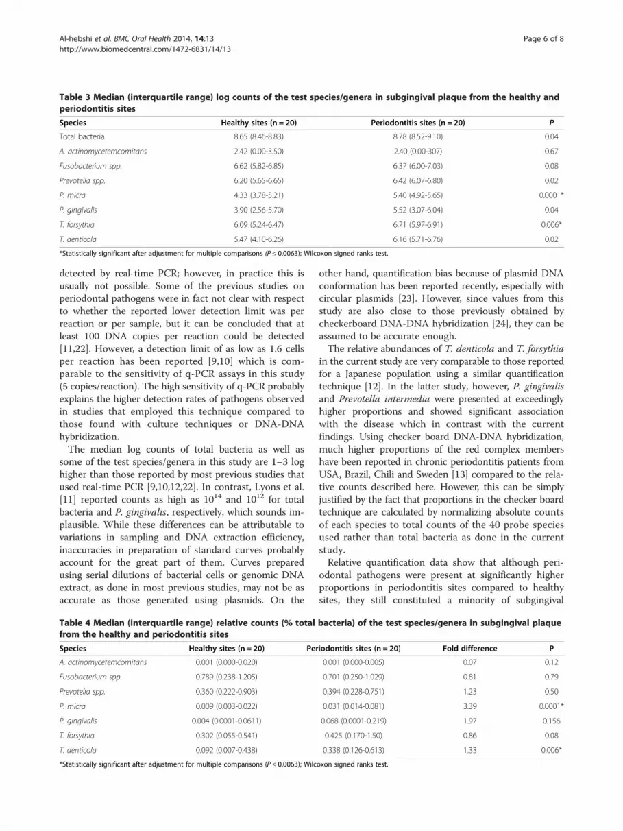
Table 3 Median (interquartile range) log counts of the test species/genera in subgingival plaque from the healthy andperiodontitis sites
Species Healthy sites (n = 20) Periodontitis sites (n = 20) P
Total bacteria 8.65 (8.46-8.83) 8.78 (8.52-9.10) 0.04
A. actinomycetemcomitans 2.42 (0.00-3.50) 2.40 (0.00-307) 0.67
Fusobacterium spp. 6.62 (5.82-6.85) 6.37 (6.00-7.03) 0.08
Prevotella spp. 6.20 (5.65-6.65) 6.42 (6.07-6.80) 0.02
P. micra 4.33 (3.78-5.21) 5.40 (4.92-5.65) 0.0001*
P. gingivalis 3.90 (2.56-5.70) 5.52 (3.07-6.04) 0.04
T. forsythia 6.09 (5.24-6.47) 6.71 (5.97-6.91) 0.006*
T. denticola 5.47 (4.10-6.26) 6.16 (5.71-6.76) 0.02
*Statistically significant after adjustment for multiple comparisons (P ≤ 0.0063); Wilcoxon signed ranks test.
Al-hebshi et al. BMC Oral Health 2014, 14:13 Page 6 of 8http://www.biomedcentral.com/1472-6831/14/13
detected by real-time PCR; however, in practice this isusually not possible. Some of the previous studies onperiodontal pathogens were in fact not clear with respectto whether the reported lower detection limit was perreaction or per sample, but it can be concluded that atleast 100 DNA copies per reaction could be detected[11,22]. However, a detection limit of as low as 1.6 cellsper reaction has been reported [9,10] which is com-parable to the sensitivity of q-PCR assays in this study(5 copies/reaction). The high sensitivity of q-PCR probablyexplains the higher detection rates of pathogens observedin studies that employed this technique compared tothose found with culture techniques or DNA-DNAhybridization.The median log counts of total bacteria as well as
some of the test species/genera in this study are 1–3 loghigher than those reported by most previous studies thatused real-time PCR [9,10,12,22]. In contrast, Lyons et al.[11] reported counts as high as 1014 and 1012 for totalbacteria and P. gingivalis, respectively, which sounds im-plausible. While these differences can be attributable tovariations in sampling and DNA extraction efficiency,inaccuracies in preparation of standard curves probablyaccount for the great part of them. Curves preparedusing serial dilutions of bacterial cells or genomic DNAextract, as done in most previous studies, may not be asaccurate as those generated using plasmids. On the
Table 4 Median (interquartile range) relative counts (% totalfrom the healthy and periodontitis sites
Species Healthy sites (n = 20) Per
A. actinomycetemcomitans 0.001 (0.000-0.020)
Fusobacterium spp. 0.789 (0.238-1.205)
Prevotella spp. 0.360 (0.222-0.903)
P. micra 0.009 (0.003-0.022)
P. gingivalis 0.004 (0.0001-0.0611)
T. forsythia 0.302 (0.055-0.541)
T. denticola 0.092 (0.007-0.438)
*Statistically significant after adjustment for multiple comparisons (P ≤ 0.0063); Wilc
other hand, quantification bias because of plasmid DNAconformation has been reported recently, especially withcircular plasmids [23]. However, since values from thisstudy are also close to those previously obtained bycheckerboard DNA-DNA hybridization [24], they can beassumed to be accurate enough.The relative abundances of T. denticola and T. forsythia
in the current study are very comparable to those reportedfor a Japanese population using a similar quantificationtechnique [12]. In the latter study, however, P. gingivalisand Prevotella intermedia were presented at exceedinglyhigher proportions and showed significant associationwith the disease which in contrast with the currentfindings. Using checker board DNA-DNA hybridization,much higher proportions of the red complex membershave been reported in chronic periodontitis patients fromUSA, Brazil, Chili and Sweden [13] compared to the rela-tive counts described here. However, this can be simplyjustified by the fact that proportions in the checker boardtechnique are calculated by normalizing absolute countsof each species to total counts of the 40 probe speciesused rather than total bacteria as done in the currentstudy.Relative quantification data show that although peri-
odontal pathogens were present at significantly higherproportions in periodontitis sites compared to healthysites, they still constituted a minority of subgingival
bacteria) of the test species/genera in subgingival plaque
iodontitis sites (n = 20) Fold difference P
0.001 (0.000-0.005) 0.07 0.12
0.701 (0.250-1.029) 0.81 0.79
0.394 (0.228-0.751) 1.23 0.50
0.031 (0.014-0.081) 3.39 0.0001*
0.068 (0.0001-0.219) 1.97 0.156
0.425 (0.170-1.50) 0.86 0.08
0.338 (0.126-0.613) 1.33 0.006*
oxon signed ranks test.

Al-hebshi et al. BMC Oral Health 2014, 14:13 Page 7 of 8http://www.biomedcentral.com/1472-6831/14/13
microbiota. This is, however, not surprising since assess-ment of earlier cultivation-based studies and more re-cent studies employing molecular techniques clearlyreveals that periodontal pathogens, particularly membersof the red complex, have almost always been detected atlow abundance [3,24,25]. What has not been addressedadequately, on the other hand, is how these pathogenscan cause periodontitis at such low abundance. One in-teresting, currently evolving view is that low abundanceperiodontal pathogens orchestrate periodontitis by indu-cing a dysbiotic “pathogenic” microbial community thatin turn mediates bone destruction [26]. This is thoughtto result from ability of these pathogens to subvert somecomponents of the host response rather than to actdirectly as proinflammatory bacteria as has been veryrecently demonstrated for P. gingivalis in vitro [27].Accordingly, low abundant periodontal pathogens areclaimed to function as keystone pathogens, a hypoth-esis that challenges the role of red complex membersas conventional pathogens [28].The current study did not show an association between
P. gingivalis and periodontal destruction, which is veryhard to defend against the existing overwhelming evi-dence. However, this can simply be a failure to detectexisting association due to lack of adequate power, espe-cially that there was a significant difference in absolutecount at the 0.05 level. On the other hand, given the poly-microbial nature of periodontitis, and in view of the newkeystone pathogen hypothesis, it is also plausible topropose that other members of the pathogenic team canin certain circumstances take over the role of P. gingivalisas a keystone pathogen. In fact, P. gingivalis has not al-ways showed the strongest association with periodon-titis [3,29,30]. In the current study, T. denticola and T.forsythia, both members of the red complex, did showsignificant association with periodontitis, which is con-sistent with the literature. However, P. micra (previ-ously known as Peptostreptococcus micros) showed thestrongest association with the disease being present atsignificantly higher absolute and relative counts in peri-odontitis sites in all study subjects. This species is amember of the orange microbial complex [6], and thereis an expanding evidence on its role, along with otherpeptostreptococci, as a periodontal pathogen [3,29,31,32].
ConclusionDespite its presence in very low relative counts, P. micrashowed the strongest association with periodontal de-struction, which is suggestive of a potential role as key-stone pathogen in place of P. gingivalis. However, thisneeds to be validated in a larger-scale study before it canbe claimed to represent ethnic variations in pathogens’association with periodontitis.
Competing interestsThe authors declare that they have no competing interests.
Authors’ contributionsNA designed and carried out the laboratory work, performed the statisticalanalysis of data and wrote the manuscript. HS was responsible of the fieldwork, collection of specimens, and data entry. AA contributed to the studydesign, supervision of clinical data collection, and writing of the manuscript.IG was involved in the study design, and overall supervision of the researchproject. All authors read and approved the final manuscript.
AcknowledgementsThis study was partially funded by Al-saeed Foundation for Science andTechnology. All laboratory work was conducted at the Molecular ResearchLaboratory, UST, Sana’a, Yemen. We would like to thank Dr. MohammedSultan for his help with sample collection.
Author details1Department of Preventive Dentistry, Faculty of Dentistry, Jazan University,P.O. box: 114, Jazan, Saudi Arabia. 2Molecular Research Laboratory, Faculty ofMedical Sciences, University of Science and Technology, Sana’a, Yemen.3Department of Periodontology, Oral Pathology, Oral Medicine andRadiology; Faculty of Dentistry, University of Sana’a, Sana’a, Yemen.4Department of Periodontology, Faculty of Dentistry, Khartoum University,Khartoum, Sudan.
Received: 12 November 2013 Accepted: 14 February 2014Published: 18 February 2014
References1. Darveau RP: Periodontitis: a polymicrobial disruption of host
homeostasis. Nat Rev Microbiol 2010, 8(7):481–490.2. Moore WE, Moore LV: The bacteria of periodontal diseases. Periodontol
2000 1994, 5:66–77.3. Kumar PS, Griffen AL, Moeschberger ML, Leys EJ: Identification of
candidate periodontal pathogens and beneficial species by quantitative16S clonal analysis. J Clin Microbiol 2005, 43(8):3944–3955.
4. Paster BJ, Boches SK, Galvin JL, Ericson RE, Lau CN, Levanos VA,Sahasrabudhe A, Dewhirst FE: Bacterial diversity in human subgingivalplaque. J Bacteriol 2001, 183(12):3770–3783.
5. Dewhirst FE, Chen T, Izard J, Paster BJ, Tanner AC, Yu WH, Lakshmanan A,Wade WG: The human oral microbiome. J Bacteriol 2010, 192(19):5002–5017.
6. Socransky SS, Haffajee AD, Cugini MA, Smith C, Kent RL Jr: Microbialcomplexes in subgingival plaque. J Clin Periodontol 1998, 25(2):134–144.
7. Dewhirst FE, Tamer MA, Ericson RE, Lau CN, Levanos VA, Boches SK, Galvin JL,Paster BJ: The diversity of periodontal spirochetes by 16S rRNA analysis.Oral Microbiol Immunol 2000, 15(3):196–202.
8. Rylev M, Kilian M: Prevalence and distribution of principal periodontalpathogens worldwide. J Clin Periodontol 2008, 35(8 Suppl):346–361.
9. Mineoka T, Awano S, Rikimaru T, Kurata H, Yoshida A, Ansai T, Takehara T:Site-specific development of periodontal disease is associated withincreased levels of Porphyromonas gingivalis, Treponema denticola,and Tannerella forsythia in subgingival plaque. J Periodontol 2008,79(4):670–676.
10. Yoshida A, Kawada M, Suzuki N, Nakano Y, Oho T, Saito T, Yamashita Y:TaqMan real-time polymerase chain reaction assay for the correlation ofTreponema denticola numbers with the severity of periodontal disease.Oral Microbiol Immunol 2004, 19(3):196–200.
11. Lyons SR, Griffen AL, Leys EJ: Quantitative real-time PCR for Porphyromonasgingivalis and total bacteria. J Clin Microbiol 2000, 38(6):2362–2365.
12. Kuboniwa M, Amano A, Kimura KR, Sekine S, Kato S, Yamamoto Y, Okahashi N,Iida T, Shizukuishi S: Quantitative detection of periodontal pathogens usingreal-time polymerase chain reaction with TaqMan probes. Oral MicrobiolImmunol 2004, 19(3):168–176.
13. Haffajee AD, Bogren A, Hasturk H, Feres M, Lopez NJ, Socransky SS:Subgingival microbiota of chronic periodontitis subjects from differentgeographic locations. J Clin Periodontol 2004, 31(11):996–1002.
14. World Health Organization: Oral health surveys: basic methods. 4th edition.Geneva: World Health Organization; 1997.

Al-hebshi et al. BMC Oral Health 2014, 14:13 Page 8 of 8http://www.biomedcentral.com/1472-6831/14/13
15. Silness J, Loe H: Periodontal disease in pregnancy. Ii. Correlation betweenoral hygiene and periodontal condtion. Acta Odontol Scand 1964,22:121–135.
16. Holland PM, Abramson RD, Watson R, Gelfand DH: Detection of specificpolymerase chain reaction product by utilizing the 5′––3′ exonucleaseactivity of Thermus aquaticus DNA polymerase. Proc Natl Acad Sci U S A1991, 88(16):7276–7280.
17. Al-Hebshi NN, Al-Sharabi AK, Shuga-Aldin HM, Al-Haroni M, Ghandour I:Effect of khat chewing on periodontal pathogens in subgingival biofilmfrom chronic periodontitis patients. J Ethnopharmacol 2010, 132(3):564–569.
18. Suzuki N, Yoshida A, Saito T, Kawada M, Nakano Y: Quantitativemicrobiological study of subgingival plaque by real-time PCR showscorrelation between levels of Tannerella forsythensis and Fusobacteriumspp. J Clin Microbiol 2004, 42(5):2255–2257.
19. Martin FE, Nadkarni MA, Jacques NA, Hunter N: Quantitativemicrobiological study of human carious dentine by culture and real-timePCR: association of anaerobes with histopathological changes in chronicpulpitis. J Clin Microbiol 2002, 40(5):1698–1704.
20. Al-Otaibi M, Al-Harthy M, Gustafsson A, Johansson A, Claesson R, Angmar-Mansson B: Subgingival plaque microbiota in Saudi Arabians after use ofmiswak chewing stick and toothbrush. J Clin Periodontol 2004,31(12):1048–1053.
21. Darout IA, Skaug N, Albandar JM: Subgingival microbiota levels and theirassociations with periodontal status at the sampled sites in an adultSudanese population using miswak or toothbrush regularly. Acta OdontolScand 2003, 61(2):115–122.
22. Nonnenmacher C, Dalpke A, Mutters R, Heeg K: Quantitative detection ofperiodontopathogens by real-time PCR. J Microbiol Methods 2004,59(1):117–125.
23. Lin CH, Chen YC, Pan TM: Quantification bias caused by plasmid DNAconformation in quantitative real-time PCR assay. PLoS One 2011,6(12):e29101.
24. Ximenez-Fyvie LA, Haffajee AD, Socransky SS: Microbial composition ofsupra- and subgingival plaque in subjects with adult periodontitis.J Clin Periodontol 2000, 27(10):722–732.
25. Moore WE, Holdeman LV, Smibert RM, Hash DE, Burmeister JA, Ranney RR:Bacteriology of severe periodontitis in young adult humans. Infect Immun1982, 38(3):1137–1148.
26. Hajishengallis G, Lamont RJ: Beyond the red complex and into morecomplexity: the polymicrobial synergy and dysbiosis (PSD) model ofperiodontal disease etiology. Mol Oral Microbiol 2012, 27(6):409–419.
27. Hajishengallis G, Liang S, Payne MA, Hashim A, Jotwani R, Eskan MA,McIntosh ML, Alsam A, Kirkwood KL, Lambris JD, et al: Low-abundancebiofilm species orchestrates inflammatory periodontal disease throughthe commensal microbiota and complement. Cell Host Microbe 2011,10(5):497–506.
28. Hajishengallis G, Darveau RP, Curtis MA: The keystone-pathogen hypothesis.Nat Rev Microbiol 2012, 10(10):717–725.
29. Colombo AP, Boches SK, Cotton SL, Goodson JM, Kent R, Haffajee AD,Socransky SS, Hasturk H, Van Dyke TE, Dewhirst F, et al: Comparisons ofsubgingival microbial profiles of refractory periodontitis, severeperiodontitis, and periodontal health using the human oral microbeidentification microarray. J Periodontol 2009, 80(9):1421–1432.
30. Kumar PS, Leys EJ, Bryk JM, Martinez FJ, Moeschberger ML, Griffen AL:Changes in periodontal health status are associated with bacterialcommunity shifts as assessed by quantitative 16S cloning andsequencing. J Clin Microbiol 2006, 44(10):3665–3673.
31. Rams TE, Feik D, Listgarten MA, Slots J: Peptostreptococcus micros inhuman periodontitis. Oral Microbiol Immunol 1992, 7(1):1–6.
32. Belstrom D, Fiehn NE, Nielsen CH, Kirkby N, Twetman S, Klepac-Ceraj V,Paster BJ, Holmstrup P: Differences in bacterial saliva profile betweenperiodontitis patients and a control cohort. J Clin Periodontol 2014,41(2):104–112.
doi:10.1186/1472-6831-14-13Cite this article as: Al-hebshi et al.: Subgingival periodontal pathogensassociated with chronic periodontitis in Yemenis. BMC Oral Health2014 14:13.
Submit your next manuscript to BioMed Centraland take full advantage of:
• Convenient online submission
• Thorough peer review
• No space constraints or color figure charges
• Immediate publication on acceptance
• Inclusion in PubMed, CAS, Scopus and Google Scholar
• Research which is freely available for redistribution
Submit your manuscript at www.biomedcentral.com/submit


