Embed Size (px)
Citation preview
www.elsevier.com/locate/ytaap
Toxicology and Applied Pharmacology 200 (2004) 159–168
Carvedilol-mediated antioxidant protection against doxorubicin-induced
cardiac mitochondrial toxicity
Paulo J. Oliveira,a James A. Bjork,b Maria S. Santos,a Richard L. Leino,c M. Kent Froberg,d
Antonio J. Moreno,a and Kendall B. Wallaceb,*
aCentre of Neurosciences and Cellular Biology of Coimbra, Department of Zoology, University of Coimbra, PortugalbDepartment of Biochemistry and Molecular Biology, School of Medicine, University of Minnesota, Duluth, MN 55812-2496, USA
cDepartment of Anatomy and Cell Biology, School of Medicine, University of Minnesota, Duluth, MN 55812-2496, USAdDepartment of Pathology, School of Medicine, University of Minnesota, Duluth, MN 55812-2496, USA
Received 21 November 2003; accepted 12 April 2004
Available online 20 July 2004
Abstract
The cardiotoxicity associated with doxorubicin (DOX) therapy limits the total cumulative dose and therapeutic success of active
anticancer chemotherapy. Cardiac mitochondria are implicated as primary targets for DOX toxicity, which is believed to be mediated by the
generation of highly reactive free radical species of oxygen from complex I of the mitochondrial electron transport chain. The objective of
this study was to determine if the protection demonstrated by carvedilol (CV), a h-adrenergic receptor antagonist with strong antioxidant
properties, against DOX-induced mitochondrial-mediated cardiomyopathy [Toxicol. Appl. Pharmacol. 185 (2002) 218] is attributable to its
antioxidant properties or its h-adrenergic receptor antagonism. Our results confirm that DOX induces oxidative stress, mitochondrial
dysfunction, and histopathological lesions in the cardiac tissue, all of which are inhibited by carvedilol. In contrast, atenolol (AT), a h-adrenergic receptor antagonist lacking antioxidant properties, preserved phosphate energy charge but failed to protect against any of the
indexes of DOX-induced oxidative mitochondrial toxicity. We therefore conclude that the cardioprotective effects of carvedilol against DOX-
induced mitochondrial cardiotoxicity are due to its inherent antioxidant activity and not to its h-adrenergic receptor antagonism.
D 2004 Elsevier Inc. All rights reserved.
Keywords: Mitochondria; Heart; Doxorubicin; Carvedilol; Atenolol; Permeability transition pore; Oxidative damage; Mitochondrionopathy
Introduction damage to the cardiac mitochondria and to the cardiomyo-
The anthracycline quinone doxorubicin (DOX) has been
widely prescribed in the treatment of several human tumors
and leukemias. Despite its effectiveness, the clinical use of
DOX is limited by a dose-dependent and cumulative car-
diotoxicity (Hrdina et al., 2000). Many hypotheses have
been advanced to explain the cardioselective toxicity of
DOX (Olson and Mushlin, 1990). Because DOX undergoes
redox cycling on mitochondrial Complex I to liberate highly
reactive oxygen free radicals (Doroshow, 1983), oxidative
0041-008X/$ - see front matter D 2004 Elsevier Inc. All rights reserved.
doi:10.1016/j.taap.2004.04.005
Abbreviations: DOX, doxorubicin; DC, mitochondrial transmembrane
potential; TPP+, tetraphenylphosphonium cation; DMSO, dimethylsulf-
oxide; CV, carvedilol; AT, atenolol; SAL, saline; DTPA, 4,4-Dithio-
bis(phenylazide).
* Corresponding author. Department of Biochemistry and Molecular
Biology, School of Medicine, University of Minnesota, 1035 University
Drive, Room 255, Duluth, MN 55812-2496. Fax: +1-218-726-8014.
E-mail address: [email protected] (K.B. Wallace).
cyte has been widely implicated as a primary cause for
doxorubicin-induced cardiac toxicity (Lee et al., 1991; Xu et
al., 2001). Several authors have shown diverse DOX-
induced damage to mitochondrial membranes (Mimnaugh
et al., 1985), proteins (Demant, 1991), and nucleic acids
(Palmeira et al., 1997). One particular element is the
triggering of the calcium-dependent mitochondrial perme-
ability transition (MPT). This phenomenon, caused by
enhanced oxidative stress and mitochondrial calcium over-
load (Bernardi, 1999; Kowaltowski and Vercesi, 1999), has
been implicated to be responsible for mitochondrial and
cellular dysfunction caused by DOX (Sokolove and Shina-
berry, 1988; Solem et al., 1994, 1996; Zhou et al., 2001a,
2001b). Beyond cessation of mitochondrial ATP synthesis,
another consequence associated with the MPT is the trig-
gering of apoptotic cell death due to the release of pro-
apoptotic factors from the dysfunctional mitochondria (Ber-
nardi, 1999; Kowaltowski and Vercesi, 1999).
P.J. Oliveira et al. / Toxicology and Applied Pharmacology 200 (2004) 159–168160
Several authors have demonstrated that doxorubicin and
its metabolites decrease mitochondrial calcium loading
capacity both in vitro (Sokolove and Shinaberry, 1988)
and in vivo (Solem et al., 1994, 1996; Zhou et al., 2001a,
2001b). The use of cyclosporin-A, a specific inhibitor of the
MPT (Broekemeier et al., 1989), was critical in identifying
the MPT as the principal cause for mitochondrial dysfunc-
tion caused by DOX exposure in vivo (Solem et al., 1994).
Recent results demonstrate that cardiomyocytes isolated
from DOX-treated animals are more susceptible to calci-
um-induced MPT and cell killing, both of which are
prevented by cyclosporin-A (Solem et al., 1996; Zhou et
al., 2001a). Evidence indicates that by inducing the MPT,
DOX interferes with mitochondrial calcium regulation and
that this is a principal feature in the pathogenesis associated
with DOX-induced cardiomyopathy.
Because liberation of free radicals is central to the
mechanism of DOX-induced damage to the myocardium,
it is logical to consider antioxidants as primary candidates to
counteract such toxic effects. In fact, several compounds
with antioxidant properties have been investigated in vitro
with some degree of success (DeAtley et al., 1999; Monti et
al., 1996). However, the rate of success when performing in
vivo studies has been less gratifying. In fact, molecules with
antioxidant characteristics in vitro such as vitamin E and
selenium (Van Vleet et al., 1980) or nimesulid (Kotsinas et
al., 1999) were found to be of limited value in counteracting
doxorubicin cardiotoxicity in vivo.
Carvedilol (CV, {1-(carbazolyl-(4)-oxy)-3-(2-methoxy-
phenoxyethyl) amino)-propanol-(2)}) is used clinically for
the treatment of congestive heart failure, mild to moderate
hypertension, and myocardial infarction. Carvedilol com-
petitively blocks h1, h2, and a1-adrenergic receptors while
displaying vasodilating properties. A distinctive character-
istic of carvedilol is its potent antioxidant properties that are
not shared by other h-adrenergic receptor antagonists (Yue
et al., 1992). This antioxidant activity of carvedilol is
attributed to its ability to chelate free iron (Noguchi et al.,
2000), which is widely implicated in enhancing the free
radical-mediated toxicity caused by doxorubicin. Carvedilol
is superior to atenolol (AT), a h-adrenergic receptor antag-
onist without antioxidant properties, in diminishing DOX-
induced negative impact on systolic blood pressure and left
ventricular fractional shortening as well on increased lipid
peroxidation (Matsui et al., 1999), which could either be
explained by a direct effect on cardiac tissue or to vascular
effects of the drug. More recently, the balance tilted for a
predominant effect on the cardiac tissue as Santos et al.
(2002) showed that carvedilol diminishes both the cardiac
mitochondrial toxicity and histopathology caused by DOX.
What has not yet been determined is whether this cardio-
specific protection can be ascribed to the antioxidant or the
general h-adrenergic receptor antagonism properties of
carvedilol. The objective of the present work was to
differentiate the relative contributions of the antioxidant
and h-adrenergic receptor antagonism properties of carve-
dilol in affording protection against the cardiac toxicity of
DOX.
Materials and methods
Chemicals. Ultrapure sucrose was obtained from Schwarz/
Mann Biotech (Cleveland, OH). DOX was purchased from
Pharmacia and Upjohn Co. (Kalamazoo, MI). Cyclosporin-
A was a gift from Sandoz Pharmaceuticals (East Hanover,
NJ) and carvedilol was obtained from Roche Pharmaceut-
icals (Lisbon, Portugal). All other chemicals were of the
highest grade available from Sigma (St. Louis, MO). Both
atenolol (AT) and carvedilol (CV) were dissolved in dime-
thylsulfoxide (DMSO). Doxorubicin (DOX) was dissolved
in saline (SAL) solution.
Animals and treatment protocol. Male Sprague–Dawley
rats (Harlan Labs, Madison, WI) weighing 200–300 g were
maintained in AAALAC-accredited, climate-controlled fa-
cilities and allowed free access to food (Purina Chow) and
water. All studies were carried out in accordance with the
Guide for the Care and Use of Laboratory Animals. Rats
were randomly divided among a total of six experimental
groups (initial weights ranged from 173.5 to 186.5 g): SAL +
DMSO, SAL + CV, SAL + AT, DOX + DMSO, DOX + CV,
DOX + AT. DOX-injected rats received seven weekly sc
injections of either DOX (2 mg kg�1) or an equivalent
volume of SAL solution (1 ml kg�1). Where indicated, CV
(1 mg kg�1, ip), AT (1 mg kg�1, ip), or an equivalent
volume of DMSO (1 ml kg�1, ip) was co-administrated with
DOX or saline solution. DOX dosage was established from
previous studies that showed cardiac mitochondrial damage
and histopathology (Solem et al., 1994; Zhou et al., 2001b).
The CV regimen was according to the work of Santos et al.
(2002), where convincing protection against DOX cardio-
selective toxicity was afforded by carvedilol administration.
Clinical trials demonstrated that both compounds, at the
same dosages, were equally effective at reducing blood
pressure in patients (Hall et al., 1991; McTavish et al.,
1993), so an equivalent dosage of both compounds was
administrated. The animals were killed by decapitation 1
week after the last injection and the hearts immediately
excised to cold buffer for mitochondrial isolation. Body and
heart weights were recorded on the day of the experiments.
Isolation of cardiac mitochondria. Cardiac mitochondria
were prepared by differential centrifugation (Oliveira et al.,
2000). Briefly, the hearts were finely minced in an ice-cold
isolation medium containing 250 mM sucrose, 1 mM
EGTA, 10 mM HEPES-KOH (pH 7.4), and 0.1% defatted
BSA. The minced tissue was suspended in 40 ml of isolation
medium containing 0.5 mg protease Type VIII (Sigma P-
5390) per g of tissue and homogenized with a tightly fitted
homogenizer (Teflon: glass pestle). The suspension was
incubated for 1 min (4 jC) and then re-homogenized. The
P.J. Oliveira et al. / Toxicology and Applied Pharmacology 200 (2004) 159–168 161
homogenate was then centrifuged at 9500 � g for 10 min.
The supernatant was decanted and the pellet was gently
homogenized to its original volume with a loose-fitting
homogenizer. The suspension was centrifuged at 900 � g
for 10 min and the resulting supernatant was centrifuged at
9000 � g for 10 min. This pellet was resuspended using a
brush and re-pelleted twice at 9000 � g for 10 min. EGTA
and defatted BSA were omitted from the final washing
medium. Mitochondrial protein content was determined by
the Bradford method calibrated with BSA (Bradford, 1976).
TPP+ uptake. The mitochondrial accumulation of tetra-
phenylphosphonium (TPP+) (indirect measurement of mito-
chondrial electrical potential, DC) was performed using a
TPP+-selective electrode and a AgCl reference electrode
(Kamo et al., 1979). Mitochondrial protein (0.5 mg) was
suspended in 2 ml of reaction buffer (200 mM sucrose, 10
mM TRIS-MOPS, 10 AM EGTA, and 5 mM KH2PO4, pH
7.4, 25 jC) supplemented with 2 AM rotenone and 0.5 AMTPP+. Sequential additions of 0.5 AM TPP+ were made to
calibrate the system and calculate the amount of TPP+ taken
by mitochondria. Absolute values for membrane potential
(in millivolts) were determined from the equation originally
proposed by Kamo et al. (1979). Succinate was added to the
mitochondrial suspension containing a total of 3 AM TPP+
in the absence and presence of 20 AM of calcium.
Oxygen consumption. Oxygen consumption by isolated
heart mitochondria was monitored polarographically with
a Clark oxygen electrode connected to a suitable recorder.
Reactions were carried out at 25 jC in 1 ml of the media
containing 200 mM sucrose, 10 mM TRIS-MOPS, 10 AMEGTA, and 5 mM KH2PO4 (pH 7.4). Mitochondria were
suspended at a concentration of 0.5 mg ml�1 in the
respiratory medium. State 4 respiration was measured in
the presence of 5 mM glutamate/malate. ADP (100 nmol)
was added to induce state 3 respiration. The respiratory
control ratio (RCR) was calculated as the ratio between state
3 and state 4 respiration and the ADP/O was calculated
according to standard procedures (Estabrook, 1967).
Mitochondrial swelling. Mitochondrial volume changes
were followed by the decrease of optical density measured
at 540 nm with a Beckman DU 7400 spectrophotometer
equipped with a magnetic stirrer assembly. The assays were
performed in 2 ml of 200 mM sucrose, 10 mM TRIS-
MOPS, 10 AM EGTA, 5 mM KH2PO4 (pH 7.4, 25 jC), and2 AM rotenone to which was added 0.5 mg of mitochondrial
protein. Succinate was added after calcium addition. Swell-
ing amplitude was calculated as the difference between
initial (pre-succinate addition) and final optical density.
Calcium loading capacity. Extramitochondrial free Ca2+
was measured with the hexapotassium salt of the fluores-
cence probe Calcium Green 5-N (Rajdev and Reynolds,
1993). Heart mitochondria (15 ng) were suspended in 300
Al of buffer containing 200 mM sucrose, 10 mM TRIS-
MOPS, 1 AM EGTA, 1 mM KH2PO4 (pH 7.4), 200 nM
Calcium Green 5-N, 1 AM rotenone, and 2.1 pmol calci-
um. Calcium Green 5-N fluorescence was continuously
recorded in a Lab-systems type 374 plate reader with
excitation and emission wavelengths of 485 and 538 nm,
respectively. Calcium loading was initiated by the addition
of 1 mM succinate. To calculate the MPT-dependent
decrease in calcium loading capacity, the same measure-
ments were repeated in the presence of cyclosporin-A to
inhibit the MPT (Zhou et al., 2001b). Mitochondrial
calcium loading capacity was expressed as the ratio of
the maximum fluorescence change in the absence and
presence of cyclosporin-A. At the end of each assay,
EGTA was added in excess to complex all free calcium
and calibrate the base line.
Tissue adenine nucleotide quantification. Tissue adenine
nucleotide extraction was performed with an acidic extrac-
tion procedure with 0.625 N HClO4 and neutralized with 5
N KOH (Jones, 1981). The resulting supernatant was
filtered through a Millipore 0.45 Am filter and the pH was
set to 7. The nucleotides were separated by reverse-phase
high performance liquid chromatography consisting of a
Beckman 126 Binary Pump Model and a 166 Variable UV
detector controlled by a computer. The detection wavelength
was 254 nm, and the column was a Lichrospher 100RP-18
(5 Am) from Merck (Darmstadt, Germany). An isocratic
elution with 100 mM phosphate buffer (KH2PO4), pH 6.5
and 1% methanol was performed with a flow rate of 1 ml
min�1. The time required for each analysis was 5 min.
Quantification of protein-bound carbonyls. Protein-reac-
tive carbonyls in tissue samples were analyzed according to
Levine et al. (1990) with slight modifications. A sample of
tissue (100–300 mg) was homogenized in three volumes of
buffer composed of 10 mM HEPES, 137 mM NaCl, 4.6
mM KCl, 2 mM KH2PO4, 0.6 mM MgSO4, and 1.1 mM
EDTA (pH 7.4). The insoluble material was discarded. Two
50-Al aliquots from each sample were taken and the protein
precipitated with 0.5 ml ice-cold trichloroacetic acid (10%
w/v). After a 10-min incubation at 4 jC, samples were
centrifuged at 11000 � g for 3 min. One of the protein
pellets was resuspended in 0.5 ml of 0.2% (w/v) 2,4-
dinitrophenylhydrazine (DNPH)/2 M HCl. The other pro-
tein sample was resuspended in 0.5 ml of 2 M HCl (and
used as blank). The pellets were gently resuspended and
continuously mixed at room temperature for 1 h. The
samples were then precipitated with 0.5 ml of 10% TCA
and centrifuged at 11000 g for 3 min. The pellets were
gently washed with 1 ml of ethanol-ethyl acetate (1:1; v/v)
to remove unreacted DNPH reagent and allowed to stand
for 10 min. The samples were centrifuged for 5 min at
11000 � g and the supernatant discarded. The washing
procedure was repeated twice. The resulting protein pellet
was gently resuspended in 1.0 ml of 6 M guanidine with 20
P.J. Oliveira et al. / Toxicology and Applied Pharmacology 200 (2004) 159–168162
mM sodium phosphate buffer (pH 6.5). The samples were
left for 10 min at room temperature before reading the
absorbance at 360 nm (DPNH-protein carbonyl adduct) and
280 nm (protein concentration). Protein carbonyl concen-
tration was estimated from the ratio A360/A280 after
subtracting the blanks.
Superoxide dismutase (SOD) activity. Total SOD activity
in homogenized heart tissue was determined according to
Prohaska (1983) by monitoring the rate of autoxidation of
pyrogallol at 320 nm. The homogenization buffer consisted
of 50 mM KPO4, pH 7.4. The reaction buffer (37 jC) was50 mM TAPS ((2-Hydroxy-1,1-bis(hydroxymethyl)ethyl)a-
mino)-1-propanesulfonic acid N-(Tris(hydroxymethyl)-
methyl)-3-aminopropanesulfonic acid), 0.1 mM 4,4-
Dithio-bis(phenylazide) (DTPA), pH 8.2. Pyrogallol (4 Al ofa 10 mM stock solution in a final volume of 1 ml) was
added to the cuvette to start the reaction and the rate of
absorbance increase was measured for 1 min. One Unit of
SOD activity was defined as the amount of sample
required to inhibit the autoxidation of pyrogallol by
50%, and the activity is expressed as Units per mg of
protein (Prohaska, 1983).
Glutathione peroxidase (GPx) activity. Tissue GPx acti-
vity was determined spectrophotometrically by the decrease
in NADPH absorbance at 340 nm. Measurements were
performed in 1 ml of reaction buffer containing 100 mM
HEPES and 0.1 mM DTPA, pH 7.4, supplemented with 1
mM GSH, NADPH, and 50 Al of commercial glutathione
reductase (Gred) 0.3 U ml�1. After sample addition (1:100
dilution from a homogenate), the reaction was started by
adding 250 AM tBHP. The difference between the rates
before and after tBHP addition was calculated and
expressed as nmol NADPH consumed per min per mg
protein (eNADPH = 6.22 mM�1 cm�1).
Glutathione reductase (Gred) activity. Gred activity was
determined according to the method of Yin et al. (1998).
Samples were prepared as described for SOD measure-
ments. The assay buffer consisted of 0.2 M KH2PO4
supplemented with 2 mM EDTA, and the reaction started
by adding NADPH in excess and, 1 min later, 1 AMGSSG. The difference between the rates before and after
GSSG addition was calculated and expressed as nmol
NADPH consumed per min per mg protein (eNADPH =
6.22 mM�1 cm�1).
Table 1
Comparison of body and heart weight among experimental groups
Saline + DMSO Saline + Carvedilol Saline + A
Heart weight (g) 1.47 F 0.04 1.49 F 0.06 1.48 FFinal body weight (g) 379.0 F 9.66 378.0 F 11.10 372.25 FHW/BW (�1000) 3.89 F 0.05 3.94 F 0.15 3.99 F
The values are the means F SEM of eight animals in each experimental group.
*P < 0.05 vs. the corresponding saline treatment.
Electron microscopy of cardiac tissue. Representative
electron micrographs from myocytes were obtained as
previously described (Santos et al., 2002; Zhou et al.,
2001b). Briefly, small blocks of cardiac muscle from the
midportion of the lateral wall of the left ventricle were fixed
in 2.5% glutaraldehyde–2% formaldehyde in 0.1 M phos-
phate buffer (pH 7.4), postfixed in 1% osmium tetroxide in
the same buffer, dehydrated in graded acetone solutions, and
finally embedded in epon-araldite. Thin sections were
stained with lead citrate and uranyl acetate. Sections were
scored from both the extent (number of damaged cardiac
myocytes) and the severity of the cellular changes according
to the same criteria as prior studies (Santos et al., 2002;
Zhou et al., 2001b). The extent of damage is reported as the
absolute number of affected cells within a 202500 Am2 total
cross-sectional region of ventricular wall. Severity was
scored on a scale of 0–3 (Santos et al., 2002; Zhou et al.,
2001b). All slides were scored independently by an exam-
iner who was blinded to the identity of each sample.
Statistical analysis. The results are presented as the
mean F SEM of at least six independent experiments.
One-way analysis of variance (ANOVA) of log-transformed
data followed by Newman–Keuls post-test was used as
statistical treatment. A P value <0.05 was considered
statistically significant.
Results
Long-term treatment with DOX caused several altera-
tions, including decreased final body weight gain and
reduced heart size (Table 1). The decrease in heart weight
was proportional to body weight reduction, resulting in an
unchanged ratio between heart and body weight (Table 1).
Although both carvedilol and atenolol prevented the de-
creased heart weight associated with DOX treatment, neither
drug prevented DOX effects on body weight. Neither
carvedilol alone nor atenolol alone (in the absence of
DOX) affected the heart or body weight.
No differences in the respiratory rates in glutamate/
malate-energized mitochondria were found between any of
the treatment groups (Table 2). State 4 respiratory activities
varied between 40 and 60 natm O min�1 mg�1 protein.
RCR values were between 6.4 F 1.0 in the SAL + CV
group and 8.0 F 1.4 in the SAL + DMSO group (Table 2).
The index for mitochondrial phosphorylation efficiency (the
tenolol DOX + DMSO DOX + Carvedilol DOX + Atenolol
0.05 1.23 F 0.04* 1.32 F 0.08 1.25 F 0.06
6.98 298.38 F 10.72* 290.61 F 6.39* 312.31 F 9.86*
0.14 4.14 F 0.18 4.55 F 0.28 4.01 F 0.18
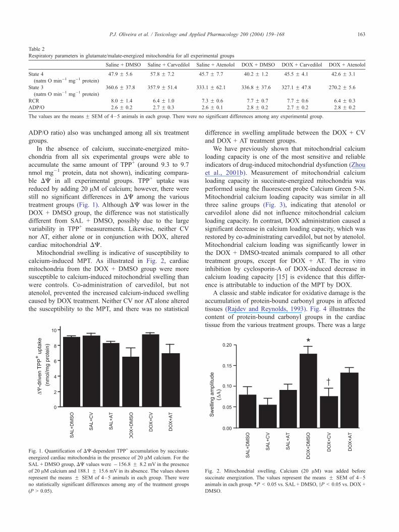
Table 2
Respiratory parameters in glutamate/malate-energized mitochondria for all experimental groups
Saline + DMSO Saline + Carvedilol Saline + Atenolol DOX + DMSO DOX + Carvedilol DOX + Atenolol
State 4
(natm O min�1 mg�1 protein)
47.9 F 5.6 57.8 F 7.2 45.7 F 7.7 40.2 F 1.2 45.5 F 4.1 42.6 F 3.1
State 3
(natm O min�1 mg�1 protein)
360.6 F 37.8 357.9 F 51.4 333.1 F 62.1 336.8 F 37.6 327.1 F 47.8 270.2 F 5.6
RCR 8.0 F 1.4 6.4 F 1.0 7.3 F 0.6 7.7 F 0.7 7.7 F 0.6 6.4 F 0.3
ADP/O 2.6 F 0.2 2.7 F 0.3 2.6 F 0.1 2.8 F 0.2 2.7 F 0.2 2.8 F 0.2
The values are the means F SEM of 4–5 animals in each group. There were no significant differences among any experimental group.
P.J. Oliveira et al. / Toxicology and Applied Pharmacology 200 (2004) 159–168 163
ADP/O ratio) also was unchanged among all six treatment
groups.
In the absence of calcium, succinate-energized mito-
chondria from all six experimental groups were able to
accumulate the same amount of TPP+ (around 9.3 to 9.7
nmol mg�1 protein, data not shown), indicating compara-
ble DC in all experimental groups. TPP+ uptake was
reduced by adding 20 AM of calcium; however, there were
still no significant differences in DC among the various
treatment groups (Fig. 1). Although DC was lower in the
DOX + DMSO group, the difference was not statistically
different from SAL + DMSO, possibly due to the large
variability in TPP+ measurements. Likewise, neither CV
nor AT, either alone or in conjunction with DOX, altered
cardiac mitochondrial DC.
Mitochondrial swelling is indicative of susceptibility to
calcium-induced MPT. As illustrated in Fig. 2, cardiac
mitochondria from the DOX + DMSO group were more
susceptible to calcium-induced mitochondrial swelling than
were controls. Co-administration of carvedilol, but not
atenolol, prevented the increased calcium-induced swelling
caused by DOX treatment. Neither CV nor AT alone altered
the susceptibility to the MPT, and there was no statistical
Fig. 1. Quantification of DC-dependent TPP+ accumulation by succinate-
energized cardiac mitochondria in the presence of 20 AM calcium. For the
SAL + DMSO group, DC values were � 156.8 F 8.2 mV in the presence
of 20 AM calcium and 188.1 F 15.6 mV in its absence. The values shown
represent the means F SEM of 4–5 animals in each group. There were
no statistically significant differences among any of the treatment groups
(P > 0.05).
difference in swelling amplitude between the DOX + CV
and DOX + AT treatment groups.
We have previously shown that mitochondrial calcium
loading capacity is one of the most sensitive and reliable
indicators of drug-induced mitochondrial dysfunction (Zhou
et al., 2001b). Measurement of mitochondrial calcium
loading capacity in succinate-energized mitochondria was
performed using the fluorescent probe Calcium Green 5-N.
Mitochondrial calcium loading capacity was similar in all
three saline groups (Fig. 3), indicating that atenolol or
carvedilol alone did not influence mitochondrial calcium
loading capacity. In contrast, DOX administration caused a
significant decrease in calcium loading capacity, which was
restored by co-administrating carvedilol, but not by atenolol.
Mitochondrial calcium loading was significantly lower in
the DOX + DMSO-treated animals compared to all other
treatment groups, except for DOX + AT. The in vitro
inhibition by cyclosporin-A of DOX-induced decrease in
calcium loading capacity [15] is evidence that this differ-
ence is attributable to induction of the MPT by DOX.
A classic and stable indicator for oxidative damage is the
accumulation of protein-bound carbonyl groups in affected
tissues (Rajdev and Reynolds, 1993). Fig. 4 illustrates the
content of protein-bound carbonyl groups in the cardiac
tissue from the various treatment groups. There was a large
Fig. 2. Mitochondrial swelling. Calcium (20 AM) was added before
succinate energization. The values represent the means F SEM of 4–5
animals in each group. *P < 0.05 vs. SAL + DMSO, yP < 0.05 vs. DOX +
DMSO.
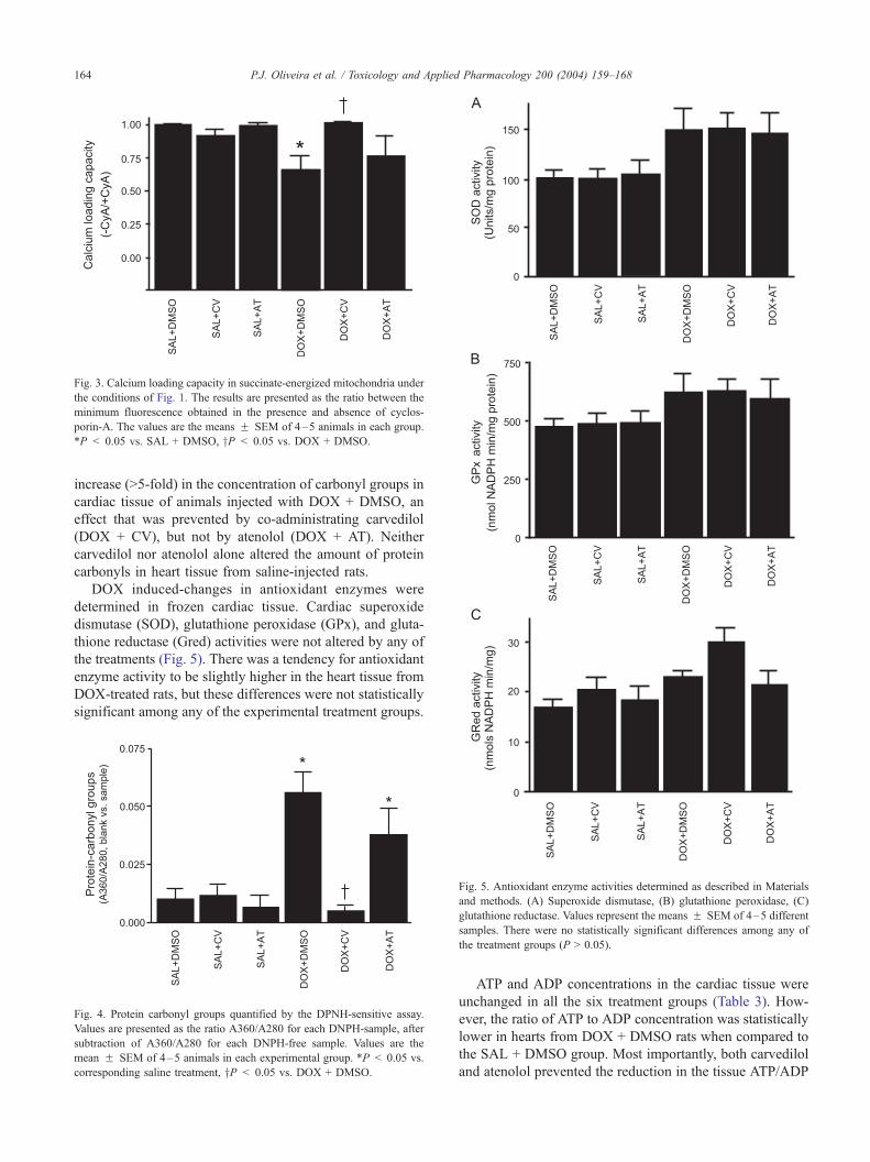
Fig. 3. Calcium loading capacity in succinate-energized mitochondria under
the conditions of Fig. 1. The results are presented as the ratio between the
minimum fluorescence obtained in the presence and absence of cyclos-
porin-A. The values are the means F SEM of 4–5 animals in each group.
*P < 0.05 vs. SAL + DMSO, yP < 0.05 vs. DOX + DMSO.
P.J. Oliveira et al. / Toxicology and Applied Pharmacology 200 (2004) 159–168164
increase (>5-fold) in the concentration of carbonyl groups in
cardiac tissue of animals injected with DOX + DMSO, an
effect that was prevented by co-administrating carvedilol
(DOX + CV), but not by atenolol (DOX + AT). Neither
carvedilol nor atenolol alone altered the amount of protein
carbonyls in heart tissue from saline-injected rats.
DOX induced-changes in antioxidant enzymes were
determined in frozen cardiac tissue. Cardiac superoxide
dismutase (SOD), glutathione peroxidase (GPx), and gluta-
thione reductase (Gred) activities were not altered by any of
the treatments (Fig. 5). There was a tendency for antioxidant
enzyme activity to be slightly higher in the heart tissue from
DOX-treated rats, but these differences were not statistically
significant among any of the experimental treatment groups.
Fig. 4. Protein carbonyl groups quantified by the DPNH-sensitive assay.
Values are presented as the ratio A360/A280 for each DNPH-sample, after
subtraction of A360/A280 for each DNPH-free sample. Values are the
mean F SEM of 4–5 animals in each experimental group. *P < 0.05 vs.
corresponding saline treatment, yP < 0.05 vs. DOX + DMSO.
Fig. 5. Antioxidant enzyme activities determined as described in Materials
and methods. (A) Superoxide dismutase, (B) glutathione peroxidase, (C)
glutathione reductase. Values represent the means F SEM of 4–5 different
samples. There were no statistically significant differences among any of
the treatment groups (P > 0.05).
ATP and ADP concentrations in the cardiac tissue were
unchanged in all the six treatment groups (Table 3). How-
ever, the ratio of ATP to ADP concentration was statistically
lower in hearts from DOX + DMSO rats when compared to
the SAL + DMSO group. Most importantly, both carvedilol
and atenolol prevented the reduction in the tissue ATP/ADP
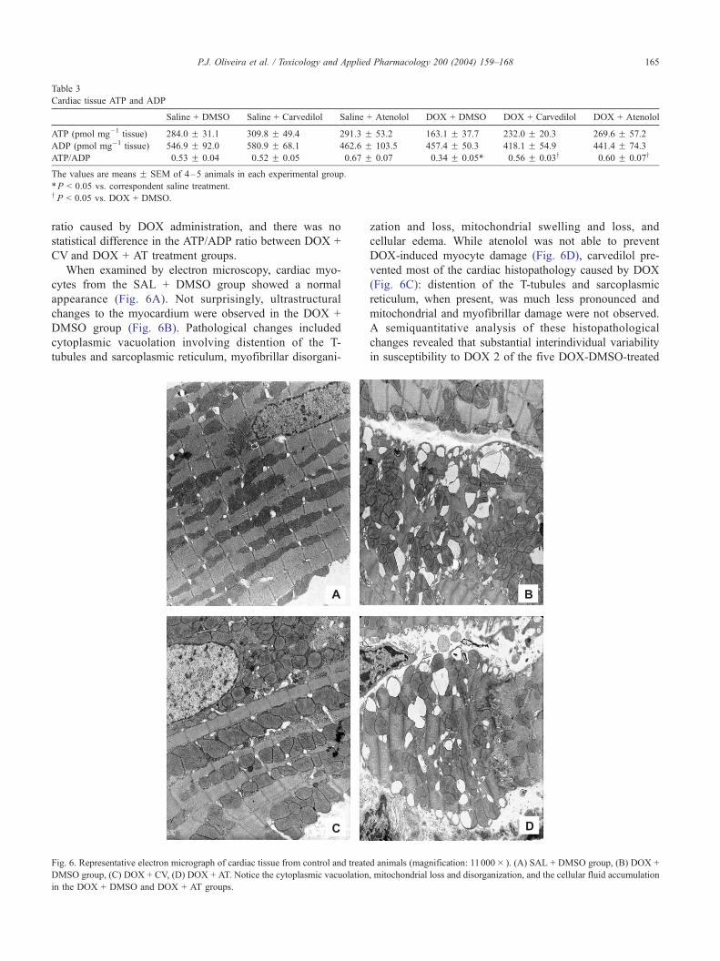
Table 3
Cardiac tissue ATP and ADP
Saline + DMSO Saline + Carvedilol Saline + Atenolol DOX + DMSO DOX + Carvedilol DOX + Atenolol
ATP (pmol mg�1 tissue) 284.0 F 31.1 309.8 F 49.4 291.3 F 53.2 163.1 F 37.7 232.0 F 20.3 269.6 F 57.2
ADP (pmol mg�1 tissue) 546.9 F 92.0 580.9 F 68.1 462.6 F 103.5 457.4 F 50.3 418.1 F 54.9 441.4 F 74.3
ATP/ADP 0.53 F 0.04 0.52 F 0.05 0.67 F 0.07 0.34 F 0.05* 0.56 F 0.03y 0.60 F 0.07y
The values are means F SEM of 4–5 animals in each experimental group.
*P < 0.05 vs. correspondent saline treatment.y P < 0.05 vs. DOX + DMSO.
P.J. Oliveira et al. / Toxicology and Applied Pharmacology 200 (2004) 159–168 165
ratio caused by DOX administration, and there was no
statistical difference in the ATP/ADP ratio between DOX +
CV and DOX + AT treatment groups.
When examined by electron microscopy, cardiac myo-
cytes from the SAL + DMSO group showed a normal
appearance (Fig. 6A). Not surprisingly, ultrastructural
changes to the myocardium were observed in the DOX +
DMSO group (Fig. 6B). Pathological changes included
cytoplasmic vacuolation involving distention of the T-
tubules and sarcoplasmic reticulum, myofibrillar disorgani-
Fig. 6. Representative electron micrograph of cardiac tissue from control and treate
DMSO group, (C) DOX + CV, (D) DOX + AT. Notice the cytoplasmic vacuolation
in the DOX + DMSO and DOX + AT groups.
zation and loss, mitochondrial swelling and loss, and
cellular edema. While atenolol was not able to prevent
DOX-induced myocyte damage (Fig. 6D), carvedilol pre-
vented most of the cardiac histopathology caused by DOX
(Fig. 6C): distention of the T-tubules and sarcoplasmic
reticulum, when present, was much less pronounced and
mitochondrial and myofibrillar damage were not observed.
A semiquantitative analysis of these histopathological
changes revealed that substantial interindividual variability
in susceptibility to DOX 2 of the five DOX-DMSO-treated
d animals (magnification: 11000� ). (A) SAL + DMSO group, (B) DOX +
, mitochondrial loss and disorganization, and the cellular fluid accumulation
Table 4
Cardiac histopathology for rats treated with doxorubicin and either carvedilol or atenolol
Saline + DMSO DOX + DMSO DOX + Carvedilol DOX + Atenolol
Extent of cell damagea 0.00 F 0.00 9.20 F 7.98 2.60 F 1.52 2.80 F 1.10
Severity of cell damageb 0.00 F 0.00 2.40 F 0.55 1.40 F 0.55* 1.60 F 0.89
a Extent of cell damage represents the mean (FSD) number of damaged myocytes in a 202500 Am2 area.b Severity of cell damage is expressed on a blinded scale of 0–3.
*P < 0.05, significantly different from doxorubicin treatment (DOX + DMSO) (n = 5).
P.J. Oliveira et al. / Toxicology and Applied Pharmacology 200 (2004) 159–168166
rats expressed little, if any, histopathology (Table 4). Ac-
cordingly, when analyzed from the extent of cardiac injury,
the experimental variability in the DOX-DMSO treatment
group precluded statistical significance for cardioprotection
by either carvedilol or atenolol. However, when analyzed by
the severity of cardiac injury, carvedilol afforded statistically
significant cardioprotection against DOX-induced cardiac
histopathology, whereas atenolol did not (Table 4).
Discussion
It is very well established that DOX, by stimulating free
radical generation on the electron transport chain, induces
mitochondrial dysfunction, particularly in the myocardium
(Santos et al., 2002; Sokolove and Shinaberry, 1988; Solem
et al., 1994, 1996; Zhou et al., 2001a, 2001b). The present
investigation confirms this mitochondrionopathy as
reflected by the substantially decreased calcium loading
capacity and increased sensitivity to calcium-induced swell-
ing of cardiac mitochondria isolated from DOX-injected rats
(Figs. 1–3), which is the same as that reported previously
from this laboratory (Santos et al., 2002; Solem et al., 1994).
However, unlike previous investigations, cardiac mitochon-
dria from DOX-treated rats did not exhibit a depression of
respiratory activities, despite receiving the same treatment
regimen. In reflecting on the source of this difference, a few
possibilities were identified. It may be because in this
particular investigation, the mitochondrial pellets were more
extensively washed than in previous studies, yielding a
more homogenous preparation containing fewer light mito-
chondria than in prior investigations. Alternatively, it may
be because the current investigation included the calcium-
chelator EGTA (10 AM) in the reaction buffer. We have
previously shown that the effects of DOX on mitochondrial
respiration are mediated through a calcium-dependent in-
duction of the mitochondrial permeability transition, which
is suppressed in the presence of EGTA. Regardless, we
interpret the present results as supporting the contention that
measurement of respiration is not a sensitive and definitive
indicator of mitochondrial dysfunction. Instead, it is the
decrease in calcium-loading capacity and loss of mitochon-
drial calcium homeostasis that is an early, more sensitive,
and consistent indication of mitochondrial dysfunction
caused by drug treatment (Zhou et al., 2001b).
DOX-induced mitochondrial cardiomyopathy is charac-
terized by the accumulation of oxidized lipids, proteins, and
DNA, mitochondrial dysfunction leading to a systolic fail-
ure, and extensive cardiac tissue histopathology character-
ized by disorganization of the sarcomeric structure, large
vacuole inclusions, and mitochondrial swelling with exten-
sive fractionation of the cristae (Iwasaki and Suzuki, 1991;
Papadopoulou et al., 1999; Santos et al., 2002; Zhou et al.,
2001b). As was shown previously (Santos et al., 2002),
carvedilol restored mitochondrial calcium loading capacity
and tissue morphology in DOX-injected animals. The nov-
elty in this investigation is that atenolol, which does not
possess antioxidant activity, was without effect against
DOX-induced cardiac mitochondrial toxicity and histopa-
thology. Atenolol did not prevent the DOX-induced de-
crease in mitochondrial calcium loading capacity or
calcium-induced mitochondrial swelling, or the cardiac
histopathology observed after repeated DOX treatments.
The results are strong evidence that it is the antioxidant
effect, rather than the h-adrenoceptor blockage activity, thatis responsible for the cardioprotective action of carvedilol.
The interference with mitochondrial calcium homeosta-
sis and mitochondrial swelling caused by DOX has been
attributed to induction of the mitochondrial permeability
transition, MPT (Solem et al., 1996; Zhou et al., 2001a,
2001b). Induction of the MPT occurs in response to
numerous conditions and is suggested to reflect the trans-
formation of a specific pore from a low-conductance to a
high-conductance state that is permeable to solutes up to
1500 Da (Bernardi, 1999). Numerous chemicals have been
demonstrated to induce this transition, the majority of
which are either direct or indirect-acting oxidants. Petro-
nilli et al. (1994) and Chernyak and Bernardi (1996)
provide evidence for the regulation of the MPT by the
redox state of critical cysteinyl thiols associated with the
pore complex. Our results confirm MPT inhibitory activity
of carvedilol demonstrated in vitro and ascribed to the
potent antioxidant properties of the drug (Oliveira et al.,
2001). The significance of the current investigation is the
demonstration of protection against the MPT by carvedilol
in vivo, which we attribute the cardioprotection afforded
by carvedilol to its antioxidant, as opposed to h-adrenergicantagonist properties.
DOX was also previously shown to decrease cardiac
tissue ATP/ADP ratio (Chacon et al., 1992), which was
confirmed by the present study (Table 3). Interestingly, both
carvedilol and atenolol exhibited an energy sparing effect. It
may be that by reducing heart rate, non-selective h-adren-ergic receptor antagonists limit cardiac energy expenditure
P.J. Oliveira et al. / Toxicology and Applied Pharmacology 200 (2004) 159–168 167
and thereby exhibit this energy saving effect (Abiko et al.,
1994), which occurs independently of any antioxidant
properties. Accordingly, by reducing heart rate, atenolol
decreases ATP consumption demands and thereby preserves
the high-energy phosphate charge of the cell, although
mitochondrial ATP synthesis may be partially compromised
by the DOX treatment. Despite the maintenance of the ATP/
ADP ratio by both carvedilol and atenolol, only carvedilol
was able to preserve mitochondrial function and myocardial
ultrastructure, suggesting that maintenance of normal energy
levels does not prevent per se the histopathology caused by
DOX. Failure of AT in counteracting DOX cardioselective
toxicity is unlikely to be due to deficient dose selection. In
fact, as previously described, clinical trials have demon-
strated equipotent effects of CV and AT regarding reduction
of blood pressure in humans (McTavish et al., 1993; Hall et
al., 1991).
In conclusion, the present investigation extends earlier
reports regarding the protective actions of carvedilol against
the DOX-induced cardiotoxicity by demonstrating that this
protective effect is due mainly to its antioxidant, and not h-adrenergic properties. Consequently, efforts to engineer new
cardioprotective agents should focus on maximizing the
antioxidant and not necessarily the autonomic properties
of the drug. The results offer important substantiation for
using this pharmacologically active agent as an adjunct to
minimize the mitochondrial cardiomyopathy that limits the
therapeutic success of DOX antineoplastic therapy.
Acknowledgments
This work was supported by the NIH grant HL 58016.
Paulo J. Oliveira is supported by a grant from the
Portuguese Foundation for Science and Technology
(PRAXIS XXI/BD/21494/99). The expert assistance of
Jamie Denninger, Jessica Berthiaume, and Tim O’Brien is
greatly acknowledged.
References
Abiko, Y., Gotoh, H., Yokoyama, T., Abiko, T., Hashizume, H., Akiyama,
K., 1994. Bopindolol and its metabolite 18-053 attenuate regional myo-
cardial acidosis during partial occlusion of the coronary artery in dogs.
Arch. Int. Pharmacodyn. Ther. 327, 40–55.
Bernardi, P., 1999. Mitochondrial transport of cations: channels exchangers
and permeability transition. Physiol. Rev. 79, 1127–1155.
Bradford, M.M., 1976. A rapid and sensitive method for the quantitation of
microgram quantities of protein utilizing the principle of protein-dye
binding. Anal. Biochem. 72, 248–254.
Broekemeier, K.M., Dempsey, M.E., Pfeiffer, D.R., 1989. Cyclosporin a is
a potent inhibitor of the inner membrane mitochondrial transition in
liver mitochondria. J. Biol. Chem. 264, 7826–7830.
Chacon, E., Ulrich, R., Acosta, D., 1992. A digitized-fluorescence-imaging
study of mitochondrial Ca2+ increase by doxorubicin in cardiac myo-
cytes. Biochem. J. 281, 871–878.
Chernyak, B.V., Bernardi, P., 1996. The mitochondrial permeability tran-
sition pore is modulated by oxidative agents through both pyridine
nucleotides and glutathione at two separate sites. Eur. J. Biochem.
238, 623–630.
DeAtley, S.M., Aksenov, M.Y., Aksenova, M.V., Harris, B., Hadley, R.,
Harper, P.C., Carney, J.M., Butterfield, D.A., 1999. Antioxidants pro-
tect against reactive oxygen species associated with adriamycin-treated
cardiomyocytes. Cancer Lett. 136, 41–46.
Demant, E.J.F., 1991. Inactivation of cytochrome c oxidase activity in
mitochondrial membranes during redox cycling of doxorubicin. Bio-
chem. Pharmacol. 41, 543–552.
Doroshow, J.H., 1983. Anthracycline antibiotic-stimulated superoxide, hy-
drogen peroxide, and hydroxyl radical production by NADH dehydro-
genase. Cancer Res. 43, 4543–4551.
Estabrook, R.W., 1967. Mitochondrial respiratory control and the polaro-
graphic measurement of ADP:O ratios. Methods Enzymol. 10, 41–47.
Hall, S., Prescott, R.I., Hallman, R.J., Dixon, S., Harvey, R.E., Ball, R.G.,
1991. A comparative study of carvedilol, slow release nifedipine, and
atenolol in the management of essential hypertension. J. Cardiovasc.
Pharmacol. 18 (Suppl. 4), S35–S38.
Hrdina, R., Gersl, V., Klimtova, I., Simunek, T., Machackova, J., Adam-
cova, M., 2000. Anthracycline-induced cardiomyopathy. Acta Med. 43,
75–82.
Iwasaki, T., Suzuki, T., 1991. Ultrastructural alterations of the myocardium
induced by doxorubicin. Virchows Arch. B Cell Pathol. 60, 35–39.
Jones, D.P., 1981. Determination of pyridine dinucleotides in cell extracts
by high-performance liquid chromatography. J. Chromatography 225,
446–449.
Kamo, N., Muratsugu, M., Hongoh, R., Kobatake, Y., 1979. Membrane
potential of mitochondria measured with an electrode sensitive to tetra-
phenyl phosphonium and relationship between proton electrochemical
potential and phosphorylation potential in steady state. J. Membr. Biol.
49, 105–121.
Kotsinas, A., Gorgoulis, V., Zacharatos, P., Zioris, H., Triposkiadis, F.,
Donta, I., Kyriakidis, M., Karayannacos, P., Kittas, C., 1999. Antioxi-
dant agent nimesulid and h-blocker metoprolol do not exert protective
effects against rat mitochondrial DNA alterations in adriamycin-induced
cardiotoxicity. Biochem. Biophys. Res. Commun. 254, 651–656.
Kowaltowski, A.J., Vercesi, A.E., 1999. Mitochondrial damage induced by
conditions of oxidative stress. Free Radical Biol. Med. 26, 463–471.
Lee, V., Randhawa, A., Singal, P.K., 1991. Adriamycin-induced myocardial
dysfunction in vitro is mediated by free radicals. Am. J. Physiol. 261,
H989–H995.
Levine, R.L., Garland, D., Oliver, C.N., Amici, A., Climent, I., Lenz, A.-G.,
Ahn, B.-W., Shaltiel, S., Stadtman, E.R., 1990. Determination of car-
bonyl content in oxidatively modified proteins. Methods Enzymol. 186,
464–478.
Matsui, H., Morishima, I., Numaguchi, Y., Toki, Y., Okumura, K., Haya-
kawa, T., 1999. Protective effects of carvedilol against doxorubicin-
induced cardiomyopathy in rats. Life Sci. 65, 1265–1274.
McTavish, D., Campoli-Richards, D., Sorkin, E.M., 1993. Carvedilol, a
review of its pharmacodynamic and pharmacokinetic properties, and
therapeutic efficacy. Drugs 45 (2), 232–258.
Mimnaugh, E.G., Trush, M.A., Bhatnagar, M., Gram, T.E., 1985. Enhance-
ment of reactive oxygen-dependent mitochondrial membrane lipid per-
oxidation by the anticancer drug adriamycin. Biochem. Pharmacol. 34,
847–856.
Monti, E., Cova, D., Guido, E., Morelli, R., Oliva, C., 1996. Protective
effect of the nitroxide tempol against the cardiotoxicity of adriamycin.
Free Radical Biol. Med. 21, 463–470.
Noguchi, N., Nishino, K., Niki, E., 2000. Antioxidant action of the anti-
hypertensive drug, carvedilol, against lipid peroxidation. Biochem.
Pharmacol. 59, 1069–1076.
Oliveira, P.J., Santos, D.L., Moreno, A.J.M., 2000. Carvedilol inhibits the
exogenous NADH dehydrogenase in rat heart mitochondria. Arch. Bio-
chem. Biophys. 374, 279–285.
Oliveira, P.J., Coxito, P.M., Rolo, A.P., Santos, D.L., Palmeira, C.M., Mo-
reno, A.J.M., 2001. Inhibitory effect of carvedilol in the high-conduc-
P.J. Oliveira et al. / Toxicology and Applied Pharmacology 200 (2004) 159–168168
tance state of the mitochondrial permeability transition pore. Eur. J.
Pharmacol. 412, 231–237.
Olson, R.D., Mushlin, P.S., 1990. Doxorubicin cardiotoxicity: analysis of
prevailing hypotheses. FASEB J. 4, 3076–3086.
Palmeira, C.M., Serrano, J., Kuehl, D.W., Wallace, K.B., 1997. Preferential
oxidation of cardiac mitochondrial DNA following acute intoxication
with doxorubicin. Biochim. Biophys. Acta 1321, 101–106.
Papadopoulou, L.C., Theophilidis, G., Thomopoulos, G.N., Tsiftsoglou,
A.S., 1999. Structural and functional impairment of mitochondria in
adriamycin-induced cardiomyopathy in mice: suppression of cyto-
chrome c oxidase II gene expression. Biochem. Pharmacol. 57,
481–489.
Petronilli, V., Constantini, P., Scorrano, L., Colonna, R., Passamonti, S.,
Bernardi, P., 1994. The voltage sensor of the mitochondrial permeability
transition pore is tuned by the oxidation-reduction state of vicinal thiols.
J. Biol. Chem. 269, 16638–16642.
Prohaska, J.R., 1983. Changes in tissue growth, concentrations of copper,
iron, cytochrome oxidase and superoxide dismutase subsequent to die-
tary or genetic copper deficiency in mice. J. Nutr. 113, 2048–2058.
Rajdev, S., Reynolds, I.J., 1993. Calcium Green-5N, a novel fluorescent
probe for monitoring high intracellular free Ca2+ concentrations asso-
ciated with glutamate excitotoxicity in cultured rat brain neurons. Neu-
rosci. Lett. 162, 149–152.
Santos, D.L., Moreno, A.J.M., Leino, R.L., Froberg, M.K., Wallace, K.B.,
2002. Carvedilol protects against doxorubicin-induced mitochondrial
cardiomyopathy. Toxicol. Appl. Pharmacol. 185, 218–227.
Sokolove, P.M., Shinaberry, R.G., 1988. Na+-independent release of Ca2+
from rat heart mitochondria-induction by adriamycin aglycone. Bio-
chem. Pharmacol. 37, 803–812.
Solem, L.E., Henry, T.R., Wallace, K.B., 1994. Disruption of mitochondrial
calcium homeostasis in vivo following chronic doxorubicin administra-
tion. Toxicol. Appl. Pharmacol. 129, 214–222.
Solem, L.E., Heller, L.J., Wallace, K.B., 1996. Dose-dependent increase
in sensitivity to calcium-induced mitochondrial dysfunction and car-
diomyocyte cell injury by doxorubicin. J. Mol. Cell. Cardiol. 28,
1023–1032.
Van Vleet, J.F., Ferrans, V.J., Weirich, W.E., 1980. Cardiac disease induced
by chronic adriamycin administration in dogs and evaluation of vitamin
E and selenium as cardioprotectants. Am. J. Pathol. 99, 13–42.
Xu, M.F., Tang, P.L., Qian, Z.M., Ashraf, M., 2001. Effects by doxorubicin
on the myocardium are mediated by oxygen free radicals. Life Sci. 68,
889–901.
Yin, X., Wu, H., Chen, Y., Kang, Y.J., 1998. Induction of antioxidants by
adriamycin in mouse heart. Biochem. Pharmacol. 56, 87–93.
Yue, T.-L., McKenna, P.J., Ruffolo Jr., R.R., Feuerstein, G., 1992. Carve-
dilol, a new h-adrenoceptor antagonist and vasodilator antihypertensive
drug, inhibits superoxide release from human neutrophils. Eur. J. Phar-
macol. 214, 277–280.
Zhou, S., Heller, L.J., Wallace, K.B., 2001a. Interference with calcium-
dependent mitochondrial bioenergetics in cardiac myocytes isolated
from doxorubicin-treated rats. Toxicol. Appl. Pharmacol. 175, 60–67.
Zhou, S., Starkov, A., Froberg, M.K., Leino, R.L., Wallace, K.B., 2001b.
Cumulative and irreversible cardiac mitochondrial dysfunction induced
by doxorubicin. Cancer Res. 61, 771–777.


