Embed Size (px)
Citation preview

CdSe Quantum Dots for Two-PhotonFluorescence Thermal ImagingLaura Martinez Maestro,† Emma Martın Rodríguez,† Francisco Sanz Rodríguez,‡M. C. Iglesias-de la Cruz,‡ Angeles Juarranz,‡ Rafik Naccache,| Fiorenzo Vetrone,§Daniel Jaque,† John A. Capobianco,*,| and Jose Garcıa Sole*,†
†Fluorescence Imaging Group, Departamento de Fısica de Materiales, C-IV, Universidad Autonoma de Madrid,C/Francisco Tomas y Valiente 7, Madrid 28049, Spain, ‡Departamento de Biologıa, Universidad Autonoma deMadrid, Madrid 28049, Spain, § Institut National de la Recherche Scientifique - Energie, Materiaux etTelecommunications, Universite du Quebec, Varennes, QC J3X 1S2, Canada, and |Department of Chemistry andBiochemistry, Concordia University, 7141 Sherbrooke Street W, Montreal, QC H4B 1R6, Canada
ABSTRACT The technological development of quantum dots has ushered in a new era in fluorescence bioimaging, which was propelledwith the advent of novel multiphoton fluorescence microscopes. Here, the potential use of CdSe quantum dots has been evaluated asfluorescent nanothermometers for two-photon fluorescence microscopy. In addition to the enhancement in spatial resolution inherentto any multiphoton excitation processes, two-photon (near-infrared) excitation leads to a temperature sensitivity of the emissionintensity much higher than that achieved under one-photon (visible) excitation. The peak emission wavelength is also temperaturesensitive, providing an additional approach for thermal imaging, which is particularly interesting for systems where nanoparticlesare not homogeneously dispersed. On the basis of these superior thermal sensitivity properties of the two-photon excited fluorescence,we have demonstrated the ability of CdSe quantum dots to image a temperature gradient artificially created in a biocompatible fluid(phosphate-buffered saline) and also their ability to measure an intracellular temperature increase externally induced in a single living cell.
KEYWORDS Nanothermometry, two-photon microscopy, quantum dots, HeLa cancer cell, thermal sensing, fluorescence imaging
Thermal sensing at the micro- and nanoscales is re-quired for high spatial resolution of temperaturegradients and is an indispensible tool for dynamical
studies of diverse small systems including electrical, photo-nic, and biological ones. Nanotechnology is providing fas-cinating approaches to solve this requirement by means ofusing nanothermal probes whose properties display a strongdependence on the local temperature.1 In these “nanother-mometers”, the thermal sensing method relies upon theparticular parameter that displays a temperature depen-dence, which gives rise to high resolution thermal imagingtechniques, such as scanning thermal and molecular fluo-rescence polarization microscopies.2-8
Fluorescent materials are particularly relevant in nano-thermometry since they operate as “non-contact” thermom-eters and can provide the dual function of imaging andtemperature sensing at the nanoscale.9 These multimodalnanothermometers constitute an excellent tool to investigatefluid systems and particularly biomedical systems, as thenanoprobes can be homogeneously dispersed in the entiremedium with minimal interaction.10 Fluorescent nanother-mometers are based on the temperature dependence of thefluorescence features (intensity, band shape, peak position,Stokes shift, or lifetime). In this respect, organic dyes were
the first exogenous multimodal fluorescent “molecularprobes” used for imaging as well as measuring the temper-ature of single living cells.11 However, organic probes areeasily damaged with the ultraviolet-visible excitation light(photobleaching).12 Thus, novel fluorescent probes weredeveloped, which are based on nanoscale inorganic materi-als (nanoparticles) and are resistant to photobleaching whileshowing high thermal sensitivity.13-22 With the commercialavailability of femtosecond (fs) lasers, these nanoprobeswere shown to produce significant emitted light under near-infrared (NIR) two-photon excitation, that is, after almostsimultaneous absorption of two excitation photons.23-26
The recent development of two-photon fluorescent micro-scopes has spurred research on two-photon excitation nano-particles since they provide a much better spectral andspatial resolution than the traditional one-photon micro-scopes.27
Quantum dots (hereafter QDs) are by far the most usedfluorescent nanoprobes for fluorescence imaging.28,29 Thisis essentially due to well-known materials engineering meth-ods to synthesize semiconductor nanocrystals with thedesired QD size and homogeneous size distribution. Thisallows for the selection of the most convenient emitted colorby means of size confinement. Moreover, the surface of theseQDs can be suitably engineered to be monodispersible in fluidsas well as to target specific cells like, for instance, a cancer cell,while having a relatively good biocompatibility.30 An additionaladvantage is that they can be efficiently two-photon excited
* To whom correspondence should be addressed.Received for review: 10/15/2010Published on Web: 11/09/2010
pubs.acs.org/NanoLett
© 2010 American Chemical Society 5109 DOI: 10.1021/nl1036098 | Nano Lett. 2010, 10, 5109–5115

in the NIR to produce visible luminescence.26 Indeed, allthese advantages have led to the commercialization of QDbiolabels at the present time. Furthermore, it has beenpreviously reported that the intensity and peak wavelengthof the QDs are influenced by the environmental tempera-ture, thus giving rise to the opportunity of using them asfluorescent nanoprobes for fluorescence thermal imag-ing.16,17,31-35 However, the reported images were acquiredby means of one-photon excitation and thus, present limitedapplicability to high-resolution experiments (like tempera-ture gradients) and particularly, to biomedical imaging,where multiphoton excitation is essential.
In this work, we have evaluated the ability of CdSe-QDsto act as fluorescent nanothermometers to realize thermalimaging by means of the modern multiphoton fluorescence-imaging microscopes. We have initially evaluated the fluo-rescence response of CdSe-QDs for thermal detection underboth one-photon and two-photon excitation by systemati-cally recording their emission spectra as a function oftemperature in the physiological temperature range. Thedifferent excitation methods lead to diverse thermal behav-iors and the fluorescence parameters that can be used forthermal sensing have been determined and calibrated. Oncethe CdSe-QDs have been fully characterized as two-photonexcited fluorescent thermal nanoprobes, they have beenused to determine the thermal gradients induced in a liquidby a focused laser beam. Finally, we demonstrate thepotential of these two-photon excited nanothermometers inbiomedical imaging by measuring the temperature evolutionof a single HeLa cancer cell. To the best of our knowledge,these constitute the first two-photon excited thermal imagesobtained using CdSe-QDs.
CdSe spherical nanoparticles of 4 nm in diameter fromInvitrogen Inc. (Qtracker 655) were dispersed in phosphatebuffered saline (PBS) at a concentration of 0.3 µM. Thesolution was placed in an open oven so that its temperaturecould be varied in the physiological range between 30 to 70°C with an accuracy of 1 °C. One-photon excitation wasachieved by using a continuous wave air cooled Argon laser(Spectra Physics 177-G02, 488 nm, and 10 mW outputpower). This wavelength lies just below the absorption edge(not included for the sake of brevity) of the CdSe-QDs/PBSsolution thus ensuring one-photon excitation. Moreover, weverified that this absorption edge is almost temperatureindependent and so no differences should be expected inthe temperature dependence of the emission when usingdifferent one-photon excitation wavelengths. In fact, weexamined this by exciting at 377 nm (pulsed N2 laser) andit led to the same results as for 488 nm excitation. As two-photon fluorescence microscopes make use of fs-pulsedexcitation laser sources, we used a mode-locked Ti:Sapphirelaser (Spectra Physics Tsunami, 100 fs pulse duration, 80 MHzrepetition rate, and 100 mW output power) for two-photonexcitation, which was set at 800 nm, the wavelength peak inthe two-photon excitation spectrum (not shown). Both excita-
tion beams were focused into the solution containing the QDsby using a 10× optical microscope objective.
Figure 1 (left side) presents digital images of the CdSe-QDs/PBS solution obtained at room temperature under bothone-photon and two-photon excitation. It can be clearly seenthat the active fluorescent volume is reduced by two-photonillumination, as this excitation becomes efficient only at thefocus of the illumination beam (where the highest photondensities are achieved) and leads to a higher confinementof the fluorescence with respect to one-photon excitation.This higher confinement allows for the achievement ofhigher spatial resolutions (below the illuminating wave-length) that is required for the imaging of micro- andnanosystems.27 Moreover, two-photon excitation allows forthe generation of visible luminescence under NIR illumina-tion, offering additional advantages for bioimaging ap-plications, such as minimization of autofluorescence,increase of penetration depths and reduction of laserinduced tissue damage.36-38 Thus, a significant improve-ment in contrast and spatial resolution of the obtainedthermal images would be achieved under two-photon NIRexcitation of the CdSe-QDs.
Figure 1 (right side) also shows the emission spectra ofthe CdSe-QDs/PBS solution at three different temperatures(30, 45, and 60 °C) as obtained under both one-photon andtwo-photon excitation. It is clear that in both cases the peakfluorescence wavelength and the integrated emitted inten-sity are strongly influenced by temperature. Independent ofthe excitation mechanism, a temperature increase causes ared shift and, simultaneously, a reduction in the opticalconversion efficiency (more remarkable under two-photon
FIGURE 1. (Left side) Digital images of the CdSe-QDs/PBS solutionobtained at room temperature under one-photon (top) and two-photon (bottom) optical excitation, respectively. (Right side) Emis-sion spectra of the CdSe-QDs/PBS solution at three different tem-peratures (30, 45, and 60 °C) as obtained under one-photon (top)and two-photon (bottom) excitation, respectively.
© 2010 American Chemical Society 5110 DOI: 10.1021/nl1036098 | Nano Lett. 2010, 10, 5109-–5115

excitation). The simultaneous existence of these two tem-perature dependent fluorescence parameters allows forCdSe-QDs to be used as “dual thermal sensors” capable oftemperature measurements by two complementary meth-ods. As will be shown below, this fact allows for thermalimaging in systems with nonhomogeneous particle distribu-tions (such as living cells).
For thermal imaging applications, an accurate calibrationof the spectral features that will be used as thermal indicatorsis required. For this purpose, the integrated emitted intensityand peak wavelength have been systematically investigatedas a function of temperature (shown in Figure 2). As can beobserved, the thermally induced spectral red shift has beenfound to be, within experimental uncertainty, independentof the number of photons involved during excitation. In bothone-photon and two-photon experiments, the peak emissionwavelength variation as a function of temperature essentiallyfollowed a linear relationship. Thus, from a linear fit we haveestimated a variation coefficient of 0.16 nm/°C, which iscomparable to previously reported values in different QDs(ranging from 0.08 to 0.2 nm/°C).17,34,35 This temperature-related shift obeys a variety of phenomena, including thethermally induced variation of the bandgap energy of theQDs, quantum yield of the emitting levels, thermal expan-sion of the QDs as well as the thermally induced variationof the solvent’s refractive index.34,39,40 The relative contribu-tion of each phenomenon has been previously estimated inthe case of CdSe-QDs, concluding that the thermally inducedvariation of the bandgap energy is the dominant one.34 Thus,it is expected that the observed thermally induced spectralshift will be independent of the particular character of theexcitation process, as observed in Figure 2a.
At variance with the thermally induced spectral shift, thereduction of the fluorescence efficiency as a function oftemperature has been found to follow different trends for
one-photon and two-photon excitations (see Figure 2b). Forone-photon excitation, the fluorescence suffers only a mod-erate reduction (25% reduction for a temperature incrementof 30 °C), whereas for two-photon excitation this reductionis three times larger (75% for the same temperature incre-ment). According to previous studies, the temperature-induced decrease of the one-photon excited QD emissioncan be attributed to the thermal activation of surface trapstates as well as to a thermally-induced increase in thenonradiative exciton recombination probability.41,42 Follow-ing this argument we can estimate, from the one-photonexcitation data, that the fluorescence quantum efficiency(Φf) of CdSe-QDs is reduced by a factor of 0.25 when thetemperature is increased from 30 to 60 °C. The thermallyinduced reduction of the two-photon excited emission in-tensity requires a different analysis. This emission intensity(I2) is proportional to the product of the two-photon absorp-tion cross section (σ2) and the fluorescence quantum ef-ficiency, so that I2 R Φf · σ2.26,43 The temperature depen-dence of the two-photon excited emission is, therefore,determined by thermally induced variations of both thefluorescence quantum efficiency and the two-photon ab-sorption cross section. From the data obtained under one-photon excitation, we have estimated a 25% reduction ofthe fluorescence quantum efficiency, which is almost a thirdof the net reduction observed in the two-photon excitedemission intensity. This fact suggests that the two-photonabsorption cross section in CdSe-QDs is also strongly tem-perature dependent, playing in fact a dominant role in thetwo-photon excited fluorescence quenching. Independentlyof the dominant mechanism for thermal quenching of thetwo-photon excited emission, its behavior is of great rel-evance for thermal imaging purposes since it leads to a muchhigher thermal sensitivity than for one-photon excitation.Thus, this enhanced thermal sensitivity together with theenhanced spatial resolution of two-photon excited fluores-cence make these CdSe-QDs nanoprobes particularly suit-able for high-resolution nanothermometry.
After we investigated the temperature influence on thetwo-photon excited emission features of the CdSe-QDs/PBSsolution, the next step was to demonstrate its use for two-photon fluorescence thermal imaging. For this purpose, wehave performed imaging experiments in two different fluidsystems.
First, by means of a pump-probe experiment we haveartificially created a light-induced thermal gradient in oursolution and this gradient has been properly imaged by usingthe temperature dependence of the CdSe-QD emissionintensity (Figure 2b). A continuous wave 980 nm laser beam(the pump) was focused into the solution. This is partiallyabsorbed by the PBS solvent but not by the QDs and theabsorbed energy is subsequently delivered as heat. As theabsorption profile is not homogeneous, greater heating isexpected at the focus compared to the surrounding area,thus a temperature gradient should be created in the solu-
FIGURE 2. Temperature variation of the peak emission wavelength(top) and integrated emitted intensity (bottom) as obtained from theCdSe-QDs/PBS solution under one and two-photon excitation.
© 2010 American Chemical Society 5111 DOI: 10.1021/nl1036098 | Nano Lett. 2010, 10, 5109-–5115

tion. This gradient is imaged using two-photon fluorescencemicroscopy of the QDs, which are resonantly excited by anorthogonal probe laser beam.
The 980 nm laser pump beam was focused down to aspot size of 200 µm. Under these conditions the maximumdensity achieved in the PBS solution was estimated to belower than 0.8 kW/cm2. For such low intensities the densityfluctuations due to optical forces can be neglected and thuswe can consider that the spatial distribution of the QDswithin the solution is homogeneous. This aspect makes itpossible to use the emitted intensity of the QDs to imagethe created thermal gradient. For this purpose, the locallyheated CdSe-QDs/PBS solution was incorporated into a fiber-coupled confocal microscope. The luminescence of theCdSe-QDs was then two-photon excited by means of a 100fs, 800 nm excitation beam (the probe) focused into thesolution orthogonally to the 980 nm pump beam. The probebeam was focused with a 10× microscope objective, whichwas also used to collect the 650 nm generated luminescenceof the CdSe-QDs. The Numerical Aperture of the 10×microscope objective was 0.25 from which we have esti-mated a spatial (lateral) resolution close to 2.5 µm. Afterpassing through confocal apertures, the two-photon excitedluminescence was analyzed by means of a fiber coupled highresolution spectrometer. The XY motorized stage on whichthe solution was mounted allowed us to scan the 800 nmprobe beam in the surroundings of the 980 nm pump focalpoint. Figure 3a shows the spatial variation of the two-photon excited emission intensity. In this image, the dottedblack lines schematically represent the 980 nm beam path.As it can be observed, the emitted intensity is stronglyreduced at focus. According to the data of Figure 2, this can
be attributed to the achievement of higher temperatures atfocus (as it was indeed expected). The data presented inFigure 2b allows for the calibration of this intensity imagein terms of temperature variations. Thus, the correspondingthermal gradient image is presented in Figure 3b anddemonstrates that the temperature at focus has been raisedby almost 20 °C. Because of the large integration timesrequired in our experimental setup for the acquisition of thetwo-photon excited spectra with low signal-to-noise ratio(above 10 s per spectrum), the acquisition of thermal imagesbased on the temperature induced spectral shifts was notpossible. Nevertheless, it should be noted that thermalmeasurement of fluids cannot be only obtained through thetemperature-induced luminescence quenching of QDs butalso from their temperature-induced spectral shift. To ac-count for this, the emission spectra generated at the focusof the heating beam were measured for different 980 nmheating beam intensities. In Figure 3c, we have included thetwo-photon emission spectra generated for two differentpump intensities. It is clear that when the 980 nm beamintensity is increased both an emission intensity decreaseand a spectral shift to longer wavelengths are simultaneouslyinduced. Figure 3d shows the induced spectral peak shift andluminescence quenching as a function of the pump beamintensity. For the largest pump intensity used in this work,a maximum spectral shift of 3.9 nm and a relative intensityreduction of 52% have been found. Thus, the maximumtemperature increment induced at focus has been found tobe close to 24 °C, when calculated based on the spectralshift, and 20 °C, when calculated from the observed emis-sion intensity reduction. Therefore, the discrepancy betweenthese two alternative methods has been found to be (2 °C,
FIGURE 3. (a) Spatial variation of the two-photon excited emission intensity as obtained from a CdSe-QDs/PBS solution in the presence of afocused 980 nm heating laser beam (its profile schematically drawn with the dashed lines). (b) Thermal image of the CdSe-QDs/PBS solutiondenoting the local temperature increment at the 980 nm beam focus. (c) Emission spectra from a CdSe-QDs/PBS solution at focus for twodifferent intensities of the pump (heating) beam. (d) Plots of the emission peak shift and emission intensity versus pump beam intensity atthe focus of this heating beam.
© 2010 American Chemical Society 5112 DOI: 10.1021/nl1036098 | Nano Lett. 2010, 10, 5109-–5115

that is, less than 10% of the measured value. The origin ofthis discrepancy could arise from the density fluctuation thatcan be generated in the fluid due to the local heating. Thus,the ability of CdSe-QDs to image thermal gradients of locallyheated liquids by two-photon excited fluorescence micros-copy is clearly demonstrated.
The CdSe-QDs were subsequently incorporated in HeLacervical cancer cells in order to investigate their thermo-metric ability in biological systems. For this purpose,immortalized human carcinoma cells were incubated for2 h with the CdSe-QD nanothermometers (using thesolution from Figure 1) and imaged by two-photon fluo-rescence microscopy. To confirm the successful internal-ization of the QDs into the cancer cells they were imagedwith a fast multiphoton microscope (Zeiss LSM510 mi-croscope), so that the images of several cells could beobtained. Figure 4 shows the optical transmission image(left column), the fluorescence image (middle column),and the superimposed images (right column) of a groupof HeLa cancer cells incubated in the absence (top row)and presence (bottom row) of QDs. Excitation and col-lection wavelengths were 800 and 650 nm, respectively.These images put into evidence a full incorporation of theQDs into the cancer cells as it has also been observed fortargeted gold nanorods.44 This was further confirmed bythe measurement of the cross-sectional multiphoton ex-cited fluorescence images of single cancer cells as well asby the analysis of the emission spectra generated whenthe NIR laser was focused inside and outside the cell.Moreover, it has been found that the multiphoton excitedemission spectrum generated from the cell well matchedthat obtained from the original QDs solutions (see Figure1). Thus, any possible contribution of autofluorescence to
the images in Figure 4 must be discarded. When the NIRexcitation beam was focused outside the cancer cells nofluorescence signal was observed. It is also clearly ob-served that the CdSe-QDs have been internalized by theHeLa cells but their distribution inside these cells isinhomogeneous. Thus, in this particular case it is moreappropriate to measure cell internal temperature changesby means of the spectral shift of the CdSe-QDs emission.At first glance it was observed that the peak shift wasessentially constant within the cell area where the nano-particles were incorporated in a sufficient concentrationto be detected by two-photon fluorescence imaging. Thisfact indicated that the temperature was essentially con-stant (25 °C) throughout the entire cell. Thus, to evaluatethe ability of the CdSe-QDs for detecting cell temperaturechanges, the cell temperature was externally varied byusing a microair-heater so that the cells could be exter-nally heated, as schematically shown in Figure 5 (leftside). The intracellular temperature increase due to thehot air flow caused a clear change in the two-photonexcited emission of the QDs incorporated inside the cell.This can be clearly observed when the emission spectrumwas recorded at heating times of 0 and 3 min (top rightside in Figure 5). In these experiments, the 800 nmexcitation beam was focused inside the cell by means ofa 100× microscope objective with a Numerical Apertureof 0.9 (giving a lateral spatial resolution close to 400 nm).Intracellular heating was evidenced by a clear red shift ofthe QDs emission band. By recording the two-photonexcited emission for different heating times and using thecalibration graph of Figure 2, we were able to measureintracellular temperature in real time. Results are shownin Figure 5 (bottom right side), which shows the intracel-
FIGURE 4. Room-temperature optical transmission images of HeLa cells nonincubated (top figures) and incubated with the CdSe-QDs/PBSsolution (bottom figures). (Left side) Images taken with the optical transmission microscope incorporated in the multiphoton fluorescencesetup. (Middle) Two-photon excited fluorescence images. (Right side) Superimposed (fluorescence and optical transmission) images.
© 2010 American Chemical Society 5113 DOI: 10.1021/nl1036098 | Nano Lett. 2010, 10, 5109-–5115
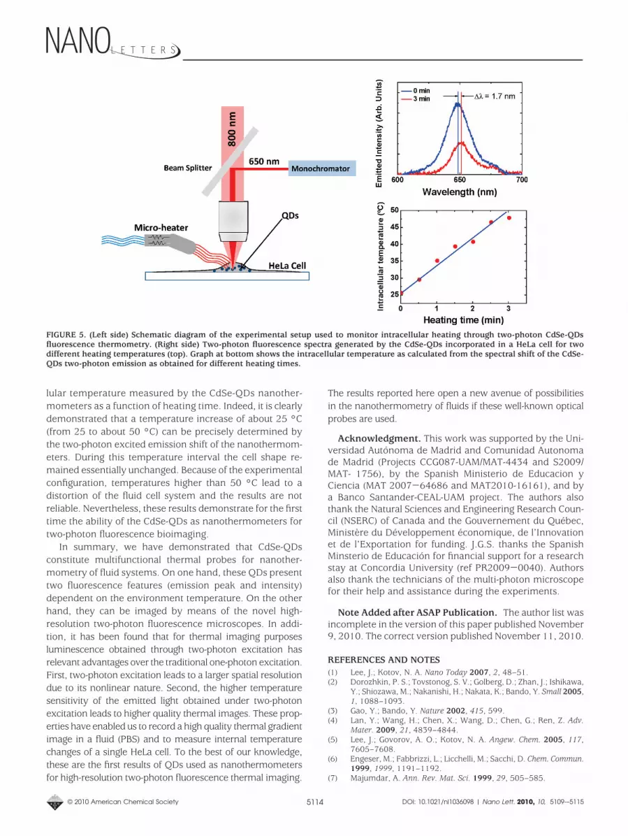
lular temperature measured by the CdSe-QDs nanother-mometers as a function of heating time. Indeed, it is clearlydemonstrated that a temperature increase of about 25 °C(from 25 to about 50 °C) can be precisely determined bythe two-photon excited emission shift of the nanothermom-eters. During this temperature interval the cell shape re-mained essentially unchanged. Because of the experimentalconfiguration, temperatures higher than 50 °C lead to adistortion of the fluid cell system and the results are notreliable. Nevertheless, these results demonstrate for the firsttime the ability of the CdSe-QDs as nanothermometers fortwo-photon fluorescence bioimaging.
In summary, we have demonstrated that CdSe-QDsconstitute multifunctional thermal probes for nanother-mometry of fluid systems. On one hand, these QDs presenttwo fluorescence features (emission peak and intensity)dependent on the environment temperature. On the otherhand, they can be imaged by means of the novel high-resolution two-photon fluorescence microscopes. In addi-tion, it has been found that for thermal imaging purposesluminescence obtained through two-photon excitation hasrelevant advantages over the traditional one-photon excitation.First, two-photon excitation leads to a larger spatial resolutiondue to its nonlinear nature. Second, the higher temperaturesensitivity of the emitted light obtained under two-photonexcitation leads to higher quality thermal images. These prop-erties have enabled us to record a high quality thermal gradientimage in a fluid (PBS) and to measure internal temperaturechanges of a single HeLa cell. To the best of our knowledge,these are the first results of QDs used as nanothermometersfor high-resolution two-photon fluorescence thermal imaging.
The results reported here open a new avenue of possibilitiesin the nanothermometry of fluids if these well-known opticalprobes are used.
Acknowledgment. This work was supported by the Uni-versidad Autonoma de Madrid and Comunidad Autonomade Madrid (Projects CCG087-UAM/MAT-4434 and S2009/MAT- 1756), by the Spanish Ministerio de Educacion yCiencia (MAT 2007-64686 and MAT2010-16161), and bya Banco Santander-CEAL-UAM project. The authors alsothank the Natural Sciences and Engineering Research Coun-cil (NSERC) of Canada and the Gouvernement du Quebec,Ministere du Developpement economique, de l’Innovationet de l’Exportation for funding. J.G.S. thanks the SpanishMinsterio de Educacion for financial support for a researchstay at Concordia University (ref PR2009-0040). Authorsalso thank the technicians of the multi-photon microscopefor their help and assistance during the experiments.
Note Added after ASAP Publication. The author list wasincomplete in the version of this paper published November9, 2010. The correct version published November 11, 2010.
REFERENCES AND NOTES(1) Lee, J.; Kotov, N. A. Nano Today 2007, 2, 48–51.(2) Dorozhkin, P. S.; Tovstonog, S. V.; Golberg, D.; Zhan, J.; Ishikawa,
Y.; Shiozawa, M.; Nakanishi, H.; Nakata, K.; Bando, Y. Small 2005,1, 1088–1093.
(3) Gao, Y.; Bando, Y. Nature 2002, 415, 599.(4) Lan, Y.; Wang, H.; Chen, X.; Wang, D.; Chen, G.; Ren, Z. Adv.
Mater. 2009, 21, 4839–4844.(5) Lee, J.; Govorov, A. O.; Kotov, N. A. Angew. Chem. 2005, 117,
7605–7608.(6) Engeser, M.; Fabbrizzi, L.; Licchelli, M.; Sacchi, D. Chem. Commun.
1999, 1999, 1191–1192.(7) Majumdar, A. Ann. Rev. Mat. Sci. 1999, 29, 505–585.
FIGURE 5. (Left side) Schematic diagram of the experimental setup used to monitor intracellular heating through two-photon CdSe-QDsfluorescence thermometry. (Right side) Two-photon fluorescence spectra generated by the CdSe-QDs incorporated in a HeLa cell for twodifferent heating temperatures (top). Graph at bottom shows the intracellular temperature as calculated from the spectral shift of the CdSe-QDs two-photon emission as obtained for different heating times.
© 2010 American Chemical Society 5114 DOI: 10.1021/nl1036098 | Nano Lett. 2010, 10, 5109-–5115

(8) Zondervan, R.; Kulzer, F.; van der Meer, H.; Disselhorst, J. A. J. M.;Orrit, M. Biophys. J. 2006, 90, 2958–2969.
(9) Wang, S.; Westcott, S.; Chen, W. J. Phys. Chem. B 2002, 106,11203–11209.
(10) Ohulchanskyy, T. Y.; Roy, I.; Yong, K.-T.; Pudavar, H. E.; Prasad,P. N. Wiley Interdiscip. Rev.: Nanomed. Nanobiotechnol. 2010, 2,162–175.
(11) Chapman, C. F.; Liu, Y.; Sonek, G. J.; Tromberg, B. J. Photochem.Photobiol. 1995, 62, 416–425.
(12) Song, L.; Hennink, E. J.; Young, T.; Tanke, H. J. Biophys. J. 1995,68, 2588–2600.
(13) Medintz, I. L.; Uyeda, T. H.; Goldman, E. R.; Mattoussi, H. Nat.Mater. 2005, 4, 435–446.
(14) Wang, H.; Huff, T. B.; Zweifel, D. A.; Wei, He; Low, P. S.; Wei, A.;Cheng, J.-X. Proc. Natl. Acad. Sci. U.S.A. 2005, 102, 15752–15756.
(15) Huang, X.; El-Sayed, I. H.; Qian, W.; El-Sayed, M. A. J. Am. Chem.Soc. 2006, 128, 2115–2120.
(16) Jorge, P. A. S.; Mayeh, M.; Benrashid, R.; Caldas, P.; Santos, J. L.;Farahi., F. Meas. Sci. Technol. 2006, 17, 1032–1038.
(17) Walker, G. W.; Sundar, V. C.; Rudzinski, C. M.; Wun, A. W.;Bawendi, M. G.; Nocera, D. G. Appl. Phys. Lett. 2003, 83, 3555–3557.
(18) Vetrone, F.; Naccache, R.; Zamarron, A.; Juarranz de la Fuente,A.; Sanz-Rodriguez, F.; Martinez Maestro, L.; Martin Rodriguez,E.; Jaque, D.; Garcia Sole, J.; Capobianco, J. A. ACS Nano 2010, 4,3254–3258.
(19) Aigouy, L.; Tessier, G.; Mortier, M.; Charlot, B. Appl. Phys. Lett.2005, 87, 184105-1–184105-3.
(20) Saıdi, E.; Samson, B.; Aigouy, L.; Volz, S.; Low, P.; Bergaud, C.;Mortier, M. Nanotechnology. 2009, 20, 115703/1–115703/8.
(21) Allison, S. W.; Gillies, G. T.; Rondinone, A. J.; Cates, M. R.Nanotechnology 2003, 14, 859–863.
(22) Wade, S. A.; Collins, S. F.; Baxter, G. W. J. Appl. Phys. 2003, 94,4743–4756.
(23) Hilderbrand, S. A.; Shao, F.; Salthouse, C.; Mahmood, U.; Weissle-der, R. Chem. Commun. 2009, 4188–4190.
(24) Yi, G.; Lu, H.; Zhao, S.; Ge, Y.; Yang, W.; Chen, D.; Guo, L.-H. NanoLett. 2004, 4, 2191–2196.
(25) Kang, H.; Jia, B.; Li, J.; Morrish, D.; Gu, M. Appl. Phys. Lett. 2010,96, No. 063702.
(26) Larson, D. R.; Zipfel, W. R.; Williams, R. M.; Clark, S. W.; Bruchez,M. P.; Wise, F. W. Science 2003, 300, 1434–1436.
(27) Xu, C.; Zipfel, W.; Shear, J. B.; Williams, R. M.; Webb, W. W. Proc.Natl. Acad. Sci. U.S.A. 1996, 93, 10763–10768.
(28) Michalet, X.; Pinaud, F. F.; Bentolila, L. A.; Tsay, J. M.; Doose, S.;Li, J. J.; Sundaresan, G.; Wu, A. M.; Gambhir, S. S.; Weiss, S.Science 2005, 307, 538–544.
(29) Chan, W. C. W.; Nie, S. Science 1998, 281, 2016–2018.(30) Derfus, A. M.; Chan, W. C. W.; Bhatia, S. N. Nano Lett. 2004, 4,
11–18.(31) Wei Liu, J.; Zhang, Y.; Ge, C. W.; Jin, Y. L.; Hu, S. L.; Gu, N. Chin.
Chem. Lett. 2009, 20, 977–980.(32) Kim, J. C.; Rho, H.; Smith, L. M.; Jackson, H. E.; Lee, S.; Dobro-
wolska, M.; Furdyna, J. K. Appl. Phys. Lett. 1999, 75, 214–216.(33) Han, B.; Hanson, W. L.; Bensalah, K.; Tuncel, A.; Stern, J. M.;
Cadeddu, J. A. Ann. Biomed. Eng. 2009, 37, 1230–1239.(34) Yu, H. C. Y.; Leon-Saval, S. G.; Argyros, A.; Barton, G. W. Appl.
Opt. 2010, 49, 2749–2752.(35) Li, S.; Zhang, K.; Yang, J.-M.; Lin, L.; Yang, H. Nano Lett. 2007, 7,
3102–3105.(36) König, K. J. Microsc. (Oxford, U.K.) 2000, 200, 83–104.(37) Venugopalan, V.; Nishioka, N. S.; Mikic, B. B. Biophys. J. 1996,
70, 2981–2993.(38) So, M. K.; Xu, C. J.; Loening, A. M.; Gambhir, S. S.; Rao, J. H. Nat.
Biotechnol. 2006, 24, 339–343.(39) Liu, T. C.; Huang, Z. L.; Wang, H. Q.; Wang, J. H.; Li, X. Q.; Zhao,
Y. D.; Luo, Q. M. Analyt. Chim. Acta. 2006, 559, 120–123.(40) Leistikow, M. D.; Johansen, J.; Kettelarij, A. J.; Lodahl, P.; Vos,
W. L. Phys. Rev. B. 2009, 79, No. 045301.(41) Biju, V.; Makita, Y.; Sonoda, A.; Yokoyama, H.; Baba, Y.; Ishikawa,
M. J. Phys. Chem. B. 2005, 109, 13899–13905.(42) Nomura, S.; Kobayashi, T. Phys. Rev. B. 1992, 45, 1305–1316.(43) He, G. S.; Yong, K.-T.; Zheng, Q.; Sahoo, Y.; Baev, A.; Ryasnyan-
skiy, A. I.; Prasad, P. N. Opt. Express 2007, 15, 12818–12833.(44) Durr, N. J.; Larson, T; Smith, D. K.; Korgel, B. A.; Sokolov, K.; Ben-
Yakar, A. Nano Lett. 2007, 7, 941–945.
© 2010 American Chemical Society 5115 DOI: 10.1021/nl1036098 | Nano Lett. 2010, 10, 5109-–5115