Embed Size (px)
Citation preview

Central Crosstalk for Somatic Tinnitus: AbnormalVergence Eye MovementsQing Yang1*, Marine Vernet1, Christophe Orssaud2, Pierre Bonfils3, Alain Londero3, Zoi Kapoula1
1 Group IRIS, CNRS, Service d’Ophtalmologie-ORL-Stomatologie, Hopital Europeen Georges Pompidou, Paris, France, 2 Service d’Ophtalmologie, Hopital Europeen
Georges Pompidou, Paris, France, 3 Service d’ORL et de Chirurgie Cervico-Faciale, Hopital Europeen Georges Pompidou, Faculte de medecine Paris-Descartes, Universite
Paris V et Laboratoire CNRS UMR 7060, Paris, France
Abstract
Background: Frequent oulomotricity problems with orthoptic testing were reported in patients with tinnitus. This studyexamines with objective recordings vergence eye movements in patients with somatic tinnitus patients with ability tomodify their subjective tinnitus percept by various movements, such as jaw, neck, eye movements or skin pressure.
Methods: Vergence eye movements were recorded with the Eyelink II video system in 15 (23–63 years) control adults and19 (36–62 years) subjects with somatic tinnitus.
Findings: 1) Accuracy of divergence but not of convergence was lower in subjects with somatic tinnitus than in controlsubjects. 2) Vergence duration was longer and peak velocity was lower in subjects with somatic tinnitus than in controlsubjects. 3) The number of embedded saccades and the amplitude of saccades coinciding with the peak velocity ofvergence were higher for tinnitus subjects. Yet, saccades did not increase peak velocity of vergence for tinnitus subjects, butthey did so for controls. 4) In contrast, there was no significant difference of vergence latency between these two groups.
Interpretation: The results suggest dysfunction of vergence areas involving cortical-brainstem-cerebellar circuits. Wehypothesize that central auditory dysfunction related to tinnitus percept could trigger mild cerebellar-brainstemdysfunction or that tinnitus and vergence dysfunction could both be manifestations of mild cortical-brainstem-cerebellarsyndrome reflecting abnormal cross-modality interactions between vergence eye movements and auditory signals.
Citation: Yang Q, Vernet M, Orssaud C, Bonfils P, Londero A, et al. (2010) Central Crosstalk for Somatic Tinnitus: Abnormal Vergence Eye Movements. PLoSONE 5(7): e11845. doi:10.1371/journal.pone.0011845
Editor: Jan Lauwereyns, Kyushu University, Japan
Received March 22, 2010; Accepted July 7, 2010; Published July 28, 2010
Copyright: � 2010 Yang et al. This is an open-access article distributed under the terms of the Creative Commons Attribution License, which permitsunrestricted use, distribution, and reproduction in any medium, provided the original author and source are credited.
Funding: The authors have no support or funding to report.
Competing Interests: The authors have declared that no competing interests exist.
* E-mail: [email protected]
Introduction
Subjective tinnitus (ST) is a sound percept without recordable
source. Its pathophysiology remains unclear but it is supposed to
result from hyperactivity and neuroplastic reorganization of
cortical-subcortical auditory and non-auditory networks [1].
Tinnitus is usually related to an auditory impairment with a good
correlation between frequency and laterality of hearing loss and
ST percept [2]. But other non-auditory medical conditions like
temporomandibular joint and upper spine disorders or whiplash
injuries have been associated with subjective tinnitus. Modulation
of tinnitus by head, neck or eye movements has been frequently
described [3] defining a ‘‘somatic tinnitus’’ subgroup of ST
patients. On another hand eye movements abnormality have
already been shown in patients with somatic tinnitus, e.g.
morphological and gain decrease in smooth pursuit, incorrect
optokinetic nystagmus, and disturbance in saccadic eye move-
ments [4,5,6]. However, there is still no study on vergence eye
movements in patients with ST.
During vergence eye movements, the eyes rotate in opposite
direction allowing an adjustment of angle of optic axes according
to the depth. Control of these complex eye movements involves
the occipital-parietal-frontal cortex [7,8,9], the superior colliculus
[10,11], the brainstem [12] and the cerebellum [13,14,15]. At the
cortical level, an EEG study [7] showed that spatial distribution of
EEG activation was more distributed bilaterally for vergence than
for saccades. Convergence targets activated a rather extended
cortical network in the central and posterior area, while
divergence targets activated a more confined posterior area
spreading ventrally from the occipital cortex. The cerebellum is
also highly involved in vergence control, both online (direct effect
[16,17,18]) and offline (adaptation effect [19,20]). For example,
the study of Nitta et al. [17,18] examined simple-spike activity of
Purkinje (P) cells in cerebellar dorsal vermis and found majority of
the vergence-related P-cells displayed both vergence eye position
and velocity sensitivity during the execution of vergence eye
movements. Takagi et al. [20] examined vergence eye movements
undergoing adaptive recalibration in response to a training
stimulus in which the initial disparity is changed just after the
vergence movement begins. They found that the dynamics of
vergence were changed after adaptation. Similar changes were
observed for saccades and the initiation of pursuit eye movements,
suggesting common neural mechanisms including cerebellar
vermis for adaptive changes in the control of all eye movements
PLoS ONE | www.plosone.org 1 July 2010 | Volume 5 | Issue 7 | e11845

[19,20]. Moreover, patients with cerebellar lesions showed deficits
of vergence [13,21,22]. Cerebellum is even more important for
vergence as not only the dorsal vermis and caudal fastigial nucleus
[22] but also the cerebellar flocculus [23] and cerebellar
hemisphere [14] are involved in the control of vergence. In
addition, neurons in the mesencephalic reticular formation (MRF)
were found to be involved in the control of vergence [12,15,22].
Therefore, vergence oculomotor system involves highly cortical-
subcortical and cerebellar areas. Moreover, it is fragile, subject to
aging, fatigue and neurological insults [22,24,25]. Lasting
vergence problems have been reported in patients with mild brain
injuries [26].
In tinnitus patients the hypothetical abnormal connections
between auditory and somatosensory centers implying dorsal root
ganglia, trigeminal ganglion and brainstem might also involve
areas important for various types of eye movements including
vergence [27]. Our recent study reported abnormalities of fixation,
smooth pursuit and optokinetic nystagmus in 5 cases of such
patients [4], particularly deficits for vertical pursuit eye movements
and fixation instability for tinnitus patients, which is line with
cerebellar dysfunction.
This study examines with objective recordings vergence eye
movements in patients presenting ST with ability to modify their
ST percept by various movements.
Materials and Methods
Ethics statementThe eye movement investigation adhered to the tenets of the
Declaration of Helsinki and was approved by the local human
experimentation committee, CPP Il de France II (No: 07035),
Hospital Necker in Paris. Written consent was obtained from all
subjects after the nature of the examination had been
explained.
PatientsAll patients attended a tertiary care tinnitus clinic at European
Hospital Georges Pompidou in Paris; tinnitus perception being stated
as their main medical complaint. They were selected because they
showed in common modulation of their tinnitus perception by oro-
facial, neck or eye movements or other musculo-skeletal activation
(skin or muscle pressure). Epidemiological data are given in Table 1.
All patients suffered from tinnitus for at least one year (mean 4.2
years). Mean age was 48.1612.7 years; 7 females and 12 males.
Tinnitus was left sided in 9 patients, right sided in 2 patient and both
sides in 8 patients (R.L for 7 and L.R for one patient). Tinnitus was
modulated by jaw movements in 14 patients, head movements in 8
patients, skin or muscle pressure in 8 patients, eye movements in 1
patient, global muscular effort in 1. One condition elicited tinnitus
modulation in 7 patients, two different conditions in 8 patients, and
three in the remaining 3 patients. Tinnitus was considered as
idiopathic in 3 patients. Unusual stressful circumstances were present
for 8 patients at tinnitus onset. Four patients had an acute otological
problem (otitis media (2), otosclerosis surgery and noise induced
hearing loss). One had a congenital sensory neural hearing loss.
Hearing loss was variable among patients. Hearing was normal in 6
patients (thresholds .15 dB from 250 to 8000 Hz). Various degree of
high or middle frequencies SNHL were present in 12 patients and
one and unilateral cophosis. None of these patients had significant
conductive hearing loss (more than 15 dB of difference on 2 adjacent
frequencies). Neurological or muscular impairments were present in 3
patients (acoustic neuroma, meningioma, cervicalgia). Patient 15 with
history of surgically treated acoustic neuroma by translabyrinthine
approach more than five years before testing experienced dizziness
during tinnitus time but had no acute clinical vestibular dysfunction at
the time of testing as attested by the absence of vertigo or dizziness or
spontaneous nystagmus. Patient T18 had unilateral SNHL (Sensori-
Neural Hearing Loss) with tinnitus and homolateral meningioma of the
posterior fossa discovered by MRI but it is impossible to be
Table 1. Clinical characteristics in tinnitus patients.
Subjects Gender Age (Year) Aetiology Side Tinnitus Pitch Audiogram Movement modulation
T1 M 42 Idiopathic R High Normal Jaw
T2 M 43 Idiopathic L High Normal Jaw
T3 M 47 Stress L High High fHZ SNHL Jaw, Skin pressure, effort
T4 F 46 Stress L High WN High fHZ SNHL Jaw, Skin pressure
T5 M 40 Stress L High Normal Head, Jaw
T6 M 61 Stress L WN High fHZ SNHL Jaw, Skin pressure
T7 M 29 Stress L High Normal Jaw
T8 F 58 Stress R.L High WN High fHZ SNHL Jaw, Head, Skin pressure
T9 M 59 Congenital SNHL R.L WN Middle fHZ SNHL Head
T10 M 43 Otitis Media Stress R High Normal Skin pressure
T11 F 41 Stress R.L WN High fHZ SNHL Jaw, Skin pressure
T12 M 51 Noise induced HL R.L High High fHZ SNHL 4 Jaw, Skin pressure
T13 F 42 Stress L WN Middle fHZ SNHL Jaw, Head
T14 F 21 Idiopathic L High Normal Effort
T15 M 54 Acoustic neuroma R.L High WN Right cophosis Head, Eye
T16 M 43 SOM, stress L.R High WN High fHZ SNHL Head
T17 F 62 Cervicalgia L High High fHZ SNHL Head, Jaw
T18 F 78 Menigioma R.L High High fHZ SNHL Head, Jaw, Skin pressure
T19 M 54 Stress R.L High WN High fHZ SNHL Head, Jaw,
doi:10.1371/journal.pone.0011845.t001
Abnormal Vergence in Tinnitus
PLoS ONE | www.plosone.org 2 July 2010 | Volume 5 | Issue 7 | e11845

affirmative regarding the causal link between these two medical
conditions. As mentioned above it should be emphasized that tinnitus
is a symptom that can be the consequence of a large variety of
diseases, including auditory (i.e. peripheral) and/or neurological (i.e.
central) disorders. Thus for all patients clinical information given in
Table 1 only indicates the medical conditions that were presented at
the onset of tinnitus; however, it is not certain that this was the
exclusive cause of tinnitus. None of them had acute clinical vestibular
dysfunction at the time of testing as attested by the absence of vertigo
or dizziness or spontaneous nystagmus.
Visual displayThe visual display on a horizontal table consisted of circular
LEDs (each LED on 2.9 mm, wavelength 636 nm with intensity of
luminous 60 mcd) placed at two viewing distance in middle line, one
at 20 cm from the subject, and the other at 150 cm. Fixation of the
first LED requires vergence angle of 17.1u and fixation of the second
LED requires vergence angle of 2.3u (Figure 1A). In a dark room,
subject was seated in an adapted chair with a chin and frontal rest.
He/she viewed binocularly and faced the visual display of the LEDs.
Vertically, all target LEDs were placed at eye level.
Oculomotor tasksEach trial started by lighting a fixation LED at the center. The
fixation LED stayed on for a random period between 1.5 and
2 sec. The target LED was kept on for 1.5 sec (Figure 1B).
Subjects were required to initiate a vergence to the other central
target LED as rapidly and accurately as possible. A black period of
500 ms separated trials. Subjects were instructed to use this period
for blinks. The total mean length of each trial was about 4 sec.
Subject performed one block which contained 30 trials for both
divergence (from 20 cm to 150 cm) and convergence (from 150 to
20 cm) interleaved randomly at equal rates.
A calibration sequence was performed at the beginning and at the
end of each block; the target made the following predictive sequence
for each viewing distance: center, 5u to left, center, 10u to left, center, 5uto right, center, 10u to right, center; the target stayed at each location
for 2 sec. From these recordings we extracted calibration factors.
Eye movement recordingHorizontal eye movements were recorded binocularly with the
EyeLink II device. Each channel was sampled at 250 Hz. The system
has a spatial resolution of 0.025u in pupil-CR mode and saccade event
resolution of 0.05u for microsaccades (see manufacturer specification).
Data analysisFrom the two individual calibrated eyes position signals we
derived the disconjugate signal (left eye-right eye). The eye velocity
of either conjugate (saccades) or disconjugate (vergence) signal was
computed using a symmetrical two-point differentiator after low-
pass filtering with a Gaussian FIR filter with a cut-off frequency of
33 Hz. The onset and the offset of the vergence eye movements
were defined as the time point when the vergence velocity exceeds
or drops 5u/s (Figure 2, point ‘i’ and ‘e’). This criterion is standard
Figure 1. The Vergence eye movement task. (A) Spatial arrangement for vergence: two diodes on an horizontal plane, one at 20 cm (requiredconvergence 17u) and another 150 cm (required vergence 2.3u) from the subject’s eyes. (B) Paradigm used for the stimulation: the fixation LED stayedon for a random period between 1.5 and 2 sec; the target LED was kept on for 1.5 sec; a black period of 500 ms was used for break.doi:10.1371/journal.pone.0011845.g001
Abnormal Vergence in Tinnitus
PLoS ONE | www.plosone.org 3 July 2010 | Volume 5 | Issue 7 | e11845

[28,29]. The process was performed automatically by the
computer, and the verification was made by visual inspection of
the individual eye position and velocity trace.
For both convergence and divergence, we measured the latency,
i.e. the time between target onset (0 ms) and vergence onset
(marker ‘i’ in Figure 2), accuracy (gain), i.e. ratio of the amplitude
Figure 2. Typical recordings of vergence eye movements. Convergence and divergence with their corresponding velocity traces plotted atdifferent scales are obtained by difference of the position signal between the two eyes (LE-RE); the arrows ‘i’ and ‘p’ indicate the onset and the offsetof convergence, respectively; the dashed line indicates the required convergence change.doi:10.1371/journal.pone.0011845.g002
Figure 3. The latency of vergence eye movements. Individual mean latency with standard deviation for convergence and divergence in controland tinnitus subjects.doi:10.1371/journal.pone.0011845.g003
Abnormal Vergence in Tinnitus
PLoS ONE | www.plosone.org 4 July 2010 | Volume 5 | Issue 7 | e11845

of vergence (‘i’ to ‘e’ at position trace, Figure 2) over the amplitude
of the target excursion in depth, and the peak velocity (value ‘v’ at
the velocity trace, Figure 2). In addition, number of embedded
saccades and amplitude of saccades coinciding with the peak
velocity of vergence were also analyzed.
Eye movements in the wrong direction, with latency shorter than
80 ms (anticipation) or longer than 800 ms, or contaminated by
blinks were rejected. For adults seven percent of trials and for elderly
subjects nine percent of trials had to be rejected using these criteria.
A two-way analysis of variance (ANOVA) was performed on
individual mean values of each parameter with the between
subjects factor-the group (control, tinnitus), and the within subjects
factor - vergence (convergence, divergence). Post-hoc comparisons
were done with the Least Significant Differences test. The
correlation was evaluated with the Spearman test.
Results
LatencyThe individual mean latencies and the standard deviation are
shown for convergence and divergence in controls and tinnitus in
Figure 3. The two-way ANOVA applied on the latency values
shows no effect of group (F1,32 = 2.31, p = 0.14), but a signifi-
cant effect of type of vergence (F1,32 = 7.99, p,0.01), i.e. longer
Figure 4. The accuracy of vergence eye movements. Individual mean of accuracy with standard deviation for convergence and divergence incontrol and tinnitus subjects.doi:10.1371/journal.pone.0011845.g004
Figure 5. The peak velocity of vergence eye movements. Individual mean peak velocity with standard deviation for convergence anddivergence in control and tinnitus subjects.doi:10.1371/journal.pone.0011845.g005
Abnormal Vergence in Tinnitus
PLoS ONE | www.plosone.org 5 July 2010 | Volume 5 | Issue 7 | e11845

latencies for convergence than for divergence. This is the case for
both control and tinnitus.
GainFigure 4 shows the individual mean gain values of vergence
with the standard deviation in control and tinnitus, respec-
tively. ANOVA applied on the mean gain values showed a
statistically significant effect of group (F1,32 = 11.54, p,0.01),
i.e. the gain was lower in tinnitus than in controls, and a signif-
icant effect of type of vergence (F1,32 = 7.59, p,0.01), i.e. the
gain is higher for divergence than for convergence. A signifi-
cant interaction is found between group and vergence
Figure 6. The duration of vergence eye movements. The Individual mean duration with standard deviation for convergence and divergence incontrol and tinnitus subjects.doi:10.1371/journal.pone.0011845.g006
Figure 7. Group mean values of each parameter. Latency (A), Gain (B), Peak velocity (C) and Duration (D) for convergence and divergence incontrol and tinnitus subjects. Asterisks show statistically significant difference between controls and tinnitus.doi:10.1371/journal.pone.0011845.g007
Abnormal Vergence in Tinnitus
PLoS ONE | www.plosone.org 6 July 2010 | Volume 5 | Issue 7 | e11845

(F1,32 = 25.01, p,0.001). The effect of group is for divergence
only (p,0.01).
Peak velocityFigure 5 shows the individual mean values of peak velocity with
the standard deviation for convergence and divergence in controls
and tinnitus, respectively. ANOVA applied on the mean values of
peak velocity shows statistically significant effect of the group
(F1,32 = 58.75, p,0.001), i.e. lower peak velocity for tinnitus
than for controls, and a significant effect of type of vergence
(F1,32 = 7.10, p,0.05), i.e. higher peak velocity for convergence
than for divergence. There is no effect of interaction between
group and vergence (F1,32 = 1.34, p,0.26).
DurationFigure 6 shows the individual mean duration of vergence with
the standard deviation in controls and tinnitus, respectively.
ANOVA applied on the mean duration shows statistically
significant effect of group (F1,32 = 9.92, p,0.01), i.e. longer
duration for tinnitus than for controls, and effect of type of
vergence (F1,32 = 47.17, p,0.001), i.e, longer duration for
divergence than for convergence. There is no effect of interaction
between group and vergence (F1,32 = 3.75, p.0.05).
Group mean values of all these parameters are summarized in
Figure 7.
Saccades during vergenceFigure 8A presents the group mean number of embedded
saccades during the total period of vergence execution; the group
mean amplitude of the subgroup of saccades coinciding with the
peak velocity of vergence is shown in Fig. 8B. ANOVA applied on
the mean numbers shows statistically significant effect of the group
(F1,32 = 16.43, p,0.001), i.e. more embedded saccades for tinnitus
than for controls. ANOVA applied on the mean amplitude of such
saccades coinciding with the peak velocity of vergence shows
statistically significant effect of the group (F1,32 = 7.42, p,0.01),
i.e. higher amplitude of coinciding saccades for tinnitus than for
controls. These significant group effects occur for both conver-
gence and divergence.
Figures 9A present the correlation between the peak velocity of
vergence and the amplitude of the saccades coinciding with the
peak velocity of vergence; the correlation between the peak
velocity of vergence and the peak velocity of the coinciding
saccades is shown in Figure 9B. The Spearman test shows
significantly positive correlation for controls only: the higher the
amplitude of the coinciding saccades, the higher the peak velocity
of vergence. Also, the higher the peak velocity of the coinciding
saccades, the higher the peak velocity of vergence. Yet, if we
exclude a few saccades with larger amplitudes than 4u, the
correlations are not significant anymore (see correlation coeffi-
cients and p values in box in each figure). For tinnitus subjects the
peak velocity of vergence is not correlated with the amplitude or
the peak velocity of coinciding saccades even though many of these
saccades are of large amplitude. In summary, in controls if a large
saccade (.4u) occurs during the peak velocity of vergence,
vergence velocity increases, while for tinnitus patients no such
increase occurs.
Figure 10 presents the correlation between the peak velocity and
the amplitude of vergence without coinciding saccades. The
Spearman test shows a significantly positive correlation for both
controls and tinnitus subjects (all p,0.05), i.e. the peak velocity of
vergence increases as the vergence amplitude increases. Yet, as it
can be seen the peak velocity values from tinnitus subjects are, in
general, lower than those from controls even for the range of
amplitudes between 12.5 and 17.5 degrees (see dotted lines).
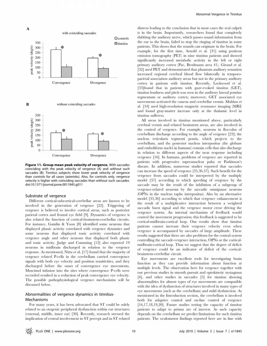
Figure 11 summarizes group mean peak velocity of vergence
with coinciding saccades (A) and vergence without coinciding
saccades (B) for controls and tinnitus. ANOVA applied on the
group mean peak velocity of vergence shows statistically significant
effect of the group for both cases with (F1,32 = 41.82, p,0.001) and
without (F1,32 = 59.41, p,0.001) coinciding saccades, i.e. the peak
velocity of vergence is lower in tinnitus than in controls for both
convergence and divergence. In addition, for controls the peak
velocity is higher for vergence with coinciding saccades than for
that without coinciding saccades ((F1,32 = 39.02, p,0.001). This is
a case for both convergence and divergence. Yet, for tinnitus there
is no difference of the peak velocity between these two cases
(F1,32 = 0.001, p = 0.92). Thus, the peak velocity of vergence is
always lower for tinnitus, with or without coinciding saccades.
Discussion
This study showed no latency abnormalities but low accuracy
for divergence, low peak velocity and long duration of both
convergence and divergence, more frequent embedded saccades
and of higher amplitude of coinciding saccades in tinnitus relative
to controls. Tinnitus subjects did not show normal saccade-
Figure 8. Embedded saccades. (A) Group mean number ofembedded saccades during complete execution of the vergence–totaltrajectory seen example in Fig. 2 from ‘i’ to ‘e’. (B) the group meanamplitude of the subgroup of saccades coinciding with the peakvelocity of vergence. Tinnitus subjects show significantly moreembedded saccades, and higher amplitude of coinciding saccadesthan controls (asterisks).doi:10.1371/journal.pone.0011845.g008
Abnormal Vergence in Tinnitus
PLoS ONE | www.plosone.org 7 July 2010 | Volume 5 | Issue 7 | e11845

Figure 9. The correlation between the vergence and the coinciding saccades. (A) The correlation between the peak velocity of vergenceand the amplitude of saccades coinciding with the peak velocity of vergence; (B) the correlation between the peak velocity of vergence and the peakvelocity of saccades coinciding with the peak velocity of vergence. Correlation coefficients ‘r’ and levels of significance are shown next to each cluster.
Abnormal Vergence in Tinnitus
PLoS ONE | www.plosone.org 8 July 2010 | Volume 5 | Issue 7 | e11845

vergence interaction, that is, saccades did contribute to increase of
the velocity of vergence as seen in controls.
Convergence–divergence differenceBefore discussing the group effects we will discuss the different
properties of convergence versus divergence confirmed for both
groups (controls and tinnitus). Note that shorter latency for
divergence than for convergence is the case for both controls and
tinnitus. This result is compatible with our previous study (Yang &
Kapoula 2004; Yang et al. 2009) and other studies (Alvarez et al.
2002, 2005). Such difference could be explained by Patel’s model (a
neural network model): two types of disparity encoders (disparity-
tuned cells) employed in this model, convergence and divergence
encoders. In other words, the divergence is not simply a negative
convergence movement (i.e. passive relaxation of convergence) but
a separate neurophysiological system. The neuro-control strategy of
these two systems may be different (Alvarez et al. 2005). Perhaps this
model can also explain the different dynamics between convergence
and divergence (shorter duration and higher peak velocity for
convergence than for divergence).
Positive significant correlations for both divergence and convergence occur for controls but for tinnitus patients. Values in boxes indicate correlationsfor the subgroup of saccades with amplitudes equal or less than 4u; no significant correlation exists for controls or tinnitus.doi:10.1371/journal.pone.0011845.g009
Figure 10. The main sequence of vergence. The correlation between the peak velocity and the amplitude of convergence (A) and of divergence(B) without coinciding saccades. All correlation coefficients and levels of significance are shown next to each cluster. Correlations are positive andsignificant, i.e. the peak velocity of vergence increases as the vergence amplitude increases for both controls and tinnitus subjects.doi:10.1371/journal.pone.0011845.g010
Abnormal Vergence in Tinnitus
PLoS ONE | www.plosone.org 9 July 2010 | Volume 5 | Issue 7 | e11845
Substrate of vergenceDifferent cortical-subcortical-cerebellar areas are known to be
involved in the generation of vergence [22]. Triggering of
vergence is believed to involve cortical areas, such as posterior
parietal cortex and frontal eye field [9]. Dynamics of vergence is
also related the function of cortical-brainstem-cerebellar circuits.
For instance, Gamlin & Yoon [8] identified some neurons that
displayed phasic activity correlated with vergence dynamics and
some neurons that displayed tonic activity correlated with
vergence angle and other neurons that displayed both phasic
and tonic activity. Judge and Cumming [12] also reported 19
neurons in midbrain discharged in relation to the vergence
response. As mentioned, Nitta et al. [17] found that the majority of
vergence related P-cells in the cerebellum carried convergence
signals with both eye velocity and position sensitivities, and they
discharged before the onset of convergence eye movements.
Muscimol infusion into the sites where convergence P-cells were
recorded resulted in a reduction of peak convergence eye velocity.
The possible pathophysiological vergence mechanisms will be
discussed below.
Abnormalities of vergence dynamics in tinnitus-Mechanisms
For many years, it has been advocated that ST could be solely
related to an otogenic peripheral dysfunction within ear structures
(external, middle, inner ear) [30]. Recently, research stressed the
implication of central involvement in ST percept and in ST related
distress leading to the conclusion that in most cases the real culprit
is in the brain. Importantly, researchers found that completely
dubbing the auditory nerve, which passes sound information from
the ear to the brain, failed to stop the ringing of tinnitus in some
patients. This shows that the sounds can originate in the brain. For
example, for the first time, Arnold et al. [31] using positron
emission tomography (PET) in nine tinnitus patients and showed
significantly increased metabolic activity in the left or right
primary auditory cortex (Pac, Brodmann area 41). Giraud et al.
[32] used PET and demonstrated that phantom auditory sensation
increased regional cerebral blood flow bilaterally in temporo-
parietal association auditory areas but not in the primary auditory
cortex in patients with tinnitus. Recently, Lockwood et al.
[33]found that in patients with gaze-evoked tinnitus (GET),
tinnitus loudness and pitch was seen in the auditory lateral pontine
tegmentum or auditory cortex; moreover, GET associated eye
movements activated the cuneus and cerebellar vermis. Muhlau et
al. [34] used high-resolution magnetic resonance imaging (MRI)
and found gray-matter increase only at the thalamic level in
tinnitus sufferers.
All areas involved in tinnitus mentioned above, particularly
cerebral vermis and related brainstem areas, are also involved in
the control of vergence. For example, neurons in flocculus of
cerebellum discharge according to the angle of vergence [23]; the
nucleus reticularis tegmenti pontis, which projects to the
cerebellum, and the posterior nucleus interpositus (the globuse
and emboliform nuclei in humans) contain cells that also discharge
in relation to different aspects of the near response including
vergence [16]. In humans, problems of vergence are reported in
patients with progressive superanuclear palsy or Parkinson’s
disease. In addition, numerous studies reported that saccades
can increase the speed of vergence [35,36,37]. Such benefit for the
vergence from saccades could be interpreted by the multiply
model [37] according to which speeding of vergence by the
saccade may be the result of the inhibition of a subgroup of
vergence-related neurons by the saccadic omnipause neurons
(OPNs in the nucleus raphe interpositus). Also by an alternative
model [35,36] according to which that vergence enhancement is
the result of a multiplicative interaction between a weighted
saccadic burst signal and the vergence motor error driving the
vergence system. An internal mechanism of feedback would
control the movement progression; this feedback is suggested to be
cortical-midbrain-cortical loop. Our results show that tinnitus
patients cannot increase their vergence velocity even when
vergence is accompanied by saccades of large amplitude. These
results suggested that there are also problems for the central system
controlling the saccade-vergence interaction, OPNs or the cortical-
midbrain-cortical loop. Thus we suggest that the degree of deficit
of vergence could be an indicator of deficit of the cortical-
brainstem-cerebellar circuit.
Eye movements are excellent tools for investigating brain
function as they can provide information about function at
multiple levels. The observation here for vergence together with
our previous studies in smooth pursuit and optokinetic nystagmus
[4], and other studies in saccades [5] for tinnitus showing
abnormalities for almost types of eye movements are compatible
with the idea of dysfunction of structures involved in many types of
eye movements (such as the cerebellum) and mild dysfunction. As
mentioned in the Introduction section, the cerebellum is involved
both for adaptive control and on-line control of vergence
[16,17,18,19,20]. Future studies testing the capacity of tinnitus
patients to adapt to prisms are of interest. As such capacity
depends on the cerebellum we predict limitations for such tinnitus
patients. The oculomotor findings reported here are in line with
Figure 11. Group mean peak velocity of vergence. With saccadescoinciding with the peak velocity of vergence (A) and without suchsaccades (B). Tinnitus subjects show lower peak velocity of vergencethan controls for all cases (asterisks). Also, for controls only, vergencevelocity is higher with coinciding saccades than without such saccades.doi:10.1371/journal.pone.0011845.g011
Abnormal Vergence in Tinnitus
PLoS ONE | www.plosone.org 10 July 2010 | Volume 5 | Issue 7 | e11845
observations of functional magnetic resonance imaging [33]
describing cerebellar activity in patients with GET. Perhaps
tinnitus and vergence dysfunction are both manifestations of mild
cerebellar syndrome including abnormality of cross-modality
interactions between vergence and auditory signals.
Given that saccades and vergence are both gifted by plasticity
and adaptive capacity, eye movement training may be used to
reduce vergence abnormality. In a retrospective analysis in
patients with acquired brain injury Ciuffreda et al. [26] observed
lasting vergence abnormalities. Ciuffreda et al. [38] showed that
vision therapy for oculomotor dysfunction can be efficient as
considerable residual neural plasticity exist. Therefore, vergence
orthoptic training could be useful alleviating visual symptoms and
perhaps abnormal oculomotor-auditory interactions. Thus useful-
ness of oculomotor reeducation in patients with somatic tinnitus
could be interestingly more widely tested in future clinical
research.
Finally, vergence abnormalities reported here could be present
only for patients who can modulate their tinnitus. It will be
interesting to study patients with tinnitus without movement
modulation. Future research is then needed to determine if the
vergence problem is due to ST itself or is related to the specific
ability of these patients to modulate their tinnitus by movements.
Acknowledgments
The authors appreciate the comments from the anonymous reviewer.
Author Contributions
Conceived and designed the experiments: QY MV CO PB AL ZK.
Performed the experiments: QY MV. Analyzed the data: QY. Contributed
reagents/materials/analysis tools: QY CO PB AL ZK. Wrote the paper:
QY MV AL ZK.
References
1. Eggermont JJ, Roberts LE (2004) The neuroscience of tinnitus. Trends Neurosci27: 676–682.
2. Norena A, Micheyl C, Chery-Croze S, Collet L (2002) Psychoacoustic
characterization of the tinnitus spectrum: implications for the underlyingmechanisms of tinnitus. Audiol Neurootol 7: 358–369.
3. Levine RA, Abel M, Cheng H (2003) CNS somatosensory-auditory interactionselicit or modulate tinnitus. Exp Brain Res 153: 643–648.
4. Kapoula Z, Yang Q, Vernet M, Bonfils P, Londero A (2009) Eye movement
abnormalities in somatic tinnitus: Fixation, smooth pursuit and optokineticnystagmus. Auris Nasus Larynx.
5. Jozefowicz-Korczynska M, Pajor A (2002) Evaluation of oculomotor tests inpatients with tinnitus. Int Tinnitus J 8: 100–103.
6. Mezzalira R, Bilecki MM, Gontijo BP, Slusser JE, Bernarde GE, et al. (2007)Can oculomotricity be altered in patients with tinnitus only? A preliminary
study. Int Tinnitus J 13: 152–156.
7. Tzelepi A, Lutz A, Kapoula Z (2004) EEG activity related to preparation andsuppression of eye movements in three-dimensional space. Exp Brain Res 155:
439–449.8. Gamlin PD, Yoon K (2000) An area for vergence eye movement in primate
frontal cortex. Nature 407: 1003–1007.
9. Kapoula Z, Isotalo E, Muri RM, Bucci MP, Rivaud-Pechoux S (2001) Effects oftranscranial magnetic stimulation of the posterior parietal cortex on saccades
and vergence. Neuroreport 12: 4041–4046.10. Walton MM, Mays LE (2003) Discharge of saccade-related superior colliculus
neurons during saccades accompanied by vergence. J Neurophysiol 90:1124–1139.
11. Suzuki S, Suzuki Y, Ohtsuka K (2004) Convergence eye movements evoked by
microstimulation of the rostral superior colliculus in the cat. Neurosci Res 49:39–45.
12. Judge SJ, Cumming BG (1986) Neurons in the monkey midbrain with activityrelated to vergence eye movement and accommodation. J Neurophysiol 55:
915–930.
13. Versino M, Hurko O, Zee DS (1996) Disorders of binocular control of eyemovements in patients with cerebellar dysfunction. Brain 119 (Pt 6): 1933–1950.
14. Richter HO, Costello P, Sponheim SR, Lee JT, Pardo JV (2004) Functionalneuroanatomy of the human near/far response to blur cues: eye-lens
accommodation/vergence to point targets varying in depth. Eur J Neurosci20: 2722–2732.
15. Mays LE (1984) Neural control of vergence eye movements: convergence and
divergence neurons in midbrain. J Neurophysiol 51: 1091–1108.16. Gamlin PD, Clarke RJ (1995) Single-unit activity in the primate nucleus
reticularis tegmenti pontis related to vergence and ocular accommodation.J Neurophysiol 73: 2115–2119.
17. Nitta T, Akao T, Kurkin S, Fukushima K (2008) Vergence eye movement
signals in the cerebellar dorsal vermis. Prog Brain Res 171: 173–176.18. Nitta T, Akao T, Kurkin S, Fukushima K (2008) Involvement of the cerebellar
dorsal vermis in vergence eye movements in monkeys. Cereb Cortex 18:1042–1057.
19. Takagi M, Tamargo R, Zee DS (2003) Effects of lesions of the cerebellaroculomotor vermis on eye movements in primate: binocular control. Prog Brain
Res 142: 19–33.
20. Takagi M, Oyamada H, Abe H, Zee DS, Hasebe H, et al. (2001) Adaptivechanges in dynamic properties of human disparity-induced vergence. Invest
Ophthalmol Vis Sci 42: 1479–1486.
21. Zee DS, Levi L (1989) Neurological aspects of vergence eye movements. RevNeurol (Paris) 145: 613–620.
22. Leigh RJ, Zee D (2006) The neurology of eye movements. New York: Oxforduniversity press.
23. Miles FA, Fuller JH, Braitman DJ, Dow BM (1980) Long-term adaptive changes
in primate vestibuloocular reflex. III. Electrophysiological observations inflocculus of normal monkeys. J Neurophysiol 43: 1437–1476.
24. Scheiman M, Mitchell GL, Cotter S, Cooper J, Kulp M, et al. (2005) Arandomized clinical trial of treatments for convergence insufficiency in children.
Arch Ophthalmol 123: 14–24.25. Scheiman M, Mitchell GL, Cotter S, Kulp MT, Cooper J, et al. (2005) A
randomized clinical trial of vision therapy/orthoptics versus pencil pushups for
the treatment of convergence insufficiency in young adults. Optom Vis Sci 82:583–595.
26. Ciuffreda KJ, Kapoor N, Rutner D, Suchoff IB, Han ME, et al. (2007)Occurrence of oculomotor dysfunctions in acquired brain injury: a retrospective
analysis. Optometry 78: 155–161.
27. Shore S, Zhou J, Koehler S (2007) Neural mechanisms underlying somatictinnitus. Prog Brain Res 166: 107–123.
28. Takagi M, Frohman EM, Zee DS (1995) Gap-overlap effects on latencies ofsaccades, vergence and combined vergence-saccades in humans. Vision Res 35:
3373–3388.29. Yang Q, Bucci MP, Kapoula Z (2002) The latency of saccades, vergence, and
combined eye movements in children and in adults. Invest Ophthalmol Vis Sci
43: 2939–2949.30. Tonndorf J (1981) Tinnitus and physiological correlates of the cochleo-vestibular
system: peripheral; central. J Laryngol Otol Suppl: 18–20.31. Arnold W, Bartenstein P, Oestreicher E, Romer W, Schwaiger M (1996) Focal
metabolic activation in the predominant left auditory cortex in patients suffering
from tinnitus: a PET study with [18F]deoxyglucose. ORL J OtorhinolaryngolRelat Spec 58: 195–199.
32. Giraud AL, Chery-Croze S, Fischer G, Fischer C, Vighetto A, et al. (1999) Aselective imaging of tinnitus. Neuroreport 10: 1–5.
33. Lockwood AH, Wack DS, Burkard RF, Coad ML, Reyes SA, et al. (2001) Thefunctional anatomy of gaze-evoked tinnitus and sustained lateral gaze.
Neurology 56: 472–480.
34. Muhlau M, Rauschecker JP, Oestreicher E, Gaser C, Rottinger M, et al. (2006)Structural brain changes in tinnitus. Cereb Cortex 16: 1283–1288.
35. Busettini C, Mays LE (2005) Saccade-vergence interactions in macaques. I. Testof the omnipause Multiply Model. J Neurophysiol 94: 2295–2311.
36. Busettini C, Mays LE (2005) Saccade-vergence interactions in macaques. II.
Vergence enhancement as the product of a local feedback vergence motor errorand a weighted saccadic burst. J Neurophysiol 94: 2312–2330.
37. Zee DS, Fitzgibbon EJ, Optican LM (1992) Saccade-vergence interactions inhumans. J Neurophysiol 68: 1624–1641.
38. Ciuffreda KJ, Rutner D, Kapoor N, Suchoff IB, Craig S, et al. (2008) Visiontherapy for oculomotor dysfunctions in acquired brain injury: a retrospective
analysis. Optometry 79: 18–22.
Abnormal Vergence in Tinnitus
PLoS ONE | www.plosone.org 11 July 2010 | Volume 5 | Issue 7 | e11845


