Embed Size (px)
Citation preview

Am. J. Hum. Genet. 45:786-792, 1989
Characterization of Rapidly Adhering Amniotic Fluid Cells byCombined Immunofluorescence and Phagocytosis Assays
K. Polgar,* R. Adany,t G. Abel,$ J. Kappelmayert L. Muszbek,T and Z. Papp*
*Department of Obstetrics and Gynecology, tDepartment of Clinical Chemistry, and linstitute of Pathophysiology, University Medical Schoolof Debrecen, Debrecen, Hungary
Summary
Culture of human amniotic-fluid cells from cases of fetal neural tube defects produces a population of rap-
idly adhering cells that were initially thought to be macrophages and later interpreted to be of neural ori-gin. In this study double and triple labeling systems for the simultaneous detection of glial and macro-
phage differentiation marker antigens have been used to demonstrate that rapidly adhering cells cannot beconsidered a homogeneous population but instead represent two distinct cell types. One of these cell popu-
lations is of glial origin and shows specific staining for glial fibrillary acidic protein, while the other popu-
lation is monocyte-derived macrophages which express marker antigens recognized by Leu M3, KiM7, andDako antimacrophage monoclonal antibodies.
Introduction
Both determination of alpha-fetoprotein (AFP) levelsand ultrasonic methods have been widely used in theprenatal diagnosis of neural tube defects (NTDs). Be-cause neither amniotic-fluid AFP estimation or ultra-sonography is entirely reliable in the diagnosis or ex-clusion of neural tube closure defects (Chapman 1982;Mizejewski and Risemberg 1985), cytological diagnos-tic methods have been developed. In cases of fetal NTDsa large number of macrophages are present in amnioticfluid. Some of these cells can be recognized on the basisof morphological properties by direct microscopical ex-amination of cytologic smears (Papp and Bell 1979;Chapman 1982; Papp et al. 1982). More recently, sim-ple color tests based on the pinocytotic properties ofthe pathognomonic cells were introduced, and thesecells are easily identified as "red cells" by light micros-copy (Polgair et al. 1984, 1985).
Although rapidly adhering (RA) cells, which settlein less than 24 h during cultivation of amniotic-fluidcells, have been identified as macrophages (Sutherland
Received May 1, 1989; revision received July 17, 1989.Address for correspondence and reprints: Dr. Katalin Polgar,
Department of Obstetrics and Gynecology, University Medical Schoolof Debrecen, P.O. Box 37, H-4012 Debrecen, Hungary.i 1989 by The American Society of Human Genetics. All rights reserved.0002-9297/89/4505-0015$02.00
et al. 1973), contradictory results have also been pub-lished. It has been suggested that most of the RA cellsare of neural origin (Gosden and Brock 1977), but somestudies have shown that RA cells are not homogeneousand that the proportion of neural cells present is vari-able (Aula et al. 1980; Cremer et al. 1981; Medina-Gomez and Bride 1986).The present study of amniotic fluid was designed to
(1) immunomorphologically characterize RA cells ap-pearing in combination with NTDs and (2) identifythe cell population involved in phagocytosis.
Material and Methods
Case Studied
Amniotic-fluid samples were obtained by transab-dominal amniocentesis from 11 patients of the GeneticCounseling Service of the Department of Obstetrics andGynecology, University School of Medicine, Debrecen,Hungary between May 10, 1985, and April 21, 1988.Amniocenteses were carried out for different indications:normal or suspicious ultrasound findings, high mater-nal serum AFP, etc. Pregnancies were terminated by in-duced abortion, and the following diagnoses wereconfirmed by embryopathological investigations: sevencases of anencephaly (two of these fetuses also had spinabifida) and four cases of spina bifida.
786
Rapidly Adhering Amniotic Fluid Cells
Samples obtained by amniocentesis were divided intotwo parts. One part was used for making cytospin prepa-rations, and the other was used for short-term cultiva-tion of amniotic-fluid cells to obtain RA cell monolayers.
Cytospin Preparations
Amniotic-fluid cell preparations were made by cyto-spin 2 centrifugation at 800 rpm for 5 min (ShandonSouthern Products, Runcorn, Cheshire).
Cell Culture
To obtain primary adherent cell monolayers, amnioticfluid was cultured in flaskette tissue culture chamberscontaining Chang C medium (HANA Biologics, Inc.,Alameda), by incubation at 370C in a humidified at-mosphere of 5% CO2 in air. After 20-24 h the super-natants, which contained mainly unattached amniotic-fluid cells and red blood cells, were removed. Slidescovered by RA cell monolayers were processed for mi-croscopy.
Fixation
Both air-dried cytospin preparations and primary ad-herent monolayers were washed in PBS, pH 7.4 andwere fixed with absolute acetone at room temperaturefor 10 min.
ProteinsHuman complement component C3 was purchased
from Cordis Laboratories (Miami). C3b was preparedby trypsin digestion of C3 according to a method de-scribed elsewhere (Ogle et al. 1985). Human IgG waspurchased from Calbiochem (Lucern).
Coupling of Proteins to MicrospheresCovalent coupling ofproteins (C3b or IgG) to fluores-
cent carboxylated microbeads ("Fluoresbrite," 0.88 gtmdiameter; Polysciences, Warrington, PA) was performedaccording to a method described elsewhere (Ogle et al.1985).
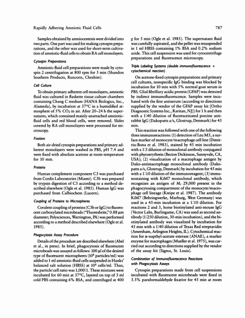
Phagocytosis Assay Procedure
Details of the procedure are described elsewhere (Abelet al., in press). In brief, phagocytosis of fluorescentmicrobeads was assayed as follows: 100 gl of the desiredtype of fluorescent microspheres (109 particles/ml) wasadded to 1 ml amniotic-fluid cells suspended in Hanks'balanced salt solution (HBSS) at 106 cells/ml. Thus,the particle:cell ratio was 1,000:1. These mixtures wereincubated for 60 min at 370C, layered on top of 3 mlcold PBS containing 6% BSA, and centrifuged at 400
g for 5 min (Ogle et al. 1985). The supernatant fluidwas carefully aspirated, and the pellet was resuspendedin 1 ml HBSS containing 1% BSA and 0.2% sodiumazide. This cell suspension was used for cytocentrifugepreparations and fluorescence microscopy.
Triple Labeling Systems (double immunofluorescence +cytochemical reaction)
On acetone-fixed cytospin preparations and primarycell cultures, nonspecific IgG binding was blocked byincubation for 10 min with 5% normal goat serum inPBS. Glial fibrillary acidic protein (GFAP) was detectedby indirect immunofluorescence. Samples were incu-bated with the first antiserum (according to directionssupplied by the vendor of the GFAP assay kit [OrthoDiagnostic Systems Inc., Raritan, NJ]) for 1 h and thenwith a 1:40 dilution of fluoresceinated porcine anti-rabbit IgG (Dakopatts a/s, Glostrup, Denmark) for 45min.
This reaction was followed with one of the followingthree immunoreactions: (1) detection of Leu M3, a sur-face marker of monocyte/macrophage cell line (Dimit-riu-Bona et al. 1983), stained by 45 min incubationwith a 1:5 dilution of monoclonal antibody conjugatedwith phycoerythrein (Becton Dickinson, Sunnyvale, CA,USA); (2) visualization of a macrophage antigen byDako-antimacrophage monoclonal antibody (Dako-patts a/s, Glostrup, Denmark) by incubation for 45 minwith a 1:10 dilution of the immunoreagent; (3) immu-nostaining with KiM7 monoclonal antibody, whichrecognizes an antigen of Mr 29,000 present in thephagocytosing compartment of the monocyte/macro-phage cell lineage (Kreipe et al. 1987). The antibodyKiM7 (Behringwerke, Marburg, West Germany) wasused in a 45-min incubation at a 1:10 dilution. Forreactions 2 and 3, horse biotinylated anti-mouse IgG(Vector Labs, Burlingame, CA) was used as second an-tibody (1:250 dilution, 30-min incubation), and the bi-otinylated antibody was visualized by incubation for45 min with a 1:40 dilution of Texas Red streptavidin(Amersham, Arlington Heights, IL). Cytochemical reac-tion for a-napthyl-acetate esterase (ANAE), a markerenzyme for macrophages (Mueller et al. 1975), was car-ried out according to directions supplied by the vendorof the assay kit (Sigma, St. Louis).
Combination of Immunofluorescence Reactionswith Phagocytosis Assays
Cytospin preparations made from cell suspensionsincubated with fluorescent microbeads were fixed in3.5% paraformaldehyde fixative for 45 min at room
787

Polgar et al.
temperature and were stained for each of the antigensdescribed above. The monoclonal reactions were car-ried out as mentioned above. The binding of primaryantibody to GFAP was detected by biotinylated anti-rabbit IgG (1:250 dilution, 30 min; Vector Labs) andTexas Red streptavidin (1:40 dilution, 30 min; Amer-sham). These preparations were also stained for ANAE.On control slides, antiserum against GFAP was
replaced with nonimmune rabbit serum, while mono-clonal antibodies were replaced with control mouse IgGfrom tumor-bearing BALB/c mice (Becton Dickinson,Sunnyvale, CA). Cytospin-prepared slides were coveredwith 50% glycerol in PBS and examined with a micro-scope equipped with an epifluorescence condenser con-taining selective filters for fluorescein and Texas Red/phycoerythrin. The excitation maximum/emission max-imum of fluorescein is 495/525 nm; that of Texas Redis 595/620 nm; and that of phycoerythrin is 496/576nm. Fluorescence photomicrographs were taken onScotch 1000 ASA color slide film. Fields photographedunder fluorescent illumination were also photographedunder bright field illumination.
Results
Cells in amniotic fluid from pregnancies with openNTDs show characteristic morphological features.Many cells showed positive staining for GFAP, and manycells were labeled for macrophage differentiation markerantigens recognized by Leu M3, KiM7, or Dako-anti-macrophage monoclonal antibodies. On RA cell mono-layers few cells (1%-2%) failed to show labeling foreither GFAP or macrophage markers. In double im-munofluorescence assays, no doubly labeled cells wereobserved in cytospin preparations; GFAP+ cells werenot observed to express macrophage differentiationmarker antigens, and macrophages did not stain forGFAP.GFAP + cells varied both in size and in morphologi-
cal appearance. Such cells were found both as relativelysmall, round cells of about 15 gm diameter and as largerpseudopodial cells of about 50 gm diameter. Cells show-ing macrophage-specific reactions mainly appeared asround mononuclear cells, but they varied markedly inapparent size.
Fluorescent latex microspheres were taken up onlyby cells recognized by Leu M3, KiM7, and Dako-antimacrophage monoclonal antibodies (fig. 1). All cellspositive for macrophage differentiation antigens in-gested IgG-coated latex microbeads, and cells whichphagocytosed fluorescent particles invariably labeled
Figure I Reaction with Dakop-antimacrophage monoclonalantibody developed by biotinylated second antibody and Texas-Redstreptavidin system on amniotic-fluid cell preparation after phago-cytosis of fluoresceinated latex microspheres. (x 240).
for macrophage antigens. GFAP + cells did not dem-onstrate phagocytic activity (fig. 2). All cells express-ing macrophage differentiation antigens were also ob-served to be positive for ANAE.
In primary cultures a significant proportion (20%-40%) of cells showed cytoplasmic fibrillar staining forGFAP. No double reactions with glial cell-specific andmacrophage-specific reagents were observed (fig. 3).
In triple labeling experiments complete codistribu-tion could be demonstrated for macrophage markers(KiM7 and Dako-antimacrophage) and ANAE, whileGFAP + cells were consistently negative for these mac-rophage markers (fig. 4).Among cells negative for the above-mentioned marker
Figure 2 Immunofluorescent labeling for GFAP (in red) in com-bination with FITC-latex microbead phagocytosis on amniotic fluidcell preparation. GFAP-positive cells do not ingest latex particles.(x 240).
788

Rapidly Adhering Amniotic Fluid Cells
Figure 3 Double immunofluorescent reaction for GFAP (ingreen) and with Dako-antimacrophage monoclonal antibody (in red)on rapidly adhering amniotic-fluid cells. (x 240).
antigens, epithelial cells and red blood cells were easilyrecognizable on the basis of their characteristic mor-phology, but a significant number of other cell typescould not be identified by morphological features alone.
Discussion
Controversy exists over the origin of mononuclearcells that are present in amniotic fluid in cases of fetalNTDs. Glial origin of the abnormal cells in the am-niotic fluid has been suggested (Aula et al. 1980; Sar-kar et al. 1980; Cremer et al. 1981; von Koskull et al.1981; Medina-Gomez and Bride 1986; Polgair et al.1987). However, others have found no evidence fordifferences among the abnormal cells with the use ofa specific antibody to GFAP (Chapman 1982).We reported earlier, from results obtained by the
peroxidase-antiperoxidase (PAP) method, the possibleneural (glial) origin of a part of amniotic-fluid patho-gnomonic cells (Polgair et al. 1987). Recently, we triedto find and apply specific markers diagnostic of mono-nuclear phagocytes (van Furth 1986; Polgar et al. 1988c).We could demonstrate (a) strong endogenous peroxi-dase and nonspecific esterase activity in these cells oncytologic smears (Polgair et al. 1987) and (b) positiveimmunostaining for both MO1 (63D3 human mono-cyte 1) and M02 (61D3 human monocyte 2) antigens(Polgair et al. 1988c). We could also demonstrate thepresence of functional markers such as Fc and C3breceptor-mediated phagocytosis and chemiluminescence(Polgair et al. 1988a). Further evidence of the mono-cytic origin of many of these cells has been presentedelsewhere (Polgair et al. 1984, 1988b).
Figure 4 Triple labeling for GFAP (top), KiM7 (middle), andANAE (bottom) GFAP-positive and KiM7-positive cells are clearlyseparated from each other. Only KiM7-positive cells show enzymecytochemical labeling for ANAE. (x 240).
GFAP has been identified elsewhere as a componentof astroglial cells, reactive microglia, and tumor-formingastrocytes (Bignami and Dahl 1974; Miyake et al. 1988).GFAP is also expressed by myelin-forming oligoden-drocytes in their early development, indicating that therelationship between astroglial and oligodendroglialcells is closer than previously has been believed (Choi
789

Polgar et al.
and Kim 1984). It is suggested that in postnatal devel-opment two distinct glioblast lines develop, one develop-ing into astrocytes and the other into oligodendrocytes.The primary glioblast is a bipotential glial progenitorcell (Bunge and Waksman 1985). Animal experimentsdemonstrated that both astroglial cells (Noske et al.1982) and microglia (Brierley and Brown 1982), ratherthan oligodendrocytes (Al-Ali and Robinson 1984), be-come active forms and may act as phagocytes in certainsituations, such as brain injury or removal to cell cul-ture. Therefore, pathological development of neuraltubes and the activation and release of glial cells or brainmacrophages may be caused by direct contact betweenneural tissue and amniotic fluid (Polgair et al. 1987).
Brain macrophages and microglial cells are consid-ered to be the most important phagocytic elementswithin the central nervous system (Esiri and Booss 1984;Gosden and Brock 1977). It is interesting that microglialcells, the only type of brain phagocytes which expressFc receptor activity similar to that of amniotic-fluid mac-rophages (Sutherland et al. 1973), do not share GFAPpositivity (Raff et al. 1979). Microglial cells showedpositive staining with nonspecific esterase stains, as domonocytes and macrophages (Esiri and Booss 1984).Later it was found that microglial cells lack macrophagemarkers (Wood et al. 1979). These cells are character-ized by 100% M01 and 10% M02 reactivity (Frank-lin et al. 1986). It is interesting that they do not sharelysozyme positivity (Esiri and Booss 1984). The con-troversy over the relationship between macrophage andmicroglia, as well as over the nature and functional roleof microglia in nervous tissue, has been renewed (Choiand Kim 1984).
According to previous work, microglial cells of thebrain are included in the mononuclear phagocyte sys-tem (van Furth 1986). Many of these macrophagesclearly originate from blood-borne monocytes and in-vade the central nervous system parenchyme via the vas-cular route (Murabe and Sano 1983). The migrationof hematogenous cells into neural tissue during embryo-genesis is a common phenomenon in human embryosand fetuses (Choi and Kim 1984). Other phagocytesmay arise from microglia resident in the brain whichare mobilized after injury (Brierley and Brown 1982).Investigators have suggested that microglia may trans-form into phagocytes (Oechmichen 1975). Other studiesfound no evidence that monocytes or macrophagesdifferentiate into microglia or that brain macrophagesare derived from microglia (Gordon 1986; Schelper andAdrian 1986). New data call into question the validity
of any hypothesis that suggests a common origin formicroglia and macrophages. Studies are underway toidentify the origin and function of true microglia.
Several studies suggest, however, that astrocytes areable to phagocytose in vivo (Noske et al. 1982). Wewere not able to demonstrate phagocytic propertiesamong either noncultured or cultured GFAP+ amni-otic-fluid cells. After we carried out C3b-coated latex-bead treatment, we could not visualize beads actuallyinternalized by pathognomonic cells in suspension orin primary culture. This may be due to the fact thatphagocytosis by astroglial cells is influenced by the stageof differentiation. During embryonic development ofthe brain, cells of various lineages may have similar mor-phologies, making it difficult to distinguish betweenthem.
Considering that the microglial cells are of mesoder-mal hematogenous origin, that macrophages have amonoblastic origin, and that both of them take partin the mononuclear phagocyte system, we conclude thatthe GFAP-negative pathognomonic cells in cases of fe-tal NTD are of hematogenous monocytic origin.The main conclusion to be drawn from the results
of the present study is that the mononuclear cell popu-lation present in amniotic fluid in cases of open NTDsis not homogeneous; two cell types can be distinguished.With double and triple immunolabeling methods incombination with fluorescent latex microsphere phago-cytosis assays, we have found that one population ofpathognomonic cells is of glial origin and lacks phago-cytic activity. We could consider this cell populationspecific for open NTDs. The other pathognomonic cellpopulation is of hematogenous-monocytic origin anddoes have phagocytic activity. This latter group of cellsforms as a result of development of NTDs. In casesof NTDs these monocytic cells occur in high numberin amniotic fluid; studies are underway to clarifywhether there are any processes causing appearance ofpathognomonic monocytic cells in even greater num-bers in amniotic fluid. We also need further confirma-tion of the fetal origin of the rapidly adherent cells inamniotic fluid.The neutral red test is a simple and rapid prenatal
diagnostic test for phagocytic cells in amniotic fluid;such cells are pathognomonic for open NTDs. How-ever, the neutral red test gives no information as to thenature or origin of the phagocytic cells. The combinedimmunofluorescence and phagocytosis assays used hereindicate that the phagocytic cells are of monocyticorigin.
790

Rapidly Adhering Amniotic Fluid Cells 791
AcknowledgmentsThe expert technical assistance of Agnes Bana, Judith
Orosz-Nagy, Emese Bede, and Emese Nagy is appreciated.We thank Professor Timothy Bestor for helpful commentson the manuscript.
ReferencesAbel G, Szollosi J, Chihara J, Fachet J. Effect of lentinan andmannan on phagocytosis of fluorescent latex microbeadsby mouse peritoneal macrophages: a flow cytometric study.Int J Immunopharmacol (in press)
Al-Ali SYA, Robinson N (1984) Neuronal and oligodendro-cytic response to cortical injury ultrastructural and cyto-chemical changes. Histochem J 16:165-178
Aula P, von Koskull H, Teramo H (1980) Glial origin of rap-idly adhering amniotic fluid cells. Br Med J 281:1456-1457
Bignami A, Dahl D (1974) Astrocyte-specific protein and neu-roglial differentiation: an immunofluorescence study withantibodies to the glial fibrillary acidic protein. J Comp Neu-rol 153:27-38
BrierleyJB, Brown AW (1982) The origin of lipid phagocytesin the central nervous sytem: the intrinsic microglia. J CompNeurol 211:397-406
Bunge RP, Waksman BH (1985) Glial development and inter-actions. Trends Neurosci 10:424-427
Chapman PA (1982) Cytology as a means of detecting neuraltube defects. Med Lab Sci 39:215-222
Choi BH, Kim RC (1984) Expression of glial fibrillary acidicprotein in immature oligodendroglia. Science 223:407-409
Cremer M, Sachner M, Cremer T, Schmid W, VoigtlanderT (1981) Demonstration of astrocytes in cultured amnioticfluid cells of three cases with neural-tube defects. Hum Ge-net 56:365-370
Dimitriu-Bona A, Burmester GR, Waters SJ, Winchester RJ(1983) Human mononuclear phagocyte differentiation an-tigens: patterns of antigenic expression on the surface ofhuman monocytes and macrophages defined by monoclo-nal antibodies. J Immunol 130:145-152
Esiri M, Booss J (1984) Comparison of methods to identifymicroglial cells and macrophages in the human central ner-vous system. J Clin Pathol 37:150-156
Franklin WA, Maison DY, Pulford K, Falini B, Bliss E, GatterKC, Stein H, et al (1986) Immunohistological analysis ofhuman mononuclear phagocytes and dendritic cells by usingmonoclonal antibodies. Lab Invest 54:322-335
Gordon S (1986) Biology of the macrophage. J Cell Sci (Suppl)4:267-286
Gosden CM, Brock DJH (1977) Morphology of rapidly ad-hering amniotic fluid cells as an aid to the diagnosis ofneural tube defects. Lancet 1:19-22
Kreipe H, Radzun HJ, Parwaresh MR, Haislip A, HansmannML (1987) Ki-M7 monoclonal antibody specific for my-
elomonocytic cell lineage and macrophages in human. JHistochem Cytochem 35:1117-1125
Medina-Gomez P, McBride (1986) Amniotic fluid macro-phages from normal and malformed fetuses. Prenat Diagn6:195-205
Miyake T, Hattori T, Fukuda M, Kitamura T, Fujita S (1988)Quantitative studies on proliferative changes of reactive as-trocytes in mouse cerebral cortex. Brain Res 451:133-138
Mizejewski GR, Risemberg HM (1985) Alpha-fetoprotein:use in predicting perinatal distress. In: Mizejewski GJ, Por-ter IH (eds) Alpha-fetoprotein and congenital disorders.Academic Press, New York, London, Toronto, pp 157-177
Mueller J, Brun del Re G, Buerki H, Keller HU, Hess MW,Cottier H (1975) Nonspecific acid esterase activity, a cri-terion for differentiation ofT and B lymphocytes in mouselymphnodes. Eur J Immunol 5:270-274
Murabe Y, Sano Y (1983) Morphological studies on neuroglia.VII. Distribution of "brain macrophages" in brains of neo-natal and adult rats as determined by means of immuno-histochemistry. Cell Tissue Res 229:85-95
Noske W, Lentzen H, Lange K, Keller K (1982) Phagocyticactivity of glial cells in culture. Exp Cell Res 142:437-445
Oechmichen M (1975) Monocytic origin of microglial cells.In: van Furth R (ed) Mononuclear phagocytes in immu-nity, infection and pathology. Blackwell, Oxford, pp 223-240
OgleJD, Ogle CK, Noel JG, Hurtubise P, AlexanderW (1985)Studies on the binding of C3b-coated microspheres to hu-man neutrophils. J Immunol Methods 47:47-62
Papp Z, BellJE (1979) Uncultured cells in amniotic fluid fromnormal and abnormal foetuses. Clin Genet 16:282-290
Papp Z, Polgair K, T6h Z, Csecsei K (1982) Prenatal diagno-sis of neural tube defects by exfoliative cytology of am-niotic fluid. Acta Cytol 26:751-752
Polgair K, Abel G, Laczk6J, Sipka S, Papp Z (1987) Immuno-cytochemical characterization of amniotic fluid macro-phages in cases of fetal neural tube defects. Am J Clin Pathol87:37-42
Polgair K, Abel G, Sipka S, CsongorJ, FachetJ, Papp Z (1988a)Immunobiological methods in the prenatal diagnosis andevaluation of fetal neural tube defects. Acta Physiol Hung71:551-555
Polgair K, Abel G, Sipka S, Laczk6 J, Papp Z (1988b) Am-niotic fluid mononuclear phagocytes: phenotypes and func-tions. Acta Paediatr Hung 29:63-67
Polgair K, Abel G, Sipka S, Papp Z (1985) On the neutralred test of amniotic fluid macrophages in neural tube defects.Invited article. Karyogram 11:39-42
(1988c) Neutral-red uptake and expression of mono-cytic antigens in amniotic fluid mononuclear phagocytes:evaluation of a novel approach for prenatal diagnosis ofneural tube defects. Am J Reprod Immunol 18:81-86
Polgair K, Sipka S, Abel G, Papp Z (1984) Neutral-red uptakeof amniotic fluid macrophages in neural-tube defects: a rapidtest. N Engl J Med 310:1463-1464

792 Polgar et al.
Raff MC, Fields KL, Hakomori KL, Mirsky R, Pruss RM,Winter J (1979) Cell-type specific markers for distinguish-ing and studying neurons and the major classes of glialcells in culture. Brain Res 174:283-308
Sarkar S, Chang HC, Porreco RP, Jones OW (1980) Neuralorigin of cells in amniotic fluid. Am J Obstet Gynecol136:67-72
Schelper RI, Adrian EK (1986) Monocytes become macro-phages; they do not become microglia: a light and electronmicroscopic study using 125-iododeoxyuridine. J Neuro-pathol 45:1-19
Sutherland GR, Brock DJH, Scrimgeour JB (1973) Amnioticfluid macrophages and anencephaly. Lancet 2:1098-1099
van Furth R (1986) The mononuclear phagocyte system: over-view. In: Weir DM, Herzenberg LA, Blackwell C, Herzen-berg LA (eds) Cellular immunology. Blackwell Scientific,Oxford, London, Edinburgh, Boston, Palo Alto, Mel-bourne, pp 42-43
von Koskull H, Virtanen I, Lehto VP, Vario T, Dahl D, AulaP (1981) Glial and neuronal cells in amniotic fluid of anen-cephalic pregnancies. Prenat Diagn 1:259-267
Wood GW, Gollakon KA, Tilzer SA, Vats T, Morantz RA(1979) The failure of microglia in normal brain to exhibitmononuclear phagocyte markers. J Neuropathol Exp Neu-rol 38:369-376


