Embed Size (px)
Citation preview

213
Kursad Turksen (ed.), Claudins: Methods and Protocols, Methods in Molecular Biology, vol. 762,DOI 10.1007/978-1-61779-185-7_15, © Springer Science+Business Media, LLC 2011
Chapter 15
Identification of Claudins by Western Blot and Immunofluorescence in Different Cell Lines and Tissues
Lorenza González-Mariscal, Erika Garay, and Miguel Quirós
Abstract
Claudins are integral proteins of the TJ. Each epithelia in the organism expresses a unique set of claudins that determines the degree of sealing of the paracellular pathway and the ionic selectivity of the tissue. TJs are dynamic structures whose organization and composition change in response to alterations in the environment as well as under physiological and pathological conditions. Changes in claudin expression and subcellular distribution can be analyzed in western blot and immunofluorescence experiments, employing a wide array of available specific antibodies against claudins. In this chapter, we describe in detail protocols used for western blot and immunofluorescence detection of claudins in epithelial cell lines and in various tissue samples.
Key words: Claudin, Western blot, Immunofluorescence, Tight junctions, Epithelia
Tight junctions (TJs) in epithelial cells localize at the most apical segment of the lateral membrane. TJ are integrated by a complex array of integral and peripheral proteins, among which claudins play a crucial role as back bone constituents of the filaments observed by freeze-fracture electron microscopy. The filaments that encircle epithelial cells below the apical microvilli are formed by lines of transmembrane particles with a 10 nm diameter that fuse when fixed with glutaraldehyde (1, 2). These particles are composed of claudin multimers, and their formation can be trig-gered upon claudin transfection, even in cells that lack TJs such as fibroblasts (3).
1. Introduction

214 L. González-Mariscal et al.
Claudins are tetraspan proteins that orient their amino and carboxyl terminal ends toward the cytosol, and expose two loops to the extracellular space. The first loop which is bigger than the second, contains several charged residues responsible for the ionic selectivity of the TJ (4–8), while the second contains aromatic and hydrophilic residues, conserved in most claudins, that are critical for the trans-interaction of claudins (9). Each particular tissue of the body of multicellular organisms exhibits a unique set of claudins that regulates by charge and size the passage of ions and molecules through the paracellular pathway.
Here we describe protocols employed to analyze the expres-sion of claudins by western blot and immunofluorescence micros-copy in diverse cell lines and tissues.
1. Phosphate buffer saline (PBS) from Gibco. 2. RIPA buffer: 40 mM Tris–HCl, pH 7.6, 150 mM NaCl, 2 mM
EDTA pH 8, 10% glycerol, 1% Triton X-100, 0.5% sodium deoxycholate, and 0.2% SDS. Store at 4°C (see Note 1).
3. Gentle lysis buffer: 20 mM Tris–HCl, pH 7.6, 50 mM NaCl, 2 mM EDTA, and 1% Triton X-100.
4. Protease inhibitors: phenyl methane sulfonyl-fluoride (PMSF) 100 mM stock dissolved in isopropanol and protease inhibi-tor cocktail Complete™ (Roche).
5. Lowry protein assay (BioRad) or BCA protein assay reagent (Pierce).
6. Laemmli sample buffer (5×): 312.5 mM Tris–HCl, 10% SDS, 50% glycerol, 25% 2-mercaptoethanol, bromophenol blue 0.5%, pH 6.8. Store at −20°C in 1 ml aliquots.
1. Separating gel buffer: 1.5 M Tris–HCl, pH 8.8. Store at 4°C.
2. Stacking gel buffer: 1 M Tris–HCl, pH 6.8. Store at 4°C. 3. Acrylamide solution: 30% (w/v) aqueous acrylamide/
bisacrylamide (37.5:1) in water. Store at 4°C (see Note 2). 4. N,N,N,N, -Tetramethyl-ethylenediamine (TEMED): Store
at 4°C. 5. Amonium persulfate (APS): prepare a 10% (w/v) solution
in water and immediately freeze and store in 500 l aliquots at −20°C.
6. Water from a Millipore MQ system with a resistivity of 18.2 M cm.
2. Materials
2.1. Cell Lysis
2.2. SDS–Polyacrylamide Gel Electrophoresis

21515 Identification of Claudins by Western Blot…
7. SDS solution: 10% (w/v) solution. Store at room temperature (see Note 3).
8. SDS–polyacrylamide gel electrophoresis (SDS–PAGE) run-ning buffer (10×): 250 mM Tris–HCl, 1.92 M glycine, pH 8.8, 1% (w/v) SDS (see Note 4). Store at room temperature.
9. Molecular weight markers: Dual Precision Plus Western blot standards (BioRad) or equivalent.
1. Transfer buffer: 48 mM Tris, 39 mM glycine, 20% v/v meth-anol, and 0.037% w/v SDS. Store at 4°C and use only once.
2. PVDF membrane (see Note 5). 3. Tris-buffer saline with Tween 20 (TBS-Tween): Prepare stock
solutions of 2 M NaCl and 500 mM Tris–HCl, pH 7.5. Store at 4°C. With them prepare a solution containing 10 mM Tris–HCl, pH 7.5, 100 mM NaCl, and 0.2% v/v Tween 20. Store at 4°C.
4. Blocking buffer: 5% (w/v) non-fat dry milk, 3% (w/v) bovine serum albumin in TBS-Tween 0.1% (see Note 6).
5. Primary antibody dilution buffer: Blocking buffer. 6. Primary antibodies: We have employed the Invitrogen rabbit
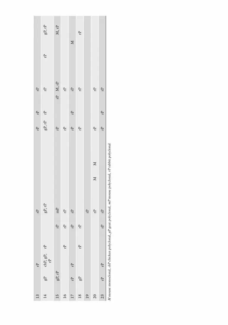
polyclonal antibodies against claudins: 1 (51–9000); 2 (51–6100); 3 (34–1700); 5 (34–1600); 7 (34–9100); and 16 (34–5400), as well as the mouse monoclonal antibody against claudin 4 (32–9400). Information on other commercial antibodies against claudins is found in Tables 1 and 2 (see Note 7). In addition, as loading control we employ antibodies against actin or tubulin.
7. Secondary antibodies: HRP goat anti-mouse or anti-rabbit IgG.
8. Chemiluminescence detection kit: ECL + Plus (GE Healthcare), Immobilon Western (Millipore) or equivalent.
9. Chemidoc Bio-Rad system can be employed to detect the chemioluminescence. However, if the signal is not too strong, we recommend the use of the autoradiography film Hyperfilm ECL (GE Healthcare).
1. Stripping buffer: sodium dithionite 2% in TBS-Tween. Store at 4°C. Light sensitive.
2. Wash buffer: TBS-Tween.
1. Glass coverslips cut in 8 mm × 8 mm squares with a diamond tip pen.
2. 2-Methylbutane (Sigma Aldrich, St Louis, MO) (see Note 8). 3. Liquid nitrogen.
2.3. Detection of Claudins by Western Blotting
2.4. Stripping and Reprobing Blots for Detection of Claudins
2.5. Immun-fluorescence for Detection of Claudins

Tabl
e 1
Type
of a
ntib
odie
s ag
ains
t cla
udin
s av
aila
ble
from
diff
eren
t man
ufac
ture
rs
Clau
din/
man
ufac
-tu
rer
Sant
a Cr
uzTh
erm
o sc
ient
ific
Invi
trog
enSi
gma
Aldr
ich
Abca
mCe
ll si
gnal
ing
Abge
ntAs
say
desi
gnBi
owor
ld
tech
nolo
gyGe
nway
bi
otec
hGe
nete
xIB
L
Life
spa
n bi
osci
-en
ces
Orig
ene
tech
-no
logi
esPr
osci
R&D
Unite
d St
ates
Bi
olog
ical
1M
, gP,
rP
rPM
, rP
M, r
PM
, rP
rPrP
MrP
rPrP
rPM
, rP
rPM
M, r
P
2rP
rPM
, rP
M, r
PM
, rP,
m
PM
, rP
rPrP
rPrP
M, r
PM
, rP
3gP
, rP
rPrP
rPrP
MrP
rPrP
rPrP
rPM
rP
4M
, gP
rPM
MM
, rP
MM
rPrP
rPrP
M, r
PM
, rP
MM
, rP
5gP
, rP
rPM
, mP,
rPrP
MrP
rPrP
rPM
, rP
M, r
P
6gP
rPrP
rPrP
M, r
PrP
7gP
, rP
rPM
, rP
M, r
PrP
rPrP
M, r
PrP
M, r
P
8gP
, rP
rPrP
rPrP
rPrP
rPM
rP
9gP
rPrP
rPrP
rPrP
10gP
, rP
rPM
, rP
rPrP
rPrP
rPM
rP
11gP
, rP
rPrP
rPrP
rPrP
rPM
, rP
M
12gP
rPrP
rPrP
MM
, rP
Clau
din/
man
ufac
-tu
rer
Sant
a Cr
uzTh
erm
o sc
ient
ific
Invi
trog
enSi
gma
Aldr
ich
Abca
mCe
ll si
gnal
ing
Abge
ntAs
say
desi
gnBi
owor
ld
tech
nolo
gyGe
nway
bi
otec
hGe
nete
xIB
L
Life
spa
n bi
osci
-en
ces
Orig
ene
tech
-no
logi
esPr
osci
R&D
Unite
d St
ates
Bi
olog
ical
13rP
rPrP
rPrP
14gP
chP,
gP,
rP
rPgP
, rP
gP, r
PrP
rPrP
gP, r
P
15gP
, rP
rPm
PrP
rPM
, rP
M, r
P
16rP
rPrP
rPrP
17rP
rPrP
rPrP
rPrP
M
18gP
rPrP
rPrP
rP
19rP
20rP
MM
rPrP
23rP
rPrP
rPrP
rPrP
M m
ouse
mon
oclo
nal,
chP
chic
ken
poly
clon
al, g
P go
at p
olyc
lona
l, m
P m
ouse
pol
yclo
nal,
rP ra
bbit
poly
clon
al

218 L. González-Mariscal et al.
Table 2 Commercial antibodies that recognize regions of Claudins different from the c-terminal segment
Claudin First loopa Second loopa Amino terminal PY Middled Isoform
1 Santa Cruz, Abcam, Abgent
Abgent
2 Abcam, Abgent Abgentb (Y195, Y224)
3 Santa Cruz Abcamc (Y219)
6 Abcamc (Y219) Abcam
7 Abcamc (Y210)
10 10-bR&D
11 Santa Cruz
14 Invitrogen
18 Invitrogena See Note 7b See Note 20c See Note 21d Undisclosed epitope in the middle part of claudins
4. Tissue freezing-mounting media: Jung tissue freezing medium or equivalent.
5. Gelatin coated or charged and precleaned (Fisherbrand® ProbeOn™ Plus) microscope slides.
6. Three Coplin jars. 7. 2% p-Formaldehyde fixation solution: add 0.25 g p-formaldehyde
to 9 ml of MiniQ water, add 20 l 2 M NaOH and stir gently on a heating block at ~60°C, until the p-formaldehyde is dissolved. Add 2.5 ml of 5× PBS and 125 l MgCl2, and allow the mixture to cool to room temperature. Adjust the pH to 7.4 with 1 M HCl and the final volume to 12.5 ml. Filter the solution through a 0.45 m membrane filter to remove any particulate matter. Make the p-formaldehyde solution fresh prior to use (see Note 9).
8. Methanol fixation: 100% methanol, ice cold. 9. Ethanol fixation: 70% ethanol, ice cold. 10. TX-100 permeabilization buffer: 0.5% Triton X-100 in PBS. 11. Acetone permeabilization: 100% acetone, ice cold. 12. Hydrophobic pen (PAP pen, Sigma Aldrich).

21915 Identification of Claudins by Western Blot…
13. Cell lines blocking solution: 0.5% bovine serum albumin IgG free in PBS.
14. Tissue blocking solution: 1% bovine serum albumin IgG free in PBS.
15. Primary antibodies:Rabbit polyclonals against claudins: 1 (51–9000, Invitrogen); 2 (51–6100, Invitrogen); 3 (34–1700, Invitrogen); 5 (34–1600, Invitrogen); 7 (34–9100, Invitrogen), 11 (sc-25711, Santa Cruz Biotechnology, Inc.), and 16 (34–5400, Invitrogen).Mouse monoclonal against claudin 4 (32–9400, Invitrogen). Information on other commercial antibodies against claudins is found in Tables 1 and 2 (see Note 7).
16. Fluorescently labeled secondary antibodies: Alexa 488- conjugated donkey Ig anti-mouse and Alexa 594-conjugated donkey Ig anti-rabbit (Molecular Probes).
17. Antifade mounting solution: Vecta Shield (Vector Laboratories, Burlingame, CA).
18. Nikon Diaphot 200 fluorescence microscope (Nikon, Tokyo, Japan) and Leica SP2 confocal microscope (Leica, Wetzlar, Germany).
1. Aspirate the medium bathing the confluent culture of cells seeded in 60 mm plates. Add 2 ml of ice-cold PBS per plate and repeat the procedure two more times.
2. Aspirate the PBS and immediately add 500 l of RIPA buffer with protease inhibitors (see Note 1).
3. Scrape the cells from the dish with a rubber policeman and transfer the viscous cell suspension to a 1.5 ml tube. Store for 15 min on ice.
4. Sonicate the lysate two times for 30 s each at low intensity in an ultrasonic processor.
5. Quantitate the proteins in the cell lysate, with the Lowry or BCA protein assay (see Note 10).
6. Dilute the lysate samples in Laemmli sample buffer. For this purpose, employ the 5× stock of Laemmli sample buffer to reach a final 1× concentration in the lysate samples.
7. Heat the samples for 10 min at 90–96°C in a heat block (see Note 11).
8. After cooling the samples briefly on ice, they are ready for loading onto SDS–PAGE or can be stored at −70°C.
3. Methods
3.1. Cell Lysis and Preparation of Samples for SDS–PAGE
3.1.1. Cell Lines (MDCK, MDA-MB231, MCF10A)

220 L. González-Mariscal et al.
1. Employ a regular razor blade to shave the mice hair off from 2 × 2 cm area of the animal back skin.
2. Dissect a portion of the epidermis with dissection scissors and transfer it to a 1.5 ml tube.
3. Wash the dissected epidermis two times with ice-cold PBS containing 1 mM PMSF.
4. Aspirate the PBS and immerse the tubes containing the tissue in liquid nitrogen. If needed, samples can next be stored at −70°C.
5. Ground the frozen tissue with a prechilled mortar and pestle, until a homogeneous powder is obtained. Transfer with a spatula the tissue powder to prechilled 1.5 ml tubes.
6. Place tissue powder in an eppendorf tube until it reaches the 100 l mark. Then add 1 ml of RIPA buffer with protease inhibitors (see Note 1). Place under gentle rotation for 15 min at 4°C.
7. Sonicate the lysate three times for 30 s each at high intensity in an ultrasonic processor.
8. Spun the lysate for 15 min at 4°C at 16,000 × g. Recover the supernatant and transfer it to a new and prechilled 1.5 ml tube (see Note 12).
9. Continue as described in Subheading 3.1.1 from steps 5 to 8.
1. Immediately after delivery, separate the placenta from the decidual tissue, and rinse it in ice-cold PBS.
2. Cut 1 cm2 biopsies, transfer the samples to 15 ml Falcon tubes, and wash again with ice-cold PBS containing 1 mM PMSF.
3. Continue as described in Subheading 3.1.2 from steps 4 to 8. 4. Continue as described in Subheading 3.1.1 from steps 5 to 8. 5. If Triton X-100 soluble and insoluble fractions wish to
be obtained, treat the powered tissue described in Sub-heading 3.1.2, step 5 with the Gentle lysis buffer containing the protease inhibitors, for 30 min at 4°C under continuous agitation. Continue with Subheading 3.1.2, steps 7 and 8, and designate the resulting supernatant as the Triton X-100 soluble fraction. To obtain the Triton X-100 insoluble frac-tion, resuspend the pellet in RIPA buffer and continue with Subheading 3.1.2, steps 7 and 8. Discard the pellet.
6. Continue as described in Subheading 3.1.1 from steps 5 to 8.
1. The following instructions are specific for the use of the Bio-Rad Mini-PROTEAN 3 gel system (Bio-Rad, Hercules, CA, USA). Start by cleaning the glass plates with a common detergent and then wash them extensively with deionized water.
3.1.2. Tissues (Skin and Placenta)
Skin
Placenta
3.2. SDS–PAGE

22115 Identification of Claudins by Western Blot…
Dry and store the clean plates until usage. Before assembly of the gel system clean the glasses with 70% ethanol and air dry.
2. Since the molecular weight of claudins is ~20 kDa, we strongly recommend preparing 15% separating gels (1.5 mm thick). To prepare two gels mix 7.5 ml acrylamide solution, 3.8 ml separating gel buffer, 3.5 ml water, 150 l SDS solution, and 150 l APS. Add 6 l TEMED and mix carefully. Immediately pour the gel by filling the space between the glass plates up to 1.5 cm below the top of the smaller glass plate. Carefully overlay with water.
3. Leave the gel to polymerize for 20 min, then prepare the stacking gel.
4. To prepare two stacking gels mix 1.3 ml acrylamide solution, 1 ml stacking gel buffer, 5.5 ml water, 80 l SDS solution, and 80 l APS. Pour off the water from the separating gel and remove residual water with blotting paper. Add 8 l TEMED to the stacking gel solution and mix carefully. Pour the stacking gel up to the top of the smaller glass plate and insert the comb.
5. After 30 min the gel should be fully polymerized. 6. Dilute 200 ml of 10× running buffer with 1,800 ml of MilliQ
water and mix well. 7. Carefully remove the comb from the stacking gels before assem-
bling the gels in the inner electrophoresis chamber, and place the chamber into the buffer tank. Fill the inner chamber completely with running buffer and ensure that the chamber is not leaky. Then fill the outer chamber making sure that the bottom of the gel is well immersed into the buffer. Eliminate the air bubbles trapped in the outer chamber at the bottom of the gel with the help of a syringe with a bent needle filled with running buffer.
8. Before loading the samples wash out the wells with running buffer applied with a 200 l pipette. Ensure that the wells are not blocked by gel slices.
9. Load 20 g of protein per well in a volume ranging from 10 to 30 l. Load the molecular weight marker to one well.
10. Cover the gel chamber with the lid and connect it to the power supply. Run the gel at 90–100 V and stop the current when the blue dye front reaches the bottom of the gel.
1. After separation by SDS–PAGE, proteins are transferred to a PVDF membrane in a Trans-Blot SD Semi-Dry Transfer Cell (Bio-Rad, Hercules, CA) (see Note 13).
2. Cut a sheet of PVDF membrane slightly larger than the size of the separating gel (8.5 × 6 cm) and leave it for 1 min to moisten in methanol (see Note 5). Then transfer the PVDF membrane
3.3. Western Blot Analysis of Claudins

222 L. González-Mariscal et al.
into blotting buffer and shake gently to wash away the methanol. Leave the membrane in the buffer for 10 min. Subsequently, immerse two sheets of gel blotting paper (ExtraThick blot paper, 2.5 mm, BioRad) in blotting buffer. Disconnect the SDS–PAGE unit from the power supply, disas-semble and separate the glass plates, remove the stacking gel, and place the separating gel in blotting buffer for 10 min.
3. Place one sheet of blotting paper on the anode plate of the blotter and remove the air bubbles trapped between the anode and the blotting paper by carefully rubbing with a pipette. Add 2 ml of blotting buffer onto the gel blotting paper, place the PVDF membrane into this buffer, and cover with 2 ml of blotting buffer. Next place the gel onto the PVDF membrane and cover with another 2 ml of blotting buffer. Place the sec-ond sheet of blotting paper on the top of the gel. Complete the blotting stack by mounting the cathode plate.
4. Connect the blotting apparatus to a power supply and carry out the transfer for 30 min at 400 mA.
5. Once the transfer is complete, switch off and disconnect the system from the power supply. Carefully disassemble the blot-ting stacks.
6. Place the membrane in a plastic container adequate for the size of the membrane and cover it with 5 ml of blocking buf-fer (see Note 6). Incubate in a rocking platform for 1 h at room temperature.
7. Remove the blocking solution and cut the PVDF membrane in two at the level of the 37 kDa molecular weight marker. Incubate the PVDF membrane segment containing the higher molecular weight markers with the antibody against actin or tubulin in blocking solution for 1 h at room temperature or overnight at 4°C. Incubate the segment of the PVDF mem-brane containing the lower molecular weight markers with the antibodies against the chosen claudin in blocking solution for a minimum of 1 h at room temperature or overnight at 4°C. We recommend the following dilutions and incubation times for claudin antibodies: (a) Tissues (1) Skin: claudin-1, 1:1,500, 1 h incubation; claudin-3, 1:250, overnight; claudin-4, 1:166, overnight; claudin-5, 1:250, overnight. (2) Placenta: claudin-1, 1:500, overnight; claudin-3. 1:125, overnight; claudin-4: 1:166, overnight; claudin-5, 1:500, overnight; claudin-15, 1:166, overnight; claudin-16, 1:125, overnight. (b) Cell lines (1) MDA-MB231 and MCF-10A: Claudin-1, 1:500, 1 h; claudin-3, 1:125 claudin-4, 1:166, overnight; claudin 7, 1:125, overnight. (2) MDCK: claudin-1, 1:1,500, 1 h; claudin-1, 1:800, overnight; claudin-3, 1:500, overnight; cluadin-4, 1:500, overnight; claudin-5, 1:250, overnight.
8. Wash the membranes six times with TBS-Tween for 5 min.

22315 Identification of Claudins by Western Blot…
9. Incubate with secondary antibody in blocking solution for 1 h at room temperature.
10. Wash the membranes as before. Remove the blot from the TBS-Tween and allow the buffer to drain from the blot almost completely, but do not allow the blot to dry up. Place the membrane on a tray with the gel side facing up, and distribute the chemiluminescence developing reagent, previously pre-pared according to the manufacturer’s instructions, over the entire membrane by gentle rocking.
11. Obtain several different exposures of the membrane either on standard chemiluminescence film or in a digitized format with a chemiluminescence detection apparatus (see Note 14). The chemiluminescence signal can next be quantified in a Chemi Doc System using the Quantity One software (see Note 15).
1. Once a positive result has been obtained with the antibody specific against a given claudin, the membrane can be stripped of the signal and reprobed with another anti claudin antibody.
2. Cover the membrane with the stripping buffer (5 ml per blot) and incubate for 2 h at room temperature in a rocking plat-form. Remember that the stripping buffer is light sensitive.
3. Wash the stripped blot three times, for 5 min each with TBS-Tween and continue as described in Subheading 3.3, steps 6–11.
1. Cells are cultured on sterile glass coverslips placed inside petri dishes (six coverslips of 8 × 8 mm/35 mm diameter petri dish).
2. Cell are washed twice with ice-cold PBS. 3. Cells are fixed with 2% p-formaldehyde fixation solution for
30 min at 4°C or 100% methanol for 20 min at −20°C (see Note 16).
4. Cells are washed three times with ice-cold PBS. 5. Cells fixed with 2% p-formaldehyde are permeabilized by
incubation in TX-100 permeabilization buffer, for 10 min at room temperature. Methanol fixed cells do not require permeabilization.
6. Cells are washed three times with ice-cold PBS. 7. Prepare a humid chamber by linking together with masking
tape as a hinge, the lids of two clean but not sterile 24-multiwell dishes. On the inside of this chamber, place on one side a humid tissue paper and in the other a Parafilm layer. On the Parafilm layer, place neatly spaced 30 l drops of cell line blocking solution, and on top of each drop gently place, with
3.4. Stripping and Reprobing the Blots for Claudins
3.5. Preparation of Samples for Immunofluorescence
3.5.1. Cell Lines (MDCK, MDA-MB231, MCF10A)

224 L. González-Mariscal et al.
the help of fine tip tweezers, a glass coverslip, taking care that the cells on the coverslip face the blocking solution. Close the humid chamber.
8. Incubated for 1 h at room temperature. 9. Prepare the dilution of claudin antibodies in cell line blocking
solution (MDCK, MDA-MB231, and MCF-10A): Claudin-1, 1:50; claudin-3, 1:50; and claudin-4, 1:50.
10. In the humid chamber, place another line of neatly spaced 30 l drops of claudins antibody solution, below the previous line of drops of blocking solution. With care, remove with the tweezers the coverslips from the blocking solution, drain the excess liquid on a filter paper, and transfer the coverslips to the antibody drops. Close the humid chamber and incubated overnight at 4°C.
11. Open the humid chamber and transfer each coverslip to an individual well in a 24-multiwell dish containing 1 ml PBS. Make sure that the cells are placed facing upward. Wash five times with PBS.
12. Dilute the secondary antibodies in blocking solution accord-ing to the manufacturer’s instructions.
13. Prepare another humid chamber with a line of drops of the secondary antibodies and transfer the coverslips from the wells to the antibody drops, making sure that the cells face the antibody solution. Close the humid chamber and cover it with aluminum foil to protect the fluorescent antibodies from light exposure. Incubate for 1 h at room temperature.
14. Repeat step 11. 15. Mount the coverslips onto glass microscope slides using
2–3 l Vecta Shield antifade mounting solution. With nail polish, seal the borders of the coverslip to avoid cell drying.
16. Store the slides in the dark at −20°C.
1. Place ~15 ml of ice-cold 2-methylbutane in a cylindrical metal container of ~6 cm height and 4.5 cm diameter (e.g., metal container of Complete™ packing).
2. Immerse for 2 min tissue samples, no bigger than 0.5 × 0.5 cm, in ice-cold 2-methylbutane (see Note 8).
3. Transfer the metal container with the tissue samples in 2-methylbutane, to liquid nitrogen for 5 min. The samples can next processed immediately for cryo-sectioning or placed in a plastic container and stored at −70°C in a Revco.
4. Make a small flat bottom mold with aluminum foil. Place a drop of tissue freezing mounting media in the bottom of the mold, move the mold to the inside of a cryostat at −20°C, and place one piece of tissue over it. Fill the mold with tissue
3.5.2. Tissues (Skin, Placenta, Uterus, and Testis)

22515 Identification of Claudins by Western Blot…
freezing mounting media. Be careful to exclude large bubbles. Leave the mold inside the cryostat at −20°C for 5 min. Alternatively, mount the tissue in the aluminum mold over a platform of dry ice pellets within a styrofoam container.
5. Remove the aluminum foil and proceed to cryosection or store the tissue samples in tightly wrapped aluminum foil envelopes, maintained within a plastic container at −70°C in a Revco.
6. Cut 6–8 m sections with a Leica Cryostat (Leica CM 1510-S) and place them onto electrocharged or gelatin-coated slides (see Note 17). Store overnight at −70°C in a Revco.
7. Place the slides with the frozen sections on a Coplin jar with 70% ethanol at −20°C for 30 min. Transfer the slides to another Coplin jar with 100% acetone for 3 min.
8. Transfer the slides three times to Coplin jars with ice-cold PBS. 9. Move the slides to a Coplin jar with TX-100 permeabilization
buffer, for 10 min at room temperature and repeat step 8. 10. Remove the slides one by one from the Coplin jars, drain
them, and dry the surface not containing the tissue sections. Draw a circle around the tissue sections with a hydrophobic pen. Place 40 l of tissue blocking solution inside the circle. Leave to quench for 1 h at room temperature (see Note 18).
11. Prepare the dilution of claudin antibodies in cell line blocking solution (a) Rat uterus: claudin-1, 1:33; claudin-3, 1:12.5; claudin-5, 1:12.5; claudin-7, 1:100. (b) Human placenta: clau-din-1, 1:25; claudin-3, 1:12.5; claudin-4, 1:167; claudin-5 1:125, claudin-16, 1:85. (c) Mouse skin: claudin-1, 1:150.
12. Drain the blocking solution and place 40 l of the claudin antibodies dilution inside the hydrophobic pen circle. Incubate overnight at room temperature.
13. Transfer the slides five times to Coplin jars with ice-cold PBS 14. Dilute the secondary antibodies in blocking solution accord-
ing to the manufacturer’s instructions. 15. Remove the slides one by one from the Coplin jars, drain
them, and dry the surface not containing the tissue section. Place 40 l of secondary antibody solution inside the hydro-phobic pen circle. Leave for 1 h at room temperature.
16. Repeat step 13. 17. Remove the slides one by one from the Coplin jars, drain
them, and dry the surface not containing the tissue section. 18. Cover the slides with 6–15 l of Vecta Shield antifade mount-
ing solution and place a coverslip on top. With nail polish seal the borders of the coverslip to avoid tissue drying.
19. Store the slides in the dark at −20°C.

226 L. González-Mariscal et al.
The main problem with claudin western blots relies in the correct identification of the claudin band as some claudin antibodies, in a nonspecific manner, recognize other low molecular weight pro-teins. In addition, some claudin antibodies recognize more than one claudin (see Note 19). To correctly identify the band of clau-dins, we suggest employing any of the following strategies: (1) Load in the SDS–PAGE an additional lane with a sample from a cell line where the specific claudin can be easily identified (Fig. 1a). (2) Employ two antibodies from different manufacturers or research groups against the same claudin, and identify the com-mon band in both blots (Fig. 1b). (3) When the specific antigenic peptide is available from the antibody manufacturer, test if its pre-incubation with the claudin antibody inhibits the appearance of a particular band ~20 kDa. (4) Do several exposure times of the claudin blots because sometimes the first band to appear ~20 kDa is not the specific claudin band (Fig. 1c).
Each claudin particle visualized by freeze-fracture electron microscopy is proposed to be composed of claudin multimers. Therefore, the detection by western blot, with claudin-specific antibodies, of bands with molecular weights higher than 20 kDa has sometimes been interpreted as the result of claudin oligomerization. In fact, the presence of 8% of the phospholipid detergent, perfluoro-octanoic acid (PFO), in the extraction buffer, followed by denaturing 13% SDS–PAGE leads to the
3.6. Interpretation of Results
3.6.1. Interpretation of Claudin Western Blots
Fig. 1. Identification of claudin-specific bands in a western Blot. (a) An additional lane with a sample from MDCK cells was included to identify the specific claudin-4 band present in the mouse skin sample. (b) Antibodies against claudin-4 from Invitrogen or generously provided by Dr M. Furuse (Kobe University, Japan) were tested in different lanes containing mouse skin samples with the purpose of identifying a common band in both lanes. (c) 1 and 4 min exposure times were employed in this blot where claudin-4 was detected in mouse skin samples. Observe that the first bands to appear are above 20 kDa and do not correspond to claudin-4.
22715 Identification of Claudins by Western Blot…
appearance of claudin-4 monomer (20.4 kDa) and several oligomers: dimers (41.7 kDa), trimers (41.7 kDa), tetramers (77.6 kDa), pentamers (89.1 kDa), and hexamers (107.2 kDa) (Mitic et al., Vol 12, 1994). This indicates that PFO permits maintenance of oligomeric claudin species and that SDS is unable to completely disrupt a complex previously exposed to PFO. In contrast, no claudin oligomers have been identified in claudin western blots of cell or tissues extracted with RIPA or gentle lysis buffers.
TJs localize at the uppermost portion of the lateral membrane, and proteins such as ZO-1, ZO-2, and occludin concentrate pre-cisely at this point. In contrast, claudin staining at the lateral membrane sometimes goes below that observed for other TJ pro-teins. Thus, while in MDCK cells, Claudin-1 expression is con-centrated at the TJ (Fig. 2a), other claudins distribute along the whole basolateral membrane. Such is the case for example of clau-din-7 in rat uterus (10) (Fig. 2b) and in rabbit renal tubules (11) (Fig. 2c). Some claudins change their localization according to the physiological state of the organism. For example, in rat uterus, claudin-3 redistributes from the TJ region to the whole basolat-eral surface as the animal progresses from diestrus to proestrus (10) (Fig. 2d). To observe claudin staining along the basolateral membrane of epithelial cell lines, confocal immunofluorescence should be done on yz sections.
The study of the subcellular distribution of claudins is highly recommended when the amount of claudin mRNA or protein detected either by microarray assays or western blot analysis, is observed to change as a result of physiological, path-ological, or experimental conditions. Care should be taken on the interpretation of results, since an increase in the amount of claudin expression does not automatically imply the appearance of more complex and sealed TJs, and in fact the opposite might be true. For example, in colon carcinoma and metastasis, clau-din-1 overexpression is accompanied by a decreased expression of the protein at the cell borders, and an enhanced nonjunc-tional claudin-1 staining, localized largely in the nucleus and cytoplasm (12). Furthermore, claudins-3 and -4 are overex-pressed in numerous carcinomas (13–16), and in the case of claudin-4 it has been observed that Tyr208, which localizes within the PDZ binding motif of the molecule, is a target of ephrin receptor EphA2, a kinase that belongs to a family of receptors frequently overexpressed in cancerous tissues (for review see ref. 17). The phosphorylation of Tyr208 diminishes claudin-4 interaction with the PDZ containing protein ZO-1, and as a result claudin-4 can no longer integrate in an efficient manner to the cell borders (18).
3.6.2. Interpretation of Claudin Immunofluorescence

228 L. González-Mariscal et al.
Fig. 2. Immunofluorescence detection of claudins. (a) Claudin-1 staining at the lateral membrane of MDCK cells is restricted to the TJ region. Observe the colocalization of claudin-1 (green) and ZO-1 (red ) in the yz sections. (b) In rat uterus (from Mendoza-Rodriguez et al. Cell Tissue Res. 2005, 319:315–329, (10) ©2005 with permission from Springer) and in (c) isolated rabbit renal tubules (from Gonzalez-Mariscal et al. Nephrol. Dial. Transplant, 2006, 21:2391–2398, (11) ©2006 with permission from Oxford University Press), claudin-7 distributes along the whole basolateral membrane, whereas ZO-1 and occludin localization is restricted to the TJ. (d) Claudin-3 localization in the uterus changes from the TJ region to the basolateral membrane, as the animal progresses from diestrus to proestrus (From Mendoza-Rodriguez et al. Cell Tissue Res. 2005, 319:315–329, (11) ©2005 with permission from Springer). LE luminal epithelium, S stroma.
1. Immediately before using the RIPA buffer for cell lysis, add 1 mM PMSF and the protease inhibitor cocktail Complete™ (for each 60 mm diameter cell culture dish add 15 l of the solution prepared by diluting 1 Complete™ pellet in 1 ml of water).
4. Notes
22915 Identification of Claudins by Western Blot…
2. Acrylamide is a neurotoxin when unpolymerized and, hence, care should be taken to avoid exposure or incorporation.
3. Storage of the 10% SDS stock solution at room temperature prevents SDS precipitation.
4. pH electrodes can be damaged when they are used with SDS-containing solutions, because dodecyl sulfates precipitate in the porous plug and on the electrode surface. So, measure the pH before adding SDS.
5. PVDF membranes are preferable to nitrocellulose membranes because of their better physical stability and the lower pro-pensity of proteins to run through the membrane. PVDF membranes should be wetted with 100% methanol just before use. Once wet they should never be allowed to dry during the blotting and detection process.
6. Bovine serum albumin for the western blot blocking buffer does not need to be IgG free.
7. Antibodies against epitopes on the first and second extracel-lular loops of claudins can be employed with live cells or in fixed cells without permeabilization. They can be used to ana-lyze the arrival of claudins to the plasma membrane and in functional assays (19).
8. 2-Methylbutane is flammable. 9. Prepare the p-formaldehyde solution in the hood to avoid
exposure to p-formaldehyde vapors. Never allow the solution to boil.
10. Alternatively at this step you can freeze at −70°C a small sample of the cell lysate for later quantification of proteins.
11. Take care not to heat above 100°C, to avoid unexpected opening of the tubes and potential loss of samples. To be on the safe side, we recommend to use safe lock reaction tubes.
12. In the skin samples, a whitish, low-density material, floating on top of the cell lysate supernatant is found. Be sure to include this material in the recovered supernatant as it is rich in claudins. In other tissues this material is also present albeit at a lower amount.
13. We favor the use of a semi-dry blotting system because it is easier, faster and requires less buffer volumes than tank blot-ting. Small proteins such as claudins (~20 kDa) are transferred with better reproducibility in this system.
14. There must be taken several different exposure times of the claudin blots since sometimes the first bands to appear around ~20 kDa are not the specific ones.
15. To have an adequate chemioluminescence quantification, the claudin signal in each sample should be normalized to that of actin or tubulin (claudin/actin or tubulin).

230 L. González-Mariscal et al.
16. p-Formaldehyde fixation is usually chosen as the best way for preserving cell morphology. However, p-formaldehyde can reduce or destroy the antigenicity of some proteins and hence 100% methanol fixation should be tried. Claudin-3 is better detected with the Invitrogen antibody, if the cells are fixed with methanol.
17. To coat slides with gelatin, heat 500 ml of distilled water to 60°C and completely dissolve 1.5 g of gelatin, Type A, with a magnetic stirrer. Add 0.25 g of chromium potassium sulfate and stir. The solution should turn pale blue. Dip racks of clean slides in the warm gelatin solution, drain the slides onto tissue paper, and then stand the slides on ends to air dry over-night on a dust-free container or cover with foil.
18. Treatment with 0.005% Evans blue for 10 min at room temperature can be used, in addition, to quench the green unspecific tissue autofluorescence and to provide a red stain-ing of the tissue to facilitate its observation.
19. Rabbit polyclonal against the carboxyl segment of claudin-1 from Invitrogen with Cat. No. 71-7800 strongly crossreacts with claudin-3; rabbit polyclonal against the first loop of claudin-1 from Santa Cruz Biotechnologies, Inc. with Cat. No. sc-28668, crossreacts with claudin-2, and rabbit poly-clonal against the first loop of claudin-3 from Santa Cruz Biotechnologies, Inc. with Cat. No. sc-28666, crossreacts with claudins 4, 6, and 9.
20. Tyrosine 195 in claudin 2 constitutes a c-Src phosphorylation site.
21. Residues Y219 in claudins 3 and 6, and Y210 in claudin-7 localize within PDZ binding motifs. Previously, it was dem-onstrated that phosphorylation of an equivalent residue in claudin-4 (Tyr208) diminishes the interaction of the claudin with ZO-1, and as a result the affected claudin can no longer integrate to the cell borders (18). This tyrosine located at position −1 is conserved in claudins 1–10, 14, 15, and 17–20 as an Eph phosphorylation site.
Acknowledgments
This work was supported by grant 98448 from the Mexican Council for Science and Technology (Consejo Nacional de Ciencia y Tecnología (CONACYT)). E.G. and M.Q. were recipi-ents of doctoral fellowships from CONACYT (203572 and 209822).

23115 Identification of Claudins by Western Blot…
References
1. Staehelin, L. A. (1973) Further observations on the fine structure of freeze-cleaved tight junctions. J. Cell Sci. 13, 763–786.
2. van Deurs, B. and Luft, J. H. (1979) Effects of glutaraldehyde fixation on the structure of tight junctions: a quantitative freeze-fracture analysis. J. Ultrastruct. Res. 68, 160–172.
3. Furuse, M., Fujita, K., Hiiragi, T., Fujimoto, K., and Tsukita, S. (1998) Claudin-1 and -2: novel integral membrane proteins localizing at tight junctions with no sequence similarity to occludin. J. Cell Biol. 141, 1539–1550.
4. Colegio, O. R., Van Itallie, C., Rahner, C., and Anderson, J. M. (2003) Claudin extracellular domains determine paracellular charge selec-tivity and resistance but not tight junction fibril architecture. Am. J. Physiol. Cell Physiol. 284, C1346–C1354.
5. Colegio, O. R., Van Itallie, C. M., McCrea, H. J., Rahner, C., and Anderson, J. M. (2002) Claudins create charge-selective channels in the paracellular pathway between epithelial cells. Am. J. Physiol. Cell Physiol. 283, C142–C147.
6. Yu, A. S., Enck, A. H., Lencer, W. I., and Schneeberger, E. E. (2003) Claudin-8 expres-sion in Madin-Darby canine kidney cells aug-ments the paracellular barrier to cation permeation. J. Biol. Chem. 278, 17350–17359.
7. Van Itallie, C. M., Rogan, S., Yu, A., Vidal, L. S., Holmes, J., and Anderson, J. M. (2006) Two splice variants of claudin-10 in the kidney create paracellular pores with different ion selectivities. Am. J. Physiol. Renal Physiol. 291, F1288–F1299.
8. Yu, A. S. (2009) Molecular basis for cation selectivity in claudin-2-based pores. Ann. N. Y. Acad. Sci. 1165, 53–57.
9. Piontek, J., Winkler, L., Wolburg, H., Muller, S. L., Zuleger, N., Piehl, C., Wiesner, B., Krause, G., and Blasig, I. E. (2008) Formation of tight junction: determinants of homophilic interaction between classic claudins. FASEB J. 22, 146–158.
10. Mendoza-Rodriguez, C. A., Gonzalez-Mariscal, L., and Cerbon, M. (2005) Changes in the distribution of ZO-1, occludin, and claudins in the rat uterine epithelium during the estrous cycle. Cell Tissue Res. 319, 315–330.
11. Gonzalez-Mariscal, L., Namorado, M. C., Martin, D., Sierra, G., and Reyes, J. L. (2006) The tight junction proteins claudin-7 and -8 display a different subcellular localization at Henle’s loops and collecting ducts of rab-bit kidney. Nephrol. Dial. Transplant. 21, 2391–2398.
12. Dhawan, P., Singh, A. B., Deane, N. G., No, Y., Shiou, S. R., Schmidt, C., Neff, J., Washington, M. K., and Beauchamp, R. D. (2005) Claudin-1 regulates cellular transfor-mation and metastatic behavior in colon can-cer. J. Clin. Invest 115, 1765–1776.
13. Lodi, C., Szabo, E., Holczbauer, A., Batmunkh, E., Szijarto, A., Kupcsulik, P., Kovalszky, I., Paku, S., Illyes, G., Kiss, A., and Schaff, Z. (2006) Claudin-4 differentiates biliary tract cancers from hepatocellular carcinomas. Mod. Pathol. 19, 460–469.
14. Kominsky, S. L., Vali, M., Korz, D., Gabig, T. G., Weitzman, S. A., Argani, P., and Sukumar, S. (2004) Clostridium perfringens enterotoxin elicits rapid and specific cytolysis of breast car-cinoma cells mediated through tight junction proteins claudin 3 and 4. Am. J. Pathol. 164, 1627–1633.
15. de Oliveira, S. S., de Oliveira, I. M., De Souza, W., and Morgado-Diaz, J. A. (2005) Claudins upregulation in human colorectal cancer. FEBS Lett. 579, 6179–6185.
16. Rangel, L. B., Agarwal, R., D’Souza, T., Pizer, E. S., Alo, P. L., Lancaster, W. D., Gregoire, L., Schwartz, D. R., Cho, K. R., and Morin, P. J. (2003) Tight junction proteins claudin-3 and claudin-4 are frequently overexpressed in ovarian cancer but not in ovarian cystade-nomas. Clin. Cancer Res. 9, 2567–2575.
17. Surawska, H., Ma, P. C., and Salgia, R. (2004) The role of ephrins and Eph receptors in can-cer. Cytokine Growth Factor Rev. 15, 419–433.
18. Tanaka, M., Kamata, R., and Sakai, R. (2005) EphA2 phosphorylates the cytoplasmic tail of Claudin-4 and mediates paracellular permea-bility. J. Biol. Chem. 280, 42375–42382.
19. Kausalya, P. J., Amasheh, S., Gunzel, D., Wurps, H., Muller, D., Fromm, M., and Hunziker, W. (2006) Disease-associated muta-tions affect intracellular traffic and paracellular Mg2+ transport function of Claudin-16. J. Clin. Invest. 116, 878–891.


