Embed Size (px)
Citation preview

BioMed CentralMolecular Cancer
ss
Open AcceResearchCharacterization of thimet oligopeptidase and neurolysin activities in B16F10-Nex2 tumor cells and their involvement in angiogenesis and tumor growthThaysa Paschoalin1, Adriana K Carmona2, Elaine G Rodrigues1, Vitor Oliveira2, Hugo P Monteiro3, Maria A Juliano2, Luiz Juliano2 and Luiz R Travassos*1,4Address: 1Department of Microbiology, Immunology and Parasitology, Experimental Oncology Unit (UNONEX), Federal University of São Paulo, São Paulo, Brazil, 2Department of Biophysics Federal University of São Paulo, São Paulo, Brazil, 3Department of Biochemistry, Federal University of São Paulo, São Paulo, Brazil and 4UNONEX, Department of Microbiology, Immunology and Parasitology (UNIFESP), Rua Botucatu, 862, 8° andar, São Paulo, SP 04023-062, Brazil
Email: Thaysa Paschoalin - [email protected]; Adriana K Carmona - [email protected]; Elaine G Rodrigues - [email protected]; Vitor Oliveira - [email protected]; Hugo P Monteiro - [email protected]; Maria A Juliano - [email protected]; Luiz Juliano - [email protected]; Luiz R Travassos* - [email protected]
* Corresponding author
AbstractBackground: Angiogenesis is a fundamental process that allows tumor growth by providingnutrients and oxygen to the tumor cells. Beyond the oxygen diffusion limit from a capillary bloodvessel, tumor cells become apoptotic. Angiogenesis results from a balance of pro- and anti-angiogenic stimuli. Endogenous inhibitors regulate enzyme activities that promote angiogenesis.Tumor cells may express pro-angiogenic factors and hydrolytic enzymes but also kinin-degradingoligopeptidases which have been investigated.
Results: Angiogenesis induced by B16F10-Nex2 melanoma cells was studied in a co-culture withHUVEC on Matrigel. A stimulating effect on angiogenesis was observed in the presence of B16F10-Nex2 lysate and plasma membrane. In contrast, the B16F10-Nex2 culture supernatant inhibitedangiogenesis in a dose-dependent manner. This effect was abolished by the endo-oligopeptidaseinhibitor, JA-2. Thimet oligopeptidase (TOP) and neurolysin activities were then investigated inB16F10-Nex2 melanoma cells aiming at gene sequencing, enzyme distribution and activity, influenceon tumor development, substrate specificity, hydrolytic products and susceptibility to inhibitors.Fluorescence resonance energy transfer (FRET) peptides as well as neurotensin and bradykininwere used as substrates. The hydrolytic activities in B16F10-Nex2 culture supernatant were totallyinhibited by o-phenanthrolin, JA-2 and partially by Pro-Ile. Leupeptin, PMSF, E-64, Z-Pro-Prolinaland captopril failed to inhibit these hydrolytic activities. Genes encoding M3A enzymes inmelanoma cells were cloned and sequenced being highly similar to mouse genes. A decreasedproliferation of B16F10-Nex2 cells was observed in vitro with specific inhibitors of theseoligopeptidases. Active rTOP but not the inactive protein inhibited melanoma cell development invivo increasing significantly the survival of mice challenged with the tumor cells. On Matrigel, rTOPinhibited the bradykinin – induced angiogenesis. A possible regulation of the homologous tumor
Published: 9 July 2007
Molecular Cancer 2007, 6:44 doi:10.1186/1476-4598-6-44
Received: 17 April 2007Accepted: 9 July 2007
This article is available from: http://www.molecular-cancer.com/content/6/1/44
© 2007 Paschoalin et al; licensee BioMed Central Ltd. This is an Open Access article distributed under the terms of the Creative Commons Attribution License (http://creativecommons.org/licenses/by/2.0), which permits unrestricted use, distribution, and reproduction in any medium, provided the original work is properly cited.
Page 1 of 14(page number not for citation purposes)

Molecular Cancer 2007, 6:44 http://www.molecular-cancer.com/content/6/1/44
enzyme in the perivascular microenvironment is suggested based on the observed rTOP inhibitionby an S-nitrosothiol NO donor.
Conclusion: Data show that melanoma cells secrete endo-oligopeptidases which have animportant role in tumor proliferation in vitro and in vivo. rTOP inhibited growth of subcutaneouslyinjected B16F10-Nex2 cells in mice. TOP from tumor cells and bradykinin in endothelial cells aretwo antagonist factors that may control angiogenesis essential for melanoma growth. A regulatoryrole of NO or S-nitrosothiols is suggested.
BackgroundAngiogenesis is a fundamental process in tumor growth,providing nutrients and oxygen to the tumor cells. Thiscomplex process involves extensive interplay betweencells, soluble factors and ECM components. Among thesoluble factors, secreted peptidases by tumor and neigh-bor cells can have a significant role in both tumor devel-opment and angiogenesis. Tumor cells express manydifferent types of proteases that are associated with tumorinvasibility [1]. Considering the various specificities ofsecreted and membrane-bound hydrolytic enzymes in theinvasive melanoma a diversity of products can be gener-ated. Peptide fragments can stimulate tumor cells to pro-duce oligo-, amino- and carboxipeptidases for furtherdegradation giving rise either to biologically active pep-tides (growth factors, regulators or signalling ligands), orto substrates accessible to be used as nitrogen source.
Presently, we describe the stimulating effect of B16F10-Nex2 melanoma cells on endothelial cells in a co-culturemodel of angiogenesis on Matrigel in vitro. In contrast, aninhibitory effect of melanoma cell culture supernatantwas observed. The agents responsible for these effects wereinvestigated.
We detected the expression of oligopeptidases in murinemelanoma cells of high invasiveness. The homologousmammalian enzymes of the M3A subfamily are generallyfound in different tissues and cellular compartments.They are neurolysin (EC 3.4.24.16) [2,3] and thimet oli-gopeptidase (TOP, EC 3.4.24.15) [4], exhibiting similarsubstrate specificities and possessing a highly conservedHEFGH metal binding motif [5,6]. They were originallydescribed as having 60% sequence identity, and distribu-tion in the cytosol, endoplasmic reticulum, mitochondriaand nucleus of different mammalian tissues and tumorcells [7-9]. Membrane-associated forms of these enzymeshave been described in corticotrophic tumor cells [10],neuronal cell lines [11] and neurons [12,13] and thesecreted forms in neuronal cell line [14-16] cultures.
Both peptidases are known to hydrolyze in vitro variousbioactive peptides, including bradykinin (BK) [17], andnumerous reports have linked the enzymes to the metab-olism of these peptides in vivo [18-23]. BK, generated
through the action of kallikreins on a precursor kininogensubstrate, induces inflammation, increased vascular per-meability, stimulation of the endothelial isoform of nitricoxide (NO) synthase, and vasodilation. Pathological con-ditions, such as myocardial ischemia, hypertension andcancer are deeply influenced by the kallikrein/kininogen/kinin system. Evidence suggests that part of the cardiopro-tective effects of specific inhibitors of the angiotensin I-converting enzyme (ACE) and neutral endopeptidase(NEP) is due to the enhanced BK activity [24,25]. Schrieferet al. [26] demonstrated that inhibition of TOP precludesdegradation of endogenous BK and provides long-lastingprotection from myocardial ischemia/reperfusion injury.TOP and neurolysin also contribute to BK metabolism inthe blood vessels [27].
The BK role on tumor-associated angiogenesis and tumorgrowth has already been addressed [28]. BK stimulatesangiogenesis in a sponge granuloma model, synergisti-cally with interleukin-1 [29]. BK has been implicated inthe enhancement of tumor growth via increased permea-bility of the tumor neo-vasculature [30,31]. Tumorgrowth and development of tumor-associated angiogen-esis are suppressed in kininogen-deficient rats [32,33].These evidences suggest that BK is a primary mediator oftumor angiogenesis and, consequently, of tumor growth.
In the present work, we have characterized TOP and neu-rolysin activities in conditioned media, lysate and mem-brane preparations of B16F10-Nex2 melanoma cells.Furthermore, we used in vivo experiments and in vitroMatrigel angiogenesis assay, to determine the role of oli-gopeptidases released by B16F10-Nex2 melanoma cellson tumor growth and BK-dependent angiogenesis. Wesuggest that locally produced NO could play a role in theregulation of anti-kinin TOP activity.
MethodsMice and cell lineagesSix- to eight-week-old female C57BL/6 mice wereobtained from the Center for Development of Experimen-tal Models (CEDEME) animal facility, Federal Universityof São Paulo (UNIFESP), and kept in isolators, with auto-claved water and food. The animal experiments were car-
Page 2 of 14(page number not for citation purposes)

Molecular Cancer 2007, 6:44 http://www.molecular-cancer.com/content/6/1/44
ried out in accordance with the UNIFESP EthicsCommittee for Animal Experimentation.
The B16F10 murine melanoma cell line is syngeneic inC57Bl/6 mice and was originally obtained from the Lud-wig Institute for Cancer Research (São Paulo Branch). Atthe Experimental Oncology Unit (UNONEX), we isolatedsublines from the original cell line with different pheno-types. The melanotic subline Nex2 (B16F10-Nex2) ischaracterized by low immunogenicity and moderate viru-lence. It forms lethal subcutaneous tumours, with nometastasis to the lung unless injected intravenously. Themelanoma cells and human umbilical vein endothelialcells (HUVEC) were maintained in complete mediumconsisting of RPMI 1640, pH 7.2, supplemented with 10mM N-2-hydroxyethylpiperazine-N'-2-ethanesulphonicacid (HEPES), 24 mM sodium bicarbonate, 10% heat-inactivated fetal calf serum (FCS) from Gibco (Minneapo-lis, MN, USA) and 40 μg/mL gentamicin sulfate (Hipola-bor Farmacêutica, Sabará, MG, Brazil).
Tumor cells growth and processingB16F10-Nex2 cells were grown in 75-cm3 flasks (CostarCorning, NY, USA) until 80–90% confluence. Spent 10%FCS-containing RPMI medium from melanoma cultureswas replaced by serum-free RPMI medium (10 mL/flask)after three washings with PBS (10 mL/wash), followingfurther incubation for 8 h. The culture supernatant fluidwas collected, centrifuged at 1,800 g for 5 min and con-centrated 10-fold using an Amicon cell (Millipore, MA,USA) with 12 kDa cut-off membrane and stirring with N2positive pressure at 4°C. The concentrated culturemedium (referred to as the 'supernatant') was used formeasurements of enzymatic activity.
To prepare cell lysates and membranes, the cells wereincubated for 8 h in serum-free medium and re-sus-pended in 50 mM Tris-HCl, pH 7.4. They were then lysedby sonication at 40 Hz (4 cycles of 60 s). After removal ofthe cell debris by centrifugation at 12,000 g for 5 min, thesupernatant was centrifuged at 100,000 g for 2 h. Thesupernatant of this centrifugation represents the cell lysateand the pellet, re-suspended in 50 mM Tris-HCl, pH 7.4,represents the cell membrane preparation.
The protein content of samples was determined as previ-ously described [34] using bovine serum albumin asstandard. The culture concentrated supernatants, celllysates and membrane preparations were used in proteo-lytic assays, in the in vitro angiogenesis assay on Matrigeland in Western blotting with anti-TOP and anti-neuro-lysin antibodies.
B16F10-Nex2 cells were irradiated at 10,000 rad and theTrypan Blue negative cells were used in the angiogenesisassay.
Western blottingFor Western blotting of TOP and neurolysin, 10 μg ofB16F10-Nex2 fractions (supernatant, lysate and mem-brane) were separated in 10% SDS-PAGE and then elec-trophoretically transferred onto nitrocellulose membrane(0.2 μm, Amersham Bioscience, England). Membraneswere incubated for 1 h in PBS and 5% dry skim milk. Theanti-TOP and anti-neurolysin antibodies (Proteimax, SãoPaulo, Brazil) at 1:1000 were used as the primary anti-body, and the secondary antibody was horse-radish per-oxidase-conjugated goat anti-rabbit antibody in PBS and1% dry skim milk. The blot was visualized using the ECLdetection system (Amersham Pharmacia Biotech).
In vitro angiogenesis assay on MatrigelBD Matrigel™ Matrix (B&D Biosciences, Bedford, MA,USA) was thawed on ice and then 15 μL per well was dis-tributed in 96-well plates, and allowed to polymerize for1 h at 37°C. HUVEC cells (5 × 10³ cells/well) suspendedin 100 μL of RPMI medium supplemented with 0.2% ofFCS were added to each well in the presence of the follow-ing inducers or inhibitors, isolated or combined: BK (1μM), B16F10-Nex2 supernatant, membrane preparationor lysate, rTOP (specific activity: 231 μM/min/mg pro-tein), JA-2 (5 μM), NT (1 μM), Angiotensin-II (1 μM) orCA-074 (100 nM). In the co-culture model consisting ofHUVEC cells and irradiated B16F10-Nex2 cells (5 × 10³
cells/well), the tumor and endothelial cells were addedtogether to Matrigel after polymerization. The co-cultureassay was standardized with live irradiated melanomacells to prevent tumor growth.
The plates were incubated at 37°C for 18 h and thenimages were captured at 8× magnification with a SonyCyber-shot camera coupled to a light inverted micro-scope. The number of angiogenic structures (closed rings)was counted from 4 different wells, and the average valuewas determined for each sample. As a control of the assayHUVEC cells were plated on Matrigel without any addi-tion.
PeptidesFRET peptides derived from neurotensin (NT) and brady-kinin (BK) were synthesized by the solid phase and classi-cal solution methods of peptide synthesis [35,36] using o-aminobenzoic acid (Abz) as fluorescent group and ethyl-enediamino-2,4-dinitrophenyl (EDDnp) as fluorescencequencher, attached respectively to the N- and C-terminalgroups of the peptides. All the obtained peptides werepurified by semi-preparative HPLC on an Econosil C-18column. The molecular mass and purity of synthesized
Page 3 of 14(page number not for citation purposes)

Molecular Cancer 2007, 6:44 http://www.molecular-cancer.com/content/6/1/44
peptides (94% or higher) were checked by MALDI-TOFmass spectrometry, using a TofSpec-E from Micromass,Manchester, UK. Nonderivatized NT and BK peptideswere purchased from Sigma, St Louis, MO, USA.
Kinetic assaysHydrolysis of the fluorogenic peptidyl substrates (approx.20 μM) at 37°C in 50 mM Tris-HCl buffer, pH 7.4, wasfollowed by measuring the fluorescence at λem. = 420 nmand λex. = 320 nm in a Hitachi F-2000 spectrofluorometer.The 1-cm-path-length cuvette containing 1 ml of the sub-strate solution was placed in a thermostatically controlledcell compartment for 5 min before the samples wereadded and the increase in fluorescence with time was con-tinuously recorded for 5–10 min. The readings were con-verted into moles of hydrolyzed substrate per minutebased on the fluorescence curves of standard peptide solu-tions before and after total enzymatic hydrolysis. The con-centration of the peptide solutions was obtained bycolorimetric determination of the 2, 4-dinitrophenylgroup (17,300 M-1.cm-1 extinction coefficient at 365 nm).The sample concentration for initial rate determinationwas chosen at a hydrolysis level less than 5% the substratepresent. Inhibitors were added to the reactions to deter-mine the putative contribution of various proteases in thecleavage of substrates using as control the inhibition bythe same inhibitors of the recombinant enzymes.
PMSF, E-64, o-phenanthrolin, Z-Pro-Prolinal, leupeptin,captopril and S-nitroso-N-acetylpenicillamine (SNAP)were purchased from Sigma (St Louis, MO, USA). Bestatinwas a gift from Kaethy B. Alves, UNIFESP, Brazil. The JA-2inhibitor [37], originally from Ian Smith of the BakerHeart Research Institute, Australia, was provided byA.C.M. Camargo, Butantan Institute, Brazil. The specificantibodies against TOP and neurolysin were purchasedfrom Proteimax (São Paulo, SP, Brazil). The results wererecorded as the percentage of residual activity relative tocontrol reactions run simultaneously in the absence of theinhibitor.
Hydrolysis of neurotensin and bradykininThe reactions of NT (pELYENKPRRPYIL) or BK (RPPGF-SPFR) (20 μM each) with mammalian recombinantenzymes and B16F10-Nex2 supernatant, with or withoutinhibitors, were carried out for 1 h at 37°C in 50 mM Tris-HCl buffer, pH 7.4. Each aliquot of reaction products wasthen analyzed by HPLC, monitoring the absorbance at220 nm.
Determination of cleaved peptide bondsThe sites of peptide cleavage were identified by isolationof the fragments in analytical HPLC. Fractions were mon-itored by UV absorbance at 220 nm and fluorescencereadings at λem. = 420 nm and λex. = 320 nm. The retention
times of the fragments produced were compared withauthentic synthetic peptide sequences and molecularmass determination by MALDI-TOF (TofSpec-E, Micro-mass) mass spectrometry.
Cloning and expression of recombinant enzymesTOP and neurolysin genes were cloned from B16F10-Nex2 melanoma cells and expressed as indicated below.The expression vectors pHis3-TOP and pHis3-Neurolysinwere constructed by insertion of the genes into pHis3plasmid, a modified pET vector. The cDNAs encoding thefull length murine melanoma TOP and neurolysin werereverse-transcribed from total B16F10-Nex2 melanomaRNA with superscript II-reverse transcriptase (Gibco BRL).The PCR mixture consisted of 1/10 of reaction-cDNA, 200μM deoxynucleoside triphosphates, 2 mM MgCl2, 50 mMKCl, 20 mM Tris-HCl (pH 8,4), 1 U taq DNA polymeraseand 50 pmoles of each of the primers: 5'-ATGAAGCCCCCCGCAG-3' and 5'-TCAGCACGCAG-GCGCCTC-3' for TOP, and 5'-ATGATCACCCTGTGCC-3'and 5'-TTACGAAGCATTCAGGCC-3' for neurolysin. ThePCR temperature cycle was 94°C for 3 min, followed by35 cycles of 94°C for 1 min, 60°C for 1 min, 72°C for 3min, and finally holding for 10 min at 72°C. The ampli-fied fragments were recovered from 1% Agarose gel usingBIOCLEAN for purification of DNA bands (BIOTOOLS,Brazil) and cloned on pGEM-T easy vector (PROMEGA,Madison, USA). The cloned gene fragment was excisedfrom the plasmid by digestion with EcoRI (Invitrogen,Carlsbad, CA, USA) and ligated into the EcoRI site ofpHis3 vector. The resulting constructs were verified byrestriction enzyme mapping and DNA sequencing. Thegene sequences were translated and compared withmouse TOP and neurolysin published sequences.
For expression, E. coli BL21 (DE3) pLysS was transformedby heat shock in expression vectors and these were grownat 37°C for 16 h, with shaking, in Luria-Bertani medium,with ampicillin (100 μg/ml) and chloramphenicol (50μg/ml). The transformed bacteria were re-inoculated infresh medium and grown with antibiotic selection to A600= 0.6, and the expression of the recombinant protein wasinduced with 1 mM isopropyl β-D-thiogalactoside(IPTG), for 4 h. Bacterial cultures were centrifuged at1,075 g for 20 min at 4°C, re-suspended in 50 mM Tris-HCl, pH 7.4 and then lysed by sonication at 40 Hz (4cycles of 60 s). After removal of the bacterial debris by cen-trifugation, the supernatants were incubated with Ni-NTA(Nickel-nitrilotriacetic acid) Agarose (Qiagen, Hilden,Germany) equilibrated in Buffer A (20 mM Tris-HCl, pH8.5, 100 mM KCl, 20 mM imidazole, 10 mM 2-mercap-toethanol, 10% [v/v] glycerol) for 3 h at 4°C with shak-ing. After washing with 10 volumes of Buffer A,recombinant protein was eluted from the column with 2volumes of Buffer C (20 mM Tris-HCl, pH 8.5, 100 mM
Page 4 of 14(page number not for citation purposes)
Molecular Cancer 2007, 6:44 http://www.molecular-cancer.com/content/6/1/44
KCl, 100 mM imidazole, 10 mM 2-mercaptoethanol, and10% [v/v] glycerol). Recombinant protein was desalted ina PD-10 column (Amersham Pharmacia Biotech), ana-lyzed by polyacrylamide gel electrophoresis after stainingwith Coomassie blue R-250 (Bio-Rad Laboratories, Rich-mond, CA, U.S.A.) and used in the assays below.
Cell proliferation assayB16F10-Nex2, 5 × 10³ cells per well, was cultivated in 96-well plates and, after cell attachment for 6 h, incubated for12, 24 and 48 h in the presence or absence of JA-2 (3 μM)and/or bestatin (50 μM) inhibitors. The cell proliferationwas measured using the Cell Proliferation Kit I (MTT)(Boehringer Mannheim), an MTT-based colorimetricassay for quantification of cell proliferation and viability.Readings were made in a plate reader at 570 nm.
In vivo grafting of tumor cellsFor in vivo experiments, C57BL/6 mice (4–5 animals pergroup) were inoculated subcutaneously on the right flankwith a suspension containing 5 × 10⁴ viable cells in 0.1 mLof serum-free RPMI medium in the presence or absence ofactive or inactive rTOP (8 μg protein and specific activityof 231 μM/min/mg protein). Tumor growth was followedby measuring its volume with a caliper three times a week,and also scoring the survival of challenged animals. Thetumor volume was calculated using the formula: V = 0.52× D1
2 × D3, where D1 and D3 are the short and long tumordiameters, respectively. Maximal volumes of 3–4 cm3
were allowed before sacrifice. Survivals of mice werescored and statistically compared.
Statistical analysisThe data are represented as means ± SE. Statistical signifi-cance was determined by the Student's t test. All experi-ments were conducted two or more times. Reproducibleresults were obtained and representative data are shown.The survival plots of animals challenged with tumor cellsand injected simultaneously with active or inactive rTOPwere analyzed by Kaplan-Meier log rank test.
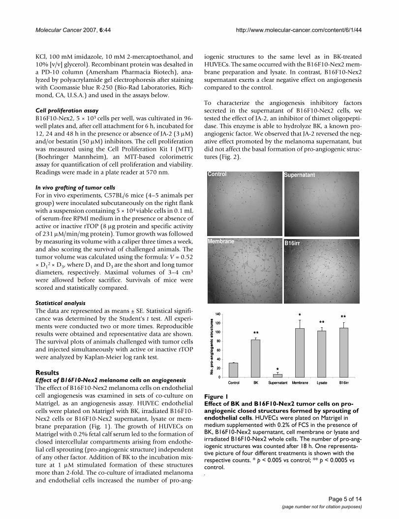
ResultsEffect of B16F10-Nex2 melanoma cells on angiogenesisThe effect of B16F10-Nex2 melanoma cells on endothelialcell angiogenesis was examined in sets of co-culture onMatrigel, as an angiogenesis assay. HUVEC endothelialcells were plated on Matrigel with BK, irradiated B16F10-Nex2 cells or B16F10-Nex2 supernatant, lysate or mem-brane preparation (Fig. 1). The growth of HUVECs onMatrigel with 0.2% fetal calf serum led to the formation ofclosed intercellular compartments arising from endothe-lial cell sprouting (pro-angiogenic structure) independentof any other factor. Addition of BK to the incubation mix-ture at 1 μM stimulated formation of these structuresmore than 2-fold. The co-culture of irradiated melanomaand endothelial cells increased the number of pro-ang-
iogenic structures to the same level as in BK-treatedHUVECs. The same occurred with the B16F10-Nex2 mem-brane preparation and lysate. In contrast, B16F10-Nex2supernatant exerts a clear negative effect on angiogenesiscompared to the control.
To characterize the angiogenesis inhibitory factorssecreted in the supernatant of B16F10-Nex2 cells, wetested the effect of JA-2, an inhibitor of thimet oligopepti-dase. This enzyme is able to hydrolyze BK, a known pro-angiogenic factor. We observed that JA-2 reversed the neg-ative effect promoted by the melanoma supernatant, butdid not affect the basal formation of pro-angiogenic struc-tures (Fig. 2).
Effect of BK and B16F10-Nex2 tumor cells on pro-angiogenic closed structures formed by sprouting of endothelial cellsFigure 1Effect of BK and B16F10-Nex2 tumor cells on pro-angiogenic closed structures formed by sprouting of endothelial cells. HUVECs were plated on Matrigel in medium supplemented with 0.2% of FCS in the presence of BK, B16F10-Nex2 supernatant, cell membrane or lysate and irradiated B16F10-Nex2 whole cells. The number of pro-ang-iogenic structures was counted after 18 h. One representa-tive picture of four different treatments is shown with the respective counts. * p < 0.005 vs control; ** p < 0.0005 vs control.
ControlControl
B16irrB16irrMembraneMembrane
SupernatantSupernatant
0
20
40
60
80
100
120
140
Control BK Supernatant Membrane Lysate B16irr
No
. p
ro-a
ng
iog
en
ic s
tru
ctu
res
*
*
****
**
0
20
40
60
80
100
120
140
Control BK Supernatant Membrane Lysate B16irr
No
. p
ro-a
ng
iog
en
ic s
tru
ctu
res
*
*
****
**
Page 5 of 14(page number not for citation purposes)

Molecular Cancer 2007, 6:44 http://www.molecular-cancer.com/content/6/1/44
Enzymatic activity of B16F10-Nex2 supernatantTo further address the enzymatic activity of B16F10-Nex2supernatant, FRET peptides derived from NT and BK, pre-viously employed to determine the specificity of recom-binant TOP and neurolysin oligopeptidases [38,39], wereused. The melanoma supernatant was able to cleave allthe assayed substrates, except Abz-GFPPFRQ-EDDnp andAbz-rRL-EDDnp (Table 1), which were also resistant torecombinant TOP and neurolysin. The fluorogenic sub-strate Abz-rRL-EDDnp was previously used to identifyneprilisin oligopeptidase [40], which was not detected inthe B16F10-Nex2 supernatant. However, the conditionedmedium cleaved preferentially Abz-GFSPFRQ-EDDnp,which is a very susceptible substrate for TOP.
HPLC analyses of Abz-GFSPFR-EDDnp and Abz-GFSP-FRQ-EDDnp degradation products by B16F10-Nex2supernatant and rTOP are shown in Figure 3. As previ-ously described for TOP and neurolysin [39], Abz-
GFSPFR-EDDnp was cleaved at Phe-Ser bond, but thepresence of glutamine in Abz-GFSPFRQ-EDDnp shiftedthe cleavage to Pro-Phe bond (Fig. 3A and 3B). Melanomasupernatant hydrolyzed these substrates as the recom-binant TOP and neurolysin do (Fig. 3C and 3D), indicat-ing that the melanoma peptidase activities could berelated to these enzymes.
Inhibition of the peptidase activityThe effects of the peptidase inhibitors on the hydrolysis ofAbz-GFSPFRQ-EDDnp by B16F10-Nex2 supernatant,recombinant TOP and neurolysin are shown in Table 2.PMSF (0.1 mM), E-64 (0.1 mM), Z-Pro-Prolinal (1 μM),leupeptin (0.2 mM) and captopril (20 μM) have noeffects. Pro-Ile (1 mM), a specific inhibitor of neurolysin[41], inhibited by ~40% the B16F10-Nex2 supernatantenzymes and ~50% the recombinant neurolysin, but doesnot inhibit TOP. o-Phenanthrolin (4 mM) and JA-2 (3μM) were the most effective inhibitors of both, the stand-ard recombinant enzymes and of the B16F10-Nex2 super-natant. o-Phenanthrolin is a specific inhibitor of metallo-proteases and JA-2 was described as a specific inhibitor ofTOP [37], but it also inhibits neurolysin as we have shownhere. The activities of B16F10-Nex2 supernatant on fluor-
HPLC analysis of FRET peptides degradation by the B16F10-Nex2 supernatantFigure 3HPLC analysis of FRET peptides degradation by the B16F10-Nex2 supernatant. Abz-GFSPFR-EDDnp (A, C) or Abz-GFSPFRQ-EDDnp (B, D) were incubated with recombinant oligopeptidase TOP (A, B), or B16F10-Nex2 supernatant (C, D) in 50 mM Tris-HCl pH 7.4 at 37°C. Reac-tion products were separated by HPLC and were identified by mass spectrometry. Chromatograms developed by fluo-rescence detection at λem. = 420 nm and λex. = 320 nm.
Time (min)
Abz
-GF
Abz
-GF
SP
Flu
ores
cenc
ein
tens
ity
0 10 20
Abz
-GF
0 10 20
Abz
-GF
SP
A B
DC
Time (min)
Abz
-GF
Abz
-GF
SP
Flu
ores
cenc
ein
tens
ity
0 10 20
Abz
-GF
0 10 20
Abz
-GF
SP
A B
DC
Effect of JA-2 on in vitro Matrigel angiogenesis assayFigure 2Effect of JA-2 on in vitro Matrigel angiogenesis assay. HUVECs were plated on Matrigel in medium supplemented with 0.2% of FCS in the presence of BK, B16F10-Nex2 super-natant and JA-2 (thimet oligopeptidase inhibitor). The number of pro-angiogenic structures was counted after 18 h. * p < 0.05 vs control; ** p < 0.005 vs control.
0
20
40
60
80
100
120
Control BK Supernatant JA-2 Supernatant+ JA-2
Supernatant+ BK + JA-2
No
. p
ro-a
ng
iog
enic
str
uct
ure
s
**
*
*
*
0
20
40
60
80
100
120
Control BK Supernatant JA-2 Supernatant+ JA-2
Supernatant+ BK + JA-2
No
. p
ro-a
ng
iog
enic
str
uct
ure
s
**
*
*
*
Table 1: Proteolytic activity of B16F10-Nex2 supernatant and recombinant enzymes on FRET peptides
Abz-peptidyl-EDDnp
Specific activity (nmoles/min/mg protein)
B16F10-Nex2 rTOP rNeurolysin
GFSPFRQ 2.7 ± 0.6 100 ± 9.8 5.5 ± 1.0GFSPFR 0.6 ± 0.1 21 ± 4.2 4.4 ± 0.5GFSIFRQ 0.6 ± 0.1 14 ± 4.4 1.6 ± 0.2GFPPFRQ 0 0 0NKPRRPQ 0.4 ± 0.4 29 ± 3.3 58 ± 8.3RPPGFSPFRQ 1.8 ± 0.1 51 ± 5.5 8.8 ± 0.7rRL 0 0 0
B16F10-Nex2 supernatant and recombinant enzymes were incubated with fluorogenic substrates (20 μM) in 50 mM Tris-HCl, pH 7.4 at 37°C. Hydrolysis was followed by measuring the fluorescence at λem. = 420 nm and λex. = 320 nm. Results are expressed as means ± SD.
Page 6 of 14(page number not for citation purposes)

Molecular Cancer 2007, 6:44 http://www.molecular-cancer.com/content/6/1/44
ogenic quenched substrates are shown in Table 1, whichwere also totally inhibited by o-phenanthrolin and JA-2.
Hydrolysis of neurotensin and bradykininNT represents the only known peptide differentiallycleaved by TOP and neurolysin, therefore it is a very usefulreagent to distinguish these peptidase activities. TOPhydrolyzes the Arg-Arg bond, producing NT1–8 and NT9–13whereas neurolysin cleaves the Pro-Tyr bond, producingNT1–10 and NT11–13. Incubation of NT with melanomasupernatant resulted in the generation of NT1–8 and NT1–
10 (Fig. 4D), the same N-terminal fragments generatedrespectively by cleavage of NT by TOP and neurolysin(Fig. 4A and 4B), that were identified by mass spectrome-try. The same assay was performed in the presence of bes-tatin (50 μM), an aminopeptidase inhibitor. In previousassays, bestatin was able to completely inhibit the super-natant peptidase activity using Phe-MCA as substrate,indicating the presence of an aminopeptidase in themelanoma supernatant (data not shown). In the presenceof bestatin, the fragment of NT10–13 (Fig. 5C) was detectedas confirmed by mass spectrometry. The aminopeptidasepresents in the B16F10-Nex2 supernatant was responsiblefor cleaving the NT C-terminal fragments. The fragmentNT9–13 was not observed in the HPLC due to the presenceof bestatin at exactly the same elution time, as shown inFigure 5A. Bestatin, as expected, was unable to modify theHPLC profile of TOP and neurolysin (Fig. 5A and 5B).
The cleavage of BK at the Phe-Ser bond by TOP and neu-rolysin was previously described [42]. The B16F10-Nex2supernatant cleaved BK at the same site, with the corre-sponding fragments being identified by mass spectrome-try (Fig. 6B). The HPLC of fragments arisen from BKhydrolysis by recombinant TOP was the control shown inFigure 6A.
Cloning of TOP and neurolysin from B16F10-Nex2 melanoma cellsBoth melanoma enzyme cDNAs were cloned, confirmingthe presence of TOP and neurolysin in melanoma cells,and the gene and translated protein sequences were com-pared to the mouse sequences (data not shown).
The cDNA sequence of TOP from melanoma showed asingle nucleotide change by comparing with the mouseenzyme. The point mutation on adenine 300 to guaninewas not able to cause alterations in the amino acidsequence.
The complete sequencing of neurolysin cDNA frommelanoma showed three modifications in relation to themouse gene: thymine 666 to cytosine, thymine 1268 tocytosine and thymine 1316 to cytosine. Only two changespromoted substitutions in the amino acid sequence: Val423 to Ala and Leu 439 to Pro. These changes did notseem to affect the active site of the enzyme and did notinclude amino acids described as important for enzymeactivity [43,44].
Peptidase activity in the culture supernatant, lysate and membrane preparation of B16F10-Nex2 cell lineThe oligopeptidase activities in the culture supernatant,lysate and membrane preparations of B16F10-Nex2melanoma cells were compared. The melanoma cells dis-
HPLC analysis of neurotensin (NT) degradation by B16F10-Nex2 supernatantFigure 4HPLC analysis of neurotensin (NT) degradation by B16F10-Nex2 supernatant. Neurotensin (20 μM) was incubated for 1 h at 37°C with recombinant TOP (A), neuro-lysin (B) or B16F10-Nex2 supernatant (D) in 50 mM Tris-HCl, pH 7.4. The reaction products were separated by HPLC. HPLC profile of melanoma supernatant without NT is shown on panel C. The neurotensin fragments were deter-mined by mass spectrometry.
NT
1-1
0
A22
0
Time (min)
NT
1-8
NT
9-1
3N
T
A
NT
11-
13
5 10 15 20 25
C
5 10 15 20 25
NT
1-1
0N
T 1
-8
NT
D
B
NT
1-1
0
A22
0
Time (min)
NT
1-8
NT
9-1
3N
T
A
NT
11-
13
5 10 15 20 25
C
5 10 15 20 25
NT
1-1
0N
T 1
-8
NT
D
B
A22
0
Time (min)
NT
1-8
NT
9-1
3N
T
A
NT
11-
13
5 10 15 20 25
C
5 10 15 20 25
NT
1-1
0N
T 1
-8
NT
D
B
Table 2: Effect of inhibitors on the hydrolysis of Abz-GFSPFRQ-EDDnp by B16F10-Nex2 supernatant and recombinant TOP and Neurolysin
Inhibitors Relative hydrolysis (%)1
B16F10-Nex2 rTOP rNeurolysin
Control (no inhibitor) 100 100 100o-phenanthrolin (4 mM) 0 0 0JA-2 (3 μM) 2.6 ± 2.1 0.6 ± 1.2 1 ± 1Pro-Ile (1 mM) 60 ± 6 87 ± 5 52 ± 4PMSF (0.1 mM) 94 ± 6 91 ± 4 93 ± 4E64 (0.1 mM) 88 ± 3 98 ± 2 99 ± 2Leupeptin (0.2 mM) 81 ± 6 96 ± 2 96 ± 4Z-Pro-Prolinal (1 μM) 93 ± 1 88 ± 3 92 ± 4Captopril (20 μM) 92 ± 3 97 ± 2 98 ± 2
1100% hydrolysis represents the cleavage of 20 μM of Abz-GFSPFRQ-EDDnp by B16F10-Nex2 supernatant and recombinant enzymes during 5 min at 37°C in 50 mM Tris-HCl, pH 7.4. The values are means of three independent experiments.
Page 7 of 14(page number not for citation purposes)

Molecular Cancer 2007, 6:44 http://www.molecular-cancer.com/content/6/1/44
play the greatest oligopeptidase activity in the lysate asshown by hydrolysis of Abz-GFSPFRQ-EDDnp in Table 3.The activity in the supernatant is 10-fold less that of thelysate, when the results are expressed in enzyme activityper cell. If the assay is standardized by specific activity, thesupernatant and lysate values were 1.39 and 3.22 μM/minper mg of protein, respectively, because the protein con-tent in the lysate is approximately 4-fold that in the super-natant. The total activity based on the volume of thefractions showed that the lysate had much more oli-gopeptidase activity than the supernatant. The activity inthe membrane fraction was very low, but the protein wasclearly detected by Western blotting (data not shown). Todemonstrate the expression levels of TOP and neurolysinin melanoma cells, B16F10-Nex2 fractions were submit-ted to immunoblotting, using specific antibodies againstthese two oligopeptidases. We detected TOP expression inthe supernatant, lysate and membrane preparations ofB16F10-Nex2 melanoma cells (data not shown). Neuro-lysin expression was not detected using this method. Thespecific anti-TOP antibody inhibited 80% of the B16F10-Nex2 catalytic activity, using the FRET peptide Abz-GFSP-FRQ-EDDnp, whereas the anti-neurolysin antibodyinhibited only 20% of the melanoma enzyme activity(Table 3).
Tumor cell proliferation assayA proliferation assay in vitro was carried out in the pres-ence of JA-2 and bestatin and the results are shown in Fig-ure 7. JA-2 and bestatin decreased B16F10-Nex2 cellgrowth 50% and 56% respectively in 48 h. A synergisticinhibitory effect (88% inhibition of proliferation in 48 h)was observed when both inhibitors were added togetherin the melanoma culture.
Effect of TOP on tumor development in vivoTo assess the effect of TOP on the tumor development invivo, tumor cells were injected subcutaneously in syn-geneic mice together with active or inactive rTOP at con-
HPLC analysis of BK degradation by B16F10-Nex2 superna-tantFigure 6HPLC analysis of BK degradation by B16F10-Nex2 supernatant. BK (20 μM) was incubated for 1 h at 37°C with recombinant TOP (A) or B16F10-Nex2 supernatant (B) in 50 mM Tris-HCl, pH 7.4. The reaction products were sep-arated by HPLC. Bradykinin fragments were determined by mass spectrometry.
� �� �� ���������
������
������
�������
�������
����
��
����
���
� �� �� ���������
������
������
�������
�������
����
��
����
���
HPLC analysis of neurotensin (NT) degradation by B16F10-Nex2 supernatant in the presence of bestatinFigure 5HPLC analysis of neurotensin (NT) degradation by B16F10-Nex2 supernatant in the presence of besta-tin. Neurotensin (20 μM) was incubated for 1 h at 37°C with recombinant TOP (A), neurolysin (B) or B16F10-Nex2 supernatant (D) in the presence of bestatin in 50 mM Tris-HCl, pH 7.4. The reaction products were separated by HPLC. HPLC profile of melanoma supernatant in the pres-ence of bestatin without NT is shown on panel C. The neu-rotensin fragments were determined by mass spectrometry.
NT
1-8
Bt
Bt B
tN
T11
-13
Bt
NT
1-8
NT
1-10
NT
11-1
3
NT
NT
Time (min)
A B
DC
5 10 15 20 25 5 10 15 20 25
A22
0
NT
1-8 B
tB
t Bt
NT
11-1
3B
t
NT
1-8
NT
1-10
NT
11-1
3
NT
NT
Time (min)
A B
DC
5 10 15 20 25 5 10 15 20 25
A22
0
Page 8 of 14(page number not for citation purposes)

Molecular Cancer 2007, 6:44 http://www.molecular-cancer.com/content/6/1/44
centrations that did not affect the growth of tumor cells invitro.
Tumor cell implantation in the presence of 8 μg of activerTOP (specific activity: 231 μM/min per mg) was followedby delayed tumor growth and prolonged survival ofinjected mice (p = 0.034) (Fig. 8A and 8B). In contrastthere was no significant difference (p = 0.32) in the tumordevelopment when B16F10-Nex2 cells were injected inthe presence of inactive rTOP (Fig. 8C and 8D).
Effect of TOP in angiogenesis assay on MatrigelWe examined the ability of rTOP to stimulate pro-ang-iogenic structures by HUVECs cultured on Matrigel. Thesestructures arise from sprouting of endothelial cells andformation of closed intercellular compartments that canbe quantified. As shown in Figure 1, BK stimulated the for-mation of pro-angiogenic structures >2-fold. TOP alonewas able to inhibit endothelial cell extensions and reversethe angiogenic stimulus promoted by BK (Fig. 9). Further,NT and angiotensin II (A-II), other peptides hydrolyzedby TOP, also stimulated angiogenesis.
Inhibition of rTOP activity by SNAPTo verify the possible inhibition of rTOP by nitric oxide,which could suggest a way of controlling the activity of theendo-oligopeptidase, we carried out a kinetic assay withthe recombinant enzyme and S-nitroso-N-acetylpenicilla-mine (SNAP), as NO donor. SNAP (1 mM to 100 μM) wasincubated with rTOP for 10 minutes and then the fluoro-genic substrate Abz-GFSPFRQ-EDDnp was added to thereaction. The result in Table 4 shows that SNAP inhibitedTOP hydrolytic activity using substrate Abz-GFSPFRQ-EDDnp, in a dose-dependent manner, but not time-dependent kinetics (data not shown). At 100 μM, SNAPwas unable to inhibit TOP. Addition of 1, 4-dithiothreitol
Effect of active and inactive rTOP on tumor cell development and animal survival after subcutaneous implantation of B16F10-Nex2 melanoma cellsFigure 8Effect of active and inactive rTOP on tumor cell development and animal survival after subcutaneous implantation of B16F10-Nex2 melanoma cells. 5 × 10⁴
viable cells were injected subcutaneously with 8 μg of active rTOP (A, B, open circle), inactive rTOP (C, D, open circle), and PBS (control, solid circle) in C57Bl/6 mice (4–5 animals per group). The tumor volume was measured every 2–3 days and a maximal volume of 3 cm3 was allowed before sacrifice. (B, D), tumor volume of individual animals; (A, C), survival plots. Statistical analysis of survivals was performed using Kaplan-Meier test.
Days after tumor cell implantation
20 30 40 50 60 70 80
% s
urvi
val
0
20
40
60
80
100
120
0
1
2
3
4
5
15 20 25 29 34 39 43 48 55
Days after tumor cell implantation
Tum
or
volu
me (
cm
3)
0
1
2
3
4
17 20 22 24 27 29 31
Days after tumor cell implantation
Tum
or
volu
me (
cm
3)
B D
Days after tumor cell implantation
20 30 40 50 60 70 80
% s
urvi
val
0
20
40
60
80
100
120A Cp=0,034 p=0,32
Days after tumor cell implantation
20 30 40 50 60 70 80
% s
urvi
val
0
20
40
60
80
100
120
0
1
2
3
4
5
15 20 25 29 34 39 43 48 55
Days after tumor cell implantation
Tum
or
volu
me (
cm
3)
0
1
2
3
4
17 20 22 24 27 29 31
Days after tumor cell implantation
Tum
or
volu
me (
cm
3)
B D
Days after tumor cell implantation
20 30 40 50 60 70 80
% s
urvi
val
0
20
40
60
80
100
120A C
Days after tumor cell implantation
20 30 40 50 60 70 80
% s
urvi
val
0
20
40
60
80
100
120
0
1
2
3
4
5
15 20 25 29 34 39 43 48 55
Days after tumor cell implantation
Tum
or
volu
me (
cm
3)
0
1
2
3
4
17 20 22 24 27 29 31
Days after tumor cell implantation
Tum
or
volu
me (
cm
3)
B D
Days after tumor cell implantation
20 30 40 50 60 70 80
% s
urvi
val
0
20
40
60
80
100
120A Cp=0,034 p=0,32
Table 3: Oligopeptidase activities in the culture supernatant, cell lysate and membrane fraction of B16F10-Nex2 cells
Cell fraction Specific activity (μM/min per mg) Activity per cell (pM/min per 103 cells) Total activity (μM/min)
Supernatant 1.39 ± 0.24 36 ± 16 3.36 ± 0.15Lysate 3.22 ± 0.96 326 ± 15 31.56 ± 0.16Lysate + Anti-TOP Ab 0.64 ± 0.19 65 ± 5 6.31 ± 0.03Lysate + Anti-Neurolysin Ab 2.58 ± 0.77 261 ± 5 25.25 ± 0.13Membrane* < 0.01 3 ± 0.5 0.2 ± 0.01
Culture supernatant, cell lysate and membrane preparation of B16F10-Nex2 cells were incubated with the fluorogenic substrate Abz-GFSPFRQ-EDDnp (20 μM) in 50 mM Tris-HCl, pH 7.4 at 37°C. Enzyme activity was followed by measuring the fluorescence at λem. = 420 nm and λex. = 320 nm. Results are expressed as means ± SD. Ab= antibodies, used at 1:300.*Although the activity of TOP in the membrane was low, the protein was clearly detected by Western blotting with anti-TOP antibody.
B16F10-Nex2 proliferation assay in the presence of JA-2 and/or bestatin inhibitorsFigure 7B16F10-Nex2 proliferation assay in the presence of JA-2 and/or bestatin inhibitors. 5 × 10³ B16F10-Nex2 cells were cultivated in 96-well plates, incubated for 12, 24 and 48 h with JA-2 (3 μM) and/or bestatin (50 μM) inhibitors, and the cell proliferation was measured using MTT in com-parison with Controls. *p < 0.05.
0
0,1
0,2
0,3
0,4
Control Bestatin JA-2 Bestatin + JA-2
A57
0
12h24h48h
**
*
0
0,1
0,2
0,3
0,4
Control Bestatin JA-2 Bestatin + JA-2
A57
0
12h24h48h
**
*
Page 9 of 14(page number not for citation purposes)

Molecular Cancer 2007, 6:44 http://www.molecular-cancer.com/content/6/1/44
(DTT) in the assay before the fluorogenic substrate pre-vented TOP inhibition by SNAP.
DiscussionWe describe in the present work the stimulating effect onangiogenesis of irradiated melanoma B16F10-Nex2tumor cells using HUVEC on Matrigel substrate. Amelanoma cell lysate and membrane preparation showedthe same effect of whole irradiated tumor cells, suggestinga distribution of pro-angiogenic factors in both mem-brane and cytoplasm of the tumor cells. In contrast,B16F10-Nex2 conditioned medium inhibited endothelialcell sprouting and the formation of pro-angiogenic struc-tures. The negative effect on angiogenesis was reversed bythe thimet oligopeptidase inhibitor JA-2, suggesting theanti-angiogenic role of this or a similar secreted oli-gopeptidase into the melanoma culture supernatant.
The presence of TOP and neurolysin in melanoma cellsand culture supernatants was confirmed by hydrolytic
assays and by cloning and sequencing the correspondingcDNAs from B16F10-Nex2 cells. The hydrolysis of NT, BKand FRET peptides were consistent with the presence ofZn-dependent oligopeptidases with a catalytic activitysimilar to that of both TOP and neurolysin. In addition,the enzymes of the melanoma supernatant hydrolyzed thesubstrate Abz-GFSPFRQ-EDDnp at the Pro-Phe bond asdescribed for TOP and neurolysin, indicating that themelanoma activity had the same specificity as of therecombinant mouse enzymes (Figs 3C and 3D).
The complete inhibition of melanoma peptidase by o-phenanthrolin as well as by JA-2, and the partial inhibi-tion by Pro-Ile give support to the functional similarity ofmelanoma enzymes with the oligopeptidases TOP andneurolysin. The absence of inhibition of peptidase activityby PMSF, E-64, Z-Pro-Prolinal, leupeptin and captoprilexcludes the presence of other peptidase classes eventuallyresponsible for the cleavage of Abz-GFSPFRQ-EDDnp, thesubstrate used in these assays. The generation of NT9–13and NT10–13 in the presence of bestatin suggested that anaminopeptidase was the most likely enzyme responsiblefor degradation of the C-terminal fragment of NT.
The cDNA sequencing of melanoma enzymes showed fewmutations in comparison with the mouse counterparts.The only modification found in the melanoma TOP genesequence did not cause an alteration in the amino acidsequence. The melanoma neurolysin gene showed threemodifications in relation to mouse gene sequence, butonly two changes promoted substitutions in the aminoacid sequence away from the conserved Zn-binding cata-lytic site (HEFGH) or from other amino acids described asimportant to enzyme activity [43,44].
We show here that significant TOP and neurolysin-likeactivities can be detected in conditioned media, lysate andmembrane preparations from melanoma cells. The oli-gopeptidase activity per cell was 10-fold greater in thelysate than supernatant. The activity in the cell membranepreparation was scarce. When the assay was normalizedby specific activity, the supernatant and lysate values weresimilar, since the protein content in the lysate is approxi-mately 4-fold that in the supernatant. Using specific anti-bodies against the studied oligopeptidases, we showedthat TOP activity in melanoma cells is significantly greaterthan that of neurolysin. TOP expression as detected byimmunoblotting was prominent in the supernatant,lysate and membrane fraction of B16F10-Nex2 cells.
The importance of the oligopeptidases for melanoma cellsin vitro was assessed by proliferation assay in the presenceof JA-2 and bestatin. We observed a decreased B16F10-Nex2 cell proliferation in the presence of JA-2, bestatinand a greater inhibition with both. Presumably both
Table 4: Effect of the NO donor SNAP on the hydrolysis of Abz-GFSPFRQ-EDDnp by rTOP
SNAP Hydrolysis (%)1
Control (no inhibitor) 1001 mM 38 ± 3500 μM 50 ± 3250 μM 83 ± 4100 μM 98 ± 21 mM + DTT 0.5 mM 100 ± 1
1100% hydrolysis represents the cleavage of 20 μM of Abz-GFSPFRQ-EDDnp by rTOP during 5 min at 37°C in 50 mM Tris-HCl, pH 7.4.
Effect of rTOP on in vitro Matrigel angiogenesis assayFigure 9Effect of rTOP on in vitro Matrigel angiogenesis assay. HUVECs were plated on Matrigel in medium supple-mented with 0.2% of FCS in the presence of BK (1 μM), rTOP (specific activity: 231 μM/min/mg), BK+ rTOP, NT (1 μM) and A-II (1 μM). The number of pro-angiogenic struc-tures was counted after 18 h. * p < 0.05 vs control.
0
20
40
60
80
100
120
140
Control BK BK + rTOP rTOP NT A-II
No
. pro
-an
gio
gen
ic s
tru
ctu
res
*
0
20
40
60
80
100
120
140
Control BK BK + rTOP rTOP NT A-II
No
. pro
-an
gio
gen
ic s
tru
ctu
res
*
Page 10 of 14(page number not for citation purposes)

Molecular Cancer 2007, 6:44 http://www.molecular-cancer.com/content/6/1/44
inhibitors may act intracellularly, inhibiting oligopepti-dases and aminopeptidases that are essential formelanoma cells. The secretion of both enzymes, TOP andneurolysin, might, however, influence tumor growth invivo by affecting angiogenesis. Mice injected with activerTOP at a concentration that was not toxic in vitro,showed a delay in tumor development and increased sur-vival of animals compared with mice challenged withtumor cells and inactive rTOP, indicating that the activeenzyme and not only the protein was necessary for thiseffect. A possible target of rTOP activity in the tumormicroenvironment could be kinins stimulating angiogen-esis.
Tumors require an adequate supply of oxygen, metabo-lites and an effective way to remove waste products [45].The generation of new blood vessels for tumor blood sup-ply is thus a rate-limiting step in tumor progression, beinga prerequisite for the rapid clonal expansion associatedwith the formation of macroscopic tumors [46]. Experi-mental data suggest that the angiogenic stimulation[46,47] is activated during the early stages of tumourdevelopment [48-51]. During development of cutaneousmelanoma in humans a similar stage-specific stimulationis also evident [52]. Tumors appear to activate the ang-iogenic switch by changing the balance of angiogenesisinducers and counteracting inhibitors, so that tumor neo-vascularization and consequent growth depends on howheavily the balance tips towards angiogenesis [53,54].One strategy for shifting the balance involves proteasesthat can control the bioavailability of angiogenic activa-tors and inhibitors.
A prototype of pro-angiogenic molecule is bradykinin.Evidence suggests that BK may be one of the primarymediators responsible for tumor angiogenesis and, conse-quently, of tumor growth [28-33]. It is well known thatendothelial cells can synthesize and secrete tissue kal-likrein [55]. Schmaier et al [56] have also shown theexpression of high molecular weight kininogen in humanumbilical vein endothelial cells (HUVEC). The generationof the vasoactive peptide bradykinin from HUVEC-boundhigh molecular weight kininogen is also known [57].Therefore, endothelial cells, particularly those used in thepresent study (HUVEC) do produce kininogen and kal-likrein and generate BK. Such functional activities ofendothelial cells are stimulated in Matrigel. The inhibitoryeffect of active rTOP inoculated with tumor cells, couldwell involve the hydrolysis of bradykinin, shifting the bal-ance of angiogenic/anti-antiangiogenic factors at an earlystage of tumor implantation. In agreement with this wehave shown in an angiogenesis assay with HUVEC onMatrigel that rTOP was able to reverse the angiogenicstimulation promoted by BK. Similarly to BK, NT and A-II, two peptides that are hydrolyzed by TOP, were able to
stimulate the endothelial cell sprouting. These peptidesare further examples of positive modulators of the ang-iogenic process.
The results suggest that TOP affects angiogenesis in vitroand in vivo, at concentrations that did not inhibitB16F10-Nex2 tumor cells directly. The secretion of TOPby tumor cells would therefore favor an anti-angiogenicresponse in balance with the pro-angiogenic stimuli byother tumor proteases expressed for instance at the cellsurface and released in the medium. We incubatedHUVEC cells with CA-074, a cathepsin B inhibitor, in theMatrigel angiogenesis assay and showed stimulation ofangiogenesis. The same inhibitor enhanced the pro-ang-iogenic effect induced by B16F10-Nex2 lysate, tumor cellmembrane, and in the co-culture of B16F10-Nex2 withHUVEC cells (data not shown). References to the role ofcathepsin B on tumor angiogenesis are contradictory. Inhuman tumors, there is evidence of a positive correlationbetween the level of cathepsin B and angiogenesis [58],and the inhibition of cathepsin B expression was associ-ated with angiogenesis suppression [59]. In contrast, thegeneration of endostatin by cathepsin B could block theangiogenesis in many tumor systems [60]. The role ofcathepsins in melanoma requires additional studies inface of the multienzymatic complex of the tumor micro-environment. Clearly, in the in vitro Matrigel invasionassays, cathepsins B and L increase the angiogenesis-inde-pendent invasive capacity of tumor cells [61]. Wedescribed a more invasive B16F10 clone (Nex2B) as com-pared to a less invasive one (2D), though with a greatercapacity of lung colonization than 2B, based on the extra-cellular rather than intracellular accumulation of cathep-sins B, D, and L [1]. Nevertheless, an intracellular role forcathepsin B in matrix degradation has been identified[62], and also, three forms of extracellularly active cathe-psin B and two forms of active cathepsin L have beendescribed in the highly invasive melanoma cell line MV3[63]. These isoforms add to the complexity of the systemand demand a careful study to unravel their role on tumorgrowth.
An additional control of angiogenesis and tumor growthmay exist based on the local release of NO. Kashiwagi etal. [64] using intravital microscopy demonstrated in a B16murine melanoma model that eNOS from vascularendothelial cells is the predominant generator of NO. NOreleased by endothelial cells could be an inhibitory ligandof TOP activity. The inhibition of enzymes by nitric oxidehas been demonstrated in the cysteine proteases [65-67],but not metallo-proteases. TOP and dipeptidylpeptidaseIV (DPP IV) were not inhibited by the NO donors sodiumnitroprusside(SNP) and 3-morpholinosydnoimine (SIN-1) in the 1–100 μM range [68]. Both compounds, how-ever, were shown to release peroxynitrite, a reactive nitro-
Page 11 of 14(page number not for citation purposes)

Molecular Cancer 2007, 6:44 http://www.molecular-cancer.com/content/6/1/44
gen species that primarily can nitrate tyrosine residues[69]. Site-directed mutation of Tyr residues at positions612 and 613 in TOP and neurolysin, respectively, stronglyreduced kcat/KM for both enzymes [70].
Nitrosothiols, which may act as NO reservoirs, could nit-rosate cysteine residues [71], such as those that play animportant role in TOP activity [72]. Presently, we exposedrTOP to SNAP a nitrosothiol, and the activity of theenzyme was inhibited in a concentration-dependent man-ner (250 μM to 1 mM range). Such inhibition was abol-ished in the presence of DTT. This result should beconsidered in the perspective of a tumor microenviron-ment with hydrophobic niches that may increase the effi-ciency of NO-based regulatory reactions [73], potentiallyinfluencing angiogenesis and tumor growth.
ConclusionThe present work describes TOP and neurolysin oli-gopeptidases in B16F10-Nex2 melanoma cells and showsthe importance of these enzymes for tumor proliferationin vitro and in vivo. Recombinant TOP protected miceagainst the subcutaneous challenge with murinemelanoma cells. The results suggest that secreted thimetoligopeptidase in tumor cells and bradykinin are twoantagonist factors that may regulate or trigger the ang-iogenic switch essential for melanoma growth. A regula-tory role of NO or S-nitrosothiols on TOP activity issuggested. Together with endogenous inhibitors, endo-oligopeptidases secreted by tumor cells can also regulateangiogenesis and might also be studied, in adequate pro-tocols, as potential anti-tumor agents.
AbbreviationsTOP or EC 24.15, metallo-endopeptidase EC 3.4.24.15;EC 24.16, metallo-endopeptidase EC 3.4.24.16; rTOP,recombinant thimet oligopeptidase; FRET, fluorescenceresonance energy transfer; HUVEC, human vein endothe-lial cell; FCS, fetal calf serum; NT, neurotensin; BK, brady-kinin; A-II, angiotensin II; JA-2, N-[1(R,S)-carboxy-3-phenylpropyl]-Ala-Aib-Tyr-p-aminobenzoate; IPTG, iso-propyl β-D-thiogalactoside; Abz, o-aminobenzoic acid;EDDnp, ethylenediamine-2,4-dinitrophenyl; E-64, trans-epoxysuccinyl-L-leucylamido-(4-guanido)butene; SNAP,S-nitroso-N-acetylpenicillamine; DTT, 1,4-dithiothreitol.
Competing interestsThe author(s) declare that they have no competing inter-ests.
Authors' contributionsTP designed and performed all biochemical and cell bio-logical experiments, carried out data analysis and draftedand conceived the manuscript. AKC designed and helpedin biochemical assays. EGR assisted in cell biological
experiments, in vivo experiments and their design. VOdesigned and performed the mass spectrometry analysisand provided recombinant TOP and neurolysin. HPMassisted in the experiments of NO inhibition and SNAPassay. MAJ provided all FRET and free peptides, used assubstrates. LJ provided the background knowledge onendo-oligopeptidases, including substrates and inhibi-tors. LRT, as Chairman of UNONEX, was the seniorauthor who conceived the study, coordinated its execu-tion, participated in its design and drafted and producedthe final version of the manuscript. All authors read andapproved the present version of the manuscript.
AcknowledgementsThis work was supported by Fundação de Amparo à Pesquisa do Estado de São Paulo (FAPESP), Brazil. We are grateful to A.C.M. Camargo for the JA-2 inhibitor, and K. Alves for bestatin. LRT, AKC, EGR, MAJ and LJ are research fellows from the CNPq.
References1. Freitas ZFO, Rodrigues EG, Oliveira V, Carmona AK, Travassos LR:
Melanoma heterogeneity: differential, invasive, metastaticproperties and profiles of cathepsin B, D and L activities insubclones of the B16F10-NEX2 cell line. Melanoma Res 2004,14:333-344.
2. Vincent B, Beaudet A, Dauch P, Vincent JP, Checler F: Distinct prop-erties of neuronal and astrocytic endopeptidase 3.4.24.16: astudy on differentiation, subcellular distribution, and secre-tion processes. J Neurosci 1996, 16:5049-5059.
3. Serizawa A, Dando PM, Barrett AJ: Characterization of a mito-chondrial metallopeptidase reveals neurolysin as a homo-logue of thimet oligopeptidase. J Biol Chem 1995,270:2092-2098.
4. Shrimpton CN, Smith AI, Lew RA: Soluble metalloendopepti-dases and neuroendocrine signaling. Endocr Rev 2002,23:647-664.
5. Chu TG, Orlowski M: Active site directed N-carboxymethylpeptide inhibitors of a soluble metalloendopeptidase fromrat brain. Biochemistry 1984, 23:3598-3603.
6. Oliveira V, Campos M, Melo RL, Ferro ES, Camargo AC, Juliano MA,Juliano L: Substrate specificity characterization of recom-binant metallo oligo-peptidases thimet oligopeptidase andneurolysin. Biochemistry 2001, 40:4417-4425.
7. Barrett AJ, Brown MA, Dando PM, Knight CG, McKie N, RawlingsND, Serizawa A: Thimet oligopeptidase and oligopeptidase Mor neurolysin. Methods Enzymol 1995, 248:529-556.
8. Krause DR, Piva TJ, Brown SB, Ellem KA: Characterization andlocalization of mitochondrial oligopeptidase (MOP) (EC3.4.24.16) activity in the human cervical adenocarcinomacell line HeLa. J Cell Biochem 1997, 66:297-308.
9. Massarelli EE, Casatti CA, Kato A, Camargo AC, Bauer JA, GlucksmanMJ, Roberts JL, Hirose S, Ferro ES: Differential subcellular distri-bution of neurolysin (EC 3.4.24.16) and thimet oligopepti-dase (EC 3.4.24.15) in the rat brain. Brain Res 1999,851:261-265.
10. Crack PJ, Wu TJ, Cummins PM, Ferro ES, Tullai JW, Glucksman MJ,Roberts JL: The association of metalloendopeptidase EC3.4.24.15 at the extracellular surface of the AtT-20 cellplasma membrane. Brain Res 1999, 835:113-124.
11. Jeske NA, Glucksman MJ, Roberts JL: EP24.15 is associated withlipid rafts. J Neurosci Res 2003, 74:468-473.
12. Fontenele-Neto JD, Massarelli EE, Gurgel Garrido PA, Beaudet A,Ferro ES: Comparative fine structural distribution ofendopeptidase 24.15 (EC3.4.24.15) and 24.16 (EC3.4.24.16)in rat brain. J Comp Neurol 2001, 438:399-410.
13. Woulfe J, Checler F, Beaudet A: Light and Electron MicroscopicLocalization of the Neutral Metalloendopeptidase EC3.4.24.16 in the Mesencephalon of the Rat. Eur J Neurosci 1992,4:1309-1319.
Page 12 of 14(page number not for citation purposes)

Molecular Cancer 2007, 6:44 http://www.molecular-cancer.com/content/6/1/44
14. Jeske NA, Glucksman MJ, Roberts JL: MetalloendopeptidaseEC3.4.24.15 is constitutively released from the exofacial leaf-let of lipid rafts in GT1-7 cells. J Neurochem 2004, 90:819-828.
15. Ferro ES, Tullai JW, Glucksman MJ, Roberts JL: Secretion of met-alloendopeptidase 24.15 (EC 3.4.24.15). DNA Cell Biol 1999,18:781-789.
16. Garrido PA, Vandenbulcke F, Ramjaun AR, Vincent B, Checler F,Ferro E, Beaudet A: Confocal microscopy reveals thimet oli-gopeptidase (EC 3.4.24.15) and neurolysin (EC 3.4.24.16) inthe classical secretory pathway. DNA Cell Biol 1999, 18:323-331.
17. Rioli V, Kato A, Portaro FC, Cury GK, te KK, Vincent B, Checler F,Camargo AC, Glucksman MJ, Roberts JL, Hirose S, Ferro ES: Neu-ropeptide specificity and inhibition of recombinant isoformsof the endopeptidase 3.4.24.16 family: comparison with therelated recombinant endopeptidase 3.4.24.15. Biochem BiophysRes Commun 1998, 250:5-11.
18. Molineaux CJ, Lasdun A, Michaud C, Orlowski M: Endopeptidase-24.15 is the primary enzyme that degrades luteinizing hor-mone releasing hormone both in vitro and in vivo. J Neuro-chem 1988, 51:624-633.
19. Barelli H, Fox-Threlkeld JE, Dive V, Daniel EE, Vincent JP, Checler F:Role of endopeptidase 3.4.24.16 in the catabolism of neuro-tensin, in vivo, in the vascularly perfused dog ileum. Br J Phar-macol 1994, 112:127-132.
20. Da Silva A, Dhuy J, Waeldele F, Bertrand C, Landry Y: Endopepti-dase 24.15 modulates bradykinin-induced contraction inguinea-pig trachea. Eur J Pharmacol 1992, 212:97-99.
21. Mentlein R, Dahms P: Endopeptidases 24.16 and 24.15 areresponsible for the degradation of somatostatin, neuro-tensin, and other neuropeptides by cultivated rat corticalastrocytes. J Neurochem 1994, 62:27-36.
22. Vincent B, Dive V, Yiotakis A, Smadja C, Maldonado R, Vincent JP,Checler F: Phosphorus-containing peptides as mixed inhibi-tors of endopeptidase 3.4.24.15 and 3.4.24.16: effect on neu-rotensin degradation in vitro and in vivo. Br J Pharmacol 1995,115:1053-1063.
23. Vincent B, Jiracek J, Noble F, Loog M, Roques B, Dive V, Vincent JP,Checler F: Effect of a novel selective and potent phosphinicpeptide inhibitor of endopeptidase 3.4.24.16 on neurotensin-induced analgesia and neuronal inactivation. Br J Pharmacol1997, 121:705-710.
24. Linz W, Wiemer G, Gohlke P, Unger T, Scholkens BA: Contribu-tion of kinins to the cardiovascular actions of angiotensin-converting enzyme inhibitors. Pharmacol Rev 1995, 47:25-49.
25. Schriefer JA, Broudy EP, Hassen AH: Endopeptidase inhibitorsdecrease myocardial ischemia/reperfusion injury in an invivo rabbit model. J Pharmacol Exp Ther 1996, 278:1034-1039.
26. Schriefer JA, Broudy EP, Hassen AH: Inhibitors of bradykinin-inactivating enzymes decrease myocardial ischemia/reper-fusion injury following 3 and 7 days of reperfusion. J PharmacolExp Ther 2001, 298:970-975.
27. Smith AI, Lew RA, Shrimpton CN, Evans RG, Abbenante G: A novelstable inhibitor of endopeptidases EC 3.4.24.15 and 3.4.24.16potentiates bradykinin-induced hypotension. Hypertension2000, 35:626-630.
28. Ishihara K, Kamata M, Hayashi I, Yamashina S, Majima M: Roles ofbradykinin in vascular permeability and angiogenesis in solidtumor. Int Immunopharmacol 2002, 2:499-509.
29. Hu DE, Fan TPD: [Leu(8)]Des-Arg(9)-Bradykinin inhibits theangiogenic effect of bradykinin and interleukin-1 in rats. Br JPharmacol 1993, 109:14-17.
30. Matsumura Y, Maruo K, Kimura M, Yamamoto T, Konno T, Maeda H:Kinin-generating cascade in advanced cancer patients and invitro study. Jpn J Cancer Res 1991, 82:732-741.
31. Wu J, Akaike T, Maeda H: Modulation of enhanced vascular per-meability in tumors by a bradykinin antagonist, a cyclooxy-genase inhibitor, and a nitric oxide scavenger. Cancer Res1998, 58:159-165.
32. Hayashi I, Amano H, Yoshida S, Kamata K, Kamata M, Inukai M, FujitaT, Kumagai Y, Furudate S, Majima M: Suppressed angiogenesis inkininogen-deficiencies. Lab Invest 2002, 82:871-880.
33. Ikeda Y, Hayashi I, Kamoshita E, Yamazaki A, Endo H, Ishihara K,Yamashina S, Tsutsumi Y, Matsubara H, Majima M: Host stromalbradykinin B2 receptor signaling facilitates tumor-associ-ated angiogenesis and tumor growth. Cancer Res 2004,64:5178-5185.
34. Bradford MM: A rapid and sensitive method for the quantita-tion of microgram quantities of protein utilizing the princi-ple of protein-dye binding. Anal Biochem 1976, 72:248-254.
35. Chagas JR, Juliano L, Prado ES: Intramolecularly quenched fluor-ogenic tetrapeptide substrates for tissue and plasma kal-likreins. Anal Biochem 1991, 192:419-425.
36. Hirata I, Cezari MHC, Nakaie CR, Boshcov P, Ito AS, Juliano MA,Juliano L: Internally quenched fluorogenic protease sub-strates: Solid-phase synthesis and fluorescent spectroscopyof peptides containing ortho-aminobenzoil-dinitrophenylgroups as donor-acceptor pairs. Lett Peptide Sci 1994, 1:299-308.
37. Shrimpton CN, Abbenante G, Lew RA, Smith AI: Development andcharacterization of novel potent and stable inhibitors ofendopeptidase EC 3.4.24.15. Biochem J 2000, 345:351-356.
38. Oliveira V, Campos M, Hemerly JP, Ferro ES, Camargo AC, JulianoMA, Juliano L: Selective neurotensin-derived internallyquenched fluorogenic substrates for neurolysin (EC3.4.24.16): comparison with thimet oligopeptidase (EC3.4.24.15) and neprilysin (EC 3.4.24.11). Anal Biochem 2001,292:257-265.
39. Camargo AC, Gomes MD, Reichl AP, Ferro ES, Jacchieri S, Hirata IY,Juliano L: Structural features that make oligopeptides suscep-tible substrates for hydrolysis by recombinant thimet oli-gopeptidase. Biochem J 1997, 324(Pt 2):517-522.
40. Medeiros MA, Franca MS, Boileau G, Juliano L, Carvalho KM: Specificfluorogenic substrates for neprilysin (neutral endopeptidase,EC 3.4.24.11) which are highly resistant to serine- and met-alloproteases. Braz J Med Biol Res 1997, 30:1157-1162.
41. Dauch P, Vincent JP, Checler F: Specific inhibition of endopepti-dase 24.16 by dipeptides. Eur J Biochem 1991, 202:269-276.
42. Norman MU, Reeve SB, Dive V, Smith AI, Lew RA: Regulation ofcardiovascular signaling by kinins and products of similarconverting enzyme systems – Endopeptidases 3.4.24.15 and24.16 in endothelial cells: potential role in vasoactive peptidemetabolism. Am J Physiol Heart Circ Physiol 2003,284:H1978-H1984.
43. Ray K, Hines CS, Coll-Rodriguez J, Rodgers DW: Crystal structureof human thimet oligopeptidase provides insight into sub-strate recognition, regulation, and localization. J Biol Chem2004, 279:20480-20489.
44. Ray K, Hines CS, Rodgers DW: Mapping sequence differencesbetween thimet oligopeptidase and neurolysin implicateskey residues in substrate recognition. Protein Sci 2002,11:2237-2246.
45. Papetti M, Herman IM: Mechanisms of normal and tumor-derived angiogenesis. Am J Physiol Cell Physiol 2002,282:C947-C970.
46. Hanahan D, Weinberg RA: The hallmarks of cancer. Cell 2000,100:57-70.
47. Folkman J: Tumor angiogenesis. In Cancer Medicine Edited by: Hol-land JF, Bast RC, Morton DL, Frei E, Kufe DW, Weichselbaum RR.Baltimore, MD: Williams and Wilkins; 1997:181-204.
48. Guidi AJ, Abu-Jawdeh G, Berse B, Jackman RW, Tognazzi K, DvorakHF, Brown LF: Vascular permeability factor (vascularendothelial growth factor) expression and angiogenesis incervical neoplasia. J Natl Cancer Inst 1995, 87:1237-1245.
49. Smith-McCune K, Zhu YH, Hanahan D, Arbeit J: Cross-speciescomparison of angiogenesis during the premalignant stagesof squamous carcinogenesis in the human cervix and K14-HPV16 transgenic mice. Cancer Res 1997, 57:1294-1300.
50. Arbeit JM: Transgenic models of epidermal neoplasia andmultistage carcinogenesis. Cancer Surveys 1996, 26:7-34.
51. Arbeit JM, Munger K, Howley PM, Hanahan D: Progressive squa-mous epithelial neoplasia in K14-human papillomavirus type16 transgenic mice. J Virol 1994, 68:4358-4368.
52. Rak JW, St Croix BD, Kerbel RS: Consequences of angiogenesisfor tumor progression, metastasis and cancer therapy. Anti-cancer Drugs 1995, 6:3-18.
53. Hanahan D, Folkman J: Patterns and emerging mechanisms ofthe angiogenic switch during tumorigenesis. Cell 1996,86:353-364.
54. Bergers G, Benjamin LE: Tumorigenesis and the angiogenicswitch. Nat Rev Cancer 2003, 3:401-410.
55. Yayama K, Kunimatsu N, Teranishi Y, Takano M, Okamoto H: Tissuekallikrein is synthesized and secreted by human vascularendothelial cells. Biochim Biophys Acta 2003, 1593:231-238.
Page 13 of 14(page number not for citation purposes)

Molecular Cancer 2007, 6:44 http://www.molecular-cancer.com/content/6/1/44
Publish with BioMed Central and every scientist can read your work free of charge
"BioMed Central will be the most significant development for disseminating the results of biomedical research in our lifetime."
Sir Paul Nurse, Cancer Research UK
Your research papers will be:
available free of charge to the entire biomedical community
peer reviewed and published immediately upon acceptance
cited in PubMed and archived on PubMed Central
yours — you keep the copyright
Submit your manuscript here:http://www.biomedcentral.com/info/publishing_adv.asp
BioMedcentral
56. Schmaier AH, Kuo A, Lundberg D, Murray S, Cines DB: The expres-sion of high molecular weight kininogen on human umbilicalvein endothelial cells. J Biol Chem 1988, 263:16327-16333.
57. Nishikawa K, Shibayama Y, Kuna P, Calcaterra E, Kaplan AP, ReddigariSR: Generation of vasoactive peptide bradykinin from humanumbilical vein endothelium-bound high molecular weightkininogen by plasma kallikrein. Blood 1992, 80:1980-1988.
58. Kruszewski WJ, Rzepko R, Wojtacki J, Skokowski J, Kopacz A, Jaskie-wicz K, Drucis K: Overexpression of cathepsin B correlateswith angiogenesis in colon adenocarcinoma. Neoplasma 2004,51:38-43.
59. Yanamandra N, Gumidyala KV, Waldron KG, Gujrati M, Olivero WC,Dinh DH, Rao JS, Mohanam S: Blockade of cathepsin B expres-sion in human glioblastoma cells is associated with suppres-sion of angiogenesis. Oncogene 2004, 23:2224-2230.
60. Im E, Venkatakrishnan A, Kazlauskas A: Cathepsin B regulates theintrinsic angiogenic threshold of endothelial cells. Mol Biol Cell2005, 16:3488-3500.
61. Colella R, Jackson T, Goodwyn E: Matrigel invasion by the pros-tate cancer cell lines, PC3 and DU145, and cathepsin L+Bactivity. Biotech Histochem 2004, 79:121-127.
62. Szpaderska AM, Frankfater A: An intracellular form of cathepsinB contributes to invasiveness in cancer. Cancer Res 2001,61:3493-3500.
63. Klose A, Zigrino P, Dennhofer R, Mauch C, Hunzelmann N: Identifi-cation and discrimination of extracellularly active cathepsinsB and L in high-invasive melanoma cells. Anal Biochem 2006,353:57-62.
64. Kashiwagi S, Izumi Y, Gohongi T, Demou ZN, Xu L, Huang PL, BuerkDG, Munn LL, Jain RK, Fukumura D: NO mediates mural cellrecruitment and vessel morphogenesis in murine melano-mas and tissue-engineered blood vessels. J Clin Invest 2005,115:1816-1827.
65. Kim YM, Talanian RV, Billiar TR: Nitric oxide inhibits apoptosisby preventing increases in caspase-3-like activity via two dis-tinct mechanisms. J Biol Chem 1997, 272:31138-31148.
66. Kim YM, Kim TH, Chung HT, Talanian RV, Yin XM, Billiar TR: Nitricoxide prevents tumor necrosis factor alpha-induced rathepatocyte apoptosis by the interruption of mitochondrialapoptotic signaling through S-nitrosylation of caspase-8.Hepatology 2000, 32:770-778.
67. Rossig L, Fichtlscherer B, Breitschopf K, Haendeler J, Zeiher AM,Mulsch A, Dimmeler S: Nitric oxide inhibits caspase-3 by S-nit-rosation in vivo. J Biol Chem 1999, 274:6823-6826.
68. Linardi A, Panunto PC, Ferro ES, Hyslop S: Peptidase activities inrats treated chronically with N(omega)-nitro-L-argininemethyl ester (L-NAME). Biochem Pharmacol 2004, 68:205-214.
69. Ischiropoulos H: Biological tyrosine nitration: a pathophysio-logical function of nitric oxide and reactive oxygen species.Arch Biochem Biophys 1998, 356:1-11.
70. Oliveira V, Araujo MC, Rioli V, de Camargo AC, Tersariol IL, JulianoMA, Juliano L, Ferro ES: A structure-based site-directed muta-genesis study on the neurolysin (EC 3.4.24.16) and thimetoligopeptidase (EC 3.4.24.15) catalysis. FEBS Lett 2003,541:89-92.
71. Jourd'heuil D, Hallen K, Feelisch M, Grisham MB: Dynamic state ofS-nitrosothiols in human plasma and whole blood. Free RadicBiol Med 2000, 28:409-417.
72. Sigman JA, Sharky ML, Walsh ST, Pabon A, Glucksman MJ, WolfsonAJ: Involvement of surface cysteines in activity and multimerformation of thimet oligopeptidase. Protein Eng 2003,16:623-628.
73. Nedospasov A, Rafikov R, Beda N, Nudler E: An autocatalyticmechanism of protein nitrosylation. Proc Natl Acad Sci USA 2000,97:13543-13548.
Page 14 of 14(page number not for citation purposes)


