Embed Size (px)
Citation preview

Biomarkers for the prediction of the bronchiolitis obliterans syndrome after lung transplantation
Annelieke W.M. Paantjens

Printing of this thesis was financially supported by:AstellasInfection & Immunity Center Utrecht
© Annelieke Paantjens 2011No part of this publication may be reprodiced or transmitted without prior permission of the author. Published papers were reprinted with permission of the publisher.
ISBN: 978-94-6169-072-2
Lay out and printing: Optima Grafische Communicatie, Rotterdam

Biomarkers for the rediction of the bronchiolitis obliterans syndrome after lung transplantation
Biomarkers ten behoeve van de voorspelling van hetbronchiolitis obliterans syndroom na longtransplantatie
(met een samenvatting in het Nederlands)
Proefschrift
ter verkrijging van de graad van doctor aan de Universiteit Utrecht op gezag van de rector magnificus, prof.dr. G.J. van der Zwaan
ingevolge het besluit van het college voor promoties in het openbaar te verdedigen op
donderdag 9 juni 2011 des middags te 12.45 uur
door
Annelieke Wilhelmina Margaretha Paantjensgeboren 10 januari 1983 te Venray

Promotoren: Prof.dr. J.C. Grutters Prof.dr. L. Meyaard
Co-promotoren: dr. H.G. Otten dr. E.A. van de Graaf

Contents
Chapter 1 General Introduction 7
Chapter 2 Serum TARC levels post lung transplantation as a predictor for the Bronchiolitis Obliterans Syndrome`
23
Chapter 3 Lung Transplantation affects expression of the Chemokine Receptor Type 4 on specific T cell Subsets
39
Chapter 4 Analysis of the Peripheral Blood Mononuclear Cell Profile after Lung Transplantation in Relation to the Bronchiolitis Obliterans Syndrome
53
Chapter 5 Chimerism of Dendritic Cell Subsets in Peripheral Blood after Lung Transplantation
65
Chapter 6 The Effect of Lung Transplantation on NKG2D Expression 79
Chapter 7 Lung Transplantation under a Tacrolimus/Mycophenolate Mofetil-Based Immunosuppressive Regimen results in Low Titers of HLA and MICA IgG Antibodies which are not related to Development of BOS
87
Chapter 8 The Occurance of IgM and IgG Antibodies against HLA or MICA afterLung Transplantation
95
Chapter 9 Identification of Allo- and Auto-Antibodies after Lung Transplantation
111
Chapter 10 Low Serum Mannose-Binding Lectin is associated with CMV Reactiovation and Survival after Lung Transplantation
131
Chapter 11 Clara Cell Secretory Protein and Surfactant Protein-D do not predict Bronchiolitis Obliterans Syndrome after Lung Transplantation
147
Chapter 12 General Discussion 163
Nederlandse samenvattingLijst van publicitiesDankwoord CV
175179181183


Chapter 1General Introduction


Chapter 1 - General Introduction
9
Lung trAnsPLAntAtion
Lung transplantation (LTx) is the final treatment option for end stage lung disease in-cluding cystic fibrosis (CF), chronic obstructive pulmonary disease (COPD), idiopathic pulmonary fibrosis (IPF) and α1-anti-trypsin deficiency emphysema (1). However LTx recipients have to live with high dosage of immune suppressive medication for the rest of their life, which results in some serious complications. During the past decades a number complications, like viral infections, malignancies, ischaemia and reperfusion injury, or rejection are more manageable due to improvements in surgical techniques, lung preservation, immune suppressive regimen, and management after LTx. Therefore, the survival rate after LTx has increased, but the average life expectancy remains poor at 7.5 years. Worldwide 49% of patients survive 5 years after LTx and 24% survive up to 10 years (1). The major cause for mortality and morbidity after LTx, reducing the life ex-pectancy severely, is the development of the bronchiolitis obliterans syndrome, which represents chronic rejection after lung transplantation.
rejeCtion After Lung trAnsPLAntAtion
The lungs are the organs which experience the most rejection problems after trans-plantation and which have the worst long-term outcome the despite radical immune suppressive regimen applied (2). After LTx three types of rejection can be identified. The first type is hyperacute rejection, which occurs within minutes or hours after connecting the donor lungs to the blood flow of the recipient. Often this occurs because of the pres-ence of pre-existing donor specific human leukocyte antigen (HLA) antibodies which in turn lead to local complement activation. This form of rejection occurs rarely nowadays because HLA antibody specificities are analyzed in detail prior to LTx, so that unsuitable donors can be excluded.
The second type of rejection is acute rejection (AR). AR occurs several days until several months after LTx. It is a cellular mechanism steered by allograft infiltrating lymphocytes. Mainly CD4+ and CD8+ T cells, and natural killer (NK) cells infiltrate. There they recognize the foreign major histocompatibility complexes (MHC) of the allograft either directly on the surface of donor cells or indirectly as processed and presented peptides on self-MHC molecules on the surface of recipient antigen presenting cells (APC) (3, 4). This form of rejection is very well treatable with immune suppressive medication such as corticosteroids.
The last type is the most severe type of rejection, chronic rejection – represented by the bronchiolitis obliterans syndrome – might develop months until many years after LTx. Chronic rejection after transplantation is irreversible and unmanageable.

10
generAL stAtistiCs
In 1963 the first human lung transplant is performed in the U.S. The patient survived 18 days. It took optimalization of surgical techniques and development of immunesuppres-sive medicine before the first succesful transplantation of a single lung was performed in 1983 followed by a double lung transplant in 1986. Worldwide 32,652 lung transplan-tations have been reported up to 30th June 2009, which were reported by 125 transplant centres of which 45 are in Europe. In the Netherlands Groningen, Utrecht/Nieuwegein and Rotterdam contribute to this. On average 2750 lung transplants are performed anu-ally (approximately 2000 bilateral and 750 single) in the recent years. The overall survival was 50% after 5 years (1994-2008). Although an increase of 10% towards a survival of 60% after 5 years was observed during the last years (2000-2008), these data indicate that the overall survival after lung transplantation remains poor (5-9). Chronic rejection or BOS is the most important cause of dysfunction of the lung allograft and limitation of long-term survival after lung transplantation (10). Although a better survival is reg-istered during the last period of lung transplantations (2000-2008), this is not the case for the incidence of BOS. After 1 year still approximately 10% of patients developed BOS (1994-2000 10.4% and 2000-2008 9.2%). Overall 32% of patients developed BOS within 3 years after lung transplantation, 46% within 5 years, 65% after 7.5 years and 75% after 10 years. It is generally assumed that all patients ultimately develop BOS.
the bronChioLitis obLiterAns syndroMe
It was in 1984 that the bronchiolitis obliterans syndrome was first described by a group of the Stanford University (USA). In a group of heart-lung transplant recipients a pro-gressive decline in lung function was observed. Biopsies of the lungs of these patients showed intraluminal polyps comprised of fibromyxoid granulation tissue and plaques of dense submucosal eosinophilic scar (11). It describes the development of progressive airflow limitation because of obstruction in the small airways due to fibrotic processes.
The term bronchiolitis obliterans (BO) is used after transplantation to describe the morphologic processes in the lung. Bronchiolitis represent the inflammation of the bronchioles, while obliterans refers to the irreversible fibrotic process (scar tissue forma-tion) partially or totally obliterating the small airways (Figure 1). The term bronchiolitis obliterans syndrome (BOS) describes the clinical and obstructive functional alterations due to the obliteration of the small airways.
Bronchiolitis obliterans syndrome after lung transplantation is characterized by infiltration of mononuclear cells, especially lymphocytes, in the basement membrane and submucosa. This leads to necrosis of cells of the epithelium and causes perivascular

Chapter 1 - General Introduction
11
and peribronchial lesions to the graft tissue. These necrotic patches are accompanied by ulceration, proliferation of fibroblasts and endothelial cells. Together with the lympho-cytes, neutrophils, and macrophages, the patches form intraluminal polypoid masses of loose fibromyxoid tissue, which lead to the obliteration of the airways and functional decline of the lungs (10, 12-16).
diAgnosis And treAtMent of the bronChioLitis obLiterAns syndroMe
Chronic rejection knows a patchy distribution throughout the lungs, therefore, histologi-cal confirmation of BOS is difficult because transbronchial biopsies are not sufficiently sensitive for diagnosis. The patchy character might lead to under diagnosis of bronchiol-itis obliterans. Furthermore, although BOS causes alterations on chest radiographs, the diagnosis cannot benefit from this because early stages of BOS are asymptomatic on them. Overall the clinical symptoms at the onset are unspecific or even absent and many patients only present with a asymptomatic fall in lung function (17). Therefore, in 1993 an international committee of the International Society of Heart and Lung Transplantation (ISHLT) proposed a clinical description recognizing chronic rejection via a decline in lung function rather than histological prove (18). The bronchiolitis obliterans syndrome de-scribed the deterioration of lung allograft function after LTx, which cannot be explained by infection and problems of the bronchial anastomosis, via the clinical marker “forced
Bronchiolitis obliteranssyndrome
Primary bronchus
Secondary bronchus
Alveoli enlarged
Tertiary
bronchus
Bronchiole
Terminal bronchiole Alveoli
Figure 1Normal
figure 1 The anatomy of bronchiolitis syndrome. Infiltration of mononuclear cells to the submucosa of the small airways leads to obliteration in the bronchioles. [Picture adapted from www.healthbase.com]
12
expiratory volume in 1 second” (FEV1) (19). The diagnosis of BOS is made when a sustained decline in FEV1 of at least 20% compared to baseline in the absence of acute rejection or infection is observed. This decline in lung function is calculated using the formula:
[(baseline FEV1 – current FEV1) / (baseline FEV1)] x 100
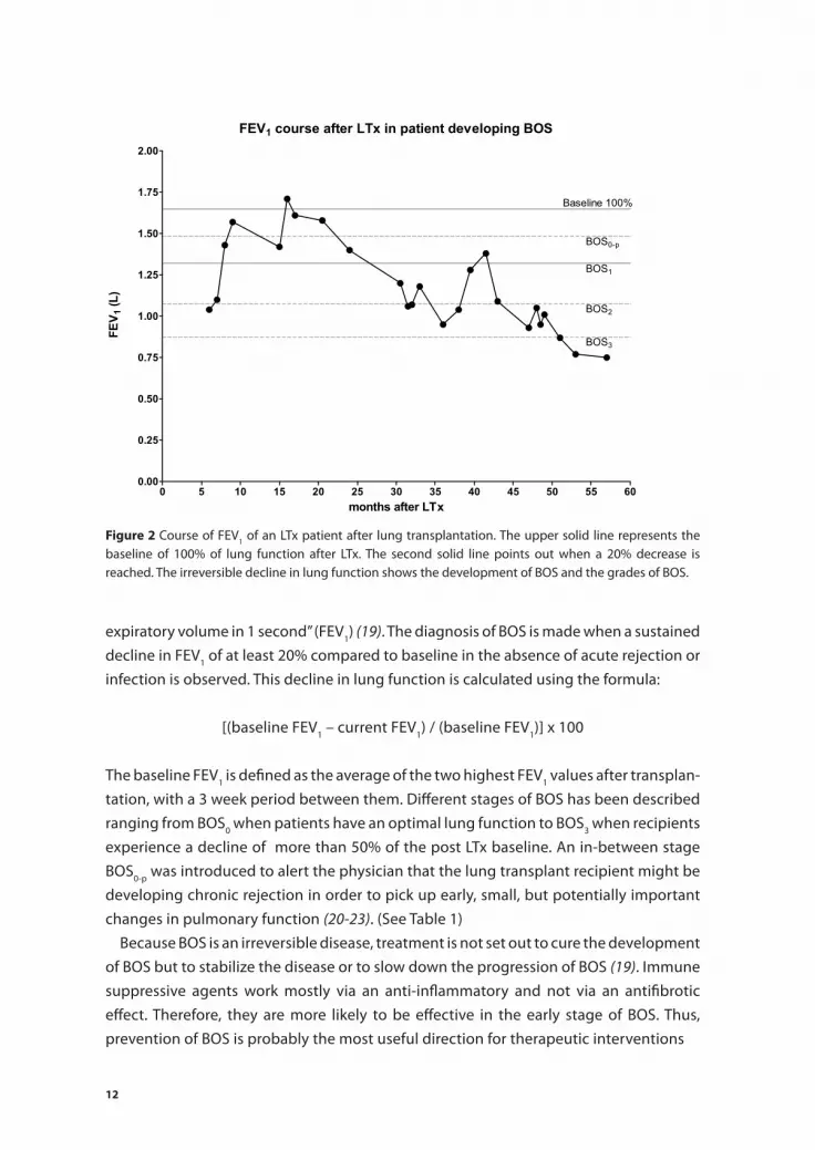
The baseline FEV1 is defined as the average of the two highest FEV1 values after transplan-tation, with a 3 week period between them. Different stages of BOS has been described ranging from BOS0 when patients have an optimal lung function to BOS3 when recipients experience a decline of more than 50% of the post LTx baseline. An in-between stage BOS0-p was introduced to alert the physician that the lung transplant recipient might be developing chronic rejection in order to pick up early, small, but potentially important changes in pulmonary function (20-23). (See Table 1)
Because BOS is an irreversible disease, treatment is not set out to cure the development of BOS but to stabilize the disease or to slow down the progression of BOS (19). Immune suppressive agents work mostly via an anti-inflammatory and not via an antifibrotic effect. Therefore, they are more likely to be effective in the early stage of BOS. Thus, prevention of BOS is probably the most useful direction for therapeutic interventions
figure 2 Course of FEV1 of an LTx patient after lung transplantation. The upper solid line represents the baseline of 100% of lung function after LTx. The second solid line points out when a 20% decrease is reached. The irreversible decline in lung function shows the development of BOS and the grades of BOS.

Chapter 1 - General Introduction
13
PossibLe MeChAnisMs LeAding to bos
BOS is a very heterogenous condition, because it probably has both humoral and cel-lular mechanisms driving the process. As a consequence the exact pathology remains unknown (24). Damage to the airway epithelium plays an important role in the cascade of events leading to BOS and as a result graft dysfunction. Therefore, BOS and its conse-quences can be seen as a “final common pathway” after initial damage to the graft. This damage might occur via several routes:
The immunologic factor
Airway epithelial cells are immunological targets. AECs express HLA class I and they also express HLA class II under inflammatory or immunological activated conditions (25-27). These donor HLA class I or II peptides are recognized by the immune system of the recipient which leads to the production of HLA antibodies (28, 29). Lung transplant recipients develop de novo antibodies against both HLA class I and class II antigens. Antibodies against the HLA molecules of the donor are ablt to fix and activate proteins of the complement system on the surface of the allograft and hence cause damage to the allograft in a donor specific manner (30, 31). It is described that increased soluble C4d in the BAL of LTx patients correlated with the presence of HLA antibodies (32). HLA class I antibodies stimulate the proliferation of endothelial cells and smooth muscle cells as well as stimulation of increased fibroblast growth factor production by these cells upon binding of the antibody to antigens on the allograft and hence contributing to the development of BOS (33, 34).
In chapter 7 and 8 of this thesis it is described how HLA antibodies of both the IgG and IgM isotype influence our cohort of LTx recipients. A role for immune suppressive regimen is suggested. Chapter 9 goes one step further, reviewing the possibility of non-HLA antibodies influencing the development of BOS. Non-HLA antibodies could target antigens on the endothelial or epithelium and the binding of these antibodies to the endothelial cells colocalizes with the deposition of C1q, C3, C5b-9 and C4d (35). Contribution of the lectin pathway of the complement system to the development of
table 1 Pulmonary function FEV1 as percentage of the post LTx stable baseline used to describe the different stages of BOS (22).
bos grade Pulmonary function
BOS0 >90%
BOS0-p 81-90%
BOS1 66-80%
BOS2 51-65%
BOS3 < 50%
14
BOS is studied in chapter 10, relating serum MBL levels to overall survival, freedom of BOS, and CMV infection.
Respiratory viruses may also contribute to the development of BOS. Although, there are no clear studies to specific respiratory viruses and their relation to BOS, a seasonal influ-ence is observed. In one study, the seasonal onset of BOS corresponded with the seasonal peak of various respiratory viruses (36, 37). Furthermore, it is reported that stable lung transplant recipients might suddenly develop BOS after clearing an acute respiratory viral infection (38, 39). Because of the immune suppressive medication, lung transplant recipi-ents are more prone for infection by viruses (40). These respiratory viruses, like influenza, parainfluenza, respiratory syncytial virus (RSV), coronaviruses, and adenovirus, might up regulate inflammatory cytokine production, like IL-1,6, and 8, or TNF which is initiated by viral replication and hence recruites and activates alloreactive T cells. However a direct action of the viruses which might damage the epithelium cannot be excluded (41).
Cells of the immune system and mainly lymphocytes are found in infiltrations of the allograft at time of BOS. Chapter 4 studies the presence of the different cell types early after transplantation in the periphery of patients and if changes in distribution of these cell types might be a biomarker for the development of BOS. The interactions of these cells can play a dual role after transplantation either contributing to rejection or tolerance. Dendritic cells (DC) can prime T cells to attack the allograft via direct or indirect interaction, but they might also teach the immune system to be hyporeactive to antigens picked up from the allograft (42-45). Some LTx patients were hyporeactive to cells of donor origin when a total leukocyte microchimerism was present (46-50). One group published that high microchimerism of the unfractionated leukocyte pool in LTx recipients is protective for the development in BOS, although many other studies are unable to reproduce these results (48-51). Furthermore, preliminary work observed that infusion of donor bone marrow in combination with LTx increases donor microchime-rism, hyporeactivity, and patients develop less BOS. However this was a study with few patients enrolling and short follow-up time (52). In chapter 5 the naturally occurring donor microchimerism in LTx patients is described. As all studies only considered the unfractionated chimerism of leukocytes, is in chapter 5 the contribution of different cell types to this microchimerism studied
The non-immunologic route
Although BOS is thought to be mediated mainly by alloimmunological injury non-allo-immune factors play a role as well. The lung is consistently exposed to exterior agents like inhaled dust, toxins, and (chemical) irritants which can promote local inflammation or increase the incidence of acute rejection and therefore the risk of developing BOS.
The surgical procedure during lung transplantation probably causes a higher inci-dence of gastro-oesophegeal reflux disease (GERD) (53-55). The transplantation might
Chapter 1 - General Introduction
15
increase the incidence of GERD by damage of the vagus nerve causing delayed gastric emptying. GERD after LTx might also be the result of the immune suppressive regimen, as many medications used after transplantation have an adverse effect on the gastro-intestinal system. GERD was more prevalent in patients eventually developing BOS than in patients with a stable lung function (55). However GERD might have been present prior to LTx but unrecognized. It was observed that patients with end stage lung diseases like CF and IPF had a higher incidence of GERD prior to LTx (56). Denervation of the lungs after LTx causes impaired cough and abnormal mucus clearance from the lungs and hence providing a chance for acid to contact the airways. Furthermore, foreign material might have an opportunity to move from the gastro-intestinal tract into the lungs and the small airways. This increased contact with acids could damage the lung epithelium contributing to the initial process leading to BOS (57, 58).
Ischeamia-reperfusion injury after lung transplantation involves infiltration of neu-trophils into the lung tissue which causes the release of oxygen radicals and proteases that can damage the small airways (59, 60). Ischeamic injury might eventually lead to an immunological reaction increasing the incidence of BOS. It has been shown that ischeamic-reperfusion injury after LTx correlates with up regulation of cytokines, like IL-2, TNF-α, and IFN-γ, inducing an inflammatory response and graft dysfunction (12, 61). Fur-thermore it was shown in animal models that reperfusion injury might also increase the expression of MHC complexes which can contribute to the development of BOS (62, 63).
As a result, during the initial inflammatory injury to the epithelial cells, independent of the immunological or non immunological route, the airway epithelium is partly or completely lost and the subepithelial basement membrane shows breaks and focal thickening (64, 65). The denuded surface is covered by provisional matrix proteins like fibronectin and fibrin, which will be resorbed after reconstitution of the epithelium. Recovery depends on effective repopulation of the air-bronchiole interface by epithelial cells. However, the regeneration of the epithelium via the stem cells in the small airways (Clara cells) is ineffective and there is aberrant tissue repair (66-68). As a consequence this leads in some bronchioles to myofibroblasts which migrate through gaps in the basement membrane into the provisional matrix and begin to deposit connective tissue, leading finally to the obliteration of the airway lumen.
risk fACtors for deveLoPMent of bos
Many different risk factors have been described for the development of the bronchiolitis obliterans syndrome. However, all risk factors remain to be probable and potential as reports are inconclusive due to the quality of data: small numbers as well as short follow-up hamper significant results. Many studies were performed retrospectively with

16
no, small, or badly defined control groups, and they mostly reflect the experiences of single centres. The best known and accepted risk factors are described below:² Acute rejection (AR) is a well known and well studied risk factor for the develop-
ment of BOS, especially when multiple and/or severe episodes occur (10, 12-16, 69). However, it remains to be a probable risk factor as patients with AR do not always develop BOS and patients with BOS did not always have AR (70). The last might be due to under detection as not all episodes of acute rejection are clinically diagnosed.
² Lymphocytic bronchitis (14, 16, 71, 72) was described to increase BOS when occurring later after LTx. However, lymphocytic bronchiolitis often coexists with acute rejec-tion, leaving ground for speculation which attributes to the development of BOS.
² HLA mismatching/antibodies (28, 73, 74). Matching for HLA antigens is not an option in lung transplantation because a small window of time is available to obtain the do-nor lungs and transplant them into the recipient. As a consequence of this no clear relation between BOS and HLA mismatching is found because almost no patients have less than 2 mismatches (10, 12, 14, 16, 75). Subsequently, the development of antibodies against donor HLA antigens is likely. Studies reporting the occurrence of HLA antibodies preceding the onset of BOS are abundant. The presence of HLA antibodies after LTx is found to be a major risk factor (28, 76, 77).
² CMV infection/pneumonitis (78). Cytomegalovirus (CMV) is a controversial risk factor for the development of BOS, and seems to be dependent on changed prophylactic approaches, as CMV pneumonitis became less widespread. Both studies in favour of as studies opposed CMV as a risk factor, have been published in the recent years. Although it appears that more recent studies do not find an association between CMV and BOS (13, 16, 76, 79). It is thought that CMV infection promotes allograft rejection through the production of cytokines and the increased expression of MHC molecules on the epithelium. The CMV serologic status of donor and recipient was found not to be of further influence on the development of BOS (12, 16, 77).
² Bacterial (pseudomonas or staphylococcus), fungal (aspergillus fumigatus) and (non-CMV) viral recurrent infection or permanent airway colonization. Reports on these types of infection are rare and contradictory. It might be that the infections are not directly correlated to the development of BOS, but they can increase the incidence of acute rejection episodes and hence contribute to the development of BOS (12, 16, 36). On the other hand, by change of architecture of the lung due to BOS the patients are more prone for infections (80, 81). In CF patients infections are usually caused by the pseudomonas already present in the patient before transplantation.
² GERD (gastro-eosophagal reflux disease) (82, 83). GERD is common after LTx, due to the operation and injury to the vagal nerve as well as medication induced gastroparesis. Hence microaspiration might promote chronic inflammation and bacterial infections in the lower airways, which is associated to the development of BOS (82, 84).

Chapter 1 - General Introduction
17
² Medication noncompliance is a known risk factor for rejection in other types of organ transplantation like kidney, heart and liver.
Furthermore some characteristics have been described to be of no relation to develop-ment of BOS, factors like age of the recipient as well as the donor, gender, blood group or primary disease of the recipient, donor ischaemic time and diffuse alveolar damage after LTx although, some controversy remains for the donor risk factors like age and ischaemic time (10, 85, 86).
sCoPe of thesis
Nowadays, BOS diagnosis is made via a surrogate clinical marker, an irreversible decline lung function of 20% from the post LTx baseline in absence of infections or other etiol-ogy. This means that the processes leading to the obstruction of small airways and sub-sequently the decline in lung function have time to evolve before treatment is started. Early detection of these processes or of patients at risk of development of BOS might be beneficial to early intensified immune suppressive therapy even before a decline in FEV1 can be observed (87). On the other hand, intensified immune suppressives need to be avoided as this might lead to other complications including dangerous viral infections or malignancies. Therefore, diagnosis would benefit from easily accessible biomarkers early after LTx, distinguishing patients at risk of development of BOS from those who remain stable over a longer period.
In this thesis we investigate possible early biomarkers in the blood of lung transplant recipients. In chapter 2 the chemokine TARC is identified as a possible biomarker and in chapter 3 we study the relation between this chemokine, its receptor and the clinic. Chapter 4 covers the presence of different recipient cell types in the peripheral blood of LTx patients, while chapter 5 focuses on the presence of donor cells in the peripheral blood of LTx patients (microchimerism). Chapter 6 describes the relation of activating receptor NKG2D on cells and its ligand sMICA to clinical parameters like infection and rejection. Chapter 7, 8 and 9 portray the role of humoral rejection after LTx. We inves-tigate the presence of HLA antibodies of both the IgM and IgG isotype as well as the possibility of identifying non-HLA antibodies and their role in rejection. In chapter ten the role of the lectin-dependent pathway of the complement cascade in investigated. In the last chapter (11) the focus is on non-immunologic damage to the lung, investigating whether pneumoproteins in the sera of LTx patient could be a good biomarker for the development of BOS.

18
referenCes
1. Trulock EP, Edwards LB, Taylor DO, Boucek MM, Keck BM, Hertz MI. Registry of the International Society for Heart and Lung Transplantation: twenty-second official adult lung and heart-lung transplant report--2005. J Heart Lung Transplant 2005; 24 (8): 956.
2. Trulock EP, Christie JD, Edwards LB, et al. Registry of the International Society for Heart and Lung Transplantation: twenty-fourth official adult lung and heart-lung transplantation report-2007. J Heart Lung Transplant 2007; 26 (8): 782.
3. Lechler RI, Batchelor JR. Restoration of immunogenicity to passenger cell-depleted kidney al-lografts by the addition of donor strain dendritic cells. J Exp Med 1982; 155 (1): 31.
4. Lechler R, Ng WF, Steinman RM. Dendritic cells in transplantation--friend or foe? Immunity 2001; 14 (4): 357.
5. Hertz MI, Aurora P, Christie JD, et al. Scientific Registry of the International Society for Heart and Lung Transplantation: introduction to the 2010 annual reports. J Heart Lung Transplant 2010; 29 (10): 1083.
6. Stehlik J, Edwards LB, Kucheryavaya AY, et al. The Registry of the International Society for Heart and Lung Transplantation: twenty-seventh official adult heart transplant report--2010. J Heart Lung Transplant 2010; 29 (10): 1089.
7. Kirk R, Edwards LB, Kucheryavaya AY, et al. The Registry of the International Society for Heart and Lung Transplantation: thirteenth official pediatric heart transplantation report--2010. J Heart Lung Transplant 2010; 29 (10): 1119.
8. Christie JD, Edwards LB, Kucheryavaya AY, et al. The Registry of the International Society for Heart and Lung Transplantation: twenty-seventh official adult lung and heart-lung transplant report--2010. J Heart Lung Transplant 2010; 29 (10): 1104.
9. Aurora P, Edwards LB, Kucheryavaya AY, et al. The Registry of the International Society for Heart and Lung Transplantation: thirteenth official pediatric lung and heart-lung transplantation report--2010. J Heart Lung Transplant 2010; 29 (10): 1129.
10. Sharples LD, McNeil K, Stewart S, Wallwork J. Risk factors for bronchiolitis obliterans: a systematic review of recent publications. J Heart Lung Transplant 2002; 21 (2): 271.
11. Burke CM, Theodore J, Dawkins KD, et al. Post-transplant obliterative bronchiolitis and other late lung sequelae in human heart-lung transplantation. Chest 1984; 86 (6): 824.
12. Bando K, Paradis IL, Similo S, et al. Obliterative bronchiolitis after lung and heart-lung transplan-tation. An analysis of risk factors and management. J Thorac Cardiovasc Surg 1995; 110 (1): 4.
13. Heng D, Sharples LD, McNeil K, Stewart S, Wreghitt T, Wallwork J. Bronchiolitis obliterans syn-drome: incidence, natural history, prognosis, and risk factors. J Heart Lung Transplant 1998; 17 (12): 1255.
14. Husain AN, Siddiqui MT, Holmes EW, et al. Analysis of risk factors for the development of bronchi-olitis obliterans syndrome. Am J Respir Crit Care Med 1999; 159 (3): 829.
15. Sharples LD, Tamm M, McNeil K, Higenbottam TW, Stewart S, Wallwork J. Development of bronchiolitis obliterans syndrome in recipients of heart-lung transplantation--early risk factors. Transplantation 1996; 61 (4): 560.
16. Girgis RE, Tu I, Berry GJ, et al. Risk factors for the development of obliterative bronchiolitis after lung transplantation. J Heart Lung Transplant 1996; 15 (12): 1200.
17. Boehler A, Estenne M. Post-transplant bronchiolitis obliterans. Eur Respir J 2003; 22 (6): 1007.

Chapter 1 - General Introduction
19
18. Cooper JD, Billingham M, Egan T, et al. A working formulation for the standardization of nomen-clature and for clinical staging of chronic dysfunction in lung allografts. International Society for Heart and Lung Transplantation. J Heart Lung Transplant 1993; 12 (5): 713.
19. Boehler A, Kesten S, Weder W, Speich R. Bronchiolitis obliterans after lung transplantation: a review. Chest 1998; 114 (5): 1411.
20. Patterson GM, Wilson S, Whang JL, et al. Physiologic definitions of obliterative bronchiolitis in heart-lung and double lung transplantation: a comparison of the forced expiratory flow between 25% and 75% of the forced vital capacity and forced expiratory volume in one second. J Heart Lung Transplant 1996; 15 (2): 175.
21. Estenne M, Van Muylem A, Knoop C, Antoine M. Detection of obliterative bronchiolitis after lung transplantation by indexes of ventilation distribution. Am J Respir Crit Care Med 2000; 162 (3 Pt 1): 1047.
22. Estenne M, Maurer JR, Boehler A, et al. Bronchiolitis obliterans syndrome 2001: an update of the diagnostic criteria. J Heart Lung Transplant 2002; 21 (3): 297.
23. Reynaud-Gaubert M, Thomas P, Badier M, Cau P, Giudicelli R, Fuentes P. Early detection of airway involvement in obliterative bronchiolitis after lung transplantation. Functional and bronchoal-veolar lavage cell findings. Am J Respir Crit Care Med 2000; 161 (6): 1924.
24. Halloran P, Mathew T, Tomlanovich S, Groth C, Hooftman L, Barker C. Mycophenolate mofetil in renal allograft recipients: a pooled efficacy analysis of three randomized, double-blind, clinical studies in prevention of rejection. The International Mycophenolate Mofetil Renal Transplant Study Groups. Transplantation 1997; 63 (1): 39.
25. Spurzem JR, Sacco O, Rossi GA, Beckmann JD, Rennard SI. Regulation of major histocompatibility complex class II gene expression on bovine bronchial epithelial cells. J Lab Clin Med 1992; 120 (1): 94.
26. Taylor PM, Rose ML, Yacoub MH. Expression of MHC antigens in normal human lungs and trans-planted lungs with obliterative bronchiolitis. Transplantation 1989; 48 (3): 506.
27. Taylor PM, Rose ML, Yacoub M. Expression of class I and class II MHC antigens in normal and transplanted human lung. Transplant Proc 1989; 21 (1 Pt 1): 451.
28. Jaramillo A, Smith MA, Phelan D, et al. Development of ELISA-detected anti-HLA antibodies precedes the development of bronchiolitis obliterans syndrome and correlates with progressive decline in pulmonary function after lung transplantation. Transplantation 1999; 67 (8): 1155.
29. Jaramillo A, Naziruddin B, Zhang L, et al. Activation of human airway epithelial cells by non-HLA antibodies developed after lung transplantation: a potential etiological factor for bronchiolitis obliterans syndrome. Transplantation 2001; 71 (7): 966.
30. Reznik SI, Jaramillo A, SivaSai KS, et al. Indirect allorecognition of mismatched donor HLA class II peptides in lung transplant recipients with bronchiolitis obliterans syndrome. Am J Transplant 2001; 1 (3): 228.
31. Reznik SI, Jaramillo A, Zhang L, Patterson GA, Cooper JD, Mohanakumar T. Anti-HLA antibody binding to hla class I molecules induces proliferation of airway epithelial cells: a potential mecha-nism for bronchiolitis obliterans syndrome. J Thorac Cardiovasc Surg 2000; 119 (1): 39.
32. Miller GG, Destarac L, Zeevi A, et al. Acute humoral rejection of human lung allografts and eleva-tion of C4d in bronchoalveolar lavage fluid. Am J Transplant 2004; 4 (8): 1323.
33. Harris PE, Bian H, Reed EF. Induction of high affinity fibroblast growth factor receptor expression and proliferation in human endothelial cells by anti-HLA antibodies: a possible mechanism for transplant atherosclerosis. J Immunol 1997; 159 (11): 5697.

20
34. Bian H, Harris PE, Mulder A, Reed EF. Anti-HLA antibody ligation to HLA class I molecules ex-pressed by endothelial cells stimulates tyrosine phosphorylation, inositol phosphate generation, and proliferation. Hum Immunol 1997; 53 (1): 90.
35. Magro CM, Ross P, Jr., Kelsey M, Waldman WJ, Pope-Harman A. Association of humoral immunity and bronchiolitis obliterans syndrome. Am J Transplant 2003; 3 (9): 1155.
36. Hohlfeld J, Niedermeyer J, Hamm H, Schafers HJ, Wagner TO, Fabel H. Seasonal onset of bronchi-olitis obliterans syndrome in lung transplant recipients. J Heart Lung Transplant 1996; 15 (9): 888.
37. Vilchez RA, Dauber J, Kusne S. Infectious etiology of bronchiolitis obliterans: the respiratory viruses connection - myth or reality? Am J Transplant 2003; 3 (3): 245.
38. Kumar D, Erdman D, Keshavjee S, et al. Clinical impact of community-acquired respiratory viruses on bronchiolitis obliterans after lung transplant. Am J Transplant 2005; 5 (8): 2031.
39. Kumar D, Husain S, Chen MH, et al. A prospective molecular surveillance study evaluating the clinical impact of community-acquired respiratory viruses in lung transplant recipients. Trans-plantation 2010; 89 (8): 1028.
40. Anderson DJ, Jordan MC. Viral pneumonia in recipients of solid organ transplants. Semin Respir Infect 1990; 5 (1): 38.
41. Garantziotis S, Howell DN, McAdams HP, Davis RD, Henshaw NG, Palmer SM. Influenza pneumonia in lung transplant recipients: clinical features and association with bronchiolitis obliterans syn-drome. Chest 2001; 119 (4): 1277.
42. Zamoyska R, Waldmann H, Matzinger P. Peripheral tolerance mechanisms prevent the develop-ment of autoreactive T cells in chimeras grafted with two minor incompatible thymuses. Eur J Immunol 1989; 19 (1): 111.
43. Austyn JM, Larsen CP. Migration patterns of dendritic leukocytes. Implications for transplantation. Transplantation 1990; 49 (1): 1.
44. Starzl TE, Demetris AJ. Transplantation milestones. Viewed with one- and two-way paradigms of tolerance. JAMA 1995; 273 (11): 876.
45. Vermaelen K, Pauwels R. Pulmonary dendritic cells. Am J Respir Crit Care Med 2005; 172 (5): 530. 46. Monaco AP. Chimerism in organ transplantation: conflicting experiments and clinical observa-
tions. Transplantation 2003; 75 (9 Suppl): 13S. 47. Sahota A, Gao S, Hayes J, Jindal RM. Microchimerism and rejection: a meta-analysis. Clin Trans-
plant 2000; 14 (4 Pt 1): 345. 48. Saraji A, Pourmand G, Mehrsai A, et al. Microchimerism and renal transplantation: doubt still
persists. Transplant Proc 2007; 39 (4): 948. 49. McSherry C, Jackson A, Hertz MI, Bolman RM, 3rd, Savik K, Reinsmoen NL. Sequential measure-
ment of peripheral blood allogeneic microchimerism levels and association with pulmonary function. Transplantation 1996; 62 (12): 1811.
50. Reinsmoen NL, Jackson A, Hertz M, et al. Peripheral blood allogeneic microchimerism in lung and cardiac allograft recipients. J Leukoc Biol 1999; 66 (2): 306.
51. Calhoun R, SivaSai KS, Sundaresan S, et al. Development of bronchiolitis obliterans syndrome despite blood chimerism in human lung transplant recipients. Transpl Int 1999; 12 (6): 439.
52. Pham SM, Rao AS, Zeevi A, et al. Effects of donor bone marrow infusion in clinical lung transplan-tation. Ann Thorac Surg 2000; 69 (2): 345.
53. Estenne M, Hertz MI. Bronchiolitis obliterans after human lung transplantation. Am J Respir Crit Care Med 2002; 166 (4): 440.
54. Young LR, Hadjiliadis D, Davis RD, Palmer SM. Lung transplantation exacerbates gastroesopha-geal reflux disease. Chest 2003; 124 (5): 1689.

Chapter 1 - General Introduction
21
55. Hadjiliadis D, Duane Davis R, Steele MP, et al. Gastroesophageal reflux disease in lung transplant recipients. Clin Transplant 2003; 17 (4): 363.
56. Tobin RW, Pope CE, 2nd, Pellegrini CA, Emond MJ, Sillery J, Raghu G. Increased prevalence of gastroesophageal reflux in patients with idiopathic pulmonary fibrosis. Am J Respir Crit Care Med 1998; 158 (6): 1804.
57. Schachter LM, Dixon J, Pierce RJ, O’Brien P. Severe gastroesophageal reflux is associated with reduced carbon monoxide diffusing capacity. Chest 2003; 123 (6): 1932.
58. Stanbrook MB, Kesten S. Bronchial hyperreactivity after lung transplantation predicts early bron-chiolitis obliterans. Am J Respir Crit Care Med 1999; 160 (6): 2034.
59. Ross SD, Tribble CG, Gaughen JR, Jr., Shockey KS, Parrino PE, Kron IL. Reduced neutrophil infiltration protects against lung reperfusion injury after transplantation. Ann Thorac Surg 1999; 67 (5): 1428.
60. Eppinger MJ, Deeb GM, Bolling SF, Ward PA. Mediators of ischemia-reperfusion injury of rat lung. Am J Pathol 1997; 150 (5): 1773.
61. Snell GI, Esmore DS, Williams TJ. Cytolytic therapy for the bronchiolitis obliterans syndrome complicating lung transplantation. Chest 1996; 109 (4): 874.
62. Serrick C, Giaid A, Reis A, Shennib H. Prolonged ischemia is associated with more pronounced rejection in the lung allograft. Ann Thorac Surg 1997; 63 (1): 202.
63. Waddell TK, Gorczynski RM, DeCampos KN, Patterson GA, Slutsky AS. Major histocompatibility complex expression and lung ischemia-reperfusion in rats. Ann Thorac Surg 1996; 62 (3): 866.
64. Siddiqui MT, Garrity ER, Martinez R, Husain AN. Bronchiolar basement membrane changes associ-ated with bronchiolitis obliterans in lung allografts: a retrospective study of serial transbronchial biopsies with immunohistochemistry [corrected]. Mod Pathol 1996; 9 (3): 320.
65. Yousem SA, Suncan SR, Ohori NP, Sonmez-Alpan E. Architectural remodeling of lung allografts in acute and chronic rejection. Arch Pathol Lab Med 1992; 116 (11): 1175.
66. McDowell EM, Keenan KP, Huang M. Restoration of mucociliary tracheal epithelium following deprivation of vitamin A. A quantitative morphologic study. Virchows Arch B Cell Pathol Incl Mol Pathol 1984; 45 (2): 221.
67. Evans MJ, Shami SG, Cabral-Anderson LJ, Dekker NP. Role of nonciliated cells in renewal of the bronchial epithelium of rats exposed to NO2. Am J Pathol 1986; 123 (1): 126.
68. Yousem SA, Berry GJ, Cagle PT, et al. Revision of the 1990 working formulation for the classifica-tion of pulmonary allograft rejection: Lung Rejection Study Group. J Heart Lung Transplant 1996; 15 (1 Pt 1): 1.
69. Keller CA, Cagle PT, Brown RW, Noon G, Frost AE. Bronchiolitis obliterans in recipients of single, double, and heart-lung transplantation. Chest 1995; 107 (4): 973.
70. Jackson CH, Sharples LD, McNeil K, Stewart S, Wallwork J. Acute and chronic onset of bronchiolitis obliterans syndrome (BOS): are they different entities? J Heart Lung Transplant 2002; 21 (6): 658.
71. Ross DJ, Marchevsky A, Kramer M, Kass RM. “Refractoriness” of airflow obstruction associated with isolated lymphocytic bronchiolitis/bronchitis in pulmonary allografts. J Heart Lung Transplant 1997; 16 (8): 832.
72. El-Gamel A, Sim E, Hasleton P, et al. Transforming growth factor beta (TGF-beta) and obliterative bronchiolitis following pulmonary transplantation. J Heart Lung Transplant 1999; 18 (9): 828.
73. Lau CL, Palmer SM, Posther KE, et al. Influence of panel-reactive antibodies on posttransplant outcomes in lung transplant recipients. Ann Thorac Surg 2000; 69 (5): 1520.
74. Palmer SM, Davis RD, Hadjiliadis D, et al. Development of an antibody specific to major histo-compatibility antigens detectable by flow cytometry after lung transplant is associated with bronchiolitis obliterans syndrome. Transplantation 2002; 74 (6): 799.
22
75. Quantz MA, Bennett LE, Meyer DM, Novick RJ. Does human leukocyte antigen matching influence the outcome of lung transplantation? An analysis of 3,549 lung transplantations. J Heart Lung Transplant 2000; 19 (5): 473.
76. Smith MA, Sundaresan S, Mohanakumar T, et al. Effect of development of antibodies to HLA and cytomegalovirus mismatch on lung transplantation survival and development of bronchiolitis obliterans syndrome. J Thorac Cardiovasc Surg 1998; 116 (5): 812.
77. Sundaresan S, Mohanakumar T, Smith MA, et al. HLA-A locus mismatches and development of antibodies to HLA after lung transplantation correlate with the development of bronchiolitis obliterans syndrome. Transplantation 1998; 65 (5): 648.
78. Kroshus TJ, Kshettry VR, Savik K, John R, Hertz MI, Bolman RM, 3rd. Risk factors for the develop-ment of bronchiolitis obliterans syndrome after lung transplantation. J Thorac Cardiovasc Surg 1997; 114 (2): 195.
79. Ettinger NA, Bailey TC, Trulock EP, et al. Cytomegalovirus infection and pneumonitis. Impact after isolated lung transplantation. Washington University Lung Transplant Group. Am Rev Respir Dis 1993; 147 (4): 1017.
80. Vos R, Vanaudenaerde BM, Geudens N, Dupont LJ, Van Raemdonck DE, Verleden GM. Pseudomonal airway colonisation: risk factor for bronchiolitis obliterans syndrome after lung transplantation? Eur Respir J 2008; 31 (5): 1037.
81. Vos R, Vanaudenaerde BM, De Vleeschauwer SI, Van Raemdonck DE, Dupont LJ, Verleden GM. De novo or persistent pseudomonal airway colonization after lung transplantation: importance for bronchiolitis obliterans syndrome? Transplantation 2008; 86 (4): 624.
82. Palmer SM, Miralles AP, Howell DN, Brazer SR, Tapson VF, Davis RD. Gastroesophageal reflux as a reversible cause of allograft dysfunction after lung transplantation. Chest 2000; 118 (4): 1214.
83. Blondeau K, Mertens V, Vanaudenaerde BA, et al. Gastro-oesophageal reflux and gastric aspira-tion in lung transplant patients with or without chronic rejection. Eur Respir J 2008; 31 (4): 707.
84. Cantu E, 3rd, Appel JZ, 3rd, Hartwig MG, et al. J. Maxwell Chamberlain Memorial Paper. Early fundoplication prevents chronic allograft dysfunction in patients with gastroesophageal reflux disease. Ann Thorac Surg 2004; 78 (4): 1142.
85. Norgaard MA, Andersen CB, Pettersson G. Does bronchial artery revascularization influence results concerning bronchiolitis obliterans syndrome and/or obliterative bronchiolitis after lung transplantation? Eur J Cardiothorac Surg 1998; 14 (3): 311.
86. Novick RJ, Bennett LE, Meyer DM, Hosenpud JD. Influence of graft ischemic time and donor age on survival after lung transplantation. J Heart Lung Transplant 1999; 18 (5): 425.
87. Swanson SJ, Mentzer SJ, Reilly JJ, et al. Surveillance transbronchial lung biopsies: implication for survival after lung transplantation. J Thorac Cardiovasc Surg 2000; 119 (1): 27.

Chapter 2Serum TARC levels post lung transplantation as a predictor for the bronchiolitis obliterans syndrome
A.W.M. Paantjens1, J.M. Kwakkel-van Erp2, W.G.J. van Ginkel1, D.A. van Kessel3, J.M.M. van den Bosch3, E.A. van de Graaf2, H.G. Otten1.
Clin Exp Immunol 2008;154(2):202-8
1Department of Immunology. University Medical Centre Utrecht,
Utrecht, The Netherlands 2Department of Respiratory Medicine. University Medical Centre
Utrecht, Utrecht, The Netherlands 3Department of Respiratory Medicine. St Antonius Hospital,
Nieuwegein, The Netherlands

AbstrACt
background: The main reason for mortality after lung transplantation is the bronchi-olitis obliterans syndrome, which represents chronic rejection. Because soluble CD30, which is mainly produced by activated Th2 cells, was shown to be related to develop-ment of BOS, we aimed to investigate the relation between development of BOS and Th2 chemoattractant thymus and activation regulated chemokine (TARC/CCL17). Methods: For 54 patients we measured serum TARC levels prior to transplantation by ELISA and in for 44 of them, sera were analyzed at month 1, 2 and 3 after LTx. In addi-tion, longitudinal measurements were performed in sera from 8 healthy controls and 14 patients; taken over a period of 2 years post transplantation from 7 patients developing BOS plus 7 clinically matched BOS-free patients. results: Median serum TARC levels post transplantation of patients who developed BOS were significantly lower than those of the matched BOS-free patients (p=0.05). A ROC analysis (AUC 0.77) together with a Kaplan Meyer analysis showed that serum TARC levels below 325 pg/ml in the first month post transplantation can predict development of BOS post transplantation (p=0.001). In contrast, pre transplant serum TARC levels were not significantly different between patients developing BOS, BOS-free patients or healthy controls.Conclusion: pre transplantation serum TARC levels do not predict the development of BOS post transplantation but measurement of the serum TARC levels in the first month directly after transplantation can provide us with a tool to identify the group at risk of developing BOS.

Chapter 2 - TARC, a predicting factor for BOS
25
introduCtion
Lung transplantation (LTx) is the final treatment option in end stage lung disease. The proportion of patients living 5 years after LTx is limited to approximately 50% and the main cause of long-term morbidity and mortality is the bronchiolitis obliterans syn-drome (BOS), which represents chronic lung allograft rejection (1-3). Data have shown that 58% of the recipients are diagnosed with BOS within 5 years post LTx with a median of diagnosis between 16-20 months, and it is generally considered that most recipients that survive operative and infectious complications will ultimately develop BOS (4,5).
Due to airflow obstruction and decline of graft function, BOS manifests as the devel-opment in a progressive deterioration in forced expiratory volume in 1s (FEV1) and it can be diagnosed by the definitive decline of 20% in FEV1 of the baseline value with no indication for other complications including infections, AR, and suture problems among others (6-8). Although the pathogenesis of BOS is unclear, the disease has a patchy char-acter of fibroproliferation and obliteration of the small airways (9). Several risk factors are identified including acute rejection, primary graft dysfunction, ischeamic time of the graft during transplantation, viral infections like CMV, gastro oesophageal reflux disease (GERD) and HLA mismatches (3,10-13). None of these factors however can be used as clinical markers for the early onset of the disease.
High sCD30 levels prior to LTx were also identified as a risk factor for the development of BOS (14-16). CD30 is expressed on the surface of Th2 cells and secreted in the blood-stream as a soluble form (sCD30) upon activation (17). The relation between sCD30 and BOS led us to speculate that chemokines involved in recruitment of Th2 cells might also be associated with the development of BOS. The thymus and activation regulated che-mokine (TARC/CCL17) can act as a chemoattractant for T helper cells type 2 by binding to the chemokine receptor CCR4 on the surface of these cells (18). TARC induces recruit-ment and migration of Th2 cells (18-19). The chemokine is expressed by various cells including endothelial cells, dendritic cells, epidermal keratinocytes, fibroblasts, platelets and activated bronchial epithelial cells and can be up regulated by pro-inflammatory cytokines like TNF-α, IL-1 and IFN-γ (20-23).
The objective of this study was to investigate whether TARC levels prior to and post lung transplantation can predict the onset and development of the bronchiolitis oblit-erans syndrome.

26
MAteriAL And Methods
Patients
A total of 57 patients (M/F 28/29, average age 46 years, range 18-61) who underwent lung transplantation at the Heart Lung Center in Utrecht, The Netherlands, between October 2001 and July 2007 and survived more than three months were included in this study.
BOS was defined as a decline of the FEV1 from the post-operative baseline at two dis-tinctive time-points of more than 20% in the absence of infection or other etiology (7).
Standard immunosuppressive therapy consisted of basiliximab, tacrolimus, myco-phenolate mofetil and prednisone for all patients. No surveillance bronchoscopies were performed. In patients who had a decline in lung function infections were diagnosed by cultures of BALF and PCR for CMV and EBV. When infections were excluded as the cause of FEV1 decline, the patients were treated with corticosteroids and azithromycine. When no increase in long function was observed the diagnosis BOS was made.
Patient follow up started in September 2004, after approval by the medical-ethical committee and informed consent was obtained from each patient. Forty-four patients donated blood every month in the first year post transplantation and once every three months in the following years. Sera stored for diagnostic purposes from 13 other patients were also included in this study, although they were either transplanted before this date or the serum sampling was not performed systematically as described above. From 54 out of 57 patients pre-transplant serum was present and from 44 out 57 patients sera were available taken monthly after transplantation up to month 3. TARC levels were determined in these sera and also in sera collected longitudinally up to 25 months post transplantation in a group of 14 patients consisting of 7 patients who developed BOS, which could be closely matched for gender, age, primary disease and follow up to 7 patients who did not develop BOS. Three patients that developed BOS were not included in this longitudinal analysis; due to lack of follow up time, lack of available serum samples or no clinical match to a non BOS patient.
Eight healthy (M/F=5/3, mean age 35 years (range 26-46)) non allergic and non smok-ing controls donated blood every two weeks for six months and once five years later. In total, 442 samples were measured for serum TARC levels.
ELISA
Serum TARC levels were measured in duplo as described before (24). 96-well ELISA-plates (Becton Dickinson, Franklin Lakes, NJ) were coated with a murine monoclonal capturing antibody directed against anti-human TARC (MAB364, R&D Systems, Abingdon, United Kingdom). Human serum diluted (1/2) was added and standard concentrations (range: 4000 pg/ml – 16 pg/ml) were prepared with recombinant human TARC (364-DN, R7D Systems) in PBS containing 1% BSA. Goat polyclonal biotinylated anti-human TARC

Chapter 2 - TARC, a predicting factor for BOS
27
antibody (BAF364, R&D Systems) was used as detecting antibody. HRP-Streptavidin Conjugate (Zymed, San Fransisco, CA) and substrate (TMB substrate, Pierce, Rockford, IL) were used according the manual of the manufacturer. Optical densities were measured at 450 nm with a Thermo labsystems Multiskan RC plate reader. The minimal concentra-tion of TARC that could be detected was 16 pg/ml.
Statistical analysis
To compare the healthy controls with the group of patients for the data prior to trans-plantation the Mann-Whitney rank-sum test was used. In order to evaluate the median of the non BOS versus BOS group post transplantation, or the patients between before and after transplantation the Wilcoxon signed rank test was performed. To asses whether serum TARC levels post transplantation can serve as a BOS predicting factor a receiver operating curve and Kaplan-Meyer curve with a Logrank test were used.
table 1 Characteristics of 14 matched patients for longitudinal study, and the 43 other LTx patients included in this study. A division is made between the patients that developed BOS and the patients that have not developed BOS. 3 Patients had a CMV infection; 2 patients were CMV negative and received lungs from a CMV positive donor, 1 CMV positive patient received lungs from a CMV negative donor. One patient had a EBV reactivation.
Matched patient group other Ltx Patients
bos non bos bos non bos
total number 7 7 3 40
bos gradeiiiiii
034
N.A.N.A.N.A.
003
N.A.N.A.N.A.
Mean follow-up(months) 38 (69-9) 39 (65-32) 21 (17-33) 17 (76-6)
Mean age(years) 51 (24-61) 50 (22-61) 39 (23-58) 41 (17-64)
Primary diseaseCfemphysemafibrotic disease
1 (14%)4 (57%)2 (29%)
1 (14%)4 (57%)2 (29%)
2 (67%)0 (0%)
1 (33%)
16 (40%)14 (35%)10 (25%)
infectionsCMvebvPseudomonas
0 (0%)0 (0%)
3 (42%)
1 (14%)0 (0%)
1 (14%)
0 (0%)0 (0%)
2 (67%)
2 (5%)1 (2.5%)18 (45%)

28
resuLts
In order to study the relation between serum TARC levels in LTx patients and the development of BOS, 57 patients and 8 healthy controls were included in this study. Characteristics are shown in Table 1. The median follow up time of the patients after transplantation was 11 months with a range from 4 till 75 months. Ten patients (19%) developed BOS and five patients died during the course of the study, three of which were associated with BOS. The median age of the patient population was 50 years (range 17-64 years), their gender was equally divided (M/F=27/27) and 35%, 36% and 29% suffered from cystic fibrosis, emphysema or fibrotic diseases (fibrosis, sarcoidosis and connective tissue diseases), respectively.
Pre transplant serum TARC and BOS
Analysis of TARC concentrations showed no difference between amounts of TARC present in serum taken prior to transplantation (605 pg/ml ± 380) compared to those in healthy controls (685 pg/ml ± 430). No associations were found between serum TARC concentra-tions, prior or post transplantation, and age, gender or primary disease. Furthermore, the 10 patients eventually developing BOS had the same amounts of pre-transplant
pre transplantation post transplantation0
250
500
750
1000
1250
1500
1750
seru
m T
AR
C (p
g/m
l)
figure 1 The transplantation procedure does not influence serum TARC levels. Serum TARC levels were measured through ELISA for 44 LTx patients pre transplantation (triangles) and 1 month post transplantation (cirkels). 10 Patients that eventually developed BOS are indicated with the filled triangles and cirkels.

Chapter 2 - TARC, a predicting factor for BOS
29
serum TARC levels as those not developing BOS. These data indicate that pre-transplant serum TARC levels were not associated with any of the clinical parameters investigated.
Effect of transplantation and immune suppression on serum TARC levels
To determine whether the transplantation procedure in combination with immunosup-pressive therapy had an effect on the serum TARC levels of LTx patients, 44 patients were selected in whom serum TARC was measured prior to and 1 month post transplanta-tion. As shown in Figure 1, serum TARC levels decreased in 15 patients; in 14 patients it remained constant whereas in 15 patients an increase was found in serum TARC levels. Overall, no significant difference was found between pre and 1 month post transplanta-tion TARC levels. The patients that developed BOS are marked by the closed symbols. For this group also no differences were found prior to and 1 month post transplantation as 5 patients had a decrease, 2 patients remained and 3 patients had an increase in serum TARC levels. Apparently, the transplantation procedure did not have an effect on serum TARC levels.
14 Patients were followed over time and donated blood once every month in the first year and once every three months in the following years post transplantation. The median follow up time was 40.5 months (range 9-74 months). Seven patients who de-
figure 2 The serum TARC levels of LTx patients do not differ from those of healthy controls over time. Longitudinally, average of 13 (9-17) measurements, ELISA for serum TARC determined the bandwidth for 22 persons. Patients (P1 to P14) and healthy controls (N1 to N8; non smoking non allergic) were ordered by increasing median of serum TARC.
30
veloped BOS and could be followed longitudinally were closely matched for underlying disease (CF 14%, Fibrotic disease 29% and emphysema 57%), age (median age 49, range 22-61), gender (Male 29%) and follow up time to 7 BOS-free patients.
Serum TARC levels were determined up to 25 months post transplantation and for comparison, levels were also measured in 8 non allergic non smoking healthy controls every two weeks for six months and once five years later. This resulted in an average of 13 (9-17) measurements per individual, providing a band width of serum TARC concen-tration from 22 persons which are depicted in Figure 2. The values of serum TARC levels post transplantation in patients (P1 to P14) are in the same range of the healthy controls (N1 to N8), indicating that the transplantation plus immune suppression employed did not cause the patient’s serum TARC levels to differ from that in healthy individuals.
Post transplant serum TARC and BOS
We next examined whether the course of serum TARC levels is associated with develop-ment of BOS, CMV or EBV reactivation or colonization with Pseudomonas. No change was found between the serum TARC levels post transplantation prior, during or after onset of BOS or CMV reactivation or Pseudomonas colonization, in the group of 14 patients followed longitudinally. A relation with EBV appearance could not be investigated as none of the 14 patients experienced a primary EBV infection or reactivation (data not shown). Patients experiencing a decline in FEV1, which were treated with corticosteroids and azithromycine, showed no alteration in serum TARC levels. Also no difference in TARC levels could be found between the 3 patients that developed BOS grade II versus the 4 patients that developed BOS grade III.
non BOS BOS0
250
500
750
1000
1250
P=0.05
Med
ian
seru
m T
AR
C (p
g/m
l)
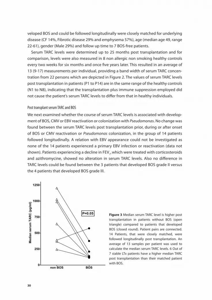
figure 3 Median serum TARC level is higher post transplantation in patients without BOS (open triangle) compared to patients that developed BOS (closed round). Patient pairs are connected. 14 Patients, that were closely matched, were followed longitudinally post transplantation. An average of 13 samples per patient was used to calculate the median serum TARC levels. 6 Out of 7 stable LTx patients have a higher median TARC post transplantation than their matched patient with BOS.

Chapter 2 - TARC, a predicting factor for BOS
31
To study whether there was a difference in serum TARC levels post transplantation be-tween the group of 7 patients who did develop BOS versus those 7 who did not develop BOS, the median of all measurements per person post transplantation was calculated and the result of the matched patient pairs is displayed in Figure 3. As shown, for 6 out of 7 fully matched patient pairs, the median serum TARC levels of the patients who de-veloped BOS were lower compared to the serum TARC levels of the patients who did not develop BOS. Statistical analysis indicated that median levels of TARC were significantly lower in the patients eventually developing BOS compared to the BOS-free patients (p=0.05, Wilcoxon rank sum test), indicating that low levels of TARC post transplant are a risk factor for BOS.
Serum TARC level as a BOS predicting factor
A receiver operating characteristics (ROC) curve was used to asses the possibility of pre-dicting BOS by serum TARC levels prior to or in the first 3 months post transplantation. Measurements of serum TARC at the fixed time points month 0, 1, 2 or 3 of 44 patients that survived at least 6 months post transplantation were included.
Prior to tranplanstation the ROC curve showed an AUC of 0.53, with a cut-off value of 590 pg/ml. This however did not result in a significant difference between the high or low serum TARC group prior to transplantation, neither did any other cut-off value. (Figure 4A)
For the 1 month post transplantation time point the curve resulted in an AUC of 0.77 (0.59-0.94), which indicates a good predicting factor for the development of BOS within 5 years post transplantation. The cut-off value defining whether a patient is at risk of developing BOS in the first years post transplantation with highest specificity as well as sensitivity was found at approximately 325 pg/ml serum (range 290- 326 pg/ml) TARC showing a specificity of 71% and a sensitivity of 80%.
This value was used for a Kaplan Meyer analysis, which is shown in Figure 4B. The difference between the group with high serum TARC level in the first month post transplantation from those with low serum TARC levels at this time point was significant. (p=0.001, Logrank test) The group with low serum TARC levels in the first month post transplantation show a higher incidence of developing BOS within the first five years post transplantation. However analysis at the time points 2 or 3 months post transplanta-tion did not reveal significant differences between the high and low serum TARC groups. Therefore low serum TARC level, <325 pg/ml, in the first month post transplantation is a risk factor for developing BOS.

32
disCussion
As patients with BOS generally respond poorly to augmented immunosuppressive therapy, a need for markers that predict the decline in graft performance is clearly present, allowing development of a strategy for treatment of patients at risk before onset of BOS. The object of this study was to investigate whether serum TARC levels are
0 10 20 30 40 500
10
20
30
40
50
60
70
80
90
100
TARC <325 pg/mlTARC >325 pg/ml
p=0.0010
Months post transplantation
Free
dom
of B
OS
0 10 20 30 40 500
10
20
30
40
50
60
70
80
90
100
< 590 (pg/ml)> 590 (pg/ml)
N.S.
Months post transplantation
Free
dom
of B
OS
A
B
figure 4 TARC is a predicting factor for development of BOS. (A) Prior to transplantation no significant differences can be seen with regard to freedom from BOS between, respectively, the 21 vs 23 patients with serum TARC levels above vs below 590 pg/ml. (b) The 30 patients with serum TARC levels above 325pg/ml in the first month post transplantation show a significant higher freedom of BOS (P-0.001) compared to the 14 patients with serum TARC levels below this concentration.

Chapter 2 - TARC, a predicting factor for BOS
33
associated with the onset and development of BOS. This is the first study showing that measurement of serum TARC levels after lung transplantation has a predictive value for the development of BOS.
Although the immunosuppressive regimen consisting of tacrolimus and mycopheno-late mofetil used in this study is known to suppress cellular (allo) immune responses efficiently, their influence on TARC production is not well known. In studies with AD and allergic asthma patients it was shown that TARC protein and mRNA levels decreased upon treatment with either cyclosporin A, tacrolimus and dexamethasone, or in combi-nation (24-27).
It is unknown however, whether this decrease in TARC levels was due to a direct effect on TARC production or an indirect effect caused by diminishment of disease activity. Furthermore, TARC can be produced by endothelial cells, dendritic cells, fibroblasts, epidermal keratinocytes and activated bronchial epithelial cells, all which can be differ-entially affected by immunosuppressives. The main source of TARC in atopic dermatitis seems to be keratinocytes in skin lesions whereas in allergic asthma it appears to be mainly produced by lung macrophages, indicating that the source of circulating TARC could be actually dependent on clinical conditions.
In our study we did not see a difference in serum TARC levels measured in a period without or with immune suppression c.q. prior versus 1 month post transplantation. Moreover, the levels of serum TARC of LTx patients measured longitudinally after trans-plantation were comparable to those found in healthy controls. This is an unexpected finding, as up regulation shortly after organ transplantation has been shown for many other cyto- and chemokines including IP-10, MCP1, IL-1β, IL-2, IL-12p40, IL-15, IL-2R, IL-6, IL-8 and IL-1Rα, although IL10 was found to be decreased (28-31). We assume that TARC production after transplantation is upregulated by vigorous allogeneic responses leading to production of known TARC-stimulatory cytokines like IL-1, INF-g and TNFa (20-23) but inhibited by the immune suppression employed, resulting in serum levels similar to those found in healthy controls. The actual reason for the low serum TARC lev-els directly after transplantation in patients, who will eventually develop BOS, remains unknown. It has been suggested that pre-existing subclinical inflammation - with its associated chemokine production - present in the donor lungs prior to transplantation, is associated with graft dysfunction and poorer prognosis after transplantation (31). Alternatively, lowered serum TARC levels also could be due to functional polymorphisms in the promoter region, such as found previously in Japanese individuals (32).
The relation found between low levels of circulating TARC and the development of BOS may be explained by its role as chemoattractant. Recently, it was shown that a subpopulation of Th2 cells expressing CCR4, the receptor for TARC, is characterized by CD4+CD25+, Treg cells and it was postulated that antigen presenting cells in the lungs and activated bronchial epithelium cells can recruit Treg cells towards a site of inflam-

34
mation through the secretion of TARC (33). Recruitment of Tregs down regulates inflam-matory responses limits tissue damage or autoimmunity. A lowered local production of TARC in the lung after transplantation might lead, according to the model described above, to an insufficient recruitment of Treg cells to the sites of ongoing inflammation, which would result in a deficient clearing of the chronic inflammatory responses in BOS. The role of Tregs in allograft rejection was also supported in a mice model using cardiac allografts. In this model, up regulation of Foxp3 expression was shown in the allografts displaying donor specific tolerance combined with recruitment of Tregs to the allografts through action of CCR4 and its ligands. (34). Interestingly, both the Th2 cytokine IL-10 known to suppress inflammatory responses and IL-12 were also found to be decreased in the broncho-alveolar lavage of patients with BOS (30, 35). This Treg-hypothesis may not seem to fit with published data showing up regulation of sCD30 prior to BOS (14), However, in our patient cohort we were not able to reproduce this finding and found instead unaltered sCD30 levels prior to BOS under the current immune suppressive regi-ment (36). Moreover, shedding of CD30 from Tregs resulting in increased serum sCD30 levels has not been reported yet.
As TARC is a small molecule of 10.5kD and leaks to the circulation without restriction, it can be expected that serum levels measured after lung transplantation reflect quantities locally produced in the lung e.g. by mature dendritic cells, monocytes and activated macrophages. This notion is supported by a recent study, showing that TARC levels in serum correlate well with those in broncho-alveolar lavage in acute eosinophilic pneu-monia (37). A small-scale study showing up regulation of CCL 19, CCL20 and CCL22 in patients developing BOS did not show an indication TARC levels in BAL predictive for BOS at month 3 and 6 after transplantation.(38) These data are in line with our results showing no predictive value for serum TARC levels 3 months after transplantation. We conclude that median serum TARC levels post transplantation in LTx patients without BOS is significantly higher that in those who developed BOS within 5 years after trans-plantation and that low serum TARC levels in the first month after lung transplantation is a predicting factor for the development of BOS. These data need to be confirmed in a larger cohort of patients, and the cut-off value of 325 pg/ml with a range of 290-326 pg/ml should be set more precisely in such a study.
Measurement of serum TARC levels in combination with other known risk factors may allow identification LTx patients at risk for development of BOS.

Chapter 2 - TARC, a predicting factor for BOS
35
referenCes
1. Trulock EP, Edwards LB, Taylor DO, Boucek MM, Keck BM, Hertz MI. Registry of the International Society for Heart and Lung Transplantation: twenty-second official adult lung and heart-lung transplant report--2005. J Heart Lung Transplant 2005; 24 (8): 956.
2. Hertz MI, Mohacsi PJ, Boucek MM, et al. The Registry of the International Society for Heart and Lung Transplantation: past, present and future. J Heart Lung Transplant 2002; 21 (9): 945.
3. Boehler A, Estenne M. Post-transplant bronchiolitis obliterans. Eur Respir J 2003; 22 (6): 1007. 4. Trulock EP. Lung transplantation. Am J Respir Crit Care Med 1997; 155 (3): 789. 5. Boehler A, Kesten S, Weder W, Speich R. Bronchiolitis obliterans after lung transplantation: a
review. Chest 1998; 114 (5): 1411. 6. Burke CM, Theodore J, Dawkins KD, et al. Post-transplant obliterative bronchiolitis and other late
lung sequelae in human heart-lung transplantation. Chest 1984; 86 (6): 824. 7. Estenne M, Maurer JR, Boehler A, et al. Bronchiolitis obliterans syndrome 2001: an update of the
diagnostic criteria. J Heart Lung Transplant 2002; 21 (3): 297. 8. Estenne M, Hertz MI. Bronchiolitis obliterans after human lung transplantation. Am J Respir Crit
Care Med 2002; 166 (4): 440. 9. Al-Githmi I, Batawil N, Shigemura N, et al. Bronchiolitis obliterans following lung transplantation.
Eur J Cardiothorac Surg 2006; 30 (6): 846. 10. Daud SA, Yusen RD, Meyers BF, et al. Impact of immediate primary lung allograft dysfunction on
bronchiolitis obliterans syndrome. Am J Respir Crit Care Med 2007; 175 (5): 507. 11. Sharples LD, McNeil K, Stewart S, Wallwork J. Risk factors for bronchiolitis obliterans: a systematic
review of recent publications. J Heart Lung Transplant 2002; 21 (2): 271. 12. Jaramillo A, Smith MA, Phelan D, et al. Development of ELISA-detected anti-HLA antibodies
precedes the development of bronchiolitis obliterans syndrome and correlates with progressive decline in pulmonary function after lung transplantation. Transplantation 1999; 67 (8): 1155.
13. Reinsmoen NL, Nelson K, Zeevi A. Anti-HLA antibody analysis and crossmatching in heart and lung transplantation. Transpl Immunol 2004; 13 (1): 63.
14. Fields RC, Bharat A, Steward N, et al. Elevated soluble CD30 correlates with development of bronchiolitis obliterans syndrome following lung transplantation. Transplantation 2006; 82 (12): 1596.
15. Bauwens AM, van de Graaf EA, van Ginkel WG, van Kessel DA, Otten HG. Pre-transplant soluble CD30 is associated with bronchiolitis obliterans syndrome after lung transplantation. J Heart Lung Transplant 2006; 25 (4): 416.
16. Golocheikine AS, Saini D, Ramachandran S, Trulock EP, Patterson A, Mohanakumar T. Soluble CD30 levels as a diagnostic marker for bronchiolitis obliterans syndrome following human lung transplantation. Transpl Immunol 2008; 18 (3): 260.
17. Del Prete G, De Carli M, D’Elios MM, et al. CD30-mediated signaling promotes the development of human T helper type 2-like T cells. J Exp Med 1995; 182 (6): 1655.
18. Imai T, Baba M, Nishimura M, Kakizaki M, Takagi S, Yoshie O. The T cell-directed CC chemokine TARC is a highly specific biological ligand for CC chemokine receptor 4. J Biol Chem 1997; 272 (23): 15036.
19. Bonecchi R, Bianchi G, Bordignon PP, et al. Differential expression of chemokine receptors and chemotactic responsiveness of type 1 T helper cells (Th1s) and Th2s. J Exp Med 1998; 187 (1): 129.
20. Fujisawa T, Fujisawa R, Kato Y, et al. Presence of high contents of thymus and activation-regulated chemokine in platelets and elevated plasma levels of thymus and activation-regulated che-

36
mokine and macrophage-derived chemokine in patients with atopic dermatitis. J Allergy Clin Immunol 2002; 110 (1): 139.
21. Panina-Bordignon P, Papi A, Mariani M, et al. The C-C chemokine receptors CCR4 and CCR8 iden-tify airway T cells of allergen-challenged atopic asthmatics. J Clin Invest 2001; 107 (11): 1357.
22. Berin MC, Eckmann L, Broide DH, Kagnoff MF. Regulated production of the T helper 2-type T-cell chemoattractant TARC by human bronchial epithelial cells in vitro and in human lung xenografts. Am J Respir Cell Mol Biol 2001; 24 (4): 382.
23. Sekiya T, Miyamasu M, Imanishi M, et al. Inducible expression of a Th2-type CC chemokine thy-mus- and activation-regulated chemokine by human bronchial epithelial cells. J Immunol 2000; 165 (4): 2205.
24. Hijnen D, De Bruin-Weller M, Oosting B, et al. Serum thymus and activation-regulated chemokine (TARC) and cutaneous T cell- attracting chemokine (CTACK) levels in allergic diseases: TARC and CTACK are disease-specific markers for atopic dermatitis. J Allergy Clin Immunol 2004; 113 (2): 334.
25. Kakinuma T, Nakamura K, Wakugawa M, et al. Thymus and activation-regulated chemokine in atopic dermatitis: Serum thymus and activation-regulated chemokine level is closely related with disease activity. J Allergy Clin Immunol 2001; 107 (3): 535.
26. Furukawa H, Nakamura K, Zheng X, et al. Enhanced TARC production by dust-mite allergens and its modulation by immunosuppressive drugs in PBMCs from patients with atopic dermatitis. J Dermatol Sci 2004; 35 (1): 35.
27. Kurokawa M, Kokubu F, Matsukura S, et al. Effects of corticosteroid on the expression of thymus and activation-regulated chemokine in a murine model of allergic asthma. Int Arch Allergy Im-munol 2005; 137 Suppl 1: 60.
28. Belperio JA, Keane MP, Burdick MD, et al. Critical role for the chemokine MCP-1/CCR2 in the pathogenesis of bronchiolitis obliterans syndrome. J Clin Invest 2001; 108 (4): 547.
29. Scholma J, Slebos DJ, Boezen HM, et al. Eosinophilic granulocytes and interleukin-6 level in bronchoalveolar lavage fluid are associated with the development of obliterative bronchiolitis after lung transplantation. Am J Respir Crit Care Med 2000; 162 (6): 2221.
30. Bharat A, Narayanan K, Street T, et al. Early posttransplant inflammation promotes the develop-ment of alloimmunity and chronic human lung allograft rejection. Transplantation 2007; 83 (2): 150.
31. Reynaud-Gaubert M, Marin V, Thirion X, et al. Upregulation of chemokines in bronchoalveolar lavage fluid as a predictive marker of post-transplant airway obliteration. J Heart Lung Transplant 2002; 21 (7): 721.
32. Sekiya T, Tsunemi Y, Miyamasu M, et al. Variations in the human Th2-specific chemokine TARC gene. Immunogenetics 2003; 54 (10): 742.
33. Iellem A, Mariani M, Lang R, et al. Unique chemotactic response profile and specific expression of chemokine receptors CCR4 and CCR8 by CD4(+)CD25(+) regulatory T cells. J Exp Med 2001; 194 (6): 847.
34. Lee I, Wang L, Wells AD, Dorf ME, Ozkaynak E, Hancock WW. Recruitment of Foxp3+ T regulatory cells mediating allograft tolerance depends on the CCR4 chemokine receptor. J Exp Med 2005; 201 (7): 1037.
35. Meloni F, Vitulo P, Cascina A, et al. Bronchoalveolar lavage cytokine profile in a cohort of lung transplant recipients: a predictive role of interleukin-12 with respect to onset of bronchiolitis obliterans syndrome. J Heart Lung Transplant 2004; 23 (9): 1053.

Chapter 2 - TARC, a predicting factor for BOS
37
36. Kwakkel-van Erp JM, Otten HG, Paantjens AW, et al. Soluble CD30 measured after lung trans-plantation does not predict bronchiolitis obliterans syndrome in a tacrolimus/mycophenolate mofetil-based immunosuppressive regimen. J Heart Lung Transplant 2008; 27 (10): 1172.
37. Miyazaki E, Nureki S, Ono E, et al. Circulating thymus- and activation-regulated chemokine/CCL17 is a useful biomarker for discriminating acute eosinophilic pneumonia from other causes of acute lung injury. Chest 2007; 131 (6): 1726.
38. Meloni F, Solari N, Miserere S, et al. Chemokine redundancy in BOS pathogenesis. A possible role also for the CC chemokines: MIP3-beta, MIP3-alpha, MDC and their specific receptors. Transpl Immunol 2008; 18 (3): 275.


Chapter 3Lung Transplantation affects expression of the Chemokine Receptor Type 4 on specific T cell Subsets
A.W.M. Paantjens1, E.A. van de Graaf2, H.D. Heerkens1, J.M. Kwakkel-van Erp2, T. Hoefnagel1, D.A. van Kessel3, J.M.M van den Bosch3, H.G. Otten1
Submitted
1Department of Immunology. University Medical Centre Utrecht,
Utrecht, The Netherlands 2Department of Respiratory Medicine. University Medical Centre
Utrecht, Utrecht, The Netherlands 3Department of Respiratory Medicine. St Antonius Hospital,
Nieuwegein, The Netherlands

40
AbstrACt
background: Alloreactive T-cells that infiltrate the graft after lung transplantation (LTx) play a role in chronic rejection. Chemokines like TARC, MCD and MCP-1 are locally produced in the lung and attract T-cells via chemokine receptor 4 (CCR4). In a TARC gradient, cells expressing CCR4++ migrate more efficiently than CCR4+ expressing cells. In this study, we compared the CCR4 expression of T-cells in blood from 20 lung transplant recipients to healthy controls. We then examined this association with concentrations of TARC in peripheral blood and the occurrence of chronic rejection.results: No relation was found between the CCR4 expression on T-cells and the concen-trations of TARC in peripheral blood. However, the CCR4++ expression decreased on CD4 T-cells from LTx patients (p<0.0001) when compared to healthy controls. The analysis of CD4 T-cell subsets showed this decrease was present on central memory, effector memory and terminally differentiated T-cells (p=0.0007, p<.0001 and p=0.05, respec-tively), while a trend was found for naïve CD4 T-cells (p=0.06). The expression of CCR4+ on Tregs was decreased in LTx patients when compared to healthy controls (p=0.02). Interestingly, the CCR4++ expression on CD4 effector memory T-cells was decreased in patients developing chronic rejection sometimes more than a year before the clinical diagnosis when compared to patients who did not (p=0.04). The analysis of CD8 T-cell subsets only showed the CCR4+ expression to be significantly increased on effector memory and terminally differentiated CD8 T-cells (p=0.02, p=0.03, respectively) in LTx patients, but no relation was found in chronic rejection. Conclusion: No correlation was found between serum TARC levels and the expression of its CCR4 receptor. However, the expression of CCR4 on T-cell subsets was altered after LTx and appears to be related to chronic rejection.

Chapter 3 - CCR4 expression influenced by LTx
41
introduCtion
Lung transplantation (LTx) is a final treatment option for end-stage lung diseases, even though the success of LTx is still restrained by occurrence of chronic rejection. The pathogenesis of chronic rejection, also known as the bronchiolitis obliterans syndrome (BOS), is unclear. BOS is characterized by the influx of leukocytes, including alloreactive T-cells, that leads to fibrosis and obliteration of the airways (1, 2).
In order to translocate T-cells to transplanted lungs, chemoattractive signals must be provided. The chemokine receptor type 4 (CCR4) expressed on T-cells ligates to a number of locally produced chemokines in the lungs, like thymus and activation-regulated chemokine (TARC/CCL17), monocyte chemotactic protein-1 (MCP-1/CCL2) and macrophage derived chemokine (MDC/CCL22). TARC, MCP-1 and MDC have all been studied in relation to the development of BOS. Concentrations of TARC present in the circulation one month after LTx were decreased in patients who eventually developed BOS (3). MCP-1 was elevated prior to BOS (4), while high MDC levels in BALF at 6 months post-transplantation were speculated to be predictive for BOS development (5, 6). The up-regulation of these chemokines at the site of inflammation might stimulate the migration of leukocytes from the periphery into the allograft.
CCR4 is expressed on T-cell subsets infiltrating the skin, synovial fluid, and on bron-chial T-cells. Less CCR4 expression is seen on interstitial T-cells and T-cells in the tonsils. CCR4 expression was not observed in the intestine (7). It has been shown that infiltrating T-cells’ CCR4 expression varies between different tissues. T-cells infiltrating the skin have a higher expression of CCR4 (CCR4++) than cells found within the bronchial cavity or the synovial fluid (CCR4+) (7). Additionally, it was shown that cells expressing CCR4++ migrate more efficiently towards a TARC gradient (7). In lung transplant patients, T-cells with CCR4 expression have been described to be present in the lungs at time of rejection (8). Tregs in BALF expressing CCR4 did not influence the development of BOS. TARC may not be critical for maintenance of Tregs in the allograft because it can selectively recruit other T-cells expressing CCR4 (9-11).
The finding that high levels of TARC in early post-transplant were associated with free-dom of BOS led us to speculate that this chemokine may attract CCR4++ T-cells, which are not involved in (or prevent) the pathogenesis of BOS. In this study, we explored the relationship between systemic TARC levels and the expression of CCR4 on T-cells. In ad-dition, we examined whether CCR4 expression on T-cell subsets, including its relation with BOS, was affected by lung transplantation.

42
MAteriAL And Methods
Patients and controls
Included in this study were 20 LTx patients who survived more than 3 months and who received transplants between September 2003 and March 2008 at the Heart Lung Cen-ter in Utrecht, The Netherlands. Five patients developed BOS during follow-up. BOS was defined as a decline in FEV1, from post-operative baseline at two distinctive time-points, of more than 20% in the absence of infection or other etiologies. Standard immuno-suppressive therapy consisted of basiliximab, tacrolimus, mycophenolate-mofetil and prednisone. Furthermore, blood obtained at one time point from 11 healthy controls was included in the measurements.
The study design was approved by the Medical Ethical Committee. Informed consent was obtained from each patient.
Sampling
The blood samples from these 20 patients, taken at approximately 5 months after LTx (range 4.2-6.1 months), were used in a cross-sectional study. Peripheral blood mono-nuclear cells (PBMC) were isolated from 40 mL of heparinized whole blood by Ficoll Paque Plus (GE healthcare, Sweden). All samples were frozen and preserved in liquid nitrogen until measurement. During a longitudinal study including 10 patients, samples were collected once prior to LTx and monthly up to 12 months after LTx, as described above. In parallel, serum samples were also collected.
Flow cytometry
Cryopreserved PBMC were rapidly thawed in a 37°C water bath and added to a medium containing 10% fetal calf serum (FCS); the cells were then centrifuged and resuspended in PBS. Two million PBMC were incubated with either relevant antibodies or isotype controls for 30 minutes on ice in the dark, which was then followed by washing and measurement.
Data acquisition and analysis were performed on a BD FACS Canto II with 8 color de-tection (BD Bioscience, CA). Each recipient sample measured had approximately 450,000 events (range 70,000 – 900,000 events) in the lymphocyte gate, which was defined by CD45+ and side-scatter (SSC) and analyzed with FACS DIVA software (BD Bioscience, CA).
Antibodies and gating strategy
Lymphocytes were identified with a scatter dot of CD45- and sideward scatter. Within this lymphocyte gate, both CD4 and CD8 T-cells were distinguished with CD4- and CD8-. CD45RO-Pe Cy7 (Becton Dickinson) and CD27-PerCP (BioLegend) were used to discrimi-nate the naïve T-cells (N), central memory T-cells (CM), effector memory T-cells (EM) and
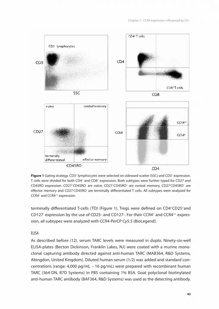
Chapter 3 - CCR4 expression influenced by LTx
43
terminally differentiated T-cells (TD) (Figure 1). Tregs were defined on CD4+CD25+and CD127- expression by the use of CD25- and CD127-. For their CCR4+ and CCR4++ expres-sion, all subtypes were analyzed with CCR4-PerCP Cy5.5 (BioLegend).
ELISA
As described before (12), serum TARC levels were measured in duplo. Ninety-six-well ELISA-plates (Becton Dickinson, Franklin Lakes, NJ) were coated with a murine mono-clonal capturing antibody directed against anti-human TARC (MAB364, R&D Systems, Abingdon, United Kingdom). Diluted human serum (1/2) was added and standard con-centrations (range: 4,000 pg/mL – 16 pg/mL) were prepared with recombinant human TARC (364-DN, R7D Systems) in PBS containing 1% BSA. Goat polyclonal biotinylated anti-human TARC antibody (BAF364, R&D Systems) was used as the detecting antibody.
CD3+ lymphocytes
CD4+ T cells
CD8+ T cells
SSC
CD3
CD8
CD4
CD27
CD45ROCD4
CCR4
naive central memory
effector memoryterminallydifferentiated
CCR4++
CCR4+
figure 1 Gating strategy. CD3+ lymphocytes were selected on sideward scatter (SSC) and CD3+ expression. T cells were divided for both CD4+ and CD8+ expression. Both subtypes were further typed for CD27 and CD45RO expression. CD27+CD45RO- are naïve, CD27+CD45RO+ are central memory, CD27-CD45RO+ are effector memory and CD27-CD45RO- are terminally differentiated T cells. All subtypes were analyzed for CCR4+ and CCR4++ expression.

44
According to the manufacturer’s manual, HRP-Streptavidin Conjugate (Zymed, San Fransisco, CA) and substrate (TMB substrate, Pierce, Rockford, IL) were used. A Thermo Labsystems Multiskan RC plate reader measured optical densities at 450 nm. The mini-mal detectable concentration of TARC was 16 pg/mL.
Statistics
A p-value below or equal to 0.05 was considered significant. The differences of CCR4 expression between LTx patients and healthy controls were analyzed using the Mann-Whitney rank-sum test (MW). Correlations were tested using Spearman’s rank correlation and were significant when the p-value was below or equal to 0.05 and the R-value was greater than (-) 0.4.
resuLts
Twenty LTx patients were included in this study, 5 of whom developed BOS. Patient characteristics are displayed in Table 1. A total of 3 patients died during follow-up. Two of these patients died as result of BOS, and one patient due to heart failure. None of the patients with CF in this study developed BOS. The distribution of primary disease was significantly different (p=0.018) between the groups with and without BOS. No CF patients who eventually developed BOS were included in the group. The non-BOS group
table 1 Patient characteristics.
bos non bos
total number 5 15
deceased 2 (40%) 1 (7%)
Age years (range) 56.2 (48-62) 43.8 (19-63)
follow-up time months (range) 35.8 (9-65) 42.6 (29-66)
gender Male Female
2 (40%)3 (60%)
10 (66%)5 (34%)
Primary disease Cystic fibrosis Emphysema Fibrotic disease
0 (0%)3 (60%)2 (40%)
8 (53%)1 (7%)
6 (40%)
bos onset Months (range)bos grade I II III
24.2 (9-41)
2 (40%)2 (40%)1 (40%)
N/A

Chapter 3 - CCR4 expression influenced by LTx
45
mainly consisted of patients with CF and only one patient with emphysema. The age of the non BOS group was slightly younger than the BOS group (44 years vs. 56 years). This was probably associated with the presence of CF patients, who are often transplanted at a younger age, in the non BOS group. Overall, the differences between both groups did not contribute to the outcome, BOS development. As previously shown for our cohort, the development of BOS was not correlated with HLA antibodies because they are only scarcely present in both pre- and post-transplant (13).
CCR4 expression differs after transplantation on T cells (subsets) from healthy controls
To examine whether CCR4 expression on T-cells and T-cell subsets was altered due to lung transplantation, blood from 20 LTx patients, taken at approximately 5 months after LTx, was analyzed and compared to results found in 11 healthy controls. As shown in Fig-ure 2A, CD4+ T-cells from LTx patients have a decreased expression of CCR4++ (p<0.0001, MW), when compared to healthy controls (HC). CCR4+ expression was not different between these groups. This decrease in CCR4 expression was present in patient groups developing BOS or not (p=0.002 and p<0.0001 respectively, data not shown). For CD8+
figure 2 Expression of CCR4+ (left y-axis) and CCR4++ (right y-axis) on peripheral CD4+ (A) and CD8+ (b) T cells in healthy controls and LTx patients. Expression of CCR4++ on CD4+ T cells was decreased in LTx patients (p=0.002). No differences were observed for CCR4 expression on CD8+ T cells between healthy controls and LTx patients.

46
figu
re 3
CCR
4+ (lef
t y-
axis
) and
CCR
4++ (r
ight
y-a
xis)
exp
ress
ion
was
affe
cted
at
5 m
onth
s af
ter
LTx.
For
CD
4+ T c
ell s
ubse
ts, C
CR4+ e
xpre
ssio
n di
d no
t di
ffer
betw
een
heal
thy
cont
rols
and
LTx
pat
ient
s; h
owev
er, C
CR4++
exp
ress
ion
(rig
ht y
-axi
s) w
as d
ecre
ased
in L
Tx p
atie
nts
for a
ll su
btyp
es (p
=0.0
007
CM, p
=0.0
001
EM, p
=0.0
5 TD
, and
a
tren
d w
as fo
und
for n
aïve
p=0
.06)
. In
CD8+ T
cel
ls, C
CR4+ e
xpre
ssio
n (le
ft y
-axi
s) w
as in
crea
sed
in LT
x pa
tient
s w
hen
com
pare
d to
hea
lthy
cont
rols
on
effec
tor m
emor
y an
d te
rmin
ally
diff
eren
tiate
d eff
ecto
r T c
ells
(p=0
.02
and
p=0.
03 re
spec
tivel
y).

Chapter 3 - CCR4 expression influenced by LTx
47
T-cells, no differences were observed in CCR4 expression between LTx patients and HC, as shown in Figure 2B.
It was then determined whether this decrease in CCR4++ expression on CD4+ T-cells could be contributed to a specific subset of T-cell, or whether all subsets contributed equally. This analysis also examined subsets of CD8+ T-cells and regulatory T-cells (Tregs). As shown in Figure 3, CCR4 expression was examined on naïve (N), central memory (CM), effector memory (EM) and terminally differentiated (TD) CD4+ and CD8+ T-cells. The CCR4++ expression was decreased on CM, EM and TD effector CD4+ T-cells (p=0.0007, P<0.0001 and p=0.05, respectively, MW) in lung transplant recipients compared to HC. Meanwhile, a trend was found for naïve CD4+ T-cells (p=0.06, MW), which indicated that all CD4+ T-cell subsets contributed to decreased CCR4++ expression on patient CD4+ T-cells. Although CCR4+ expression on CD4+ T-cell subsets was not different between patients and controls, it was increased on EM CD8+ T-cells and TD CD8+ T-cells of LTx pa-tients (p=0.02 and p=0.03 respectively, MW). There was no difference between patients’ and controls’ CCR4++ expression on CD8+ T-cell subsets.
Analysis of Tregs (CD4+CD25+CD127-) showed a significant decrease in CCR4+ expres-sion on T-cells from LTx patients when compared to HC (p=0.02, MW), as shown in Figure 4. CCR4++ expression was not different between patients and controls.
figure 4 CCR4+ expression (left y-axis) on Tregs was decreased 5 months after LTx in LTx patients when compared to healthy controls (p=0.02). CCR4++ expression (right y-axis) was not affected.

48
Relation between CCR4 expression, TARC and BOS
To study the correlation between the CCR4 receptor and its ligand TARC, a longitudinally sampled patient cohort was analyzed for both CCR4 expression on CD4+ T- cells and se-rum TARC concentration, in order to circumvent the influences of immune suppressives. Blood samples of 10 patients were analyzed. These samples were taken during the first 12 months (range 9-12, on average 10.6 months) after LTx, which resulted in a total of 61 samples (6.1 samples per patient). Serum TARC was measured at the same time points. CCR4+ or CCR4++ expression on lymphocytes, CD4+ T-cells, CD4+CD25+ T-cells or Tregs was not correlated to the levels of TARC in the serum samples of LTx patients (data not shown). In order to investigate a possible role of T-cell migration via CCR4 and TARC in the development of BOS, the level of CCR4 expression in patients with BOS was compared to the levels in patients without BOS. There was no difference in CCR4+ expression levels on lymphocytes, CD4+ and CD8+ T-cells, their subsets and Tregs between patients with or without BOS. However, CCR4++ expression levels on CD4+ EM T-cells was decreased for patients with BOS when compared to patients without BOS (p=0.04), as shown in Figure 5. CCR4++ expression in other investigated cells like lymphocytes, CD4+ and CD8+ T-cells, their subsets and Tregs did not differ between patients with and without BOS.
figure 5 CCR4++ expression (right y-axis) on CD4+ effector memory T cells was decreased 5 months after LTx in patients developing BOS when compared to patients without BOS (p=0.04). CCR4+ expression (left y-axis) was not affected.

Chapter 3 - CCR4 expression influenced by LTx
49
disCussion
The reported association between high serum concentrations of TARC one month after LTx and freedom from BOS thereafter stimulated the analysis of whether expression of its ligand CCR4 was also related with BOS. In this study, lung transplantation was shown to affect CCR4 expression on specific T-cell subsets 5 months after LTx. A decrease in CCR4++ expression on CD4+ T-cells from LTx patients was present in all CD4+ subsets. The finding that a higher percentage of CM CD4 T-cells express CCR4++ in the circulation of patients remaining BOS-free when compared to those that develop BOS suggested that CCR4++ expression and TARC production are somehow linked during the pathogenesis of chronic rejection.
The difference in expression of CCR4++ on CD4+ T-cells between LTx patients and healthy controls was reflected in all four subsets. In CD8+ T-cells, only the EM and TD subsets were different between LTx patients and HC, which indicated that the immune suppressive regimen did not specifically change CCR4 expression on all T-cells. This dif-ference in expression on T-cell subsets between healthy controls and LTx patients might partly reflect the allo-response. Furthermore, it is important to consider the T-cells’ developmental stage after LTx. The expression of CCR4 in early stadia did not correlate with its functionality. In the later stadia of TH2 differentiation, this correlation was pres-ent (14). The CCR4+ expression on Tregs was decreased in LTx patients when compared to healthy controls (p=0.02). The response of Tregs was potent and efficient but was less specific to gradients of TARC and MDC. This CCR4 expression was not restricted to Tregs, as was CCR8. . Blood-born Tregs preferentially express both CCR4 and CCR8, but as mature DCs produce TARC and MDC and almost no CCR8 chemokines, the main route of Treg attraction is via CCR4 (19). This supports the hypothesis that decreased levels of TARC produced by mature DC in the lungs at time of inflammation would result in reduced attraction of Tregs, thereby contributing to ongoing inflammation leading to BOS development (3).
TARC not only functioned as a chemoattractant for CCR4 expressing TH2 cells but its production by immature dendritic cells also polarized TH2 development from CD4+ CM cells. TH2 cells shed CD30 upon activation. In several studies, associations were found between circulating concentrations of soluble CD30 and BOS occurrence after lung transplantation (15-18). In our study, no correlation was found between serum concen-trations of TARC, sCD30 (data not shown) and expression of CCR4 on peripheral T-cells in a longitudinal analysis. Although the immune suppressive regimen used after trans-plantation was mainly pointed to T-cell suppression, it has been described that TARC levels are also suppressed by immune suppressive regimens (20). Therefore, it is possible that a relationship between CCR4 expression and TARC concentrations was affected by the medication prescribed after LTx. In addition, TARC and CCR4 expression on T-cells
50
are both measured in the periphery, which might not completely reflect the ongoing situation at the local inflammation within the lungs. Nevertheless, comparison between patients whom eventually did or did not develop BOS showed that a higher expression level of CCR4++ on these CD4+ cells was related to freedom from BOS after LTx. Thus, it was speculated that sufficient TARC production by lung dendritic cells attracted benign CM TH2 CD4+, which expressed high levels of CCR4 toward the site of inflammation.
In conclusion, expression of CCR4 on T-cell subsets was altered after LTx and appeared to be related to chronic rejection. Patients developing BOS showed a lower percentage in CCR4++ CM T-cells than patients remaining free from BOS. Although the difference found cannot be used to reliably identify patients at risk for developing BOS, the differ-ence was present more than a year before FEV1 decline. The presence of these cells, in combination with high production of TARC, may reflect a process of local inflammation in the lung, not causing fibrosis and obliteration of the airways.

Chapter 3 - CCR4 expression influenced by LTx
51
referenCes
1. Burke CM, Theodore J, Dawkins KD, et al. Post-transplant obliterative bronchiolitis and other late lung sequelae in human heart-lung transplantation. Chest 1984; 86 (6): 824.
2. Estenne M, Maurer JR, Boehler A, et al. Bronchiolitis obliterans syndrome 2001: an update of the diagnostic criteria. J Heart Lung Transplant 2002; 21 (3): 297.
3. Paantjens AW, Kwakkel-van Erp JM, van Ginkel WG, et al. Serum thymus and activation regulated chemokine levels post-lung transplantation as a predictor for the bronchiolitis obliterans syn-drome. Clin Exp Immunol 2008; 154 (2): 202.
4. Belperio JA, Keane MP, Burdick MD, et al. Critical role for the chemokine MCP-1/CCR2 in the pathogenesis of bronchiolitis obliterans syndrome. J Clin Invest 2001; 108 (4): 547.
5. Meloni F, Solari N, Miserere S, et al. Chemokine redundancy in BOS pathogenesis. A possible role also for the CC chemokines: MIP3-beta, MIP3-alpha, MDC and their specific receptors. Transpl Immunol 2008; 18 (3): 275.
6. Bhorade SM, Chen H, Molinero L, et al. Decreased percentage of CD4+FoxP3+ cells in bron-choalveolar lavage from lung transplant recipients correlates with development of bronchiolitis obliterans syndrome. Transplantation; 90 (5): 540.
7. Kunkel EJ, Boisvert J, Murphy K, et al. Expression of the chemokine receptors CCR4, CCR5, and CXCR3 by human tissue-infiltrating lymphocytes. Am J Pathol 2002; 160 (1): 347.
8. Gregson AL, Hoji A, Palchevskiy V, et al. Protection against bronchiolitis obliterans syndrome is associated with allograft CCR7+ CD45RA- T regulatory cells. PLoS One; 5 (6): e11354.
9. Vijayanand P, Durkin K, Hartmann G, et al. Chemokine receptor 4 plays a key role in T cell recruit-ment into the airways of asthmatic patients. J Immunol; 184 (8): 4568.
10. Stolberg VR, Chiu BC, Schmidt BM, Kunkel SL, Sandor M, Chensue SW. CC Chemokine Receptor 4 Contributes to Innate NK and Chronic Stage T Helper Cell Recall Responses during Mycobacte-rium bovis Infection. Am J Pathol; 178 (1): 233.
11. Yogo Y, Fujishima S, Inoue T, et al. Macrophage derived chemokine (CCL22), thymus and activa-tion-regulated chemokine (CCL17), and CCR4 in idiopathic pulmonary fibrosis. Respir Res 2009; 10: 80.
12. Hijnen D, De Bruin-Weller M, Oosting B, et al. Serum thymus and activation-regulated chemokine (TARC) and cutaneous T cell- attracting chemokine (CTACK) levels in allergic diseases: TARC and CTACK are disease-specific markers for atopic dermatitis. J Allergy Clin Immunol 2004; 113 (2): 334.
13. Paantjens AW, van de Graaf EA, van Ginkel WG, van den Bosch JM, Otten HG. Lung transplantation under a tacrolimus/mycophenolate mofetil-based immunosuppressive regimen results in low titers of HLA and MICA IgG antibodies which are not related to development of BOS. J Heart Lung Transplant 2010; 29 (5): 596.
14. Morimoto Y, Bian Y, Gao P, et al. Induction of surface CCR4 and its functionality in mouse Th2 cells is regulated differently during Th2 development. J Leukoc Biol 2005; 78 (3): 753.
15. Bauwens AM, van de Graaf EA, van Ginkel WG, van Kessel DA, Otten HG. Pre-transplant soluble CD30 is associated with bronchiolitis obliterans syndrome after lung transplantation. J Heart Lung Transplant 2006; 25 (4): 416.
16. Kwakkel-van Erp JM, Otten HG, Paantjens AW, et al. Soluble CD30 measured after lung trans-plantation does not predict bronchiolitis obliterans syndrome in a tacrolimus/mycophenolate mofetil-based immunosuppressive regimen. J Heart Lung Transplant 2008; 27 (10): 1172.

52
17. Fields RC, Bharat A, Steward N, et al. Elevated soluble CD30 correlates with development of bronchiolitis obliterans syndrome following lung transplantation. Transplantation 2006; 82 (12): 1596.
18. Golocheikine AS, Saini D, Ramachandran S, Trulock EP, Patterson A, Mohanakumar T. Soluble CD30 levels as a diagnostic marker for bronchiolitis obliterans syndrome following human lung transplantation. Transpl Immunol 2008; 18 (3): 260.
19. Iellem A, Mariani M, Lang R, et al. Unique chemotactic response profile and specific expression of chemokine receptors CCR4 and CCR8 by CD4(+)CD25(+) regulatory T cells. J Exp Med 2001; 194 (6): 847.
20. Kwakkel-van Erp JM, Haeck IM, Paantjens AW, et al. Differential usefulness of biomarkers thymus and activation-regulated chemokine and soluble CD30 during enteric coated mycophenolate sodium and cyclosporine therapy in atopic dermatitis. J Am Acad Dermatol 2010; 63 (3): e70.

Chapter 4Analysis of the Peripheral Blood Mononuclear Cell Profile after Lung Transplantation in Relation to the Bronchiolitis Obliterans Syndrome
A.W.M. Paantjens1, E.A. van de Graaf2, J.M. Kwakkel-van Erp2, T.Hoefnagel1, D.A. van Kessel3, J.M.M van den Bosch3, H.G. Otten1
Submitted
1Department of Immunology. University Medical Centre Utrecht,
Utrecht, The Netherlands 2Department of Respiratory Medicine. University Medical Centre
Utrecht, Utrecht, The Netherlands 3Department of Respiratory Medicine. St Antonius Hospital,
Nieuwegein, The Netherlands

54
AbstrACt
background: After lung transplantation, mononuclear cells including T cells may play a role in the development of BOS. To investigate whether the mononuclear cell composi-tion or specific T cell subsets in peripheral blood of patients with and without BOS are different, a cross sectional study was performed on blood taken 5 months after lung transplantation.Methods: PBMC samples of 11 patients with BOS, 39 patients without BOS and 8 healthy controls were analyzed by FACS for monocytes, B-, NK-, NKT- and T cells as well as CD4+ and CD8+ naïve, central memory, effector memory and terminally differentiated effector T cells. Patients were treated with a tacrolimus/mycophenolate-mofetil based regimen.results: Between lung transplant recipients and healthy controls no differences were found in percentage of circulating monocytes, T cells, B cells, NK cells or NKT cells. How-ever, central memory CD4+ T cells were decreased (p=0.04) and terminally differentiated effector CD4+ T cells were increased (p=0.05) in the patients remaining free from BOS compared to healthy controls. NKT cells were increased and central memory CD8+ T cells were decreased in patients developing BOS versus those remaining BOS-free (p=0.002 and p=0.02 respectively). Central memory CD8+ T cells were also decreased in patients developing BOS compared to healthy controls (p=0.02). The difference in NKT cells was not predictive of BOS while for central memory CD8+ T cells a trend was found (p=0.09).Conclusion: Differences in mononuclear cell compositions are found between healthy controls and patients who underwent lung transplantation. Some T cell subsets differ between patients developing BOS and patients remaining BOS free.

Chapter 4 - PBMC distribution after LTx
55
introduCtion
Lung transplantation (LTx) is the final treatment option for several end stage lung diseases. The outcome after LTx is influenced and hampered by the development of the bronchiolitis obliterans syndrome (BOS), which represents chronic rejection of the allograft (1-3). Approximately 50% of patients survive five years after LTx (4, 5). BOS manifests as the development in a progressive deterioration in forced expiratory volume in 1s (FEV1) due to airflow obstruction and decline of graft function. As the disease has a patchy character of fibro proliferation and obliteration of the small airways diagnosis only based on biopsy proven chronic rejection is not sufficient as this might lead to under diagnosis (6). Therefore it is diagnosed via a surrogate marker consisting of the definitive decline of 20% in FEV1 of the baseline value with no indication for other complications including infections, AR, and suture problems among others (7-9). Several risk factors for the development of BOS after LTx have been described, such as acute rejection, primary graft dysfunction, ischemic time of the graft during transplantation, viral infections like CMV, gastro oesophageal reflux disease (GERD) and HLA mismatches and HLA antibodies (3, 10-13).
Although the pathogenesis of BOS is mainly unknown, the immune system probably contributes to chronic graft rejection. For instance, BOS after LTx is characterized by infiltration of immune cells into the allograft. Accumulation of dendritic cells have been reported as well as submucosal lymphocyte and plasma cell infiltrates (14-18). These submucosal lymphocyte infiltrates consisted of mainly T cells, both CD4+ and CD8+ T cells, but also NK cells have sometimes been identified (15, 18-21).
Bronchoalveolar lavage fluid (BALF) has been increasingly used to study the patho-genesis of BOS and also for the prediction of BOS using biomarkers. Overall it was found for BOS that in BALF alveolar macrophages are decreased (22, 23). For neutrophils, CD4+ and CD8+ T cells an increase is described in BALF of patients with BOS compared to patients without BOS, although the opposite is also described (22-25).
Few studies on peripheral blood cell composition after lung transplantation have been published. CD19+ B cells were described to be dramatically decreased and hardly present in the peripheral blood of patients with BOS while CD8+ T cells were increased (26). NK cells were found to be activated but decreased in the peripheral blood of patients with BOS compared to LTx patients without BOS, although others report this decrease of NK cells in all LTx patients (21, 27). More detailed research is performed on T regulatory cells showing that levels of CD4+CD25+CD69- and CD4+CD25++ were decreased in patients with BOS (28).
This study focuses on the mononuclear cell composition, such as T cells, B cells, monocytes, NK cells, NKT cells, and T cell subset profiles found in peripheral blood approximately 5 months after lung transplantation. A comparison was made between

56
patients eventually developing BOS, patients who did not, and healthy controls In addi-tion, differences found are tested for their ability of identification of patients at risk for development of BOS, whether they could be biomarkers for development of BOS early after LTx.
Methods
Patients
50 LTx patients transplanted between September 2003 and March 2008 at the Heart Lung Centre in Utrecht, the Netherlands whom survived more than three months and had a follow-up time of 24 months were included in this study. Two patients who died because of BOS before the 24 month follow-up was completed were also included. Eleven patients developed BOS during follow-up. BOS was defined as a decline of the FEV1 from the post-operative baseline at two distinctive time-points of more than 20% in the absence of infection or other ethiology. Standard immunosuppressive therapy consisted of basiliximab, tacrolimus, mycophenolate-mofetil and prednisone.
The study design was approved by the medical ethical committee. Informed consent was obtained from each patient.
Blood sampling
In a cross-sectional study blood samples at approximately 5 months after LTx (range 4.2-6.1 months) were used. Blood of 8 healthy controls taken at 1 time point was also included in the measurements. From 40 ml heparinized whole blood peripheral blood mononuclear cell (PBMC) were isolated by Ficoll Paque Plus (GE healthcare, Sweden). All samples were frozen and preserved in liquid nitrogen until measurement.
Flow cytometry
Cryopreserved PBMC were rapidly thawed in a 37°C water bath and added to medium containing 10% fetal calf serum (FCS) after which the cells were centrifuged and resus-pended in PBS. Two million PBMC were incubated with either relevant antibodies or iso-type controls for 30 minutes on ice in the dark followed by washing and measurement.
Data acquisition and analysis was performed on a BD FACS Canto II with 8 color detec-tion (BD Bioscience, CA). Each recipient sample that was measured had approximately 194,000 events (range 69,000– 573,000 events) in the lymphocyte gate, which was defined by CD45+ and side-scatter (SSC), and analyzed with FACS DIVA software (BD Bioscience, CA).

Chapter 4 - PBMC distribution after LTx
57
Antibodies
Lymphocyte subsets were distinguished by the use of CD45-PE Cy7(BioLegend) for lym-phocytes, CD3-eFluor 450 (eBioscience) for T cells, CD19-PerCP (BioLegend) for B cells, CD14-APC-H7 (Becton Dickinson) for monocytes, and CD16/CD56-PE (BioLegend) for NK cells and their respective isotype controls (BioLegend and Becton Dickinson). NKT cells were defined as CD3+ and CD16/56+ (Figure 1A).
T cell subsets were identified by CD3-eFluor 450 (eBioscience), CD4-APC H7 (Becton Dickinson), CD8a-PE (BioLegend), CD45RO-Pe Cy7 (Becton Dickinson) and CD27-PerCP (BioLegend) (Figure 1B).
Statistics
A p-value of < 0.05 was considered to be significant. Mann-Whitney rank-sum tests were used to study the differences between healthy controls, patients with BOS and patients without BOS. A receiver operating curve (ROC) analysis was used to calculate the most discriminative cut-off point for cells found to be different between patients with BOS and patients without BOS. This cut-off value was than validated in a Kaplan-Meyer analysis.
figure 1 Gating strategy. Different cell types (A) are identified based on CD45 expression (lymphocytes and monocytes) and additional markers CD19+ (B cells), CD14 (monocytes), CD3+CD16+56- (T cells), CD3+CD16+56+ (NKT cells) and CD3-CD16+56+ expression (NK cells).The difference between CD16+56+ was made based on a histogram of the expression on the lymphocyte gate. T cell subsets (b) are discriminated by gating for CD3+ (T cells) with additional marker CD4+ and CD8+ and subsets were further identified by CD27 and CD45RO expression. CD27+CD45RO- are naïve, CD27+CD45RO+ are central memory, CD27-CD45RO+ are effector memory and CD27-CD45RO- are terminally differentiated effector T cells.

58
resuLts
Eleven out of 50 patients included in this study eventually developed BOS. Patient characteristics are presented in Table 1. No differences exist between the patient group developing BOS and the patients without BOS based on age, gender or primary disease, type of graft, CMV reactivation or CMV status. The average time to develop BOS was 22.5 months after LTx (range 5-49 months). The average follow-up time of BOS patients was shorter than patients without BOS (47.5 months (range 9-80) and 57.5 months (range 31-82) respectively). More patients with BOS than patients without BOS died during follow-up (p=0.01). HLA antibodies do not influence the development of BOS or the survival outcome, as only low titers of IgG anti-HLA antibodies are present in our cohort both pre- and post-transplant (29).
table 1 Patient characteristics
bos non bos
total number (n=50) 11 (22%) 39 (78%)
deceased 5 (46%) 2 (5%)
Age years (range) 46.4 (17-62) 43.8 (18-64)
follow-up time months (range) 47.5 (9-80) 57.5 (31-82)
gender Male Female
4 (36%)7 (64%)
23 (59%)16 (41%)
Primary disease Cystic fibrosis Emphysema Fibrotic disease
3 (27%)5 (46%)3 (27%)
18 (46%)10 (26%)11 (28%)
type of graft Single Double
29
435
CMv Reactivation No reactivation
47
1425
CMv status (donor/recipient) Negative/negative Negative/positive Positive/negative Positive/positive Unknown/positive
15212
1111944
bos onset Months (range)bos grade I II III
22.5 (5-50)
3 (27%)3 (27%)5 (46%)
Not applicable

Chapter 4 - PBMC distribution after LTx
59
Mononuclear cell composition
Fractions of different mononuclear cells were measured at month 5 after LTx in the blood of LTx patients developing BOS and without BOS as well as in the blood of healthy controls in order to analyze each contribution to the total PBMC. All samples contained approximately the same amount of total leukocytes per ml blood upon freezing and thawing. No differences were found between healthy controls and lung transplant recipients, either developing BOS or not, in percentage of monocytes, T cells, B cells, NK cells or NKT cells. Also between LTx patients developing BOS and without BOS most mononuclear cell fractions did not differ, although NKT cells (CD3+CD16/56+) were de-creased (p=0.02) in patients without BOS (Figure 2).
T cell subsets
Next it was examined whether all T cell subsets within the CD4+ and CD8+ T cell popula-tion differ between healthy controls and LTx patients. To this end 4 subsets were dis-criminated based on CD27 and CD45RO expression; naïve T cells (N), central memory T cells (CM), effector memory T cells (EM) and terminally differentiated effector T cells (TD, effector T cells).
For CD4 + T cells, as shown in Figure 3A, the central memory subset was significantly decreased in LTx patients without BOS compared to healthy controls (p=0.04), while the terminally differentiated effector CD4+ T cells were significantly increased in patients without BOS compared to healthy controls (p=0.05). No differences were found between
figure 2 FACS analysis of the mononuclear cell composition. The levels of T cells, B cells, monocytes and NK cells are comparable between healthy controls (n=8), patients developing BOS (n=11) and patients without BOS (n=39). NKT cells are significantly decreased in patients with BOS compared to patients without BOS (p=0.02).

60
healthy controls and LTx patients eventually developing BOS but trends were seen for decreased central memory and effector memory T cells and terminally differentiated effector T cells in patients developing BOS (p=0.07, p=0.09 and p=0.06, respectively). CD4+ T cells were not different between patients developing BOS and without BOS.
In the CD8+ subtype central memory T cells were significantly decreased in LTx patients developing BOS compared to healthy controls and LTx patient without BOS (p=0.02 and p=0.02, respectively), as shown in Figure 3B.
NKT cells and CM CD8+ T cells are no predictors of BOS
Significant differences between patients whom developed BOS and patients without BOS were found for the levels of NKT cells and central memory CD8+ T cells at 5 months after LTx. To investigate whether the levels of these cells might be an early predictor of BOS, a ROC analysis was performed. For NKT cells an area under the curve (AUC) of 0.725 was obtained in a ROC analysis with 3.9% (percentage of lymphocytes) as best discriminative cut-off value at month 5 after LTx. However, a Kaplan-Meyer (log rank) analysis showed no significant difference between patients with high or low levels NKT cells (data not shown), and therefore measurement of NKT cells at 5 months after LTx is not a predictive marker.
Central memory CD8+ T cells had an AUC of 0.738 with 7.3% as best discriminative cut-off level at month 5 after LTx. In a Kaplan-Meyer analysis, a trend was found that patients with low levels (<7.3% of CD8+ T cells) at 5 months after transplantation more often developed BOS than patients with high levels of CM CD8+ T cells (p=0.09, log rank test), as shown in Figure 4.
figure 3 FACS analysis of T cell subsets. Healthy controls (n=8) have more central memory CD4+ T cells than patients without BOS (n=39, p=0.04) and more central memory CD8+ T cells than patients developing BOS (n=11, p=0.05), while central memory CD8+ T cells of patients developing BOS are also decreased compared to patients without BOS (p=0.02).

Chapter 4 - PBMC distribution after LTx
61
disCussion
This study is novel with regard to the analysis of the mononuclear cell composition and T cell subsets in peripheral blood after lung transplantation. This is the first study to show significant differences in levels CM CD4+ and CD8+ between healthy controls and LTx patients approximately 5 months after LTx, next to demonstrating significant differences for NKT cells and CM CD8+ T cells between patients whom eventually developed BOS versus patients whom did not.
Differences found in blood between LTx patients in time developing BOS versus those who do not is worthwhile to investigate in more detail as they can result in powerful diagnostic tools for the prediction of patients at risk of developing BOS (26). Cellular differences found in this study are present relatively early after lung transplantation and might be an indication of ongoing inflammatory responses. These processes might be ‘silent rejection’ and go unnoticed until deterioration of lung function is measured when there is no return for BOS. Previous studies in peripheral blood after lung transplanta-tion have been mainly focused on analysis of a certain hematopoietic lineage and its activation status, and/or some subsets analysis in peripheral blood, after the diagnosis BOS has been made. For instance, in a small study using 2 color FACS technology the composition of lymphocytes after LTx was reported, showing a decrease in B cells and CD4+ T cells and an increase in CD8+ T cells in patients diagnosed with BOS (26). Also dif-ferences were present between previous studies with regard to sampling methods and immune suppressive regime employed, which hampers comparison of data. However, no elaborate study has been reported on the composition of all mononuclear cells pres-ent in blood prior to the diagnosis BOS (21, 27).
figure 4 CCR4+ expression (left y-axis) on Tregs was decreased 5 months after LTx in LTx patients when compared to healthy controls (p=0.02). CCR4++ expression (right y-axis) was not affected.

62
Although, BALF might reflect the local environment of inflammation in the lung more truly than peripheral blood, several studies made clear that the procedures for analysis of BALF are hard to standardize and results did not reach conformity (22-25, 30). A small study investigated the differences between BALF and blood after LTx. CD3+ and CD4+ T cells were decreased while CD8+ T cells were increased in BALF compared to peripheral blood. Differences between patients developing BOS and patients without BOS were not conclusive as few samples were available for the measurements in peripheral blood (25). Fluctuations over time of especially the amount of T cells are documented in BALF (31). The composition of PBMC may also change in time after lung transplantation, hence is it is important that our cross-sectional study results are further verified in longitudinal studies
We have used a 6 color staining on a larger patient cohort with a state of the art immune suppressive regimen early after transplantation resulting in an overview of mononuclear cell composition early after transplantation. The increase in NKT cells after LTx in patients developing BOS is interesting. Although NKT cells represent only a small population (0.5%) of the total CD3+ T cells, they produce both Th1 and Th2 cytokines and interact with cells from the innate immune system. In case of staphylococcus pneumo-nia, NKT cells were able to attract neutrophils to the lungs and an increase in neutrophils has been detected in patients whom developed BOS after LTx (32-34). Additionally, NKT cells have been shown to contribute to renal and hepatic ischemia-reperfusion injury, a process which has been assigned a risk factor for development of BOS (12, 35).
NKT and CM CD8+ T cells were found to be different between patients whom in time would develop BOS and patients without BOS. However, as ROC analysis shows the division is not black and white in this cohort. An overlap of cell content between both patient groups is the reason that the differences are not predictive in this cohort. These findings need to be validated in a much larger cohort.
It is important to consider PBMC composition found in LTx patients is influenced both by ongoing alloimmune responses in combination with the immune suppressive regimen based on tacrolimus and mycophenolate mofetil (31). Our data indicate that, compared to healthy controls, naïve and effector memory CD4+ are not changed while central memory CD4+ T cells are decreased and terminally differentiated CD4+ effector T cells are increased in LTx patients. The CD8+ naïve T cells remain stable compared to healthy controls, central memory CD8+ T cells are decreased, effector memory cells seem slightly increased, while terminally differentiated CD8+ effector T cells are increased. This gives rise to a more cytotoxic effector T cell profile in peripheral blood of LTx patients compared to healthy controls.
In conclusion CM CD4+ and CD8+ T cells are affected after lung transplantation compared to healthy controls. CM CD8+ T cells are decreased and NKT cells are increased at 5 months after transplantation in patients developing BOS compared to patients without BOS.

Chapter 4 - PBMC distribution after LTx
63
referenCes
1. Trulock EP, Edwards LB, Taylor DO, Boucek MM, Keck BM, Hertz MI. Registry of the International Society for Heart and Lung Transplantation: twenty-second official adult lung and heart-lung transplant report--2005. J Heart Lung Transplant 2005; 24 (8): 956.
2. Hertz MI, Mohacsi PJ, Boucek MM, et al. The Registry of the International Society for Heart and Lung Transplantation: past, present and future. J Heart Lung Transplant 2002; 21 (9): 945.
3. Boehler A, Estenne M. Post-transplant bronchiolitis obliterans. Eur Respir J 2003; 22 (6): 1007. 4. Trulock EP. Lung transplantation. Am J Respir Crit Care Med 1997; 155 (3): 789. 5. Boehler A, Kesten S, Weder W, Speich R. Bronchiolitis obliterans after lung transplantation: a
review. Chest 1998; 114 (5): 1411. 6. Al-Githmi I, Batawil N, Shigemura N, et al. Bronchiolitis obliterans following lung transplantation.
Eur J Cardiothorac Surg 2006; 30 (6): 846. 7. Estenne M, Hertz MI. Bronchiolitis obliterans after human lung transplantation. Am J Respir Crit
Care Med 2002; 166 (4): 440. 8. Burke CM, Theodore J, Dawkins KD, et al. Post-transplant obliterative bronchiolitis and other late
lung sequelae in human heart-lung transplantation. Chest 1984; 86 (6): 824. 9. Estenne M, Maurer JR, Boehler A, et al. Bronchiolitis obliterans syndrome 2001: an update of the
diagnostic criteria. J Heart Lung Transplant 2002; 21 (3): 297. 10. Reinsmoen NL, Nelson K, Zeevi A. Anti-HLA antibody analysis and crossmatching in heart and
lung transplantation. Transpl Immunol 2004; 13 (1): 63. 11. Jaramillo A, Smith MA, Phelan D, et al. Development of ELISA-detected anti-HLA antibodies
precedes the development of bronchiolitis obliterans syndrome and correlates with progressive decline in pulmonary function after lung transplantation. Transplantation 1999; 67 (8): 1155.
12. Sharples LD, McNeil K, Stewart S, Wallwork J. Risk factors for bronchiolitis obliterans: a systematic review of recent publications. J Heart Lung Transplant 2002; 21 (2): 271.
13. Daud SA, Yusen RD, Meyers BF, et al. Impact of immediate primary lung allograft dysfunction on bronchiolitis obliterans syndrome. Am J Respir Crit Care Med 2007; 175 (5): 507.
14. Yousem SA, Ray L, Paradis IL, Dauber JA, Griffith BP. Potential role of dendritic cells in bronchiolitis obliterans in heart-lung transplantation. Ann Thorac Surg 1990; 49 (3): 424.
15. Uyama T, Winter JB, Groen G, Wildevuur CR, Monden Y, Prop J. Late airway changes caused by chronic rejection in rat lung allografts. Transplantation 1992; 54 (5): 809.
16. Uyama T, Winter JB, Sakiyama S, Monden Y, Groen G, Prop J. Replacement of dendritic cells in the airways of rat lung allografts. Am Rev Respir Dis 1993; 148 (3): 760.
17. Yousem SA, Paradis IL, Dauber JA, et al. Large airway inflammation in heart-lung transplant recipients--its significance and prognostic implications. Transplantation 1990; 49 (3): 654.
18. Hasegawa S, Ockner DM, Ritter JH, et al. Expression of class II major histocompatibility complex antigens (HLA-DR) and lymphocyte subset immunotyping in chronic pulmonary transplant rejec-tion. Arch Pathol Lab Med 1995; 119 (5): 432.
19. Holland VA, Cagle PT, Windsor NT, Noon GP, Greenberg SD, Lawrence EC. Lymphocyte subset populations in bronchiolitis obliterans after heart-lung transplantation. Transplantation 1990; 50 (6): 955.
20. Gelman AE, Okazaki M, Sugimoto S, et al. CCR2 regulates monocyte recruitment as well as CD4 T1 allorecognition after lung transplantation. Am J Transplant; 10 (5): 1189.

64
21. Fildes JE, Yonan N, Tunstall K, et al. Natural killer cells in peripheral blood and lung tissue are associated with chronic rejection after lung transplantation. J Heart Lung Transplant 2008; 27 (2): 203.
22. Slebos DJ, Postma DS, Koeter GH, Van Der Bij W, Boezen M, Kauffman HF. Bronchoalveolar lavage fluid characteristics in acute and chronic lung transplant rejection. J Heart Lung Transplant 2004; 23 (5): 532.
23. Reynaud-Gaubert M, Thomas P, Gregoire R, et al. Clinical utility of bronchoalveolar lavage cell phenotype analyses in the postoperative monitoring of lung transplant recipients. Eur J Cardio-thorac Surg 2002; 21 (1): 60.
24. Ward C, Whitford H, Snell G, et al. Bronchoalveolar lavage macrophage and lymphocyte pheno-types in lung transplant recipients. J Heart Lung Transplant 2001; 20 (10): 1064.
25. Whitehead BF, Stoehr C, Finkle C, et al. Analysis of bronchoalveolar lavage from human lung transplant recipients by flow cytometry. Respir Med 1995; 89 (1): 27.
26. Fattal-German M, Frachon I, Cerrina J, et al. Particular phenotypic profile of blood lymphocytes during obliterative bronchiolitis syndrome following lung transplantation. Transpl Immunol 1994; 2 (3): 243.
27. Meehan AC, Sullivan LC, Mifsud NA, et al. Natural killer cell activation in the lung allograft early posttransplantation. Transplantation 2009; 89 (6): 756.
28. Meloni F, Vitulo P, Bianco AM, et al. Regulatory CD4+CD25+ T cells in the peripheral blood of lung transplant recipients: correlation with transplant outcome. Transplantation 2004; 77 (5): 762.
29. Paantjens AW, van de Graaf EA, van Ginkel WG, van den Bosch JM, Otten HG. Lung transplantation under a tacrolimus/mycophenolate mofetil-based immunosuppressive regimen results in low titers of HLA and MICA IgG antibodies which are not related to development of BOS. J Heart Lung Transplant 2010; 29 (5): 596.
30. Clelland C, Higenbottam T, Stewart S, et al. Bronchoalveolar lavage and transbronchial lung bi-opsy during acute rejection and infection in heart-lung transplant patients. Studies of cell counts, lymphocyte phenotypes, and expression of HLA-DR and interleukin-2 receptor. Am Rev Respir Dis 1993; 147 (6 Pt 1): 1386.
31. Slebos DJ, Scholma J, Boezen HM, et al. Longitudinal profile of bronchoalveolar lavage cell char-acteristics in patients with a good outcome after lung transplantation. Am J Respir Crit Care Med 2002; 165 (4): 501.
32. Bendelac A, Savage PB, Teyton L. The biology of NKT cells. Annu Rev Immunol 2007; 25: 297. 33. Kawakami K. Role of natural killer T cells in host defence against cryptococcal infection. Rev
Iberoam Micol 2002; 19 (3): 149. 34. Wallace KL, Marshall MA, Ramos SI, et al. NKT cells mediate pulmonary inflammation and dys-
function in murine sickle cell disease through production of IFN-gamma and CXCR3 chemokines. Blood 2009; 114 (3): 667.
35. Lappas CM, Day YJ, Marshall MA, Engelhard VH, Linden J. Adenosine A2A receptor activation reduces hepatic ischemia reperfusion injury by inhibiting CD1d-dependent NKT cell activation. J Exp Med 2006; 203 (12): 2639.
Chapter 5Chimerism of Dendritic Cell Subsets in Peripheral Blood after Lung Transplantation
A.W.M. Paantjens1, E.A. van de Graaf2, H.D. Heerkens1, J.M. Kwakkel-van Erp2, T. Hoefnagel1, D.A. van Kessel3, J.M.M van den Bosch3, H.G. Otten1
In press J. Heart Lung Transplant 2011
1Department of Immunology. University Medical Centre Utrecht,
Utrecht, The Netherlands 2Department of Respiratory Medicine. University Medical Centre
Utrecht, Utrecht, The Netherlands 3Department of Respiratory Medicine. St Antonius Hospital,
Nieuwegein, The Netherlands

66
AbstrACt
background: Passenger leukocytes of donor origin are transferred to the patient result-ing in circulatory microchimerism after lung transplantation (LTx). This chimeric state has been shown to occur in the total leukocyte fraction as well as unseparated periph-eral blood mononuclear cells (PBMCs). In this study we determined the microchimerism levels of B cells, monocytes, NK+T cells, and dendritic cell subsets (DCs: mDC1, mDC2 and pDC) during the first year after lung transplantation. Methods: In order to identify circulating donor cells, 11 patient-donor combinations were selected which were mismatched for HLA-B8. Analysis took place by flow cytom-etry on a minimum of 1 million PBMC taken monthly up to 1 year after LTx.results: Levels of microchimerism were found to be stable after LTx for all cell types investigated, although for NK+T cells an above baseline chimerism of donor cells from the donor lung was observed the first month after transplantation. Circulating PBMCs consisted on average of 0.002%, 1.7%, 0.03% and 0.001% of B cells, monocytes, NK+T cells and DCs, respectively, indicating that overall levels of microchimerism differ be-tween the cell types investigated. In 2 patients no B cell chimerism and in 1 patient no DC chimerism could be detected. Cell types and DC subsets of recipient origin were dis-tributed normal. Conversely, monocytes, B cells and DC of donor origin were increased while donor NK+T cells were decreased compared to the recipient ratios. Analysis of circulating recipient DCs showed a normal distribution of mDC1 (70%), mDC2 (5%) and pDC (25%). However, circulating donor DCs consisted of 80%, 20% and <1% of mDC1, MDC2 and pDC, indicating that donor plasmacytoid dendritic cells were not detectable in the circulation.Conclusion: The first year after lung transplantation a stable microchimerism was de-tected for all cell types investigated. However, donor pDCs were consistently absent in all samples investigated which may be linked with graft rejection often observed after LTx.

Chapter 5 - PBMC chimerism after LTx
67
introduCtion
In the first period after organ transplantation, cells of donor origin leave the graft and enter the circulation of the recipient. Although the levels of these passenger leukocytes decrease dramatically in the circulation within a few weeks after transplantation, a small fraction remains detectable even years after transplantation, resulting in stable circu-latory microchimerism (1-4). This chimeric state has been shown to occur in the total leukocyte fraction as well as unseparated peripheral blood mononuclear cells (PBMCs) as detected by PCR and flow cytometry analysis (5, 6).
Although many studies on microchimerism focused on total leukocytes or unfrac-tioned lymphocytes, individual cell types might make different contributions to this total microchimerism and might be of different interest with regard to graft survival. For instance, dendritic cells (DC) can play a dual role after transplantation; on one side as DC are potent antigen presenting cells they might induce an alloreactive response against the graft on the other hand they could possibly provoke donor specific toler-ance (7-10). Based on expression of blood DC antigens (BDCA1,2,3) and other markers, different DC subsets can be distinguished including myeloid DCs (mDC1 and mDC2) and plasmacytoid DCs (pDC) (11). Both mDC subsets produce IL-12 and elicit a Th1 response, while pDCs are able to produce IFN-α and trigger a Th2 response and induce FoxP3 expression. All three subsets are present in the lung according flow cytometry and immunohistochemistry (12-14). It was also shown that donor DC migrate from the graft after LTx resulting in DC-microchimerism detectable at least 1 year after LTx (15). However, whether all DC-subsets migrate equally to the periphery after transplantation and whether all subsets of donor DCs are able to survive in the host and form microchi-merism is unknown.
In this study we sought to determine in more detail how the levels of microchime-rism after LTx differ among cell types especially for the DC subtypes mDC1, mDC2 and pDC, but also of monocytes, B cells and NK+T cells. Therefore we analyzed from 11 LTx patients peripheral mononuclear blood cells (PBMCs) taken each month during the first year after LTx.
MAteriAL And Methods
Recipients
Eleven recipients who underwent lung transplantation between September 2004 and September 2007 at the University Medical Centre Utrecht, The Netherlands were includ-ed in this study. All recipients survived more than three months after transplantation and were HLA-B8 negative while their respective donors were HLA-B8 positive. Char-

68
acteristics are presented in Table 1. Standard immunosuppressive therapy consisted of basiliximab, tacrolimus, mycophenolate mofetil and prednisone for all recipients
Four recipients developed BOS, which was defined as a decline of the FEV1 from the post-operative baseline of more than 20% in the absence of infection or other etiology in accordance with the ISHLT criteria (16). Two recipients died because of BOS.
Sampling
Recipient follow up started in September 2003, after approval by the medical-ethical committee (project number 03/130) and informed consent was obtained from each recipient. Blood samples were collected routinely once prior to, once every month during first 12 months after transplantation. Blood from heart beating donors was col-lected prior to organ donation. From 40 ml heparinized whole blood peripheral blood mononuclear cell (PBMC) were isolated by Ficoll Paque Plus (GE healthcare, Sweden) All samples were frozen and preserved in liquid nitrogen until measurement.
Flow cytometric analysis
Cryopreserved PBMC were rapidly thawed in a 37°C water bath and added to medium containing 10% fetal calf serum (FCS) after which the cells were centrifuged and resus-pended in PBS. Two million PBMC were incubated with either relevant antibodies or iso-type controls for 30 minutes on ice in the dark followed by washing and measurement.
table 1
hLA mismatch
recipient gender Age*(years)
Primary disease
type graft
follow-up**
(months)
bos(months)
deceased(months)
Class i Class ii
1 m 31 CF bilateral 52 none n.a. 3 2
2 m 37 CF bilateral 25 5 25 4 2
3 m 52 FD bilateral 52 none n.a. 3 2
4 m 33 CF bilateral 28 none n.a. 4 2
5 m 54 EMF single 26 none n.a. 3 1
6 m 24 FD bilateral 45 none n.a. 2 2
7 f 49 EMF bilateral 9 8 9 4 2
8 m 24 CF bilateral 38 26 n.a. 4 2
9 f 59 EMF bilateral 56 16 n.a. 2 1
10 f 40 FD bilateral 50 none n.a. 4 2
11 f 57 EMF bilateral 65 none n.a. 4 2
Patient characteristics. Abbreviations: HLA: human leukocyte antigen, BOS: bronchiolitis obliterans syndrome, m: male, f: female, CF: cystic fibrosis, FD: fibrotic disease, EMF: emphysema, n.a.: not applicable.* Age at moment of lung transplantation** Clinical follow-up from time of lung transplantation until November 2009 or time of death

Chapter 5 - PBMC chimerism after LTx
69
Data acquisition and analysis was performed on a BD FACS Canto II with 8 color detec-tion (BD Bioscience, CA). Each recipient sample that was measured had approximately 450 000 events (range 70 000 – 900 000 events) in the R1 gate, which was defined by CD45+ and side-scatter (SSC), and analyzed with FACS DIVA software (BD Bioscience, CA).
Antibodies
DC were identified using a combination of antibodies; CD45-PE-Cy7, CD19-PerCP, BDCA-1-pacific-blue (BioLegend, CA) CD14-APC-H7 (BD Biosciences, CA) BDCA-2-PE, BDCA-3-APC (Miltenyi Biotec, Germany) and HLA-B8-Fitc (One lambda, CA), and isotype controls; Mouse-IgG1-pacific blue, Mouse IgG1-PE (BioLegend, CA), Mouse-IgG1-APC and Mouse
Figure 1
figure 1 Gating strategy. CD14 and CD19 were used to exclude monocytes and B cells respectively. BDCA1 distinguishes mDC1, BDCA2 marks pDC and BDCA3 identifies mDC2. Back gating was performed to confirm that BDCA1,2,3 expressing cells were present in the R1 and R2 gate. All cell types were analyzed for HLA-B8 staining which identifies cells of donor origin. Isotype controls were included.

70
IgG2B-Fitc (BD Biosciences, CA), or with a combination of HLA-DR-Alexa fluor 700, CD19-PerCP, CD123-PE-Cy7, and CD11c-pacific blue, (BioLegend, CA), CD45RA-PE (Sanquin, The Netherlands) and HLA-B8-Fitc (One lambda, CA) and isotype controls; Mouse-IgG1-PE-Cy7, Mouse-IgG1-Pacific blue, Mouse-IgG1-PE (Biologend, CA).
Cell type identification
As shown in Figure 1, monocytes were identified based on their CD14 expression. DC, some monocytes but mainly lymphocytes reside in the R1 gate which was defined on CD45+ and SSC. B cells were all cells within the R2 gate expressing CD19. Cells in the R2 which were CD14- and CD19- were divided in mDC1, based on BDCA-1 (CD1c) expression, mDC2, which express BDCA-3 (CD141), and pDC which are positive for BDCA-2 (CD303). Remaining cells within the R2 gate were titled NK and T cells. Back gating was performed to confirm that BDCA1,2,3 expressing cells were present in the R1 and R2 gate. In the alternative DC staining pDCs are defined as HLA-DR+/CD19-/CD123hi/ CD45RA+/CD11c- while cDCs were identified by HLA-DR+/CD19-/CD123lo/CD45RA-/CD11c+.
table 2
ratio Mix Measured (in Cd45+ gate; percentage)
hLA-b8- hLA-b8+ hLA-b8- hLA-b8+
1 0 99.2 0.8
0 1 8.7 91.3
1 1 37.9 62.1
10 1 83.0 17.0
100 1 96.8 3.2
1000 1 99.2 0.8
3000 1 99.5 0.5
10000 1 99.6 0.4
HLA-B8 positive cells in an artificial microchimerism. 2,000,000 Cryopreserved PBMC of a HLA-B8 negative and HLA-B8 positive healthy control were quickly thawed in a 37°C water bath and mixed 1:0, 0:1, 1:1, 1:10, 1:100, 1:1.000, 1:3,000, or 1:10,000 to create artificial microchimerism mixtures. Cells were stained with CD45, CD14, CD19, BDCA-1, BDCA-2, BDCA-3 and HLA-B8. HLA-B8 positive cells were determined for all cell types; positive cells within the CD45+ gate are displayed. This artificial microchimerism was created twice and analyzed. The negative control gives a background of 0.8% which is reached again in the 1:1000 diluted sample. Therefore the threshold is just above 1:1000. Data are consistent for two separate artificial mixture experiments and using different cell types/gates as basis.

Chapter 5 - PBMC chimerism after LTx
71
resuLts
Detection Limit of Chimerism
To study the detection limit for a specific donor cell type within the PBMC fraction of a recipient, an artificial microchimerism was created. PBMC of 2 healthy persons, of which one was positive for HLA-B8 and the other was negative for HLA-B8, were mixed in ratios ranging from 1:1 to 1:10,000. Samples containing either only HLA-B8 negative or HLA-B8 positive cells served as a negative and positive controls. Flow cytometry analysis for dendritic cells and HLA-B8 (as described above) was performed on the mixtures resulting in a background staining of the negative control of 0.8% within the R2 gate. Percentages of HLA-B8+ DC detected in the R2 gate of the different cell mixtures dropped according dilutions and reached background levels of 0.8% in the 1:1000 dilution. The 1:100 dilu-tion had 3.2% cells which were HLA-B8 positive, while the dilutions 1:3000 and 1:10,000 fell below the background level as shown in Table 2. Therefore, it can be concluded that the threshold for detection of HLA-B8 positive DC in a HLA-B8 negative setting is just above 1:1000. These background levels and threshold were not different for any of the other cell types investigated, using another gate than the R2 gate.
Levels of Microchimerism after LTx
In order to study the levels of microchimerism after lung transplantation in several cell types, samples from 11 recipients taken monthly were analyzed during the first 12 months after LTx. The average percentage of donor cells in these 121 blood samples in the lymphocyte/DC/monocyte gate was below 2%, of which monocytes make up the largest compartment (Table 3.) Microchimerism was detected in all recipients although not for all cell types on each time point. For instance, 1 recipient showed low levels microchimerism for monocytes only at month 8 after transplantation and NK+T cell chimerism only at month 9 after transplantation.
All recipients had detectable microchimerism for monocytes and NK+T cells, 1 recipi-ent lacked microchimerism for DC while 2 recipients showed no donor B cells.
table 3 Microchimerism of different cell types or all donor cells calculated to percentages of all cells (lymphocytes/DC/monocytes gate). *The percentages are the average of 11 recipients during month 1 until month 12 after LTx.
Cell type Microchimerism (percentage* of all cells)
Monocytes 1.744%
B Cells 0.002%
Dendritic cells 0.001%
NK and T cells 0.030%
All donor cells 1.777%

72
Stability of microchimerism during the first year after lung transplantation
To investigate the course of microchimerism during the first year after lung transplanta-tion for the different cell types, the percentage of microchimerism per cell type of all 11 recipients were averaged from month 1 until month 12 after transplantation and data from individual patients were normalized (see Figure 2), to compensate for inter individual differences (17, 18). For each individual patient and/or cell type the highest percentage of microchimerism was denoted as 1, all other time points of a patient and/or cell type were related to these samples with highest microchimerism. Each point represents the average of 11 lung transplant recipients on that time point. For NK and T cells an above baseline chimerism period, which might be due to the out flow of donor cells from the allograft, is still present in the first month after transplantation after which a stable level of microchimerism remains. For monocytes, B cells and DC the level of mi-crochimerism is already stable in the first month after lung transplantation and remains stable at least up to 12 months after lung transplantation.
Ratios of donor cell types and DC subsets are altered
It was determined whether the cell types of the recipient had a normal composition within the PBMCs and whether the levels of donor cell types reflected this distribution. To avoid any overrepresentation of cell types by the above baseline chimerism period of passenger leukocytes, the first month after LTx was excluded from this analysis. The average of all recipient and donor cell types as percentages of total recipient or donor cells (lymphocyte/DC/monocyte gate with HLA-B8- and HLA-B8+ gate respectively) per recipient during the first year after LTx was calculated. The PBMC fraction of the recipient had a normal composition with NK and T cells making up approximately
figure 2 Stable course of microchimerism after lung transplantation. For each recipient the point with the highest percentage of microchimerism, of the cell type investigated, was defined as 1, all other time points were related to this. All normalized data of the 11 recipients combined show relatively stable levels of microchimerism up to 1 year after LTx. For NK and T cells a flow out during the first months after LTx is shown

Chapter 5 - PBMC chimerism after LTx
73
70%, monocytes 25%, B cells 5% and DC less than 1%, see Figure 3A. However the composition of donor cells was different. In most patients monocytes were the main contributing cell type, (median of 53%) followed by B cells (median of 8%). The NK+T cell compartment (median of 5%) of the donor was decreased compared to recipient levels. Although dendritic cells (median of <1%) are still the smallest population within the donor PBMCs increased levels were found when compared to the recipient levels (data presented in Figure 3B). Furthermore, the composition of microchimerism between individuals was markedly different. For instance, in some patients the circulating donor cells were composed almost exclusively of monocytes whereas in others B cells were
figure 3 Percentage recipient and donor cell subtypes in PBMC. A and b recipient and donor cell types percentage of all recipient and donor cells (based on lymphocyte/DC/monocyte gate); recipient cells are normally distributed in the blood while levels of donor cells are changed. Donor monocytes, DC and B cells are enriched. C and d recipient and donor DC subtypes as the percentage of all recipient and donor DC; recipient DC subsets are normally distributed while donor pDC cannot be found in the blood of the recipient after transplantation. Each dot represents the average percentage cells during month 2 until 12 in one recipient, median is displayed by line

74
most predominant. These data indicate that although patients may show comparable percentages of microchimerism within the total PBMC fraction, the underlying cell type responsible may be of a different hematopoietic lineage.
Three subsets of dendritic cells can be distinguished, and these subsets were analyzed in the same way as the cell types described above, as percentages of the total recipient or donor DC population. Recipient DC subsets have a normal composition; mDC1, mDC2 and pDC make up 70%, 5% and 25% of the total recipient DC population, respectively as shown in Figure 3C. The donor DC subset composition in the DC compartment is clearly different when compared to the distribution of recipient DC subsets. Donor pDC cannot be detected in any recipient this is reflected in the levels of mDC1 and mDC2 which are increased and make up the donor DC compartment, as shown in Figure 3D.
As BDCA 2 (CD303), the marker to identify pDCs, can be internalized upon activation (19), the results were confirmed by an alternative staining for HLA-DR, CD19, CD123, CD45RA, and CD11c, identifying conventional DC (cDC, composed of mDC1 and mDC2, HLA-DR+/CD19-/CD123lo/CD45RA-/CD11c+) and pDC (HLA-DR+/CD19-/CD123hi/CD45RA+/CD11c-). Samples of the month in which an individual recipient displayed the highest levels of microchimerism in previous staining was available for 9/11 recipients. The results of this additional staining confirmed that donor pDC were absent in the PBMC fractions of the recipient (data not shown).
disCussion
This is the first study measuring microchimerism of different hematopoietic cell types and DC subsets in time after lung transplantation. The first month after transplantation NK and T cells still show an above baseline chimerism, but at month 2 after transplanta-tion B cells, monocytes, DC, NK and T cells reach stable levels of microchimerism, which is detectable at least up to 1 year after transplantation.
The identification of the different DC subsets is based on the DC markers BDCA1 (CD1c), BDCA2 (CD303) and BDCA3 (CD141). BDCA2 staining is used to distinguish pDCs from mDC1 and mDC2 (11). It is known that upon activation of pDCs approximately 50% of BDCA2 is internalized (19). However, pDCs of donor origin also could not be de-tected in any of the recipients using a different staining with antibodies directed against HLA-DR, CD19, CD123, CD45RA and CD11c, indicating that donor pDCs are either not transplanted along with the graft or they cannot survive in the host. However, as previ-ous studies showed pDC present within lungs, the latter explanation is very plausible because pDC are sensitive to stress and might not survive the hostile environment of the host.

Chapter 5 - PBMC chimerism after LTx
75
In this study, chimerism was quantified using flow cytometry. An alternative approach would be to purify DC subsets for instance by MACS and perform quantitative PCR for HLA-B8. However, isolation of cells is inherently associated with loss of cells and/or cell death which we felt would compromise quantification. In the experiment in which microchimerism was simulated with cells of a HLA-B8- and HLA-B8+ healthy donor the detection limit using flow cytometry was determined. In subpopulations when 1 in every 500 cells with a specific fenotypic make up is of donor origin (0.2%) chimerism can be detected. Measurements are performed purposely with large numbers of cells, ap-proximately 400,000 events in our R1 gate, making sure that donor pDCs are not missed due to technical inability to detect low numbers.
Ratios of DC subtypes, B cells, monocytes and NK+T cells were different between autologous and allogeneic cells in peripheral blood of the patients investigated. Specifi-cally, monocytes and mDC of donor origin are present in relatively higher proportion present than B, NK and T cells compared to the normal distribution of patient cells, while pDCs of donor origin cannot be found in lung transplant recipients. To examine whether differences between donor and patient cell ratio’s were also present in T cell subsets we analyzed this in a pilot study in blood derived from 8 lung transplant recipients taken at approximately 5 months after transplantation. Using CD3, CD4, CD8, CD27, CD45RO and HLA-B8 we found that less donor naïve CD4+ T cells but more donor central memory CD4+ and CD8+ T cells were present compared to recipients’ own levels of CD4+ or CD8+ T cell subsets (data not shown). This indicates that growth or maintenance of donor hematopoietic cells within recipients is regulated differently than that of autologous cells. Microchimerism persists in the recipient’s body longer than the average lifespan of leukocytes. In renal transplantation cells of donor origin were detected up to 30 years after transplantation (20). This led to believe that donor stem cells migrate from the transplanted organ and survive in the host (9, 21). The fact that in this study monocytes of donor origin are found 1 year after transplantation is in concert with this concept, as this cell type has a high turnover. The place where these cells reside is not clear. Stem cells might migrate from the organ to the bone marrow of the recipient or self renewing precursor populations survive in the organ (22).
In literature there are two sides regarding microchimerism and clinical outcome of solid organ transplantation (6, 23, 24). Speculations are made in literature that the clinical outcome and levels of microchimerism might be dependent on the type of transplanted organ (25). Specifically for lung transplantation, there are few reports on chimerism and rejection and mostly consisting of few individuals. On the one hand studies report recipients with high microchimerism to be hyporesponsive towards the donor in lung transplantation (23, 24, 26-28). On the other hand papers describe development of BOS regardless of the presence of microchimerism (29). Four of the 11 recipients developed BOS; however no differences concerning microchimerism are detected compared to the

76
7 recipients without BOS. Thus, in this small study cohort we do not find any relation between microchimerism levels overall or of any cell type and the clinical outcome of the recipients (data not shown).
In conclusion, microchimerism can be detected at least up to 1 year after lung trans-plantation for different cell types en subsets. Although the levels of microchimerism differ largely between cell types, they are relatively stable at 2 months after transplanta-tion. Remarkably, donor pDCs cannot be detected in lung transplant recipients, while mDC1 and mDC2 of donor origin are present.

Chapter 5 - PBMC chimerism after LTx
77
referenCes
1. Starzl, T. E., A. J. Demetris, M. Trucco, H. Ramos, A. Zeevi, W. A. Rudert, M. Kocova, C. Ricordi, S. Ildstad, and N. Murase. 1992. Systemic chimerism in human female recipients of male livers. Lancet 340:876.
2. Schlitt, H. J., H. Kanehiro, G. Raddatz, G. Steinhoff, N. Richter, B. Nashan, B. Ringe, K. Wonigeit, and R. Pichlmayr. 1993. Persistence of donor lymphocytes in liver allograft recipients. Transplantation 56:1001.
3. Richter, N., G. Raddatz, T. Graeter, H. J. Schafers, and H. J. Schlitt. 1995. Allogeneic lymphocyte chimerism after clinical lung transplantation. Transpl Immunol 3:74.
4. Hisanaga, M., J. Hundrieser, K. Boker, K. Uthoff, G. Raddatz, T. Wahlers, K. Wonigeit, R. Pichlmayr, and H. J. Schlitt. 1996. Development, stability, and clinical correlations of allogeneic microchime-rism after solid organ transplantation. Transplantation 61:40.
5. Suberbielle, C., S. Caillat-Zucman, C. Legendre, C. Bodemer, L. H. Noel, H. Kreis, and J. F. Bach. 1994. Peripheral microchimerism in long-term cadaveric-kidney allograft recipients. Lancet 343:1468.
6. Hisanaga, M., H. J. Schlitt, J. Hundrieser, K. Wonigeit, Y. Nakajima, H. Kanehiro, H. Nakano, and R. Pichlmayr. 1997. Frequency and clinical correlations of allogeneic microchimerism after heart, liver, and lung transplantation. Transplant Proc 29:1215.
7. Zamoyska, R., H. Waldmann, and P. Matzinger. 1989. Peripheral tolerance mechanisms prevent the development of autoreactive T cells in chimeras grafted with two minor incompatible thymuses. Eur J Immunol 19:111.
8. Austyn, J. M., and C. P. Larsen. 1990. Migration patterns of dendritic leukocytes. Implications for transplantation. Transplantation 49:1.
9. Starzl, T. E., and A. J. Demetris. 1995. Transplantation milestones. Viewed with one- and two-way paradigms of tolerance. Jama 273:876.
10. Vermaelen, K., and R. Pauwels. 2005. Pulmonary dendritic cells. Am J Respir Crit Care Med 172:530. 11. Dzionek, A., A. Fuchs, P. Schmidt, S. Cremer, M. Zysk, S. Miltenyi, D. W. Buck, and J. Schmitz. 2000.
BDCA-2, BDCA-3, and BDCA-4: three markers for distinct subsets of dendritic cells in human peripheral blood. J Immunol 165:6037.
12. Demedts, I. K., G. G. Brusselle, K. Y. Vermaelen, and R. A. Pauwels. 2005. Identification and charac-terization of human pulmonary dendritic cells. Am J Respir Cell Mol Biol 32:177.
13. Tsoumakidou, M., N. Tzanakis, H. A. Papadaki, H. Koutala, and N. M. Siafakas. 2006. Isolation of myeloid and plasmacytoid dendritic cells from human bronchoalveolar lavage fluid. Immunol Cell Biol 84:267.
14. Tsoumakidou, M., K. P. Karagiannis, I. Bouloukaki, S. Zakynthinos, N. Tzanakis, and N. M. Siafakas. 2009. Increased Bronchoalveolar Lavage Fluid CD1c Expressing Dendritic Cells in Idiopathic Pul-monary Fibrosis. Respiration 78:446.
15. Van der Meulen, J., N. A. Mifsud, D. Abud, E. Paul, M. D. Varney, S. R. Lewin, P. U. Cameron, and T. C. Kotsimbos. 2009. Differential dynamics of donor DC and non-DC peripheral blood mononuclear cell microchimerism in lung transplantation. Clin Immunol 133:179.
16. Estenne, M., J. R. Maurer, A. Boehler, J. J. Egan, A. Frost, M. Hertz, G. B. Mallory, G. I. Snell, and S. Yousem. 2002. Bronchiolitis obliterans syndrome 2001: an update of the diagnostic criteria. J Heart Lung Transplant 21:297.
17. Schlitt, H. J., J. Hundrieser, M. Hisanaga, K. Uthoff, M. Karck, T. Wahlers, K. Wonigeit, and R. Pichl-mayr. 1994. Patterns of donor-type microchimerism after heart transplantation. Lancet 343:1469.

78
18. Rudert, W. A., M. Kocova, A. S. Rao, and M. Trucco. 1994. Fine quantitation by competitive PCR of circulating donor cells in posttransplant chimeric recipients. Transplantation 58:964.
19. Wu, P., J. Wu, S. Liu, X. Han, J. Lu, Y. Shi, J. Wang, L. Lu, and X. Cao. 2008. TLR9/TLR7-triggered down-regulation of BDCA2 expression on human plasmacytoid dendritic cells from healthy individuals and lupus patients. Clin Immunol 129:40.
20. Starzl, T. E., A. J. Demetris, M. Trucco, A. Zeevi, H. Ramos, P. Terasaki, W. A. Rudert, M. Kocova, C. Ricordi, S. Ildstad, and et al. 1993. Chimerism and donor-specific nonreactivity 27 to 29 years after kidney allotransplantation. Transplantation 55:1272.
21. Starzl, T. E., A. J. Demetris, N. Murase, A. W. Thomson, M. Trucco, and C. Ricordi. 1993. Donor cell chimerism permitted by immunosuppressive drugs: a new view of organ transplantation. Im-munol Today 14:326.
22. Bogunovic, M., F. Ginhoux, A. Wagers, M. Loubeau, L. M. Isola, L. Lubrano, V. Najfeld, R. G. Phelps, C. Grosskreutz, E. Scigliano, P. S. Frenette, and M. Merad. 2006. Identification of a radio-resistant and cycling dermal dendritic cell population in mice and men. J Exp Med 203:2627.
23. Monaco, A. P. 2003. Chimerism in organ transplantation: conflicting experiments and clinical observations. Transplantation 75:13S.
24. Sahota, A., S. Gao, J. Hayes, and R. M. Jindal. 2000. Microchimerism and rejection: a meta-analysis. Clin Transplant 14:345.
25. Reinsmoen, N. L., A. Jackson, C. McSherry, D. Ninova, R. H. Wiesner, M. Kondo, R. A. Krom, M. I. Hertz, R. M. Bolman, 3rd, and A. J. Matas. 1995. Organ-specific patterns of donor antigen-specific hyporeactivity and peripheral blood allogeneic microchimerism in lung, kidney, and liver trans-plant recipients. Transplantation 60:1546.
26. Saraji, A., G. Pourmand, A. Mehrsai, M. Taherimahmodi, M. Nikoobakht, A. Asadpour, B. Nikbin, N. Tajik, and A. Emamzadeh. 2007. Microchimerism and renal transplantation: doubt still persists. Transplant Proc 39:948.
27. McSherry, C., A. Jackson, M. I. Hertz, R. M. Bolman, 3rd, K. Savik, and N. L. Reinsmoen. 1996. Sequential measurement of peripheral blood allogeneic microchimerism levels and association with pulmonary function. Transplantation 62:1811.
28. Reinsmoen, N. L., A. Jackson, M. Hertz, K. Savik, S. Kubo, S. Ormaza, L. Miller, and C. McSherry. 1999. Peripheral blood allogeneic microchimerism in lung and cardiac allograft recipients. J Leukoc Biol 66:306.
29. Calhoun, R., K. S. SivaSai, S. Sundaresan, E. P. Trulock, J. P. Lynch, G. A. Patterson, J. D. Cooper, and T. Mohanakumar. 1999. Development of bronchiolitis obliterans syndrome despite blood chimerism in human lung transplant recipients. Transpl Int 12:439.

Chapter 6The Effect of Lung Transplantation on NKG2D Expression
A.W.M. Paantjens1, H.D. Heerkens1, J.M. Kwakkel-van Erp2, T. Hoefnagel1, D.A. van Kessel3, J.M.M van den Bosch3, E.A. van de Graaf2, H.G. Otten1
Submitted
1Department of Immunology. University Medical Centre Utrecht,
Utrecht, The Netherlands 2Department of Respiratory Medicine. University Medical Centre
Utrecht, Utrecht, The Netherlands 3Department of Respiratory Medicine. St Antonius Hospital,
Nieuwegein, The Netherlands

80
AbstrACt
background: NKG2D has been proposed to mediate clearance of Pseudomonas aeru-ginosa by natural killer cells (NK), we examined whether NKG2D expression is related to pulmonary infections with these bacteria. Therefore, 151 sequentially taken blood samples were analyzed. Furthermore, interaction between activating receptor NKG2D and soluble MICA (sMICA) inhibits NK cell activation and might contribute to chronic rejection (bronchiolitis obliterans syndrome; BOS) after lung transplantation (LTx).results: Analysis of NKG2D expression on NK-, NKT- and T-cells determined prior to and 1 month after LTx showed that NKG2D+ expression on NK cells increased significantly (p=0.01). Overall NKG2D expression of LTx patients was stable and comparable to healthy controls. sMICA could be detected in serum of 2 patients, which did not correlate sMICA and NKG2D expression levels. No relation was found between NKG2D expression and P. aeruginosa infectionConclusion: We conclude that after lung transplantation NKG2D expression levels and sMICA are not correlated with BOS or with each other. However, NKG2D expression on NK cells is influenced by LTx.

Chapter 6 - Effect of LTx on NKG2D
81
introduCtion
NK cells have a central role in the innate immune system and have been shown to contribute to the outcome of lung transplantation (LTx) (1, 2). Peripheral NK cells in LTx recipients infiltrate into the lungs of those patients with bronchiolitis obliterans syndrome (BOS), which is considered to be the consequence of chronic lung allograft rejection (3). In addition, the frequency and phenotype of peripheral NK cells drastically changes after LTx, with immature NK cells being more prominent and mature NK cells being more activated but overall less cytotoxic (CD16-CD56dim) (1).
NK cells are regulated by a balance of inhibitory and activating signals derived from killer immunoglobulin-like receptors, adhesion molecules and costimulatory receptors including NKG2D (4). One of the ligands of NKG2D is MICA, which is expressed on en-dothelial cells, epithelial cells, fibroblasts, activated monocytes, and tumor cells and is up regulated under stress. The ectodomain of MICA can be shed into the periphery as soluble MICA (sMICA), and subsequent binding of sMICA to NKG2D leads to internaliza-tion of the complex, thereby inhibiting the function of NKG2D as well as inhibiting the activation and cytotoxic potential of NK cells (5, 6). It has been shown in heart transplant patients that the presence of circulating sMICA in the first year after transplantation correlates with a decreased incidence of acute rejection (7, 8). In addition, pooled sera of these patients containing sMICA could down regulate NKG2D on NK cell lines in vitro, thereby inhibiting NK cell function. Furthermore, it has been shown in mice that the blockade of NKG2D inhibits pulmonary clearance of Pseudomonas aeruginosa, a bacte-rium that causes respiratory problems in LTx patients with cystic fibrosis (9, 10).
In this study, we hypothesized that the expression of NKG2D on NK cells and/or serum concentrations of sMICA are related to chronic rejection and P. aeruginosa infection in patients after lung transplantation.
Methods
Ten patients were selected based on clinical matching for sex, age, primary disease, and time of LTx (Table 1); five of them developed BOS, and five counterparts did not. BOS was defined as a decline of the FEV1 of more than 20% from the post-operative baseline at two distinctive time-points in the absence of infection or other etiology in accordance with the International Society for Heart & Lung Transplantation (ISHLT) criteria (11). Blood samples were taken once before LTx, monthly during the first year and once every three months during the second year after LTx. In total, 151 blood samples were analyzed (average: 15 samples per patient range 9-21 samples). Standard immunosuppressive therapy consisted of basiliximab, tacrolimus, mycophenolate mofetil and prednisone for

82
tabl
e 1
Patie
nt c
hara
cter
istic
s. Re
latio
n be
twee
n Ps
eudo
mon
as a
erug
inos
a in
fect
ion,
oth
er c
linic
al c
hara
cter
istic
s an
d N
KG2D
exp
ress
ion.
Clin
ical
ly m
atch
ed p
atie
nts
(5 w
ith B
OS
and
5 w
ithou
t, ba
sed
on s
ex, a
ge, p
rimar
y di
seas
e an
d fo
llow
-up
time
afte
r LTx
) are
pre
sent
ed a
s pa
irs b
etw
een
dott
ed li
nes.
Show
n ar
e th
e pe
rcen
tage
s of
N
KG2D
-exp
ress
ing
cells
(mea
n pe
rcen
tage
) aft
er tr
ansp
lant
atio
n an
d sM
ICA
mea
n le
vels
aft
er L
Tx (s
tand
ard
devi
atio
n); t
he K
IR h
aplo
type
can
be
eith
er A
, mea
ning
AA
, w
hich
is in
hibi
tory
, or B
+, m
eani
ng A
B or
BB,
whi
ch is
act
ivat
ing,
as
defin
ed b
y LU
MIN
EX S
SO (O
ne L
ambd
a In
c, C
anog
a Pa
rk, C
A).
Abb
revi
atio
ns: F
: fem
ale,
M: m
ale,
CF:
cy
stic
fibr
osis
, EM
F: e
mph
ysem
a, F
D: fi
brot
ic d
isea
se, Y
: yes
, N: n
o.
Pseu
do-m
onas
in
fect
ion
sex
Prim
ary
dise
ase
Age
bos
(mon
th a
fter
Ltx)
% n
kg2d
+ exp
ress
ion
sMiC
A p
g/m
lki
r ha
plot
ype
CMv
reac
tiva
tion
nk
Cells
nk
t ce
llst
cells
1+
FCF
16Y
(50)
70.4
66.5
41.1
441
(177
)B+
N
2+
FCF
17N
50.5
30.2
31.8
995
(524
)B+
Y
3+
MCF
25Y
(26)
81.5
32.9
41.1
<200
AY
4-
MCF
24N
59.0
58.9
38.1
<200
B+N
5+
FFD
56Y
(9)
68.9
74.0
34.0
<200
AY
6-
FFD
56N
66.8
51.2
27.9
<200
B+Y
7-
FEM
F58
Y (8
)55
.033
.620
.0<2
00A
N
8-
FEM
F53
N66
.359
.721
.7<2
00B+
N
9-
FEM
F60
Y (1
6)53
.245
.821
.4<2
00B+
N
10-
FEM
F57
N44
.750
.934
.0<2
00B+
Y
aver
age
--
-42
(22)
61.6
50.4
31.1
--
-

Chapter 6 - Effect of LTx on NKG2D
83
all patients. In addition, blood samples were taken from five healthy controls and ten randomly selected patients prior to LTx. The study design was approved by the medical-ethical committee, and informed consent was obtained from each patient.
resuLts And disCussion
NKG2D expression levels on NK cells, NKT cells, and T cells of LTx patients determined by flow cytometry before and after LTx did not differ from NKG2D expression observed in healthy controls with the exception that a trend was found for increased NKG2D expres-sion on NK cells after transplantation compared to healthy controls (p=0.09, Figure 1).
Comparison of NKG2D expression levels on the NK cells, NKT cells, or T cells of LTx pa-tients before and the first month after transplantation revealed an increase in NKG2D on NK cells after transplantation (p=0.01 Figure 1). This finding is consistent with previously published results showing an activated NK cell phenotype after LTx (1). In the samples taken up to two years after LTx, NKG2D expression was found on average on 62% of NK cells (range 45%-70%, standard deviation (SD) range 7.4%-20.5%); on average, 50% of NKT cells expressed NKG2D (range 30%-74%, SD range 1.5%-20.9%). Analysis of NKG2D expression on T cells yielded an average of 31% positive T cells (range 20%-41%, SD range 4.8%-13.1%). No changes in NKG2D expression on the cells were observed at the time of BOS diagnosis or CMV infection. A comparison between the clinically matched patients, five with and five without BOS, showed that no differences were found be-tween the patient group with BOS and the group without BOS with respect to NKG2D expression on NK cells, NKT cells, or T cells.
As sMICA might down regulate NKG2D and thereby inhibit NK cell function, high levels of sMICA could be protective against BOS. Circulating levels of sMICA after LTx were below the detection limit of 200 pg/ml in 8 out of 10 patients. sMICA could be detected in two patients that were matched counterparts (one who developed BOS and one did not; Table 1) during the first two years after LTx. Longitudinally, these two patients showed high variability in sMICA levels after transplantation, with 7 out of 38 samples having undetectable levels of sMICA. These data indicate that sMICA levels are not related to the development of BOS. In addition, no correlation could be found between the concentrations of sMICA and the expression levels of NKG2D on NK cells, NKT cells, or T cells.
The relation between the expression of NKG2D and Pseudomonas aeruginosa infec-tion was studied in 20 patients using blood taken prior to LTx. Ten patients experienced Pseudomonas aeruginosa colonization, and 10 patients did not. Although NKG2D expression on NK cells, NKT cells, and T cells was slightly lower for patients without infection, this difference was not significant (p=0.11, p=0.27 and p=0.11, respectively).

84
After transplantation, samples from 6 patients without and 4 patients with Pseudomonas aeruginosa colonization were also analyzed for NKG2D expression levels. No differences were found in NKG2D expression on NK and NKT cells, although a trend was observed on T cells showing slightly decreased levels of NKG2D expression in patients without infection (p=0.09, data not shown). Thus, we have no indication that colonization with P. aeruginosa in the lungs of (CF) patients prior or post lung transplantation influences the expression of NKG2D.
figure 1 NKG2D expression levels on NK, NKT and T cells prior and after LTx compared to healthy controls. Approximately 1*106 thawed PBMCs were stained for CD3Fitc/CD16PE+CD56PE (Beckman Coulter, CA), CD45PerCP and NKG2DAPC (Becton Dickinson, CA). NKG2D expression was analyzed on NK cells (CD3-
CD16+56+), NKT cells (CD3+CD16+56+) and T cells (CD3+CD16+56-) in 5 healthy controls and 10 LTx recipients prior to and 1 month after LTx. NKG2D expression was increased after LTx on NK cells, while expression levels did not differ for NKT and T cells compared with levels prior to LTx.
Chapter 6 - Effect of LTx on NKG2D
85
In conclusion, after lung transplantation, NKG2D expression on NK cells was increased while expression on NKT and T cells was stable. For all the cells examined, NKG2D ex-pression did not differentiate between patients with and without BOS. We observed no relation between NKG2D expression or sMICA serum levels and P. aeruginosa infection, nor was there an association between NKG2D expression or sMICA serum levels and the development of BOS after LTx.

86
referenCes
1. Meehan AC, Sullivan LC, Mifsud NA, et al. Natural Killer Cell Activation in the Lung Allograft Early Posttransplantation. Transplantation 2009.
2. Kwakkel-van Erp JM, van de Graaf EA, Paantjens AW, et al. The killer immunoglobulin-like receptor (KIR) group A haplotype is associated with bronchiolitis obliterans syndrome after lung trans-plantation. J Heart Lung Transplant 2008; 27 (9): 995.
3. Uehara S, Chase CM, Kitchens WH, et al. NK cells can trigger allograft vasculopathy: the role of hybrid resistance in solid organ allografts. J Immunol 2005; 175 (5): 3424.
4. Lopez-Botet M, Llano M, Navarro F, Bellon T. NK cell recognition of non-classical HLA class I mol-ecules. Semin Immunol 2000; 12 (2): 109.
5. Bahram S, Bresnahan M, Geraghty DE, Spies T. A second lineage of mammalian major histocom-patibility complex class I genes. Proc Natl Acad Sci U S A 1994; 91 (14): 6259.
6. Suarez-Alvarez B, Lopez-Vazquez A, Diaz-Molina B, et al. The predictive value of soluble major histocompatibility complex class I chain-related molecule A (MICA) levels on heart allograft rejec-tion. Transplantation 2006; 82 (3): 354.
7. Suarez-Alvarez B, Lopez-Vazquez A, Diaz-Pena R, et al. Post-transplant soluble MICA and MICA antibodies predict subsequent heart graft outcome. Transpl Immunol 2006; 17 (1): 43.
8. Borchers MT, Harris NL, Wesselkamper SC, et al. The NKG2D-activating receptor mediates pulmo-nary clearance of Pseudomonas aeruginosa. Infect Immun 2006; 74 (5): 2578.
9. Wesselkamper SC, Eppert BL, Motz GT, Lau GW, Hassett DJ, Borchers MT. NKG2D is critical for NK cell activation in host defense against Pseudomonas aeruginosa respiratory infection. J Immunol 2008; 181 (8): 5481.

Chapter 7Lung Transplantation under a Tacrolimus/Mycophenolate Mofetil-Based Immunosuppressive Regimen results in Low Titers of HLA and MICA IgG Antibodies which are not related to Development of BOS
A.W.M. Paantjens1, E.A. van de Graaf2, W.G.J. van Ginkel1, J.M.M. van den Bosch3, H.G. Otten1.
J Heart Lung Transplant. 2010 May;29(5):596-8
1Department of Immunology. University Medical Centre Utrecht,
Utrecht, The Netherlands 2Department of Respiratory Medicine. University Medical Centre
Utrecht, Utrecht, The Netherlands 3Department of Respiratory Medicine. St Antonius Hospital,
Nieuwegein, The Netherlands

88
AbstrACt
HLA antibodies are a well described risk factor for development of the bronchiolitis obliterans syndrome. However, in our patient group we there were almost no HLA antibodies detected using ELISA. Using a much more sensitive technique, Luminex, on our cohort of 49 patients periodically after lung transplantation we find only very low levels of HLA antibodies. The weak positivity in not related to the development of BOS, but probably the immune suppressive regimen applied, consisting of tacrolimus and mycophenolate mofetil, is the cause of weak HLA antibody positivity.

Chapter 7 - HLA antibody occurrence under immune suppressive regimen
89
The main cause for morbidity and mortality after lung transplantation (LTx) is the bron-chiolitis obliterans syndrome (BOS), which is generally thought to represent chronic rejection. Although the appearance of antibodies against HLA-antigens present on donor cells after LTx was identified as one of the major predisposing factors for BOS, large differences in range of anti-HLA antibodies have been reported between several studies. For instance, anti-HLA antibodies could be detected using flow-PRA in 11% of patients (1) after lung transplantation whereas they were found in 50% of patients using a complement dependent cytotoxicity (CDC) assay (2). It has also been shown that less anti-HLA antibodies were detected in patients treated with cyclosporine/mocypheno-late-mofetil compared to patients on a cyclosporine/azathioprine based therapy (3). These data indicate that the sensitivity and specificity of detection techniques used for anti-HLA antibodies and/or the immunosuppression used may result in marked differ-ences in results with regard to identification of patients at risk to develop BOS.
The effect of a tacrolimus/mycophenolate-mofetil based immunosuppressive regi-men on the appearance of anti-HLA antibodies after lung transplantation is unknown. We sought to determine the percentage of patients developing donor-specific anti-HLA antibodies under this regimen within 2 years after LTx, and whether antibodies appear-ing after LTx are associated with the development of BOS. To this end, we have used the Luminex assay is considered to be one of the most sensitive assays for detection of anti-HLA antibodies compared to CDC, flowcytometry and ELISA (3, 4).
All forty-nine LTx patients transplanted between September 2003 and March 2008 at the Heart Lung Centre in Utrecht, the Netherlands, and who survived more than three months, and of whom samples were available regularly during year 1 to 2 after trans-plantation, were included in this study. Nine patients developed BOS during follow-up, defined as a decline of the FEV1 from the post-operative baseline at two distinctive time-points of more than 20% in the absence of infection or other ethiology. For lung preservation perfadex was used. Standard immunosuppressive therapy consisted of basiliximab (immediately after surgery), tacrolimus, mycophenolate-mofetil and prednisone, combined with prophylactic treatment against cytomegalovirus using valganciclovir. Patients donated blood every month in the first year after transplanta-tion and once every three months in the following years. Sera of patients with known HLA type were screened for the presence of anti-HLA and anti-MICA antibodies prior to transplantation and longitudinally, on average 20.7 months (range 10-29 months), after transplantation, with an average of 7.8 samples (range 5-10 samples) per patient.
Patient characteristics are presented in Table 1. There are no differences between patients with or without BOS concerning gender, age, primary disease, HLA mismatches, type of graft and ischemic times. While by ELISA (LATTM, One Lambda, CA) only 1 serum sample was positive for HLA class-I antibodies, the Luminex assay showed mostly weak positivity in 105 out of 382 serum samples (HLA class-I 60, HLA class-II 41 and MICA 27)

90
investigated.(details in legends to Figure 1) The mean fluorescence intensity (MFI) values of beads containing donor-specific HLA antigens (DSA) were compared to those coated with third party antigens (TPA) for each time-point and we found no differences over time between average MFI levels of DSA and TPA beads (Figure 1) (5).
In order to examine whether the appearance of HLA or MICA-antibodies is associated with BOS, levels were compared with those found in non-BOS patients. No relation could be found between the MFI course of DSA/TPA HLA class-I, -II and the development of BOS. In addition, MFI levels in post transplant sera from patient with BOS were not higher than those found in pre-transplant sera (Table 1). In general, very low MFI values were found (average 209; range 7-10,551). The low titers of anti-HLA antibodies found
table 1 Patient demographics are given for all LTx patients divided in two groups; patients with BOS and patients without BOS. None of the characteristics were significantly different between the patients with BOS and patients without BOS, except for number of deceased, which was higher in the patient group with BOS.
bos non bos
total number deceased
94
400
gender Male Female
3 (33%)6 (67%)
22 (55%)18 (45%)
Mean age (years) 42.7 (16-59) 43.3 (17-63)
Mean follow-up (months) 37.1 (11-62) 42.2 (16-63)
type of graft Bilateral Single
9 (100%)0 (0%)
36 (90%)4 (10%)
ischemic times (min)(mean, range) Bilateral Single
317 (240-405)N.A.
322 (190-540)211 (161-265)
Primary disease Cystic fibrosis Emphysema Fibrotic diseases
3 (33%)3 (33%)3 (33%)
19 (48%)11 (28%) 10 (25%)
onset of bos (months) 19.7 (5-50) N.A.
bos grade Grade 1 Grade 2 Grade 3
216
N.A.
hLA mismatches (mean) Class I Class II
3.61.7
3.11.8
Mean fluorescent intensity (mean, range) HLA class-I HLA class II MICA
Prior to Ltx
56 (12-180) 35 (12-78)
67 (15-192)
After Ltx
36 (13-75) 29 (11-338) 75 (12-577)
Prior to Ltx
138 (19-1513) 55 (11-191) 109 (21-576)
After Ltx
135 (8-3005) 56 (8-259) 92 (17-408)

Chapter 7 - HLA antibody occurrence under immune suppressive regimen
91
in our patient cohort are probably due to the immunosuppressive regimen. MFI levels of beads with MICA antigens also did not differ between sera taken prior to or after LTx from patients developing BOS (Table 1). Seven patients with BOS and their respective donors were genotyped for MICA and MICA antibodies were analyzed by single antigen
figure 1 Average mean fluorescent intensity (MFI) of donor specific antigen (DSA) or third party antigen (TPA) in time. Fourty-nine patients were analyzed once prior to LTx and longitudinally after transplantation resulting in a total of 382 samples analyzed by Luminex according to the manufactures’ instructions (LABScreen Mixed, LSM12, One Lambda, CA) , with an average of 7.8 samples (range 5-10 samples) per patient. Data were analyzed by the program ‘Fusion” supplied with the kits, which determines positive and negative cut-off values using controls present within the kit. Shown are results of Luminex analysis with beads containing HLA class I (A) or Class II (b) MICA antigens (C), each averaged from all LTx patients per quarter after lung transplantation. No difference in antibody profile was found between DSA and TPA

92
beads (LABScreen® MICA, LSMICA001, One Lambda, CA). Five out of 7 patients had received a donor lung mismatched for MICA. Serum obtained prior to transplantation, 3 months after transplantation and 3 months prior to development of BOS showed MICA antibodies in respectively 3, 4 and 5 patients. However, only 1 patient had donor specific antibodies <3 months prior to BOS diagnosis, indicating that most of the MICA antibod-ies found were not donor specific and not related to MICA mismatches.
We conclude that overall low titers of antibodies against HLA class-I and -II and MICA are present after lung transplantation in sera from patients treated with tacrolimus/mycophenolate-mofetil, and that these antibodies are not associated with BOS.

Chapter 7 - HLA antibody occurrence under immune suppressive regimen
93
referenCes
1. Palmer, S. M., R. D. Davis, D. Hadjiliadis, M. I. Hertz, D. N. Howell, F. E. Ward, K. Savik, and N. L. Reinsmoen. 2002. Development of an antibody specific to major histocompatibility antigens detectable by flow cytometry after lung transplant is associated with bronchiolitis obliterans syndrome. Transplantation 74:799.
2. Schulman, L. L., E. K. Ho, E. F. Reed, C. McGregor, C. R. Smith, E. A. Rose, and N. M. Suciu-Foca. 1996. Immunologic monitoring in lung allograft recipients. Transplantation 61:252.
3. Terasaki, P. I., and M. Ozawa. 2004. Predicting kidney graft failure by HLA antibodies: a prospective trial. Am J Transplant 4:438.
4. Amico, P., G. Honger, M. Mayr, J. Steiger, H. Hopfer, and S. Schaub. 2009. Clinical relevance of pretransplant donor-specific HLA antibodies detected by single-antigen flow-beads. Transplanta-tion 87:1681.
5. Higgins, R., D. Lowe, M. Hathaway, F. Lam, H. Kashi, L. C. Tan, C. Imray, S. Fletcher, K. Chen, N. Krishnan, R. Hamer, D. Zehnder, and D. Briggs. 2009. Rises and falls in donor-specific and third-party HLA antibody levels after antibody incompatible transplantation. Transplantation 87:882.


Chapter 8The Occurance of IgM and IgG Antibodies against HLA or MICA after Lung Transplantation
A.W.M. Paantjens1, E.A. van de Graaf2, J.M. Kwakkel-van Erp2, T. Hoefnagel1, W.G.J. van Ginkel1, F. Fakhry1 D.A. van Kessel3, J.M.M van den Bosch3, H.G. Otten1
Submitted
1Department of Immunology. University Medical Centre Utrecht,
Utrecht, The Netherlands 2Department of Respiratory Medicine. University Medical Centre
Utrecht, Utrecht, The Netherlands 3Department of Respiratory Medicine. St Antonius Hospital,
Nieuwegein, The Netherlands

96
AbstrACt
background: The production of IgG HLA antibodies after lung transplantation (LTx) is considered to be a major risk factor for the development of chronic rejection, repre-sented by the bronchiolitis obliterans syndrome (BOS). It has recently been observed that elevated levels of IgM HLA antibodies may also correlate with the development of chronic rejection in heart and kidney transplantation. In this study, the relationship between IgM and IgG antibodies against HLA and MICA after lung transplantation is investigated in the context of chronic rejection after lung transplantation.Methods: Serum was collected from 49 patients once prior to transplantation and monthly for up to 1 year after lung transplantation. Luminex analysis of 477 samples was used to detect the presence of IgM and IgG antibodies against HLA and MICA. results: The presence of either IgM or IgG HLA and/or MICA antibodies prior to or after transplantation was not related to survival, gender, primary disease or the develop-ment of BOS. The only exception to this finding occurred in patients diagnosed with cystic fibrosis that exhibited lower levels of (HLA) antibodies prior to LTx compared with patients diagnosed with other primary diseases (p=0.002). Sera from patients suffering from BOS exhibited significantly lower titers of IgG antibodies and higher titers of IgM antibodies compared to patients without BOS (p<0.0001 and p=0.04, respectively). Additionally, the production of IgG alloantibodies was not preceded by an increase in levels of IgM, and IgM levels were not followed by an increase in IgG, indicating that there is no relationship between IgM and IgG antibodies against HLA.Conclusion: Under this current immune suppressive regimen, we found no evidence of class-switching of alloreactive B cells. Additionally, although the presence of IgM antibodies does not correlate with BOS after LTx, IgMhigh IgGlow HLA class I antibody titers were observed with a higher frequency in patients with BOS compared to patients with-out BOS. IgM antibodies are more efficient than IgG antibodies at fixing complement, suggesting that IgM HLA antibodies could be relevant to the pathogenesis of BOS.

Chapter 8 - HLA IgG and IgM antibody titers associated with BOS
97
introduCtion
The Bronchiolitis Obliterans Syndrome (BOS) represents chronic rejection that accounts for the majority of mortality after lung transplantation (LTx). Almost 50% of lung trans-plant recipients develop BOS within five years after LTx (1-3). BOS consists of damage and fibrosis within the airways, leading to decreased lung function which is used to diagnose chronic rejection after LTx (4). Although the mechanisms underlying the pathogenesis of BOS remain unclear, several risk factors have been identified. The appearance of IgG antibodies against human leukocyte antigens (HLA) after lung transplantation is one of the major risk factors for chronic rejection (5-8).
The presence of IgG anti-HLA in patient sera reactive with antigens present on the donor lung prior to transplantation, is a contraindication for transplantation (9, 10). The majority of HLA-diagnostics identify HLA antibody specificity prior to or after transplan-tation of the IgG isotype. The presence of this isotype is indicative of T cell reactivity, as T cells are required to facilitate the isotype switch from IgM to IgG. Current immune suppressive regimens used after transplantation are focused on inhibiting T cell func-tion, including help for isotype switching (11). We recently described the absence of IgG anti-HLA after lung transplantation when patients were treated with a tacrolimus/mycophenolate mofetil immunosuppressive regimen (12). Therefore, we hypothesize that the low levels of IgG antibodies observed during this regimen may be the result of repression of IgM to IgG class switching. IgM antibodies develop early during the immune response and are able to fix complement efficiently. Although the presence of IgM antibodies prior to or after transplantation was initially considered to be harmless, it has recently been demonstrated that the presence of these antibodies in heart or kidney transplant patients may be predictive of rejection (13).
The goal of this study was to determine the relationship between IgM HLA antibod-ies after lung transplantation and the development of BOS. Additionally, we examined whether a correlation existed between IgM and IgG HLA antibodies after lung trans-plantation to determine if the isotype switch is inhibited by the immune suppressive regimen of tacrolimus/mycophenolate mofetil.
Methods
Patients
A total of 49 LTx patients transplanted between September 2003 and March 2008 at the Heart Lung Centre in Utrecht, the Netherlands, who exhibited a greater than three months survival were included in this study. Eleven patients developed BOS during follow-up. BOS was defined as an irreversible decline in FEV1 of more than 20%

98
compared to the post-operative baseline in the absence of infection or other etiology (14, 15). Standard immunosuppressive therapy consisted of basiliximab, tacrolimus, mycophenolate-mofetil and prednisone. No standard surveillance bronchoscopies were performed. In patients where a decline in lung function was observed, infections were diagnosed by cultures or BALF, and PCR was used for the diagnosis of CMV and EBV. When infections were excluded as the cause of FEV1 decline, patients were treated with corticosteroids and azithromycine.
The study design was approved by the medical ethical committee. Informed consent was obtained from each patient. Patients donated blood every month during the first year after transplantation and once every three months in the following years.
HLA antibodies
Sera of 49 patients with known HLA type were screened for the presence of anti-HLA and anti-MICA antibodies prior to transplantation and then longitudinally, with an average of 20.7 months (range 10-29 months), after transplantation. A total of 382 samples were analyzed for IgG, and 477 samples were analyzed for IgM antibodies against HLA using Luminex beads according to the manufacturer’s protocol (LABScreen Mixed, LSM12, OneLambda). For the IgM assay, the IgG detecting antibody was replaced with an IgM detecting antibody (R-phycoerythrin-conjugated affiniPure F(ab) Fragment Donkey anti-human IgM obtained from Jackson ImmunoResearch). Three hundred eighty two samples obtained from 49 patients were analyzed for both IgG and IgM.
The IgM tests were validated by measurement of 30 unimmunized males and mea-surements of samples obtained from kidney transplantation patients known to have high titers of IgM using both ELISA and Luminex. Optimal serum dilutions, conjugate dilutions and incubation times were determined.
Depletion of IgM from sera
Dithiothreitol (DTT) was used to deplete serum IgM as previously described (16). Sera were incubated with an equal volume of 0.01 M DTT for 30 minutes at 37°C before test-ing as described above. A negative control (sample containing high levels of IgG and no IgM that was treated with DTT) and a positive control (sample containing high IgM and no DTT) were included in addition to sera that were diluted with PBS and not depleted of IgM by DTT.
Definition of antibody positivity
Positivity for IgG anti-HLA or -MICA was defined using the default settings of the soft-ware (Fusion) provided by the manufacturer (OneLambda). For IgM positive HLA and MICA antibodies, a threshold was defined. Thirty healthy HLA-unimmunized males were analyzed to determine background levels. For each bead, the average background, in-

Chapter 8 - HLA IgG and IgM antibody titers associated with BOS
99
cluding the range and standard deviation, was determined in these healthy controls. A patient was considered to have positive antibodies when the mean fluorescent intensity (MFI) (corrected for the negative bead) was >5 times the average background MFI of the unimmunized males.
Titers of IgM and IgG antibodies were defined as IgMhigh/IgMlow when MFI values were above or below the overall average of all beads in thirty healthy HLA-unimmunized males, or IgGhigh/IgGlow when MFI levels were above or below the average background levels of all beads for IgG as defined by the manufacturer.
Possible donor specific antigens and third party antigens
For each patient, it was determined which beads contained at least 1 antigen from a mismatched donor HLA. These beads were considered to possess possible donor spe-cific antigens (DSA). Beads that were negative for mismatched donor HLA antigens were identified as third party antigens (TPA).
Statistics
A p value of < 0.05 was considered to be significant. For correlation analysis, a p value of < 0.05 combined with -0.4 < r > 0.4 was considered significant. Correlations were ana-lyzed using spearman rank correlation analysis and differences in patient characteristics were analyzed by Mann-Whitney rank-sum, Fisher exact or chi-square tests. These tests were also used to analyze differences in IgM and IgG titers between patients with and without BOS.
resuLts
Patient characteristics
This study involved 49 patients whose details are displayed in Table 1. Four patients died as a result of BOS and 1 patient died due to other causes. As expected, the mortality rate was higher in patients diagnosed with BOS compared to patients without BOS (p=0.02, fisher exact test). It should be noted that the group of BOS patients did not differ from the patients without BOS with respect to gender, age, primary disease, type of graft, ischemic time, or HLA mismatches for Class I and Class II. The average onset of BOS was 22.5 months after lung transplantation.
Presence of antibodies before and after transplantation
We obtained serum from 41 patients prior to transplantation. Twelve of these patients tested positive for IgM anti-HLA class-I and/or class-II, and 5 patients tested positive for IgG HLA class-I or -II antibodies, indicating that IgM antibodies against HLA occurred

100
more frequently in our study population compared to IgG antibodies. Additionally, anti-bodies against MICA were more frequently of the IgM isotype (n=7) compared with the IgG (n=2) isotype. After transplantation, serum was collected monthly from 49 patients. It was observed that antibodies against HLA class-I and/or class-II were more frequently of the IgG (n=23) isotype than of the IgM (n=16) isotype. MICA antibodies, however, were more frequently of the IgM (n=14) isotype (IgG, n=7). No relationship was observed between presence of antibodies before and after lung transplantation. The majority of patients who, prior to LTx, have antibodies against HLA or MICA also express these anti-bodies after transplantation. Some of these antibodies are not persistently reactive with the same HLA-coated Luminex beads, and some patients expressing antibodies prior to LTx do not express them after LTx. In 25 patients, de novo antibodies against HLA or MICA appeared after transplantation, with an average first appearance after transplanta-tion at 7.5 months (HLA class-I), 17.6 months (HLA class-II) and 8.4 months (MICA). A longitudinal analysis was performed to determine if specific patterns in the elevation or reduction of titers could be observed. Although, on average, low but stable titers of HLA class I, class II and MICA IgM antibodies were detected after LTx, average titers
table 1 Patient characteristics. Abbreviations: BOS: bronchiolitis obliterans syndrome, SD: standard deviation, CF: cystic fibrosis, EMF: emphysema, FD: fibrotic disease.
bos non bos
total number Deceased
114 (36%)
381 (3%)
gender Male Female
3 (27%)8 (73%)
22 (58%)16 (42%)
Average age (years; SD) 45 (+15) 42 (+13)
Primary disease CF EMF FD
3 (27%)5 (46%)3 (27%)
19 (50%)9 (24%)
10 (26%)
type of graft Single Bilateral
2 (18%)9 (82%)
4 (11%)34 (89%)
Average ischemic time (minutes; SD) Single Bilateral
216 (+70)317 (+51)
209 (+43)322 (+72)
hLA mismatches (average; SD) Class I Class II
3.6 (+0.7)1.7 (+0.5)
3.1 (+0.7)1.7 (+0.5)
bos onset (months; SD) 22.5 (+13.9) ND
bos grade I II III
3 (27%)2 (18%)6 (55%)
ND

Chapter 8 - HLA IgG and IgM antibody titers associated with BOS
101
were elevated compared to background titers (Figure 1). The MFI levels of 30 healthy unimmunized males were used to establish the background levels. For HLA class I, an average background MFI of 107 was observed, while LTx patients on average exhibited an MFI of 186. For HLA class II, a background level MFI of 62 was observed, while the LTx patient average was a MFI of 96. MICA titers were highest in LTx patients as compared to HLA class I and II. MFI was 385 in LTx patients compared with a background of MFI 148 observed in unimmunized healthy males. MFI levels of the 3 antibody groups were relatively stable after LTx. Although a peak in the levels of MICA antibody was observed in the 8th quarter after LTx, this was the result of a low sample number in that quarter and 1 patient exhibiting a high titer of MICA antibodies after LTx.
Correlation between IgM and IgG antibodies and isotype switching
A total of 341 serum samples taken after LTx were analyzed for both HLA and MICA an-tibodies of IgM and IgG isotype, with a total of 12,958 bead-serum combinations being measured. The MFI values of beads containing either HLA class I, HLA class II or MICA antigens are displayed in Figure 2, with each dot representing MFI values derived from IgM/IgG assays of 1 Luminex bead tested against 1 serum. Average background values for IgM and IgG are displayed as grey bars, with each graph divided into four quadrants. In Figure 2A, the correlation between IgM and IgG for HLA class I after transplantation
figure 1 Average mean fluorescent intensity (MFI) of IgM isotype HLA class I, classII and MICA antibodies over time a quarter is equal to 4 months). Forty-one patients were analyzed once prior to LTx and 49 patients were measured longitudinally after transplantation resulting in a total of 477 samples that were analyzed by Luminex (LABScreen Mixed, LSM12, One Lambda, CA), with an average of 9.7 samples (range 5-10 samples) per patient. Shown are results of Luminex analysis with beads containing HLA class I (closed dot) or Class II (closed square) or MICA antigens (triangle with dotted line). These data were averaged from all LTx patients per quarter after lung transplantation.

102
is indicated by 8,182 bead measurements. Most samples titers of IgM and IgG are near background levels. Although some sera show elevated titers for either IgM or IgG, almost no sample is positive for both IgM and IgG. A small percentage of the data points did exhibit elevated IgM and extremely elevated IgG MFI values; however, these data were all derived from the same individual. This was a female (40 years) who already exhibited elevated MFI values prior to transplantation and had also experienced two pregnancies. As fewer beads in the kit are coated with HLA class II or MICA antigens, graphs depicting the results of these studies contain a reduced number of data points (Figure 2B and 2C, respectively). In agreement with previous results, samples containing both IgM and IgG antibodies against HLA class-II antigens were rare. This was also observed in studies of
IgM
(MFI
)
IgG (MFI)
1 10 100
1000
1000
0
1000
001
1010
100
1000
10000Class I
1 10 100
1000
1000
01
1010
100
1000
10000Class II
1 101
10
10 100
1000
1000
0
10
100
1000
10000
100000MICA
Figure 2
figure 2 Correlation plots of IgM and IgG HLA class I (a), HLA class II (b) and MICA (c) antibodies after LTx. Grey lines represent the average background as detected in 27 unimmunized males (IgM) or as defined by the manufacturer (IgG). The background range of the individual beads is displayed by the grey area.

Chapter 8 - HLA IgG and IgM antibody titers associated with BOS
103
antibodies against MICA. These findings indicate no correlation between IgM and IgG levels of anti–HLA class–I, -II or MICA antibodies after LTx. The majority (approximately 80%) of samples exhibit an average or low MFI compared to the average background observed in unimmunized controls (Figure 2A, 2B and 2C).
To investigate whether the lack of positivity for IgG anti-HLA antibodies may result from the physical inhibition by IgM anti-HLA antibody binding to the beads, 10 sera samples exhibiting elevated levels of MFI specific for IgM anti-HLA class I antibodies were selected and treated with DTT to disrupt IgM. One sample exhibiting high IgG MFI levels and low expression of IgM was used. IgM anti-HLA was not detected following incubation with DTT, and MFI levels of IgG anti-HLA did not increase. The IgG anti-HLA
figure 3 Examples of no correlation between levels of IgM and IgG anti-HLA. Elevated levels of IgM are not followed in time by high levels of IgG levels (A), and increased levels of IgG are not preceded by high levels of IgM (b). Figures are indicative of 1 bead obtained from 1 patient in time.
104
control sample still exhibited elevated MFI values following DTT treatment, indicating that IgG binding was not impaired by DTT (data not shown). Given these findings, the lack of IgG anti-HLA detection in serum samples with IgM anti-HLA antibodies is not the result of inhibition by antibodies of the IgM isotype.
In samples containing elevated levels of IgM or IgG antibodies against HLA, it was in-vestigated whether high IgM was followed in time by high IgG or whether high IgG was preceded by high IgM. To this end, beads were identified recognized by patient sera and the other sera collected from the same patients at other time points were analyzed for reactivity against the same beads. Figure 3 illustrates a longitudinal analysis recognition of a specific bead by IgM and IgG anti-HLA antibodies. Figure 3A provides an example of high IgM levels which is not are not followed by IgG through the means of isotype switching, as IgG levels remain very low. Figure 3B indicates that elevated levels of IgG are not preceded by high IgM expression. Longitudinal analysis of all sera exhibiting either high IgM or IgG MFI values provided comparable results, indicating that, in these samples of lung transplant patients; there is no relation between IgM and IgG antibodies against HLA or MICA.
Relation between clinical parameters, antibodies and antibody titers
We next investigated if clinical characteristics were related to IgM or IgG anti-HLA anti-bodies present prior to lung transplantation. There was no difference in the gender of patients that tested positive for IgM or IgG isotype HLA antibodies prior to transplanta-tion. The percentage of males expressing IgM antibodies against MICA prior to trans-plantation was slightly higher than that of females (p=0.09, fisher exact test). Patients lacking IgM antibodies were younger than patients expressing MICA IgM antibodies (p=0.04, Mann-Whitney rank-sum test). Transplant patients suffering from end stage cystic fibrosis prior to LTx were younger than patients diagnosed with emphysema or fibrotic diseases. These cystic fibrosis patients exhibited reduced levels of IgM antibod-ies compared to the other patients (p=0.014, chi square), and also showed a reduction in HLA class I and/or class II IgM antibodies (p=0.002, chi square). These differences in clinical characteristics in patients with and without IgM antibodies were not observed after lung transplantation.
Analysis of antibody titers after LTx indicated no relationship to the development of BOS (data not shown), as no rise or fall in antibody titers was observed prior to BOS diagnosis. The presence of IgM or IgG antibodies against HLA class I, HLA class II and/or MICA prior to or after lung transplantation is not related to the development of BOS. Donor specific antibodies may better correlate with development of BOS, so we desig-nated DSA for beads with possible donor specific antigens and TPA beads lacking these antigens. Our results indicated no differences between patients with BOS and without BOS for IgM anti-HLA, although a trend was observed in patients lacking IgG anti-HLA,
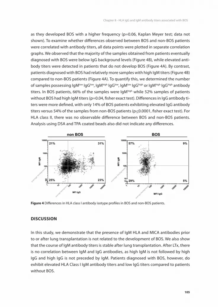
Chapter 8 - HLA IgG and IgM antibody titers associated with BOS
105
as they developed BOS with a higher frequency (p=0.06, Kaplan Meyer test; data not shown). To examine whether differences observed between BOS and non-BOS patients were correlated with antibody titers, all data points were plotted in separate correlation graphs. We observed that the majority of the samples obtained from patients eventually diagnosed with BOS were below IgG background levels (Figure 4B), while elevated anti-body titers were detected in patients that do not develop BOS (Figure 4A). By contrast, patients diagnosed with BOS had relatively more samples with high IgM titers (Figure 4B) compared to non-BOS patients (Figure 4A). To quantify this, we determined the number of samples possessing IgMlow IgGlow, IgMhigh IgGlow, IgMlow IgGhigh or IgMhigh IgGhigh antibody titers. In BOS patients, 66% of the samples were IgMhigh while 52% samples of patients without BOS had high IgM titers (p=0.04, fisher exact test). Differences in IgG antibody ti-ters were more defined, with only 14% of BOS patients exhibiting elevated IgG antibody titers versus 54% of the samples from non-BOS patients (p<0.0001, fisher exact test). For HLA class II, there was no observable difference between BOS and non-BOS patients. Analysis using DSA and TPA coated beads also did not indicate any differences.
disCussion
In this study, we demonstrate that the presence of IgM HLA and MICA antibodies prior to or after lung transplantation is not related to the development of BOS. We also show that the course of IgM antibody titers is stable after lung transplantation. After LTx, there is no correlation between IgM and IgG antibodies, as high IgM is not followed by high IgG and high IgG is not preceded by IgM. Patients diagnosed with BOS, however, do exhibit elevated HLA Class I IgM antibody titers and low IgG titers compared to patients without BOS.
figure 4 Differences in HLA class I antibody isotype profiles in BOS and non-BOS patients.

106
IgM antibodies against HLA have been considered to be clinically irrelevant, but recent findings challenge this concept. In heart transplantation, it was shown that non-HLA antibodies of IgM isotype were correlated with a reduction in graft survival (13, 17). In the context of heart and kidney transplantation, it has also been demonstrated that expression of HLA antibodies of the IgM isotype correlates with transplant rejection (13). The sensitive Luminex method used in this study made it possible to detect the presence of low HLA IgM antibody titers. Consistent with the results of HLA IgG antibody studies, no relationship could be detected between the presence of low HLA IgM antibody titers and chronic rejection or overall survival in patients treated with an immune suppressive regimen consisting of tacrolimus and mycophenolate mofetil after lung transplantation (12). In concert with this, a study of kidney transplantation also showed that low titers of DSA HLA IgG antibodies present prior to transplantation are not detrimental to overall survival and do not correlate with acute or chronic rejection (18).
One study detected a correlation between MICA antibodies and development of rejec-tion after kidney transplantation. However, a number of other studies of heart or kidney transplantation did not find such a relation (19-22). Although MICA antibodies appear to contain minor complement fixing abilities, MICA is expressed in high levels in the renal system and intestines and is not detected in the heart or aorta, raising the possibility that the ability of these antibodies to bind complement to induce antibody mediated rejection is organ specific (22). Although MICA expression can be induced on airway epithelial cells by stress, we observed no relationship between the presence of MICA-antibodies and the chronic rejection of graft survival. In our study, MICA antibodies were present in both males and females prior to transplantation, although levels were slightly higher in males. These results and previous studies indicate that it is unlikely that MICA antibodies develop due to pregnancy.
High levels of IgM antibodies in serum may physically inhibit the binding of IgG antibodies to the Luminex beads (23); however, experiments using DTT treatment indicated that IgM antibodies could be depleted from the serum and that MFI levels of IgG did not increase. This indicates that the lack of IgGhigh antibody titers in BOS patients is not an artifact caused by high concentrations of IgM antibodies. Elevated levels of IgG anti-HLA that are detected after lung transplantation are also commonly observed prior to transplantation. The production of these antibodies is therefore not hampered by the immunosuppressive treatment with tacrolimus and mycophenolate mofetil administered after transplantation. We also observed no indication of IgM replacement by IgG alloantibodies, suggesting a lack of class-switching of B cells that may be due to the immunosuppression of T cells. These findings revealed that de novo IgG anti-HLA antibodies after lung transplantation yielded low levels of MFI.
Although titers of HLA class I IgMhigh IgGlow antibodies are elevated in patients with BOS, this cannot be used as a possible marker for the development of BOS because some

Chapter 8 - HLA IgG and IgM antibody titers associated with BOS
107
patients without BOS also have antibody titers exhibiting elevated IgM and low IgG. Additionally, although patients without BOS express higher HLA IgG antibody levels compared to patients with BOS, some considerations have to be placed. First, HLA IgG antibody titers are low and may not be capable of activating the complement cascade. Second, it is unknown if IgG HLA antibodies are of the IgG1, IgG3 subclasses able to fix complement, or of the IgG2 and IgG4 subclasses. If HLA antibodies are sequestered to the transplanted lung rendering them undetectable in BOS patients, they could fix the complement system and hence contribute to the development of BOS after lung transplantation (13, 24, 25). Furthermore, there are reports that low titers of HLA anti-bodies may be protective instead of harmful (22, 26). It is also unclear if the relationship between BOS and HLA class-I antibody levels contributes to the disease process or is an epiphenomenon. In our assays, we used beads with multiple antigens coated on them which did not allow us to detect specific donor reactivity. It should be noted that using beads specific to either DSA or TPA did not reveal any significant difference, and therefore a test with single antigen beads probably does not contribute to our findings. We also could not exclude cross reactivity between the antibodies.
In conclusion, lung transplant patients develop IgM HLA and MICA antibodies prior to and after transplantation. The presence of these antibodies and their titers are not relat-ed to chronic rejection under the current immune suppressive regime, and the isotype switch is inhibited. Patients diagnosed with BOS, however, exhibit elevated levels of IgM and reduced IgG HLA expression compared to patients without BOS. HLA antibodies are thought to be involved in chronic graft rejection as a result of their complement fixing ability as observed by C4D deposition within the graft. Mechanistically, our data could imply that IgM HLA antibodies are relevant to the pathogenesis of BOS because they are able to fix complement more efficiently than IgG.
ACknoWLedgeMents
We would like to thank OneLambda for kindly providing the Luminex kits LABScreen Mixed.

108
referenCes
1. Estenne M, Hertz MI. Bronchiolitis obliterans after human lung transplantation. Am J Respir Crit Care Med 2002; 166 (4): 440.
2. Boehler A, Estenne M. Post-transplant bronchiolitis obliterans. Eur Respir J 2003; 22 (6): 1007. 3. Christie JD, Edwards LB, Aurora P, et al. The Registry of the International Society for Heart and Lung
Transplantation: Twenty-sixth Official Adult Lung and Heart-Lung Transplantation Report-2009. J Heart Lung Transplant 2009; 28 (10): 1031.
4. Al-Githmi I, Batawil N, Shigemura N, et al. Bronchiolitis obliterans following lung transplantation. Eur J Cardiothorac Surg 2006; 30 (6): 846.
5. Trulock EP, Edwards LB, Taylor DO, Boucek MM, Keck BM, Hertz MI. Registry of the International Society for Heart and Lung Transplantation: twenty-second official adult lung and heart-lung transplant report--2005. J Heart Lung Transplant 2005; 24 (8): 956.
6. Daud SA, Yusen RD, Meyers BF, et al. Impact of immediate primary lung allograft dysfunction on bronchiolitis obliterans syndrome. Am J Respir Crit Care Med 2007; 175 (5): 507.
7. Jaramillo A, Smith MA, Phelan D, et al. Development of ELISA-detected anti-HLA antibodies precedes the development of bronchiolitis obliterans syndrome and correlates with progressive decline in pulmonary function after lung transplantation. Transplantation 1999; 67 (8): 1155.
8. Sharples LD, McNeil K, Stewart S, Wallwork J. Risk factors for bronchiolitis obliterans: a systematic review of recent publications. J Heart Lung Transplant 2002; 21 (2): 271.
9. Smith MA, Sundaresan S, Mohanakumar T, et al. Effect of development of antibodies to HLA and cytomegalovirus mismatch on lung transplantation survival and development of bronchiolitis obliterans syndrome. J Thorac Cardiovasc Surg 1998; 116 (5): 812.
10. Gammie JS, Pham SM, Colson YL, et al. Influence of panel-reactive antibody on survival and rejec-tion after lung transplantation. J Heart Lung Transplant 1997; 16 (4): 408.
11. Bhorade SM, Stern E. Immunosuppression for lung transplantation. Proc Am Thorac Soc 2009; 6 (1): 47.
12. Paantjens AW, van de Graaf EA, van Ginkel WG, van den Bosch JM, Otten HG. Lung transplantation under a tacrolimus/mycophenolate mofetil-based immunosuppressive regimen results in low titers of HLA and MICA IgG antibodies which are not related to development of BOS. J Heart Lung Transplant; 29 (5): 596.
13. Stastny P, Ring S, Lu C, Arenas J, Han M, Lavingia B. Role of immunoglobulin (Ig)-G and IgM an-tibodies against donor human leukocyte antigens in organ transplant recipients. Hum Immunol 2009.
14. Estenne M, Maurer JR, Boehler A, et al. Bronchiolitis obliterans syndrome 2001: an update of the diagnostic criteria. J Heart Lung Transplant 2002; 21 (3): 297.
15. Burke CM, Theodore J, Dawkins KD, et al. Post-transplant obliterative bronchiolitis and other late lung sequelae in human heart-lung transplantation. Chest 1984; 86 (6): 824.
16. Taylor CJ, Chapman JR, Ting A, Morris PJ. Characterization of lymphocytotoxic antibodies causing a positive crossmatch in renal transplantation. Relationship to primary and regraft outcome. Transplantation 1989; 48 (6): 953.
17. Smith JD, Hamour IM, Burke MM, et al. A reevaluation of the role of IgM Non-HLA antibodies in cardiac transplantation. Transplantation 2009; 87 (6): 864.
18. Aubert V, Venetz JP, Pantaleo G, Pascual M. Low levels of human leukocyte antigen donor-specific antibodies detected by solid phase assay before transplantation are frequently clinically irrel-evant. Hum Immunol 2009; 70 (8): 580.

Chapter 8 - HLA IgG and IgM antibody titers associated with BOS
109
19. Zou Y, Stastny P, Susal C, Dohler B, Opelz G. Antibodies against MICA antigens and kidney-transplant rejection. N Engl J Med 2007; 357 (13): 1293.
20. Lemy A, Andrien M, Wissing KM, et al. Major Histocompatibility Complex Class 1 Chain-Related Antigen A Antibodies: Sensitizing Events and Impact on Renal Graft Outcomes. Transplantation; 90 (2): 168.
21. Pavlova YA, Malek I, Honsova E, et al. Hepatocyte growth factor and antibodies to HLA and MICA antigens in heart transplant recipients. Tissue Antigens.
22. Smith JD, Brunner VM, Jigjidsuren S, et al. Lack of Effect of MICA Antibodies on Graft Survival Following Heart Transplantation. Am J Transplant 2009.
23. Kosmoliaptsis V, Bradley JA, Peacock S, Chaudhry AN, Taylor CJ. Detection of immunoglobulin G human leukocyte antigen-specific alloantibodies in renal transplant patients using single-antigen-beads is compromised by the presence of immunoglobulin M human leukocyte antigen-specific alloantibodies. Transplantation 2009; 87 (6): 813.
24. Colvin RB, Smith RN. Antibody-mediated organ-allograft rejection. Nat Rev Immunol 2005; 5 (10): 807.
25. Adeyi OA, Girnita AL, Howe J, et al. Serum analysis after transplant nephrectomy reveals restricted antibody specificity patterns against structurally defined HLA class I mismatches. Transpl Immu-nol 2005; 14 (1): 53.
26. Lynch RJ, Platt JL. Accommodation in organ transplantation. Curr Opin Organ Transplant 2008; 13 (2): 165.


Chapter 9Identification of Allo- and Auto-Antibodies after Lung Transplantation
A.W.M. Paantjens1, E.A. van de Graaf2, J.M. Kwakkel-van Erp2, W.G.J. van Ginkel1, T. Hoefnagel1, D.A. van Kessel3, J.M.M. van den Bosch3, H.G. Otten1.
Lung Transplantation: Therapies, Complications and Outcomes
pp. 93-110
1Department of Immunology. University Medical Centre Utrecht,
Utrecht, The Netherlands 2Department of Respiratory Medicine. University Medical Centre
Utrecht, Utrecht, The Netherlands 3Department of Respiratory Medicine. St Antonius Hospital,
Nieuwegein, The Netherlands

112
AbstrACt
The bronchiolitis obliterans syndrome (BOS) is considered to be the consequence of chronic lung allograft rejection, characterized histologically by airway epithelial cell (AEC) apoptosis and luminal fibrosis in the respiratory bronchioles causing airflow ob-struction. Although the detailed etiology and pathogenesis of BOS are not clear, it has become evident that both the humoral and the cellular allogeneic immune response against AEC and endothelial cells, contributes significantly to the pathogenesis of BOS. It was demonstrated that the presence of allo-antibodies reacting with HLA and non-HLA antigens expressed on AEC may precede BOS development, suggesting that non-HLA antigenic systems may also play a role in chronic lung allograft rejection. These data are in line with results obtained in kidney transplantation, in which it was demonstrated that endothelial cell-reactive non-HLA antibodies could be found in sera of patients, which have suffered from hyperacute or acute kidney allograft rejection.
Identification of non-HLA antigens recognized by the patients’ humoral immune sys-tem after lung transplantation provides insight in the immunopathogenesis of rejection and may lead to tailor-made immune suppression. Therefore, research has focussed to-wards new methods identifying non-HLA antibodies after solid organ transplantation. In literature, 3 methods have been described for identification of previously unknown anti-gens recognized by antibodies in the sera of patients after transplantation. One method is based on protein arrays. A second, recently described technique, uses SIMT which is an immunoprecipitation followed by Matrix-assisted laser desorption/ionization-Time-of-flight mass spectrometry (MALDI-TOF). The third method is the serologic analysis of antigens by recombinant expression cloning (SEREX), which has been applied on lung transplantation and is able to screen a very large spectrum of antigens expressed by a target tissue like the bronchus in a single screening. Here, we review the advantages and disadvantages of these large-scale screening techniques which can be used to identify antigens recognized by the immune system after lung transplantation (LTx), and provide a comprehensive overview of the antigens identified so far. In addition, the possibilities of identifying patients at risk for rejection using antibody-based screening procedures will be discussed.

Chapter 9 - Identification of allo- & auto-antibodies
113
the bronChioLitis obLiterAns syndroMe After Lung trAnsPLAntAtion
For end stage lung diseases lung transplantation (LTx) is the only treatment option. Although, through the years many successful immune suppression regimens were intro-duced, the overall survival of lung transplant recipients is severely hampered by devel-opment of chronic rejection, also known as the bronchiolitis obliterans syndrome (BOS). The overall BOS free survival is 50% in 5 years after LTx (1). The exact processes leading to development of BOS are unknown, but several risk factors have been described, like primary graft dysfunction (PGD), infections, gastro-oesophageal reflux disease (GERD) and HLA antibodies (2-6).
Antibody MediAted rejeCtion
Antibodies playing a role in humoral chronic rejection of kidneys have been reported as early as 1970 (7) Nowadays, this role is confirmed and widely studied. After antibod-ies bind to the graft they have the ability to fix and activate complement, and thereby damaging the graft (8-10). In the classic pathway of complement activation, after C1q interacts with antibodies, C4d is a product of complement activation, which is cova-lently bound to the graft and is a marker for antibody mediated rejection (AMR) (11), before C5b-C9 can be fixed in the membrane and form pores. The reactivity against epithelium cells activated the production of growth factors and stress proteins which in turn activates fibroproliferation (12). The damage done by complement to the allograft leads to graft dysfunction and the bronchiolitis syndrome, although this route has not been documented extensively in lung transplantation and some conflicts between stud-ies are present (13-18). Deposits of C4d and C1q were found on the bronchial wall in patients with BOS, in addition C4d deposits and immunoglobulins have been detected on the bronchial epithelium as well providing the proof that the complement cascade is activated via the classical pathway (13, 19, 20).
HLA antibodies
Antibody mediated rejection (AMR) mainly is ascribed to the presence of HLA antibod-ies. Evidence was provided by studies showing that HLA antibodies reactive with donor cells prior to renal- and later also lung-transplantation correlated with hyperacute rejec-tion (21-23). Many studies describe the relation between HLA antibodies prior to or after LTx and rejection either acute or chronic and the HLA antibodies are considered to be a major predisposing factor of BOS (6, 24-28). It is beyond the scope of this review to describe these relations in detail. More recently the focus has been on donor specific

114
antibodies (DSA), and it was shown that these donor-specific HLA alloantibodies can ini-tiate rejection through complement-mediated and antibody-dependent, cell-mediated cytotoxicity (29, 30). In lung transplantation the immunosuppressive regime might be of major influence as recently described in two studies analyzing HLA antibodies after lung transplantation by luminex. Under a regime consisting of tacrolimus, mycophenolate mofetil and prednisone HLA antibodies were present at low titers and did not correlate with BOS, while under another regime a correlation between DSA HLA antibodies and BOS was described (31, 32), hence HLA antibodies cannot always serve as marker for patients at risk of development of BOS.
Non-HLA antibodies
From studies on renal transplantation it became apparent that not only HLA antibodies contribute to AMR. Associations between non-HLA antibodies directed against endothe-lial cells were found in some patients whom had a kidney transplantation that rejected their allograft and later also confirmed for heart and lung transplantation (33-38). In lung transplantation anti-epithelial cell (anti-AEC) antibodies were detected in patients with-out HLA-reactivity prior to transplantation, and the presence of these anti-AEC antibod-ies was related to a poorer graft survival, indicating non-HLA antibodies are important in BOS (39, 40). But this became really apparent with a study on renal transplantation between HLA-identical siblings also lead to chronic rejection via the antibody mediated pathway, and in 2005 Opelz et al. revealed that non-HLA immunity plays an important role in chronic rejection in kidney transplant recipients from HLA-identical siblings (41). Furthermore, it was reported that only 18% of renal graft failure could be contributed to HLA antibodies, while 38% was due to non-HLA antibodies and 43% were associated to non-immunologic factors (42).
Extensive research on non-HLA antibodies has concentrated to the MHC class I polypeptide related chain A (MICA). Zwirner et al was the first to report the non-HLA antibody against MICA to be present in kidney transplantation (43). In lung transplanta-tion elaborate studies on MICA antibodies and BOS are absent. We were able to show in a longitudinal study of 50 lung transplant recipients, with an immunosuppressive regime consisting of mycophenolate mofetil, tacrolimus and prednisone that MICA antibodies are present and increased after lung transplantation but they are not related to the bronchiolitis obliterans syndrome. Furthermore, the MICA antibodies detected in 7 lung transplant recipients with BOS after lung transplantation were not donor specific (31). However, recently it was shown that patients under a different immune suppressive regime (cyclosporine, azathioprine and prednisone) did have MICA antibodies which correlated with the development of BOS (32). Therefore, the immunosuppressive regime applied is of importance as well whether certain antibodies can be applied as biomark-ers as described above for HLA antibodies.

Chapter 9 - Identification of allo- & auto-antibodies
115
Testing both HLA and non-HLA antibodies is of clinical importance as both HLA and non HLA antibodies, rise early after transplantation and their appearance was reported well before the rise in serum creatinin, an indication for rejection in kidney transplanta-tion (44). Another non-HLA antibody found in renal transplant patients with allograft rejection but not in other patients is against the angiotensin II type 1 (AT1) receptor (45), and in heart transplantation antibodies recognizing vimentin, myosin and phospholipids have been detected after transplantation (46-48). Although testing for (donor specific) HLA antibodies is routine prior to and post transplantation, methodological options for identification and characterization of non-HLA antigens targeted during rejection after transplantation are scarce.
Screening strategies
Growing evidence that next to HLA antibodies non-HLA antibodies might be part of processes leading to chronic rejection called for techniques to identify such autoanti-bodies. Therefore, measurements of antibodies post transplantation might provide an early biomarker for detection of patients at risk of rejection. The prognostic significance of HLA antibody detection of transplantation is somewhat limited as they appear in patients after solid organ transplantation who reject this organ but also some patients who do not experience rejection episodes. In addition, patients without HLA antibodies still can develop chronic rejection. This indicates that there is a need to find relevant biomarkers that are highly sensitive and very specific for early diagnosis and prognosis of chronic rejection by humoral mechanisms.
Using antibodies as possible biomarkers for chronic rejection after lung transplanta-tion has some practical advantages. They are relatively easy accessible by a non invasive approach using sera from patients Moreover, autoantibodies are naturally stable and persist in the serum for a relatively long period of time because they are not subjected to the types of proteolysis observed for other polypeptides (49). A disadvantage that needs to be considered is probably the heterogenicity of antibodies. A high variety of antibodies against different antigens is found between patients. Therefore, lacks a single antibody test both sensitivity and specificity and need the test to be repeated or the combination of several autoantibodies can be used (50).
When researchers in the transplantation field became more interested in the role and possibilities of autoantibodies, as it became apparent they might contribute to the development of rejection, new techniques had to be developed. Systematic screening methods were needed to be able to identify possible new antigens in a high throughput manner which was not labor intensive. For that reason different techniques have been developed and amended. One of the first studies in renal transplantation describing the usefulness of such techniques showed that harvesting endothelial cells from peripheral blood using TIE-2 antibodies on dynabeads used for easy patient anti-endothelial cell

116
crossmatch (51). Here some of the techniques will be briefly reviewed and their contri-bution to the field of human (lung) transplantation will be enlighted.
Serological proteome analysis
Serological proteome analysis (SERPA) also known as Proteomex is a combination of 2-DE gels and western blotting. The technique was originally developed for the identification of tumor antigens in kidney cancer (52, 53). And was one of the first techniques allowing identification of proteins/antigens in a high throughput manner. SERPA has been widely used in identification of tumor antigens, antigens in autoimmune diseases and possible vaccine strategies of infectious diseases (54, 55). Although, it has been very successful identifying several possible markers for different lung cancers it has not been used in the research to identify antigens for autoantibodies in lung transplantation or other forms of solid organ transplantation (56-58).
Every technique has advantages and disadvantages compared to other techniques. For SERPA one major advantage is the use of isolated protein as starting material which makes it possible to identify post-translational modifications and protein isoforms. Ad-ditionally as no library has to be constructed, SERPA is less time consuming than SEREX (described later) which uses a cDNA library. On the other hand, the techniques has its limitations, for instance it has a bias to abundant proteins because of the sensitivity of the staining methods. Furthermore hydrophobic and insoluble transmembrane proteins as well as small proteins (<10kDA) or proteins with extreme isoelectric points are dif-ficult to detect (59-61). Due to the use of western blotting as staining method only linear epitodes are detected (50).
The SERPA technique has proved the possibilities of identifying new target antigens, and based on this technique protein arrays were designed.
Protein arrays
Protein arrays are based in antigen immobilization on a support where sera of patients can react. Commercially there are arrays available coated with over 5000 different pro-teins developed as screening for (ovarian) cancer and therefore at present probably not the best starting point to screen for (organ) specific allograft rejection related antigens. However, these arrays have been used in pediatric kidney transplantation studies, and proven the feasibility of using protein arrays to detect new target antigens during rejec-tion (62). Antigens detected in these studies include MICA and antibodies against the renal pelvis area and cortex specific antigens. One of the antigens related to renal area was Protein kinase Cζ (PKCζ) which was shown to be related to development of acute rejection (63).
An advantage of using protein arrays to identify unknown antigenic structures is that they are not labor intensive as well as they allow analysis of a large number of targets

Chapter 9 - Identification of allo- & auto-antibodies
117
in one step and therefore are high-throughput (64-66). Another advantage of the small arrays is that only little material is needed to perform one array (67).
On the other side, protein arrays are limited by the availability of commercial or home made proteins on the arrays. They do not cover the whole proteome. Furthermore with protein arrays it is not possible to differentiate between intracellular and surface expressed proteins, while especially surface expressed antigens are of interest in the context of allograft rejection (68). In addition, the protein arrays themselves have some disadvantages as well. It is difficult to produce and purify native protein targets and once bound to the array they have a short shelf-life (69, 70).
SIMT
Recently a new technique was developed called Sequential analysis of Immunopre-cipitation followed by Matrix-assisted laser desorption/ionization-Time-of-flight mass spectrometry (SIMT) (71). SIMT is a combination between immunoprecipitation and mass spectrometry to identify possible new target antigens for allograft rejection. An advantage of this method over the previously described methods is the usage of native human cells and indirectly measuring the clinically more relevant antigens expressed on the surface of these cells.
However, as the technique has only been used as prove of principle it still has to prove its benefits for transplantation research. In a HLA-B27 or -B7 setting the technique was able to pickup these antigens as targets of an immune response, other HLA molecules and even non-HLA targets need to be investigated. The technique has only been used in a setup phase where samples with proven reactivity in lymphocytotoxic assays were used opposed to autologous material. As SIMT is a relatively new technique it might be optimized. At present, there are problems with detecting bands of non specific serum antigens binding to the beads. And a major drawback is the use of material as much serum is needed for one screening, as well as the relatively laborious steps of which the method is composed.
SEREX
The technique Serological identification of antigens by recombinant expression cloning (SEREX) using a cDNA expression library has been developed to explore the humoral response in sera from patients with their own tumor as source in 1995 (59, 72). Since the development of the technique numerous tumor associated antigens reactive with many cancer types have been identified by SEREX and over 2300 are documented in a database (73-75). As the technique has proven its strength in the field of cancer research it had been utilized in other fields like auto-immune diseases and transplantation.
One of the strengths of the SEREX technique is the use of target tissue, like a spe-cific tumor or a specific organ as source of the mRNA from which a cDNA library is con-
118
structed. The SEREX techniques knows some disadvantages as well, the work needed to construct a cDNA library is quite laborious. It uses an artificial expression system and therefore potentially representing denatured or improperly glycosylated proteins. It cannot discriminate between intracellular and surface proteins like SIMT does. SEREX might produce numerous false positives due to the identification of a humoral response specific to patient but unrelated to tumor (59). This is in concert with our study after lung transplantation (data shown below), however using patient and respective donor mate-rial the false positive results might be decreased (76). There might be a bias towards antigens that are highly expressed in the target tissues used to generate the cDNA libraries (77). And small proteins (<120 aa) are less well incorporated in the expression system (78).
We made use of the SEREX system in the setting of stem cell, kidney and lung trans-plantation. The results obtained are briefly summarized below.
Lung transplantationFor the identification of antigens after lung transplantation several screenings have been performed: 3 months after lung transplantation, 6 months after lung transplanta-tion and less than three months before development of BOS.
An epithelial-cell cDNA expression library was made from trachea of 15 lung donors. In the first pilot sera of 11 random patients from the cohort were taken and each screened against 3000 plaques from the library (79). In a later stage the screening was elaborated and sera derived from 7 patients, taken 3 months after LTx or <3 months prior to BOS was screened against 4x10⁴ plaques from the library. Recognized plaques were isolated, further seeded and rescreened until 100% of the reseeded plaques were recognized by the serum (Figure 1).
Rescreening as shown in Figure 1, resulted in 28 plaques recognized, and alignment of all inserts showed several identical amino-acid sequence motifs occurring in different inserts. Next to identification of the inserts in the plaques 15 amino acid long peptides were designed on the possible motifs and tested on ELISA. This however did not result in positive signals above background, and therefore it was concluded that these peptide were no possible epitopes.
After sequencing some of the plaques were identified as non coding regions (n=22) and 3 were found to hold genes without start codon and 3 plaques contained genes with start codon. (Table 1)
The genes without start codon were identified as XP_931864, LOC 284058 and PSMC 4, and the genes with start codon were PLUNC, F3, and ZNF 33A. These possible an-tigens are all internally expressed with the exception of PLUNC and F3. Because this first protein palate lung nasal clone (PLUNC) is expressed in the upper respiratory tract and oral cavity it was of special interest regarding a possible antigen target after lung

Chapter 9 - Identification of allo- & auto-antibodies
119
transplantation (80-82). PLUNC’s putative function is thought to be as an immune defence protein of epithelial surfaces against pathogenic microorganisms because its sequence is homologue to lipopolysaccharidebinding protein (LBP) and bacteriocidal/permeability-increasing protein (BPI) (83-85).
The cDNA of PLUNC was excreted from the plaque and cloned into SOLR bacteria. Lysates from these bacteria were tested on Western Blot with different sera of lung transplantation patients and a commercially available antibody. As shown in Figure 2, patient serum was able to detect PLUNC in the lysate of SOLR bacteria and therefore antibodies were directed against PLUNC. From sera of 12 lung transplantation tested 4 were capable to positively recognize PLUNC.
Figure 1
figure 1 Bacteriophages were seeded out using E-coli plates as described in patients & methods, yielding approximately 4000 plaques per plate. The nitrocellulose filters were screened with patient serum diluted 1:500. Shown are A) an example of a first screening result from 1 patient serum on 1 plate in which 1 positive plaque was detected B) an optical enlargement of that plaque marked by the arrow and C) the results of reseeding and rescreening of the recognized plaque.
table 1 SEREX screenings were performed at 3 different time points.
3 months post Ltx 6 months post Ltx < 3 months prior to bos
number of patients 7 patients with BOS 11 random patients 7 patients with BOS and 1 patient with rejection problems
Plaques per patient 40 000 3 000 40 000
Positive plaques 7 11 10
identification 7 non coding regions 3 genes with start codon3 genes without start codon5 non coding regions
10 non coding regions

120
Another study by Goers et al. has also detected non-HLA antigens after lung transplanta-tion (12). This was not after elaborate screenings but a quick scan in a few individuals. A combination of PRA and western blotting on airway epithelial cells, followed by protein isolation and sequencing identified K-α1 tubulin as a possible target antigen. Further analysis revealed that 36 BOS and 36 non BOS together with 10 HC had no reactivity against HLA, but 12 out of 36 BOS had specific reactivity against AEC via K-α1 tubulin.
Kidney transplantationFor kidney transplantation non-HLA antibodies have been reported to influence al-lograft survival. One of the non-HLA antigens identified is the angiotensin II type 1 (AT1) receptor and in a recent study, the presence of activating IgG antibodies targeting this receptor was examined in 33 kidney-transplant recipients with refractory vascular rejec-tion (45, 86). Activating IgG anti-AT1 receptor antibodies were detected in serum from all 16 patients with malignant hypertension - in absence of anti-HLA antibodies - but not in the other patients.
Antibodies have also been described against MHC class-I related A antigens (MICA), which are expressed on endothelial and epithelial cells, monocytes and fibroblasts (87). In a large scale multicenter study on 1329 patients with functioning kidney transplants, it was shown that the presence of anti-MICA antibodies in post-transplant sera is signifi-cant correlated to kidney allograft loss (88). In this study, donor graft survival was 72% vs 81% in patients which had or did not have anti-MICA antibodies, respectively.
Here, the main purpose of was to identify non-HLA antigens recognized by antibodies from patients awaiting kidney retransplantation with SEREX. To this end, 7 patients (M/F = 3/4; median age = 46; range = 29-60 years) were selected having rejected their kidney. Serum was taken after nephrectomy and analyzed for reactivity against an epithelial cell protein-expression library SEREX. Serum of every individual was screened against approximately 3x10^4 plaques. A total of 8 plaques were recognized by the sera of 4 patients. After rescreening as shown above in Figure 1, five inserts were very small (5-67
Figure 2
figure 2 Lysate of SOLR bacteria or SOLR bacteria with PLUNC were separated on a 12% SDS-PAGE gel before blotted onto a nitrocellulose membrane and incubated with either serum of a lung transplantation patient or a polyclonal antibody against PLUNC. The protein is detected by both the patient’s serum and antibody at approximately 24 kDa only in the lysate of bacteria with PLUNC.

Chapter 9 - Identification of allo- & auto-antibodies
121
aa) of which the original encoding gene could not be identified. The other inserts con-sisted of 3 different non-HLA antigens: tetraspanin 8, LPLUNC1 and BSCv (also known as C20ORF3 or adipocyte plasma membrane-associated protein). These were recognized by 4/7, 5/7 and 3/7 patient sera tested respectively, but not by sera from 3 healthy controls.
Stem cell transplantationTreatment with rituximab, a B-cell inhibitor, has a positive effect on the disease and in several studies allo- or autoantibodies have been detected in chronic Graft versus Host Disease (cGVHD) patients.
The aim of this study was to examine if auto- and alloantigens in cGVHD can be identi-fied by SEREX. To this end 10 sera derived from patients with cGVHD were examined by SEREX with a cDNA bank from epithelial cells of a lung. Furthermore, it was also deter-mined whether the identified antigens were also recognized by other cGVHD patient sera and a healthy control.
table 2 Abbreviations: nr, number; nd, not determined. Patients 1, 2, 3, 4 and 5 were screened differently from patients 6, 7, 8, 9 and 10 therefore not all results are available as prescreening was not performed. The number of initial positive plaques is the total amount of positives that were observed on the filter after the first screening. The number of IgG in the first screening is the number of initial positive plaques that were identified as false positives during the prescreening. More IgG was seen when the total number of plaques was higher. The number of positive plaques in the first screening where isolated and underwent a second screening. During this second screening the filters were also incubated with patient serum and with TBS. A lot of the plaques were IgG despite of the prescreening. Plaques that were found positive during the second screening were isolated and screened again; the final screening. During this screening the filter was also incubated with normal serum and when positive plaques were seen on this filter they were considered negative. Identification of the isolated plaques after the second screening was performed if they were found to be positive after the final screening.
Patient nr of total plaques
nr of positive plaques
nr of igg detected in 1st screening
nr of igg detected in 2nd screening
nr of plaques recognized by control serum
final nr ofpositive plaques
1 67.000 98 80 11 1 6
2 39.000 51 37 8 1 5
3 45.000 53 43 8 2 0
4 36.000 22 17 2 2 1
5 28.000 17 11 1 3 2
6 14.000 nd nd nd nd 0
7 13.000 nd nd nd nd 0
8 11.500 nd nd nd nd 3
9 10.000 nd nd nd nd 2
10 16.000 nd nd nd nd 0
total 279.500 246 188 30 9 19

122
Nineteen positive cloned inserts were found (details in Table 2). Nine of those were identified as interferon gamma inducible protein 16 (IFI16), one was identified as tro-phoblast glycoprotein (TPBG), another as syndecan binding protein (SDCBP), and two were unknown, one was an artifact and the other five are unidentified. IFI 16 was of special interest because it was identified by screening the sera of 3 out of 10 different patients.
Overall the SEREX technique is powerful enough to detect various possible antigens after stem cell and solid organ transplantation. Antigens identified were recognized by several different patients but not healthy controls. However, as for antigens found for GvHD and kidney transplantation, the antigens detected after lung transplantation could not be cloned into fusion proteins to be purified and used on ELISA for high trough put screening of a patient cohort. Due to instability proteins were not fused to a tag and analysis of the other possible targets as bacterial lysate on Western Blot revealed cross reactivity in the samples, multiple bands on different heights were detected, which were not multimers of the predicted size.
ConCLusion
Delineation of the non-HLA antibody spectrum after lung transplantation may facilitate our understanding of both allo- and autoimmune responses in chronic allograft rejec-tion. The screening for possible antigens after transplantation is relatively new, but examples proving the strengths of the high through put wide technologies in the cancer research field are abundant. However, many of the antigens discovered via one of the above mentioned techniques still need to prove their power as biomarkers in the clinic (89). Many antibodies against these new antigens have a low frequency and are present in approximately 20-30% of patients of specific tumors (59). The combination of detection of several of the newly discovered non-HLA antibodies or biomarkers seems to allow the uncovering of tumors with higher efficiency than isolated biomarkers (65, 66, 90-94).
Next to the clinical relevance and low frequency that the antibodies are found in patients, it should be explained why screenings result in antigens mainly originating from intracellular proteins (95). This might be circumvented by the introduction of SIMT as a new screening technique, which is able to screen only surface expressed proteins.
At the moment all screening techniques focus on finding antibodies of IgG isotype. Recently it has become clear that IgM antibodies might contribute more to rejection after transplantation than previously thought. Both IgM HLA and non-HLA antibodies were described to be deleterious for the overall survival of transplant recipients (96, 97).

Chapter 9 - Identification of allo- & auto-antibodies
123
Overall, high trough put screening technologies might contribute to the detection of possible antigens after lung transplantation. However the clinical significance and the labor intensive character are major aspects to be considered before employing these methods.
ACknoWLedgeMents
We thank the ROTRF (project number 409414018) for their financial support and mrs. M. van Loon for her excellent technical assistance.

124
referenCes
1. Christie JD, Edwards LB, Aurora P, Dobbels F, Kirk R, Rahmel AO, et al. Registry of the International Society for Heart and Lung Transplantation: twenty-fifth official adult lung and heart/lung trans-plantation report--2008. J Heart Lung Transplant 2008;27(9):957-69.
2. Boehler A, Estenne M. Post-transplant bronchiolitis obliterans. Eur Respir J 2003;22(6):1007-18. 3. Daud SA, Yusen RD, Meyers BF, Chakinala MM, Walter MJ, Aloush AA, et al. Impact of immediate
primary lung allograft dysfunction on bronchiolitis obliterans syndrome. Am J Respir Crit Care Med 2007;175(5):507-13.
4. Sharples LD, McNeil K, Stewart S, Wallwork J. Risk factors for bronchiolitis obliterans: a systematic review of recent publications. J Heart Lung Transplant 2002;21(2):271-81.
5. Jaramillo A, Smith MA, Phelan D, Sundaresan S, Trulock EP, Lynch JP, et al. Development of ELISA-detected anti-HLA antibodies precedes the development of bronchiolitis obliterans syndrome and correlates with progressive decline in pulmonary function after lung transplantation. Trans-plantation 1999;67(8):1155-61.
6. Reinsmoen NL, Nelson K, Zeevi A. Anti-HLA antibody analysis and crossmatching in heart and lung transplantation. Transpl Immunol 2004;13(1):63-71.
7. Jeannet M, Pinn VW, Flax MH, Winn HJ, Russell PS. Humoral antibodies in renal allotransplantation in man. N Engl J Med 1970;282(3):111-7.
8. Baldwin WM, 3rd, Pruitt SK, Brauer RB, Daha MR, Sanfilippo F. Complement in organ transplanta-tion. Contributions to inflammation, injury, and rejection. Transplantation 1995;59(6):797-808.
9. Baldwin WM, 3rd, Qian Z, Wasowska B, Sanfilippo F. Complement causes allograft injury by cell activation rather than lysis. Transplantation 1999;67(11):1498-9.
10. Girnita AL, Lee TM, McCurry KR, Baldwin WM, 3rd, Yousem SA, Detrick B, et al. Anti-human leuko-cyte antigen antibodies, vascular C4d deposition and increased soluble c4d in broncho-alveolar lavage of lung allografts. Transplantation 2008;86(2):342-7.
11. Nickeleit V, Zeiler M, Gudat F, Thiel G, Mihatsch MJ. Detection of the complement degradation product C4d in renal allografts: diagnostic and therapeutic implications. J Am Soc Nephrol 2002;13(1):242-51.
12. Goers TA, Ramachandran S, Aloush A, Trulock E, Patterson GA, Mohanakumar T. De novo produc-tion of K-alpha1 tubulin-specific antibodies: role in chronic lung allograft rejection. J Immunol 2008;180(7):4487-94.
13. Miller GG, Destarac L, Zeevi A, Girnita A, McCurry K, Iacono A, et al. Acute humoral rejection of human lung allografts and elevation of C4d in bronchoalveolar lavage fluid. Am J Transplant 2004;4(8):1323-30.
14. Wallace WD, Reed EF, Ross D, Lassman CR, Fishbein MC. C4d staining of pulmonary allograft biopsies: an immunoperoxidase study. J Heart Lung Transplant 2005;24(10):1565-70.
15. Ionescu DN, Girnita AL, Zeevi A, Duquesnoy R, Pilewski J, Johnson B, et al. C4d deposition in lung allografts is associated with circulating anti-HLA alloantibody. Transpl Immunol 2005;15(1):63-8.
16. Saint Martin GA, Reddy VB, Garrity ER, Simpson K, Robinson JA, Adent JK, et al. Humoral (antibody-mediated) rejection in lung transplantation. J Heart Lung Transplant 1996;15(12):1217-22.
17. Magro CM, Klinger DM, Adams PW, Orosz CG, Pope-Harman AL, Waldman WJ, et al. Evidence that humoral allograft rejection in lung transplant patients is not histocompatibility antigen-related. Am J Transplant 2003;3(10):1264-72.
18. Westall GP, Snell GI, McLean C, Kotsimbos T, Williams T, Magro C. C3d and C4d deposition early after lung transplantation. J Heart Lung Transplant 2008;27(7):722-8.

Chapter 9 - Identification of allo- & auto-antibodies
125
19. Magro CM, Pope Harman A, Klinger D, Orosz C, Adams P, Waldman J, et al. Use of C4d as a diagnos-tic adjunct in lung allograft biopsies. Am J Transplant 2003;3(9):1143-54.
20. Magro CM, Ross P, Jr., Kelsey M, Waldman WJ, Pope-Harman A. Association of humoral immunity and bronchiolitis obliterans syndrome. Am J Transplant 2003;3(9):1155-66.
21. Lau CL, Palmer SM, Posther KE, Howell DN, Reinsmoen NL, Massey HT, et al. Influence of panel-reactive antibodies on posttransplant outcomes in lung transplant recipients. Ann Thorac Surg 2000;69(5):1520-4.
22. Bittner HB, Dunitz J, Hertz M, Bolman MR, 3rd, Park SJ. Hyperacute rejection in single lung transplantation--case report of successful management by means of plasmapheresis and anti-thymocyte globulin treatment. Transplantation 2001;71(5):649-51.
23. Kissmeyer-Nielsen F, Olsen S, Petersen VP, Fjeldborg O. Hyperacute rejection of kidney allografts, associated with pre-existing humoral antibodies against donor cells. Lancet 1966;2(7465):662-5.
24. Lee PC, Terasaki PI, Takemoto SK, Lee PH, Hung CJ, Chen YL, et al. All chronic rejection failures of kidney transplants were preceded by the development of HLA antibodies. Transplantation 2002;74(8):1192-4.
25. Takemoto SK, Zeevi A, Feng S, Colvin RB, Jordan S, Kobashigawa J, et al. National conference to as-sess antibody-mediated rejection in solid organ transplantation. Am J Transplant 2004;4(7):1033-41.
26. Ozawa M, Terasaki PI, Castro R, Alberu J, Morales-Buenrostro L, Alvarez I, et al. 14th International HLA and Immunogenetics Workshop Prospective Chronic Rejection Project: a three-year follow-up analysis. Clin Transpl 2007:255-60.
27. Palmer SM, Davis RD, Hadjiliadis D, Hertz MI, Howell DN, Ward FE, et al. Development of an antibody specific to major histocompatibility antigens detectable by flow cytometry after lung transplant is associated with bronchiolitis obliterans syndrome. Transplantation 2002;74(6):799-804.
28. Girnita AL, Duquesnoy R, Yousem SA, Iacono AT, Corcoran TE, Buzoianu M, et al. HLA-specific antibodies are risk factors for lymphocytic bronchiolitis and chronic lung allograft dysfunction. Am J Transplant 2005;5(1):131-8.
29. Vongwiwatana A, Tasanarong A, Hidalgo LG, Halloran PF. The role of B cells and alloantibody in the host response to human organ allografts. Immunol Rev 2003;196:197-218.
30. Mauiyyedi S, Colvin RB. Humoral rejection in kidney transplantation: new concepts in diagnosis and treatment. Curr Opin Nephrol Hypertens 2002;11(6):609-18.
31. Paantjens AW, van de Graaf EA, van Ginkel WG, van den Bosch JM, Otten HG. Lung transplantation under a tacrolimus/mycophenolate mofetil-based immunosuppressive regimen results in low titers of HLA and MICA IgG antibodies which are not related to development of BOS. J Heart Lung Transplant 2010;29(5):596-8.
32. Angaswamy N, Saini D, Ramachandran S, Nath DS, Phelan D, Hachem R, et al. Development of antibodies to human leukocyte antigen precedes development of antibodies to major histocom-patibility class I-related chain A and are significantly associated with development of chronic rejection after human lung transplantation. Hum Immunol 2010;71(6):560-5.
33. Paul LC, Carpenter CB. Antibodies against renal endothelial alloantigens. Transplant Proc 1980;12(3 Suppl 1):43-8.
34. Perrey C, Brenchley PE, Johnson RW, Martin S. An association between antibodies specific for endothelial cells and renal transplant failure. Transpl Immunol 1998;6(2):101-6.

126
35. Magro CM, Deng A, Pope-Harman A, Waldman WJ, Bernard Collins A, Adams PW, et al. Humorally mediated posttransplantation septal capillary injury syndrome as a common form of pulmonary allograft rejection: a hypothesis. Transplantation 2002;74(9):1273-80.
36. Pierce JC, Waller M, Phibbs M. A mixed antiglobulin test with kidney cells in suspension for IgG antibody in human allograft recipients. Transplantation 1975;19(4):343-8.
37. Mohanakumar T, Waldrep JC, Phibbs M, Mendez-Picon G, Kaplan AM, Lee HM. Serological charac-terization of antibodies eluted from chronically rejected human renal allografts. Transplantation 1981;32(1):61-6.
38. Hosenpud JD, Everett JP, Morris TE, Mauck KA, Shipley GD, Wagner CR. Cardiac allograft vascu-lopathy. Association with cell-mediated but not humoral alloimmunity to donor-specific vascular endothelium. Circulation 1995;92(2):205-11.
39. Smith JD, Crisp SJ, Dunn MJ, Pomerance A, Yacoub MH, Rose ML. Pre-transplant anti-epithelial cell antibodies and graft failure after single lung transplantation. Transpl Immunol 1995;3(1):68-73.
40. Jaramillo A, Naziruddin B, Zhang L, Reznik SI, Smith MA, Aloush AA, et al. Activation of human airway epithelial cells by non-HLA antibodies developed after lung transplantation: a potential etiological factor for bronchiolitis obliterans syndrome. Transplantation 2001;71(7):966-76.
41. Opelz G. Non-HLA transplantation immunity revealed by lymphocytotoxic antibodies. Lancet 2005;365(9470):1570-6.
42. Terasaki PI. Deduction of the fraction of immunologic and non-immunologic failure in cadaver donor transplants. Clin Transpl 2003:449-52.
43. Zwirner NW, Marcos CY, Mirbaha F, Zou Y, Stastny P. Identification of MICA as a new polymorphic alloantigen recognized by antibodies in sera of organ transplant recipients. Hum Immunol 2000;61(9):917-24.
44. Mao Q, Terasaki PI, Cai J, Briley K, Catrou P, Haisch C, et al. Extremely high association between appearance of HLA antibodies and failure of kidney grafts in a five-year longitudinal study. Am J Transplant 2007;7(4):864-71.
45. Dragun D, Muller DN, Brasen JH, Fritsche L, Nieminen-Kelha M, Dechend R, et al. Angiotensin II type 1-receptor activating antibodies in renal-allograft rejection. N Engl J Med 2005;352(6):558-69.
46. Jurcevic S, Dunn MJ, Crisp S, Busing K, Rinaldi M, Pellegrini C, et al. A new enzyme-linked immuno-sorbent assay to measure anti-endothelial antibodies after cardiac transplantation demonstrates greater inhibition of antibody formation by tacrolimus compared with cyclosporine. Transplanta-tion 1998;65(9):1197-202.
47. Morgun A, Shulzhenko N, Unterkircher CS, Diniz RV, Pereira AB, Silva MS, et al. Pre- and post-transplant anti-myosin and anti-heat shock protein antibodies and cardiac transplant outcome. J Heart Lung Transplant 2004;23(2):204-9.
48. Laguens RP, Vigliano CA, Argel MI, Chambo JG, Rozlosnik JA, Perrone SV, et al. Anti-skeletal muscle glycolipid antibodies in human heart transplantation as predictors of acute rejection: comparison with other risk factors. Transplantation 1998;65(10):1345-51.
49. Tan HT, Low J, Lim SG, Chung MC. Serum autoantibodies as biomarkers for early cancer detection. Febs J 2009;276(23):6880-904.
50. Casiano CA, Mediavilla-Varela M, Tan EM. Tumor-associated antigen arrays for the serological diagnosis of cancer. Mol Cell Proteomics 2006;5(10):1745-59.
51. Vermehren D, Sumitran-Holgersson S. Isolation of precursor endothelial cells from periph-eral blood for donor-specific crossmatching before organ transplantation. Transplantation 2002;74(11):1479-86.

Chapter 9 - Identification of allo- & auto-antibodies
127
52. Klade CS, Voss T, Krystek E, Ahorn H, Zatloukal K, Pummer K, et al. Identification of tumor antigens in renal cell carcinoma by serological proteome analysis. Proteomics 2001;1(7):890-8.
53. Kellner R, Lichtenfels R, Atkins D, Bukur J, Ackermann A, Beck J, et al. Targeting of tumor associated antigens in renal cell carcinoma using proteome-based analysis and their clinical significance. Proteomics 2002;2(12):1743-51.
54. Sikaroodi M, Galachiantz Y, Baranova A. Tumor markers: the potential of “omics” approach. Curr Mol Med;10(2):249-57.
55. Greub G, Kebbi-Beghdadi C, Bertelli C, Collyn F, Riederer BM, Yersin C, et al. High throughput sequencing and proteomics to identify immunogenic proteins of a new pathogen: the dirty genome approach. PLoS One 2009;4(12):e8423.
56. Yang F, Xiao ZQ, Zhang XZ, Li C, Zhang PF, Li MY, et al. Identification of tumor antigens in human lung squamous carcinoma by serological proteome analysis. J Proteome Res 2007;6(2):751-8.
57. Huang LJ, Chen SX, Huang Y, Luo WJ, Jiang HH, Hu QH, et al. Proteomics-based identification of secreted protein dihydrodiol dehydrogenase as a novel serum markers of non-small cell lung cancer. Lung Cancer 2006;54(1):87-94.
58. Li C, Xiao Z, Chen Z, Zhang X, Li J, Wu X, et al. Proteome analysis of human lung squamous carci-noma. Proteomics 2006;6(2):547-58.
59. Desmetz C, Cortijo C, Mange A, Solassol J. Humoral response to cancer as a tool for biomarker discovery. J Proteomics 2009;72(6):982-8.
60. Balboni I, Chan SM, Kattah M, Tenenbaum JD, Butte AJ, Utz PJ. Multiplexed protein array platforms for analysis of autoimmune diseases. Annu Rev Immunol 2006;24:391-418.
61. Canelle L, Bousquet J, Pionneau C, Deneux L, Imam-Sghiouar N, Caron M, et al. An efficient proteomics-based approach for the screening of autoantibodies. J Immunol Methods 2005;299(1-2):77-89.
62. Li L, Wadia P, Chen R, Kambham N, Naesens M, Sigdel TK, et al. Identifying compartment-specific non-HLA targets after renal transplantation by integrating transcriptome and “antibodyome” measures. Proc Natl Acad Sci U S A 2009;106(11):4148-53.
63. Sutherland SM, Li L, Sigdel TK, Wadia PP, Miklos DB, Butte AJ, et al. Protein microarrays identify an-tibodies to protein kinase Czeta that are associated with a greater risk of allograft loss in pediatric renal transplant recipients. Kidney Int 2009;76(12):1277-83.
64. Bouwman K, Qiu J, Zhou H, Schotanus M, Mangold LA, Vogt R, et al. Microarrays of tumor cell de-rived proteins uncover a distinct pattern of prostate cancer serum immunoreactivity. Proteomics 2003;3(11):2200-7.
65. Wang X, Yu J, Sreekumar A, Varambally S, Shen R, Giacherio D, et al. Autoantibody signatures in prostate cancer. N Engl J Med 2005;353(12):1224-35.
66. Kijanka G, Murphy D. Protein arrays as tools for serum autoantibody marker discovery in cancer. J Proteomics 2009;72(6):936-44.
67. Robinson WH, Steinman L, Utz PJ. Protein arrays for autoantibody profiling and fine-specificity mapping. Proteomics 2003;3(11):2077-84.
68. Mattoon D, Michaud G, Merkel J, Schweitzer B. Biomarker discovery using protein microarray technology platforms: antibody-antigen complex profiling. Expert Rev Proteomics 2005;2(6):879-89.
69. Anderson KS, LaBaer J. The sentinel within: exploiting the immune system for cancer biomarkers. J Proteome Res 2005;4(4):1123-33.
70. Ramachandran N, Hainsworth E, Bhullar B, Eisenstein S, Rosen B, Lau AY, et al. Self-assembling protein microarrays. Science 2004;305(5680):86-90.

128
71. Heinold A, Kuehl B, Brenner-Weiss G, Opelz G, Tran TH. Sequential analysis by immunoprecipi-tation-MALDI-TOF: a novel method for detection and identification of alloantibody specificities. Hum Immunol;71(5):462-7.
72. Sahin U, Tureci O, Schmitt H, Cochlovius B, Johannes T, Schmits R, et al. Human neoplasms elicit multiple specific immune responses in the autologous host. Proc Natl Acad Sci U S A 1995;92(25):11810-3.
73. Chen G, Zhang W, Cao X, Li F, Liu X, Yao L. Serological identification of immunogenic antigens in acute monocytic leukemia. Leuk Res 2005;29(5):503-9.
74. Tureci O, Usener D, Schneider S, Sahin U. Identification of tumor-associated autoantigens with SEREX. Methods Mol Med 2005;109:137-54.
75. Jongeneel V. Towards a cancer immunome database. Cancer Immun 2001;1:3. 76. Krause P, Tureci O, Micke P, Buhl R, Huber C, Sahin U. SeroGRID: an improved method for the rapid
selection of antigens with disease related immunogenicity. J Immunol Methods 2003;283(1-2):261-7.
77. Fernandez Madrid F, Tang N, Alansari H, Karvonen RL, Tomkiel JE. Improved approach to identify cancer-associated autoantigens. Autoimmun Rev 2005;4(4):230-5.
78. Kurosu K, Takiguchi Y, Okada O, Yumoto N, Sakao S, Tada Y, et al. Identification of annexin 1 as a novel autoantigen in acute exacerbation of idiopathic pulmonary fibrosis. J Immunol 2008;181(1):756-67.
79. Otten HG, van den Bosch JM, van Ginkel WG, van Loon M, van de Graaf EA. Identification of non-HLA target antigens recognized after lung transplantation. J Heart Lung Transplant 2006;25(12):1425-30.
80. Ghafouri B, Kihlstrom E, Tagesson C, Lindahl M. PLUNC in human nasal lavage fluid: multiple isoforms that bind to lipopolysaccharide. Biochim Biophys Acta 2004;1699(1-2):57-63.
81. Sung YK, Moon C, Yoo JY, Moon C, Pearse D, Pevsner J, et al. Plunc, a member of the secretory gland protein family, is up-regulated in nasal respiratory epithelium after olfactory bulbectomy. J Biol Chem 2002;277(15):12762-9.
82. Bingle CD, Craven CJ. Meet the relatives: a family of BPI- and LBP-related proteins. Trends Im-munol 2004;25(2):53-5.
83. Schroder NW, Schumann RR. Non-LPS targets and actions of LPS binding protein (LBP). J Endo-toxin Res 2005;11(4):237-42.
84. Wheeler TT, Haigh BJ, McCracken JY, Wilkins RJ, Morris CA, Grigor MR. The BSP30 salivary proteins from cattle, LUNX/PLUNC and von Ebner’s minor salivary gland protein are members of the PSP/LBP superfamily of proteins. Biochim Biophys Acta 2002;1579(2-3):92-100.
85. Bingle CD, LeClair EE, Havard S, Bingle L, Gillingham P, Craven CJ. Phylogenetic and evolutionary analysis of the PLUNC gene family. Protein Sci 2004;13(2):422-30.
86. Dragun D. The role of angiotensin II type 1 receptor-activating antibodies in renal allograft vascu-lar rejection. Pediatr Nephrol 2007;22(7):911-4.
87. Zwirner NW, Dole K, Stastny P. Differential surface expression of MICA by endothelial cells, fibro-blasts, keratinocytes, and monocytes. Hum Immunol 1999;60(4):323-30.
88. Terasaki PI, Ozawa M, Castro R. Four-year follow-up of a prospective trial of HLA and MICA anti-bodies on kidney graft survival. Am J Transplant 2007;7(2):408-15.
89. Rose ML, Smith JD. Clinical relevance of complement-fixing antibodies in cardiac transplantation. Hum Immunol 2009;70(8):605-9.
90. Caron M, Choquet-Kastylevsky G, Joubert-Caron R. Cancer immunomics using autoantibody signatures for biomarker discovery. Mol Cell Proteomics 2007;6(7):1115-22.

Chapter 9 - Identification of allo- & auto-antibodies
129
91. Chapman C, Murray A, Chakrabarti J, Thorpe A, Woolston C, Sahin U, et al. Autoantibodies in breast cancer: their use as an aid to early diagnosis. Ann Oncol 2007;18(5):868-73.
92. Koziol JA, Zhang JY, Casiano CA, Peng XX, Shi FD, Feng AC, et al. Recursive partitioning as an approach to selection of immune markers for tumor diagnosis. Clin Cancer Res 2003;9(14):5120-6.
93. Zhong L, Peng X, Hidalgo GE, Doherty DE, Stromberg AJ, Hirschowitz EA. Identification of circulat-ing antibodies to tumor-associated proteins for combined use as markers of non-small cell lung cancer. Proteomics 2004;4(4):1216-25.
94. Chapman CJ, Murray A, McElveen JE, Sahin U, Luxemburger U, Tureci O, et al. Autoantibodies in lung cancer: possibilities for early detection and subsequent cure. Thorax 2008;63(3):228-33.
95. Mohebbi N, Wagner CA. The “antibodyome”: or, how to find antibodies? J Nephrol 2009;22(4):439-41.
96. Stastny P, Ring S, Lu C, Arenas J, Han M, Lavingia B. Role of immunoglobulin (Ig)-G and IgM an-tibodies against donor human leukocyte antigens in organ transplant recipients. Hum Immunol 2009;70(8):600-4.
97. Smith JD, Hamour IM, Burke MM, Mahesh B, Stanford RE, Haj-Yahia S, et al. A reevaluation of the role of IgM Non-HLA antibodies in cardiac transplantation. Transplantation 2009;87(6):864-71.


Chapter 10Low Serum Mannose-Binding Lectin is associated with CMV Reactiovation and Survival after Lung Transplantation
J.M. Kwakkel- van Erp1, A.W.M. Paantjens2, D.A. van Kessel3, J.C. Grutters3, J.M.M. van den Bosch3, E.A. van de Graaf1, H.G. Otten 2
Accepted Clin Exp Immunol
!Department of Respiratory Medicine. University Medical Centre
Utrecht, Utrecht, The Netherlands 2Department of Immunology. University Medical Centre Utrecht,
Utrecht, The Netherlands 3Department of Respiratory Medicine. St Antonius Hospital,
Nieuwegein, The Netherlands

132
AbstrACt
background: Despite the use of immunosuppressives mainly influencing T- and B-cell responses, the prevalence of the bronchiolitis obliterans syndrome (BOS) after lung transplantation is high. Mannose-binding lectin (MBL) is a pattern recognition molecule of complement and an important component of the innate immunity. MBL is associated with rejection, infection and survival in other solid organ transplantations. In this study the effect of MBL levels on CMV reactivations, the development of BOS and survival after lung transplantation was investigated.Methods: MBL levels were measured in 85 patients before and in 57 of these patients after lung transplantation. The relation of MBL on survival, CMV reactivation and the development of BOS were investigated with Kaplan Meier (log rank) survival analysis.results: MBL levels decreased on average by 20% (p<0.001) after transplantation and eventually returned to pre transplant levels. Fourteen of the 85 patients had deficient pre transplant MBL levels and these patients had a tendency towards a better survival compared to those with normal MBL levels (p=0.08). Although no correlation was found between MBL deficiency and the development of BOS, more CMV reactivations occurred in recipients with deficient versus normal levels of MBL (p=0.03).Conclusion: Our results suggest that MBL deficiency is associated with CMV reactiva-tions and a longer overall survival but not with the development of BOS.

Chapter 10 - MBL, CMV and survival after LTx
133
introduCtion
Lung transplantation is an accepted therapy for end stage lung diseases. Although survival after lung transplantation has significantly improved in the last decades, a 5 years survival of approximately 50% remains poor (1). This is mainly due to the develop-ment of the bronchiolitis obliterans syndrome (BOS) which is clinically diagnosed by a decrease in lung function (2). BOS is acknowledged to be an immune driven response. Nevertheless, despite widely used immunosuppressives like calcineurin inhibitors, pred-nisone, mycophenolate acid derivates and mTOR inhibitors, all mainly influencing T- and B-cell responses, the occurrence of chronic rejection is high suggesting that the innate immune system may contribute at least in part to the development of BOS.
The complement system plays an important role in the innate immune system and can be activated by three different pathways: the classical, the alternative and the lectin pathway. The initiator of the lectin pathway is the mannose binding lectin (MBL) which binds to carbohydrate residues including mannose, fucose and N-acetylglucosamine on the surfaces of microorganisms such as viruses, bacteria and fungi. After binding to MBL, the MBL-associated serine protease (MASP-2) is activated and leads to cleavage of C4 and C2 followed by formation of C4b2a. The latter acts as a C3 convertase and further on in the cascade the microorganism is opsonized and phagocytosed. MBL deficiencies are associated with an enlarged vulnerability to bacterial infections as seen in allogeneic stem cell transplantation, after liver and simultaneous pancreas-kidney transplantation and in neutropenic patients after chemotherapy (3-6). In addition, MBL deficiency is a risk factor for cytomegalovirus (CMV) infection after kidney transplantation (7,8).
Interestingly, MBL levels have been correlated with graft survival. In a follow up of more than 10 years, MBL deficiency was correlated with an improved graft survival in 266 kidney respectively 99 pancreas-kidney transplant recipients. (9,10). In heart trans-plantation MBL deficiency was associated with chronic rejection in a study following 38 heart transplant recipients but these results could not be confirmed in 90 heart trans-plant recipients (11,12). Maybe the permissive cold ischemia time is of influence, which is much longer in kidney and pancreas-kidney transplantation compared to heart trans-plantation. MBL deficiency prevents complement activation and it has been suggested that MBL deficiency may be beneficial in ischemic injury (13,14). In lung transplantation an association was found between donor lungs with the MBL-encoding haplotype LXPA and BOS (15). This haplotype is associated with MBL deficiency (16-18). The influence of MBL genotype from donor lungs on the development of BOS, instead the MBL genotype of the recipients, is remarkable since MBL is mainly produced in the liver. This finding suggests that in lung transplant recipients, production of extrahepatic MBL production is relevant to bronchiolar damage after lung transplantation

134
The relation between circulating levels of MBL and clinical outcome after lung trans-plantation has not been studied yet. Therefore, the aim of our study was to evaluate the role of functional MBL levels in recipients on the development of BOS and (re)occurrence of CMV-infection.
Methods
Subjects
Patients who underwent lung transplantation at the Heart Lung Center in Utrecht between September 2001 and November 2008 were included in this study. Since September 2003 all patients were included into a research protocol whereby sera and clinical information were collected regularly. Several hours prior to transplantation serum was collected and stored at -80°C. According to the standard ISHLT criteria BOS was defined as a decline of the FEV1 of more than 20% in the absence of infection or other etiology (2). To avoid the bias of postoperative complications in the diagnosis of BOS, we excluded patients who died during the period from transplantation until three months after transplantation. The immunosuppressive regimen consisted of anti-CD25 induction, tacrolimus, mycophenolate mofetil and prednisone. Patients at risk for CMV reactivation, defined as a CMV seropositive donor or recipient, were treated with val-ganciclovir for 6 months post transplantation. The study was approved by the medical ethical committee and informed consent was obtained from each patient.
MBL analysis
Serum was collected prior to transplantation, stored at -80°C and at a later time point thawed for qualitative determination of MBL performed by a commercially available enzyme immunoassay (WieslabTM COMPL MP320, Land, Sweden) (19,20). Samples were not frozen and thawed more than once. MBL values are expressed in percentages and a cut off value for MBL deficiency below 10% corresponds with MBL levels below 300 ng/ml, which has clinical implications for patients at risk for infections (19,21). No MBL levels were measured in the donor.
CMV (re)activation
CMV serostatus of patient and donor were determined by enzyme-linked immunoassay (ELISA; VIDAS-biomerieux, Marcy L’Etoile, France). Post transplant monitoring for CMV (re)activation was performed by PCR. We used the PCR (Taqman) whereby a serological (re)activation was defined as a successive assay detecting more than 400 copies/ml in serum. After October 2003 we increased our volume and used 3 ml serum in stead of 1 ml and therefore our threshold for a successive assay could be lowered to 50 CMV
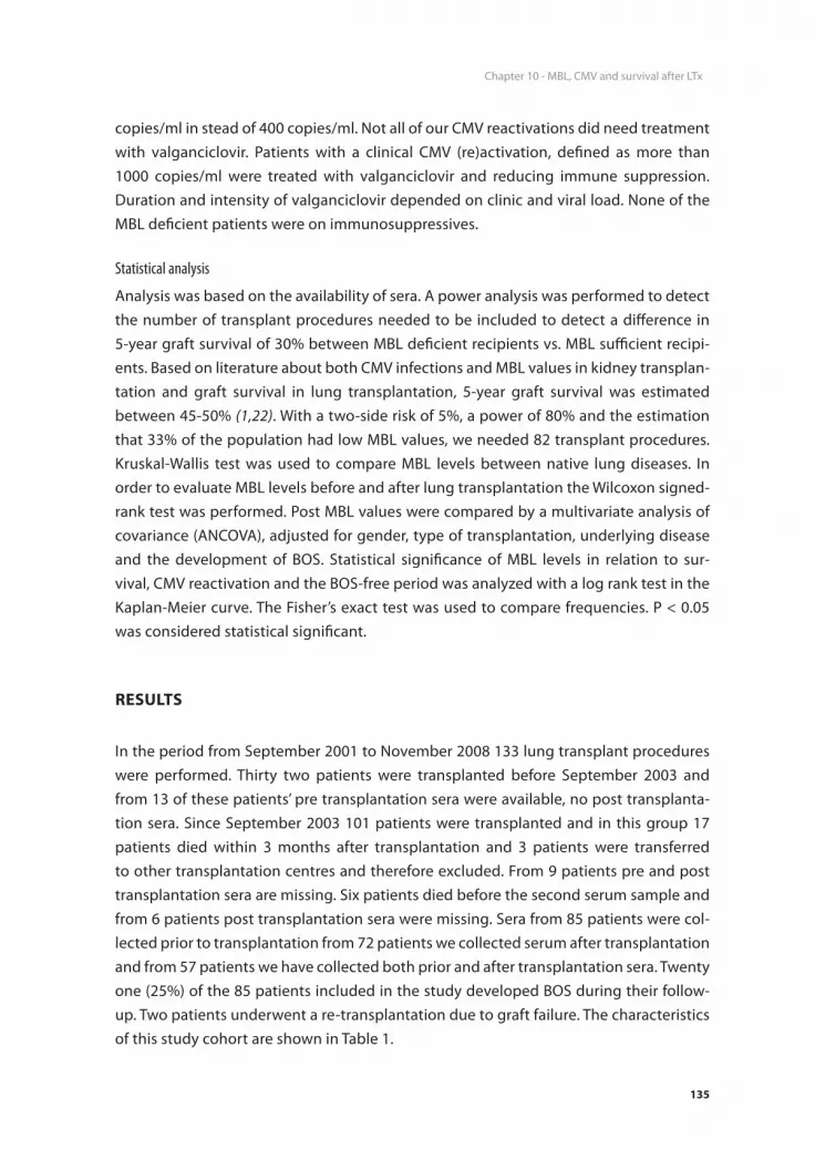
Chapter 10 - MBL, CMV and survival after LTx
135
copies/ml in stead of 400 copies/ml. Not all of our CMV reactivations did need treatment with valganciclovir. Patients with a clinical CMV (re)activation, defined as more than 1000 copies/ml were treated with valganciclovir and reducing immune suppression. Duration and intensity of valganciclovir depended on clinic and viral load. None of the MBL deficient patients were on immunosuppressives.
Statistical analysis
Analysis was based on the availability of sera. A power analysis was performed to detect the number of transplant procedures needed to be included to detect a difference in 5-year graft survival of 30% between MBL deficient recipients vs. MBL sufficient recipi-ents. Based on literature about both CMV infections and MBL values in kidney transplan-tation and graft survival in lung transplantation, 5-year graft survival was estimated between 45-50% (1,22). With a two-side risk of 5%, a power of 80% and the estimation that 33% of the population had low MBL values, we needed 82 transplant procedures. Kruskal-Wallis test was used to compare MBL levels between native lung diseases. In order to evaluate MBL levels before and after lung transplantation the Wilcoxon signed-rank test was performed. Post MBL values were compared by a multivariate analysis of covariance (ANCOVA), adjusted for gender, type of transplantation, underlying disease and the development of BOS. Statistical significance of MBL levels in relation to sur-vival, CMV reactivation and the BOS-free period was analyzed with a log rank test in the Kaplan-Meier curve. The Fisher’s exact test was used to compare frequencies. P < 0.05 was considered statistical significant.
resuLts
In the period from September 2001 to November 2008 133 lung transplant procedures were performed. Thirty two patients were transplanted before September 2003 and from 13 of these patients’ pre transplantation sera were available, no post transplanta-tion sera. Since September 2003 101 patients were transplanted and in this group 17 patients died within 3 months after transplantation and 3 patients were transferred to other transplantation centres and therefore excluded. From 9 patients pre and post transplantation sera are missing. Six patients died before the second serum sample and from 6 patients post transplantation sera were missing. Sera from 85 patients were col-lected prior to transplantation from 72 patients we collected serum after transplantation and from 57 patients we have collected both prior and after transplantation sera. Twenty one (25%) of the 85 patients included in the study developed BOS during their follow-up. Two patients underwent a re-transplantation due to graft failure. The characteristics of this study cohort are shown in Table 1.

136
MBL values before and after lung transplantation
Different cut off values to discriminate between MBL deficient and normal MBL patients were used but the cut off value of MBL values less than 10% turned out to be the most discriminative. Prior to lung transplantation 14 out of 85 patients had MBL values less than 10% (MBL deficient), corresponding with MBL levels below 300 ng/ml. No sig-nificant differences in recipients’ age, HLA-mismatches, sputum cultures shortly before transplantation, CRP or gender distribution were observed between the two groups with sufficient and MBL deficient values. Of the 85 patients, 24 patients suffered from fibrotic disease with mean MBL values of 92.5% (95% CI 71.6- 113.4%), 38 from emphysema with mean MBL values of 63.5 % (95% CI 47.2-79.7%) and 23 from CF with mean MBL values of 73.8% (95% CI 54.5-93.3%). Analysis of MBL values showed no association between pre transplant MBL values and native lung diseases.
To analyze whether the transplantation procedure in combination with immunosup-pressive therapy had an effect on MBL levels, in 57 patients MBL values were measured before and ± 20 months after transplantation. As shown in Figure 1A, MBL values decreased significantly after transplantation (p<0.0001) (95% CI 9.9-23.8). On average
table 1 Patient characteristics. Abbreviations MBL: Mannose Binding Lectin; CF: Cystic Fibrosis; CMV: cytomegalovirus; IgG: immunoglobulin G; Donor? means that the CMV status of the donor was not determined; CMV reactivation: detecting more than 50 copies/ml in serum; HLA: Human Leucocyte Antigen; BOS: Bronchiolitis Obliterans Syndrome
Pre transplantationn=85
MbL < 10%n=14
MbL ≥ 10%n=71
MbL values (in %) 95% CI (63.5-84.7)
Mean follow up (in months) 55 (5-102) 55 (5-102) 51 (30-102)
Age (in years) 45 (16-64) 48 (17-62) 50 (16-64)
gender m/f 42/43 8/6 34/37
bilateral/unilateral 69/16 9/5 60/11
native disease CF Emphysema Fibrotic disease
23 (27 %)38 (45 %)24 (28 %)
4 (29%)9 (64%)1 (7%)
21 (30%)29 (40%)21 (30%)
CMv igg status Donor+/recipient- Donor+/recipient+ Donor-/recipient+ Donor-/recipient- Donor?/recipient +Copies in CMv reactivationMean (range)
1382-
2651 (50-2651)
197
161712
2558 (0-10724)
hLA Mismatch Mean (SD) Class I Class II
3.1 ± 0.81.6 ± 0.5
3.2 ± 0.81.7 ± 0.5
bos 21 6 15

Chapter 10 - MBL, CMV and survival after LTx
137
MBL levels were reduced by 20% after transplantation. Although in most patients MBL values decreased after transplantation, in 12 patients an increase in MBL values was detected after transplantation. No correlation was found between an increase in MBL values and sputum cultures before transplantations, native disease or CMV copies after transplantation.
To examine whether post-transplant MBL levels would normalize to reach pre trans-plant levels, we additionally analyzed post transplantation samples taken more than 40 months after lung transplantation from 6 patients and this showed that the MBL values which decreased after transplantation, eventually increased and seemed to return to values before transplantation (Figure 1B).
figure 1 (A) In 57 lung transplant recipients serum MBL values were measured before and 20 months after lung transplantation. Each circle represents the MBL measured in one serum. The decrease in MBL values was significant and medians and 25%-75% intervals are depicted prior and post lung transplantation. In the 45 patients that showed a decrease in MBL values after transplantation, no differences in native lung diseases were detected. Twelve patients (6 CF and 6 emphysema) demonstrated an increase in MBL values after transplantation.(B) In 6 lung transplant recipients MBL values were measured before and after transplantation at 2 separate time points. Every line represents MBL measurements of a single lung transplant recipient.

138
MBL values and CMV reactivation
In 24 of the 71 patients with normal pre transplant MBL values (33%), CMV copies were detected during follow up, which was not significantly different compared to 7 of the 14 patients with low MBL values (50%) (p=0.30). None of the CMV reactivations occurred while patients received valganciclovir prophylaxes. It is remarkable that in 50% of patients with low pre transplant MBL levels CMV copies were detected since only one patient was considered a high risk patient for CMV reactivation (CMV serostatus donor+/recipient-). This may suggest that patients with pre transplant MBL deficiency have a high risk of CMV reactivation. Therefore we analyzed CMV reactivation in recipients with a CMV IgG positive status. Prior to transplantation 35 out of 71 patients (49%) with normal MBL levels and 11 out of 14 patients (79%) with MBL deficient levels had IgG antibodies against CMV (p=ns). In the CMV seropositive patients, CMV reactivation after transplantation was detected in 9 of 35 recipients with normal pre transplant MBL levels (26%), compared to 7 of the 11 with MBL deficient levels (64%) (p=0.03). Kaplan-Meier analysis showed a non-significant tendency towards more CMV reactivations in patients with pre transplant MBL deficient levels and a positive IgG status (Figure 2A) (p=0.06; HR 0.3 (95% CI 0.1-1.0 ) ).
Post transplant sera were available in 72 patients but no influence of post transplanta-tion MBL on CMV reactivation was detected (p=0.4).
MBL values, BOS and survival
During the study 13 patients died of whom 6 were diagnosed with BOS. Fourteen of the 71 patients with normal pre transplant MBL values (20%) developed BOS, compared to 7 of the 14 patients in the group with MBLdeficient values (50%) (p=0.02). In a Kaplan-Meier analysis no correlation was found between pretransplant MBL values and the development of BOS (Figure 2B) (p=0.13; HR 0.4 (95% CI 0.1-1.3)). However, there was a non-significant tendency towards a better survival in patients with MBL deficient levels prior to transplantation (Figure 2C) (p=0.08; HR 3.4 (95% CI 0.9-4.0).
From 72 patients post transplant sera were available but no influence of post-trans-plant MBL values on survival (p=0.82; HR 1.32 (95% CI 0.3-5.6)) nor BOS was detected (p=0.17; HR 0.4 (95% CI 0.1-1.4)).
Analyzing the 57 patients for whom pre- and post transplantation sera were available showed that no patients with MBL deficiency died whereas 83% of patients with normal MBL survived (p=0.25; HR 0.4 (95% CI 0.4-24.9)). In this group MBL deficient patients had a freedom from BOS of 53% vs. 78% in normal MBL patients (p=0.32; HR 2.2 (95%CI 0.5-11.0)).
Chapter 10 - MBL, CMV and survival after LTx
139
figure 2 (A) Kaplan Meier analysis of cytomegalovirus (CMV) reactivation showed a trend towards more CMV reactivations in recipients with deficient pre transplant MBL values (solid line). Patients with normal pre transplant MBL values are depicted with a dashed line. CMV reactivation was defined as a successive assay in the polymerase chain reaction after transplantation in those patients who had a seropositive CMV status before transplantation. (B) Kaplan Meier analysis of freedom from BOS showed no significant difference in recipients with deficient pre transplant MBL values (solid line) compared to normal pre transplant MBL values (dashed line).(C) Kaplan Meier analysis of survival showed a trend towards a better survival in recipients with deficient pre transplant MBL values (solid line) compared to normal pre transplant MBL values (dashed line).

140
disCussion
Long term survival after lung transplantation is poor, mainly due to the development of BOS. Although BOS is caused largely by alloimmunity, there is hardly any clinical improvement to augmented therapy compromising the adaptive immune system. Therefore we presume that the innate immunity also plays a role in the development of BOS. Earlier studies suggested an influence of MBL levels on infections and survival in solid organ transplants (8-10,15). In this study we investigated the effects of MBL values on clinical outcome (CMV reactivation and (graft) survival) after lung transplantation.
Since immunosuppressive therapy was tapered in the first year after transplantation and maintained thereafter, we presumed that MBL levels would be stable 20 months after transplantation. However, MBL levels dropped in this time frame after transplan-tation by 20% and later on, MBL-values increased and reached values comparable to those before transplantation. We assume that due to the immunosuppressives which are mostly cleared by the liver, the MBL synthesis by the liver is compromised. For liver transplantation it was demonstrated that MBL levels increase after transplantation and are stable for more than one year (4,23). The immunosuppressive regimen in liver trans-plantation is mild compared to lung transplantation, which may explain the differences in post transplant MBL levels found between these types of organ transplantation. In the aforementioned studies it was also shown that MBL is mainly produced by the trans-planted liver (4). Analysis on extra-hepatic production of MBL showed mRNA expression in the small intestine and testis, but not in the lung (24). Munster et al demonstrated that donor haplotype LXPA correlated with a superior lung transplant outcome but only during an immunosuppressive regimen of cyclosporine, azathioprine and prednisone. After introducing the new immunosuppressive protocol the influence of MBL haplo-type on graft survival disappeared. Since the study of Munster et al. was performed in the Netherlands and the majority of recipients and donors in our cohort are from the Netherlands and matched according the same Eurotransplant algoritms, we presume that the distribution of the MBL-haplotypes is similar to the cohort of Munster et al. In addition their new immunosuppressive protocol is similar to ours with the exception that we use mycophenolate mofetil instead of azathioprine. Nevertheless, MBL has been found in bronchoalveolar lavage (BAL) fluid, although only during inflammation, which may be caused by local production of activated alveolar cells or caused by “leakage” from the serum (25). Nevertheless, it seems unlikely that small amounts of MBL locally produced in the lung contribute significantly to the amount present in the circulation. Until today no (functional) RNA fragments in the lung have been described in the data-base of Online Mendelian Inheritance in Man (OMIM) (26).
MBL deficiency is associated with viral infections(26-28). The results of our study demonstrated more CMV reactivations in patients with pre transplant MBL deficient

Chapter 10 - MBL, CMV and survival after LTx
141
values. This is in line with the results of Manuel et al. who studied plasma MBL levels in 16 kidney transplant patients with a high risk CMV status (donor-positive/receptor negative) and reported an association with MBL status and CMV reactivation (7). Also in stem cell transplantation an association was found between MBL levels and CMV infection/reactivation in 131 patients (29). Although we observed a constantly higher amount of CMV reactivations in the group with MBL deficient levels, this did not reach statistical significance in a Kaplan-Meier analysis (p=0.06). In our daily clinical practice we use CMV reactivation as a tool to detect an over suppressed immune system and if CMV copies are detected, we reduce immunosuppression. During CMV prophylaxis no CMV reactivations occurred. In addition, during the study 13 patients died of which 6 had BOS, indicating that survival and BOS are not linked in this study. This might explain why low MBL values could be associated with a better survival, but not with BOS. Our results on the non-significant correlation between MBL levels and CMV reactivation/development of BOS should be interpreted with caution, since we have a relatively small number of patients.
Not only MBL deficiency but also the development of BOS and survival are associated with viral, bacterial and fungal infections (30-32). Unfortunately, in our follow up of lung transplant recipients no surveillance bronchoscopies, sputum cultures nor surveillance viral swabs are implemented and therefore we could not explore the association of MBL values and fungal, mycobacterium and common viral infections. The association between CMV infections and BOS is disputed. Some studies showed an association but others do not (33-35). We found an association between MBL deficiency and the development of BOS (p=0.02), although this did not reach statistical significance in a Kaplan-Meier analysis. This is in line with the results of Munster et al. who found no longer a correlation between MBL and the development of BOS after switching to an immunosuppressive regimen comparable to the one used in this study (15). Since the use of azithromycin some patients with BOS have a reversible or a less decreasing lung function and this may have an impact on survival after lung transplantation (36-38). Patients with normal MBL values before transplantation had a non-significant tendency towards a worse survival, suggesting that the contribution of MBL in local complement activation is related to graft survival.
A limitation of our study may be the missing data concerning multiple post transplan-tation sera. Although we included 6 patients with post transplantation samples taken more than 40 months after lung transplantation and showed that MBL values seemed to return to values before transplantation we would like to relate CMV reactivations with actual MBL values. Another remark is that in our study only 14 out of 85 patients had MBL deficient levels (in stead of the expected one third). This could have led to the non-significant result, and a type II error.
142
In conclusion, the findings of the present study suggest that MBL deficiency is associ-ated with CMV reactivations and a longer overall survival but not with the development of BOS. Larger studies are needed to further confirm these findings including the influ-ence of drugs that are commonly used in the transplantation field on the production and or clearance of MBL.
ACknoWLedgeMents
We thank Mrs. Coby v.d. Velde for her excellent technical assistance.

Chapter 10 - MBL, CMV and survival after LTx
143
referenCes
1. Christie JD, Edwards LB, Aurora P et al. Registry of the International Society for Heart and Lung Transplantation: Twenty-fifth official adult lung and heart/lung transplantation report-2008. Journal of Heart and Lung Transplantation 2008;27: 957-969.
2. Estenne M, Hertz MI. Bronchiolitis obliterans after human lung transplantation. American Journal of Respiratory and Critical Care Medicine 2002;166: 440-444.
3. Mullighan CG, Heatley SL, Danner S et al. Mannose-binding lectin status is associated with risk of major infection following myeloablative sibling allogeneic hematopoietic stem cell transplanta-tion. Blood 2008;112: 2120-2128.
4. Bouwman LH, Roos A, Terpstra OT et al. Mannose binding lectin gene polymorphisms confer a major risk for severe infections after liver transplantation. Gastroenterology 2005;129: 408-414.
5. Verschuren JJW, Roos A, Schaapherder AFM et al. Infectious complications after simultaneous pancreas-kidney transplantation: A role for the lectin pathway of complement activation. Trans-plantation 2008;85: 75-80.
6. Schlapbach LJ, Aebi C, Otth M et al. Serum levels of mannose-binding lectin and the risk of fever in neutropenic pediatric cancer patients. Pediatric Blood & Cancer 2007;49: 11-16.
7. Manuel O, Pascual M, Trendelenburg M, Meylan PR. Association between mannose-binding lectin deficiency and cytomegalovirus infection after kidney transplantation. Transplantation 2007;83: 359-362.
8. Sagedal S, Thiel S, Hansen TK, Mollnes TE, Rollag H, Hartmann A. Impact of the complement lectin pathway on cytomegalovirus disease early after kidney transplantation. Nephrology Dialysis Transplantation 2008;23: 4054-4060.
9. Berger SP, Roos A, Mallat MJK, Fujita T, de Fijter JW, Daha MR. Association between mannose-binding lectin levels and graft survival in kidney transplantation. American Journal of Transplan-tation 2005;5: 1361-1366.
10. Berger SP, Roos A, Mallat MJK, de Fijter JW, Daha MR. Pre-transplantation MBL levels predict patient and graft survival after simultaneous pancreas - kidney transplantation. Molecular Im-munology 2007;44: 156.
11. Fiane AE, Ueland T, Simonsen S et al. Low mannose-binding lectin and increased complement activation correlate to allograft vasculopathy, ischaemia, and rejection after human heart trans-plantation. European Heart Journal 2005;26: 1660-1665.
12. Fildes JE, Shaw SM, Walker AH et al. Mannose-binding Lectin Deficiency Offers Protection From Acute Graft Rejection After Heart Transplantation. Journal of Heart and Lung Transplantation 2008;27: 1353-1356.
13. de VB, Walter SJ, Peutz-Kootstra CJ, Wolfs TG, van Heurn LW, Buurman WA. The mannose-binding lectin-pathway is involved in complement activation in the course of renal ischemia-reperfusion injury. Am J Pathol 2004;165: 1677-1688.
14. Zhou W, Farrar CA, Abe K et al. Predominant role for C5b-9 in renal ischemia/reperfusion injury. J Clin Invest 2000;105: 1363-1371.
15. Munster JM, van der Bij W, Breukink MB et al. Association Between Donor MBL Promoter Haplo-type and Graft Survival and the Development of BOS After Lung Transplantation. Transplantation 2008;86: 1857-1863.
16. Madsen HO, Satz ML, Hogh B, Svejgaard A, Garred P. Different molecular events result in low protein levels of mannan-binding lectin in populations from Southeast Africa and South America. Journal of Immunology 1998;161: 3169-3175.

144
17. Garred P, Voss A, Madsen HO, Junker P. Association of mannose-binding lectin gene variation with disease severity and infections in a population-based cohort of systemic lupus erythematosus patients. Genes and Immunity 2001;2: 442-450.
18. Lee SG, Yum JS, Moon HM et al. Analysis of mannose-binding lectin 2 (MBL2) genotype and the serum protein levels in the Korean population. Molecular Immunology 2005;42: 969-977.
19. Seelen MA, Roos A, Wieslander J et al. Functional analysis of the classical, alternative, and MBL pathways of the complement system: standardization and validation of a simple ELISA. Journal of Immunological Methods 2005;296: 187-198.
20. Roos A, Bouwman LH, Munoz J et al. Functional characterization of the lectin pathway of comple-ment in human serum. Mol Immunol 2003;39: 655-668.
21. Vekemans M, Robinson J, Georgala A et al. Low mannose-binding lectin concentration is associ-ated with severe infection in patients with hematological cancer who are undergoing chemo-therapy. Clinical Infectious Diseases 2007;44: 1593-1601.
22. Ghods FJ, Solgi G, Amirzargar AA, Nikbin B, Ghods AJ. High frequency of clinically significant infections and cytomegalovirus disease in kidney transplant recipients with serum mannose-binding lectin deficiency. Iran J Kidney Dis 2009;3: 28-33.
23. Worthley DL, Johnson DF, Eisen DP et al. Donor Mannose-Binding Lectin Deficiency Increases the Likelihood of Clinically Significant Infection after Liver Transplantation. Clinical Infectious Diseases 2009;48: 410-417.
24. Seyfarth J, Garred P, Madsen HO. Extra-hepatic transcription of the human mannose-binding lectin gene (mbl2) and the MBL-associated serine protease 1-3 genes. Molecular Immunology 2006;43: 962-971.
25. Eisen DP. Mannose-Binding Lectin Deficiency and Respiratory Tract Infection. Journal of Innate Immunity 2010;2: 114-122.
26. Online Mendelian Inheritance in Man, OMIMTM website. Available: http://www.ncbi.nlm.nih.gov/omim/. Accessed 2010 September 21
27. Chong WP, To YF, Ip WK et al. Mannose-binding lectin in chronic hepatitis B virus infection. Hepa-tology 2005;42: 1037-1045.
28. Tan Y, Liu L, Luo P et al. Association between mannose-binding lectin and HIV infection and progression in a Chinese population. Mol Immunol 2009;47: 632-638.
29. Seppanen M, Lokki ML, Lappalainen M et al. Mannose-binding lectin 2 gene polymorphism in recurrent herpes simplex virus 2 infection. Hum Immunol 2009;70: 218-221.
30. Neth OW, Bacher U, Das P et al. Influence of mannose-binding lectin genotypes and serostatus in allo-SCT: analysis of 131 recipients and donors. Bone Marrow Transplantation 2010;45: 13-19.
31. Khalifah AP, Hachem RR, Chakinala MM et al. Respiratory viral infections are a distinct risk for bronchiolitis obliterans syndrome and death. American Journal of Respiratory and Critical Care Medicine 2004;170: 181-187.
32. Gottlieb J, Schulz TF, Welte T et al. Community-Acquired Respiratory Viral Infections in Lung Transplant Recipients: A Single Season Cohort Study. Transplantation 2009;87: 1530-1537.
33. Valentine VG, Gupta MR, Walker JE et al. Effect of Etiology and Timing of Respiratory Tract Infec-tions on Development of Bronchiolitis Obliterans Syndrome. Journal of Heart and Lung Trans-plantation 2009;28: 163-169.
34. Luckraz H, Sharples L, McNeil K, Wreghitt T, Wallwork J. Cytomegalovirus antibody status of donor/recipient does not influence the incidence of bronchiolitis obliterans syndrome in lung transplantation. J Heart Lung Transplant 2003;22: 287-291.

Chapter 10 - MBL, CMV and survival after LTx
145
35. nziger-Isakov LA, DelaMorena M, Hayashi RJ et al. Cytomegalovirus viremia associated with death or retransplantation in pediatric lung-transplant recipients. Transplantation 2003;75: 1538-1543.
36. Tamm M, Aboyoun CL, Chhajed PN, Rainer S, Malouf MA, Glanville AR. Treated cytomegalovirus pneumonia is not associated with bronchiolitis obliterans syndrome. Am J Respir Crit Care Med 2004;170: 1120-1123.
37. Verleden GM, Dupont LJ. Azithromycin therapy for patients with bronchiolitis obliterans syn-drome after lung transplantation. Transplantation 2004;77: 1465-1467.
38. Yates B, Murphy DM, Forrest IA et al. Azithromycin reverses airflow obstruction in established bronchiolitis obliterans syndrome. American Journal of Respiratory and Critical Care Medicine 2005;172: 772-775.
39. Vanaudenaerde BM, Meyts I, Vos R et al. A dichotomy in bronchiolitis obliterans syndrome after lung transplantation revealed by azithromycin therapy. European Respiratory Journal 2008;32: 832-843.


Chapter 11Clara Cell Secretory Protein and Surfactant Protein-D do not predict Bronchiolitis Obliterans Syndrome after Lung Transplantation
A.W.M. Paantjens1, H.G. Otten1, W.G.J. van Ginkel1, D.A. van Kessel2, J.M.M. van den Bosch2, J.M. Kwakkel-van Erp3, E.A. van de Graaf3.
An adapted version has been published
Transplantation 2010;90(3):340-2.
1Department of Immunology. University Medical Centre Utrecht,
Utrecht, The Netherlands 2Department of Respiratory Medicine. St Antonius Hospital,
Nieuwegein, The Netherlands 3Department of Respiratory Medicine. University Medical Centre
Utrecht, Utrecht, The Netherlands

148
AbstrACt
background: Serum concentrations of Clara cell secretory protein (CCSP) decrease after lung transplantation in patients who develop the bronchiolitis obliterans syndrome (BOS), whereas the relation between serum concentrations of surfactant protein D (Sp-D) and BOS is unknown. This study investigates whether CCSP and Sp-D can be used as biomarkers for development of BOS.Methods: Twenty seven patients (M/F 11/16, mean age 44 +/- 16 years) were included of whom 11 developed BOS. Patients had a follow up of at least 2 years (mean 50 +/- 13 months) after transplantation and concentrations of CCSP and Sp-D were measured longitudinally in a total of 516 serum samples.results: A significant difference (P=0.0001) was found between concentrations of CCSP in patients with BOS (median 4.4 ng/ml; range 0.8-21.0) versus patients without BOS (median 6.3 ng/ml; range 1.5-37.0). At the moment of BOS diagnosis, patients with BOS had on average 0.5 times less serum CCSP than patients without BOS (p<0.001). However, due to the fluctuation of CCSP concentrations in serum over time in individual patients, no algorithm could be defined discriminating patients developing BOS prior to the clinical diagnosis of BOS. No significant difference was found between serum concentrations of Sp-D in patients with BOS (median 6.5 ng/ml; range 17.7-290.8) versus patiens without BOS (median 97.3; range 23.5-625.2), nor did levels differ between these 2 patient groups at the time of BOS diagnosis.Conclusion: Serum CCSP concentrations, but not Sp-D, decrease before the functional diminishment in lung capacity. However, due to the fluctuation of concentrations over time, CCSP can not be used as a biomarker predictive for BOS.

Chapter 11 - CCSP does not predict BOS
149
introduCtion
Bronchiolitis obliterans (BO) is the main cause of mortality after lung transplantation (LTx). The diagnosis can only be made after taking lung biopsies and is considered to represent chronic lung allograft rejection.(1-3). To avoid potentially harmful invasive techniques the bronchiolitis obliterans syndrome (BOS), which is defined as an irrevers-ible 20% or more decline in forced expiratory volume in 1 second (FEV1) from baseline, is used as a surrogate marker for the development of BO. It is generally considered that all recipients who survive operative and infectious complications will ultimately develop BOS. Within 5 years after lung transplantation, only 58% of the recipients are still free from BOS (4, 5). When BOS is diagnosed the fibrotic process caused by chronic rejec-tion has already irreversibly affected the bronchioles, and therefore responds poorly to augmented therapy. In an early stage, the processes that lead to BOS may possibly be reversible and therefore we postulate that the early detection could lead to more successful interventions.
The Clara cell secretory protein (CCSP/CC16) is produced by the non ciliated Clara cells on the epithelium of the small airways (8). The Inflammatory injury, fibroproliferation and obliteration of the small airways occurring during the pathogenesis of BOS may influence the production of CCSP level and its leakage into the circulation. It has been described that serum Clara cell secretory protein decreases in patients developing BOS, either after lung transplantation or during graft versus host disease (GvHD) after stem cell transplantation (SCT), whereas no decline was observed in patients not developing BOS (6, 7). For that reason, CCSP might be a biomarker for development of BOS.
Another pneumoprotein, which is expressed by the type II cells in the alveoli, is sur-factant protein D (Sp-D). Although it is mentioned as possible damage marker in other obstructive lung diseases (8-11) nothing is known about its relation to BOS. Due the much larger size of Sp-D than CCSP (respectively clusters up to 650 kDa and 16 kDa), the protein only leaks in small amounts into the circulation in healthy persons (12-14). However, inflammation and damage of the respiratory membrane as seen during BOS may cause increased production and loss of size selectivity of the respiratory membrane leading to enhanced and leakage of Sp-D.
In order to examine in detail whether the pneumoproteins CCSP and Sp-D can be used as (early) biomarkers for the development of BOS, we have measured serum concentra-tions longitudinally over 2 years, with an average of 15 time points for 27 patients (11 with BOS and 16 without BOS).

150
MAteriAL And Methods
Patients
Between October 2001 and July 2007, 46 patients underwent lung transplantation at the University Medical Centre Utrecht, The Netherlands. Patients were selected who survived more than 2 years and those who died earlier due to BOS (n=3) were included. Encompassing a study population of 27 patients (M/F 11/16, mean age 44+/- 16 years) with a mean follow-up time of 50 months. In total, eleven patients developed BOS, which was defined as a decline of the FEV1 from the post-operative baseline at two distinctive time-points of more than 20% in the absence of infection or other etiology in accordance with the ISHLT criteria (15).
Standard immunosuppressive therapy consisted of basiliximab, tacrolimus, myco-phenolate mofetil and prednisone for all patients. When patients showed a decline in lung function with more than 20% of baseline, efforts were made to diagnose a possible reversibility of the lung function due to infection. To this purpose, cultures were taken from sputum, bronchoalveolar lavage fluid or blood and PCR for EBV and CMV. When an infection was diagnosed in these cultures, no bronchoalveolar lavage was performed. Infections or reactivation of CMV was treated according to good clinical practice. When despite treatment of a diagnosed infection the lung function did not improve, BOS was diagnosed according to the ISHLT criteria. Subsequently the patients were treated with high dose corticosteroids and azithromycine. No surveillance bronchoscopy was performed. Patient follow up started in September 2004, after approval by the medical-ethical committee and informed consent was obtained from each patient. After trans-plantation, 9 CF patients remained colonized with Pseudomonas aeruginosa of whom finally 2 developed BOS. When BOS was diagnosed 4 additional patients were colonized with Pseudomonas. Twenty two patients donated blood every month in the first year post transplantation and once every three months in the following years. BALF was neither collected systematically nor stored when collected for diagnostic means. Sera stored for diagnostic purposes from 5 other patients were also included in this study, although they were either transplanted before this date or the serum sampling was not performed systematically as described above. Eight healthy (M/F=5/3, mean age 35 years (range 26-46)) non allergic and non smoking controls donated blood every two weeks for six months and once five years later.
ELISA
CCSP and Sp-D levels were determined in sera collected longitudinally until 20 months up to 60 months post transplantation. FEV1 data were available for all patients during this period. The pneumoproteins were measured using a 96-well ELISA-assay (BioVendor Laboratory Medicine, Czech Republic), according to the protocols of the manufacturer. In

Chapter 11 - CCSP does not predict BOS
151
total, 516 samples were measured in duplicate for serum CCSP and Sp-D levels. Standard dilutions and controls from the manufacturer were included in each assay. The minimal concentration of serum CCSP that could be detected was 2 ng/ml (range 100 – 2 ng/ml). The range in which serum Sp-D concentration could be measured was 1100.0 – 16.5 ng/ml.
Statistical analysis
The significance of differences in serum CCSP and Sp-D levels after transplantation between LTx patients and healthy controls and the patient groups BOS vs. non BOS, BOS vs. healthy controls and non BOS vs. healthy controls were calculated according the
table 1 Patient characteristics are given for all patients or divided in a group patients who developed BOS and patients without BOS. The statistical significance was calculated using the Fisher’s exact test for total numbers, gender, type of graft, and presence of HLA antibodies. For all other characteristics the Mann-Whitney rank sum test was performed. BOS, Bronchiolitis Obliterans Syndrome; HLA, human leukocyte antigen; CMV, cytomegalovirus; EBV, Epstein-Barr virus; N.A., not applicable.
All bos non bos p-value
total number deceased
273 (11%)
113 (27%)
160 (0%) 0.06
gender Male Female
11 (41%)16 (59%)
4 (36%)7 (64%)
7 (44%)9 (56%)
1.0
Mean age (years) 44 (17-61) 46 (17-61) 43 (21-63) 0.4
Mean follow-up (months) 50 (11-84) 48 (11-84) 50 (39-76) 0.6
type of graft Bilateral Single
26 (96%)1 (4%)
11 (100%)0 (0%)
15 (94%)1 (6%)
1.0
Primary disease Cystic Fibrosis Emphysema Fibrotic diseases
10 (37%)9 (33%)8 (30%)
3 (28%)4 (36%)4 (36%)
7 (44%)5 (31%)4 (25%)
0.41.00.7
hLA Antibodies Positive Negative
1 (4%)26 (96%)
0 (0%)11 (100%)
1 (6%)15 (94%)
1.0
hLA Mismatches (mean) Class I Class II
3.11.7
3.01.6
3.11.8
0.40.3
infections CMV EBV Pseudomonas
2 (7%)0 (0%)
13 (48%)
0 (0%)0 (0%)
6 (55%)
2 (13%)0 (0%)
7 (44%)
0.51.00.7
onset of bos (months) N.A. 21 (9-52) N.A. N.A.
bos grade 1 2 3
N.A.0 (0%)1 (9%)
10 (91%)
N.A. N.A.

152
Mann-Whitney rank sum test (MWW). The difference of CCSP and Sp-D between BOS and non BOS at the moment of BOS development was analyzed by the linear mixed model (LMM). In Table 1, significance was calculated with the Fisher’s exact test for total number, gender, type of graft, and presence of HLA antibodies. The significance of all other characteristics in Table 1 was analysed using the Mann-Whitney rank sum test.
resuLts
Patients
Patient demographics are shown in Table 1. The patients who developed BOS are similar to the group of patients without BOS with respect to gender, age, primary disease, type of graft, HLA antibodies and mismatches, and infections. Three patients with BOS died because of BOS. No patients died in the group without BOS. None of the patients had an EBV infection, 2 experienced a CMV infection and there were no difference between patients with or without BOS with regard to pseudomonas colonization. No acute re-jections were observed. The patient group without BOS had more patients with cystic fibrosis as primary disease (44% vs. 28%). None of these characteristics were significantly different between the patient group with BOS and the patient group without BOS. As expected, the graft survival was significantly different between the two groups. (BOS, mean 21 months; range 9-52 months and non BOS, mean 50 months; range 39-76 months; p=0.0001 MWW).
Post-transplant Serum CCSP, Sp-D and BOS
Serum CCSP and Sp-D levels were measured for a mean of 50 (SD 13) months starting from 1 month after transplantation in 27 lung transplant patients. CCSP and Sp-D were also measured in the serum of 8 healthy controls approximately every two weeks for 6 months and once 5 years later to examine whether serum pneumoprotein levels in lung transplantation patients differ from that in healthy controls. The individual bandwidths of CCSP and Sp-D serum levels of 27 LTx patients (11 patients with BOS and 16 non-BOS patients) as well as 8 healthy controls are shown in Figure 1.
Serum concentrations of CCSP were found to be significantly different between all three groups (Figure 1A). Non BOS patients have higher levels of serum CCSP after transplantation than BOS patients (p=0.0001 MWU test), whereas patients either with or without BOS have higher serum CCSP levels compared to the healthy controls. (BOS p=0.0056 and non BOS p=0.0001 MWU test). Hence, all lung transplant patients taken together have higher serum CCSP than healthy controls (p=0.0001 MWU test). Serum CCSP levels between patients with and without a pseudomonas colonization were not

Chapter 11 - CCSP does not predict BOS
153
different. A comparison of serum concentrations of Sp-D indicated that the patients also have higher levels of Sp-D than that in healthy controls (p=0.0019 MWU test, Figure 1B).
When analyzed separately, only the lung transplantation patients without BOS showed higher serum Sp-D than the healthy controls (p=0.0006 MWU test). However, serum Sp-D levels of the non BOS patients did not differ from levels found in patients with BOS.
figure 1 The serum levels of clara cell secretory protein (A), and not surfactant protein D (B), in lung transplant patients with BOS do differ from the levels of lung transplant patients without BOS. Serum levels of CCSP and Sp-D were obtained as previously described in materials and methods. 516 serum samples were analysed, patients with BOS had a mean of 13.8 +/- 5.1 samples, patients without BOS had an mean of 16.4 +/- 3.8 samples and the healthy control group individuals had 16.0 +/- 1.1 samples. Bandwidth is displayed as boxes and whiskers with the boxes covering the 75% interval. Individuals are arranged according to increasing median CCSP or Sp-D levels.
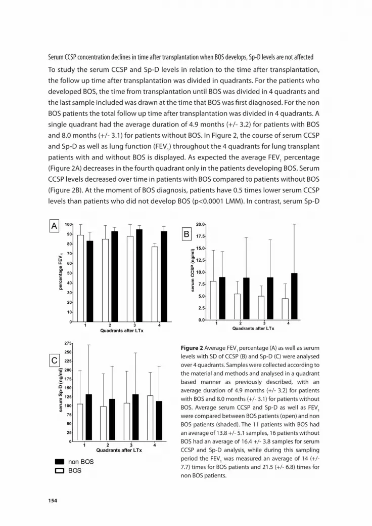
154
Serum CCSP concentration declines in time after transplantation when BOS develops, Sp-D levels are not affected
To study the serum CCSP and Sp-D levels in relation to the time after transplantation, the follow up time after transplantation was divided in quadrants. For the patients who developed BOS, the time from transplantation until BOS was divided in 4 quadrants and the last sample included was drawn at the time that BOS was first diagnosed. For the non BOS patients the total follow up time after transplantation was divided in 4 quadrants. A single quadrant had the average duration of 4.9 months (+/- 3.2) for patients with BOS and 8.0 months (+/- 3.1) for patients without BOS. In Figure 2, the course of serum CCSP and Sp-D as well as lung function (FEV1) throughout the 4 quadrants for lung transplant patients with and without BOS is displayed. As expected the average FEV1 percentage (Figure 2A) decreases in the fourth quadrant only in the patients developing BOS. Serum CCSP levels decreased over time in patients with BOS compared to patients without BOS (Figure 2B). At the moment of BOS diagnosis, patients have 0.5 times lower serum CCSP levels than patients who did not develop BOS (p<0.0001 LMM). In contrast, serum Sp-D
figure 2 Average FEV1 percentage (A) as well as serum levels with SD of CCSP (B) and Sp-D (C) were analysed over 4 quadrants. Samples were collected according to the material and methods and analysed in a quadrant based manner as previously described, with an average duration of 4.9 months (+/- 3.2) for patients with BOS and 8.0 months (+/- 3.1) for patients without BOS. Average serum CCSP and Sp-D as well as FEV1 were compared between BOS patients (open) and non BOS patients (shaded). The 11 patients with BOS had an average of 13.8 +/- 5.1 samples, 16 patients without BOS had an average of 16.4 +/- 3.8 samples for serum CCSP and Sp-D analysis, while during this sampling period the FEV1 was measured an average of 14 (+/- 7.7) times for BOS patients and 21.5 (+/- 6.8) times for non BOS patients.

Chapter 11 - CCSP does not predict BOS
155
levels did not diminish over time and at moment of BOS development; concentrations between patients with or without BOS were not different from each other (Figure 2C).
CCSP is not a predictive biomarker for development of BOS
In order to study whether serum CCSP levels in the first two years after transplantation are predictive for patients at risk to develop BOS, or of diagnostic use, we used different models in comparing CCSP levels post transplantation in patients developing BOS to patients without BOS. To analyze the predictive value of serum CCSP levels, receiver operating characteristic (ROC) curves were made using concentrations determined every month after transplantation and from averaged concentrations of the first 3 or 6 months after transplantation. The ROC curve analysis never met an area under the curve (AUC) higher than 0.69 for single time points, and never higher than 0.63 over a period after transplantation, indicating that CCSP measurements are not predictive for BOS. In Figure 3, examples are shown of individual courses of serum CCSP and FEV1 from two patients with BOS (Figure 3A and 3b) and two patients without BOS (Figure 3C and 3D).
figure 3 Example of FEV1 (left vertical axis) and serum CCSP (right vertical axis) course over time in 4 individual patients. Two patients developed BOS (A and B); time of BOS diagnosis is shown as a solid line when the FEV1 was decreased more than 20%, represented by the dotted line. C and D are examples of FEV1 and serum CCSP courses in LTx patients, who did not develop BOS.

156
Although all individual patients have variable serum CCSP levels over time, a final decline in serum CCSP can be seen in patients with BOS starting before FEV1 decline. A similar decline in CCSP was observed in 9 out of 11 patients with BOS and in 1 out of 16 patients without BOS. However, neither the decline of 10% or more from top level, nor the decline over time in individuals could be used as a predictive model for the develop-ment of BOS. This indicates that, due to inter-individual variability and fluctuating differ-ences in CCSP levels, CCSP measurements in serum can not be used to identify patients at risk for developing BOS. However, as 81% of BOS patients and only 6% of non BOS patients show a decrease in serum CCSP during or just prior to BOS, monitoring serum concentrations of CCSP might be useful to distinguish the diagnosis BOS from other causes responsible for a more than 20% decrease in FEV1. In order to examine whether serum CCSP measurements are of diagnostic use, we compared serum concentrations of CCSP between patients with or without BOS at fixed time points after transplanta-tion and averaged over several post transplant periods. This analysis did not result in a discriminative cut off value indicating that CCSP serum concentrations can not be used as a diagnostic marker for BOS.
disCussion
In this study we investigated the possibility of using a decrease in serum CCSP as a biomarker for the development of BOS. 27 LTx patients were analysed longitudinally (average of 15 samples per patient) for serum CCSP and serum Sp-D. Although non BOS patients showed higher levels of serum concentrations CCSP after transplantation compared to BOS patients, an overlap was found between serum CCSP values of these patient groups at various time-points investigated, indicating that absolute CCSP values do not provide additional information in the diagnosis of BOS. In line with previous pub-lications, we have shown a significant decrease in serum CCSP after lung transplantation in a cohort of patients who develop BOS at time of BOS diagnosis (6, 7), while serum Sp-D levels do not differ between patients with and without BOS. This decrease in serum CCSP was found in 9 out of 11 patients with BOS and 1 out of 16 patients without BOS and was observed in patients developing BOS before FEV1 decreased >20%. However, the decrease in serum CCSP could not be used for prediction due to fluctuations in indi-vidual patients over time. Therefore, serum CCSP measurements cannot be employed as a predictive or diagnostic biomarker for the development of BOS.
Several studies have provided data suggesting a role for CCSP in the modulation of inflammatory responses after lung injury (16-18). For instance, CCSP can inhibit production of interferon-γ (19) by airway epithelial cells whereas CCSP deficient mice
showed increased inflammatory response after lung injury and viral infection. However,

Chapter 11 - CCSP does not predict BOS
157
a recently emerging field relates to a completely other role of CCSP in lung homeostasis. In the normal situation, intrapulmonary stem cells are considered the primary source for epithelial regeneration upon injury. After lung transplantation however, epithelial chimerism occurs within bronchi, type II pneumocytes and seromucous peribronchial gland, and it has always been assumed that these recipient chimeric cells originate from the bone marrow (20). Recently, a subpopulation of CD45+ CCSP-expressing bone-marrow derived cells has been discovered which contributes to lung epithelial repair and which is able to differentiate into various epithelial cell lineages (21). Lung damage transiently induced an increased expression of CCSP of these cells in bone marrow and peripheral circulation, suggesting that mechanisms are in place at the lung for stimu-lating immigration and maintenance of tissue-repairing cells derived from the bone marrow. These data are in line with immunohistochemical findings showing enhanced integration of recipient cells taking place into chronically damaged epithelial structures (22). In vitro studies have shown that CCSP indirectly influences migration of fibroblasts by inhibiting the activity of pospholipase 2 (23), and it has been suggested that CCSP is a rescue factor for endogenous/autologous progenitor cells, stimulating survival and/or proliferation in an injurious environment (21). Collectively, these data indicate that CCSP has a role in the establishment and maintenance of lung chimerism.
The decrease of serum CCSP observed in LTx patients developing BOS might be the result of several processes, including loss of Clara cells, decreased production of CCSP by Clara cells and blockade of transfer into the circulation due to fibrosis. CCSP is a small protein supposedly not restricted by size selectivity of respiratory membranes, which is in line with data showing a direct relation between CCSP concentrations in BALF versus serum from patients (6, 24). As the fibrotic process in patients with BOS occurs in the small airway, we assume that the decrease in serum CCSP levels is mainly a result of reduced production due to destruction of the non ciliated Clara cells in the bronchiolar epithelium. New in this paper are the data on SP-D serum levels in relation to the devel-opment of BOS. Sp-D is a large protein, produced by type II alveolar cells, of which under normal conditions the passage to serum is restricted by size selectivity of the respiratory membrane (24-26). In pulmonary obstructive diseases like COPD the decrease of Sp-D in BALF and consequently elevated serum levels are caused by increased leakage due to damage of the respiratory membrane (8, 9, 25, 27, 28). Thus, we postulated that this would also be observed also in patients prior to the diagnosis of BOS. However, signifi-cant differences in levels of Sp-D could not be observed in sera analyzed longitudinally after lung transplantation and therefore, measurements of this pneumoprotein do not provide an indication which patients are at risk for the development of BOS.
Measurement of CCSP is a basically different approach for the prediction of BOS than measurements of immunological biomarkers related to in the inflammatory process involved in chronic rejection. First, immunological biomarkers can reflect immune

158
processes ultimately leading to BOS, whereas the serum concentration of CCSP will decrease late in the fibrotic process when Clara cells are actually destroyed during the pathogenesis of BOS. This is in line with our finding that measurement of CCSP early after transplantation is not predictive of BOS in individual patients, while in the cohort studied CCSP decrease was observed prior to the diagnosis of BOS. Second, the use of immunological biomarkers in the prediction of BOS is limited to the immunological mechanism(s) involved in chronic rejection. Conversely, as there is not one mechanism that causes chronic rejection an immunological biomarker studied may only be of use in the specific mechanism involved, thereby lowering its generic sensitivity for predicting BOS (29). For instance, as the HLA system plays a pivotal role in rejection and acceptance of a donated organ the assumption that development of HLA antibodies predicts rejec-tion is obvious (29-33). However, BOS is often developed without HLA antibodies being detected, and a rise in HLA antibodies does not necessarily lead to BOS and therefore make HLA antibodies not suitable to be a biomarker. In recent studies in renal, heart and lung transplantation patients with high levels of sCD30 prior to transplantation were at risk for the development of chronic rejection after transplantation (34). In addition, sCD30 preceded BOS after lung transplantation in a cyclopsporin/azathioprine based immunosuppressive regimen, but this observation could not be repeated in a tacro-limus/mycophenolate based regimen demonstrating that the underlying mechanism may be influenced by therapy and thus invalidating sCD30 as a biomarker (35, 36). A similar observation was made with respect to TARC. Low levels of serum TARC, the first month after transplantation, were a risk for a shorter freedom of BOS but does not corre-late with the development of BOS (37). As chronic rejection after transplantation can be caused by different mechanisms of the immune system, different predictive biomarkers will be found in these underlying processes. This indicates that immunosuppressive treatment might very well be adaptive based on the biomarkers found in an individual patient. In contrast to most biomarkers decreasing levels of CCSP do not lead to BOS but are caused by BOS, and therefore its measurement is supportive next to lung function testing in all patients developing BOS independent of the underlying pathogenesis.
In this paper we have studied serum levels of the pneumoproteins Sp-D and CCSP as possible biomarkers of BOS after lung transplantation. Sp-D was not found to be de-creased or increased in advance of BOS. In addition we have investigated the feasibility of CCSP as a predictive or diagnostic marker. However, as the decrease in CCSP is caused by BOS, we consider it a late biomarker but not a predictive one.
ACknoWLedgeMents
We thank sir P. Zanen for his help with the statistical analyses.

Chapter 11 - CCSP does not predict BOS
159
referenCes
1. Boehler A, Estenne M. Post-transplant bronchiolitis obliterans. Eur Respir J. 2003 Dec;22(6):1007-18.
2. Hertz MI, Henke CA, Nakhleh RE, Harmon KR, Marinelli WA, Fox JM, et al. Obliterative bronchiolitis after lung transplantation: a fibroproliferative disorder associated with platelet-derived growth factor. Proc Natl Acad Sci U S A. 1992 Nov 1;89(21):10385-9.
3. Trulock EP, Edwards LB, Taylor DO, Boucek MM, Keck BM, Hertz MI. Registry of the International Society for Heart and Lung Transplantation: twenty-second official adult lung and heart-lung transplant report--2005. J Heart Lung Transplant. 2005 Aug;24(8):956-67.
4. Boehler A, Kesten S, Weder W, Speich R. Bronchiolitis obliterans after lung transplantation: a review. Chest. 1998 Nov;114(5):1411-26.
5. Trulock EP. Lung transplantation. Am J Respir Crit Care Med. 1997 Mar;155(3):789-818. 6. Nord M, Schubert K, Cassel TN, Andersson O, Riise GC. Decreased serum and bronchoal-
veolar lavage levels of Clara cell secretory protein (CC16) is associated with bronchiolitis oblit-erans syndrome and airway neutrophilia in lung transplant recipients. Transplantation. 2002 Apr 27;73(8):1264-9.
7. Mattsson J, Remberger M, Andersson O, Sundberg B, Nord M. Decreased serum levels of clara cell secretory protein (CC16) are associated with bronchiolitis obliterans and may permit early diagnosis in patients after allogeneic stem-cell transplantation. Transplantation. 2005 May 27;79(10):1411-6.
8. Pastva AM, Wright JR, Williams KL. Immunomodulatory roles of surfactant proteins A and D: implications in lung disease. Proc Am Thorac Soc. 2007 Jul;4(3):252-7.
9. Mutti A, Corradi M, Goldoni M, Vettori MV, Bernard A, Apostoli P. Exhaled metallic elements and serum pneumoproteins in asymptomatic smokers and patients with COPD or asthma. Chest. 2006 May;129(5):1288-97.
10. Sims MW, Tal-Singer RM, Kierstein S, Musani AI, Beers MF, Panettieri RA, et al. Chronic obstructive pulmonary disease and inhaled steroids alter surfactant protein D (SP-D) levels: a cross-sectional study. Respir Res. 2008;9:13.
11. Sin DD, Leung R, Gan WQ, Man SP. Circulating surfactant protein D as a potential lung-specific biomarker of health outcomes in COPD: a pilot study. BMC Pulm Med. 2007;7:13.
12. Reid KB. Interactions of surfactant protein D with pathogens, allergens and phagocytes. Biochim Biophys Acta. 1998 Nov 19;1408(2-3):290-5.
13. Crouch EC. Surfactant protein-D and pulmonary host defense. Respir Res. 2000;1(2):93-108. 14. Holter JF, Weiland JE, Pacht ER, Gadek JE, Davis WB. Protein permeability in the adult respira-
tory distress syndrome. Loss of size selectivity of the alveolar epithelium. J Clin Invest. 1986 Dec;78(6):1513-22.
15. Estenne M, Maurer JR, Boehler A, Egan JJ, Frost A, Hertz M, et al. Bronchiolitis obliterans syndrome 2001: an update of the diagnostic criteria. J Heart Lung Transplant. 2002 Mar;21(3):297-310.
16. Singh G, Katyal SL. Clara cells and Clara cell 10 kD protein (CC10). Am J Respir Cell Mol Biol. 1997 Aug;17(2):141-3.
17. Jorens PG, Sibille Y, Goulding NJ, van Overveld FJ, Herman AG, Bossaert L, et al. Potential role of Clara cell protein, an endogenous phospholipase A2 inhibitor, in acute lung injury. Eur Respir J. 1995 Oct;8(10):1647-53.

160
18. Lesur O, Bernard A, Arsalane K, Lauwerys R, Begin R, Cantin A, et al. Clara cell protein (CC-16) induces a phospholipase A2-mediated inhibition of fibroblast migration in vitro. Am J Respir Crit Care Med. 1995 Jul;152(1):290-7.
19. Dierynck I, Bernard A, Roels H, De Ley M. Potent inhibition of both human interferon-gamma production and biologic activity by the Clara cell protein CC16. Am J Respir Cell Mol Biol. 1995 Feb;12(2):205-10.
20. Herzog EL, Van Arnam J, Hu B, Krause DS. Threshold of lung injury required for the appearance of marrow-derived lung epithelia. Stem Cells. 2006 Aug;24(8):1986-92.
21. Wong AP, Keating A, Lu WY, Duchesneau P, Wang X, Sacher A, et al. Identification of a bone mar-row-derived epithelial-like population capable of repopulating injured mouse airway epithelium. J Clin Invest. 2009 Feb;119(2):336-48.
22. Kleeberger W, Versmold A, Rothamel T, Glockner S, Bredt M, Haverich A, et al. Increased chimerism of bronchial and alveolar epithelium in human lung allografts undergoing chronic injury. Am J Pathol. 2003 May;162(5):1487-94.
23. Shijubo N, Kawabata I, Sato N, Itoh Y. Clinical aspects of Clara cell 10-kDa protein/ uteroglobin (secretoglobin 1A1). Curr Pharm Des. 2003;9(14):1139-49.
24. Bernard A, Carbonnelle S, Nickmilder M, de Burbure C. Non-invasive biomarkers of pulmonary damage and inflammation: Application to children exposed to ozone and trichloramine. Toxicol Appl Pharmacol. 2005 Aug 7;206(2):185-90.
25. Shijubo N, Itoh Y, Yamaguchi T, Shibuya Y, Morita Y, Hirasawa M, et al. Serum and BAL Clara cell 10 kDa protein (CC10) levels and CC10-positive bronchiolar cells are decreased in smokers. Eur Respir J. 1997 May;10(5):1108-14.
26. Felgenhauer K. Protein filtration and secretion at human body fluid barriers. Pflugers Arch. 1980 Mar;384(1):9-17.
27. Shijubo N, Itoh Y, Yamaguchi T, Abe S. Development of an enzyme-linked immunosorbent assay for Clara cell 10-kDa protein: in pursuit of clinical significance of sera in patients with asthma and sarcoidosis. Ann N Y Acad Sci. 2000;923:268-79.
28. Robin M, Dong P, Hermans C, Bernard A, Bersten AD, Doyle IR. Serum levels of CC16, SP-A and SP-B reflect tobacco-smoke exposure in asymptomatic subjects. Eur Respir J. 2002 Nov;20(5):1152-61.
29. Le Moine A, Goldman M, Abramowicz D. Multiple pathways to allograft rejection. Transplantation. 2002 May 15;73(9):1373-81.
30. SivaSai KS, Smith MA, Poindexter NJ, Sundaresan SR, Trulock EP, Lynch JP, et al. Indirect recog-nition of donor HLA class I peptides in lung transplant recipients with bronchiolitis obliterans syndrome. Transplantation. 1999 Apr 27;67(8):1094-8.
31. Jaramillo A, Smith MA, Phelan D, Sundaresan S, Trulock EP, Lynch JP, et al. Development of ELISA-detected anti-HLA antibodies precedes the development of bronchiolitis obliterans syndrome and correlates with progressive decline in pulmonary function after lung transplantation. Trans-plantation. 1999 Apr 27;67(8):1155-61.
32. Reznik SI, Jaramillo A, Zhang L, Patterson GA, Cooper JD, Mohanakumar T. Anti-HLA antibody binding to hla class I molecules induces proliferation of airway epithelial cells: a potential mecha-nism for bronchiolitis obliterans syndrome. J Thorac Cardiovasc Surg. 2000 Jan;119(1):39-45.
33. Jaramillo A, Naziruddin B, Zhang L, Reznik SI, Smith MA, Aloush AA, et al. Activation of human airway epithelial cells by non-HLA antibodies developed after lung transplantation: a potential etiological factor for bronchiolitis obliterans syndrome. Transplantation. 2001 Apr 15;71(7):966-76.

Chapter 11 - CCSP does not predict BOS
161
34. Bauwens AM, van de Graaf EA, van Ginkel WG, van Kessel DA, Otten HG. Pre-transplant soluble CD30 is associated with bronchiolitis obliterans syndrome after lung transplantation. J Heart Lung Transplant. 2006 Apr;25(4):416-9.
35. Kwakkel-van Erp JM, Otten HG, Paantjens AW, van Kessel DA, van Ginkel WG, van den Bosch JM, et al. Soluble CD30 measured after lung transplantation does not predict bronchiolitis obliterans syndrome in a tacrolimus/mycophenolate mofetil-based immunosuppressive regimen. J Heart Lung Transplant. 2008 Oct;27(10):1172-5.
36. Fields RC, Bharat A, Steward N, Aloush A, Meyers BF, Trulock EP, et al. Elevated soluble CD30 cor-relates with development of bronchiolitis obliterans syndrome following lung transplantation. Transplantation. 2006 Dec 27;82(12):1596-601.
37. Paantjens AW, Kwakkel-van Erp JM, van Ginkel WG, van Kessel DA, van den Bosch JM, van de Graaf EA, et al. Serum thymus and activation regulated chemokine levels post-lung transplantation as a predictor for the bronchiolitis obliterans syndrome. Clin Exp Immunol. 2008 Nov;154(2):202-8.


Chapter 12General Discussion


Chapter 12 - General Discussion
165
disCussion
Biomarkers- Lost and Found
In this thesis we show the discovery of possible new biomarkers identifying patients at risk for the development of BOS. One of the best known risk factors for the develop-ment of BOS is the presence of HLA antibodies either prior to LTx or as produced de novo after LTx (1, 2). In Chapters 7 and 8 the presence of HLA antibodies of the IgG and IgM isotypes are studied in our cohort of 49 patients transplanted between 2003 and 2008. In an earlier pilot study the same patients were analyzed for the presence of HLA antibodies via ELISA and only 1 out of 382 samples was found positive. After screening the same samples with a much more sensitive luminex technique, 105 samples were found to be positive for either HLA class I, class II, MICA or a combination. However, the titers were very low and therefore only weak positivity could be detected. No relation with survival or development of BOS could be detected. In the same patient samples also IgM isotype HLA antibodies were measured. Although only weak positivity for IgG type antibodies was detected, higher titers of IgM were measured in these samples. In this study it was shown that the isotype switching was hampered, since high titers of IgM were not followed by high titers of IgG, and high titers of IgG were not preceded by high IgM HLA antibodies. Furthermore it was described that patients with BOS have lower titers of HLA IgG antibodies but higher titers of IgM HLA antibodies compared to patients without BOS. The contradiction of our results with the known literature will be further discussed in “The influence of immune suppressives on the outcome after LTx”.
Antibodies against molecules on the allograft, like HLA antibodies, can activate the classical pathway of the complement system and hence cause damage to the allograft (3). However, the lectin pathway of complement has been shown to play a role in infec-tions and long-term outcome after transplantation. Therefore, in chapter 10 we studied the lectin pathway of the complement system. MBL the mediator of this pathway rec-ognizes carbohydrate residues on the surface of viruses, bacteria and fungi after which the complement cascade is activated. The role of functional MBL levels was evaluated in recipients on the development of BOS and (re)occurrence of CMV-infection. The results of our study demonstrated more CMV reactivations in patients with low MBL values prior to LTx and longer overall survival. However, no relation with the development of BOS was found. Patients with normal MBL values before transplantation showed a trend towards a worse survival, suggesting that the contribution of MBL in local complement activation is related to graft survival.
Whether complement activation after binding of HLA or non-HLA antibodies on the allograft has an important role in the development of BOS, or even graft survival, after lung transplantation should be studied in more detail. The results do however suggest

166
that MBL does not play an important role in chronic allograft rejection after lung trans-plantation in contrast to results shown in literature for kidney transplantation.
Early versus late biomarkers
The time after transplantation at which a predictive biomarker is found is of essence for the possible intervention in the treatment in order to prevent the development of BOS instead of stabilizing the disease to slow down progression. Furthermore, an early biomarker might be reflective of the cause or mechanism of BOS, while a late biomarker might merely be representative of the consequence of BOS.
From earlier research in our cohort it is known that serum concentrations of sCD30 are associated with the development of BOS (4). Because, sCD30 is produced by activated Th2 cells (5), in Chapter 2 of this thesis we investigated serum concentration of TARC a chemokine which attracts Th2 cells in relation to development of BOS. In addition, we studied whether the expression of the receptor for this chemokine, CCR4, also plays a role in the development of BOS in chapter 3. Low levels of serum TARC (< 325 pg/ml) at month 1 after lung transplantation were predictive of development of BOS. Not all patients with low levels of TARC developed BOS but some of these non BOS patients might eventually still develop BOS. Serum TARC levels are promising to function as an early biomarker for the development of BOS.
TARC is a chemokine which might play a role in development of BOS by attracting T cells to the site of inflammation in the allograft. TARC is one of the ligands for the CCR4 receptor which is expressed on T cells and high expression of this receptor is usually found in T cells infiltrating the skin by efficiently moving along a gradient of TARC (6, 7). The expression of the receptor was described only to correlate with functionality of the cells when they were in the later stages of Th2 development (8). TARC produced by dendritic cells is also known to polarize Th2 development. Analyzing the expression of this receptor it was found that CCR4++ expression was decreased on patients developing BOS compared to patients without BOS. This indicates a link between soluble CD30, TARC and its receptor – all suggestive for a role of Th2 cells - in the development of BOS (4, 9-12).
In Chapter 11 a biomarker is described which is decreased in all patients developing BOS but not in patients remaining free of BOS. CCSP is a protein secreted by Clara cells on the epithelium of the small airways (13). As consequence of BOS the Clara cells are destructed and therefore a drop in the pneumoprotein CCSP was detected in the serum of all patients developing BOS. However, due to the fact that the decrease in CCSP is a consequence of BOS - therefore late in the development of BOS, as well as variability in the expression of the protein in each patient - the protein is a late biomarker but not a predictive. Overall, a late biomarker might be able to discriminate patients whom

Chapter 12 - General Discussion
167
developed BOS from patients whom have a temporarily low lung function but who will recover and from showing incidentally a FEV1 below 80% again.
The influence of immune suppressives on the outcome after LTx
The differences between our results and those of older studies, in which much higher frequencies of patients positive for HLA antibodies were found, is probably caused by a different immune suppressive regimen. It was described that patients receiving a regimen based on tacrolimus and mycophenolate mofetil have less HLA antibodies than patients with a cyclosporine and azathioprine based regimen (14). All patients in our cohort received an immune suppressive regimen base of tacrolimus and mycopheno-late mofetil and therefore the absence of HLA antibodies and only weak positivity can be a result of the medication after lung transplantation. Furthermore it is known that Mycophenolate Mofetil (MMF) blocks the de novo pathway of purine synthesis, which is essential for lymphocytes, inhibiting T- and B-cell proliferation and antibody formation (15). Additionally, the influence of the immune suppressive regimen is also seen in our study for sCD30. sCD30 was described to be predictive for BOS prior to LTx in our cohort, while another group also showed an increase at time of BOS development, which could not be confirmed in our patient group. An important difference between the patient groups was the medication after LTx. Furthermore, in a setting of atopic dermatitis we were able to show that the use of MMF influenced the levels of sCD30 but not TARC, when compared to cyclosporine (4, 9-12).
Overall, new immune suppressives probably influence the applicability of biomark-ers as well as new and established risk factors predictive for BOS occurring after lung transplantation.
If the immune suppressive regimen has an influence on the established risk factors and new biomarkers, what about the overall clinical results? In the literature some studies suggested that MMF might be a superior over azathioprine after lung transplantation as patients treated with MMF showed decreased incidence of acute lung allograft rejec-tion, which is a risk factor for the development of BOS (16-21).
In the last decade of LTx it is also acknowledged by the ISHLT that the overall survival of LTx patients is better when compared to the first decades of lung transplantation. Also the incidence of BOS after LTx is slightly decreased compared to the first decade of lung transplantation. Therefore, it was analysed whether our patients also had a better chance of survival after LTx when compared to the average world wide. This average world wide include immune suppressive regimens like regimens based on cyclosporine and azathioprine as well as the regimen as employed in our centre based on tacrolimus and MMF. As can be seen in Figure 1A the overall survival of 187 patients transplanted between July 2001 and December 2010 is better than the average worldwide. The aver-

168
age for the last decade (2000-2008) world wide is 60% of LTx patients surviving 5 years after LTx while in our cohort approximately 73% of the LTx patients survive 5 years after LTx (22).
To study whether the overall survival could be due to a lower incidence of BOS in our cohort, our results of freedom from BOS were compared the results of LTx world wide in the last decade of transplantation. At the ISHLT it was registered that 46% of patients de-veloped BOS within 5 years after LTx world wide. In our patient cohort of 187 patients 32 patients developed BOS. In Figure 1B the freedom from BOS is displayed, showing that 25% of our patients developed BOS within 5 years after lung transplantation compared to 46% on average world wide.
Overall comparing the results of the ISHLT to our cohort it can be concluded that if the treatment of lung transplantation evolves the processes leading to development of BOS will be influenced by new immune suppressives and therefore the incidence of BOS and survival after lung transplantation.
PBMCs after lung transplantation
After lung transplantation a small fraction of blood cells found in the periphery of the recipient is from donor origin. This leukocyte microchimerism was described in several studies published by one group to be protective for development of BOS, although other groups could not repeat these results (23-26). In Chapter 5, we studied the contribution of the different cell types in this leukocyte fraction in relation to microchimerism and development of BOS. The most remarkable finding was that the microchimerism found in individual recipients was established by one major cell type. For instance, in some recipients the peripheral microchimerism consisted for more than 90% by monocytes,
figure 1 Kaplan-Meyer analyses of overall survival (a) as well as incidence of BOS (b) after LTx. 187 Patients transplanted in Utrecht and Nieuwegein between July 2001 and December 2010 were included (solid lines) compared to data of the ISHLT of all recorded lung transplantations world wide (dashed lines). Figures were not death censored.

Chapter 12 - General Discussion
169
while in other patients the cells of donor origin consisted of 5% monocytes and over 50% of B cells. As the microchimerism was stable at least up to one year after transplan-tation, we believe that stem cells of donor origin find their way through the periphery of the recipient towards the bone marrow. Where they survive and proliferate. Another interesting finding was that no plasmacytoid dendritic cells of donor origin could be detected in lung transplant recipients, which indicates that cells giving rise to these pDCs do not integrate to the bone marrow, or that these cells do not survive in the recipients environment. In our small study, no relation was found between the pres-ence of microchimerism and the development of BOS, but taken together all this data, it might be important to consider the main to microchimerism contributing cell type when studying the relation between microchimerism with the development of BOS in the future.
All roads lead to BOS
One of the difficulties when searching for a biomarker for the development of BOS after lung transplantation, preferably being specific and sensitive as well as already detect-able well before the fibrosis of the allograft is irreversible is the unknown pathology of BOS. As described in the Chapter 1 there are two hypotheses for the etiology of chronic rejection after lung transplantation. On the one hand immunological factors like acute rejection or HLA antibodies play an extensive role (27). On the other hand immunological independent factors like aspiration or reperfusion injury are suggested to lead to injury of the allograft (28). Both types of damage to the allograft leading to progressive injury are not mutually exclusive and probably overlap each other frequently (29). BOS is prob-ably the final common pathway of fibrosis and remodelling of the small airways causing long-term airflow limitation (30). Therefore BOS can be seen as a complex symphony of various overlapping allograft damaging processes. As a result, there probably is not only one biomarker which is part of the underlying processes which is able to discriminate all patients developing BOS from the patients remaining stable after lung transplantation. It might turn out that every processes show a different biomarker identifying the patients at risk of BOS development via that specific process. And a combination of biomarkers is needed to identify the patients at risk. Furthermore it has to be considered that it is
table 1. Staging of BOS grades
bos grade Pulmonary function
BOS0 >90%
BOS0-p 81-90%
BOS1 66-80%
BOS2 51-65%
BOS3 < 50%

170
generally acknowledged that in the end all patients will develop BOS sooner or later. Whether a patient develops BOS already a few months after LTx or whether this happens several years after transplantation might be caused by different underlying processes or even the same event for both types of patients only occurring at a different period after LTx.
In conclusion, it might be important to monitor patients very closely at intervals after lung transplantation for the rest of their lives, as well as testing for several biomark-ers and combining this in a multivariate analysis. The presence of several biomarkers combined could be the threshold for positively and sensitively identifying the patients at risk for of BOS development. Indicating an individually based monitoring, analyzing and treatment programme after lung transplantation.

Chapter 12 - General Discussion
171
referenCes
1. Belperio JA, Weigt SS, Fishbein MC, Lynch JP, 3rd. Chronic lung allograft rejection: mechanisms and therapy. Proc Am Thorac Soc 2009; 6 (1): 108.
2. McDyer JF. Human and murine obliterative bronchiolitis in transplant. Proc Am Thorac Soc 2007; 4 (1): 37.
3. Watanabe J, Scornik JC. Measuring human complement activation by HLA antibodies. Arch Pathol Lab Med 2006; 130 (3): 368.
4. Bauwens AM, van de Graaf EA, van Ginkel WG, van Kessel DA, Otten HG. Pre-transplant soluble CD30 is associated with bronchiolitis obliterans syndrome after lung transplantation. J Heart Lung Transplant 2006; 25 (4): 416.
5. Del Prete G, De Carli M, D’Elios MM, et al. CD30-mediated signaling promotes the development of human T helper type 2-like T cells. J Exp Med 1995; 182 (6): 1655.
6. Kunkel EJ, Boisvert J, Murphy K, et al. Expression of the chemokine receptors CCR4, CCR5, and CXCR3 by human tissue-infiltrating lymphocytes. Am J Pathol 2002; 160 (1): 347.
7. Imai T, Nagira M, Takagi S, et al. Selective recruitment of CCR4-bearing Th2 cells toward antigen-presenting cells by the CC chemokines thymus and activation-regulated chemokine and macrophage-derived chemokine. Int Immunol 1999; 11 (1): 81.
8. Morimoto Y, Bian Y, Gao P, et al. Induction of surface CCR4 and its functionality in mouse Th2 cells is regulated differently during Th2 development. J Leukoc Biol 2005; 78 (3): 753.
9. Kwakkel-van Erp JM, Otten HG, Paantjens AW, et al. Soluble CD30 measured after lung trans-plantation does not predict bronchiolitis obliterans syndrome in a tacrolimus/mycophenolate mofetil-based immunosuppressive regimen. J Heart Lung Transplant 2008; 27 (10): 1172.
10. Fields RC, Bharat A, Steward N, et al. Elevated soluble CD30 correlates with development of bronchiolitis obliterans syndrome following lung transplantation. Transplantation 2006; 82 (12): 1596.
11. Golocheikine AS, Saini D, Ramachandran S, Trulock EP, Patterson A, Mohanakumar T. Soluble CD30 levels as a diagnostic marker for bronchiolitis obliterans syndrome following human lung transplantation. Transpl Immunol 2008; 18 (3): 260.
12. Kwakkel-van Erp JM, Haeck IM, Paantjens AW, et al. Differential usefulness of biomarkers thymus and activation-regulated chemokine and soluble CD30 during enteric coated mycophenolate sodium and cyclosporine therapy in atopic dermatitis. J Am Acad Dermatol 2010; 63 (3): e70.
13. Singh G, Singh J, Katyal SL, et al. Identification, cellular localization, isolation, and characterization of human Clara cell-specific 10 KD protein. J Histochem Cytochem 1988; 36 (1): 73.
14. Terasaki PI, Ozawa M. Predicting kidney graft failure by HLA antibodies: a prospective trial. Am J Transplant 2004; 4 (3): 438.
15. Fieguth HG, Simon A, Scherer M, Klesius AA, Wiedenmann DE. Immunosuppressive regimen in lung transplantation: initial intravenous mycophenolate mofetil and cyclosporine avoid cytolytic induction and allow for rapid steroid tapering. Transplant Proc 2002; 34 (5): 1879.
16. Izbicki G, Shitrit D, Aravot D, et al. Improved survival after lung transplantation in patients treated with tacrolimus/mycophenolate mofetil as compared with cyclosporine/azathioprine. Transplant Proc 2002; 34 (8): 3258.
17. Gerbase MW, Spiliopoulos A, Fathi M, Nicod LP. Low doses of mycophenolate mofetil with low doses of tacrolimus prevent acute rejection and long-term function loss after lung transplanta-tion. Transplant Proc 2001; 33 (3): 2146.

172
18. Neurohr C, Huppmann P, Zimmermann G, et al. Tacrolimus and mycophenolate mofetil as first line immunosuppression after lung transplantation. Transpl Int 2009; 22 (6): 635.
19. O’Hair DP, Cantu E, McGregor C, et al. Preliminary experience with mycophenolate mofetil used after lung transplantation. J Heart Lung Transplant 1998; 17 (9): 864.
20. Ross DJ, Waters PF, Levine M, Kramer M, Ruzevich S, Kass RM. Mycophenolate mofetil versus azathioprine immunosuppressive regimens after lung transplantation: preliminary experience. J Heart Lung Transplant 1998; 17 (8): 768.
21. Zuckermann A, Klepetko W, Birsan T, et al. Comparison between mycophenolate mofetil- and azathioprine-based immunosuppressions in clinical lung transplantation. J Heart Lung Trans-plant 1999; 18 (5): 432.
22. Hertz MI, Aurora P, Christie JD, et al. Scientific Registry of the International Society for Heart and Lung Transplantation: introduction to the 2010 annual reports. J Heart Lung Transplant 2010; 29 (10): 1083.
23. Reinsmoen NL, Jackson A, Hertz M, et al. Peripheral blood allogeneic microchimerism in lung and cardiac allograft recipients. J Leukoc Biol 1999; 66 (2): 306.
24. McSherry C, Jackson A, Hertz MI, Bolman RM, 3rd, Savik K, Reinsmoen NL. Sequential measure-ment of peripheral blood allogeneic microchimerism levels and association with pulmonary function. Transplantation 1996; 62 (12): 1811.
25. Monaco AP. Chimerism in organ transplantation: conflicting experiments and clinical observa-tions. Transplantation 2003; 75 (9 Suppl): 13S.
26. Sahota A, Gao S, Hayes J, Jindal RM. Microchimerism and rejection: a meta-analysis. Clin Trans-plant 2000; 14 (4 Pt 1): 345.
27. Boehler A, Kesten S, Weder W, Speich R. Bronchiolitis obliterans after lung transplantation: a review. Chest 1998; 114 (5): 1411.
28. Tullius SG, Tilney NL. Both alloantigen-dependent and -independent factors influence chronic allograft rejection. Transplantation 1995; 59 (3): 313.
29. Ward C, Whitford H, Snell G, et al. Bronchoalveolar lavage macrophage and lymphocyte pheno-types in lung transplant recipients. J Heart Lung Transplant 2001; 20 (10): 1064.
30. Trulock EP. Lung transplantation. Am J Respir Crit Care Med 1997; 155 (3): 789.

Nederlandse samenvatting Lijst van publicities Dankwoord CV


175
Nederlandse samenvatting
Nederlandse samenvattingLongtransplantatie is een laatste behandelingsmogelijkheid voor verschillende levens-bedreigende longaandoeningen zoals cystic fibrose (taaislijmziekte), COPD (Chronische Obstructive longziekte) of IPF (Interstitiële Pulmonaire Fibrose). Echter een longtrans-plantatie brengt ook veel problemen met zich mee. De gemiddelde levensverwachting na een longtransplantatie is slechts 7,5 jaar. Deze levensverwachting wordt voornamelijk gelimiteerd door afstoting van het orgaan.
Na een longtransplantatie kunnen er verschillende vormen van afstoting optreden; hyperacute afstoting, acute afstoting en chronische afstoting. De eerste twee zijn te-genwoordig goed te voorkomen/behandelen. Echter, chronische afstoting – ook wel bekend als het bronchiolitis obliterans syndroom (BOS) – is niet te genezen en slecht te behandelen, ook al krijgen patiënten veel medicijnen welke het afweersysteem (deels) uitschakelen.
Bij het ontstaan van BOS ontstaan er beschadigingen in de longen die leiden naar uitgebreide vorming van littekenweefsel en ontstekingen. Hierdoor worden de kleine luchtwegen in de longen opgevuld met cellen en andere producten, en wordt de adem-haling gehinderd. Hoe BOS precies ontstaat na longtransplantatie is onbekend. Wel zijn er factoren bekend die een rol kunnen spelen, namelijk: antilichamen tegen HLA (humane leukocyten antigeen) eiwitten, infecties, oprisping en inhalering van zure maagsappen, eerder opgetreden acute afstoting, het niet nemen van medicijnen en de tijd dat donor longen uit het lichaam zijn geweest. Al deze factoren kunnen leiden tot schade aan het orgaan en ontsteking waarna de schade moet worden hersteld. Het wordt verondersteld dat dit laatste niet goed gebeurd wat bijdraagt aan de verstopping van de kleine luchtwegen en dus de ademhaling bemoeilijkt.
De diagnose van chronische afstoting is moeilijk te maken. Het zou kunnen aan de hand van longbiopsies. Echter, is dit zeer ingrijpend en omdat de ziekte zich niet gelijkmatig verdeeld over de longen, is het moeilijk een biopsie te nemen op de juiste plek. Daarom wordt BOS gediagnoseerd aan de hand van de longfunctie. Na een longtransplantatie worden geregeld longfunctietesten gedaan om de FEV1 (forced expiratory volume) te meten. Dit is het volume dat je in 1 seconde kunt uitblazen. De twee hoogste resultaten worden gebruikt om de top te definiëren. Als er vervolgens een daling is van > 20% zonder dat er een duidelijke reden voor deze daling is, wordt er gesproken over BOS.
In Nederland worden jaarlijks ongeveer 60 longtransplantaties uitgevoerd in Utrecht/Nieuwegein, Groningen en Rotterdam. Wereldwijd zijn dit er ongeveer 2750. Uit regi-stratie van al die transplantaties wereldwijd en hoe het verloop is, zien we dat ongeveer 50% van de patiënten binnen 5 jaar na transplantatie BOS heeft ontwikkeld.
176
Doel van het proefschrift
In dit proefschrift beschrijven we de zoektocht naar een biologische marker (biomar-ker) in het bloed van longtransplantatie patiënten die het ontstaan van BOS al vroeg na transplantatie kan voorspellen. Dit kan niet alleen helpen in de diagnose, maar als een biomarker voldoende vroeg na longtransplantatie meetbaar is zou er ingegrepen kunnen worden in de medicatie en worden geprobeerd de ontwikkeling van BOS te stoppen.
In hoofdstuk 2 en 3 wordt er gekeken naar het eiwit TARC en de receptor die aan TARC bindt, CCR4, welke op witte bloedcellen (specifiek T cellen) wordt aangemaakt. TARC is een eiwit dat ervoor kan zorgen dat T cellen zich naar de plaats van een ontsteking bewegen. We vinden in onze groep patiënten, bestaande uit 45 patiënten, dat patiënten die uiteindelijk BOS ontwikkelen 1 maand na transplantatie vele lagere niveaus van TARC in hun bloed hebben dan patiënten die stabiel bleven. Verder zien we dat er verschillen zijn in de hoeveelheid CCR4 op T cellen tussen longtransplantatie patiënten en gezonde mensen. Bovendien wordt op een bepaalde soort T cellen (CD4+ central memory) een verschil in expressie van de CCR4 receptor gevonden tussen patiënten die BOS ontwik-kelen en patiënten die stabiel blijven.
In hoofdstuk 4, 5 en 6 kijken we of de verschillende cellen die te vinden zijn in het bloed van longtransplantatie patiënten zouden kunnen functioneren als biomarkers voor het ontstaan van BOS. We beschrijven wat de percentages zijn van verschillende cellen die 5 maanden na transplantatie in het bloed van longtransplantatie patiënten zitten. We zien dat verschillende cellen van het immuunsysteem (NKT cellen) zijn verhoogd en een bepaalde soort van T cellen (central memory CD8+ T cellen) zijn verlaagd in het bloed van patiënten die BOS zullen ontwikkelen vergeleken met stabiele patiënten.
Na een transplantatie is een klein deel cellen in het bloed afkomstig van de donor. Witte bloedcellen die zich nog in het orgaan bevonden tijdens de transplantatie kunnen overleven in de ontvanger, er ontstaat een microchimerisme. Wij hebben onderzocht welke cellen konden overleven in de ontvanger. Wij beschrijven dat de verschillende witte bloedcellen van donor herkomst (monocyten, B cellen, dendritische cellen, NK en T cellen) twee maanden na transplantatie tot ten minste 1 jaar na transplantatie aan-toonbaar zijn. Verder zien we dat niet alle patiënten dezelfde soort/percentages cellen van de donor hebben. Verder zien we dat 1 bepaalde soort van de dendritische cellen (welke belangrijke cellen van het immuunsysteem zijn voor de presentatie van o.a. eiwitten) niet kan overleven in de ontvanger, namelijk de zogenaamde plasmacytoïde dendritische cel.
Uiteindelijk kijken we ook nog specifiek naar NK cellen. NKG2D is een receptor op NK cellen welke een activerende werking heeft als deze bindt aan het bijbehorende

177
Nederlandse samenvatting
eiwit MICA. MICA kan tijdens stressvolle omstandigheden ook losgemaakt worden van de cellen waar het op voorkomt en in de bloedstroom terecht komen als sMICA. Wanneer sMICA bindt aan NKG2D wordt de functie gehinderd en zal dit niet leiden tot activatie van NK cellen. In hoofdstuk 6 worden een aantal bevindingen beschreven over de onderlinge relaties tussen NK cellen, NKG2D, sMICA, BOS en Pseudomonas aeruginosa infectie. Ten eerste zien we dat na longtransplantatie niet veel sMICA voorkomt en dat dit niet correleert met de hoeveelheid NKG2D op o.a. NK cellen. Ten tweede laten we zien dat de hoeveelheid van NKG2D geen relatie heeft tot het ontstaan van BOS of de aanwezigheid van de bacterie Pseudomonas aeruginosa. Tot slot zien we wel dat 1 maand na transplantatie de hoeveelheid van NKG2D op NK cellen is verhoogd vergeleken met het bloedmonster voor transplantatie. Dit geeft aan dat NK cellen meer geactiveerd zijn, wat overeenkomt met eerder beschreven literatuur.
In hoofdstuk7, 8, 9 en 10 wordt humorale afstoting bestudeerd. Hoofdstuk 7 en 8 kijken naar de aanwezigheid van HLA antilichamen na transplantatie. Er zijn twee isotypes HLA antilichamen IgM en IgG. IgM is de voorloper van IgG. Hoewel HLA antilichamen (van IgG isotype) beschreven zijn in de literatuur als risico factor voor het ontstaan van BOS zien wij bij onze patiënten bijna geen IgG HLA antilichamen, en als we ze zien zijn het hele kleine hoeveelheden. We zien in dezelfde monsters wel meer HLA IgM antilichamen, de aanwezigheid van dit isotype is in onze groep patiënten echter niet gerelateerd aan het ontstaan van BOS. Ook zien we dat IgM HLA antilichamen niet worden gevolgd door IgG antilichamen. Wij denken dat deze tegenstelling met de literatuur te maken heeft met de soort medicatie die wij gebruiken. Wel zien we als we naar de hoeveelheden antilichamen kijken dat patiënten die BOS ontwikkelen een kleinere hoeveelheid IgG HLA antilichamen maar meer IgM HLA antilichamen hebben.
Naast HLA komen er ook andere eiwitten voor op de cellen van het donor orgaan die kunnen verschillen tussen donor en ontvanger. Het afweersysteem kan ook deze eiwitten als vreemd herkennen en antilichamen hier tegen maken. Deze non-HLA antilichamen zijn nog onbekend en in hoofdstuk 9 onderzoeken we de literatuur op verschillende mogelijkheden om de non-HLA antilichamen te kunnen identificeren. Wij beschrijven de SEREX techniek. Dit is een zeer bewerkelijke procedure, welke een enkel resultaat geeft maar ook veel achtergrond ruis oplevert. Wel wordt duidelijk dat inderdaad non-HLA antilichamen worden gemaakt na longtransplantatie tegen verschillende eiwitten.
Als antilichamen aan een cel binden kan het complement systeem in werking worden gesteld. Het complement systeem plakt verschillende eiwitten op de cel waardoor een buisje in de celwand ontstaat en de cel vernietigd kan worden. Een van de routes die het complement systeem volgt is de MBL (mannose bindende lectine) route. Hierin worden vooral eiwitten van oa. virussen herkend waarna de eiwitafzetting begint. In hoofdstuk 10 onderzoeken we de niveaus van MBL na longtransplantatie en het klinische resul-

178
taat. Weinig MBL voor transplantatie leidde tot meer infecties van het cytomegalovirus (CMV), maar ook een betere overleving. Echter werd er geen relatie met het ontstaan van BOS ontdekt.
In conclusie is het duidelijk dat antilichamen een grote rol kunnen spelen bij het ontstaan van BOS, maar wel onderhevig zijn aan de medicatie die wordt gebruikt. Wat de precieze rol van complement is, moet nog nader worden bekeken.
In het laatste hoofdstuk kijken we naar een biomarker die zijn oorsprong niet in het afweersysteem heeft. clara cell secretory protein (CCSP) is een eiwit dat wordt gepro-duceerd door clara cellen in de kleine luchtwegen. Bij schade aan de longen tijdens het ontstaan van BOS wordt deze productie beïnvloed d.m.v. afbraak van de clara cellen. Als we kijken naar de niveaus van dit eiwit in het bloed van longtransplantatie patiënten zien we dat patiënten die BOS ontwikkelen een afname laten zien ten tijde van het ontstaan van BOS terwijl stabiele patiënten geen afname laten zien. Maar omdat de verlaging pas optreedt als het proces van BOS ontwikkeling al aan de gang is, is het ook de vraag of deze biomarker niet te laat is voor interventie in de medicatie om BOS een halt toe te roepen.
Conclusie
In dit proefschrift laten we zien dat al bekende risicofactoren onderhevig kunnen zijn aan het type medicatie dat wordt gebruikt, biomarkers vroeg na longtransplantatie of ten tijde van BOS ontwikkeling kunnen worden gevonden en dat meerder takken van het afweersysteem een rol lijken te spelen. TARC is een veel belovende vroege biomarker die verder onderzocht dient te worden naar de toepasbaarheid in de kliniek.
Tot slot
Als we als laatste kijken naar de resultaten van Utrecht over bijna 10 jaar (juli 2001 tot en met december 2010) zien we een overleving van 73% van de patiënten 5 jaar na trans-plantatie. Verder zien we dat 5 jaar na transplantatie 25% van de patiënten BOS hebben ontwikkeld, wat betekent dat dit beter is dan de wereldwijde cijfers laten zien. Mogelijk heeft dit te maken met de redelijk nieuwe vormen van medicatie die na transplantatie worden gegeven, iets wat nog niet in elke longtransplantatiecentrum is ingevoerd.

179
Lijst van publicaties
Lijst van publicatiesPaantjens AWM, van de Graaf EA, Heerkens HD, Kwakkel-van Erp JM, Hoefnagel T, van Kessel DA, van den Bosch JMM, Otten HG. Chimerism of dendritic cell subsets in periph-eral blood after lung transplantation. J Heart Lung Transplant 2011.
Paantjens AWM, van de Graaf EA, Kwakkel-van Erp JM, van Ginkel WGJ, Hoefnagel T, van Kessel DA, van den Bosch JMM, Otten HG. Identification of Allo- and Auto-Antibodies after Lung Transplantation. “Lung Transplantation: Therapies, Complications and Out-come”Chapter 3, isbn: 978-1-61122-760-4. Review
Kwakkel-van Erp JM, Haeck IM, Paantjens AWM, van de Graaf EA, van Ginkel WGJ, Knol MJ, de Bruin-Weller MS, Lammers JJ, Knol EF, Bruijnzeel-Koomen C, Otten HG. Differen-tial usefulness of biomarkers thymus and activation-regulated chemokine and soluble CD30 during enteric coated mycophenolate sodium and cyclosporine therapy in atopic dermatitis. J Am Acad Dermatol 2010;63(3):e70-2.
Paantjens AWM, Otten HG, van Ginkel WGJ, van Kessel DA, van den Bosch JMM, Kwakkel-van Erp JM, van de Graaf EA. Clara cell secretory protein and surfactant protein-d do not predict bronchiolitis obliterans syndrome after lung transplantation. Transplantation 2010;90(3):340-2.
Paantjens AWM, van de Graaf EA, van Ginkel WGJ, van den Bosch JMM, Otten HG. Lung Transplantation under a Tacrolimus/Mycophenolate Mofetil-Based Immunosuppressive Regimen Results in Low Titers of HLA and MICA IgG Antibodies which are not Related to Development of BOS. J Heart Lung Transplant 2010.
Kwakkel-van Erp JM, van de Graaf EA, Paantjens AWM, van Ginkel WGJ, Schellekens J, van Kessel DA, van den Bosch JMM, Otten HG. The killer immunoglobulin-like receptor (KIR) group A haplotype is associated with bronchiolitis obliterans syndrome after lung transplantation. J Heart Lung Transplant 2008;27(9):995-1001
Paantjens AWM, Kwakkel-van Erp JM, van Ginkel WGJ, van Kessel DA, van den Bosch JMM, van de Graaf EA, Otten HG. Serum thymus and activation regulated chemokine levels post-lung transplantation as a predictor for the bronchiolitis obliterans syndrome. Clin Exp Immunol 2008;154(2):202-8

180
Kwakkel-van Erp JM, Otten HG, Paantjens AWM, van Kessel DA, van Ginkel WGJ, van den Bosch JMM, van de Graaf EA. Soluble CD30 measured after lung transplantation does not predict bronchiolitis obliterans syndrome in a tacrolimus/mycophenolate mofetil-based immunosuppressive regimen. J Heart Lung Transplant 2008;27(10):1172-5
Rzymski T, Paantjens AWM, Bod J, Harris AL. Multiple pathways are involved in the anoxia response of SKIP3 including HuR-regulated RNA stability, NF-kappaB and ATF4. Onco-gene 2008;27(33):4532-43

181
Dankwoord
DankwoordHet dankwoord, in ieder geval het meest gelezen stukje uit een proefschrift. Ik schrijf dit nu vanachter mijn bureau in Utrecht, waar ik 4 jaar met plezier heb gezeten, gewerkt en gedacht terwijl mijn ogen dwaalden over het uitzicht. Mijn opvolger aan dit bureau wens ik veel plezier met de zomer zon, stormen, regenbogen, prachtige winterse tafere-len en files op de A28. Ook nu moet ik niet te lang kijken, want dit zijn de laatste loodjes, het dankwoord:
Henny en Ed, mijn co-promotoren. Wat heb ik 4 jaar met veel plezier met jullie gewerkt. Onze werkbesprekingen vond ik altijd nuttig maar ook gezellig, en dat is misschien al het halve werk wat betreft enthousiasme. Henny, niet alleen heb ik van jou veel geleerd over de immunologische kanten van transplantaties, misschien nog veel belangrijker heb jij mij gestimuleerd te controleren, plannen, managen en beslissen. Dit zijn taken waar ik zeker altijd wat aan zal hebben. Ed, jij liet mij zien wat voor klinische kanten er om de hoek komen bij longtransplantaties, en bleef altijd de kant van de kliniek in de gaten houden wat leidde tot geslaagde discussies. Dank jullie wel!
Ter nagedachtenis aan prof. dr. Jules van den Bosch.
Jan Grutters en Linde Meyaard, mijn promotoren, dank jullie dat jullie deze functie over hebben willen nemen tijdens de laatste maanden. Jan, ook al heb je pas aan het einde de functie over moeten nemen, was je altijd bereid geïnteresseerd en kritisch te zijn als ik mijn presentaties gaf tijdens research meetings met de kliniek uit het UMC en het AZN. Linde ik heb vooral met je te maken gehad tijdens de gesprekken van mijn AIO begeleidingscommissie. Samen met Debbie dwongen jullie mij om met een helikopter blik naar het onderzoek te kijken en om knopen door te hakken, beslissingen te nemen. Ik vond het altijd erg verhelderende gesprekken. Dank, voor jullie inzet.
Groep Otten, klein maar fijn. Walter en Tineke, jullie hebben mij ieder de helft van mijn onderzoek bijgestaan. En ik wil jullie daarvoor dan ook bedanken, zonder jullie inzet was het voor mij een stuk moeilijker geweest om alles te kunnen doen. Daarom denk ik dat jullie ook erg trots kunnen zijn op het resultaat. Hanneke, ik vond het fijn met jou te kunnen werken, ik vanuit het lab en jij vanuit de kliniek. En wil je danken voor je gezel-lige discussies over zowel de inhoud als de procesgang van onze projecten. Mooi werk!
Anna en Hanne, jullie hebben met jullie stageprojecten ook ieder op eigen manier bijgedragen aan het ontstaan van dit proefschrift. Anna, helaas bleek de SEREX niet zo succesvol gezellig was het wel. En Hanne wat heb jij een werk verricht en ik hoop dat je

182
ook een leuke tijd op het lab hebt gehad en misschien nog een keer twijfelt om naast kliniek ook onderzoek te gaan doen.
Collega’s van de immunologie, zowel van de research als de AMI.Kamergenootjes, in vier jaar tijd hebben er veel veranderingen plaats gevonden. Kees,
Suzanne, Floor, Kristof, Cordula, Peter, Hilde, Ellen, Thijs, Bart, Wouter en Kirsten, zo’n beetje de laatste bezetting. Mensen kwamen en gingen maar er was altijd tijd voor werk en tijd voor koffie of in mijn geval (veel) thee.
Saskia, Yvonne en Erna (AZN), dank voor jullie ondersteunend werk.
De volledige transplantaties teams van het UMC Utrecht en het Sint Antonius ziekenhuis in Nieuwegein. Samen leveren jullie goed werk wat ten grondslag ligt aan dit proef-schrift.
Maar ook mensen die wat minder snappen waar ik mee bezig was afgelopen 4 jaar (ja, iets met onderzoek en longtransplantaties…) zijn erg belangrijk voor mij. Evelien, wat waren we blij elkaar die eerste dag van onze studie te zien en wat leuk dat jij ook vanuit Wageningen naar ’s-Hertogenbosch bent verhuisd! Ik vind het geweldig dat je vandaag mijn paranimf wil zijn.
De kippen (en “hanen”), ik vind onze avonden altijd heerlijk en kijk er altijd weer naar uit om bij elkaar te komen. Jullie zijn voor mij een erg bijzondere groep en wisten jullie dat we nu precies 10 jaar geleden druk bezig waren met onze eindexamens.Gelukkig zijn we nog altijd diezelfde ‘kippen’. Tijd voor een feestje!
Martin, Marijke, Sabine en Jeroen, ik voel me altijd thuis bij jullie. Ik vind het fijn dat jullie vier jaar geïnteresseerd zijn geweest en ons graag de helpende hand bieden in vanalles en nog wat.
Marjolein, mijn liefste grote (enige ☺) zus. Totaal verschillend en toch een groot voor-beeld voor mij! Wat leuk dat je vandaag als paranimf bij mij zal staan.
Pap en mam, dankzij jullie heb ik dit kunnen doen. Inhoudelijk zijn jullie misschien afgehaakt toen ik wiskunde B, scheikunde en natuurkunde op het VWO koos maar jul-lie zijn altijd geïnteresseerd gebleven. Dank jullie voor jullie onvoorwaardelijke steun, vertrouwen en liefde. Jullie hebben mij kansen geboden waardoor ik nu dit proefschrift kan verdedigen.
Bart, dank je dat jij ‘jij’ bent. Dat je mij uitdaagt en steunt, met me kibbelt en met me lacht, maar bovenal bij mij staat dat maakt alles gewoon leuker.
Liefs, Annelieke

183
Curriculum Vitae
Curriculum VitaeAnnelieke Wilhelmina Margaretha Paantjens is op 10 januari 1983 geboren in Venray. Nadat zij daar in 2001 het VWO afrondde aan het Raayland College is zij in Wageningen moleculaire wetenschappen gaan studeren aan de Wageningen University and Research Centre. In 2002 behaalde ze haar propedeuse en 31 maart 2004 werd de bachelor fase afgerond. Na afstudeer onderzoeken bij onder andere de groep van Metabolism, Nutri-tion and Genomics van de vakgroep Human Nutrition aan de Wageningen University and Research Centre en de groep Molecular Biology van het NCMLS in Nijmegen is zij stage gaan lopen in Oxford in de Growth factor groep van de Cancer Research UK aan het Weatherall institute of Molecular Medicine verbonden aan Oxford University onder begeleiding van prof.dr. A. Harris en dr. T Rzymski. In augustus 2006 studeerde ze uitein-delijk af voor de master Molecular Sciences met als specialisatie biomedisch onderzoek. In maart 2007 is zij begonnen aan een promotie onderzoek aan het UMC Utrecht onder begeleiding van dr. Henny Otten en dr. Ed van de Graaf waarvan dit proefschrift het resultaat is.


