Embed Size (px)
Citation preview

Respiratory Reovirus 1/L Induction of IntraluminalFibrosis, a Model of Bronchiolitis ObliteransOrganizing Pneumonia, Is Dependent on TLymphocytes
Elizabeth I. Majeski,* Manjeet K. Paintlia,*Andrea D. Lopez,* Russell A. Harley,†
Steven D. London,* and Lucille London*From the Departments of Microbiology and Immunology,* and
Pathology and Laboratory Medicine,† Medical University of South
Carolina, Charleston, South Carolina
Bronchiolitis obliterans organizing pneumonia(BOOP) is a clinical syndrome characterized byperivascular/peribronchiolar leukocyte infiltrationleading to the development of intraalveolar fibrosis.We have developed an animal model of BOOP whereCBA/J mice infected with 1 � 106 plaque-formingunits (PFU) reovirus 1/L develop follicular bronchioli-tis and intraalveolar fibrosis similar to human BOOP.In this report, we demonstrate a role for T cells in thedevelopment of intraluminal fibrosis associated withBOOP. Corticosteroid treatment of reovirus 1/L-in-fected mice both inhibited the development of fi-brotic lesions when administered early in the time-course and promoted the resolution of fibroticlesions when corticosteroid administration was de-layed. Further, the depletion of either CD4� or CD8�
T cells before reovirus 1/L infection also inhibitedfibrotic lesion development. Both corticosteroid treat-ment and depletion of CD4� or CD8� T cells alsoresulted in decreased expression of the proinflamma-tory and profibrotic cytokines, interferon (IFN)-� andmonocyte chemoattractant protein-1 (MCP-1). Fur-ther, treatment of mice with a neutralizing monoclo-nal antibody to IFN-� also significantly inhibited thedevelopment of fibrosis. Taken together, these resultssuggest a significant role for T cells in the develop-ment of reovirus 1/L-induced BOOP fibrotic lesions inCBA/J mice and suggests that TH1-derived cytokines,especially IFN-� , may play a key role in fibrotic lesiondevelopment. (Am J Pathol 2003, 163:1467–1479)
Bronchiolitis obliterans organizing pneumonia (BOOP),first described in 1985, is a pattern of injury characterizedhistologically as “patchy plugs of fibrous tissue (Massonbodies) filling bronchiolar lumens (bronchiolitis obliter-ans) and alveolar ducts and spaces (organizing pneu-
monis).”1–3 This patchy fibrosis may begin as focal le-sions within the alveoli and the terminal bronchioles of thelung and progress bilaterally over time.1 Other histologi-cal features include clusters of mononuclear inflamma-tory cells, chronic inflammation in the walls of the sur-rounding alveoli with reactive type II cells, increasednumbers of foamy macrophages in the alveoli, and pre-served lung architecture.2,4 The development of BOOP isoften of unknown etiology (idiopathic BOOP) but BOOPhas also been associated as a consequence of lunginjury due to environmental toxins, bacterial infections,viral infections, and lung or bone marrow transplanta-tion.3,5 BOOP is responsive to corticosteroids and treat-ment with prednisone continues to be the primary treat-ment for patients with symptomatic and progressivedisease.2–4
Since infiltrating lymphocytes are associated with theinitiation of BOOP lesions,6,7 it is possible that these cellsplay an active role in the progression of inflammatory fociinto lesions that are progressively dominated by fibroblasts.In patients with BOOP, there is an increase of activatedbronchoalveolar lavage (BAL) lymphocytes with up to 80%to 95% of this cellular infiltrate being comprised of cytotoxic/suppressor CD8� T cells.6–9 These cells may be involved inthe inflammation and subsequent fibrosis occurring inBOOP patients.2,5,7,10–12 Several studies have shown thatthe infiltration of T lymphocytes may be important in thedevelopment of other forms of pulmonary fibrosis, althoughthe data from both animal models and patients has beenequivocal.13–18 Although these existing models of experi-mental pulmonary fibrosis have been useful for histopatho-logical and functional investigations of other types of fibroticevents in the lung, the process of fibrotic lesion develop-ment in these models may be distinct from BOOP lesiondevelopment. Thus, differences in the phenotype of theinflammatory cell infiltrate, expression of soluble mediators,and response to various treatments may be different and,
Supported by a U.S. Public Health Service grant AI R01 40175 and a grantfrom the American Lung Association (both to L. L.).
Accepted for publication June 26, 2003.
Address reprint requests to Lucille London, Ph.D., Department of Mi-crobiology and Immunology, Medical University of South Carolina, P.O.Box 250504, 173 Ashley Avenue, Charleston, South Carolina 29425.E-mail: [email protected].
American Journal of Pathology, Vol. 163, No. 4, October 2003
Copyright © American Society for Investigative Pathology
1467

therefore, may fail to accurately reflect the intraluminal andfibroblastic nature of the bronchoalveolar obliteration ob-served in BOOP lesions.19–21
We have described a spectrum of inflammatory lungdiseases after respiratory infection with reovirus serotype1, strain Lang (reovirus 1/L), which is dependent on thestrain of mice used.22–27 In this spectrum CBA/J miceinfected with 1 � 106 PFU reovirus 1/L develop a histo-logically severe inflammation characterized by an infiltra-tion of lymphocytes organized adjacent to the pulmonaryvasculature of the lung.23 This pattern of mononuclearcell organization without the involvement of intraluminalfibrosis results in lesions histopathologically consistentwith the non-fibrotic human syndrome termed follicularbronchiolitis (FB). However, accompanying the develop-ment of FB in CBA/J mice are the presence of foamymacrophages and the elicitation of a non-specific fibroticresponse of the lung characteristic of BOOP fibrotic le-sions.23 To investigate the role of the inflammatory cellinfiltrate, especially T cells, in the development of reovirus1/L-induced fibrotic lesions, the effect of either cortico-steroid treatment or the depletion of CD4� or CD8� Tcells before reovirus 1/L infection was determined. Ourresults indicate that corticosteroid treatment of reovirus1L-infected mice both inhibited the development of fi-brotic lesions when administered early in the time-courseand promoted the resolution of fibrotic lesions when cor-ticosteroid administration was delayed. In addition, thedepletion of either CD4� or CD8� T cells before reovirus1/L infection also inhibited fibrotic lesion development.Both corticosteroid treatment and depletion of CD4� orCD8� T cells also resulted in decreased expression ofthe proinflammatory and profibrotic cytokines, interferon(IFN)-� and monocyte chemoattractant protein-1 (MCP-1). Finally, treatment of mice with a neutralizing monoclo-nal antibody to IFN-� also significantly inhibited the de-velopment of fibrosis. Taken together, the results suggesta significant role for T cells in the development of reovirus1/L-induced BOOP fibrotic lesions in CBA/J mice and thatTH1 derived cytokines, especially IFN-�, may play a keyrole in fibrotic lesion development.
Materials and Methods
Animals
Four- to 5-week-old female CBA/J mice (The JacksonLaboratory, Bar Harbor, ME) were maintained in mi-croisolator cages under specific pathogen-free condi-tions in a BL-2 facility. Cages were housed in an HEPA-filtered animal isolator clean room (Nuaire Inc., Plymouth,MN). All animal manipulations were performed in class IIbiological safety cabinets. Virally primed mice were keptphysically isolated from all other mice.
Virus
Reovirus 1/L was originally obtained from Dr. W. Joklik(Duke University School of Medicine, Durham, NC).Third-passage gradient-purified stocks were obtained by
re-cloning and amplifying parental stocks on L-929 fibro-blast cells [American Type Culture Collection (ATCC),Rockville, MD] as previously described.23 Following thepurification of new stocks, infectious viral titers were ob-tained by limiting dilution on L-929 monolayers.23
Inoculation Protocol
Animals were lightly anesthetized with an i.p. injection of0.08 ml of 20% ketamine (Vetalar 100 mg/ml; Fort DodgeLaboratories, Inc., Fort Dodge, IA) and 2% PromAce(acepromazine maleate 10 mg/ml; Ayerst Laboratories,New York, NY) before immunization. Animals were in-fected by the intranasal (i.n.) application of 1 � 106 PFUof reovirus 1/L in 30 �l (15 �l in each nostril) in sterileinjectable grade 0.9% NaCl (Baxter Healthcare Corp.,Deerfield, IL). Control animals were inoculated with 30 �l(15 �l in each nostril) of sterile injectable grade 0.9%NaCl. After the indicated timepoints, animals were sacri-ficed with an i.p. injection of 0.2 ml sodium Nembutal (50mg/ml; Abbott Laboratories, North Chicago, IL).
Methylprednisolone Administration
As an initial dosing regimen either 10 mg/kg or 20 mg/kgmethylprednisolone (MPS) (�0.1 to 0.2 mg/mouse) (Sig-ma Chemicals, St. Louis, MO) dissolved in PBS wasadministered i.p. to mice beginning on either days 0, 5,10, or 14 post-reovirus 1/L infection and given daily untilthe completion of the time-course. Since these initialstudies indicated that treatment with MPS (20 mg/kg)either beginning at day 0 or day 5 post infection oradministration of MPS (10 mg/kg) before day 5 postreovirus 1/L infection (beginning at day 0) resulted in anincreased mortality rate as compared to that observed inuntreated, reovirus 1/L-infected mice (Table 1), all addi-tional studies were performed using MPS at a concentra-tion of 10 mg/kg beginning 5 days post-reovirus 1/Linfection unless otherwise noted.
CD4 and CD8 Depletion
Adult CBA/J mice were treated i.p. with either 0.5 mg ofpurified GK1.5 monoclonal antibody (mAb)28 for deple-tion of CD4� lymphocytes or 0.25 mg of purified 53–6.72mAb29 for depletion of CD8� lymphocytes for three con-secutive days. Depleted mice were then infected i.n. with1 � 106 PFU of reovirus 1/L in 30 �l (15 �l in each nostril)in sterile injectable grade 0.9% NaCl. Control, depletedanimals were inoculated with 30 �l (15 �l in each nostril)of sterile injectable grade 0.9% NaCl. The depleted statewas maintained by treating with either 0.5 mg purifiedGK1.5 or 0.25 mg of purified 53–6.72 mAb every 6 days.Depletion of the appropriate subset of T cells was verifiedby flow cytometry of cells obtained from the lymph nodeand spleen before infection with reovirus 1/L on day 0and on days 7 and 14 postinfection. Depleted mice wereevaluated for the development of BOOP fibrotic lesions atday 21 post-reovirus 1/L infection.
1468 Majeski et alAJP October 2003, Vol. 163, No. 4

In Vivo Interferon-� Depletion
An anti-IFN-� mAb (R4–6A2, rat IgG1, ATCC HB170) wasobtained from ATCC30–32 and ascites fluid was gener-ated for in vivo use (Strategic Biosolutions, Newark, NJ).Reovirus 1/L-infected (106 PFU BOOP) CBA/J mice weretreated i.p. every 3 days beginning on day 3 postinfectionwith either 100 �g anti-IFN-� antibody in PBS or 100 �gnormal rat IgG (Sigma) in PBS. Mice were evaluated ondays 14 and 21 postinfection for the development offibrotic lesions by hematoxylin and eosin (H&E) and Ma-son’s trichrome stain.
Bronchoalveolar Lavage (BAL)
BAL was performed in situ by injecting and withdrawing a0.5 ml aliquot of Hank’s balanced salt solution (HBSS)twice through an intubation needle (21 gauge). A total of1.5 ml of HBSS was used. BAL fluid was frozen at �70°Cuntil use. Cells collected by BAL were washed threetimes with HBSS containing 5% fetal calf serum (FCS)and 0.05% azide, and resuspended at 1 � 106 cells/ml.
Histology
Lungs were inflated in situ with 10% neutral bufferedformalin (0.5 mls) (Richard-Allan Scientific, Kalamazoo,MI) by intratracheal (i.t.) intubation, removed, and sus-pended in an additional 10% neutral buffered formalinovernight before being embedded in paraffin. H&E stainand Mason’s trichrome stain, which was used to visualizecollagen deposition, were performed on 4-�m sections.Inflammatory infiltration with the development of FB,which is defined as a mononuclear cell infiltrate that
condenses into prominent peribronchiolar lymphoid ac-cumulations, was blindly evaluated. FB was scored on ascale of 0 to 3: 0, normal; 1, mild (� 4 follicles per lobe);2, moderate (between 5 and 8 follicles per lobe); 3,severe (� 8 follicles per lobe). Fibrosis was scored on ascale of 0 to 4: 0, normal; 1, mild; 2, moderate; 3, severe;4, very severe.
Hydroxyproline (HP) Assay
The extent of pulmonary fibrosis was also determined byestimating total lung collagen as reflected by the mea-surement of the HP content of the lung as previouslydescribed.25,26,33 Mice were sacrificed at various inter-vals after infection with reovirus 1/L and the lungs wereremoved, lyophilized, and weighed. Differences betweengroups were examined for statistical significance usingtwo-tailed Student’s t-test. A P value less than 0.05 wasconsidered significant.
Antibodies
The following monoclonal antibodies were used in thisstudy: Cy-Chrome-conjugated rat anti-mouse CD45 (30-F11, leukocyte common antigen, Ly-5); fluorescein iso-thiocyanate (FITC)-conjugated hamster anti-mouse CD3(145–2C11, CD3 � chain); FITC-conjugated rat anti-mouse CD8a (53–6.7, Ly-2); R-phycoerythrin (PE)-conju-gated rat anti-mouse CD4 (GK1.5, L3T4) (Caltag, Burlin-game, CA); R-PE-conjugated rat anti-mouse Pan-NKcells (DX5); FITC-conjugated rat anti-mouse CD45R/B220 (RA3–6B2); R-PE-conjugated rat anti-mouseCD11b (M1/70, integrin�m chain, Mac-1 � chain); andFITC-conjugated rat anti-mouse Ly6G (RB6–8C5, Gr-1,neutrophils) (Pharmingen, San Diego, CA); hamster anti-rat CD3 (�-chain, 48–2B) (Santa Cruz Biotechnology,Santa Cruz, CA); rat anti-mouse CD11b (Mac-1 � chain)(Serotec, Westbury, NY); and rat anti-mouse neutrophil(MCA 771F) (Serotec).
Flow Cytometric Analysis
Cells collected by BAL were washed three times withHBSS containing 5% FCS and 0.05% azide, and resus-pended at 1 � 106 cells/ml. Cells were stained for cellsurface marker expression as previously described ex-cept that all cells were also stained with anti-CD45 (30-F11), leukocyte common antigen Ly-5, and only anti-CD45-positive cells were acquired for analysis.24,26
Isotype-matched controls were run for each sample(Caltag and Pharmingen). The total number of PMNs wasobtained by adding the anti-Gr-1 (Ly6G) single-positivecells and the anti-Gr-1/anti-Mac-1 (integrin�m chain) dou-ble-positive cells. The total number of B cells was ob-tained by adding the anti-B220 (CD45R) single-positivecells plus the anti-B220/anti-Mac-1 double-positive cells.The total number of macrophages was obtained by enu-merating those cells stained only with anti-Mac-1. Flowcytometric analysis was performed using a dual-laserFACS Caliber flow cytometer and the Cell Quest acqui-
Table 1. Modulation of Reovirus 1/L-Induced BOOP FibroticLesions in CBA/J Mice
Day* Dose†%
Mortality‡ FB§ Fibrosis¶
Untreated 0.0 mg/kg 20% ��� ����Day 0� 20 mg/kg 60% ��� NDDay 0� 10 mg/kg 40% ��� NDDay 5� 20 mg/kg 40% �� �Day 5** 10 mg/kg 20% �� �Day 10†† 10 mg/kg 20% ��� ��Day 14†† 10 mg/kg 20% ��� ��Untreated‡‡
CD4�0.0 mg/kg 0% �§§ �
Untreated‡‡
CD8�0.0 mg/kg 0% ��§§ �
*Day treatment with methylprednilosone was begun postinfectionwith 1 � 106 PFU reovirus 1/L.
†Dose of methylprednilosone administered daily i.p. in 100 �l PBS.‡Percent mortality on day 14 postreovirus 1/L infection.§Follicular bronchiolitis severity on day 14 postreovirus 1/L infection.¶Fibrotic lesion severity on day 21 postreovirus 1/L infection.�Experiment performed once with two mice per timepoint.**Experiment performed five times with two mice per timepoint.††Experiment performed twice with two to four mice per timepoint.‡‡Mice were depleted of CD4� or CD8� cells prior to reovirus 1/L
infection as described in Materials and Methods.§§Follicular bronchiolitis severity on day 21 post-reovirus 1/L
infection.ND, not determined.
Reovirus 1/L-Induced BOOP Fibrosis Requires T Cells 1469AJP October 2003, Vol. 163, No. 4

sition and analysis software program (BD Biosciences,San Jose, CA).
RNase Protection Assay
Total cellular RNA was isolated from whole lungs byguanidium denaturation using TRI-reagent (MolecularResearch Center, Cincinnati, OH). Riboquant multiproberibonuclease protection assay (RPA) mouse templatesets mCK-1b, mCK-2b, mCK-3b and mCK-5 were pur-chased from Pharmingen. Template set mCK-1b con-tained probes for Interleukin (IL)-2 - 5, -9, -10, -13, -15,and IFN-�. Template set mCK-2b contained probes forIL-1�, -1�, -1Ra, -6, -10, -12, IFN-� inducing factor (IGIF),IFN-�, and migration inhibitory factor (MIF). Template setmCK-3b contained probes for tumor necrosis factor(TNF)-�, lymphotoxin (LT)-�, TNF-�, IL-6, IFN-�, IFN-�,transforming growth factor (TGF)-�1, TGF-�2, TGF-�3,and MIF. Template set mCK-5 contained probes for thechemokines, lymphotactin (Ltn), regulated on activationnormal T cells expressed and secreted (RANTES),eotaxin, macrophage inflammatory protein (MIP)-1�,MIP-1�, MIP-2, interferon inducible protein (IP)-10, mono-cyte chemoattractant protein (MCP)-1, and T-cell activa-tion factor (TCA)-3. All template sets also containedprobes for the control genes GAPDH and L32. RPA anal-ysis was performed as previously described26 using ra-diolabeled RNA transcripts from the multiprobe sets gen-erated by in vitro transcription (Pharmingen) following themanufacturer’s instructions. Gels were dried and ex-posed to Fuji RX film at �70°C with DuPont CronexQuanta III intensifying screens for 1 to 5 days. Bandintensities on scanned RPA gels were analyzed using thepublic domain NIH Image program developed at the U.S.National Institutes of Health. Specific cytokine or chemo-kine band intensities were normalized to L32 controls toaccount for differences in total RNA loading in eachsample. The mean � SD of the densitometric measure-ments from two independent experiments with two miceper time point (four independent autoradiographs) overthe indicated timepoints were determined. Differences inexpression level between uninfected controls and reovi-rus 1/L-infected groups were examined for statistical sig-nificance using a two-tailed Student’s t-test. A P valueless than 0.05 was considered significant.
ELISA
100 �l of BAL fluid was analyzed for mouse IFN-� andMCP-1 in duplicate using the R&D Systems Quantikine Mimmunoassay systems (R&D Systems, Minneapolis, MN).The results were expressed as the mean � SD. Differ-ences between groups were examined for statistical sig-nificance using a two-tailed Student’s t-test. A P valueless than 0.05 was considered significant.
Results
Methylprednisolone Modulates theDevelopment of Fibrosis After i.n. Infection withReovirus 1/L
To determine the efficacy of treatment with MPS on thedevelopment of reovirus 1/L-induced BOOP fibrotic le-sions, CBA/J mice were infected i.n with 1 � 106 PFUreovirus 1/L and treated with various doses of MPS dailyat various timepoints post-reovirus 1/L infection. Miceinoculated with saline and treated with MPS beginningeither on day 0, 5, 10, or 14 postinoculation did notdevelop any abnormalities and 100% of treated animalssurvived (data not shown). In reovirus 1/L-infected mice,when treatment with MPS was begun simultaneously toinfection with reovirus 1/L at day 0, a three-fold increasein mortality of reovirus 1/L-infected animals (60% versus20%) was observed when animals were treated with 20mg/kg MPS daily (Table 1). Even when the dose of MPSwas reduced to 10 mg/kg daily, a two-fold increase inmortality of reovirus 1/L-infected animals (40% versus20%) was observed as compared to untreated, reovirus1/L-infected animals (Table 1). Due to the high degree ofmortality associated with these treatment regimens, wewere unable to evaluate fibrotic lesion development atday 21 postinfection (Table 1). However, FB formation,which is characterized as a mononuclear cell infiltratethat condenses into peribronchiolar lymphoid accumula-tions, was prominent.
To prevent the increase in mortality of MPS-treated,reovirus 1/L-infected mice that was observed, and todetermine whether MPS treatment effects fibrotic lesiondevelopment, the administration of MPS was delayeduntil day 5 postinfection. Mice treated with 20 mg/kg MPSdaily still exhibited an increase in mortality (40% versus20%) (Table 1). However, mice treated with 10 mg/kgMPS daily exhibited a similar mortality rate of 20% ascompared to untreated, reovirus 1/L-infected mice (Table1). As determined by H&E (Figure 1, A and C) andMason’s trichrome staining (Figure 1, B and D), a signif-icant inhibition of fibrotic lesion development was ob-served in MPS-treated, reovirus 1/L-infected mice (Figure1, C and D), as compared to untreated, reovirus 1/L-infected mice (Figure 1, A and B). However, both FB andfoamy macrophages are still prominently observed in thelungs of MPS-treated reovirus 1/L-infected mice at days14 and 21 postinfection (Figure 1, C and D). In addition,to support the histological evaluation, total lung collagenwas estimated by the biochemical measurement of HPcontent of the lungs on days 14 and 21 from reovirus1/L-infected mice to evaluate the extent of pulmonaryfibrosis. Values were expressed as the percentage of thatobtained in control mice. As shown in Figure 2, a two-fold(day 14) to three-fold (day 21) increase in HP accumula-tion in the lungs was observed postinfection with reovirus1/L as compared to saline, inoculated controls. In con-trast, less than a 1.5-fold increase in HP content wasobserved in MPS-treated, reovirus 1/L-infected mice ascompared to untreated, reovirus 1/L-infected mice on
1470 Majeski et alAJP October 2003, Vol. 163, No. 4

either day 14 or day 21 post-reovirus 1/L infection (Figure2). The significant decrease in HP content in the lungs ofMPS treated, reovirus 1/L-infected mice is consistent withthe observation of limited areas of fibrotic polyps ob-served in Figure 1, C and D. Therefore, our results dem-onstrate both histologically and biochemically that the
administration of MPS beginning on day 5 postinfection tomice receiving 1 � 106 PFU reovirus 1/L was effective atinhibiting fibrosis associated with reovirus 1/L-inducedBOOP. While fibrotic lesion development was signifi-cantly inhibited by MPS treatment beginning on day 5postinfection, prominent FB lesions (condensing lym-phoid follicles) were still present (Table 1).
To evaluate the effect of MPS administration on theresolution of BOOP lesions, mice were i.n. infected with1 � 106 PFU of reovirus 1/L, and MPS treatment wasbegun 10 or 14 days postinfection when fibrotic lesiondevelopment had already begun. Treatment with MPS at10 mg/kg daily beginning on days 10 or 14 post-reovirus1/L infection also demonstrated 20% mortality (Table 1).At day 21 postinfection, although fibrotic lesions are stillobserved in mice treated with MPS beginning on day 14postinfection (Figure 1, K and L), these areas of fibrosisare smaller, more discrete, and less severe then the fibroticlesions observed in untreated, reovirus 1/L-infected mice onday 14 (Figure 1, I and J) or day 21 (Table 1; Figure 1, A andB). In both untreated and MPS-treated, reovirus 1/L-infectedmice, both fibrotic lesions and FB resolved by day 28 to 35postinfection (data not shown)23.
Depletion of Either CD4� or CD8� T CellsBefore Reovirus 1/L Infection Inhibits theDevelopment of Fibrotic Lesions
To determine whether the infiltration of T cells plays asignificant role in reovirus 1/L-induced BOOP fibrotic le-sions, CBA/J mice were depleted of either CD4� or CD8�
T cells before infection with 1 � 106 PFU reovirus 1/L.Verification of depletion of either CD4� or CD8� T cellswas determined by flow cytometry before infection withreovirus 1/L and maintenance of the depleted state wasverified at both days 7 (data not shown) and 14 postin-
Figure 1. Inhibition of fibrosis associated with BOOP after corticosteroidtreatment or after depletion of CD4� or CD8� T cells. Normal (A, B, I, J),MPS-treated (C, D, K, L), CD4-depleted (E, F), and CD8-depleted (G, H)CBA/J mice were i.n. infected with reovirus 1/L and paraffin-embedded lungsections were stained with H&E (A, C, E, G, I, K) or Mason’s trichrome (B,D, F, H, J, L) for determination of collagen deposition on days 14 (J) or 21(B, D, F, H, L) post-reovirus 1/L infection. With Mason’s trichrome the nucleistain a dark blue/purple, muscle stains red, and collagen stains blue. Reovi-rus 1/L-infected CBA/J lung on day 21 postinfection (A and B); Reovirus1/L-infected CBA/J mice treated daily with MPS (beginning on day 5 postin-fection) on day 21 postinfection (C and D); CD4-depleted, reovirus 1/L-infected CBA/J lung (E and F); CD8-depleted, reovirus 1/L-infected CBA/Jlung (G and H); Reovirus 1/L-infected CBA/J lung on day 14 postinfection (Iand J); Reovirus 1/L-infected CBA/J mice treated daily with MPS (beginningon day 14 postinfection) on day 21 postinfection (K and L). Representativeof four independent experiments containing 2 mice per timepoint (A to D, I,J). Representative of one independent experiment containing 2 mice pertimepoint (E to H). Representative of two independent experiments contain-ing 4 mice per timepoint (K and L). Objective magnification, �20.
Figure 2. Modulation of total hydroxyproline content in the lungs of reovirus1/L-infected mice. CBA/J mice were i.n. infected with 1 � 106 PFU reovirus1/L and HP content as a measurement of total collagen content in the lungswas determined. Results are expressed as a percentage of HP as compared tosaline-inoculated control mice (open bar) on either day 14 (solid bar) orday 21 (striped bar) post-reovirus 1/L infection. Each data-point representsthe mean � SD of four mice. *P � 0.05 as compared to saline-inoculatedcontrol mice. **P � 0.05 as compared to reovirus 1/L-infected mice.
Reovirus 1/L-Induced BOOP Fibrosis Requires T Cells 1471AJP October 2003, Vol. 163, No. 4
fection (Figure 4E). Both CD4- and CD8-depleted miceinoculated with saline did not develop any abnormalitiesand 100% of the animals survived through day 21 (datanot shown). In addition, no mortality was observed ineither CD4- or CD8-depleted reovirus 1/L-infected mice(Table 1). As determine by H&E (Figure 1, E and G) andMason’s trichrome (Figure 1, F and H) staining, a signif-icant inhibition of fibrotic lesion development on day 21was observed in both reovirus 1/L-infected CD4- (Figure1, E and F) and CD8-depleted (Figure 1, G and H) mice,as compared to untreated, reovirus 1/L-infected mice(Figure 1, A and B). However, both FB and foamy mac-rophages are still observed in the lungs of CD4- andCD8-depleted reovirus 1/L-infected mice at day 21postinfection (Figure 1, E and G), although these lesionswere not prominent. In addition, the measurement of totallung collagen on either day 14 or 21 from CD4- or CD8-depleted reovirus 1/L-infected mice was significantlylower than that observed in reovirus 1/L-infected mice(Figure 2). This significant decrease in HP content in thelungs of CD4- or CD8-depleted reovirus 1/L-infectedmice is consistent with the observation of limited areas offibrotic polyps observed in Figure 1, E and G.
The Administration of MPS or Depletion ofEither CD4� or CD8� T Cells Modifies theInflammatory Response to Reovirus 1/L
To determine the percentage over time of different leu-kocyte subsets present in the inflammatory infiltrate afterMPS treatment beginning 5 days postinfection with 1 �106 PFU reovirus 1/L cells obtained from the BAL were
analyzed by flow cytometry using monoclonal antibodiesspecific for T-cell subsets (CD3, CD4, CD8), B cells(B220), macrophages (CD11b), and NK cells (pan-NK).BAL cells were stained with the leukocyte common anti-gen (Ly-5) anti-CD45 mAb and only anti-CD45-positivecells were acquired for analysis. Isotype-matched con-trols were run for each sample (data not shown).
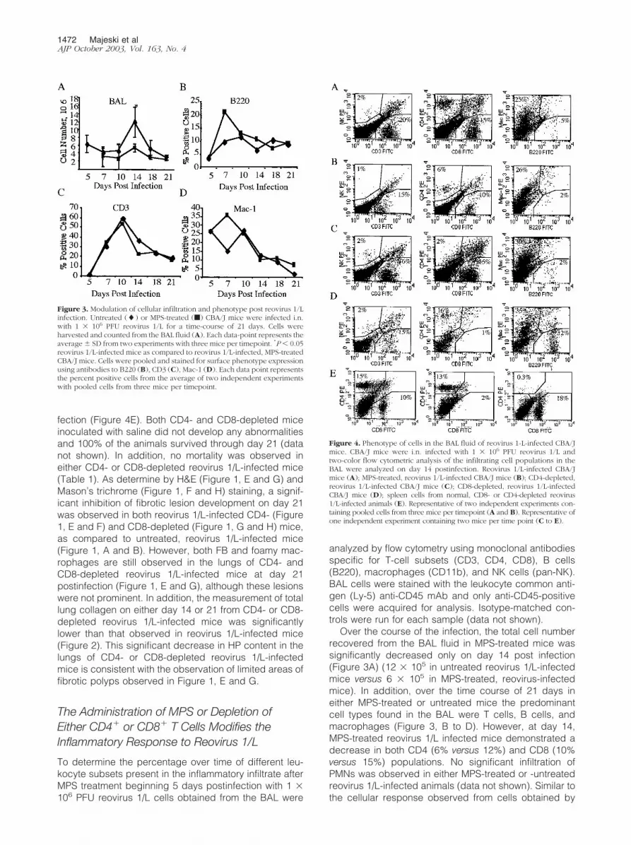
Over the course of the infection, the total cell numberrecovered from the BAL fluid in MPS-treated mice wassignificantly decreased only on day 14 post infection(Figure 3A) (12 � 105 in untreated reovirus 1/L-infectedmice versus 6 � 105 in MPS-treated, reovirus-infectedmice). In addition, over the time course of 21 days ineither MPS-treated or untreated mice the predominantcell types found in the BAL were T cells, B cells, andmacrophages (Figure 3, B to D). However, at day 14,MPS-treated reovirus 1/L infected mice demonstrated adecrease in both CD4 (6% versus 12%) and CD8 (10%versus 15%) populations. No significant infiltration ofPMNs was observed in either MPS-treated or -untreatedreovirus 1/L-infected animals (data not shown). Similar tothe cellular response observed from cells obtained by
Figure 3. Modulation of cellular infiltration and phenotype post reovirus 1/Linfection. Untreated (�) or MPS-treated (f) CBA/J mice were infected i.n.with 1 � 106 PFU reovirus 1/L for a time-course of 21 days. Cells wereharvested and counted from the BAL fluid (A). Each data-point represents theaverage � SD from two experiments with three mice per timepoint. *P � 0.05reovirus 1/L-infected mice as compared to reovirus 1/L-infected, MPS-treatedCBA/J mice. Cells were pooled and stained for surface phenotype expressionusing antibodies to B220 (B), CD3 (C), Mac-1 (D). Each data point representsthe percent positive cells from the average of two independent experimentswith pooled cells from three mice per timepoint.
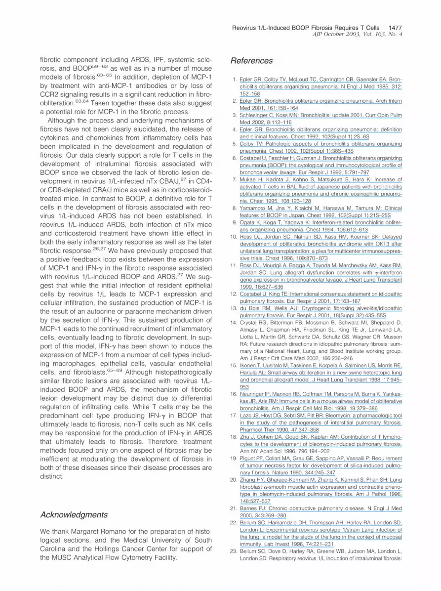
Figure 4. Phenotype of cells in the BAL fluid of reovirus 1-L-infected CBA/Jmice. CBA/J mice were i.n. infected with 1 � 106 PFU reovirus 1/L andtwo-color flow cytometric analysis of the infiltrating cell populations in theBAL were analyzed on day 14 postinfection. Reovirus 1/L-infected CBA/Jmice (A); MPS-treated, reovirus 1/L-infected CBA/J mice (B); CD4-depleted,reovirus 1/L-infected CBA/J mice (C); CD8-depleted, reovirus 1/L-infectedCBA/J mice (D); spleen cells from normal, CD8- or CD4-depleted reovirus1/L-infected animals (E). Representative of two independent experiments con-taining pooled cells from three mice per timepoint (A and B). Representative ofone independent experiment containing two mice per time point (C to E).
1472 Majeski et alAJP October 2003, Vol. 163, No. 4

BAL, few differences in the percentages of T or B lym-phocytes and macrophages were observed in the inter-stitial areas of untreated reovirus 1/L-infected mice versusMPS-treated, reovirus 1/L-infected mice (data notshown).
In either CD4- or CD8-depleted reovirus 1/L-infectedmice, the predominant cell types found in the BAL weremacrophages (30% - 35% in CD4- or CD8-depleted re-ovirus 1/L-infected mice versus 25% reovirus 1/L-infectedmice or 26% MPS-treated reovirus 1/L infected mice)(Figure 4). CD4-depleted, reovirus 1/L-infected miceshowed an infiltration of CD8� cells (15%) without asignificant infiltration of CD4� cells (2%) (Figure 4C) inthe BAL fluid. A similar response was observed withCD8-depleted, reovirus 1/L-infected mice (16% CD4�
cells versus 1% CD8� cells) (Figure 4, C and D). Nosignificant infiltration of PMNs was observed in eitherCD4- or CD8-depleted reovirus 1/L-infected animals(data not shown). Depletion of either CD4� or CD8� cellswas verified on both day 7 (data not shown) and day 14(Figure 4E) post-reovirus 1/L infection. As shown in Fig-ure 4E, the phenotype of spleen cells from normal micewas 15% CD4� and 10% CD8�cells, while animals de-pleted of either CD8, or CD4 demonstrated the appropri-ate phenotype (CD8-depleted animals: 13% CD4� and2% CD8� cells; CD4-depleted animals: 0.3% CD4� and18% CD8� cells).
Cytokine and Chemokine Expression areModulated in the Lungs of MPS-Treated CBA/JMice Receiving 1 � 106 PFU Reovirus 1/L
Cytokine and chemokine mRNA expression in total lungtissue was evaluated by RPA. Three cytokine RPA tem-plate sets (Figure 5, A to C) and one chemokine RPAtemplate set (Figure 5D) were evaluated in MPS-treatedor untreated CBA/J mice after i.n. inoculation with 1 � 106
PFU of reovirus 1/L over a time-course of 21 to 28 days.Together, these four template sets allow an analysis of theexpression and modulation of mRNAs for cytokines andchemokines which have been implicated to play a role inthe pulmonary fibrotic process (such as MCP-1, IL-6, IFN-�)as well as others contained within the template sets.
Substantial expression of mRNA for a number of cyto-kines and chemokines were observed in the lungs ofCBA/J mice infected i.n. with 1 � 106 PFU reovirus 1/L.(Figure 5). These include IL-9 (Figure 5A), the proinflam-matory cytokines IL-1�, IL-1 �, IL-1 Ra, IL-6 (Figure 5B),IFN-�, (Figure 5, A to C), MIF (Figure 5, B and C), and thechemokines, MIP-1�, MIP-1�, MCP-1, RANTES, andIP-10 (Figure 5D). In comparison to CBA/J mice infectedwith 1 � 106 PFU reovirus 1/L, MPS-treated reovirus1/L-infected i.n. CBA/J mice, in general, demonstrated adecreased expression of most of the cytokines and che-mokines present on the four template sets. Expression ofthe following cytokines and chemokines was observed inMPS-treated reovirus 1/L-infected CBA/J mice: IL-9 (Fig-ure 5A) IL-1�, IL-1 �, IL-1 Ra, IL-6 (Figure 5B), IFN-�,(Figure 5, A to C), MIF (Figure 5, B and C), MCP-1,RANTES, and IP-10 (Figure 5D). Cytokine mRNA expres-
sion was not induced for a number of cytokine genes ineither MPS-treated or untreated reovirus 1/L-infectedmice. These included IL-2, IL-3, and IL-13. In addition,mRNA expression for the chemokines eotaxin, MIP-2,and TCA-3 were not significantly induced.
Since both IFN-� and MCP-1 have been implicated inthe fibrotic process induced by reovirus 1/L,26,27 wequantitated the autoradiographs and found a significantup-regulation of both IFN-� and MCP-1 (Figure 6A)mRNA in CBA/J mice infected with 1 � 106 PFU reovirus1/L. However, in CBA/J mice infected with 1 � 106 PFUreovirus 1/L and treated with MPS beginning on day 5postinfection, a significant decrease in the mRNA expres-sion of IFN-� (Figure 6A) was observed. Although theexpression of MCP-1 was decreased after MPS treat-ment, this decrease as compared to control mice was notstatistically significant (Figure 6A). In a similar manner,while a significant increase in IFN-� protein in the BALfluid was observed in both MPS treated and untreatedreovirus 1/L-infected mice as compared to uninfectedcontrols (Figure 6B), a decrease in IFN-� protein expres-sion was observed between MPS-treated and untreated,reovirus 1/L-infected mice (Figure 6B). Similar to the ex-pression of mRNA for MCP-1, a decrease (although notstatistically significant) in the protein expression ofMCP-1 in the BAL fluid of MPS-treated reovirus 1/L-in-fected was observed (Figure 6B). In contrast, in bothCD4- and CD8-depleted mice, a reduction in the proteinexpression of both MCP-1 and IFN-� was observed (Fig-ure 6C). In all cases IFN-� and MCP-1 was not detectedin control, saline-inoculated mice (data not shown).
To demonstrate a role for IFN-� in the development offibrotic lesions in reovirus 1/L induced BOOP, reovirus1/L-infected (106 PFU BOOP) CBA/J mice were treatedi.p. every 3 days beginning on day 3 postinfection witheither 100 �g anti-IFN-� antibody (R4–6A2) in PBS (Fig-ure 7, C and D) or 100 �g normal rat IgG (Figure 7, A andB). Mice were evaluated on days 14 (Figure 7, A and C)and 21 (Figure 7, B and D) postinfection for the develop-ment of fibrotic lesions by H&E staining. As can be ob-served with H&E staining, significant fibrotic lesion devel-opment was observed in reovirus 1/L-infected mice whowere treated with normal rat IgG on both days 14 and 21(Figure 7, A and B). However, in reovirus 1/L-infected,anti-IFN-� treated mice, although follicular bronchiolitis(FB) lesions were observed on day 14 and to a lesserextent on day 21 (Figure 7D) postinfection, this was ac-companied by little to no fibrotic lesion development(Figure 7, C and D).
Discussion
In this study, we have used a reovirus 1/L-induced modelof BOOP to study cellular and molecular events importantin the development of this type of pulmonary fibrosis.Using reovirus 1/L-infected mice that were either treatedwith corticosteroids or depleted of either CD4� or CD8�
T cells, our results demonstrate that T cells play a majorrole in the development of intraluminal fibrosis associatedwith BOOP. Corticosteroid treatment of reovirus 1/L-in-
Reovirus 1/L-Induced BOOP Fibrosis Requires T Cells 1473AJP October 2003, Vol. 163, No. 4

fected mice both inhibited the development of fibroticlesions when administered early in the time-course andpromoted the resolution of fibrotic lesions when cortico-steroid administration was delayed. Daily treatment withcorticosteroids inhibited the total cellular infiltration intothe lung post reovirus 1/L infection, and also resulted inan inhibition of the proinflammatory and profibrotic cyto-kines, IFN-� and MCP-1. Further, the depletion of eitherCD4� or CD8� T cells before reovirus 1/L infection alsoinhibited fibrotic lesion development as well as IFN-� andMCP-1 protein expression. Consistent with these results,T-cell-deficient neonatally thymectomized (nTx) CBA/Jmice do not develop intraluminal fibrosis after infectionwith 1 � 106 PFU reovirus 1/L and both the phenotype of
the infiltrating cells and the expression of both IFN-� andMCP-1 were significantly altered in these nTx CBA/J miceas compared to normal CBA/J mice.27 Taken together,this study combined with our previous studies suggest asignificant role for T cells in the development of reovirus1/L-induced BOOP fibrotic lesions and suggests that TH1derived cytokines, especially IFN-�, may play a key rolein fibrotic lesion development.
Currently few small animal models of BOOP exist.However, several studies have shown that the infiltrationof T lymphocytes may be important in the development ofother forms of pulmonary fibrosis although the data fromboth animal models and patients has been equivocal.Intratracheal administration of bleomycin in rodents, a
Figure 5. Differential cytokine and chemokine mRNA expression from the lungs of untreated and MPS-treated reovirus 1/L-infected CBA/J mice. CBA/J mice werei.n. infected with 1 � 106 PFU reovirus 1/L and treated with MPS i.p. daily beginning on day 5 postinfection. RNA was harvested from the whole lungs at theindicated timepoints, and probed by RPA analysis using the following multiprobe template sets: mCK-1b (T-cell-derived cytokine panel) (A); mCK-2b(inflammatory cytokine panel) (B); mCK-3b (cytokines associated with fibrosis) (C); and mCK-5 (chemokine panel) (D). The position of individual cytokine orchemokine mRNA expression is indicated on the left side of each panel. Representative of three independent experiments containing two mice per timepoint.
1474 Majeski et alAJP October 2003, Vol. 163, No. 4

model for idiopathic pulmonary fibrosis (IPF), results ininterstitial fibrosis accompanied by a significant infiltra-tion of both T and B lymphocytes.17,18 In this model,some reports have demonstrated that the inhibition ordepletion of lymphocytes by anti-lymphocyte antibody,mAb to T-cell subsets, or treatment with steroids inhibitedthe development of bleomycin-induced fibrosis,34–36
while other studies found no effect of T-cell depletion onfibrotic lesions induced by bleomycin instillation.37–39
Similarly, in nude mice lacking T cells or SCID micedeficient in both T and B cells, conflicting evidence for arole of T cells in the fibrotic process has been report-ed.40–42 A role for T cells has also been proposed forBronchiolitis Obliterans Syndrome (BOS) which is themajor limitation to survival postlung transplantation and ischaracterized by persistent peribronchiolar inflammationthat leads to airway fibrosis/obliteration.3,16 Models ofBOS demonstrate increases in CD4� and CD8� T cells,B cells, and macrophages post allograft transplanta-tion.15,16,43,44 Further, in SCID allograft models of BOS noinflux of lymphocytes or fibrosis was observed, suggest-ing that lymphocytes may play an important role in thedevelopment of fibrosis during chronic graft rejection.16
While these models have been used to evaluate potentialmechanisms related to fibrosis, they may not fully reca-pitulate what occurs in similar human pathologies. As anexample, while bleomycin-induced fibrosis in rodents isused as a model for human IPF, many features of bleo-mycin-induced fibrosis are not shared with the humancondition.45 However, in this regard, the reovirus 1/L-induced model of BOOP recapitulates the histological(intraluminal fibrosis) and phenotypic characteristics ofhuman BOOP and thus, is an excellent small animalmodel of human BOOP. Furthermore, unlike other animalmodels for pulmonary fibrosis, reovirus-induced pulmo-nary fibrosis offers the advantage in that it also providesa model that recapitulates the response of human dis-ease to clinical treatments currently in use. Since theselesions occur in a well-defined temporal sequence thatproceeds from initial peribronchiolar inflammatory lesionsto characteristic, fibrotic cellular BOOP lesions, thismodel can be used to evaluate the cellular and molecularsignals that may lead to fibrotic lesion development inhuman BOOP.
The results described in this manuscript as well as inour previous publications all consistently demonstrate a
Figure 6. Expression of IFN-� and MCP-1 in corticosteroid-treated or CD4- or CD8-depleted CBA/J mice infected with 1 � 106 PFU reovirus 1/L. Relativedifferences in mRNA expression of either IFN-� or MCP-1 (A) over time were determined by comparing the ratio of cytokine/chemokine mRNA with thehousekeeping gene, L32 from RPA autoradiograms. IFN-� expression was evaluated between days 5 and 10 postinfection. MCP-1 expression was evaluatedbetween days 3 and 10 postinfection. Control, uninfected mice (open bar); reovirus 1/L-infected CBA/J mice (solid bar); MPS-treated, reovirus 1/L-infectedCBA/J mice (striped bar). The mean � SD of the densitometric measurements from two independent experiments with two mice per timepoint (fourindependent autoradiographs) over the indicated timepoints are presented. Detection of IFN-� or MCP-1 protein by ELISA in the BAL fluid over time from reovirus1/L-infected CBA/J mice (�) and MPS-treated reovirus 1/L-infected CBA/J mice (f) (B). The mean � SD of two independent experiments, evaluated in duplicate,is shown. Detection of MCP-1 (f) or IFN-� (�) proteins by ELISA in the BAL fluid evaluated on day 14 post-reovirus 1/L infection from reovirus 1/L-infectednormal, CD4-depleted, or CD8-depleted CBA/J mice (C). Representative of one experiment with two animals per timepoint, performed in duplicate *P � 0.05 ascompared to saline-inoculated control mice. **P � 0.05, as compared to reovirus 1/L-infected mice.
Reovirus 1/L-Induced BOOP Fibrosis Requires T Cells 1475AJP October 2003, Vol. 163, No. 4

clear dependence of T cells on the fibrotic process as-sociated with reovirus 1/L-induced BOOP.26 Thus, weanalyzed cytokines and chemokines that have previouslybeen associated with fibrotic lesion development. Webelieve that our data supports a role for IFN-� in thefibrotic process and that it acts as a profibrotic agent inthe spectrum of fibrosis induced in reovirus 1/L-infectedmice.22,25–27 Our data demonstrate that when the inflam-matory and fibrotic process in BOOP is inhibited either bycorticosteroid treatment or CD4 or CD8 depletion, theconcentration of IFN-� in the BAL fluid decreases. Both adecrease in IFN-� expression and limited fibrotic lesiondevelopment was also demonstrated in T-cell-deficientnTx mice that were infected with reovirus 1/L-inducedBOOP.27 However, in reovirus 1/L-induced ARDS, thefibrotic component was not inhibited and IFN-� expres-sion levels remained high in either corticosteroid-treatedor nTx animals.25–27 Further, treatment of reovirus 1/L-induced BOOP with an anti-IFN-� neutralizing monoclo-nal antibody inhibited fibrotic lesion development on bothdays 14 and 21 postinfection, further supporting our hy-pothesis that IFN-� plays a significant role in fibroticlesion development. Our data also demonstrate an in-creased expression of MCP-1 in reovirus-1/L inducedBOOP that was modified after corticosteroid treatment orwithin CD4- or CD8-depleted mice. This data are also inagreement with our previous studies that demonstratedan increase in MCP-1 expression in reovirus 1/L-inducedARDS whose fibrosis is not modified either in nTx mice orby corticosteroid treatment.26,27 However, in reovirus 1/L-induced BOOP a decrease in MCP-1 expression as wellas fibrosis was observed in nTx mice.27 Taken together,
these data and our previously published results demon-strate a direct correlation of proinflammatory cytokineexpression such as IFN-� and MCP-1 and the develop-ment of fibrosis.
Our results, demonstrating a role for IFN-� and MCP-1in reovirus 1/L-induced BOOP are in agreement withother studies in which both IFN-� and MCP-1 have beenimplicated in the fibrotic process in both patients andanimal models.46–48 A role for IFN-� in bleomycin-in-duced interstitial fibrosis is supported by the observa-tions that susceptible versus non-susceptible mousestrains produce high amounts of IFN-�,42 depletion of Tcells down-regulates both IFN-� expression and fibro-sis,35,49 and high levels of IFN-� expression and fibrosisare observed in SCID mice, which are susceptible tobleomycin-induced fibrosis.42 Bleomycin treatment ofIFN-� knockout mice (IFN-� -/-) also resulted in both asignificant inhibition of pulmonary inflammation and fibro-sis.50 IFN-� has also been implicated in the fibrotic pro-cess associated with the tracheal transplant model ofBOS.11,16,51,52 Expression of TH1 cytokines includingIFN-� and IL-10 were up-regulated to a greater extentthan TH2 cytokines (IL-4), suggesting that although cy-tokine production by all T-lymphocyte subsets (TH1, TH2)may be involved in the development of BOS, the TH1cytokine products may be more important in the devel-opment of fibrosis.52 INF-� has also been implicated as aprofibrotic factor in lung fibrosis that occurs in patients oranimal models with fibrosing alveolitis, IPF, sarcoidosis,chronic beryllium disease, silicosis, and lung allograftfibrosis.53–58 MCP-1 is also expressed in a number ofinflammatory conditions in patients that demonstrate a
Figure 7. Inhibition of fibrosis associated with BOOP after treatment with a neutralizing monoclonal antibody to interferon-�. CBA/J mice were i.n. infected withreovirus 1/L and treated i.p. every 3 days beginning on day 3 postinfection with either 100 �g normal rat IgG (A and B) or 100 �g anti-IFN-� antibody (C andD) in PBS. Mice were evaluated both on days 14 (A and C) and 21 (B and D) postinfection for the development of fibrotic lesions by H&E stain. A and B: Reovirus1/L-infected CBA/J mice treated with normal Rat IgG. C and D: Reovirus 1/L-infected CBA/J mice treated with an anti-IFN-� antibody. Representative of oneindependent experiment containing four mice per timepoint (magnification �20).
1476 Majeski et alAJP October 2003, Vol. 163, No. 4
fibrotic component including ARDS, IPF, systemic scle-rosis, and BOOP59–63 as well as in a number of mousemodels of fibrosis.63–65 In addition, depletion of MCP-1by treatment with anti-MCP-1 antibodies or by loss ofCCR2 signaling results in a significant reduction in fibro-obliteration.63,64 Taken together these data also suggesta potential role for MCP-1 in the fibrotic process.
Although the process and underlying mechanisms offibrosis have not been clearly elucidated, the release ofcytokines and chemokines from inflammatory cells hasbeen implicated in the development and regulation offibrosis. Our data clearly support a role for T cells in thedevelopment of intraluminal fibrosis associated withBOOP since we observed the lack of fibrotic lesion de-velopment in reovirus 1/L-infected nTx CBA/J,27 in CD4-or CD8-depleted CBA/J mice as well as in corticosteroid-treated mice. In contrast to BOOP, a definitive role for Tcells in the development of fibrosis associated with reo-virus 1/L-induced ARDS has not been established. Inreovirus 1/L-induced ARDS, both infection of nTx miceand corticosteroid treatment have shown little effect inboth the early inflammatory response as well as the laterfibrotic response.26,27 We have previously proposed thata positive feedback loop exists between the expressionof MCP-1 and IFN-� in the fibrotic response associatedwith reovirus 1/L-induced BOOP and ARDS.27 We sug-gest that while the initial infection of resident epithelialcells by reovirus 1/L leads to MCP-1 expression andcellular infiltration, the sustained production of MCP-1 isthe result of an autocrine or paracrine mechanism drivenby the secretion of IFN-�. This sustained production ofMCP-1 leads to the continued recruitment of inflammatorycells, eventually leading to fibrotic development. In sup-port of this model, IFN-� has been shown to induce theexpression of MCP-1 from a number of cell types includ-ing macrophages, epithelial cells, vascular endothelialcells, and fibroblasts.65–69 Although histopathologicallysimilar fibrotic lesions are associated with reovirus 1/L-induced BOOP and ARDS, the mechanism of fibroticlesion development may be distinct due to differentialregulation of infiltrating cells. While T cells may be thepredominant cell type producing IFN-� in BOOP thatultimately leads to fibrosis, non-T cells such as NK cellsmay be responsible for the production of IFN-� in ARDSthat ultimately leads to fibrosis. Therefore, treatmentmethods focused only on one aspect of fibrosis may beinefficient at modulating the development of fibrosis inboth of these diseases since their disease processes aredistinct.
Acknowledgments
We thank Margaret Romano for the preparation of histo-logical sections, and the Medical University of SouthCarolina and the Hollings Cancer Center for support ofthe MUSC Analytical Flow Cytometry Facility.
References
1. Epler GR, Colby TV, McLoud TC, Carrington CB, Gaensler EA: Bron-chiolitis obliterans organizing pneumonia. N Engl J Med 1985, 312:152–158
2. Epler GR: Bronchiolitis obliterans organizing pneumonia. Arch InternMed 2001, 161:158–164
3. Schlesinger C, Koss MN: Bronchiolitis: update 2001. Curr Opin PulmMed 2002, 8:112–116
4. Epler GR: Bronchiolitis obliterans organizing pneumonia: definitionand clinical features. Chest 1992, 102(Suppl 1):2S–6S
5. Colby TV: Pathologic aspects of bronchiolitis obliterans organizingpneumonia. Chest 1992, 102(Suppl 1):38S–43S
6. Costabel U, Teschler H, Guzman J: Bronchiolitis obliterans organizingpneumonia (BOOP): the cytological and immunocytological profile ofbronchoalveolar lavage. Eur Respir J 1992, 5:791–797
7. Mukae H, Kadota J, Kohno S, Matsukura S, Hara K: Increase ofactivated T cells in BAL fluid of Japanese patients with bronchiolitisobliterans organizing pneumonia and chronic eosinophilic pneumo-nia. Chest 1995, 108:123–128
8. Yamamoto M, Jna Y, Kitaichi M, Haraswa M, Tamura M: Clinicalfeatures of BOOP in Japan. Chest 1992, 102(Suppl 1):21S–25S
9. Ogata K, Koga T, Yagawa K: Interferon-related bronchiolitis obliter-ans organizing pneumonia. Chest 1994, 106:612–613
10. Ross DJ, Jordan SC, Nathan SD, Kass RM, Koerner SK: Delayeddevelopment of obliterative bronchiolitis syndrome with OKT3 afterunilateral lung transplantation: a plea for multicenter immunosuppres-sive trials. Chest 1996, 109:870–873
11. Ross DJ, Moudgil A, Bagga A, Toyoda M, Marchevsky AM, Kass RM,Jordan SC: Lung allograft dysfunction correlates with �-interferongene expression in bronchoalveolar lavage. J Heart Lung Transplant1999, 18:627–636
12. Costabel U, King TE: International consensus statement on idiopathicpulmonary fibrosis. Eur Respir J 2001, 17:163–167
13. du Bois RM, Wells AU: Cryptogenic fibrosing alveolitis/idiopathicpulmonary fibrosis. Eur Respir J 2001, 18(Suppl 32):43S–55S
14. Crystal RG, Bitterman PB, Mossman B, Schwarz MI, Sheppard D,Almasy L, Chapman HA, Friedman SL, King TE Jr, Leinwand LA,Liotta L, Martin GR, Schwartz DA, Schultz GS, Wagner CR, MussonRA: Future research directions in idiopathic pulmonary fibrosis: sum-mary of a National Heart, Lung, and Blood Institute working group.Am J Respir Crit Care Med 2002, 166:236–246
15. Ikonen T, Uusitalo M, Taskinen E, Korpela A, Salminen US, Morris RE,Harjula AL: Small airway obliteration in a new swine heterotopic lungand bronchial allograft model. J Heart Lung Tranplant 1998, 17:945–953
16. Neuringer IP, Mannon RB, Coffman TM, Parsons M, Burns K, Yankas-kas JR, Aris RM: Immune cells in a mouse airway model of obliterativebronchiolitis. Am J Respir Cell Mol Biol 1998, 19:379–386
17. Lazo JS, Hoyt DG, Sebti SM, Pitt BR: Bleomycin: a pharmacologic toolin the study of the pathogenesis of interstitial pulmonary fibrosis.Pharmcol Ther 1990, 47:347–358
18. Zhu J, Cohen DA, Goud SN, Kaplan AM: Contribution of T lympho-cytes to the development of bleomycin-induced pulmonary fibrosis.Ann NY Acad Sci 1996, 796:194–202
19. Piguet PF, Collart MA, Grau GE, Sappino AP, Vassalli P: Requirementof tumour necrosis factor for development of silica-induced pulmo-nary fibrosis. Nature 1990, 344:245–247
20. Zhang HY, Gharaee-Kermani M, Zhang K, Karmiol S, Phan SH: Lungfibroblast �-smooth muscle actin expression and contractile pheno-type in bleomycin-induced pulmonary fibrosis. Am J Pathol 1996,148:527–537
21. Barnes PJ: Chronic obstructive pulmonary disease. N Engl J Med2000, 343:269–280
22. Bellum SC, Hamamdzic DH, Thompson AH, Harley RA, London SD,London L: Experimental reovirus serotype 1/strain Lang infection ofthe lung: a model for the study of the lung in the context of mucosalimmunity. Lab Invest 1996, 74:221–231
23. Bellum SC, Dove D, Harley RA, Greene WB, Judson MA, London L,London SD: Respiratory reovirus 1/L induction of intraluminal fibrosis:
Reovirus 1/L-Induced BOOP Fibrosis Requires T Cells 1477AJP October 2003, Vol. 163, No. 4

a model for the study of bronchiolitis obliterans organizing pneumo-nia. Am J Pathol 1997, 150:2243–2254
24. Thompson AH, London L, Bellum SC, Hamamdzic D, Harley RA,London SD: Respiratory-mucosal lymphocyte populations inducedby reovirus serotype 1 infection. Cell Immunol 1996, 169:278–287
25. London L, Majeski EI, Paintlia MK, Harley RA, London SD: Respiratoryreovirus 1/L induction of diffuse alveolar damage: a model of acuterespiratory distress syndrome. Exper Mol Pathol 2002, 72:24–36
26. London L, Majeski EI, Altman-Hamamdzic S, Enockson C, PaintliaMK, Harley RA, London SD: Respiratory reovirus 1/L induction ofdiffuse alveolar damage: pulmonary fibrosis is not modulated bycorticosteroids in acute respiratory distress syndrome in mice. ClinImmunol 2002, 103:284–295
27. Majeski EI, Harley RS, Bellum SC, London SD, London L: Differentialrole for T cells in the development of fibrotic lesions associated withreovirus 1/L induced BOOP versus ARDS. Am J Respir Cell Mol Bio2003 28:208–217
28. Dialynas DP, Quan ZS, Wall KA, Pierres A, Quintans J, Loken MR,Pierre M, Fitch FW: Characterization of the murine T cell surfacemolecule designated L3T4, identified by monoclonal antibody GK1.5:similarities to the human Leu3/T4 molecule. J Immunol 1983, 131:2445–2451
29. Ledbetter JA, Herzenberg LA: Xenogeneic antibodies to mouse lym-phoid differentiation antigens. Immuno Rev 1979, 47:63–90
30. Spitalny GL. Havell EA: Monoclonal antibody to murine � interferoninhibits lymphokine-induced antiviral and macrophage tumoricidalactivities. J Exp Med 1984, 159:1560–1565
31. Tay CH, Welsh RM: Distinct organ-dependent mechanisms for thecontrol of murine cytomegalovirus infection by natural killer cells.J Virol 1997, 71:267–275
32. Sayles PC, Johnson LL: Intact immune defenses are required for miceto resist the ts-4 vaccine strain of Toxoplasma gondii. Infect Immun1996, 64:3088–3092
33. Reddy GK, Enwemeka CS: A simplified method for the analysis ofhydroxyproline in biological tissues. Clin Biochem 1996, 29:225–229
34. Phan SH, Thrall RS, Williams C: Bleomycin-induced pulmonaryfibrosis: effects of steroid on lung collagen metabolism. Am RevRespir Dis 1981, 124:428–434
35. Sharma SK, MacLean JA, Pinto C, Kradin RL: The effect of an anti-CD3 monoclonal antibody on bleomycin-induced lymphokine pro-duction and lung injury. Am J Respir Crit Care Med 1996, 154:193–200
36. Koenig WJ, Cross CE, Hesterberg TW, Last JA: The smoking gun:mechanism of methylprednisolone prevention of bleomycin-inducedpulmonary fibrosis. Chest 1983, 83:5S–7S
37. Sterling KM, DiPetrillo T, Cutroneo KR, Prestayko A: Inhibition ofcollagen accumulation by glucocorticoids in rat lung after intratra-cheal bleomycin instillation. Cancer Res 1982, 42:405–408
38. Janick-Buckner D, Ranges GE, Hacker MP: Effect of cytotoxic mono-clonal antibody depletion of T-lymphocyte subpopulations on bleo-mycin-induced lung damage in C57BL/6J mice. Toxicol Appl Pharm1989, 100:474–484
39. Khalil N, Whitman C, Zuo L, Danielpour D, Greenberg A: Regulationof alveolar macrophage transforming growth factor-� secretion bycorticosteroids in bleomycin-induced pulmonary inflammation in therat. J Clin Invest 1993, 92:1812–1818
40. Szapiel SV, Elson N, Fulmer JD, Hunninghake GW, Crystal RG: Bleo-mycin-induced interstitial pulmonary disease in the nude, athymicmouse. Am Rev Respir Dis 1979, 120:893–899
41. Schrier DJ, Phan SH, McGarry BM: The effects of the nude (nu/nu)mutation on bleomycin-induced pulmonary fibrosis: a biochemicalevaluation. Am Rev Respir Dis 1983, 127:614–617
42. Lake-Bullock HM, Zhu V, Hao J, Cohen H, Kaplan AM: T cell inde-pendence of bleomycin-induced pulmonary fibrosis. J Leuk Biol1999, 65:187–195
43. Koskinen P, Kallio EA, Bruggeman CA, Lemstrom KB: Cytomegalo-virus infection enhances experimental obliterative bronchiolitis in rattracheal allografts. Am J Respir Crit Care Med 1997, 155:2078–2088
44. Boehler A, Chamberlain D, Kesten S, Slutsky AS, Liu M, Keshavjee S:Lymphocytic airway infiltration as a precursor to fibrous obliteration ina rat model of bronchiolitis obliterans. Transplantation 1997, 64:311–317
45. Cooper JAD III: Pulmonary fibrosis: pathways are slowly coming ontolight. Am J Respir Cell Mol Biol 2000, 520–523
46. Lukacs NW, Hogaboam C, Chensue SW, Blease K, Kunkel SL: Type1/type 2 cytokine paradigm and the progression of pulmonary fibro-sis. Chest 2001, 120(Suppl 1):5S–8S
47. Kuwano K, Hagimoto N, Hara N: Molecular mechanisms of pulmonaryfibrosis and current treatment. Curr Mol Med 2001, 1:551–573
48. Lasky JA, Brody AR: Interstitial fibrosis and growth factors. EnvironHealth Perspect 2000, 108(Suppl 4):751–762
49. Tager A, Luster A, Kradin R: T-cell chemokines interferon-inducibleprotein-10 and monokine induced by interferon-� are upregulated inbleomycin-induced lung injury. Chest 1999, 116:90S
50. Chen ES, Greenlee BM, Wills-Karp M, Moller DR: Attenuation of lunginflammation and fibrosis in interferon-�-deficient mice after intratra-cheal bleomycin. Am J Respir Cell Mol Biol 2001, 24:545–555
51. Boehler A, Bai XH, Liu M, Cassivi S, Chamberlain D, Slutsky AS,Keshavjee S: Upregulation of T-helper 1 cytokines and chemokineexpression in post-transplant airway obliteration. Am J Respir CritCare Med 1999, 159:1910–1917
52. Neuringer IP, Walsh SP, Mannon RB, Gabriel S, Aris RM: Enhanced Tcell cytokine gene expression in mouse airway obliterative bronchi-olitis. Transplantation 2000, 69:399–405
53. Davis GS, Pfeiffer LM, Hemenway DR: Interferon-� production byspecific lung lymphocyte phenotypes in silicosis in mice. Am J RespirCell Mol Biol 2000, 22:491–501
54. Robinson BW, Rose AH: Pulmonary � interferon production in patientswith fibrosing alveolitis. Thorax 1990, 45:105–108
55. Shaw RJ, Benedict SH, Clark RA, King TE Jr: Pathogenesis of pul-monary fibrosis in interstitial lung disease: alveolar macrophagePDGF(B) gene activation and up-regulation by interferon-�. Am RevRespir Dis 1991, 143:167–173
56. Awad M, Pravica V, Perrey C, El Gamel A, Yonan N, Sinnott PJ,Hutchinson IV: CA repeat allele polymorphism in the first intron of thehuman interferon-� gene is associated with lung allograft fibrosis.Human Immunol 1999, 60:343–346
57. Shigehara K, Shijubo N, Ohmichi M, Takahashi R, Kon S, Okamura H,Kurimoto M, Hiraga Y, Tatsuno T, Abe S, Sato N: IL-12 and IL-18 areincreased and stimulate IFN-� production in sarcoid lungs. J Immunol2001, 166:642–649
58. Wallace WAH, Howie SEM: Immunoreactive interleukin 4 and inter-feron-� expression by type II alveolar epithelial cells in interstitial lungdisease. J Pathol 1999, 187:475–480
59. Iyonaga K, Takeya M, Saita N, Sakamoto O, Yoshimura T, Ando M,Takahashi K: Monocyte chemoattractant protein-1 in idiopathic pul-monary fibrosis and other interstitial lung diseases. Hum Pathol 1994,25:455–463
60. Suga M, Iyonaga K, Ichiyasu H, Saita N, Yamasaki H, Ando M:Clinical significance of MCP-1 levels in BALF and serum in patientswith interstitial lung diseases. Eur Respir J 1999, 14:376–382
61. Hasegawa M, Sato S, Takehara K: Augmented production of chemo-kines (monocyte chemotactic protein-1 (MCP-1), macrophage inflam-matory protein-1� (MIP-1�) and MIP-1�) in patients with systemicsclerosis: mCP-1 and MIP-1� may be involved in the development ofpulmonary fibrosis. Clin Exper Immunol 1999, 117:159–165
62. Car BD, Meloni F, Luisetti M, Semenzato G, Gialdroni-Grassi G, WalzA: Elevated IL-8 and MCP-1 in the bronchoalveolar lavage fluid ofpatients with idiopathic pulmonary fibrosis and pulmonary sarcoid-osis. Am J Respir Crit Care Med 1994, 149:655–659
63. Belperio JA, Keane MP, Burdick MD, Lynch JP 3rd, Xue YY, Berlin A,Ross DJ, Kunkel SL, Charo IF, Strieter RM: Critical role for the che-mokine MCP-1/CCR2 in the pathogenesis of bronchiolitis obliteranssyndrome. J Clin Invest 2001, 108:547–556
64. Smith RE, Strieter RM, Phan SH, Kunkel SL: C-C chemokines: novelmediators of the profibrotic inflammatory response to bleomycin chal-lenge. Am J Respir Cell Mol Biol 1996, 15:693–702
65. Moore BB, Paine R III, Christensen PJ, Moore TA, Sitterding S, NganR, Wilke CA, Kuziel WA, Toews GB: Protection from pulmonary fibro-sis in the absence of CCR2 signaling. J Immunol 2001, 167:4368–4377
66. Andoh A, Takaya H, Makino J, Sato H, Bamba S, Araki Y, Hata K,Shimada M, Okuno T, Fujiyama Y, Bamba T: Cooperation of interleu-kin-17 and interferon-� on chemokine secretion in human fetal intes-tinal epithelial cells. Clin Exper Immunol 2001, 125:56–63
1478 Majeski et alAJP October 2003, Vol. 163, No. 4

67. van der Velden VH, Verheggen MM, Bernasconi S, Sozzani S, NaberBA, van der Linden-van Beurden CA, Hoogsteden HC, Mantovani A,Versnel M: Interleukin-1� and interferon-� differentially regulate re-lease of monocyte chemotactic protein-1 and interleukin-8 by humanbronchial epithelial cells. Eur Cytokine Netw 1998, 9:269–277
68. Bauermeister K, Burger M, Almanasreh N, Knopf HP, Schumann RR,Schollmeyer P, Dobos GJ: Distinct regulation of IL-8 and MCP-1 by
LPS and interferon-�-treated human peritoneal macrophages. Neph-rol Dial Transplant 1998, 13:1412–1419
69. Brown Z, Gerritsen M, Carley WW, Strieter RM, Kunkel SL, WestwickJ: Chemokine gene expression and secretion by cytokine-activatedhuman microvascular endothelial cells: differential regulation ofmonocyte chemoattractant protein-1 and interleukin-8 in response tointerferon-�. Am J Pathol 1994, 145:913–921
Reovirus 1/L-Induced BOOP Fibrosis Requires T Cells 1479AJP October 2003, Vol. 163, No. 4


