Embed Size (px)
Citation preview

DaltonTransactions
PAPER
Cite this: Dalton Trans., 2014, 43,8395
Received 4th January 2014,Accepted 24th February 2014
DOI: 10.1039/c4dt00024b
www.rsc.org/dalton
Colloidal assemblies of oriented maghemitenanocrystals and their NMR relaxometricproperties†
Athanasia Kostopoulou,a Sabareesh K. P. Velu,b Kalaivani Thangavel,b
Francesco Orsini,b Konstantinos Brintakis,a,c Stylianos Psycharakis,a,d Anthi Ranella,a
Lorenzo Bordonali,e Alexandros Lappas*a and Alessandro Lascialfari*b
An elevated-temperature polyol-based colloidal-chemistry approach allows for the development of
size-tunable (50 and 86 nm) assemblies of maghemite iso-oriented nanocrystals, with enhanced magne-
tization. 1H-nuclear magnetic resonance (NMR) relaxometric experiments show that the ferrimagnetic
cluster-like colloidal entities exhibit a remarkable enhancement (4–5 times) in transverse relaxivity when
compared to that of the superparamagnetic contrast agent Endorem®, over an extended frequency
range (1–60 MHz). The marked increase in the transverse relaxivity r2 at a clinical magnetic field strength
(∼1.41 T), which is 405.1 and 508.3 mM−1 s−1 for small and large assemblies, respectively, makes it possible
to relate the observed response to the raised intra-aggregate magnetic material volume fraction. Further-
more, cell tests with a murine fibroblast culture medium confirmed cell viability in the presence of the
clusters. We discuss the NMR dispersion profiles on the basis of relaxivity models to highlight the
magneto-structural characteristics of the materials for improved T2-weighted magnetic resonance
images.
A. Introduction
Over the past decade, there has been considerable progress inthe synthesis of colloidal inorganic nanocrystals and theirexploitation for applications in various fields, from electronicsand optoelectronics1 to nanomedicine and clinical practice.2
An interesting development in this direction involves the syn-thesis of colloidal nanocrystals made of multiple subunitsarranged in a controlled topological fashion3,4 or self-assembled in cluster-like structures (e.g. γ-Fe2O3, Fe3O4, PbS,ZnO, TiO2, etc.).
5–9 Their synthesis is either the result of direct
specific interactions (e.g. van der Waals attractions, stericrepulsions, attractive depletion or capillary forces, Coulombforces, etc.) of their discrete components, or of some externalstimulus, such as light, magnetic or electric fields.10 Thecoexistence within the same nanoscale entity of distinctmaterial sections, directly interconnected through inorganicinterfaces, or secondary structures communicating throughstrong interactions, enables multifunctionality. This is becausecoupling between mechanisms is established across the inter-faced material domains,11–13 while the physical properties mayalso have a collective nature.14 Based on these appealing fea-tures, such multifunctional systems are promising for techno-logical areas such as biology and biomedicine. Furthermore,those materials featuring a soft magnetic state in conjunctionwith some other non-homologous property (e.g. plasmonic orexcitonic emission) and with appropriate surface biocompati-ble coatings, occupy a prominent position in this scenario:4,9
they can be exploited for in vivo biomedical applications, suchas magnetically-driven contrast enhancement,15,16 selectivehyperthermia treatment,17 and even targeted drug delivery.18
Specifically, the afore-mentioned iron-oxide based nano-materials have emerged as promising probes in bionanotech-nology as they allow for a novel approach to the diagnosis ofvarious diseases with magnetic resonance imaging (MRI).During the last two decades, several commercial MRI contrast
†Electronic supplementary information (ESI) available: Fluorescence microscopyimages, calculation of the magnetic material volume fraction, table of transverserelaxivities of different ferrite-based samples. See DOI: 10.1039/c4dt00024b
aInstitute of Electronic Structure and Laser, Foundation for Research and Technology
– Hellas, Vassilika Vouton, 71110 Heraklion, Greece. E-mail: [email protected];
Fax: +30 2810 301305; Tel: +30 2810 391344bDipartimento di Fisica, Università degli studi di Milano and INSTM, via Celoria 16,
I-20133 Milano, Italy. E-mail: [email protected];
Fax: +39 02 50317208; Tel: +39 02 50317383cDepartment of Physics, Aristotle University of Thessaloniki, 54124 Thessaloniki,
GreecedDepartment of Medicine, University of Crete, Voutes, 71003 Heraklion, GreeceeDipartimento di Fisica, Università degli studi di Pavia and INSTM, via Bassi 6,
I-27100 Pavia, Italy
This journal is © The Royal Society of Chemistry 2014 Dalton Trans., 2014, 43, 8395–8404 | 8395

agents have been developed for clinical studies. The majorityof such systems are based on superparamagnetic iron-oxidenanoparticles, coated with hydrophilic polymers or sugars,including Endorem®, Resovist®, and Combidex®. However,poly-dispersity and poor crystallinity are often the mostcommon limitations of such systems, originating from the syn-thesis protocol; as a result, the magnetic properties and con-trast efficiencies vary from one batch to another.19,20
To develop new magnetic compounds for improved MR-based diagnostics, it is desirable to adjust the magnitude ofthe particle’s net magnetic moment μ (because R2 = 1/T2 ∝ μ2,where R2 is the transverse relaxation rate and T2 is the spin–spin relaxation time) by controlling the structural character-istics of the nano-object, i.e. the nanoparticle’s size, shape,composition and crystallinity.21 Rapid advances in the chemi-cal routes for the synthesis of size- and shape-controlled,surface functionalised magnetic nanoparticles22,23 have led toincreased particle magnetization and enhanced image con-trast, preserving biocompatibility at the same time. As indivi-dual superparamagnetic iron oxide nanocrystals (SPIONs) tendto give relatively low magnetization,21 to improve the MRI con-trast, larger colloidal entities composed of primary nanocrys-tals (PNCs), which are self-assembled24 or incorporated intopolymer matrices, have been investigated. The latter approach,for instance, entails SPIONs encapsulated in polyacrylamine,25
silica,26 phospholipid micelles,27 block polymers28 or amphi-philic polymers,29,30 while the former assembly-approacheslead to efficient “clustering” of individual PNCs, and promoteclose contact. In turn, this pathway further boosts the magneti-zation of such nanostructures, while the proximity of nanopar-ticles within the cluster promotes coupling of the neighboringmagnetic moments and results in a stronger perturbation ofthe local magnetic field in their vicinity. Indeed, the hydrogennuclear moments relax faster, leading to a strong contrast inthe MRI images. Magnetic nanoclusters of this type have beendeveloped with capping agents of different nature, includingoleylamine/oleic acid,31,32 citrate,33–35 polymers7,9,24,36–44 orblock copolymers.45–47 When the enlarged cluster-like entitycontains a biocompatible polymer-coating such as polyvinylalcohol (PVA), poly(ethylene glycol)-co fumarate (PEGF) orcross linked PEGF, wide-spread opportunities in the biomedi-cal field become possible.24 These colloidal nanocluster agentsfeature an improved contrast efficiency with respect to com-mercial compounds, or at least their MRI contrast response iscomparable to that of the widely available compoundEndorem®.
In the present work we report on the size-controlled assem-blies of pure maghemite (γ-Fe2O3) PNCs, which are crystallo-graphically aligned within the hydrophilic colloidalnanoclusters (CNCs). Our system displays a weak ferrimagneticbehavior, in contrast to the majority of the known nanoparti-cle-based aggregates devised for MR-based applications. Wesuggest that careful engineering of the CNCs’ magneto-struc-tural characteristics produces a remarkable improvement of r2(about four-to-five times higher) compared to Endorem®.From the point of view of design criteria we suggest that an
increased intra-aggregate magnetic material volume fractionand the proper choice of the surfactant, allowing water protonpenetration, are necessary conditions for superior MR-relatedcontrast enhancing features of such low-cytotoxicitynanoarchitectures.
B. ExperimentalB.1 Materials
All reagents were used as received without further purification.Anhydrous iron chloride (FeCl3, 98%), was purchased fromAlfa Aesar (United States). Anhydrous sodium hydroxide(NaOH, 98%) and polyacrylic acid (PAA, Mw = 1800), were pur-chased from Sigma Aldrich (United States), while diethyleneglycol (DEG, (HOCH2CH2)2O) of reagent (<0.3%) and labora-tory (<0.5%) grades were purchased from Fisher (UnitedStates). Absolute ethanol was purchased from Sigma Aldrich.
B.1.1 Cell cultures. NIH/3T3 murine fibroblasts (ATCC –
American Type Culture Collection) were suspended at a con-centration of 105 cells mL−1 in Dulbecco’s modified Eagle’smedium (DMEM) supplemented with 10% fetal bovine serum(FBS) and 1% antibiotic solution (GIBCO, Invitrogen, Karls-ruhe, Germany); cells were then cultured at 37 °C, in an atmos-phere of 5% CO2, and detached when growth reached 75%confluence using 0.05% trypsin/EDTA (GIBCO, Invitrogen,Karlsruhe, Germany).
B.2 Synthesis of γ-Fe2O3 nanoparticle clusters
The CNCs were synthesized by a modified high-temperaturecolloidal chemistry protocol based on a one-step polyolprocess first reported by Yin and co-workers.48 All syntheseswere carried out under an argon atmosphere in 100 mL round-bottom three-neck flasks connected via a reflux condenser to astandard Schlenk line setup, equipped with immersion temp-erature probes and digitally-controlled heating mantles. Thereactants, FeCl3, NaOH, polyacrylic acid (PAA, Mw = 1800),diethylene glycol (DEG), except ethanol, were stored andhandled under anaerobic conditions in an Ar-filled glove-box(MBRAUN, UNILab). Crucially, utilising two types of DEGgrades allowed for assemblies of iron-oxide nanocrystals withsmall and large diameters to be prepared upon increasingwater content in the reaction mixture. Observation of the puri-fied nanocluster samples by transmission electron microscopy,at regular time-intervals over a period of a year, have shownthat they are stable with no aggregation among their units.Details of the synthesis protocol can be found elsewhere.49
B.3 Characterization techniques
B.3.1 Transmission electron microscopy (TEM). Low-magni-fication and high-resolution TEM images were recorded on aLaB6 JEOL 2100 electron microscope operating at an accelerat-ing voltage of 200 kV. For the purposes of the TEM analysis, adrop of a diluted colloidal nanoclusters aqueous solution wasdeposited onto a carbon-coated copper TEM grid. All theimages were recorded by a Gatan ORIUS™ SC 1000 CCD
Paper Dalton Transactions
8396 | Dalton Trans., 2014, 43, 8395–8404 This journal is © The Royal Society of Chemistry 2014

camera and the structural features of the nanostructures werestudied by two-dimensional (2D) fast Fourier transform (FFT)analysis.
B.3.2 Magnetic measurements. The magnetic propertiesof the samples were studied by means of a SuperconductingQuantum Interference Device (SQUID) magnetometer(Quantum Design MPMS XL5). The magnetic measurementswere performed on the dried powder of the nanomaterials.The magnetic data were normalized to the mass of theγ-Fe2O3, as derived by inductively coupled plasma atomic emis-sion spectroscopy (ICP-AES) analysis ([Fe]50.2 nm CNCs = 44.9 ±0.3 mM, [Fe]85.6 nm CNCs = 42.3 ± 0.3 mM).
B.3.3 Nuclear magnetic resonance (NMR). Two solutions ofCNCs were prepared for the NMR investigation, having an ironconcentration C = 1.225 and 1.208 mmol L−1, as determinedby ICP-AES measurements for the small and large CNCs,respectively. NMR longitudinal and transverse relaxation timeswere measured by using two different pulsed Fourier trans-form (FT)-NMR spectrometers: (i) a Smartracer Stelar relax-ometer (using the Fast-Field-Cycling, FFC, technique) forfrequencies in the range 0.01 ≤ ν ≤ 10 MHz, and (ii) a StelarSpinmaster relaxometer for ν > 10 MHz. We used a commonsaturation recovery sequence to measure the longitudinal(spin–lattice, T1) nuclear relaxation time. Standard radio fre-quency excitation pulse sequences, CPMG for ν > 3 MHz andpre-polarized Hahn echo sequence for ν < 3 MHz, were used tomeasure the transverse (spin–spin, T2) nuclear relaxation time.The investigated frequency range 0.01 ≤ ν ≤ 60 MHz corre-sponds to an external magnetic field H = 0.00023–1.41 Tesla(ω = γH, where ω is the Larmor frequency and γ/2π =42.58 MHz T−1). The magnetic field range was chosen in orderto cover the typical fields used in both clinical and researchlaboratory MRI tomography (H = 0.2, 0.5 and 1.5 Tesla). Themeasurements at room and physiological temperatures gavethe same results within 10%.
The efficiency of the CNCs as MRI contrast agents wasdetermined by calculating the nuclear longitudinal and trans-verse relaxivities, r1 and r2 respectively, defined as
ri ¼ ð1=TiÞmeas � ð1=TiÞdiamC
; i ¼ 1; 2 ð1Þ
where (1/Ti)meas is the value measured for the solutionswith concentration C (mmol L−1) of the magnetic center and(1/Ti)diam represents the nuclear relaxation rate of the diamag-netic host solution, which in our case is water.
B.3.4 Cell viability assay. The interaction of the cells withthe CNCs was evaluated using a live-dead cell staining kit (Bio-vision). The assay protocol has been described in previousstudies.50,51 The cell suspension (105 cells mL−1) was de-posited in 24-well plates and cultivated for 1, 3, 5 and 7 days.In parallel, different concentrations of CNCs (25, 50, 100 and200 μg mL−1 Fe) were added into the cell suspension for eachdifferent culture period. Cells that remained alive after certaindays of cultivation were stained green whilst dead cells werestained yellow-red. The images were recorded in a Zeiss fluo-rescence microscope equipped with the Laser scanning system
Radiance 2100 (400–700 nm) and a Carl-Zeiss Axio CameraHR. Experiments were repeated at least three times for eachsize and available concentration of the CNCs.
B.3.5 Cell proliferation assay. The quantification of live cellsafter each day of culture was performed by using an ATP-Glo™bioluminometric cell viability assay kit (Biotium, Inc.). Thishighly sensitive homogenous assay for quantifying adenosinetriphosphate (ATP) involves an addition of an ATP-Glo™ detec-tion cocktail to cells cultured in a serum-supplementedmedium. Since ATP is an indicator of metabolically activecells, the number of viable cells can be assessed based on theamount of ATP available. This ATP detection kit takes advan-tage of Firefly luciferase’s use of ATP to oxidize D-luciferin andthe resulting production of light in order to assess the amountof ATP available. Luminescence measurements were per-formed using a Synergy™ HT Multi-Mode Microplate Reader(BioTek). The different CNCs concentrations were tested induplicate in each individual cell culture experiment and theseexperiments were repeated at least three times. For statisticalanalysis, the data were subjected to one way ANOVA followedby Tukey tests for multiple comparisons between pairs ofmeans, using commercially available software (SPSS 21, IBM).
C. Results and discussionC.1 Morphology and crystal structure
Iron-oxide colloidal assemblies of two different sizes were pre-pared on the basis of a high-temperature wet chemistry polyolpathway. The influence of different synthesis parameters49 onthe structural characteristics of the CNCs were carefully exam-ined in order to attain a significant degree of size/shape homo-geneity. 57Fe Mössbauer spectra (MS) recorded for the twonanocrystal assemblies at 10, 77 and 300 K show similar fea-tures, confirming the maghemite (γ-Fe2O3) nature49 of theseiron-oxides. Furthermore, bright-field TEM images for tworepresentative samples are shown in Fig. 1a and c. Both CNCsamples have a flower-like, almost spherical shape withoutaggregation between units. Each nanocluster is an assembly ofsmall PNCs, with no isolated nanoparticles left out of theaggregate. The average diameters of the entities are 50.2 ± 5.4and 85.6 ± 13.3 nm (Fig. 1a and c). Further evidence for thetopological arrangement and the crystallographic orientationof the assembled nanocrystals is provided by HRTEM imagesand the calculated FFT patterns taken from individual CNCs(Fig. 1b and d). The observed diffraction spots suggest that theindividual nanocrystals are iso-oriented. Analysis of the FFTpatterns allowed us to identify that the crystal symmetry of theCNCs as a whole is similar to that of the individual, smallPNCs, which crystallize in a cubic spinel structure. The attrac-tive magnetic dipolar interactions (vide infra) of the PNCs arestrong enough to counter-balance the electrostatic repulsionsand allow for the controlled assembly of the PNCs in a second-ary structure. During the growth, the as-formed PNCs re-orientin the colloid and minimize their surface energy by aggregat-
Dalton Transactions Paper
This journal is © The Royal Society of Chemistry 2014 Dalton Trans., 2014, 43, 8395–8404 | 8397

ing through oriented attachment, and form a single-crystal-line-like secondary nanostructure.38
C.2 Magnetic properties
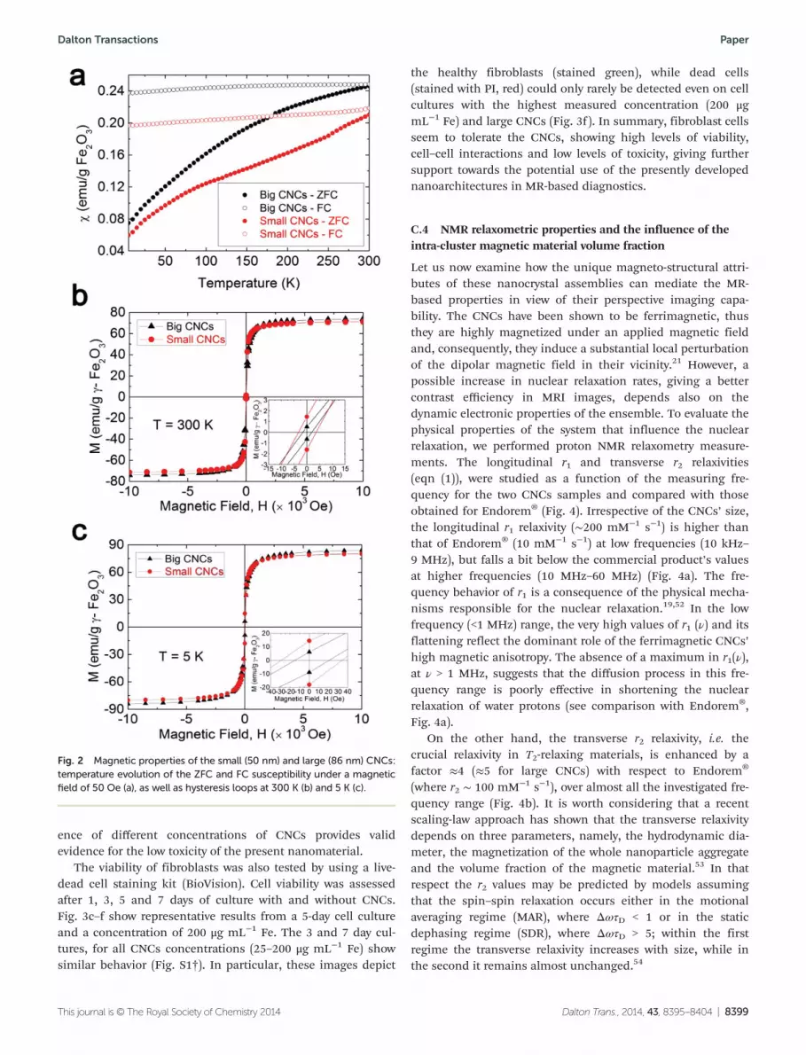
In Fig. 2b the room temperature hysteresis loops indicate thatthe dried powders of the CNCs are ferrimagnetic (FiM) withcoercive fields (Hc) of 5.4 and 2.1 Oe, for the small and largeCNCs, respectively, while the saturation magnetizations (MS)were 71.2 and 73.4 emu g−1 γ-Fe2O3, very close to the bulkvalue of 74 emu g−1 for γ-Fe2O3.
The hysteretic behavior in the temperature dependent zero-field and field-cooled (ZFC-FC) susceptibility (χ) indicatesblocked particles below 300 K for both cases (Fig. 2a). Since noobvious difference between the Mössbauer spectra of the driedpowder and those of the frozen solutions of the CNCs couldbe resolved, we conclude that the ferrimagnetism of theCNCs can be attributed to intra-cluster dipolar interactionsonly.49
C.3 Cytotoxicity
The CNCs appear to satisfy the first prerequisite of tailoredmagnetic properties for improved MR properties. In addition,the purposeful choice of PAA as the surfactant rendered the
assemblies negatively charged (z-potential was −65.4 ± 10.8and −50.0 ± 6.5 mV for the small and large CNCs, respectively)with good colloidal stability. However, a second condition fortheir implementation in MR-based diagnostics requires verifi-cation of their toxicity on living cells. For this purpose, murinefibroblast cells (NIH/3T3) were exposed to the CNCs tothoroughly check the cell-nanocluster interactions. An ATP-based Glo™ cell viability assay was used for the luminometricmeasurement of the NIH/3T3 cells’ growth. Since the ATP indi-cates the metabolically active cells, we quantified the numberof viable cells based on the concentration of the available ATP.The ATP concentrations, recorded over a regular period of cultiva-tion time (1, 3, 5 or 7 days), are plotted in Fig. 3a and b. A seriesof different concentrations (25, 50, 100 and 200 μg mL−1 Fe)was used for both small and large CNCs.
Specifically, the cell growth for different concentrations ofCNCs, for the same number of days of culture, displayed nostatistical difference when compared to the respective controlsamples (cells without CNCs). The significance levels (p) wereestimated using one way ANOVA followed by Tukey tests (p <0.05). Additionally, the difference in the size of the CNCs didnot affect the cell proliferation after 1, 3, 5 or 7 days of culture.The unaffected proliferation of NIH/3T3 cells under the pres-
Fig. 1 Low- (a, c) and high- (b, d) resolution TEM images of colloidal nanoclusters (CNCs) of maghemite. Insets of panels a and c show the size dis-tribution for CNCs with diameters of 50 and 86 nm. Insets of panels b and d show the calculated FFT patterns from an isolated cluster-like structure;the reflections are indexed on the basis of a cubic spinel (fcc) iron-oxide crystal structure. The corresponding zone-axis is marked by B.
Paper Dalton Transactions
8398 | Dalton Trans., 2014, 43, 8395–8404 This journal is © The Royal Society of Chemistry 2014
ence of different concentrations of CNCs provides validevidence for the low toxicity of the present nanomaterial.
The viability of fibroblasts was also tested by using a live-dead cell staining kit (BioVision). Cell viability was assessedafter 1, 3, 5 and 7 days of culture with and without CNCs.Fig. 3c–f show representative results from a 5-day cell cultureand a concentration of 200 μg mL−1 Fe. The 3 and 7 day cul-tures, for all CNCs concentrations (25–200 μg mL−1 Fe) showsimilar behavior (Fig. S1†). In particular, these images depict
the healthy fibroblasts (stained green), while dead cells(stained with PI, red) could only rarely be detected even on cellcultures with the highest measured concentration (200 μgmL−1 Fe) and large CNCs (Fig. 3f). In summary, fibroblast cellsseem to tolerate the CNCs, showing high levels of viability,cell–cell interactions and low levels of toxicity, giving furthersupport towards the potential use of the presently developednanoarchitectures in MR-based diagnostics.
C.4 NMR relaxometric properties and the influence of theintra-cluster magnetic material volume fraction
Let us now examine how the unique magneto-structural attri-butes of these nanocrystal assemblies can mediate the MR-based properties in view of their perspective imaging capa-bility. The CNCs have been shown to be ferrimagnetic, thusthey are highly magnetized under an applied magnetic fieldand, consequently, they induce a substantial local perturbationof the dipolar magnetic field in their vicinity.21 However, apossible increase in nuclear relaxation rates, giving a bettercontrast efficiency in MRI images, depends also on thedynamic electronic properties of the ensemble. To evaluate thephysical properties of the system that influence the nuclearrelaxation, we performed proton NMR relaxometry measure-ments. The longitudinal r1 and transverse r2 relaxivities(eqn (1)), were studied as a function of the measuring fre-quency for the two CNCs samples and compared with thoseobtained for Endorem® (Fig. 4). Irrespective of the CNCs’ size,the longitudinal r1 relaxivity (∼200 mM−1 s−1) is higher thanthat of Endorem® (10 mM−1 s−1) at low frequencies (10 kHz–9 MHz), but falls a bit below the commercial product’s valuesat higher frequencies (10 MHz–60 MHz) (Fig. 4a). The fre-quency behavior of r1 is a consequence of the physical mecha-nisms responsible for the nuclear relaxation.19,52 In the lowfrequency (<1 MHz) range, the very high values of r1 (ν) and itsflattening reflect the dominant role of the ferrimagnetic CNCs’high magnetic anisotropy. The absence of a maximum in r1(ν),at ν > 1 MHz, suggests that the diffusion process in this fre-quency range is poorly effective in shortening the nuclearrelaxation of water protons (see comparison with Endorem®,Fig. 4a).
On the other hand, the transverse r2 relaxivity, i.e. thecrucial relaxivity in T2-relaxing materials, is enhanced by afactor ≈4 (≈5 for large CNCs) with respect to Endorem®
(where r2 ∼ 100 mM−1 s−1), over almost all the investigated fre-quency range (Fig. 4b). It is worth considering that a recentscaling-law approach has shown that the transverse relaxivitydepends on three parameters, namely, the hydrodynamic dia-meter, the magnetization of the whole nanoparticle aggregateand the volume fraction of the magnetic material.53 In thatrespect the r2 values may be predicted by models assumingthat the spin–spin relaxation occurs either in the motionalaveraging regime (MAR), where ΔωτD < 1 or in the staticdephasing regime (SDR), where ΔωτD > 5; within the firstregime the transverse relaxivity increases with size, while inthe second it remains almost unchanged.54
Fig. 2 Magnetic properties of the small (50 nm) and large (86 nm) CNCs:temperature evolution of the ZFC and FC susceptibility under a magneticfield of 50 Oe (a), as well as hysteresis loops at 300 K (b) and 5 K (c).
Dalton Transactions Paper
This journal is © The Royal Society of Chemistry 2014 Dalton Trans., 2014, 43, 8395–8404 | 8399

To calculate the expected Larmor frequency shift we firstestimated the values of the intra-cluster volume fraction, φintra,employing the following relation (refer to S2†):
ργ-Fe2O3ð1� fmγ-Fe2O3Þ=ðρPAAfmγ-Fe2O3Þ ¼ ð1� φintraÞ=φintra ð2Þwhere ργ-Fe2O3
= 4900 kg m−3 is the density of bulk maghemite,ρPAA = 1150 kg m−3 is the density of polyacrylic acid, andfmγ-Fe2O3
is the weight fraction of the iron-oxide in a knownmass of dried nanocluster powder, estimated from the ther-mogravimetric (TGA) measurements.2 Eqn (2) yields φintra =0.60 and 0.72 for small and large CNCs, respectively.
Furthermore, the Larmor frequency shift is expressed as
Δω ¼ γμ0M�V=3 ð3Þ
where M*V is the normalized magnetization (see Table 1), while
the translational diffusion time τD is defined as τD = d2/4D,where d is the diameter of the CNCs and D = 3 × 10⁻⁹ m2 s−1 isthe water translational diffusion coefficient. τD amounts to∼0.2 and ∼0.6 μs, for the small and large CNCs.
Thus, finally:
ΔωτD ¼ 4:9 and ΔωτD ¼ 18:2 ð4Þfor small (50 nm) and large (86 nm) CNCs, respectively, indi-cating that the investigated samples fall at the extremes of theSDR regime (5 < ΔωτD < 20). We note that, within the SDR
regime, the straddling water molecules feel a relatively con-stant dipolar magnetic field in their vicinity and it is not sur-prising that the experimentally observed r2(ν) are marginallydependent on the CNCs’ average size. The small difference inthe sample volume magnetization, M*
V, and φintra, is anadditional reason behind the little variation at ν > 4 MHz(Fig. 4b).
The result of eqn (4) suggests that the transverse relaxivity(r2) values for both CNCs samples may be predicted by modelsassuming that the spin–spin relaxation occurs in the SDRregime.
Firstly, if we calculate the expected r2 value with the theore-tical (approximate) expression found in ref. 53,
rtheo2 ¼ 2πγPμ0νmatM*V=9
ffiffiffi
3p ð5Þ
where νmat is the volume fraction to iron concentration conver-sion factor (νmat = 1.57 × 10⁻⁵ m3 mol−1 for maghemite), andcompare it to the experimental result rexp2 , we observe a reason-able agreement, since rtheo2 /rexp2 = 1.11 and 1.10 for the smalland large CNCs, respectively. Conversely, the expected normal-ized relaxivity r*2 = r2φintra/M
*v2 for 50 nm and 86 nm clusters is
about 2 × 10⁻⁸ and 8 × 10⁻⁸ s−1 mM−1 m2 A−2, respectively, butthe real values characterizing the two samples are 5.5 × 10⁻⁹
and 5.4 × 10⁻⁹ s−1 mM−1 m2 A−2, i.e. an order of magnitudedifferent. Such a marked difference evidently hints at poor
Fig. 3 Cytotoxicity profiles of the 50 (a) and 86 nm (b) CNCs samples. NIH/3T3 fibroblast cells were incubated with CNCs at the indicated doses for 1,3, 5 and 7 days. An ATP-Glo TM Bioluminometric cell viability assay kit was used for luminometric measurement of the fibroblasts’ growth. Each his-togram reflects the luminescence (mean value in RLU – relative luminescence units, ±Standard Deviation – SD) as derived from three independentexperiments. Fluorescence microscopy images of live (green) and dead (orange-red) fibroblasts, cultured without CNCs (control) (c, e) and withsmall (d) or large (f ) CNCs, after 5 days of culture.
Paper Dalton Transactions
8400 | Dalton Trans., 2014, 43, 8395–8404 This journal is © The Royal Society of Chemistry 2014

agreement between theory and experiment; however, one mayargue that samples located at the edges of the SDR regime areprone to misbehavior: the 50 nm sample is neither in themotional averaging regime (MAR) nor completely in the SDRrange, i.e. in a region not described by any existing model; the86 nm sample is very close to the ΔωτD ∼ 20 limit, where therefocusing pulses used in the T2 sequence become effective, aneffect which is not accounted for by the SDR model. The
experimental evidence proves, however, that Endorem® is notaffected by these issues at all, since for the commercial com-pound ΔωτD = 4.4 and r*2 = 7.8 × 10⁻⁸ s−1 mM−1 m2 A−2, whichis perfectly in line with the theory.
Finally, a justification for the significant (4 to 5 times)increase of r2 with respect to that of Endorem®, can beinferred by considering the almost two-times higher volumemagnetization in conjunction with the roughly three-timeslarger intra-cluster volume fraction (Endorem®: M*
V = 0.77 ×10⁵ A m−1, φintra = 0.23; Table 1).
The above discussed points suggest that the φintra
parameter has a crucial role in the improvement of MRproperties.
C.5 CNCs relaxivities and their relevance to other ferritenanoarchitectures
Two broad categories, depending on the nature of the mag-netic state, entailing either superparamagnetic (SPM) or ferri-magnetic (FiM) functional structures, were considered. A clearenhancement in the transverse relaxivity r2 was shown againstindividual SPM nanocrystals with different capping agentssuch as polymers,19 dendrons55 or DHAA.56 Their size-onlydependent relaxivity in the MAR regime renders them lessefficient T2-relaxation agents compared to the CNCs, whoseimproved faster reduction of T2 is determined by the synergeticaction of MS, size and φintra.
53 However, a progressive increasein the size of individual nanoparticles can lead to FiMnanoarchitectures which may provide enhanced relaxivitieswith the advantages of the SDR regime. The only FiM systemsof large size and enhanced magnetic anisotropy are the iron-oxide nanocubes, with an edge length of 22 nm, encapsulatedin PEG-phospholipids, with good colloidal stability; thesesystems have shown a value r2 = 761 mM−1 s−1, higher thanour system57 (see Table S2† for a more comprehensivecomparison).
Alternatively, controlled aggregation of nanoparticles in sec-ondary structures can further boost the relaxivities with anoutcome equivalent to SDR. In this respect, to the best of ourknowledge, the only ferrimagnetic secondary structurethat has been studied so far for its outstanding visualizing(T2-weighted contrast properties) as well as drug-deliveringactions, is a newly designed liposome-encapsulated magneticnanoparticle cluster.58 This system shows an unprecedented r2of 1286 mM−1 s−1 but at a higher magnetic field of 2.35 T,thus leaving the CNCs a valid alternative.
Nevertheless, the strategy to embed nanoparticles inmatrices/polymers44,59 or aggregating them in well-controlledmorphologies32,33,36,40,45,46,60–62 can also afford SPM nanoarchi-tectures and shows relaxivities explainable within the SDRregime. In this case, our system shows larger r2 (405 and510 mM−1 s−1 for the small and large assemblies, respectively)compared to most of the SPM cluster-type particles, such asthose capped with citrate,33 PVP,36 polystyrene,63 Dextran,44
PEG,39 (Mal)mPEG-PLA copolymer,46 amine,32 and TREG.62 Anexception is the SPM magnetite-based cluster-analogue ofhigher r2 relaxivity, which is covered by the same surfactant as
Fig. 4 Room temperature longitudinal r1 (a) and transverse r2 (b) relax-ivities as a function of proton Larmor frequency (or the external mag-netic field, H) for the 50 (circles) and 86 nm (triangles) CNCs. Thecorresponding data for the until recently commercial contrast agentEndorem® (squares) are also shown.
Table 1 Parameters utilized for the calculation of the intra-clustermagnetic material volume fraction and the determination of the trans-verse relaxivity regime to which CNCs belong. These entail: Dhydro, thehydrodynamic diameter of the CNCs; fmγ-Fe2O3
, the weight fraction of theiron-oxide in a known mass of dried nanocluster powder as derived bythe thermogravimetric (TGA) measurements; φintra, the intra-clustervolume fraction of the magnetic material; MS, the saturation magnetiza-tion of the CNCs at room temperature; M*
V, the volume magnetization;Δω, the Larmor frequency shift; τD, the translational diffusion time. Note:parameter values for Endorem® tabulated in this table were taken fromref. 53
SampleDhydro(nm)
fmγ-Fe2O3
(%) φintra
M*V
(105 A m−1) ΔωτD Regime
Endorem® 80 63.8 0.23 0.77 4.4 SDR50.2 nm CNCs 78.6 86.8 0.60 1.95 4.9 SDR85.6 nm CNCs 121.8 92.1 0.72 2.37 18.2 SDR
Dalton Transactions Paper
This journal is © The Royal Society of Chemistry 2014 Dalton Trans., 2014, 43, 8395–8404 | 8401
ours, but synthesized in an autoclave, through a polyol processfor an extended period of time (12 h).60,61 Amongst them,those clusters of diameter 34 and 63 nm (with r2 of 540 and630 mM−1 s−1, correspondingly) have a φintra of 0.30 and 0.50and MV of 1.23 × 10⁵ and 1.79 × 10⁵ A m−1, respectively, bothsmaller than the value for the CNCs of the present study. Theenhanced values of their relaxivities suggest that r2 is mediatednot only by the latter two magneto-structural parameters, butit must also be a function of the surface properties of theassemblies (e.g. thickness of surface coating, L, over the hydro-dynamic diameter, Dhydro, of the assembly), as the interactionsbetween the water protons and the assemblies could occurprimarily on their surface.64,65
A further observation is that, in general, r2 can be thoughtto originate from the variation in the diffusion length of thewater molecules, relative to the dimensions of the surfactant-coordinated inorganic entities themselves. Then, the r2 relaxi-vity is expected to decrease as the molecular weight of thecapping agent increases,64 but this appears not to be the casein our system since the clusters are capped with a surfactant(PAA) of lower molecular weight (Mw = 1800 versus Mw =500060). The possible lower steric hindrance amongst shorterchains, allows for a higher packing density of a larger numberof polyacrylate chains to coordinate the nanoclusters’ surface.In turn, this complies with the higher z-potential attained bythe CNCs in the present case (−65.4 and −50.0 mV for thesmall and the large clusters, compared to −38 and −43 mV, for34 and 63 nm clusters by Li et al.60). Such a dense surface cov-erage may impede the penetration ability (diffusivity) of thewater molecules. As a consequence, the water-proton nuclearmoments are less strongly perturbed by the local magneticfield generated by the inorganic entities at the surface of thenanoclusters themselves which generates a lower r2.
Other differences may also be attributed to the likely modi-fication of the chemical bonding (entailing the surface functio-nalised nanocrystals of the assembly) in the two cases. Thisbonding can mediate the particles’ magnetic anisotropy oreven enhance their surface spin disorder, thus having animpact on lowering the relaxivity values.66,67
D. Conclusions
The suggested colloidal chemistry pathway allows ferrimag-netic assemblies of crystallographically oriented primary nano-crystals of maghemite to be obtained. They are not only well-dispersed in aqueous media and have low cytotoxicity, butshow an enhanced transverse NMR relaxivity, r2. The resultssuggest that the pronounced enhancements in the magnitudeof r2 have their origin in the ability to guide the assembly ofindividual nanocrystals so that they become crystallographi-cally oriented, allowing for an increased magnetic materialvolume fraction (φintra) within the larger-grown nanoclusterentities. The favourable high intra-cluster volume fraction ofthe magnetic moments appears to make them efficientlycoupled to each other in the CNCs. In turn, this permits a
coherent and intense perturbation of the dipolar magneticfield in the near vicinity of the straddling water molecules, acondition that enhances the relaxation of the associatedproton nuclear spins. The polyacrylate surface coordinatinggroups of the CNCs further mediate the efficient penetrationof the water protons and in conjunction with the surface spindisorder of the inorganic nanocrystals determine the magni-tude of r2. Such nanoarchitectures appear to have potential forimproved diagnostic quality in T2-weighted MR imagingtechniques.
Finally, the observations presented in section C.4 lead tothe conclusion that a definitive theory for r2 relaxivity still hasto be formulated, and further effort needs to be undertaken toaccount for overlooked effects that may indeed have a strongimpact on the relaxometric properties of a nanoparticle-basedcontrast agent. Specifically, we would like to highlight theexistence of classes of compounds that do not strictly obey theuniversal scaling law envisioned by Vuong et al.53 for clusters,since when ΔωτD is at the lower and upper limits of the SDRrange, predictions based on ref. 53 cannot be reliable.
Acknowledgements
This work was supported by the European Commissionthrough the Marie-Curie Transfer of Knowledge programNANOTAIL (grant no. MTKD-CT-2006-042459). SKPV, KVT, FO,LB and AL thank the Italian projects INSTM-Regione lombar-dia “Mag-NANO”, FIRB “Riname” and Fondazione Cariplo no.2010-0612. We thank Tomas Orlando and Paolo Arosio forexperimental help.
Notes and references
1 D. V. Talapin, J. S. Lee, M. V. Kovalenko andE. V. Shevchenko, Chem. Rev., 2010, 110, 389–458.
2 P. Zrazhevskiy, M. Sena and X. Gao, Chem. Soc. Rev., 2010,39, 4326–4354.
3 L. Carbone and P. D. Cozzoli, Nano Today, 2010, 5, 449–493.
4 N. C. Bigall, W. J. Parak and D. Dorfs, Nano Today, 2012, 7,282–296.
5 C. G. Li, Y. Zhao, F. F. Li, Z. Shi and S. H. Feng, Chem.Mater., 2010, 22, 1901–1907.
6 X. L. Hu, J. M. Gong, L. Z. Zhang and J. C. Yu, Adv. Mater.,2008, 20, 4845–7850.
7 X. L. Fang, C. Chen, M. S. Jin, Q. Kuang, Z. X. Xie, S. Y. Xie,R. B. Huang and L. S. Zheng, J. Mater. Chem., 2009, 19,6154–6160.
8 Y. Zhou and M. Antonietti, J. Am. Chem. Soc., 2003, 125,14960–14961.
9 S. Xuan, Y. J. Wang, J. C. Yu and K. C. Leung, Chem. Mater.,2009, 21, 5079–5087.
10 M. Grzelczak, J. Vermant, E. M. Furst and L. M. Liz-Marzán, ACS Nano, 2010, 4, 3591–3605.
Paper Dalton Transactions
8402 | Dalton Trans., 2014, 43, 8395–8404 This journal is © The Royal Society of Chemistry 2014

11 K. H. Su, Q. H. Wei, X. Zhang, J. J. Mock, D. R. Smith andS. Schultz, Nano Lett., 2003, 3, 1087–1090.
12 J. Lee, A. O. Govorov and N. A. Kotov, Nano Lett., 2005, 5,2063–2069.
13 A. Kostopoulou, F. Thétiot, I. Tsiaoussis, M. Androulidaki,P. D. Cozzoli and A. Lappas, Chem. Mater., 2012, 24, 2722–2732.
14 Z. Nie, A. Petukhova and E. Kumacheva, Nat. Nanotechnol.,2010, 5, 15–25.
15 H. B. Na, I. C. Song and T. Hyeon, Adv. Mater., 2009, 21,2133–2148.
16 M. Lévy, C. Wilhelm, M. Devaud, P. Levitz and F. Gazeau,Contrast Media Mol. Imaging, 2012, 7, 373–383.
17 C. S. S. R. Kumar and F. Mohammad, Adv. Drug DeliveryRev., 2011, 63, 789–808.
18 S. R. Deka, A. Quarta, R. Di Corato, A. Riedinger,R. Cingolani and T. Pellegrino, Nanoscale, 2011, 3, 619–629.
19 M. F. Casula, P. Floris, C. Innocenti, A. Lascialfari,M. Marinone, M. Corti, R. A. Sperling, W. J. Parak andC. Sangregorio, Chem. Mater., 2010, 22, 1739–1748.
20 C. W. Jung and P. Jacobs, Magn. Reson. Imaging, 1995, 13,661–674.
21 J. Cheon and J. H. Lee, Acc. Chem. Res., 2008, 41, 1630–1640.
22 U. Jeong, X. W. Teng, Y. Wang, H. Yang and Y. N. Xia, Adv.Mater., 2007, 19, 33–60.
23 A. Figuerola, R. Di Corato, L. Manna and T. Pellegrino,Pharmacol. Res., 2010, 62, 126–143.
24 H. Amiri, M. Mahmoudi and A. Lascialfari, Nanoscale,2011, 3, 1022–1030.
25 B. A. Moffat, G. R. Reddy, P. McConville, D. E. Hall,T. L. Chenevert, R. R. Kopelman, M. Philbert,R. Weissleder, A. Rehemtulla and B. D. Ross, Mol. Imaging,2003, 2, 324–332.
26 E. Taboada, R. Solanas, E. Rodriguez, R. Weissleder andA. Roig, Adv. Funct. Mater., 2009, 19, 2319–2324.
27 B. A. Larsen, M. A. Haag, N. J. Serkova, K. R. Shroyer andC. R. Stoldt, Nanotechnology, 2008, 19, 265102.
28 J. F. Berret, N. Schonbeck, F. Gazeau, D. El Kharrat,O. Sandre, A. Vacher and M. Airiau, J. Am. Chem. Soc., 2006,128, 1755–1761.
29 R. Di Corato, P. Piacenza, M. Musarò, R. Buonsanti,P. D. Cozzoli, M. Zambianchi, G. Barbarella, R. Cingolani,L. Manna and T. Pellegrino, Macromol. Biosci., 2009, 9,952–958.
30 R. Di Corato, N. C. Bigall, A. Ragusa, D. Dorfs, A. Genovese,R. Marotta, L. Manna and T. Pellegrino, ACS Nano, 2011, 5,1109–1121.
31 T. Isojima, S. K. Suh, J. B. Vander Sande and T. AlanHatton, Langmuir, 2009, 25, 8292–8298.
32 K. C. Barick, M. Aslam, Y. P. Lin, D. Bahadur, P. V. Prasadand V. P. Dravid, J. Mater. Chem., 2009, 19, 7023–7029.
33 L. Lartigue, P. Hugounenq, D. Alloyeau, S. P. Clarke,M. Lévy, J. C. Bacri, R. Bazzi, D. F. Brougham, C. Wilhelmand F. Gazeau, ACS Nano, 2012, 6, 10935–10949.
34 F. Dong, W. Guo, J. H. Bae, S. H. Kim and C. S. Ha, Chem. -Eur. J., 2011, 17, 12802–12808.
35 C. Cheng, Y. Wen, X. Xu and H. Gu, J. Mater. Chem., 2009,19, 8782–8788.
36 S. Xuan, F. Wang, Y. X. Wang, J. C. Yu and K. C. F. Leung,J. Mater. Chem., 2010, 20, 5086–5094.
37 M. A. Daniele, M. L. Shaughnessy, R. Roeder, A. Childress,Y. P. Bandera and S. Foulger, ACS Nano, 2013, 7, 203–213.
38 C. Cheng, F. Xu and H. Gu, New J. Chem., 2011, 35, 1072–1079.
39 F. Hu, K. W. MacRenaris, E. A. Waters, E. A. Schultz-Sikma,A. L. Eckermann and T. J. Meade, Chem. Commun., 2010,46, 73–75.
40 J. Cha, Y. S. Kwon, T. J. Yoon and J. K. Lee, Chem.Commun., 2013, 49, 457–459.
41 S. M. Lai, J. K. Hsiao, H. P. Yu, C. W. Lu, C. C. Huang,M. J. Shieh and P. S. Lai, J. Mater. Chem., 2012, 22, 15160–15167.
42 M. Mahmoudi, A. Simchi, M. Imani, A. S. Milani andP. Stroeve, J. Phys. Chem. B, 2008, 112, 14470–14481.
43 B. P. Jia and L. Gao, J. Phys. Chem. C, 2008, 112, 666–671.44 E. K. Lim, E. Jang, B. Kim, J. Choi, K. Lee, J. S. Suh,
Y. M. Huh and S. Haam, J. Mater. Chem., 2011, 21, 12473–12478.
45 N. Pothayee, S. Balasubramaniam, N. Pothayee, N. Jain,N. Hu, Y. Lin, R. M. Davis, N. Sriranganathan,A. P. Koretsky and J. S. Riffle, J. Mater. Chem. B, 2013, 1,1142–1149.
46 C. Zhang, X. Xie, S. Liang, M. Li, Y. Liu and H. Gu, Nano-medicine, 2012, 8, 996–1006.
47 H. Chen, J. Yeh, L. Wang, H. Khurshid, N. Peng, A. Y. Wangand H. Mao, Nano Res., 2010, 3, 852–862.
48 J. Ge, Y. Hu, M. Biasini, W. P. Beyermann and Y. Yin,Angew. Chem., Int. Ed., 2007, 46, 4342–4345.
49 A. Kostopoulou, K. Brintakis, M. Vasilakaki,K. N. Trohidou, A. P. Douvalis, A. Lascialfari, L. Manna andA. Lappas, Nanoscale, 2014, 6, 3764–3776.
50 A. Ranella, M. Barberoglou, S. Bakogianni, C. Fotakis andE. Stratakis, Acta Biomater., 2010, 6, 2711–2720.
51 S. Psycharakis, A. Tosca, V. Melissinaki, A. Giakoumaki andA. Ranella, Biomed. Mater., 2011, 6, 045008.
52 S. Laurent, D. Forge, M. Port, A. Roch, C. Robic, L. V. Elstand R. N. Muller, Chem. Rev., 2008, 108, 2064–2110.
53 Q. L. Vuong, J. F. Berret, J. Fresnais, Y. Gossuin andO. Sandre, Adv. Healthcare Mater., 2012, 1, 502–512.
54 E. Pöselt, H. Kloust, U. Tromsdorf, M. Janschel, C. Hahn,C. Maßlo and H. Weller, ACS Nano, 2012, 6, 1619–1624.
55 B. Basly, D. Felder-Flesch, P. Perriat, C. Billotey, J. Taleb,G. Pourroy and S. Begin-Colin, Chem. Commun., 2010, 46,985–987.
56 L. Xiao, J. Li, D. F. Brougham, E. K. Fox, N. Feliu,A. Bushmelev, A. Schmidt, N. Mertens, F. Kiessling,M. Valldor, B. Fadeel and S. Mathur, ACS Nano, 2011, 5,6315–6324.
57 N. Lee, Y. Choi, Y. Lee, M. Park, W. K. Moon, S. H. Choiand T. Hyeon, Nano Lett., 2012, 12, 3127–3131.
Dalton Transactions Paper
This journal is © The Royal Society of Chemistry 2014 Dalton Trans., 2014, 43, 8395–8404 | 8403

58 G. Mikhaylov, U. Mikac, A. A. Magaeva, V. I. Itin,E. P. Naiden, I. Psakhye, L. Babes, T. Reinheckel, C. Peters,R. Zeiser, M. Bogyo, V. Turk, S. G. Psakhye, B. Turk andO. Vasiljeva, Nat. Nanotechnol., 2011, 6, 594–602.
59 M. S. Martina, J. P. Fortin, C. Menager, O. Clement,G. Barratt, C. Grabielle-Madelmont, F. Gazeau, V. Cabuiland S. Lesieur, J. Am. Chem. Soc., 2005, 127, 10676–10685.
60 M. Li, H. Gu and C. Zhang, Nanoscale Res. Lett., 2012, 7,204.
61 F. Xu, C. Cheng, D. X. Chen and H. Gu, ChemPhysChem,2012, 13, 336–341.
62 D. Maity, P. Chandrasekharan, P. Pradhan, K. H. Chuang,J. M. Xue, S. S. Feng and J. Ding, J. Mater. Chem., 2011, 21,14717–14724.
63 Y. T. Wang, F. H. Xu, C. Zhang, D. Lei, Y. Tang, H. Xu,Z. Zhang, H. Lu, X. Du and G. Y. Yang, Nanomedicine, 2011,7, 1009–1019.
64 S. Tong, S. J. Hou, Z. L. Zheng, J. Zhou and G. Bao, NanoLett., 2010, 10, 4607–4613.
65 D. Yoo, J. H. Lee, T. H. Shin and J. Cheon, Acc. Chem. Res.,2011, 44, 863–874.
66 L. Bordonali, T. Kalaivani, K. P. V. Sabareesh, C. Innocenti,E. Fantechi, C. Sangregorio, M. F. Casula, L. Lartigue,J. Larionova, Y. Guari, M. Corti, P. Arosio and A. Lascialfari,J. Phys.: Condens. Matter, 2013, 25, 066008.
67 H. Duan, M. Kuang, X. Wang, Y. A. Wang, H. Maoand S. Nie, J. Phys. Chem. C, 2008, 112, 8127–8131.
Paper Dalton Transactions
8404 | Dalton Trans., 2014, 43, 8395–8404 This journal is © The Royal Society of Chemistry 2014


