Embed Size (px)
Citation preview
Cell, Vol. 102, 753–763, September 15, 2000, Copyright 2000 by Cell Press
Combinatorial Roles of the Nuclear ReceptorCorepressor in Transcription and Development
through ligand-dependent activation and through activerepression by unliganded nuclear receptors (reviewedin McKenna et al., 1999; Glass and Rosenfeld, 2000; Hu
Kristen Jepsen,1,2 Ola Hermanson,1
Thandi M. Onami,2,8 Anatoli S. Gleiberman,1
Victoria Lunyak,1 Robert J. McEvilly,1
and Lazar, 2000). Investigation of active repression byRiki Kurokawa,3 Vivek Kumar,1 Forrest Liu,1
T3R and RAR led to the identification of the nuclearEdward Seto,4 Stephen M. Hedrick,2
receptor corepressor (N-CoR) (Horlein et al., 1995) andGail Mandel,5 Christopher K. Glass,3
the closely related factor, silencing mediator for retinoicDavid W. Rose,6 and Michael G. Rosenfeld1,7
acid and thyroid hormone receptors (SMRT) (Chen and1 Howard Hughes Medical InstituteEvans, 1995). N-CoR and SMRT both contain a con-2 Department of Biologyserved bipartite nuclear receptor interaction domain3 Department of Cellular and Molecular Medicine(Seol et al., 1996; Zamir et al., 1996) and three indepen-University of California, San Diegodent repressor domains that are capable of transferringLa Jolla, California 92093their active repression function to a heterologous DNA4 H. Lee Moffitt Cancer Centerbinding domain.
and Research Institute While steroid hormone receptors do not appear toUniversity of South Florida interact with N-CoR or SMRT in the presence or absenceTampa, Florida 33612 of agonists, both the estrogen receptor (ER) and the5 Howard Hughes Medical Institute progesterone receptor (PR) can interact with these core-State University of New York, Stony Brook pressors in the presence of their respective antagonistsStoney Brook, New York 11794 (Xu et al., 1996; Jackson et al., 1997; Smith et al., 1997;6 Division of Endocrinology and Metabolism Lavinsky et al., 1998). The ability of N-CoR or SMRTUniversity of California, San Diego to serve as corepressors has also been suggested forLa Jolla, California 92093 several other members of the nuclear receptor super-
family (reviewed in Glass and Rosenfeld, 2000). In addi-tion, N-CoR and SMRT have been implicated as core-pressors for a variety of unrelated transcription factors,Summaryincluding MAD (Heinzel et al., 1997), CBF1/RBP-Jkappa/Su(H) (Kao et al., 1998), and homeodomain factors (XuTranscriptional repression plays crucial roles in di-et al., 1998).verse aspects of metazoan development, implying
Although N-CoR and SMRT can interact with mSin3critical regulatory roles for corepressors such as(Alland et al., 1997; Heinzel et al., 1997; Nagy et al., 1997)N-CoR and SMRT. Altered patterns of transcription inand with certain HDAC proteins, including HDAC 4, 5,tissues and cells derived from N-CoR gene–deletedand 7 (Huang et al., 2000; Kao et al., 2000), neithermice and the resulting block at specific points in CNS,N-CoR nor SMRT are components of the mSin3 complex
erythrocyte, and thymocyte development indicated(Hassig et al., 1997; Laherty et al., 1998) or of the HDAC-
that N-CoR was a required component of short-term containing NuRD complex (Xue et al., 1998). In fact,active repression by nuclear receptors and MAD and purification of a SMRT complex (Guenther et al., 2000)of a subset of long-term repression events mediated or HDAC3 complex (Wen et al., 2000) reveals that N-CoRby REST/NRSF. Unexpectedly, N-CoR and a specific and SMRT form a stable complex with HDAC3 and atdeacetylase were also required for transcriptional ac- least one additional protein, TBL1 (Guenther et al., 2000).tivation of one class of retinoic acid response element. These data imply either a redundancy or combinatorialTogether, these findings suggest that specific combi- usage of N-CoR-associated deacetylases in active re-nations of corepressors and histone deacetylases me- pression.diate the gene-specific actions of DNA-bound repres- In this manuscript, we investigated the biologicalsors in development of multiple organ systems. role of the corepressor N-CoR in vivo and found that
N-CoR2/2 embryos exhibit defects in developmental pro-gression of specific erythrocyte, thymocyte, and neuralIntroductionevents. N-CoR proves to be required for repression byT3R and RAR, for the function of ER antagonists, and forActive repression of gene expression by sequence-spe-the repressive actions of several other classes of DNAcific transcription factors plays a critical role in the regu-binding repressors. We have also established a role forlation of diverse biological processes, including cell pro-N-CoR in long-term repression mediated by repressorliferation, development, and homeostasis (Mannervik etelement silencing transcription factor/neuron-restrictiveal., 1999). Nuclear receptors, including the retinoic acidsilencer factor (REST/NRSF). Additionally, our data sug-(RAR) and thyroid hormone receptors (T3R), are essentialgest that in particular contexts, N-CoR, with HDAC3,for the regulation of development and homeostasis bothhas a role in activation of gene expression. Our findingsprovide evidence that N-CoR is a required develop-7 To whom correspondence should be addressed (e-mail: mrosenfeld@mental regulator, linking short- and long-term repressionucsd.edu).events on a subset of gene targets for nuclear receptors8 Present address: Emory Vaccine Center and Department of Micro-and other classes of transcription factors to specificbiology and Immunology, Emory University School of Medicine, At-
lanta, GA 30322. combinations of corepressors.
Cell754
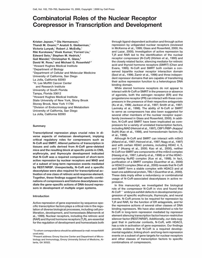
Figure 1. Targeted Deletion of the N-CoRGenomic Locus
(A) Schematic of interaction domains ofN-CoR. The sites of HDAC, mSin3 (SID), CBF/SuH, and nuclear receptor interaction do-mains (I and II) are indicated, as are the re-pression domains (RI, RII, RIII).(B) Homologous recombination replaced the59 SID with the PGKneopA cassette. Locationof 59 and 39 probes are shown.(C) After NheI digestion of genomic DNA frommice of each genotype, a 6.5 kb wild-typeallele or a 5.3 kb targeted allele was detectedby Southern blot analysis with the 59 probe.(D) Western blot analysis of protein fromMEFs of each genotype probed with anti-N-CoR antibody shows the absence of detect-able protein in cells from the N-CoR2/2
embryo.
Results half of the N-CoR2/2 embryos exhibited secondaryedema. Additionally, N-CoR2/2 embryos were on aver-age 80% the size of their wild-type and heterozygoteGeneration of N-CoR Gene–Deleted Mice
The murine N-CoR protein is encoded by a 7.4 kb mRNA littermates. The severity of the anemia and edema in-and is highly homologous to the related factor SMRT creased with age and appeared to be the ultimate cause(Figure 1A). In situ hybridization revealed that both genes of death. At E14.5, the hematocrit of the N-CoR2/2 em-were widely expressed but that gene-specific differ- bryos was severely reduced (14.5 6 0.83; mean 6 SEM)ences in their developmental expression patterns did relative to heterozygote or wild-type littermates (44 6 0.88)exist (data not shown). In order to characterize the bio- (Figure 2B). Peripheral blood smears from N-CoR2/2 em-logical role of N-CoR, we designed a targeting construct bryos showed fewer nonnucleated erythrocytes butto delete a portion of the N-CoR genomic locus. The many more nucleated erythroblasts than wild-type con-strategy employed replaced 3 kb of genomic sequence trols (Figure 2C).encoding the 59 Sin3 interaction domain (SID1) with the Primitive erythropoiesis is initiated in yolk sac bloodneomycin resistance gene (Figure 1B). Two clonal ES islands on E7 and results in the production of nucleatedcell lines heterozygous for the mutant allele were used red blood cells. Definitive erythropoiesis is character-to generate chimeric mouse strains from which we gen- ized by the production of mature, nonnucleated erythro-erated mice homozygous for the N-CoR mutation (Figure cytes, and normally occurs in the fetal liver beginning on1C). Disruption of the N-CoR gene was confirmed by
E12. N-CoR2/2 embryos survived until E15.5 and anemiathe absence of detectable transcripts and protein inwas observed only after E13.5, suggesting that thesecells derived from N-CoR2/2 embryos (Figure 1D andembryos have a defect in definitive rather than primitivedata not shown). In situ hybridization revealed no differ-erythropoiesis. Indeed, fetal livers of N-CoR2/2 embryosence in levels of SMRT transcripts between wild-typewere approximately one half the size of their wild-typeand N-CoR2/2 embryos (data not shown). The majority ofand heterozygous littermates (Figure 2D). N-CoR tran-N-CoR2/2 embryos died by day 15.5 of gestation (E15.5),scripts were highly expressed in E14.5 fetal liver, whilewith occasional embryos surviving 1–2 days longer.expression of SMRT transcripts was relatively low (Fig-ure 2E), suggesting that definitive erythropoiesis mightDefinitive Erythropoiesis Was Impairedbe sensitive to ablation of the N-CoR gene.in N-CoR2/2 Embryos
To determine the ability of N-CoR2/2 fetal liver progeni-N-CoR2/2 embryos were markedly pale from E13.5 (Fig-ure 2A), suggesting a severe anemia, and approximately tors to give rise to red cell colonies in vitro, an equal
N-CoR in Transcription and Development755
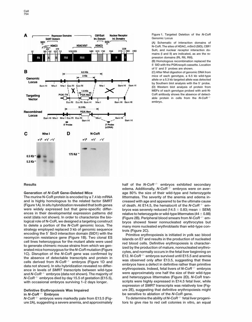
Figure 2. Block in Definitive Erythropoiesis inN-CoR2/2 Embryos
(A) Micrographs of wild-type and N-CoR2/2
E14.5 embryos with intact visceral yolk sacs,illustrating the severe anemia of N-CoR2/2
embryos.(B) Mean hematocrit of E14.5 wild-type andheterozygote animals (n 5 5) was 44.5, ascompared to a mean of 14.5 for N-CoR2/2
embryos (n 5 4).(C) Giemsa staining of E14.5 wild-type bloodshowed predominantly nonnucleated redcells, with occasional nucleated erythro-blasts and yolk sac erythrocytes. N-CoR2/2
blood contained fewer nonnucleated redcells, yolk sac erythrocytes, and a large num-ber of nucleated erythroblasts.(D) The average number of nucleated cellsin wild-type or heterozygous fetal livers wasz2-fold greater than in N-CoR2/2 fetal liver.(E) In situ hybridization of wild-type E14.5 fe-tal liver using 35S-labeled N-CoR and SMRTprobes showed that N-CoR transcripts werehighly expressed compared to SMRT tran-scripts.(F) In vitro differentiation of wild-type or het-erozygote and N-CoR2/2 E12.5 and E13.5 fetalliver cells. BFU-E cells give rise to red cellcolonies within 7–10 days and require Epo,stem cell factor, and IL-3 and/or GM-CSF.CFU-E cells require only Epo and give rise tored cell colonies within 2–3 days. N-CoR2/2
embryos showed a defect in BFU-E colonyformation.(G) Immunohistochemistry with anti-CA II an-tibody indicated increased CAII immunoreac-tivity in N-CoR2/2 fetal liver-derived cells.
number of fetal liver cells from embryos of each geno- difference in Epo receptor (EpoR) transcripts (data notshown).type were plated in methylcellulose media containing
growth factors appropriate for the development of either To assess a transcriptional role for N-CoR in erythro-cytes, we examined expression of carbonic anhydraseburst-forming unit-erythroid (BFU-E) or colony-forming
unit-erythroid (CFU-E) colonies (Figure 2F) (Gregory and II (CA II), which is upregulated in the presence of T3 andrepressed in erythrocytes transformed by the constitu-Eaves, 1978). As shown in Figure 2F, there was no appar-
ent difference in BFU-E colony formation by fetal liver tive repressor v-ErbA (Pain et al., 1990). An increase inCA II immunoreactivity was observed in erythroid cellscells derived from E12.5 N-CoR2/2, heterozygous, or
wild-type embryos. However, the ability of cells derived in N-CoR2/2 mice (Figure 2G), suggesting that in theabsence of N-CoR, the ability of T3R to mediate repres-from N-CoR2/2 fetal livers to form BFU-E colonies was
reduced 3-fold at E13.5, and this difference was even sion of specific target genes was lost.more pronounced by E14.5 (Figure 2F and data notshown). T Cell Development in N-CoR-Deficient Embryos
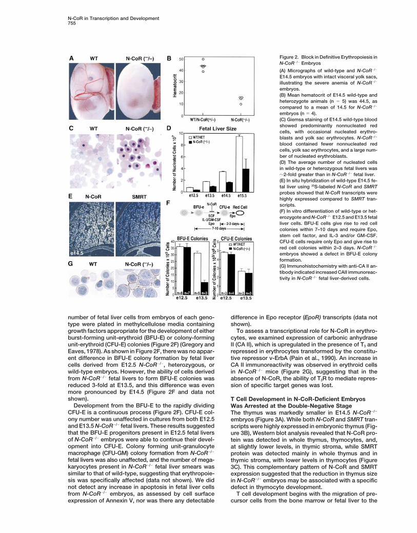
Development from the BFU-E to the rapidly dividing Was Arrested at the Double-Negative StageCFU-E is a continuous process (Figure 2F). CFU-E col- The thymus was markedly smaller in E14.5 N-CoR2/2
ony number was unaffected in cultures from both E12.5 embryos (Figure 3A). While both N-CoR and SMRT tran-and E13.5 N-CoR2/2 fetal livers. These results suggested scripts were highly expressed in embryonic thymus (Fig-that the BFU-E progenitors present in E12.5 fetal livers ure 3B), Western blot analysis revealed that N-CoR pro-of N-CoR2/2 embryos were able to continue their devel- tein was detected in whole thymus, thymocytes, and,opment into CFU-E. Colony forming unit-granulocyte at slightly lower levels, in thymic stroma, while SMRTmacrophage (CFU-GM) colony formation from N-CoR2/2 protein was detected mainly in whole thymus and infetal livers was also unaffected, and the number of mega- thymic stroma, with lower levels in thymocytes (Figurekaryocytes present in N-CoR2/2 fetal liver smears was 3C). This complementary pattern of N-CoR and SMRTsimilar to that of wild-type, suggesting that erythropoie- expression suggested that the reduction in thymus sizesis was specifically affected (data not shown). We did in N-CoR2/2 embryos may be associated with a specificnot detect any increase in apoptosis in fetal liver cells defect in thymocyte development.from N-CoR2/2 embryos, as assessed by cell surface T cell development begins with the migration of pre-
cursor cells from the bone marrow or fetal liver to theexpression of Annexin V, nor was there any detectable
Cell756
(DN) to CD41/CD81 (DP) and then analyzed by flow cy-tometry. The total number of live thymocytes, as as-sessed by forward scatter versus side scatter profiles,was reduced z8-fold in the cultured N-CoR2/2 thymii(Figure 3E). An increased rate of cell death, as evidencedby detection of a large number of dead cells in culturesfrom N-CoR2/2 thymii, may account for this reduction.Additionally, in contrast to wild-type and heterozygotelittermates, in which z58% of thymocytes were CD41/CD81 after 3 days of culture, thymii from N-CoR2/2 em-bryos were largely blocked in the CD42/CD82 stage,with only 3% of thymocytes expressing both CD4 andCD8 (Figure 3F). To determine whether this small numberof CD41/CD81 cells could progress to the single-posi-tive stage, thymii from E15.5 N-CoR2/2 embryos wereanalyzed after 5 days of FTOC, during which time differ-entiation to single-positive cells occurs. Thymocytesfrom E15.5 N-CoR2/2 embryos were able to progress tothe single-positive stage; however, thymocyte numberswere drastically reduced (data not shown).
The double-negative stage of thymocyte developmentcan be further subdivided on the basis of surface ex-pression of CD25 and CD44, in which maturation pro-ceeds through the CD252CD441 → CD251CD441 →CD251CD442 → CD252CD442 stages (Figure 3D). Tofurther analyze the CD42/CD82 compartment, surfaceexpression of CD25 and CD44 was determined. After72 hr in culture, there was an z2-fold increase (from31% to 54%) in the CD251/CD442 population, and anz3-fold decrease (from 38% to 13%) in the CD252/CD442 population in the thymocytes derived fromN-CoR2/2 embryos versus those from wild-type littermates(Figure 3F). These data indicated a block in thymocytedevelopment beyond the CD251/CD442 stage.
Studies in a number of mutant mouse strains haveFigure 3. Defect in T Cell Development in N-CoR2/2 Miceled to the proposal of a cascade of events that are(A) Hematoxylin and eosin (H&E)–stained sagittal sections of wild-required for progression in and beyond the CD251/type and N-CoR2/2 thymii on E14.5. Thymii of N-CoR2/2 embryos
were consistently 25%–30% the size of their wild-type and heterozy- CD442 stage. Briefly, cells must undergo productivegote littermates. Size marker 5 100 mm. V(D)J recombination of the T cell receptor (TCR) b locus,(B) In situ hybridization of wild-type E14.5 thymus sections using express TCRb polypeptides, assemble a TCRb-pTa/35S-labeled N-CoR and SMRT probes. Both N-CoR and SMRT tran- CD3 complex, and activate signaling cascades thoughtscripts were present in E14.5 thymus. to be dependent on protein tyrosine kinases of the Src(C) Western blot analysis of whole thymus, isolated thymocytes, (Lck; Fyn) and Syk (ZAP-70; Syk) families (reviewed inand thymic stroma. N-CoR protein was expressed in thymocytes,
Fischer and Malissen, 1998). The expression of TCRbwhole thymus, and at slightly lower levels in thymic stroma; in con-on the surface of CD251/CD442 cells did not differ be-trast, SMRT protein was expressed in whole thymus, thymic stroma,tween N-CoR2/2 embryos and wild-type or heterozygousand at lower levels in thymocytes.littermates, suggesting productive recombination of the(D) Schematic diagram of the progression from double-negative (DN)
to double-positive (DP) T cells, with stages affected in N-CoR2/2 TCRb locus (data not shown). CD3 expression was alsoembryos indicated. unaltered, further suggesting that N-CoR2/2 embryos(E) Recovery of live T cells after fetal thymic organ culture. were capable of assembling a pre-TCR complex (data(F) Flow cytometric analysis of viable cells from E14.5 fetal thymic not shown). Antibodies to CD3e can induce CD42/CD82
organ cultures after 3 days in culture. In the top panels, antibodies thymocytes to differentiate into CD41/CD81 cells in aagainst mouse CD4 and CD8 were used and showed a block from manner that is independent of TCRb expression (LeveltDN to DP T cells in thymii from N-CoR2/2 embryos. In the bottom
et al., 1993) but dependent on signal transduction bypanel, CD25 and CD44 expression was analyzed in DN cells andLck and Ras (Swat et al., 1996). Following a 72 hr treat-showed a block at the CD251CD442 stage in thymii from N-CoR2/2
ment with anti-CD3e antibody, N-CoR2/2 thymocytesembryos.progressed to the CD41/CD81 stage, and the numberof CD251/CD441 thymocytes was similar to that of het-
thymus, where maturation is characterized by a “double- erozygote and wild-type littermates (comparable to Fig-negative” stage (DN) where CD4 and CD8 coreceptors ure 3F, left-hand panels), indicating that events down-are absent, a “double-positive” stage (DP) where both stream of pre-TCR assembly were unaffected. Thecoreceptors are expressed, and finally a “single-posi- recovery of live cells from N-CoR2/2 thymii after FTOCtive” mature stage (SP) where either CD4 or CD8 core- with anti-CD3e antibody was also increased comparedceptor is expressed (Figure 3D). N-CoR2/2 thymii were to cultures without anti-CD3e treatment (data notanalyzed by fetal thymic organ cultures (FTOC) (Rams- shown). This is consistent with the hypothesis that adell, 1992). E14.5 fetal thymus lobes were cultured for specific block in progression underlies the decreased
number of cells found in cultured thymii from N-CoR2/272 hours to allow for differentiation from CD42/CD82
N-CoR in Transcription and Development757
embryos. Therefore, N-CoR expression was required forprogression of thymocyte development from the CD251/CD442 stage to the double-positive stage.
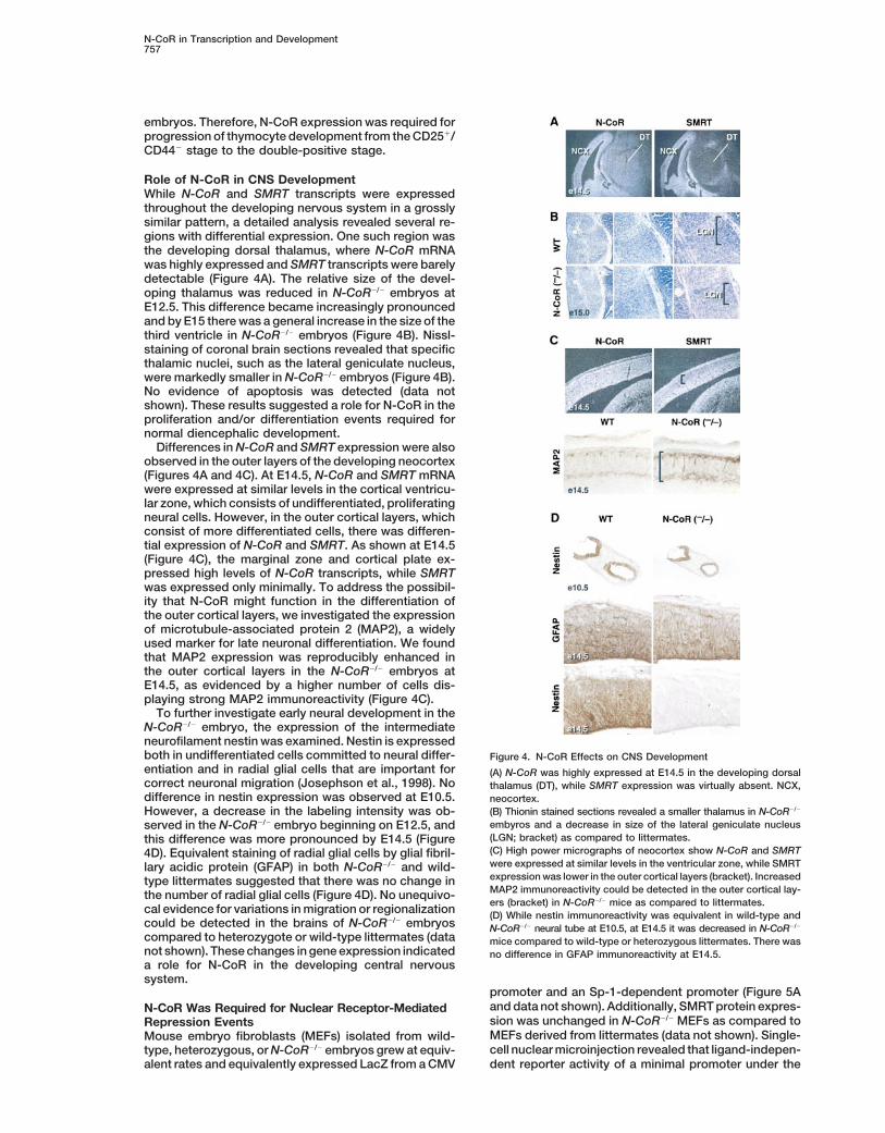
Role of N-CoR in CNS DevelopmentWhile N-CoR and SMRT transcripts were expressedthroughout the developing nervous system in a grosslysimilar pattern, a detailed analysis revealed several re-gions with differential expression. One such region wasthe developing dorsal thalamus, where N-CoR mRNAwas highly expressed and SMRT transcripts were barelydetectable (Figure 4A). The relative size of the devel-oping thalamus was reduced in N-CoR2/2 embryos atE12.5. This difference became increasingly pronouncedand by E15 there was a general increase in the size of thethird ventricle in N-CoR2/2 embryos (Figure 4B). Nissl-staining of coronal brain sections revealed that specificthalamic nuclei, such as the lateral geniculate nucleus,were markedly smaller in N-CoR2/2 embryos (Figure 4B).No evidence of apoptosis was detected (data notshown). These results suggested a role for N-CoR in theproliferation and/or differentiation events required fornormal diencephalic development.
Differences in N-CoR and SMRT expression were alsoobserved in the outer layers of the developing neocortex(Figures 4A and 4C). At E14.5, N-CoR and SMRT mRNAwere expressed at similar levels in the cortical ventricu-lar zone, which consists of undifferentiated, proliferatingneural cells. However, in the outer cortical layers, whichconsist of more differentiated cells, there was differen-tial expression of N-CoR and SMRT. As shown at E14.5(Figure 4C), the marginal zone and cortical plate ex-pressed high levels of N-CoR transcripts, while SMRTwas expressed only minimally. To address the possibil-ity that N-CoR might function in the differentiation ofthe outer cortical layers, we investigated the expressionof microtubule-associated protein 2 (MAP2), a widelyused marker for late neuronal differentiation. We foundthat MAP2 expression was reproducibly enhanced inthe outer cortical layers in the N-CoR2/2 embryos atE14.5, as evidenced by a higher number of cells dis-playing strong MAP2 immunoreactivity (Figure 4C).
To further investigate early neural development in theN-CoR2/2 embryo, the expression of the intermediateneurofilament nestin was examined. Nestin is expressedboth in undifferentiated cells committed to neural differ- Figure 4. N-CoR Effects on CNS Developmententiation and in radial glial cells that are important for (A) N-CoR was highly expressed at E14.5 in the developing dorsalcorrect neuronal migration (Josephson et al., 1998). No thalamus (DT), while SMRT expression was virtually absent. NCX,difference in nestin expression was observed at E10.5. neocortex.
(B) Thionin stained sections revealed a smaller thalamus in N-CoR2/2However, a decrease in the labeling intensity was ob-embyros and a decrease in size of the lateral geniculate nucleusserved in the N-CoR2/2 embryo beginning on E12.5, and(LGN; bracket) as compared to littermates.this difference was more pronounced by E14.5 (Figure(C) High power micrographs of neocortex show N-CoR and SMRT4D). Equivalent staining of radial glial cells by glial fibril-were expressed at similar levels in the ventricular zone, while SMRTlary acidic protein (GFAP) in both N-CoR2/2 and wild-expression was lower in the outer cortical layers (bracket). Increasedtype littermates suggested that there was no change inMAP2 immunoreactivity could be detected in the outer cortical lay-the number of radial glial cells (Figure 4D). No unequivo-ers (bracket) in N-CoR2/2 mice as compared to littermates.
cal evidence for variations in migration or regionalization (D) While nestin immunoreactivity was equivalent in wild-type andcould be detected in the brains of N-CoR2/2 embryos N-CoR2/2 neural tube at E10.5, at E14.5 it was decreased in N-CoR2/2
compared to heterozygote or wild-type littermates (data mice compared to wild-type or heterozygous littermates. There wasnot shown). These changes in gene expression indicated no difference in GFAP immunoreactivity at E14.5.a role for N-CoR in the developing central nervoussystem.
promoter and an Sp-1-dependent promoter (Figure 5Aand data not shown). Additionally, SMRT protein expres-N-CoR Was Required for Nuclear Receptor-Mediatedsion was unchanged in N-CoR2/2 MEFs as compared toRepression EventsMEFs derived from littermates (data not shown). Single-Mouse embryo fibroblasts (MEFs) isolated from wild-cell nuclear microinjection revealed that ligand-indepen-type, heterozygous, or N-CoR2/2 embryos grew at equiv-
alent rates and equivalently expressed LacZ from a CMV dent reporter activity of a minimal promoter under the
Cell758
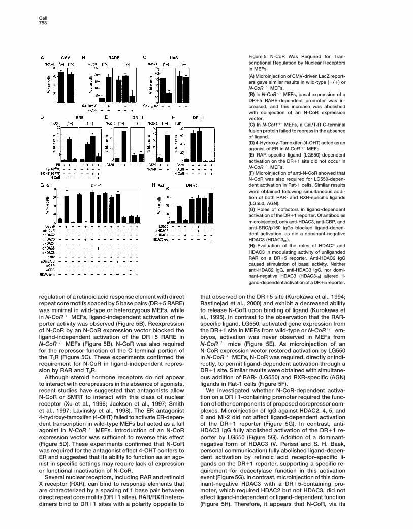
Figure 5. N-CoR Was Required for Tran-scriptional Regulation by Nuclear Receptorsin MEFs
(A) Microinjection of CMV-driven LacZ report-ers gave similar results in wild-type (1/1) orN-CoR2/2 MEFs.(B) In N-CoR2/2 MEFs, basal expression of aDR15 RARE-dependent promoter was in-creased, and this increase was abolishedwith coinjection of an N-CoR expressionvector.(C) In N-CoR2/2 MEFs, a Gal/T3R C-terminalfusion protein failed to repress in the absenceof ligand.(D) 4-Hydroxy-Tamoxifen (4-OHT) acted as anagonist of ER in N-CoR2/2 MEFs.(E) RAR-specific ligand (LG550)-dependentactivation on the DR11 site did not occur inN-CoR2/2 MEFs.(F) Microinjection of anti-N-CoR showed thatN-CoR was also required for LG550-depen-dent activation in Rat-1 cells. Similar resultswere obtained following simultaneous addi-tion of both RAR- and RXR-specific ligands(LG550, AGN).(G) Roles of cofactors in ligand-dependentactivation of the DR11 reporter. Of antibodiesmicroinjected, only anti-HDAC3, anti-CBP, andanti-SRC/p160 IgGs blocked ligand-depen-dent activation, as did a dominant-negativeHDAC3 (HDAC3DN).(H) Evaluation of the roles of HDAC2 andHDAC3 in modulating activity of unligandedRAR on a DR15 reporter. Anti-HDAC2 IgGcaused stimulation of basal activity. Neitheranti-HDAC2 IgG, anti-HDAC3 IgG, nor domi-nant-negative HDAC3 (HDAC3DN) altered li-gand-dependent activation of a DR15reporter.
regulation of a retinoic acid response element with direct that observed on the DR15 site (Kurokawa et al., 1994;Rastinejad et al., 2000) and exhibit a decreased abilityrepeat core motifs spaced by 5 base pairs (DR15 RARE)
was minimal in wild-type or heterozygous MEFs, while to release N-CoR upon binding of ligand (Kurokawa etal., 1995). In contrast to the observation that the RAR-in N-CoR2/2 MEFs, ligand-independent activation of re-
porter activity was observed (Figure 5B). Reexpression specific ligand, LG550, activated gene expression fromthe DR11 site in MEFs from wild-type or N-CoR1/2 em-of N-CoR by an N-CoR expression vector blocked the
ligand-independent activation of the DR15 RARE in bryos, activation was never observed in MEFs fromN-CoR2/2 mice (Figure 5E). As microinjection of anN-CoR2/2 MEFs (Figure 5B). N-CoR was also required
for the repressor function of the C-terminal portion of N-CoR expression vector restored activation by LG550in N-CoR2/2 MEFs, N-CoR was required, directly or indi-the T3R (Figure 5C). These experiments confirmed the
requirement for N-CoR in ligand-independent repres- rectly, to permit ligand-dependent activation through aDR11 site. Similar results were obtained with simultane-sion by RAR and T3R.
Although steroid hormone receptors do not appear ous addition of RAR- (LG550) and RXR-specific (AGN)ligands in Rat-1 cells (Figure 5F).to interact with corepressors in the absence of agonists,
recent studies have suggested that antagonists allow We investigated whether N-CoR-dependent activa-tion on a DR11-containing promoter required the func-N-CoR or SMRT to interact with this class of nuclear
receptor (Xu et al., 1996; Jackson et al., 1997; Smith tion of other components of proposed corepressor com-plexes. Microinjection of IgG against HDAC2, 4, 5, andet al., 1997; Lavinsky et al., 1998). The ER antagonist
4-hydroxy-tamoxifen (4-OHT) failed to activate ER-depen- 6 and Mi-2 did not affect ligand-dependent activationof the DR11 reporter (Figure 5G). In contrast, anti-dent transcription in wild-type MEFs but acted as a full
agonist in N-CoR2/2 MEFs. Introduction of an N-CoR HDAC3 IgG fully abolished activation of the DR11 re-porter by LG550 (Figure 5G). Addition of a dominant-expression vector was sufficient to reverse this effect
(Figure 5D). These experiments confirmed that N-CoR negative form of HDAC3 (V. Perissi and S. H. Baek,personal communication) fully abolished ligand-depen-was required for the antagonist effect 4-OHT confers to
ER and suggested that its ability to function as an ago- dent activation by retinoic acid receptor–specific li-gands on the DR11 reporter, supporting a specific re-nist in specific settings may require lack of expression
or functional inactivation of N-CoR. quirement for deacetylase function in this activationevent (Figure 5G). In contrast, microinjection of this dom-Several nuclear receptors, including RAR and retinoid
X receptor (RXR), can bind to response elements that inant-negative HDAC3 with a DR15-containing pro-moter, which required HDAC2 but not HDAC3, did notare characterized by a spacing of 1 base pair between
direct repeat core motifs (DR11 sites). RAR/RXR hetero- affect ligand-independent or ligand-dependent function(Figure 5H). Therefore, it appears that N-CoR, via itsdimers bind to DR11 sites with a polarity opposite to
N-CoR in Transcription and Development759
recruitment of enzymatically active HDAC3, was re-quired to mediate ligand-dependent activation on aDR11 element. Antibodies against several of the coacti-vators required for DR15-dependent activation in thisassay also abolished ligand-induced activation on theDR11 site (Figure 5G), indicating that the same familiesof coactivators were required for ligand-dependent acti-vation of both DR11 and DR15 reporters.
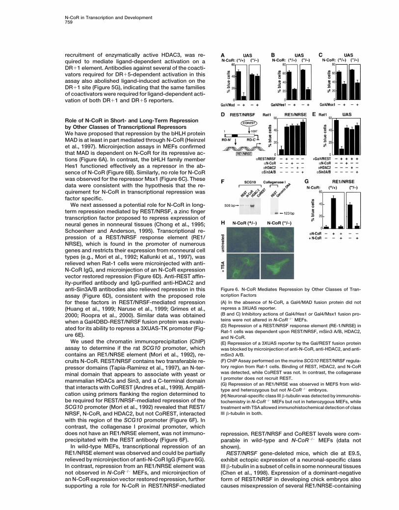
Role of N-CoR in Short- and Long-Term Repressionby Other Classes of Transcriptional RepressorsWe have proposed that repression by the bHLH proteinMAD is at least in part mediated through N-CoR (Heinzelet al., 1997). Microinjection assays in MEFs confirmedthat MAD is dependent on N-CoR for its repressive ac-tions (Figure 6A). In contrast, the bHLH family memberHes1 functioned effectively as a repressor in the ab-sence of N-CoR (Figure 6B). Similarly, no role for N-CoRwas observed for the repressor Msx1 (Figure 6C). Thesedata were consistent with the hypothesis that the re-quirement for N-CoR in transcriptional repression wasfactor specific.
We next assessed a potential role for N-CoR in long-term repression mediated by REST/NRSF, a zinc fingertranscription factor proposed to repress expression ofneural genes in nonneural tissues (Chong et al., 1995;Schoenherr and Anderson, 1995). Transcriptional re-pression of a REST/NRSF response element (RE1/NRSE), which is found in the promoter of numerousgenes and restricts their expression from nonneural celltypes (e.g., Mori et al., 1992; Kallunki et al., 1997), wasrelieved when Rat-1 cells were microinjected with anti-N-CoR IgG, and microinjection of an N-CoR expressionvector restored repression (Figure 6D). Anti-REST affin-ity-purified antibody and IgG-purified anti-HDAC2 and
Figure 6. N-CoR Mediates Repression by Other Classes of Tran-anti-Sin3A/B antibodies also relieved repression in thisscription Factorsassay (Figure 6D), consistent with the proposed role(A) In the absence of N-CoR, a Gal4/MAD fusion protein did notfor these factors in REST/NRSF-mediated repressionrepress a 3XUAS reporter.(Huang et al., 1999; Naruse et al., 1999; Grimes et al.,(B and C) Inhibitory actions of Gal4/Hes1 or Gal4/Msx1 fusion pro-2000; Roopra et al., 2000). Similar data was obtainedteins were not altered in N-CoR2/2 MEFs.when a Gal4DBD-REST/NRSF fusion protein was evalu-(D) Repression of a REST/NRSF response element (RE-1/NRSE) inated for its ability to repress a 3XUAS-TK promoter (Fig-Rat-1 cells was dependent upon REST/NRSF, mSin3 A/B, HDAC2,
ure 6E). and N-CoR.We used the chromatin immunoprecipitation (ChIP) (E) Repression of a 3XUAS reporter by the Gal/REST fusion protein
assay to determine if the rat SCG10 promoter, which was blocked by microinjection of anti-N-CoR, anti-HDAC2, and anti-mSin3 A/B.contains an RE1/NRSE element (Mori et al., 1992), re-(F) ChIP Assay performed on the murine SCG10 REST/NRSF regula-cruits N-CoR. REST/NRSF contains two transferable re-tory region from Rat-1 cells. Binding of REST, HDAC2, and N-CoRpressor domains (Tapia-Ramirez et al., 1997), an N-ter-was detected, while CoREST was not. In contrast, the collagenaseminal domain that appears to associate with yeast orI promoter does not recruit REST.mammalian HDACs and Sin3, and a C-terminal domain(G) Repression of an RE1/NRSE was observed in MEFS from wild-
that interacts with CoREST (Andres et al., 1999). Amplifi- type and heterozygous but not N-CoR2/2 embryos.cation using primers flanking the region determined to (H) Neuronal-specific class III b-tubulin was detected by immunohis-be required for REST/NRSF-mediated repression of the tochemistry in N-CoR2/2 MEFs but not in heterozygous MEFs, whileSCG10 promoter (Mori et al., 1992) revealed that REST/ treatment with TSA allowed immunohistochemical detection of class
III b-tubulin in both.NRSF, N-CoR, and HDAC2, but not CoREST, interactedwith this region of the SCG10 promoter (Figure 6F). Incontrast, the collagenase I proximal promoter, whichdoes not have an RE1/NRSE element, was not immuno- repression. REST/NRSF and CoREST levels were com-precipitated with the REST antibody (Figure 6F). parable in wild-type and N-CoR2/2 MEFs (data not
In wild-type MEFs, transcriptional repression of an shown).RE1/NRSE element was observed and could be partially REST/NRSF gene-deleted mice, which die at E9.5,relieved by microinjection of anti-N-CoR IgG (Figure 6G). exhibit ectopic expression of a neuronal-specific classIn contrast, repression from an RE1/NRSE element was III b-tubulin in a subset of cells in some nonneural tissuesnot observed in N-CoR2/2 MEFs, and microinjection of (Chen et al., 1998). Expression of a dominant-negativean N-CoR expression vector restored repression, further form of REST/NRSF in developing chick embryos also
causes misexpression of several RE1/NRSE-containingsupporting a role for N-CoR in REST/NRSF-mediated
Cell760
neural-specific genes, including SCG10 and Ng-Cam(Chen et al., 1998). To determine if N-CoR could be acomponent of this repression, expression of class IIIb-tubulin was assessed in MEFs using the TuJ1 anti-body. Class III b-tubulin expression was observed inz20 percent of N-CoR2/2 MEFs but not in heterozygousMEFs (Figure 6H). After incubation with TSA for 24–48hr, z20% of both heterozygous and N-CoR2/2 MEFsexpressed class III b-tubulin (Figure 6H). This was con-sistent with effects of TSA on REST/NRSF-repressedgenes in NIH 3T3 cells (Naruse et al., 1999). Thus, inaddition to its role in mediating short-term repressionevents, N-CoR may also be involved in long-term repres-sion events mediated by REST/NSRF on a subset of itstarget genes in specific cell types.
Discussion
N-CoR Was Required for Active Repressionby Several Classes of DNA BindingTranscription FactorsTranscriptional activation and repression mediated bysequence-specific DNA binding factors underlie the bi-nary decisions necessary for orchestration of develop-mental programs in metazoans. Transcriptional activa-tion by different classes of DNA binding proteins isdependent upon recruitment of a series of multisubunitcoactivator complexes, exemplified by the alternativerequirements for SAGA, ADA, and other complexes bydifferent transcription units in yeast (Grant et al., 1997).The discovery of a large and ever increasing numberof putative vertebrate corepressors and of a family ofhistone deacetylases has raised the question of whethercombinations of repressors and corepressors are re-
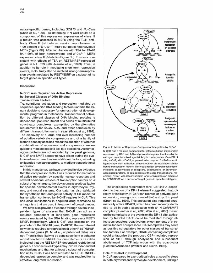
Figure 7. Model of Repressor-Corepressor Integration by N-CoRquired to mediate specific cell fate decisions. As homol-
N-CoR was a required component for effective ligand-independentogous proteins are not encoded by the yeast genome,repression by RAR and T3R and prevented agonist functions by the
N-CoR and SMRT appear to have arisen during the evo- estrogen receptor mixed agonist 4-hydroxy-tamoxifen. On a DR11lution of metazoans to allow additional factors, including site, N-CoR, with HDAC3, appeared to be required for RAR-specificunliganded nuclear receptors, to mediate transcriptional ligand-dependent activation, either directly or via modulation of site-repression. bound transcription factors. This could reflect several mechanisms,
including deacetylation of receptors, coactivators, or chromatin-In this manuscript, we have provided in vivo evidenceassociated proteins, or components of the core transcriptional ma-that the corepressor N-CoR was required for mediationchinery. N-CoR was also involved in long-term repression mediatedof active repression by specific nuclear receptors andby REST/NRSF on a subset of target genes in specific cell types.several additional classes of transcription factors on a
subset of gene targets, thereby acting as a critical factorThe unexpected requirement for N-CoR in RA-depen-for specific developmental events in erythrocytic, thy-
dent activation of a DR11 element suggested that, di-mic, and neural systems. Our data has also validatedrectly or indirectly, N-CoR can repress or activate genethe hypothesis that antagonist activity of ER-mediatedexpression, analogous to roles of Sin3 and rpd3 in yeasttranscription conferred by 4-OHT requires N-CoR. This(Struhl et al., 1998). This activation also required enzy-has clear implications in acquired drug resistance tomatically active HDAC3, which has been recently identi-antagonists that are used in treatment of breast cancer.fied to be in stable association with an N-CoR/SMRTWe have also provided evidence through several inde-complex (Guenther et al., 2000; Wen et al., 2000). Basedpendent types of analyses that N-CoR can act as aon the complexity of the events on the DR11 site, activa-required component of long-term gene repressiontion by N-CoR/HDAC3 could be mediated through ef-events mediated by the DNA binding repressor REST/fects on receptors, coactivators, or components of chro-NRSF. Interestingly, while N-CoR was present on thematin. Indeed, corepressor/HDAC complexes may servepromoter of the SCG10 gene, CoREST, the recruitmentas positive coregulators for other classes of transcrip-of which is required for repression of other REST/NRSF-tion factors. For example, HDAC-containing complexesdependent genes (G. M. et al., unpublished data), wascould antagonize the proposed CBP-mediated repres-not. There is thus likely to be gene specificity in cofactorsion of dTCF through acetylation and subsequentrecruitment to REST/NRSF response elements. Our dataabolishment of TCF interaction with the coactivatorindicated that the REST/NRSF-dependent restriction ofb-catenin/Armadillo (Waltzer and Bienz, 1998).genes out of specific cell types may involve independent
mechanisms and that for at least a subset of transcrip-tion units, N-CoR was both recruited to a REST/NRSF- Role of N-CoR in Development
N-CoR appeared to exert critical roles at specific stepsdependent repression complex and was required for itseffective long-term repression. in both erythroid and thymocyte development, linking a

N-CoR in Transcription and Development761
Experimental Proceduresspecific corepressor to discrete cell-type specificationevents. Studies in primary avian erythroblasts showed
Generation of N-CoR2/2 Micethat overexpression of T3R in the absence of T3 resultedGenomic DNA clones encoding the murine N-CoR gene were iso-in sustained proliferation and tightly arrested differentia-lated from a 129Sv lFIXII library (Stratagene, La Jolla, CA). Thetion of erythroblasts, while addition of T3 caused losstargeting construct, designed to delete amino acids 282–308 and
of self-renewal capacity and induced terminal differenti- induce a frameshift mutation if the resulting mRNA were translated,ation (Bauer et al., 1998). As N-CoR2/2 erythroblasts had was assembled using a 1.8 kb EcoRI fragment and a 5.0 kb XhoI–SalIenhanced levels of the CA II, unliganded T3R appeared PCR fragment. Transfected R1 ES cells (J. D. Marth, University ofto require N-CoR for repression events critical to expan- California, San Diego) were selected in medium containing 0.4 mg/mlsion of erythroblast progenitors. Proliferation in the ab- G418 and 10 mg/ml gancyclovir. Two clones containing the targeted
allele were identified by Southern blotting. Cells were injected intosence of T3 appears to require cooperation with c-kit,3.5-day-old C57BL/6 blastocysts, which were transferred into Swissbecause in the absence of the c-kit ligand SCF, differen-foster mothers. Chimeric males were crossed with C57BL/6 females.tiation occurs (Bauer et al., 1998). This suggests that
stages of development that require SCF may also in-volve T3R. It is then interesting to note that CFU-E colony Immunohistochemistry and In Situ Hybridization Analysisformation, which does not require SCF, was normal in Immunohistochemistry and in situ hybridization were performed asN-CoR2/2 embryos at the ages tested, while SCF-depen- previously described (Hermanson et al., 1994) using probes of base
pairs 2766–3171 of SMRT and 3954–4689 of N-CoR. The followingdent BFU-E colony formation was impaired.antisera were used: mouse anti-MAP2 (Sigma), rabbit anti-nestin (R.In N-CoR2/2 embryos, the block in thymocyte develop-McKay), mouse anti-nestin (PharMingen), TuJ1 (Babco), and mousement at the CD251/CD442 stage during the DN to DPanti-GFAP (ICN).transition was similar to that seen in mice with deletion
of particular transcription factors. Mice lacking the tran-scriptional repressor Hes1 exhibit blocks at both CD252/ Erythropoietic Analyses
Hematocrit were determined using heparinized Micro-HematocritCD441 and CD251/CD442 stages of development (Tom-Capillary Tubes (Fisher). Giemsa staining of blood smears was per-ita et al., 1999), but preliminary results with a Gal4-Hes1formed according to manufacturer’s instructions using Accustain,fusion protein suggested that in MEFs Hes1 maintainedGiemsa Stain, Modified (Sigma). For colony-forming assays, individ-its repressive function in the absence of N-CoR. TCF/ual fetal livers were dissected, disaggregated into single-cell sus-LEF double gene-deleted mice exhibit a block at thepensions, and passed through a cell strainer (70 mm, Falcon). CellsCD251/CD442 stage, as well as at the immature CD8were diluted in 2% acetic acid to lyse nonnucleated mature erythro-
single-positive (CD81 ISP) stage, raising the possibility cytes and the remaining cells counted. Cells amounting to 1 3 105
that N-CoR-associated deacetylase activity may affect (BFU-E) or 1 3 104 (CFU-E) from each fetal liver were plated inTCF/LEF-dependent activation events (Waltzer and methylcellulose medium containing erythropoietin with (for BFU-EBienz, 1998). assays) or without (for CFU-E assays) SCF, IL-3, and IL-6 (Stem Cell
The early events of neural induction appeared to be Technologies, Vancouver, BC). Benzidine-positive BFU-E colonieswere counted after 10 days; CFU-E colonies were counted after 3largely maintained as assessed by the normal onsetdays. Sheep anti-human Carbonic Anhydrase II antibody (The Bind-of nestin expression in N-CoR2/2 embryos. While theing Site, UK) was used for detection of CA II in fetal liver cells.mechanism of downregulation of nestin at midgestation
remains to be elucidated, it is intriguing that the en-hancer that drives nestin gene expression in the CNS Thymocyte Analysesharbors binding sites for both POU factors and nuclear Thymocytes were released by straining lobes through 70 mm Nylonreceptors (Josephson et al., 1998). The finding that Cell Strainers (Falcon). Thymic stroma devoid of thymocytes was
prepared by culturing E14.5 or E15.5 fetal thymic lobes for 5 daysMAP2, which can be induced by retinoic acid and har-in the presence of 1.35 mM dGUO (Sigma). Western blot analysisbors RAREs in its promoter (Neuman et al., 1995), waswas performed as previously described using anti-rabbit antibodiesupregulated in the outer layers of neocortex of N-CoR2/2
to N-CoR (Heinzel et al., 1997) or anti-guinea pig antibodies to SMRTembryos identifies MAP2 as a putative target gene for(Lavinsky et al., 1998). For experiments with anti-CD3e, thymii wereN-CoR-mediated repression through unliganded RAR.cultured 3 days with 25 mg/ml purified anti-CD3e (PharMingen). FlowExperiments in cell lines have suggested that decreasedcytometry was performed using 1 3 106 cells that were washed inlevels of MAP2 inhibit neuronal differentiation and neu-FACS buffer (PBS/2% fetal calf serum/0.1% NaN3) and incubated
rite formation (Dinsmore and Solomon, 1991). in 100 ml FACS buffer plus antibody for 20 min. The process wasIn conclusion, N-CoR appeared to exert corepressor repeated with 28 antibodies as necessary. Cells were collected prior
roles for subsets of genes under control of specific DNA to analysis on a FACScan (Becton Dickinson). Collection and analy-binding repressors and was involved in repression sis were performed using CellQuest software (Becton Dickinson).events mediated by specific nuclear receptors and sev- Directly conjugated antibodies specific for mouse CD4 and CD8eral other classes of DNA binding transcription factors (Caltag) and CD25, CD44, TCRb, and CD3 (PharMingen) were used.(Figure 7). Because of the linkage between N-CoR andREST/NRSF-dependent repression, our evidence sug- Single-Cell Nuclear Microinjectiongested that N-CoR participates in both transient and Each experiment was performed on three independent coverslipslong-term repression events. Additionally, N-CoR ap- consisting of z1000 cells. Where no experimental antibody waspeared to serve as a cofactor required, directly or indi- used, preimmune rabbit or guinea pig IgG was coinjected. Antibod-rectly, for gene activation on certain nuclear receptor ies to N-CoR, Sin3A/B, and HDAC2 (Heinzel et al., 1997), to CBPresponse elements (Figure 7). Interestingly, both short- and SRC-1 (Torchia et al., 1997), to REST and CoREST (Andres et
al., 1999), to HDAC3, 4, 5, and 6, and to Mi-2 (Santa Cruz Biotechnol-and long-term repressor functions and specific activa-ogy) were used. The RXR-specific ligand AGN 194204 was a gifttion functions appeared to require the actions of distinctfrom R. Chandraratna (Allergan Pharmaceuticals).HDACs, suggesting that there may be DNA site-, pro-
moter-specific usage of N-CoR-associated HDACs.These studies functionally link the corepressor N-CoR Chromatin Immunoprecipitation (ChIP) Assayto repressor-mediated determination of lineage pro- Rat-1 cells were fixed with 1% formaldehyde for 30 min at RT andgression in distinct cell types in mammalian devel- treated as described (Hecht and Grunstein, 1999). Cross-linked ad-
ducts were resuspended and sonicated resulting in DNA fragmentsopment.
Cell762
of 500–600 bp. Immunoprecipitation was performed using antibody- Guenther, M.G., Lane, W.S., Fischle, W., Verdin, E., Lazar, M.A., andShiekhattar, R. (2000). A core SMRT corepressor complex containingcoated tosylactivated Dynabeads M-280 (Dynal, Oslo, Norway). Pro-
tein-bound, immunoprecipitated DNA was dissolved in TE buffer HDAC3 and TBL1, a WD40- repeat protein linked to deafness. GenesDev. 14, 1048–1057.and treated at 658C overnight. Digestion buffer (100 mM NaCl;10
mM Tris-HCL, pH 8; 25 mM EDTA, pH 8; 0.5% SDS) was added to Hassig, C.A., Fleischer, T.C., Billin, A.N., Schreiber, S.L., and Ayer,the sample and incubated 2 hr at 508C with 0.1 mg/ml Proteinase D.E. (1997). Histone deacetylase activity is required for full transcrip-K (Sigma). Following extraction and precipitation, 50 cycles of PCR tional repression by mSin3A. Cell 89, 341–347.(Ta 5 568C) were performed. Primers for SCG10 were as described
Hecht, A., and Grunstein, M. (1999). Mapping DNA interaction sites(Mori et al., 1992). ChIP experiments on the collagenase I promoter
of chromosomal proteins using immunoprecipitation and polymer-were performed as above, except Hela cell extract was used. Oligos
ase chain reaction. Methods Enzymol. 304, 399–414.were as follows: 59-CAAATAATCTGCTAGGAGTCACCA; 39-ATATA
Heinzel, T., Lavinsky, R.M., Mullen, T.M., Soderstrom, M., Laherty,GAGTCCTTGCCCTTCCAGA.C.D., Torchia, J., Yang, W.M., Brard, G., Ngo, S.D., Davie, J.R., etal. (1997). A complex containing N-CoR, mSin3 and histone deacety-Acknowledgmentslase mediates transcriptional repression. Nature 387, 43–48.
We thank V. Perissi and S. H. Baek for dominant-negative HDAC3; Hermanson, O., Ericson, H., Sanchez-Watts, G., Watts, A.G., andB. S. Katzenellenbogen for CMV-ER; R. McKay for nestin antibodies; Blomqvist, A. (1994). Autoradiographic visualization of 35S-labeledJ. D. Marth for R1 ES cells; A. Krones and T. Herman for sequencing; cRNA probes combined with immunoperoxidase detection of chol-M. Frazer and H. Taylor for animal care; A. Hazra and C. Nelson for eragenoid: a double-labeling light microscopic method for in situassistance; M. Fisher for help in preparing the manuscript; P. Meyer hybridization and retrograde tract tracing. J. Histochem. Cytochem.for preparation of the figures; B. Andersen for useful discussions; 42, 827–831.and A. K. Ryan for comments on the manuscript. This research was Horlein, A.J., Naar, A.M., Heinzel, T., Torchia, J., Gloss, B., Kuro-supported by grants to D. W. R. (NIH 1 RO1 DK54802-O1A1); E. S., kawa, R., Ryan, A., Kamei, Y., Soderstrom, M., Glass, C.K., et al.S. M. H., G. M., and C. K. G. (NIH); and M. G. R. (NIH, CAPCURE, (1995). Ligand-independent repression by the thyroid hormone re-and California Cancer Research Program). ceptor mediated by a nuclear receptor co-repressor. Nature 377,
397–404.Received June 30, 2000; revised August 14, 2000.
Hu, I., and Lazar, M.A. (2000). Transcriptional repression by nuclearhormone receptors. Trends Endocrinol. Metab. 11, 6–10.
ReferencesHuang, Y., Myers, S.J., and Dingledine, R. (1999). Transcriptionalrepression by REST: recruitment of Sin3A and histone deacetylaseAlland, L., Muhle, R., Hou, H., Jr., Potes, J., Chin, L., Schreiber-to neuronal genes. Nat. Neurosci. 2, 867–872.Agus, N., and DePinho, R.A. (1997). Role for N-CoR and histone
deacetylase in Sin3-mediated transcriptional repression. Nature Huang, E.Y., Zhang, J., Miska, E.A., Guenther, M.G., Kouzarides, T.,387, 49–55. and Lazar, M.A. (2000). Nuclear receptor corepressors partner with
class II histone deacetylases in a Sin3-independent repression path-Andres, M.E., Burger, C., Peral-Rubio, M.J., Battaglioli, E., Anderson,way. Genes Dev. 14, 45–54.M.E., Grimes, J., Dallman, J., Ballas, N., and Mandel, G. (1999).
CoREST: a functional corepressor required for regulation of neural- Jackson, T.A., Richer, J.K., Bain, D.L., Takimoto, G.S., Tung, L.,specific gene expression. Proc. Natl. Acad. Sci. USA 96, 9873–9878. and Horwitz, K.B. (1997). The partial agonist activity of antagonist-
occupied steroid receptors is controlled by a novel hinge domain-Bauer, A., Mikulits, W., Lagger, G., Stengl, G., Brosch, G., and Beug,binding coactivator L7/SPA and the corepressors N-CoR or SMRT.H. (1998). The thyroid hormone receptor functions as a ligand-oper-Mol. Endocrinol. 11, 693–705.ated developmental switch between proliferation and differentiation
of erythroid progenitors. EMBO J. 17, 4291–4303. Josephson, R., Muller, T., Pickel, J., Okabe, S., Reynolds, K., Turner,P.A., Zimmer, A., and McKay, R.D. (1998). POU transcription factorsChen, J.D., and Evans, R.M. (1995). A transcriptional co-repressorcontrol expression of CNS stem cell-specific genes. Developmentthat interacts with nuclear hormone receptors. Nature 377, 454–457.125, 3087–3100.Chen, Z.F., Paquette, A.J., and Anderson, D.J. (1998). NRSF/RESTKallunki, P., Edelman, G.M., and Jones, F.S. (1997). Tissue-specificis required in vivo for repression of multiple neuronal target genesexpression of the L1 cell adhesion molecule is modulated by theduring embryogenesis. Nat. Genet. 20, 136–142.neural restrictive silencer element. J. Cell Biol. 138, 1343–1354.Chong, J.A., Tapia-Ramirez, J., Kim, S., Toledo-Aral, J.J., Zheng,
Y., Boutros, M.C., Altshuller, Y.M., Frohman, M.A., Kraner, S.D., and Kao, H.Y., Ordentlich, P., Koyano-Nakagawa, N., Tang, Z., Downes,Mandel, G. (1995). REST: a mammalian silencer protein that restricts M., Kintner, C.R., Evans, R.M., and Kadesch, T. (1998). A histonesodium channel gene expression to neurons. Cell 80, 949–957. deacetylase corepressor complex regulates the Notch signal trans-
duction pathway. Genes Dev. 12, 2269–2277.Dinsmore, J.H., and Solomon, F. (1991). Inhibition of MAP2 expres-sion affects both morphological and cell division phenotypes of Kao, H.Y., Downes, M., Ordentlich, P., and Evans, R.M. (2000). Isola-neuronal differentiation. Cell 64, 817–826. tion of a novel histone deacetylase reveals that class I and class II
deacetylases promote SMRT-mediated repression. Genes Dev. 14,Fischer, A., and Malissen, B. (1998). Natural and engineered disor-55–66.ders of lymphocyte development. Science 280, 237–243.Kurokawa, R., DiRenzo, J., Boehm, M., Sugarman, J., Gloss, B.,Glass, C.K., and Rosenfeld, M.G. (2000). The coregulator exchangeRosenfeld, M.G., Heyman, R.A., and Glass, C.K. (1994). Regulationin transcriptional functions of nuclear receptors. Genes Dev. 14,of retinoid signalling by receptor polarity and allosteric control of121–141.ligand binding. Nature 371, 528–531.Grant, P.A., Duggan, L., Cote, J., Roberts, S.M., Brownell, J.E., Can-Kurokawa, R., Soderstrom, M., Horlein, A., Halachmi, S., Brown, M.,dau, R., Ohba, R., Owen-Hughes, T., Allis, C.D., Winston, F., etRosenfeld, M.G., and Glass, C.K. (1995). Polarity-specific activitiesal. (1997). Yeast Gcn5 functions in two multisubunit complexes toof retinoic acid receptors determined by a co-repressor. Nature 377,acetylate nucleosomal histones: characterization of an Ada complex451–454.and the SAGA (Spt/Ada) complex. Genes Dev. 11, 1640–1650.
Laherty, C.D., Billin, A.N., Lavinsky, R.M., Yochum, G.S., Bush, A.C.,Gregory, C.J., and Eaves, A.C. (1978). Three stages of erythropoieticSun, J.M., Mullen, T.M., Davie, J.R., Rose, D.W., Glass, C.K., et al.progenitor cell differentiation distinguished by a number of physical(1998). SAP30, a component of the mSin3 corepressor complexand biologic properties. Blood 51, 527–537.involved in N-CoR-mediated repression by specific transcriptionGrimes, J.A., Nielsen, S.J., Battaglioli, E., Miska, E.A., Speh, J.C.,factors. Mol. Cell 2, 33–42.Berry, D.L., Atouf, F., Holdener, B.C., Mandel, G., and Kouzarides,
T. (2000). The co-repressor mSin3A is a functional component of the Lavinsky, R.M., Jepsen, K., Heinzel, T., Torchia, J., Mullen, T.M.,Schiff, R., Del-Rio, A.L., Ricote, M., Ngo, S., Gemsch, J., et al. (1998).REST-CoREST repressor complex. J. Biol. Chem. 275, 9461–9467.
N-CoR in Transcription and Development763
Diverse signaling pathways modulate nuclear receptor recruitment p/CIP binds CBP and mediates nuclear-receptor function. Nature387, 677–684.of N-CoR and SMRT complexes. Proc. Natl. Acad. Sci. USA 95,
2920–2925. Waltzer, L., and Bienz, M. (1998). Drosophila CBP represses thetranscription factor TCF to antagonize Wingless signalling. NatureLevelt, C.N., Mombaerts, P., Iglesias, A., Tonegawa, S., and Eich-395, 521–525.mann, K. (1993). Restoration of early thymocyte differentiation in
T-cell receptor beta-chain-deficient mutant mice by transmembrane Wen, Y.-D., Perissi, V., Staszewski, L.M., Yang, W.-M., Krones, A.,signaling through CD3 epsilon. Proc. Natl. Acad. Sci. USA 90, 11401– Glass, C.K., Rosenfeld, M.G., and Seto, E. (2000). The histone deace-11405. tylase-3 complex contains nuclear receptor corepressors. Proc.
Natl. Acad. Sci. USA 97, 7202–7207.Mannervik, M., Nibu, Y., Zhang, H., and Levine, M. (1999). Transcrip-tional coregulators in development. Science 284, 606–609. Xu, J., Nawaz, Z., Tsai, S.Y., Tsai, M.J., and O’Malley, B.W. (1996).
The extreme C terminus of progesterone receptor contains a tran-McKenna, N.J., Lanz, R.B., and O’Malley, B.W. (1999). Nuclear re-scriptional repressor domain that functions through a putative core-ceptor coregulators: cellular and molecular biology. Endocr. Rev.pressor. Proc. Natl. Acad. Sci. USA 93, 12195–12199.20, 321–344.Xu, L., Lavinsky, R.M., Dasen, J.S., Flynn, S.E., McInerney, E.M.,Mori, N., Schoenherr, C., Vandenbergh, D.J., and Anderson, D.J.Mullen, T.M., Heinzel, T., Szeto, D., Korzus, E., Kurokawa, R., et al.(1992). A common silencer element in the SCG10 and type II Na1(1998). Signal-specific co-activator domain requirements for Pit-1channel genes binds a factor present in nonneuronal cells but notactivation. Nature 395, 301–306.in neuronal cells. Neuron 9, 45–54.Xue, Y., Wong, J., Moreno, G.T., Young, M.K., Cote, J., and Wang,Nagy, L., Kao, H.Y., Chakravarti, D., Lin, R.J., Hassig, C.A., Ayer, D.E.,W. (1998). NURD, a novel complex with both ATP-dependent chro-Schreiber, S.L., and Evans, R.M. (1997). Nuclear receptor repressionmatin-remodeling and histone deacetylase activities. Mol. Cell 2,mediated by a complex containing SMRT, mSin3A, and histone851–861.deacetylase. Cell 89, 373–380.Zamir, I., Harding, H.P., Atkins, G.B., Horlein, A., Glass, C.K., Rosen-Naruse, Y., Aoki, T., Kojima, T., and Mori, N. (1999). Neural restrictivefeld, M.G., and Lazar, M.A. (1996). A nuclear hormone receptor core-silencer factor recruits mSin3 and histone deacetylase complex topressor mediates transcriptional silencing by receptors with distinctrepress neuron-specific target genes. Proc. Natl. Acad. Sci. USArepression domains. Mol. Cell. Biol. 16, 5458–5465.96, 13691–13696.
Neuman, K., Soosaar, A., Nornes, H.O., and Neuman, T. (1995).Orphan receptor COUP-TF I antagonizes retinoic acid-induced neu-ronal differentiation. J. Neurosci. Res. 41, 39–48.
Pain, B., Melet, F., Jurdic, P., and Samarut, J. (1990). The carbonicanhydrase II gene, a gene regulated by thyroid hormone and erythro-poietin, is repressed by the v-erbA oncogene in erythrocytic cells.New Biol. 2, 284–294.
Ramsdell, F. (1992). Fetal thymic organ culture for T cell develop-ment studies. In Current Protocols in Immunology, J.E. Coligan,A.M. Kruisbeek, D.H. Margulies, E.M. Shevach, and W. Strober, eds.(New York: John Wiley and Sons), pp. 3.18.1–3.18.10.
Rastinejad, F., Wagner, T., Zhao, Q., and Khorasanizadeh, S. (2000).Structure of the RXR-RAR DNA-binding complex on the retinoicacid response element DR1. EMBO J. 19, 1045–1054.
Roopra, A., Sharling, L., Wood, I.C., Briggs, T., Bachfischer, U.,Paquette, A.J., and Buckley, N.J. (2000). Transcriptional repressionby neuron-restrictive silencer factor is mediated via the Sin3-histonedeacetylase complex. Mol. Cell. Biol. 20, 2147–2157.
Schoenherr, C.J., and Anderson, D.J. (1995). The neuron-restrictivesilencer factor (NRSF): a coordinate repressor of multiple neuron-specific genes. Science 267, 1360–1363.
Seol, W., Mahon, M.J., Lee, Y.K., and Moore, D.D. (1996). Two recep-tor interacting domains in the nuclear hormone receptor corepressorRIP13/N-CoR. Mol. Endocrinol. 10, 1646–1655.
Smith, C.L., Nawaz, Z., and O’Malley, B.W. (1997). Coactivator andcorepressor regulation of the agonist/antagonist activity of themixed antiestrogen, 4-hydroxytamoxifen. Mol. Endocrinol. 11,657–666.
Struhl, K., Kadosh, D., Keaveney, M., Kuras, L., and Moqtaderi, Z.(1998). Activation and repression mechanisms in yeast. Cold SpringHarb. Symp. Quant. Biol. 63, 413–421.
Swat, W., Shinkai, Y., Cheng, H.L., Davidson, L., and Alt, F.W. (1996).Activated Ras signals differentiation and expansion of CD4181
thymocytes. Proc. Natl. Acad. Sci USA 93, 4683–4687.
Tapia-Ramirez, J., Eggen, B.J., Peral-Rubio, M.J., Toledo-Aral, J.J.,and Mandel, G. (1997). A single zinc finger motif in the silencingfactor REST represses the neural-specific type II sodium channelpromoter. Proc. Natl. Acad. Sci. USA 94, 1177–1182.
Tomita, K., Hattori, M., Nakamura, E., Nakanishi, S., Minato, N., andKageyama, R. (1999). The bHLH gene Hes1 is essential for expansionof early T cell precursors. Genes Dev. 13, 1203–1210.
Torchia, J., Rose, D.W., Inostroza, J., Kamei, Y., Westin, S., Glass,C.K., and Rosenfeld, M.G. (1997). The transcriptional co-activator


