Embed Size (px)
Citation preview
51
COMPARATIVE ASPECTS OF FISH TASTE BUDULTRASTRUCTURE
Klaus Reutterr and Martin Witt2
rDepartment of AnatomyUniversity of TübingenOsterbergstraße 3
D-7 207 4 Tübingen, Germany2Department
of AnatomyTechnical UniversityFetscherstrasse 74D-01307 Dresden, Germany
Abstract
Taste buds are composed of several cell types and occur in all classes of vertebrates. Infish, these organs are found in members of all systematic groups which have been exam-ined. Taste buds are intraepithelial organs whose size, shape, position and cellular compo-sition varies greatly between systematic groups. Taste bud sensory cells may be classifiedinto electron-lucent light cells and electron-dense dark cells; some taxa may have alsosubtypes of light and dark cells. In some cases, basal cells are Merkel cell-like. In sometaxa the taste buds have afferent and a few efferent synapses. Taste buds possibly providedifferent fishes with different information from the environment.
1. INTRODUCTION
Our knowledge about the gustatory system and the ultrastructure of taste buds in fishhas increased greatly during about the past three decades. The results ofthese studies havebeen reviewed several times (Kapoor et al., 1975; Reutter, 197g, l9g6; Jakubowski andwhitear, 1990; Reutter and witt, 1993; Sorensen and caprio, l99g). Most of this work hasbeen conducted in teleosts, the modern bony fishes. During the more recent years, the gus-tatory system of non-tdleostean fish became of interest, especially from comparative mor-phological and physiological perspectives. For example, it has iecently been shown thatthe taste bud of a shark (dogfish) is quite different to that of a bullhead (Whitear and
Advances in Chemical Signals in Vertebrates, edited by Johnston el a/.Kluwer Academic / Plenum Publishers, New York, 1999. 573

57{
Moate, 1994a: R.rrrra- , ^^,, ' Reutter and M. witt
urodelan ,ur,l' I:,ult:t l ee4) while I
3*i*,fgül$,',,frl;ffi ##itilp;ä::#r1lä'{i;;ryr';ö:rasrebudo,r"l1ll:.i"0*',i,lnär,äl.f,äTi;1ffi ;;1.,139'':iiili.:ä:ilJ,;T-; ":iT.H" lff #-T {lJ Ei ::,,'.'"T
o'*'^ "ol'
"ö'i}
i n' u''r' "" l äi"" 1'
"' i n
usua r ry 0,.. u,äil,..u r a.tu i r, är,',"#fl l", "Jräjffiillffi .,:ä$ffi
"*r[:;l;:'ä*li+t l'ffi *.:;n**lj rxl*'r,m ,rällä iTil':$";"f ;l**;"*"Ti::lftiip'Ti:i:yr "o' o'oi oioäi :it!'liö;:: il * ,?t:e sroups
"ri"n,..ä,'i#fi:",,f,jlil.|o""oi#,n"r and jt ',
il",.,r.i"*"
2. MATERIALS AND METHODSThe results
iff g;iiffi:#: I l.l,:':öffij:':::.t
d uring,h e ras,
*idfi,m**H*ääffifir#**ffiqftrathin sections *.."'.äl^]::p,t o1 ethanol, the specimen-;:::'::".u' t"h ptcric acid. Af-,,*"$nl ".,*,,T;:ää"Tii'J,[:{üj::".'ü::'ä"if:::H":,,,:1i,;'ili,'.il i,
. s e, ac h i i,"il::ä,r**:::l;:J:t ä'.:""H*:: """ rhe sec'fion s were
. Holostei: T.rereosrei:rl.^.fl 1*oL^,,'iii,"ii::2::I::::,;:;;';:::::stigated;ru.o"un
"lli:;,,::;:;:ä:,;:, rn,i,li,"i"*turus) nebutoszs and rhe wers or' Dipnoi (Choanichthv..;f;';";rtrarian
rungfi sh, Neoceratodus forsteri.
3. RESULTS
0.,.0lj?J,i.ili[,iT;n:f the taste buds o
*:f; il""::ffi #$ü +:tilHi,'3 #ltl"1r ö:t T: :r'' :' a re n' w c' m.
fr*i'':"j',;tffi *fiiftm*l;i",ffi flä*rli;tr
ffi*-*fffifigffffiffiffffi

,ratiye Aspects ofFish Taste Bud Ultrastructure
Table 1. comparison of the main taste bud (TB) components of fishes which belong todifferent systematic groups
575
Systematic order Selachii Holostei Teleostei Dipnoi
/
Genus
TB size(heighvwidth, pm)
TB positionIn epidermal hillockIn flat epitheliumOn top of dermal papilla
Cell typesLight cellDark cellMarginal cellBasal cell
LocationWith spines
Basal nerve fiber plexus
Ascending nerve fibersSynapses
AfferentEfferent
References
Scyliorhinus
50/30
?
I type2 types
?
outside TB base
small
+
Reutter 1994
Whitear &Moate 1994a
Lepisosteus
65/4s
+
+
2 typesI type
++
at TB base
small
++
Reutter & Witt 1996
Ameiurus (A)Si/urzs (S)
ssl35 (A)80/50 (s)
I
+
+
I typet1o"
i
at TB base(+;A); + (S)
r1"
++ (A); _ (S)
Desgranges 1966Reutter 1971,'78,'86,
'87
Neoceratodus
r 00/80
+(+)+
I typeI type
f
+
at TB base
small
++
Reutter l99l
Royer& Kinnamon, 1996
picted in Figure 5. Because of space limitations, only the apices of taste buds belonging tothe four named taxa are shown with original electron micrographs (Figures l-4).
3.1. Selachian Taste Bud
In Scyliorhinas, the taste buds are relatively small, sitting in epidermal hillocks and donot rest on a dermal papi[a as they are totally surrounded by marginal or epidermal cells. Thesensory epithelium comprises light and dark cells; the light cells terminate apically either withone big, conus-like receptor villus (not shown) or with a big and divided microvillar, stereo-cilia-like structure (Figure l). The dark cells comprise two subtypes: one type comprises cellsof moderate electron density which are apically rich in secretory vesicles and which have longand divided receptor villi. The second fype is highly electron-dense and contributes to thetaste buds receptor area with long and stereocilia-like microvilli. Characteristically, the basalcells are situated outside the taste bud and rest on top of the basal lamina which surrounds thedermal papilla. The taste bud's nerve fiber plexus is small; light (and dark) cells are afferentlyinnervated. Efferent synapses have not been found.
3.2. Holostean Taste Bud
In Lepisosteus, the taste buds are located in epidermal hillocks and always rest on adermal papilla. The sensory epithelium consists of light and dark cells; the light cells com-

K. Reutter and M. Witt
Figure l' Apical part of a Scyliorhinus caniculus (Selachii) - taste bud in longitudinal section. The receptor area(RA) is formed by the very long, thin and partly divided receptor villi ofdark cells (Cd) and shorter, but also di-vided villi of light cells (cl)' Light and dark celis are apically connected to each other by tightjunctions (Jt). Bar:I um.
prlse one subtype of numerous cells which end apically with one big receptor villus andanother subtype which bears several small receptor villi (Figure 2). The dark cells areequipped with small villi and are rich in electron-dense vesicles of different sizes, espe-cially in their apical cytoplasm. The nerve fiber plexus is small; the bases of light and darkcells are afferently innervated. Efferent synapses are rarely found between light cells andnerve fibers' The basal cells form the taste buds base and rest directly on the basal laminawhich surrounds the dermal papilla. They are afferently innervated and possess arm-likeprocesses which contain numerous dense_cored vesicles.
3.3. Teleostean Thste Bud
In teleosteans, the taste buds of different species are often of different sizes, evenwhen they belong to the- same systematic group, as seen in the silurid s Ameiurus and, silu-ras' Teleostean taste buds may be situatedln more or less elevated epidermal hillocks or inflat epithelia. They always rest on top of a dermal papilla. The sensory epithelium of thetaste bud consists oflight and dark cells; the light ".tt,
b.u, apically one relatively big andlong receptor villus, the dark cells contribute tä the receptor u..u *ittr several small recep-tor villi (Figure 3). Regularly the taste buds nerve fiber flexus is well developed and its fi-
T

rtive Aspects ofFish Taste Bud Ultrastructure 577
Figure 2. Apical part of a Lepisosteus oculatus (Holostei) - taste bud in longitudinal section. The sensory epithe-
lium ofthe organ consists oftwo types oflight cells and one type ofdark cells. The light cell a (Cla) terminates
with several short microvilli, the light cell b (Clb) bears one long and thick microvillus. The dark cells (Cd) are
apically rich in electron-dense vesicles ofdifferent sizes and end with several small receptor vilti. Jt - tightjunc-
tion; RA - receptor area. Bar: I Pm.
bers synapse onto light cells and dark cells (seldom) afferently; efferent synapses have
been found on light cells, too (Ameiurus; Desgranges, 1966). The basal cells are Merkel-
cell-like cells (see Reutter and Witt, 1993). They are typically disk-like, contain vesicular
structures ofdifferent sizes and electron-densities and synapse afferently onto nerve fibers
of the taste buds plexus. Light cells and dark cells may be connected afferently to the ba-
sal cells as well (Ameiurus). Basal cells may have microvillar processes (spikes or spines)
which project into the organ's nerve fiber plexus; they always rest in a depression of the
basal lamina which surrounds the dermal papilla.
3.4. Dipnoan Taste Bud
Neoceratodus-taste buds are the biggest ones we found among the fish species ex-
amined. They are situated mainly in epidermal hillocks and rest on top of a big dermal pa-
pilla. The sensory epithelium consists of big, elongated cells which belong to two cell
types: The light cells end apically with one big receptor villus (which may be more than
6pm long and 0.5 pm wide) and are rich in organelles, especially in microtubules and mi-
crofilaments. The dark cells terminate with several small, divided or bundled receptor

lEK. Reutter and M. Witt
t\
#t
e
Figure 3. Apical part of a Silurus g/anrs (Teleostei) - taste bud in longitudinal section. The receptor area (RA) isbror'rgh,t together by the single large receptor villi oflight cells (cl) and the numerous small receptor villi ofdarkcells (Cd)' one small receptor villus is elongated, too (anow). Note also the surface coat ofthe receptor area. cm -marginal cell; Jt - tight junction. Bar: I pm.
villi; they are apically rich in secretory vesicles and bundles of tonofilaments (Figure 4).The nerve fiber plexus of the bud is relatively small and some nerve fibers may reach upto the apical part of the organ. Afferent rynupr., are commonly seen between the basalprocesses oflight cells and nerve fibers; in a few cases they occur between the basal proc-esses of dark cells and nerve fibers. Efferent synapses occur infrequently between nervefibers and the bases of light cells. The basal cälls are disk-shaped'and resemble Merkelcells. occasionally, some nerve fibers lie between a basal cell and the basal lamina.
4. DISCUSSION
The comparison of the taste buds from fish belonging to different systematicgroups clearly shows that there exists more than one type of taste buds. It is likely that indifferent taxa of fish the. taste buds belong to taxa-specific types. But unfortunately, atthe moment rhe taste buds of only a few siecies of fish that berong to the same system_atic group have been investigated. It is not clear if our knowledge Äout the taste bud-ul-trastructure in Scyliorhinzs, for example, is representative of all other Chondrichthyes;some of them seem to have no taste buds and in Raja only solitary chemosensory cells

dtive Aspects of Fish Taste Bud Ultrastructure 579
Figure 4. Apical part of a Neoceratodus forsteri (Dipnoi) - taste bud in longirudinal section. Each light cell (Cl)contributes to the receptor area (RA) with one very large and undivided microvillus; one large and broad microvil-lus is obliquely cut (asterisk). The dark cells (Cd) project into the receptor area with several thin, often dividedand relatively long "small" microvilli. Jt - tight junction. Bar: I pm.
were found (Whitear and Moate, 1994b). Further, taste bud ultrastructure varies in mod-ern bony fishes. Besides the typical teleostean taste buds of cyprinids (Hirata, 1966), si-lurids (Reutter, 1978-1987, and others), cobirids (Jakubowski, 1983) and salmonids(Ezeasor, 1982), the characid Astyanax (Boudriot and Reutter, 1997) and the cyprinidDanio (Hansen et al., 1997) have a third type of taste bud cell within their sensory epi-thelium. We do not know the significance of this variation; possibly it is a sign of a spe-cial adaptation to the fish's habitat and food-intake behavior. So we should know what isa highly evolved fish taste bud and what a more primitive organ looks like. Neither canwe exclude a species-specific variability of taste bud-cells which may allow specializedreceptive functions. Thus, we are not yet able to make firm conclusions about taste budevolution in fish. We know only about the taste buds of few representatives of some maintaxa and we know nothing about the taste buds in most of the 25.000 fish species whichbelong to other taxa. We should respect the diversity of the micromorphologies of tastebuds in selachian, holostean, teleostean and dipnoan fish. Following our comparative re-sults, future investigations will-hopefully-continue to speak of distinct taxa-specificfish taste buds rather than only of "fish taste buds". Clearly, we are now ready to eluci-date the functional significance of differences in taste buds and how they relate to the
J80
K. Reutter and M. Wifl
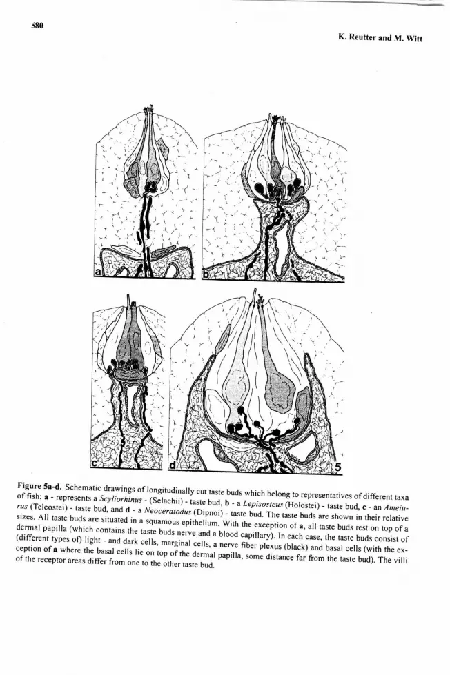
Figure 5a-d' Schematic drawings oflongitudinally cut taste buds which belong to representatives ofdifferent taxaof fish: a-represents ascvliorhinus-(sJlachii)-il;q: b-aLepisosteus(Horostei)-tasrebud, c-anAmeiu-rzs (Teleostei) - taste bud' ^11 I .- "
w"or"roroa*Tiiioi) - taste bud. The taste buds are shown in rheir relativesizes' All taste buds are situated in a squamous "oi,i.iiuri. wi,r, tn"
"*""ption oi", ",r
,"r,. buds rest on top of adermal papilla (which contains the taste uua, n.*, "nJ'a
brood capiilary). In each case, the taste buds consist of(different types of) tight - and aart cerrs, ,nurgi;;i;;il; I ner"e fib"r ptexus (black) and basal cells (with the ex_iiHä:il1".::ii:,f:ifi '
,j":ffi :il:Jnx.;ui nuo' r'",,o,"" di.tu*. *, ;"; rhe rasre bud). rhe v i r i

581
CKNOWLEDGMENTS
The invaluable help from Gerd Geiger.(electron microscopy), Manfred Mauz (pho_tography) and Mihnea Nicolescu (drawing-s) is greatly ""k"";l;ä;;d. Many thanks also toBob Johnston and peter Sorensen for editäi, correcting and improving this text.
REFERENCES
Boudriot'F Reutter,K'(1997)'ultrastructureofthetasrebudsintheblindcave fishAstyanaxjordani(,,Anoptich_thys") and its sighted rerative Astyanax mexicanus (Tereostei). crr.nl. s..rr., z-r (lggg; in press).cummings' T-A', Delay, R'J., Roper, s.D. (1987). ultrastructure of apical specializations of taste cells in the mud_puppy, Necturus maculosus. J. Comp. Neurol. 261, 604415.
Desgranges' J'c' ( | 966)' Sur la double innervation des cellules-sensorielles des bourgons du goüt des barbillons dupoisson-chat. C. R. Acad. Sci. [D] paris 263, I 103_t t06.Ezeasor' D'N' (1982)' Distribution and ultrastructure of taste buds in the oro- pharyngeal cavity of the rajnbowtrout, Salmo gairdneri Richardson. J. Fish Biol. 2/i 51_6g.t"totT;l1jl,"Hr*: (1971). Fine structure of the taste bud in the mudpuppv, Necturus macuto.sus. A- J.
Hansen'
l;:f ,- ii;r,l""J,jler' K' ( 1997)' chemical senses in the zebrafish. r st. rübingen Zebrafish Meeting, r 9.
Hirata'Y'(1966)'Finestructureoftheterminalbudsonthebarbelsofsomefishes.Arch.histol.jap.26,s0!-s23.Jakubowski' M' (1983)' New details of the ultrastructure (TEM, SEM) of taste buds in fishes. Z. mikrosk.-anat.Forsch. 97, 849-862.Jakubowski, M', whitear, M (1990). comparative morphology and cytology of taste buds in teleosts. Z. mikosk.-anat. Forsch. I 04. 529_560.Kapoor, 8.G., Evans, H.8., pevzner, R.A. (1975). The gustatory system in fish. Adv. mar. Biol. /j, 53_10g.Lindemann, B. (1996). Taste receprion. physiol Rev. 76,.719_766.Reutter' K' (1971 )' Die Geschmacksknospen des Zwergwelses,{z iurus nebulosus(Lesueur); Morphologische undhistochemische Untersuchungen. Z. Zellforsch. 120, 2g0_30g.Reutter,K'(1978).Tasteorganinthebullhead(Teleostei).Adv.Anat.Embryol.cell
Biot. jJ. l_9g.Reutter' K' (1986)' chemoreceptors. ln: Biologt of the integument Vol. II, yertebrates(J. Bereiter-Hahn, A.G.Matoltsy and K.S. Richards, Eds.). Springer, Berlin, Heidetberg, New york, pp.-iSG{Oa.Reutter' K' (1987)' Specialized receptor villi and basal cells within the taste bud of,h".urop.un sirurid fish,.si/a-rus glanis (Teleostei) In: olfaction and Taste IX (s.D. Roper and J. Atema, eds.). Annars of the New yorkAcademy ofSciences, New york, pp. 57G_573.*t"*"tbl;.!lnio1)' ultrastructure of taste buds in the Australian.lungfish, Neoceratodus forsreri (Dipnoi). chem.
Reutter'K'(1994)'ultrastruc'trtre.oftastebudsinthesporteddogfish,scylioräinuscaniculus(Selachii). ln:olfac-tion and raste Xr (K. Kurihara, N. Suzuki and H. ogawa, Eds.). Springer, Tokyo, aerlin, Heiderberg, NewYork, London, paris, Hongkong, Barcelona, p. 754.
Reutter' K'' witt, M' (t993)' Morphology of vertebrare taste organs and their nerve supply. ln: Mechanisms of';;':tr#o*"'n (S.A. simon and s.D. Roper. eds.). cRö press, Bo"u R;;;n,;rn Arbor, London, rokyo,
Reutter' K'' witt' Y;
jl:::l*fstructure of the taste buds in the spotted gar, Lepisosteus ocutatus(Holostei).
Royer' S'M'' Kinnamon' J'c' (1996) comparison of high-pressure freezing/freezesubstitution and chemical fixa-tion of catfish barbel taste buds. Microsc. Res. TJchn. 35,3g5412.Sorensen, P.w., caprio, J. ( | 99g). chemoreception. In: Evans, D.H. (ed.) The physiorogr of Fishes.2nd. ed. cRCPress, Boca Raton, New york, pp. 375!;OS.whitear, M', Moate, R.M. ( I 994a). Microanatomy of taste buds in the dogfish, scyliorhinus canicala. J. Submicr.Cytol. Pathol., 26, 357167.*nttt"}#;:
[:ä';yjgib)' chemosensory cells in the oral epitheriu m or Raja ctavara (chondrichthyes). J.