Embed Size (px)
Citation preview
Comparison of blood proteins and some hormonal levels in pregnant and non pregnant cows
األبقار في الهرمونات بعض ومستويات الدم بروتينات بين مقارنةالحوامل وغير الحوامل
TEAMA, F. E.
Biological Applications Department, Radioisotopes Applications Division, Nuclear Research Center, Atomic Energy Authority, Inshas, Cairo, Egypt, P.O. 13759.
Key words: Protein fraction, T3, T4, progesterone, pregnant and non cowsIsotope and Radiation Research, 2009
طعيمة إدريسإبراهيم فاطمةالخالصة
الفصل بأستخدام ومكوناتها الدم بروتينات فى التغيرات تقدير الدراسة هذة من الهدفاليود ورباعى ثالثى الدرقية الفدة من كل هرمونات مستوى وتقدير والكهربى
خالل المختلفة أشهرالبروجستيرون . الحمل حوامل الغير بالألبقار مقارنتها األبقارمع فىالحوامل األبقار من الدم عينات اخذ من تم ومن الثالثالشهر بداية التاسـع الشهر حتى
. حوامل الغير الكهربى األبقار الفصل وتم الدم سيرم فى الكلية البروتينات تقدير تماأللفا , الألبيومين من كل فى متمثال األلفا 1لمكوناتها . 2و كما جلوبيولين والجاما والبيتا
طرق بأستخدام اليود ورباعى ثالثى الدرقية الفدة هرمونات من كل مستوى تقدير تم . المناعية الطرق بأستخدام البروجستيرون هرمون تقدير تم كما األشعاعية المناعة
نقص وجد و معنوى وقد األولى أشـهر الثالثة فى الكلية البروتينات تركيز فىقرب الطبيعى من مستوياتها اقتربت بينما حوامل الغير بمثيالتها مقارنة الجلوبيولينات
. . الحمل من األخير الشهر حتى سجل الأللبيومين فى معنوى نقص هناك األخيرة الشهورالش حتى الحمل بداية من جلوبيولين الجاما فى معنوى نقص هناك أن الخامس هكما ـر
. األخير الشهر فى الزيادة فى بدأ األلفا كما بينما فى معنوى غير تغير األلفا 1وجد 2ومثيالتها مع مقارنة الحوامل األبقار فى المختلفة الحمل شهور خالل جلوبيولين والبيتا
. . اليود ورباعى ثالثى الدرقية الفدة هرمونات فى معنوية زيادة وجد كما حوامل كما الفيرالى ووصل األولى الشهور فى بالزيادة البروجستيرون هرمون فى معنوى تغير وجداقل الى ليصل األخير الشهر فى معنويا نقص ثم السابع الشهر فى المنحنى اقصى
. الوالدة قبل لة مستوى
واجزائها الكلية البروتينات من كل على يوئثر األبقار فى الحمل أن نستخلص سبق مماالكلية البروتينات فى نقص و الهرمونات فى التغيرات بعض محدثا ايضا المختلفة
فى والجلوبيولين نقص الى باألضافة الأللبيومين نقص و الحمل من األولى الشهور فى . الحمل من األخيرة الشهور فى جلوبيولين الجاما
Abstract
The aim of this study was to determine the blood protein changes, its fractions and thyroid hormonal levels during different stages of gestation as compared to non pregnant
1

cows. The blood samples were taken from pregnant cows at different stage of gestation (1 – 9 months) and from non pregnant cows. Serum total proteins was estimated and serum protein electrophoresis exhibiting albumin, α 1, α 2, β, and γ globulins were determined. Also, the level of thyroid hormones (T3 and T4) was determined using antibodies labeled with radioactive isotope element (125I) and progesterone determined using ELSA. A decrease of total protein and globulin concentrations were observed at the first 3 months while reached to the near normal at the last months. A significant decrease in albumin concentration was recorded during the months of pregnancy. A significant decrease in γ globulin concentrations were recorded at the beginning of pregnancy until the fifth month while it starts to increase during the last months. A non significant change in α - 1, α - 2, and β globulins, during the stages of pregnancy as compared to the non pregnant cows. The thyroid hormones showed a significant increase. There is a significant increase in progesterone hormone level in the first three months and the pick reach to high level in the 7th month, and decreased during the last month to reach the lowest level before parturition.
In conclusion, pregnancy in cows influences the protein fractions, progesterone and thyroid hormones). A decreasing of total proteins and globulins in the first months and a decreasing of albumin during the pregnancy months in addition to an increase of γ globulin fractions in the last four months of pregnancy.
Introduction
Overall body protein status is usually assessed through the levels of total proteins in blood which include albumin and globulins. Several physiological factors play an important role in the level of blood component mainly proteins and hormones including age (Rao and Sheshagiri 1998), body weight (USH, 1998), pregnancy and lactation (AIN, 1993). Pregnancy represents the most anabolic period of the female life cycle. During pregnancy, protein as a nutrient is an essential component for the growing of fetus in addition to that cows don't intake sufficient diet to fulfill the needs of fetus besides their own requirements, consequently, the pregnant animals often suffer from nutrition deficiency which may affect on the blood component concentration ( Habeen et al, 1999). It was reported that pregnancy significantly influences the protein fractions in all animal species (BARTA and ARNOLD1993) . During pregnancy there is a tendency for a decreasing of albumin and increasing of globulins (YOSHIDA, 1986).
The present study was undertaken to assess the serum protein fractions and thyroid hormone changes during different gestational ages and to compare with non pregnant cows.
Materials and Methods
The research was carried out on 40 clinically healthy Holstein pregnant cows aged 2-3 years, with average body weight 330 ± 18 kg. The animals were kept on farms of this number and 10 cows not pregnant, 40 cows were in various stages of pregnancy. The remaining 10 cows were not gravid during August, 2007. The animals were divided into
2

eight groups. Group one comprised non-gravid 10 cows, group two comprised cows in the 3ed month of pregnancy, those in group three were 4th months of pregnant, those in group four were 5 months of pregnant, those in group five were 6 months pregnant, those in group six were 7 months pregnant, those in group seven were 8 months, while those in group eight were in their 9th months of pregnant. Blood samples were collected from the jugular vein using vacutainer tubes containing anticoagulant such as heparin. Immediately after collection, blood samples were stored in a portable refrigerator at 4°C and transferred to the laboratory. The samples were centrifuged for 10 min at 3000 rpm and 4°C and the plasma was separated into vials and kept at -20°C until analysis was done. Total protein and albumin concentrations in the serum, as well as the A:G ratio, were determined in each sample, and electrophoresis was carried out.
Assay of total protein and electrophoretic patern.
Concentration of total serum proteins was determined in each serum sample by means of the biuret method (G REEN et al., 1982) on a biochemical autoanalyzer Technicon RA 1000 (Technicon Instruments Corporation, New York, USA), while the serum albumin concentration was determined by means of the bromine-cresol-green (BCG) method (BUSH, 1998) on a biochemical autoanalyzer Technicon RA 1000 using chemicals supplied by Randox (Randox Laboratories LTD., United Kingdom). The globulin concentration in the serum and the A:G ratio were calculated from the known total protein and albumin concentration in the serum.
Electrophoresis was carried out at 200 V for 30 minutes on cellulose acetate strips (Cellogel®, Chemetron, Milan, Italy). A barbitone buffer was used (Barbitone buffer 0.057 mol/l, pH 8.6, ionic strength 0.05, 10.3 g H N NaO , Mr 206.18) and 1.34 g of potassium diethylbarbiturate (C 8 11 2 3H N NaO , Mr 184.20)) was dissolved in1lof diethylbarbituric acid distilled water. The strips were coloured with Ponceau S colour (Ponceau S, Chemetron, Milan, Italy), washed out in 5% acetic acid 3-4 times, each time for five minutes, and immersed in methanol for 30 seconds to dehydrate. The strips were then immersed for one minute in a solution consisting of two parts of glacial acetic acid and eight parts of methanol in order to achieve transience. They were then dried at 95-100 °C (BARTA and ARNOLD, 1993). Protein fractions relationship was read off a densitometer and the absolute concentration of individual fractions in g/l was calculated from the percentages obtained by a densitometer, and the amount of total proteins in the sample determined on an autoanalyzer Technicon RA 1000.
Hormonal assay
T3, T4 hormones were determined by Radioimmunoassay (RIA) technique using solid phase coated tubes kits purchased from Diagnostic Systems Laboratories, (Inc., Webstir, Texas, USA). Progesterone hormone was determined by using ELISA technique.Statistical: The Student's t-test (Prism software program, v. 5) was used for data processing; the data for which P was less then or equal to 0.01 were considered to be very significantly different, whereas the data for which P was between 0.01 - 0.05 were
3

considered to be significantly different; data for which P was more than 0.05 were not considered to be significantly different.
Results and Discussion
The results displayed a visible increase in the concentration of total serum proteins, albumin, globulin and A/G ratio, in the months of pregnancy, whereas their decrease is visible in the ninth month of pregnancy (Table 1, Figs 1 and 2). If mean values of total serum protein concentrations and individual protein fractions in the serum of the month of pregnancy are compared, of non-gravid cows and cows in the 8 it is evident that there are very significant differences in total protein, albumin, globulin, α1 -globulin and α 2 -globulin concentrations, whereas the difference in the concentration of α -globulin and A:G ratio is not significant (Table 2, Figs 3 and 4). If the results obtained in the eighth and in the ninth months of pregnancy are compared in the same way, it can be noted that in the ninth month of pregnancy the concentrations of globulin, α1-globulin and α 2 -globulin are very significantly decreased, that the total serum protein concentration is significantly decreased, and that albumin concentration is significantly increased, whereas the difference in -globulin recorded in the eighth and in the ninth months of pregnancy is not significant (Table 3, Figs. 3 and 4). A:G ratio very significantly The month of pregnancy (Table 3).decreased in the 9th month.
Non pregnant pregnant 6.5 g/ l
Fig. 1: The concentration of total serum proteins, albumin, globulin and A/G ratio, months of pregnancy, whereas their decrease is visible in the ninth month of pregnancy
4

fig. 2: The concentration of total serum proteins, albumin, globulin and A/G ratio, months of pregnancy
Table1. Concentrations of total proteins and individual protein fractions in the serum of non-pregnant cows and pregnant cows (mean ± SE)
Month of pregnancy
N Total protein (g/dl)
Albumin
(g/dl)
Globulin
(g/dl)
A/G
(g/dl)
α1- globulinα2- globulinβ- globulinγ globulin
Non-preg-nant 10 8.8±3.48 3.7±.51 5.1±.33 0.76±.05 0.3 0.6 1.7 2.5
3 8 6.2±1.6 3.1±.13 3.1±.52 0.98±.01 0.3 0.3 1.0 1.5
4 5 7.4±2.96 4.1±.4 3.3±.64 1.22±.03 0.4 0.4 1.4 1.7
5 5 7.6±3.12 3.4±.34 4.2±.69 0.84±.06 0.4 0.7 0.7 2.4
6 6 7.8±4.5 3.7±.36 4.1±.23 0.9±.04 0.4 0.6 0.7 2.4
7 5 7.8±4.3 3.5±2.5 4.9±.64 0.7±.03 0.3 0.6 1.2 2.7
8 6 8.3±3.8 3.7±.81 4.7±.61 0.77±.01 0.3 0.5 2.7
9 5 7.5±0.34 3.1±.17 4.4±.52 1.23±.25 0.86 2.3 1.7 2.5
Pregnancy 7.27±0.3 3.6±26 4.2±.15 0.84±.012 0.4 0.6 1.2 2.8
% of changes 90%
-10%
80%
-20%
98%
-2%
72%
-28%
5
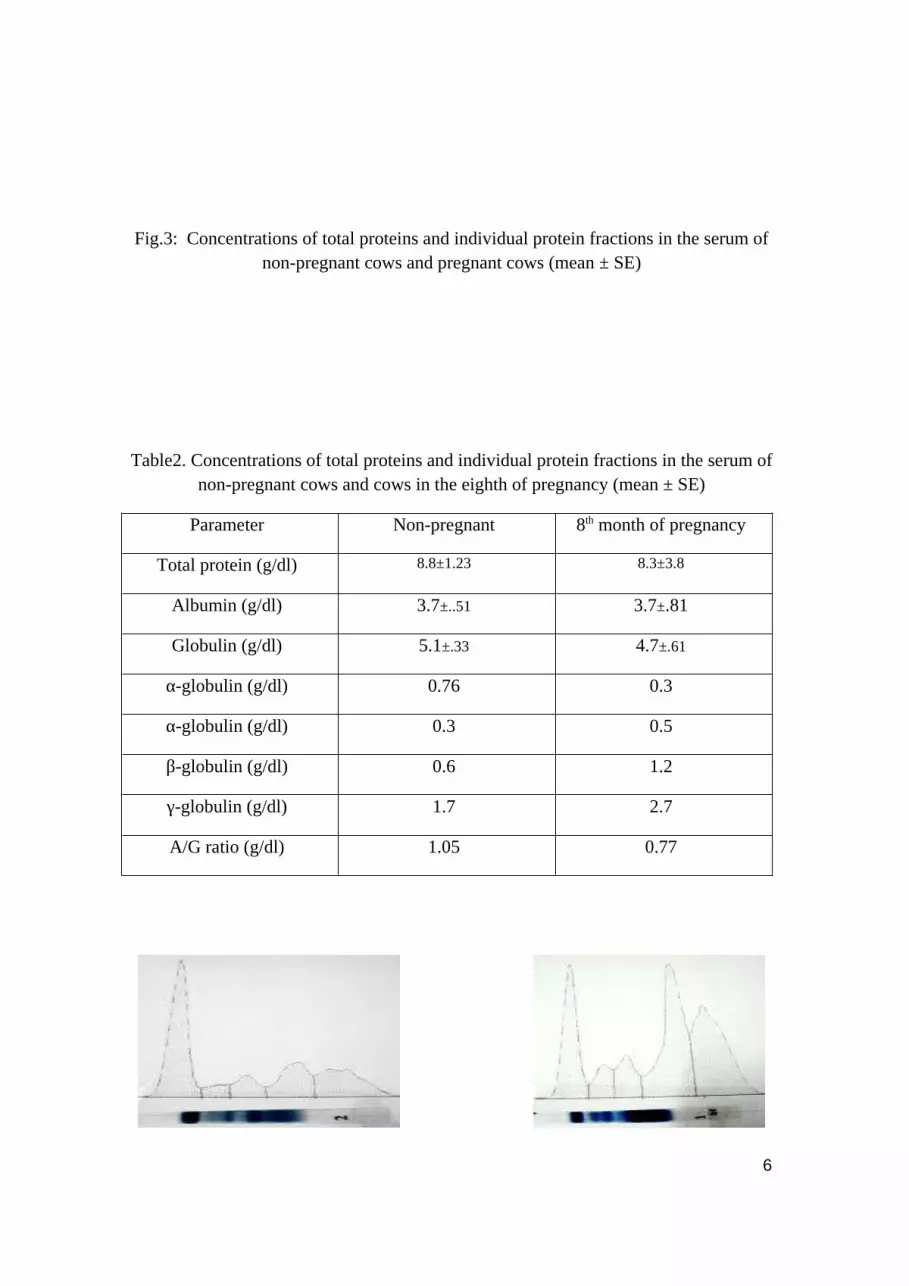
Fig.3: Concentrations of total proteins and individual protein fractions in the serum of non-pregnant cows and pregnant cows (mean ± SE)
Table2. Concentrations of total proteins and individual protein fractions in the serum of non-pregnant cows and cows in the eighth of pregnancy (mean ± SE)
Parameter Non-pregnant 8th month of pregnancy
Total protein (g/dl) 8.8±1.23 8.3±3.8
Albumin (g/dl) 3.7±..51 3.7±.81
Globulin (g/dl) 5.1±.33 4.7±.61
α-globulin (g/dl) 0.76 0.3
α-globulin (g/dl) 0.3 0.5
β-globulin (g/dl) 0.6 1.2
γ-globulin (g/dl) 1.7 2.7
A/G ratio (g/dl) 1.05 0.77
6

Fig. 4. Concentrations of total proteins and individual protein fractions in the serum of non-pregnant cows and cows in the eighth and ninth months of pregnancy (mean ± SE)
Table 3. Concentrations of total proteins and individual protein fractions in the serum of non-pregnant cows and cows in the eighth and ninth months of pregnancy (mean ± SE)
Parameter 8th month of pregnancy 9th month of pregnancy
Total protein (g/dl) 8.3±0.38 7.5±0.23
Albumin (g/dl) 3.6±.81 3.1±.17
Globulin (g/dl) 4.7 4.4±.52
α-globulin (g/dl) 0.3 1.23±.25
α-globulin (g/dl) 0.5 0.86
β-globulin (g/dl) 1.2 2.3
γ-globulin (g/dl) 2.7 1.7
A/G ratio (g/dl) 0.77 2.5
Since uterine secretory proteins play important roles in the maintenance of pregnancy, comparative studies on the total protein concentration and differential profile of proteins in uterine secretions of normal non-pregnant as well as repeat breeding animals could be very useful. The present study revealed the concentrations of total proteins in the uterine secretions of firest breeder cows to be very significantly (p<0.01) higher than those of normal healthy non – pregnant cows. The levels of proteins in uterine flushings of healthy cows have been reported to be 8.3.25 ± 0.38 mg/100 ml (DIXON, 1961) while protein levels of uterine flushings of breeder cows have been reported to be 7.5 mg/100 ml (BARTA; ARNOLD 1993) The higher concentrations of proteins in uterine flushings of repeat breeders were suggested to be due to the increased levels of secretory proteins, cellular debris and tissue damage. The total protein concentration in uterine flushings was reported to decrease significantly after treatment. In the present study, the analysis of proteins revealed some interesting patterns. On Native the protein was found to be exclusively present in the uterine secretions of healthy non – pregnant cows only whereas, the proteins p22 and p20 were present in the uterine secretions of both, the healthy non – pregnant and the repeat breeder cows. The protein p<10 was found to be exclusively present in the uterine secretions of repeat breeder cows only, whereas, p33 was present in both, the healthy non-pregnant as well as the repeat breeder cows. It was interesting to note that p22 and p20 and p33 (SDS-PAGE), common to healthy non-pregnant and repeat breeder cows, were conspicuously absent in uterine secretions from pregnant cows. We
7

have not come across any report on such studies on comparison of protein profiles of uterine secretions of repeat breeder cows with healthy non-pregnant and pregnant cows in the available literature. The exact nature and function of proteins unique to the repeat breeders or non – pregnant cows observed in our study remain to be elucidated. The complete understanding of their roles holds the key to immunological manipulation of sterility and fertility in reproduction and treatment of repeat breeding of non-infectious origin in domestic animals.
Thyroid Hormones
thT4 and triiodothyronine concentrations were at least twice as high in the serum of pregnant cows as compared with the non-pregnant cows (fig. 5) by (6.44±0.8 and 4.5±0.46 ) for T3 while, the level in T4 was(194±20.4 and 143.4±6.75 nmol/l ). It is concluded that the thyroid gland has activity in the pregnant cows and are relatively independent of one another. Thyroid hormones are involved in the overall metabolic rate and oxygen consumption of the body. Their effect on mammary development is probably indirect or via the normal requirements of cell maintenance. Hypothyroidism retards ductal and lobuloalveolar growth. Administration of thyroid hormones restores the normal developmental pattern.
Fig.5: Thyroxin and triiodothyronine levels in pregnant and non pregnant cows
Thyroid hormones are important regulators of mammalian development, cellular differentiation and metabolism (Ingbar, et al., 1986). In vivo studies as early as 1934 demonstrated that thyroid hormone administration can increase milk production in dairy cows and in vitro studies have shown that 3,3',5-triiodothyronine (T3) potentiates the activity of other lactogenic and galactopoietic hormones (Houdebine et al., 1978; Bhattacharjee and Vonderhaar, 1984). Tissue sensitivity to thyroid hormones can be altered by iodothyronine deiodination and changes in expression of nuclear receptors for
In the present study, serum progesterone (P4) level was significantly high (P<0.05) in pregnant cows in second trimester as compared with non-pregnant ones and this level was over three times (6.08 ng/ml) than non-pregnant (1.6 ng/ml) (Fig .6). This result agree with EL-Masry et al., (1997) who reported that P4 level increased with the advance of pregnancy, and decreased in non-pregnant buffaloes cows. Also, it was reported in previous study on female camels that P4 levels were increased by pregnancy and this increase reached four folds during the 1st half of pregnancy as compared to non-pregnant animals Abdel-Rahman et al., (2000). The high levels of P4 might require for maintaining of pregnancy because P4 acts directly to inhibit myometrial
8

concentrations and might prevent oxytocin release and to the increase in each of maternal body weight during pregnancy and fetus body weight (El-Fouly et al., 1998a).Fig.6: level of progesterone in pregnant and nonpregnant cows
ConclusionsThe protein profiles and total protein concentration of uterine secretions of uninfected repeat breeder cows were compared with those of healthy non-pregnant animals. The mean value of total proteins in uterine secretions of repeat breeders was found to be very significantly (p<0.01) higher than that of normal healthy non – pregnant cows. On SDS-PAGE, the protein p<10 was found to be present exclusively in the uterine secretions of repeat breeder cows but not in healthy non – pregnant cows. On Native PAGE, proteins p22 and p20, and on SDS-PAGE, proteins p>100, p68, p52, p33 and p20 were found to be common between healthy non – pregnant cows and repeat breeder cows. The proteins p27 (Native PAGE) and p24 (SDS-PAGE) were present in the uterine secretions of healthy non-pregnant cows but absent in case of repeat breeder cows. The exact physiological role and significance of characteristic presence or absence of certain proteins in uterine secretions of repeat breeder cows could not be ascertained.
AcknowledgementsThe author is grateful to the Dina farms and the staff of the Farm for allowing collecting the samples and to Dr. Samir Fathy Osman, formerly in the Department of agriculture productions for help. And many grateful for Prof . Dr. Abdel Wahab Prof of clinical biochemistry for his help.
ReferencesAbdel-Rahman, H.; A. F. Nebar; A. A. M. Habeeb; H. M. Yousef and H. E. S. Deyab (2000). Journal of Agricultural research, 25: 415-425.BARTA, O., D. F. ARNOLD (1993): St Laboratory, 1 ed. (Barta, O., Ed). Bar-Lab, Inc. Blacksburg. pp. C1-17.Bhattacharjee M, Vonderhaar BK. Thyroid hormones enhance the synthesis and secretion of alpha lactalbumin by mouse mammary tissue in vitro. Endocrinology 115:1070–1077, 1984. BUSH, B. M. (1998): Nd Animal Clinicians, 2ed., (Bush B.,M., Ed.). Blackwell Science Ltd, Oxford OEL. pp. 250-254.DIXON, F .J., W. O. WEIGLE,J.J.VASQUEZ (1961): . Lab. Invest. 10, 216-236.El-Fouly, H. A., K. A. El-Masry and M. H. Gamal (1998a).J. Zag. Vet. 26: 68-78.El-Masry, K. A., H. M. Yousef and H. A. Farghaly (1997). Buffalo J.,2: 147-156.GREEN, S. A., S. J. JENKINS,P.A.CLARK (1982): Cornell Vet. 72, 416-426.Gupta, H. C., Branton, C., Evans, D. L. and Waters, W. H. (1962) Journal of Dairy Science. 45: 668-669. Houdebine L, Delouis C, Devinoy E. Post-transcriptional stimulation of casein synthesis by thyroid hormone. Biochimie 60:809–812, 1978.Ingbar SH, Braverman LE, Eds. The Thyroid. New York: J.B. Lippincott Company, 1986.JAIN, N. B. (1993): Lea & Febiger. Philadelphia. pp. 349-379.JEFFCOTT, L. B. (1974): J. Comp. Path. 84, 93-101.KANEKO, J. J. (1989): Academic Press. San Diego. pp. 142-165. Laemmli, U. K. (1970). Nature. 227: 680-685.
9

LIBERG, P. (1977): Acta Vet. Scand. 18, 40-53.MCGUIRE, T. C., D. S. ADAMS (1982): Comp. Cont. Educ. Pract. Vet. 4, 35-40.Rao, K. S. and Sheshagiri, V. N. (1998) 75: 369-370. Reinhold, G. (1953) Academic Press, New York. Vol. I: p 88. TRUMEL, C., F. SCHELCHER,J.P.BRAUN,J.F.GUELFI (1996): Rev. Med. Vet. 147, 123-130.VAN DEN BROEK, A. H. M. (1992): Br. Vet. J. 148, 259-262.Yoshid, A. Y. (1986): J. Vet. Med. Sci. 48, 1153-1159.
10