Embed Size (px)
Citation preview

Available online at www.sciencedirect.com
Biomaterials 29 (2008) 1750e1761www.elsevier.com/locate/biomaterials
Comparison of nanoscale and microscale bioactive glasson the properties of P(3HB)/Bioglass� composites
Superb K. Misra a,1, Dirk Mohn b,1, Tobias J. Brunner b, Wendelin J. Stark b, Sheryl E. Philip c,Ipsita Roy c, Vehid Salih d, Jonathan C. Knowles d, Aldo R. Boccaccini a,*
a Department of Materials, Imperial College London, Prince Consort Road, London SW7 2AZ, UKb Institute for Chemical and Bioengineering, Department of Chemistry and Applied Biosciences, ETH Zurich, 8093 Zurich, Switzerland
c Department of Molecular and Applied Biosciences, University of Westminster, London W1W 6UW, UKd Division of Biomaterials and Tissue Engineering, UCL Eastman Dental Institute, London WC1X 8LD, UK
Received 30 September 2007; accepted 22 December 2007
Available online 6 February 2008
Abstract
This study compares the effects of introducing micro (m-BG) and nanoscale (n-BG) bioactive glass particles on the various properties(thermal, mechanical and microstructural) of poly(3hydroxybutyrate) (P(3HB))/bioactive glass composite systems. P(3HB)/bioactive glasscomposite films with three different concentrations of m-BG and n-BG (10, 20 and 30 wt%, respectively) were prepared by a solvent castingtechnique. The addition of n-BG particles had a significant stiffening effect on the composites, modulus when compared with m-BG. However,there were no significant differences in the thermal properties of the composites due to the addition of n-BG and m-BG particles. The systematicaddition of n-BG particles induced a nanostructured topography on the surface of the composites, which was not visible by SEM in m-BG com-posites. This surface effect induced by n-BG particles considerably improved the total protein adsorption on the n-BG composites compared tothe unfilled polymer and the m-BG composites. A short term in vitro degradation (30 days) study in simulated body fluid (SBF) showed a highlevel of bioactivity as well as higher water absorption for the P(3HB)/n-BG composites. Furthermore, a cell proliferation study using MG-63cells demonstrated the good biocompatibility of both types of P(3HB)/bioactive glass composite systems. The results of this investigationconfirm that the addition of nanosized bioactive glass particles had a more significant effect on the mechanical and structural properties ofa composite system in comparison with microparticles, as well as enhancing protein adsorption, two desirable effects for the application ofthe composites in tissue engineering.� 2008 Elsevier Ltd. All rights reserved.
Keywords: Poly(3hydroxybutyrate); MG-63 osteoblasts; Nanoparticles; Bioactive glass; Bioactive composites; Osteoblast
1. Introduction
A number of combinations of biodegradable polymers andbioactive ceramics in the form of biocompatible scaffolds havebeen used in the field of tissue engineering, with particularemphasis towards hard tissue regeneration [1e4]. Polyhydrox-yalkanoates (PHA) are among the group of biodegradable
* Corresponding author. Tel.: þ44 207 594 6731; fax: þ44 207 584 3194.
E-mail address: [email protected] (A.R. Boccaccini).1 Both authors contributed equally towards the completion of this work.
0142-9612/$ - see front matter � 2008 Elsevier Ltd. All rights reserved.
doi:10.1016/j.biomaterials.2007.12.040
polymers that have been used in combination or alone for bio-medical applications (tissue regeneration, drug delivery,patches) [5,6]. One popular member within the PHA familyis poly(3hydroxybutyrate), known as P(3HB), which exhibitsa much longer degradation time than polymers of thepoly(a-hydroxyacid) group (e.g. PLA or PLGA). P(3HB) isa microbial derived polyester that has been reported to beproduced by a wide variety of organisms and under variouscarbon sources [7]. P(3HB) has also been shown to havea good degree of biocompatibility with various cell lines[5,8]. In addition to its biocompatibility and biodegradability,P(3HB) has been reported to have piezoelectric properties,

Table 1
Chemical composition (wt%) of n-BG and m-BG particles used in this study
and their respective BET-specific surface area (SSA) measurements
SiO2 Na2O CaO P2O5 SSA (m2 g�1)
m-BG 45 24.5 24.5 6 <1
n-BG 46.08 22.96 27.18 3.77 79
1751S.K. Misra et al. / Biomaterials 29 (2008) 1750e1761
which can possibly play a vital role to stimulate bone growthand regeneration [9,10]. Various composites of P(3HB) andbioactive inorganic phases, such as hydroxyapatite, wollaston-ite and Bioglass�, have been prepared to impart strength andbioactivity to the composites, as reviewed elsewhere [11].Inorganic phases can in principle be added to the polymermatrix in their micro- or nanoscale. However, for PHAcomposites, mainly microparticles have been investigated[11,12].
Presently nanofibres [13,14] as well as nanoparticles are be-ing developed for tissue engineering applications. Nanoscalehydroxyapatite [15,16], tricalcium phosphate [17], bioactiveglass [18], and titanium oxide [19], for example, have beenprepared and used as reinforcement materials in biopolymermatrix composites. The extent of the differences in the me-chanical properties and in vitro degradation characteristics ofcomposites due to micro- and nanoscale bioactive phaseadditions is an interesting topic of research but it has beeninvestigated only in few cases (in isolation), e.g., in the caseof hydroxyapatite and bioactive glass particles, respectively[15,16,18]. Loher et al. have most recently demonstrated thestrongly improved bioactivity, degradation and mechanicalproperties of nanoscale tricalcium phosphate doped PLGAwhen compared to pure polymer or composites containing mi-croparticles [17]. The size of the filler particles is an importantparameter that affects the mechanical properties of compositematerials, due to marked microstructural differences intro-duced by the micro- or nanoscale fillers that contribute to-wards different interaction between the filler particles andthe polymer matrix. The larger specific surface area of thenanoparticles should lead to increased interface effects and itshould also contribute to improved bioactivity, when com-pared to standard (mm-sized) particles [17,18,20]. Addition-ally, the use of nanoparticles in a polymeric matrix mimicsmore closely the structure of natural bone, which containsnanoscale hydroxyapatite crystallites, which are responsiblefor the high strength of bone [21], combined with the poly-meric phase of collagen. Pramanik et al. [15] have shownthat the addition of nanoparticles of hydroxyapatite to poly(-ethylene-co-acrylic acid) results in a significant increase instrength and modulus, when compared with hydroxyapatitemicroparticles. Addition of hydroxyapatite nanoparticles toPLLA scaffolds (prepared using phase separation technique)has also been reported to help the scaffolds maintain aniso-tropic and regular pore structure, compared to the irregularpore structure due to addition of mm-sized hydroxyapatite[16]. Human mesenchymal stem cells grown on electro-spunnanoscale tricalcium phosphate/PLGA composites exhibitedvery high bioactivity and showed rapid differentiation onsuch surfaces confirming an improved textural stimulationby the nanostructured composites [22]. Webster et al. [23]showed that a significant increase in protein adsorption and os-teoblast adhesion was observed on the nanoscale ceramic ma-terials compared to microscale ceramic materials. It has alsobeen shown that bioactive glass nanoparticles, when immersedin water produced a higher alkalinity than the commerciallyavailable (mm-sized) 45S5 Bioglass� [18]. This effect could
buffer to a greater extent the acidic degradation of somepolymers [2], when the nanoscale bioactive glass particlesare used as filler in a composite.
The above-mentioned examples serve to demonstrate thatthe presence of nanoscale fillers in biopolymers can affect toa great extent the microstructural, mechanical and biologicalbehaviour of the composite systems developed for tissueengineering applications as well as their in vitro degradationproperties. Keeping this evidence in mind, the present studyaims to depict, for the first time, a direct comparison of the dif-ferences in the structural, thermal, mechanical, and biologicalbehaviour of P(3HB)/bioactive glass composites due to theaddition of nanoscale (n-BG) or microscale bioactive glass(m-BG) particles. The reason for using bioactive glass parti-cles is their high bioactivity [24,25], particularly in the formof nanoparticles [18,20], as well as the demonstrated effectof bioactive glass dissolution products on osteoblast cellgene expression [26] and potential effect on angiogenesis[27], which provides a conducive environment for cell coloni-sation, proliferation and cell differentiation as well as potentialenhanced vascularisation. Melt-derived m-BG particles andflame spray synthesised n-BG particles of the same nominalcomposition (45S5 [24]) were combined with P(3HB), in con-centrations of 10, 20 and 30 wt%. A short term (30 days) invitro degradation study using simulated body fluid (SBF)was also conducted to compare the weight loss and wateruptake for the two types of composites. A protein adsorptionstudy using foetal bovine serum as well as a preliminary cellproliferation study on the various composite samples wascarried out using MG-63 osteoblast cell line.
2. Materials and methods
2.1. Materials
All chemicals for polymer production, composite preparation and simu-
lated body fluid (SBF) preparation were obtained from SigmaeAldrich Com-
pany Ltd. (England). The chemicals required for the preparation of n-BG
particles were obtained from Riedel-de Haen, Applichem, Lancaster and Acros
(Switzerland). The bacterial strain used for producing P(3HB) was provided by
the University of Westminster culture collection. The details of P(3HB)
production have been presented recently elsewhere [28]. Briefly, P(3HB)
was produced using Bacillus cereus SPV in a 20 l fermenter vessel. The culture
was grown for 72 h under nitrogen limitation conditions and the polymer was
subsequently extracted using a dispersion technique [28,29].
Two different types of bioactive glass particles, i.e., bioactive glass micro-
particles (m-BG) and bioactive glass nanoparticles (n-BG) of nominal compo-
sition ‘‘45S5’’ (composition shown in Table 1 [24]) were used in this study to
prepare the composite systems. The m-BG particles used were from a melt-
derived bioactive glass powder, with mean particle size <5 mm. This grade
of bioactive glass was developed initially by Hench et al. [30] and has been
extensively used in the last 10 years in the field of tissue engineering and

1752 S.K. Misra et al. / Biomaterials 29 (2008) 1750e1761
bone regeneration [2,4,25e27,31], commonly known as 45S5 Bioglass�.
Flame spray synthesis [32] was applied to prepare bioactive glass nanopar-
ticles, as described by Brunner et al. [20]. Briefly, corresponding amounts of
2-ethylhexanoic acid salts of calcium and sodium were mixed with hexame-
thyldisiloxane and tributylphosphate and diluted with xylene. The solution
was pumped (10 ml min�1) through a capillary (diameter 0.4 mm), dispersed
with oxygen (10 l min�1) and ignited with a methane (1.13 l min�1) and oxy-
gen (2.4 l min�1) flame. The as-formed bioactive glass particles were collected
by using baghouse filter and they were then sieved by using 250 mm meshes to
separate the agglomerates. The n-BG and m-BG particles were analysed prior
to composite preparation by X-ray diffraction (XRD) analysis (analytical pa-
rameters in Section 2.2). Scanning electron microscopy (SEM) (gold/platinum
coated samples) and transmission electron microscopy (TEM) images of the n-
BG and m-BG particles were taken using JEOL 5610LV (JEOL, USA), LEO
1530 Gemini and JEOL JEM-2000 FX II (fitted with LaB6 filament), respec-
tively. Typical images of n-BG and m-BG particles are shown in Fig. 1. The
chemical composition of the n-BG particles was determined by using laser
ablation inductively coupled plasma mass spectrometry (LA-ICP-MS). More-
over, BET-specific surface area values for the m-BG and n-BG particles were
measured by nitrogen adsorption at 77 K using a Tristar 3000 (Micrometrics,
Norcross, GA, USA).
2.2. Composite preparation
P(3HB) and P(3HB)/bioactive glass films were prepared by employing the
solvent casting technique. Chloroform was used to dissolve P(3HB) using
a polymer concentration of 3 wt%. Three different concentrations of n-BG
and m-BG particles viz. 10, 20 and 30 wt%, were added to the polymer solu-
tion and this was sonicated for 1e2 min using a sonicating probe (Ultrasonic
Homogenizers US200, Philip Harris Scientific, Leicestershire, UK) in order to
break the bioactive glass agglomerates and ensure a better (homogeneous)
distribution of n-BG and m-BG particles in the composite. The solution was
then cast onto glass Petri dishes at room temperature. Films of nominally
0.12e0.14 mm thickness were produced. Once the films were cast they were
stored in desiccators for further analyses as mentioned below.
Fig. 1. (a) SEM image of m-BG particles, (b) SEM image
2.3. Physical characterisation of materials
2.3.1. Scanning electron microscopy (SEM)The samples before and after in vitro degradation (dried) were placed on
8 mm diameter aluminium stubs and gold/platinum coated for 2 min. Images
were taken at various magnifications and acceleration voltages (max. of
20 kV) to avoid beam damage to the polymer.
2.3.2. Contact angle study
Static contact angle measurements were carried out in order to evaluate the
wettability of the composite samples. The experiment was carried out on
a KSV Cam 200 optical contact angle meter (KSV Instruments Ltd., Finland).
An equal volume of water (<20 ml) was placed on every sample by means of
a gas tight micro-syringe forming a drop. Photos (frame interval e 1 s, number
of frames e 100) were taken to record the shape of the drops. The water
contact angles on the specimens were measured by analysing the recorded
drop images (four repeats for each sample) using the Windows based KSV-
Cam software.
2.3.3. Differential scanning calorimetry (DSC)
Thermal properties of the samples were measured using a PerkineElmer
Pyris Diamond DSC (PerkineElmer Instruments, USA). The sample mass
for these measurements was in the range of 5e7 mg. Samples were encapsu-
lated in standard aluminium pans and all tests performed under nitrogen atmo-
sphere. The samples were heated/cooled/heated at a rate of 20 �C min�1
between �50 �C and 200 �C. Measurements were carried out in triplicates.
2.3.4. Mechanical tests
Tensile strength tests were conducted on flat specimens (width: 1.4 mm,
length: 7e8 mm and thickness: w100e120 mm) cut out from the solvent
cast films, using a PerkineElmer Dynamic Mechanical Analyser (DMA 7e,
PerkineElmer Instruments, USA) at room temperature. The initial load was
set to 1 mN and it was increased to 6000 mN at a rate of 200 mN min�1.
Four repeat specimens were tested for each sample during this experiment.
of n-BG particles (c) TEM image of n-BG particles.

1753S.K. Misra et al. / Biomaterials 29 (2008) 1750e1761
2.3.5. X-ray diffractionX-ray diffraction (XRD) analysis on samples before and after immersion in
SBF was performed on a Bruker D8 Advance diffractometer in flat plate
geometry, using Ni filtered Cu Ka radiation. Data were collected from 10 to
100� in qeq mode with a primary beam slit size of 0.6 mm. A Bruker Lynx
Eye silicon strip detector was used and a step size of 0.0196671� and a count
time of 0.1 s per step.
2.3.6. Protein adsorption studyA protein adsorption assay was carried out on the samples using foetal
bovine serum (FBS) (SigmaeAldrich, UK). Analysis was performed in tripli-
cates. The samples (1 cm2) were incubated with 200 ml of undiluted FBS in
1.5 ml eppendorf and incubated at 37 �C for 24 h. The serum was then
removed and the samples were washed three times with phosphate buffered
saline (PBS). The proteins adsorbed on the samples were further collected
by incubating the samples with 1 ml of 2% sodium dodecyl sulfate (SDS) in
PBS for 24 h at room temperature and under vigorous shaking. The amount
of total protein adsorbed by the samples was measured by using a commercial
protein quantification kit (Pierce, Rockford, IL). The optical density of the
samples was measured spectrophotometrically at 562 nm against a calibration
curve using bovine serum albumin as per the manufacturer’s protocol.
2.4. Acellular in vitro tests in SBFof P(3HB)/bioactive glass composites
A short term in vitro bioactivity study was carried out using acellular sim-
ulated body fluid (SBF), as developed by Kokubo et al. [33]. All the samples
were weighed (M0, dry), immersed in SBF and kept under static conditions at
37 �C for periods of up to 30 days. The SBF was exchanged every 5 days. At
specified time points (5, 10 15, 20 and 30 days), the films were collected and
analysed for water uptake (%WA) and weight loss (%WL) behaviour. For mea-
suring the water absorption (%WA) of the samples, the immersed samples
were extracted at given time points, the surface was gently wiped and the
weight was measured (Mt, wet). Similarly, for measuring the weight loss the
samples were withdrawn from the SBF, dried at 37 �C overnight and subse-
quently weighed (Mt, dry). Water absorption and weight loss were calculated
using the following equations:
%WA¼�
Mt;wet�Mt;dry
Mt;dry
�� 100
%WL¼�
M0;dry�Mt;dry
M0;dry
�� 100
ð1Þ
The P(3HB)/n-BG samples were analysed at each time point using XRD and
SEM to determine the level of bioactivity. P(3HB)/m-BG samples were not
analysed for this part of the experiments, since the bioactivity of m-BG parti-
cles (45S5 Bioglass�) has been extensively reported in the literature [4,24,25]
and that of P(3HB)/m-BG composites has also been investigated in an earlier
study [12].
2.5. Cell culture studies
In vitro cell culture studies were conducted on the P(3HB)/n-BG and
P(3HB)/m-BG films using MG-63 osteoblast cells, human osteosarcoma cell
line [34]. The cells were cultured in growth medium [Dulbecco’s modified Ea-
gles Medium (DMEM), (PAA, Germany), supplemented with 10% foetal calf
serum, 1% penicillin and streptomycin solution] and incubated at 37 �C in
a humidified atmosphere (5% CO2 in 95% air). Culture medium was changed
every 2 days.
2.5.1. Cell seeding on the test samples
The semi-confluent cells were released by trypsinisation and pelleted by
centrifugation at 1000 rpm for 3 min. A cell count was performed using trypan
blue dye and haemocytometer. The samples (1 cm2) were UV-sterilised for
30 min (each side) and passivated in culture medium for 12 h, prior to seeding
the cells. A cell density of 20,000 cells/cm2 was used to seed the samples kept
in 24 well plates, followed by incubating the samples in a humidified environ-
ment for a period of up to 7 days. The medium of the wells was changed every
second day and samples were analysed after 1, 4 and 7 days for cell prolifer-
ation and SEM observation. Cell culture studies were carried out on triplicate
samples per experiment. Standard tissue culture plastic was used as the control
surface.
2.5.2. Cell proliferation study
The cell proliferation study on the samples was carried out using Alamar
blue assay (AbD Serotec, UK). Alamar blue was added to the samples (10% v/
v of medium) and incubated at 37 �C in an incubator (with 5% CO2) for 4 h.
Aliquots of 100 ml from each sample well were transferred to a black 96-well
plate and the fluorescence of Alamar blue was measured using a fluorescence
plate reader (Fluoroskan, Lab Systems) at absorbance 560 nm (A560) and emis-
sion 590 nm (A590).
2.5.3. SEM preparation for cells
One particular composition of composites, i.e., P(3HB)/30 wt% bioactive
glass (m-BG and n-BG) and unfilled P(3HB) films were examined under
SEM to observe the MG-63 cell spreading and attachment on the surface of
the samples. Day 1, 4 and 7 specimens were fixed in 3% glutaraldehyde in
0.1 M cacodylate buffer for 12 h at 4 �C. Subsequent dehydration using a series
of graded ethyl alcohols (50%, 70%, 90% and 100%) was performed. Samples
were then critical point dried by immersion in hexamethyldisilazane for 2 min
and left in a fume cupboard for 2 h for subsequent drying. The dried samples
were then attached to aluminium stubs, gold coated and examined under SEM
(JEOL 5610LV, JEOL, USA) at an acceleration voltage of 10e15 kV.
2.6. Statistical analysis
All data are expressed as mean� standard deviation. The data were
compared using Student’s t-test and differences were considered significant
when *p< 0.05, very significant **p< 0.01 and highly significant
***p< 0.001, respectively. A p-value higher than 0.05 ( p> 0.05) was taken
as indicating no significant difference.
3. Results
3.1. Bioactive glass characterisation
The chemical composition of the m-BG particles and the n-BG particles is shown in Table 1. The chemical compositionfor the n-BG particles was measured using LA-ICP-MS andshowed a very similar composition to the commercially avail-able m-BG particles (45S5 Bioglass�). The n-BG particlesused in this study had a spherical shape with relatively smallagglomerate sizes compared to the irregular shape of the m-BG particles (as shown in Fig. 1). The specific surface area(SSA) and the BET equivalent diameter for the n-BG particleswere 79 m2 g�1 and 29 nm, respectively [20]. In contrast, forthe m-BG particles, the SSA was <1 m2 g�1. The XRD patternshowed no crystalline peaks and confirmed the amorphousnature of both n-BG and m-BG particles.
3.2. Morphology and microstructural properties
The morphology and microstructure of the P(3HB)/n-BGand P(3HB)/m-BG composites examined using SEM areshown in Fig. 2 (planar and cross sections of P(3HB)/30 wt% n-BG and P(3HB)/30 wt% m-BG composites). It canbe seen from the micrographs that the addition of n-BG parti-cles changes the surface morphology considerably and adds tothe surface a rough topography when compared to the m-BG
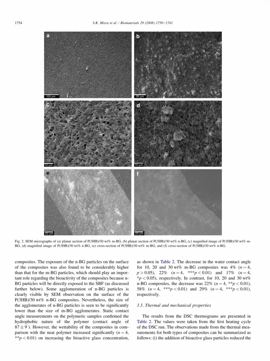
Fig. 2. SEM micrographs of (a) planar section of P(3HB)/30 wt% m-BG, (b) planar section of P(3HB)/30 wt% n-BG, (c) magnified image of P(3HB)/30 wt% m-
BG, (d) magnified image of P(3HB)/30 wt% n-BG, (e) cross-section of P(3HB)/30 wt% m-BG, and (f) cross-section of P(3HB)/30 wt% n-BG.
1754 S.K. Misra et al. / Biomaterials 29 (2008) 1750e1761
composites. The exposure of the n-BG particles on the surfaceof the composites was also found to be considerably higherthan that for the m-BG particles, which should play an impor-tant role regarding the bioactivity of the composites because n-BG particles will be directly exposed to the SBF (as discussedfurther below). Some agglomeration of n-BG particles isclearly visible by SEM observation on the surface of theP(3HB)/30 wt% n-BG composites. Nevertheless, the size ofthe agglomerates of n-BG particles is seen to be significantlylower than the size of m-BG agglomerates. Static contactangle measurements on the polymeric samples confirmed thehydrophobic nature of the polymer (contact angle of87� 9
�). However, the wettability of the composites in com-
parison with the neat polymer increased significantly (n¼ 4,**p< 0.01) on increasing the bioactive glass concentration,
as shown in Table 2. The decrease in the water contact anglefor 10, 20 and 30 wt% m-BG composites was 4% (n¼ 4,p> 0.05), 22% (n¼ 4, ***p< 0.01) and 17% (n¼ 4,*p< 0.05), respectively. In contrast, for 10, 20 and 30 wt%n-BG composites, the decrease was 22% (n¼ 4, **p< 0.01),58% (n¼ 4, ***p< 0.01) and 29% (n¼ 4, ***p< 0.01),respectively.
3.3. Thermal and mechanical properties
The results from the DSC thermograms are presented inTable 2. The values were taken from the first heating cycleof the DSC run. The observations made from the thermal mea-surements for both types of composites can be summarized asfollows: (i) the addition of bioactive glass particles reduced the

Table 2
Comparison of thermal properties and water contact angle for P(3HB)/n-BG
and P(3HB)/m-BG composite samples
Bioactive glass
concentration (wt%)
Tm (�C) DHf (J/g) Water contact angle
(degree� SD)
0 172� 2 73� 1 87� 9
10 m-BG 155/171 58� 2** 83� 6
n-BG 156/172 55� 9** 68� 1**
20 m-BG 156/172 59� 2** 68� 6**
n-BG 157/171 60� 4** 55� 1***
30 m-BG 156/170 57� 3* 71.8� 0.3*
n-BG 155/170 55� 6* 62� 2***
***p< 0.001; **p< 0.01; *p< 0.05 [data compared with P(3HB)]. Tm, melt-
ing temperature; DHf, heat of fusion.
1755S.K. Misra et al. / Biomaterials 29 (2008) 1750e1761
heat of fusion (n¼ 3, **p< 0.01) when compared to the poly-mer, which can also be used as indicator of the crystallinity ofthe material, (ii) the differences due to the addition of the n-BG and m-BG particles on the composites’ thermal properties(melting temperature, heat of fusion) was insignificant (n¼ 3,p> 0.05), as shown in Table 2, (iii) the addition of both n-BGand m-BG particles prevents the recrystallisation of the poly-mer during the cooling run (after the first heating), as investi-gated elsewhere [12]. This was evident by the absence of thepolymer melting peak during the second DSC heating run(data not shown here).
The static mechanical tests conducted on composite sam-ples, however, yielded different results. In contrast to a reduc-tion (n¼ 4, p> 0.05) in the Young’s modulus due to theaddition of the m-BG particles, the addition of 10, 20 and30 wt% n-BG particles resulted in a significant increase(n¼ 4, *p< 0.05) in the Young’s modulus compared to thevalue of the unfilled P(3HB) film, which was found to be1.01 GPa. The increase in the elastic modulus for the compos-ites (compared to the unfilled polymer) due to the addition of10, 20 and 30 wt% of n-BG particles was 57%, 14% and 20%,respectively. In comparison, there was a decrease in the elasticmoduli of the composite due to the addition of m-BG, asshown in Fig. 3. The significant increase (n¼ 4, **p< 0.01)
Fig. 3. Modulus comparison for various concentrations of m-BG and n-BG
particles in P(3HB)/bioactive glass composites. The data (n¼ 4; error
bars¼�SD) were compared using Student’s t-test and differences were con-
sidered significant when *p< 0.05, **p< 0.01 and ***p< 0.001.
in the static modulus of the P(3HB)/n-BG composites com-pared to P(3HB)/m-BG composites can be explained due tothe true reinforcement achieved using BG nanoparticles, incontrast to m-BG particles. In this last case, the reduction ofYoung’s modulus is most certainly due to the poor mixingof m-BG particles with the polymer matrix, leading to largeagglomerations and consequent to residual porosity of thefilms, as described elsewhere [12] (see also Fig. 2c and e).
3.4. In vitro degradation study
The water uptake and weight loss study for the compositeswas conducted using immersion tests in SBF over a 1-monthperiod. Bioactivity of the P(3HB)/n-BG composites was alsodemonstrated using SEM and XRD analysis over the 1-monthperiod. The bioactivity of the P(3HB)/m-BG composites is notpresented in this study, as it has been investigated in an earlierwork [12]. The bioactivity of m-BG (45S5 Bioglass�) hasbeen extensively reported in the literature as well[4,24,25,27]. The results from the in vitro bioactivity studiesdepicted in Fig. 4 and Fig. 5 can be summarized as follows.
Fig. 4. In vitro degradation study of the samples in simulated body fluid show-
ing (a) % water absorption, (b) % weight loss. Data are presented in bar graphs
to have a direct comparative analysis between the m-BG and n-BG compos-
ites. (n¼ 3; error bars¼�SD).

5 µm
50 µm
5 µm
5 µm
a b
c d
Lin
(C
ou
nts)
0100200300400500600700800900
1000110012001300140015001600170018001900
2-Theta - Scale
20 30 40
30% n-BG 0 days
30% n-BG 5 days
30% n-BG 10 days
30% n-BG 20 days
30% n-BG 30 days
e
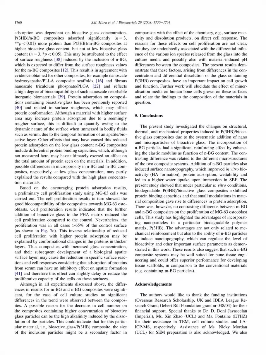
Fig. 5. SEM micrographs for (a)e(c) P(3HB)/30 wt% n-BG samples after 5, 15 and 20 days in SBF, (d) neat P(3HB) after 20 days in SBF, (e) corresponding XRD
plot with hydroxyapatite markers for P(3HB)/30 wt% n-BG composites after 0, 5, 10, 20 and 30 days.
1756 S.K. Misra et al. / Biomaterials 29 (2008) 1750e1761

Fig. 6. Total protein adsorption study on P(3HB)/bioactive glass composites
containing m-BG and n-BG particles in different concentrations (wt%) using
foetal bovine serum. The data (n¼ 3; error bars¼�SD) were compared using
Student’s t-test and differences were considered significant when *p< 0.05,
**p< 0.01 and ***p< 0.001.
1757S.K. Misra et al. / Biomaterials 29 (2008) 1750e1761
(i) The water absorption of the composite samples increasedon prolonged immersion in SBF, as well as on increasingthe bioactive glass concentration. The water uptake for then-BG composites was higher than that of the m-BG compos-ites. (ii) There was no significant change ( p> 0.05) in the wa-ter uptake and mass loss behaviour for the unfilled P(3HB)films, confirming their hydrophobic and slow degradation na-ture. (iii) The mass loss for the composite films increased withimmersion time and with bioactive glass concentration, andthe mass loss was found to be higher for the n-BG compositeswhen compared to m-BG composites. (iv) The level of bioac-tivity (given by the extent of HA formation for a determinedtime period in SBF) for the P(3HB)/n-BG composites wasconsiderably higher than for m-BG composites, which wereinvestigated previously [12], and it increased with immersiontime. Surface examination and XRD analysis of P(3HB)/30 wt% n-BG composites (Fig. 5) confirms the formation ofnanoscale hydroxyapatite on the composite’s surface within5 days of immersion in SBF. This feature was also evidentin 10 and 20 wt% n-BG samples. Although the bioactivitycomparison of the n-BG and m-BG particles on their own isnot addressed in this paper, certainly the high exposure of n-BG particles on the surface of the composite (Fig. 2b) is aidinghydroxyapatite formation and thus making the P(3HB)/n-BGcomposites ‘‘more’’ bioactive than earlier reported P(3HB)/m-BG composites [12]. In contrast, the neat P(3HB) filmshad no changes on the surface after 20 days of immersionand no evidence of bioactivity was recorded (Fig. 4d). Itshould be noted that XRD data was collected in qeq modeas opposed to thin film thus the low counts for the thin apatitefilm formed relative to the substrate.
3.5. Protein adsorption and cell culture analysis
The protein adsorption study showed that the total amountof protein adsorbed on the surface was higher for the compos-ites compared to the unfilled polymer (which was measured tobe 171� 20 mg). The presence of n-BG particles in the com-posite resulted in higher protein binding compared to thesame concentration of m-BG particles, as evident from the re-sults in Fig. 6. For example, composites with 30 wt% n-BGand m-BG particles resulted in 186% and 56% increase(n¼ 3, **p< 0.01) in total protein adsorption, respectively.Such a difference in protein adsorption capability is thoughtto be related to the marked differences in surface morphologyand surface area available for protein adsorption for each ofthe three films (P(3HB), P(3HB)/m-BG and P(3HB)n-BG),as shown in Fig. 2.
Cell proliferation study of MG-63 osteoblasts cells on thecomposite samples was carried out using Alamar blue assayas previously reported [35]. The proliferation assay showedthe cell growth to be higher on the neat polymer samplesthan on the composite samples (n¼ 3, **p< 0.01) and itwas comparable to cell growth on tissue culture plastic(n¼ 3, p> 0.05), as shown in Fig. 7. However, there seemedto be a decrease in cell growth with increasing bioactive glassconcentration. This trend was similar for both n-BG and m-BG
composites. Nevertheless, the composites as well as the neatpolymer samples showed an increase in cell proliferation dur-ing the 7 day period, in tandem with the trend for the tissueculture plastic used as a control (Fig. 7). Since the P(3HB)/n-BG and P(3HB)/m-BG samples were tested in separatebatches, the results obtained are not directly comparable.Therefore, Fig. 7c shows the cell proliferation results of allthe samples compared to the control (control is normalisedto 100%). In order to reveal the morphology of cells culturedon the P(3HB)/bioactive glass composites, cells cultured forday 4 and day 7 were examined by SEM and typical imagesare shown in Fig. 8. The attachment of cells on compositesamples and the intracellular communication of cells withone another were evident on all examined samples.
4. Discussion
This study examined the effect of the size of bioactive glassparticles (mm- and nm-sized) on the properties of P(3HB)/bio-active glass composites, which are being developed for tissueengineering applications [12]. All analyses carried out showedconsiderable and significant differences between the P(3HB)/m-BG and P(3HB)/n-BG composites in key properties relevantfor applications in tissue engineering. The nanometric size ofthe n-BG particles contributed to increasing the stiffness of thecomposites (up to a 50% increase), as previously observed innanoscale tricalcium phosphate/PLGA composites [17], by ef-ficiently filling small pores of the polymeric matrix (Fig. 2).The Young’s modulus increase in the PHA/n-BG compositescan be also related to the effect of the higher interfacial surfacearea provided by the nanoparticles, which should enhance theload transfer between the matrix and the stiff inclusions. Thisbehaviour was not achieved by the m-BG particles as theyhighly agglomerated leading to poor mixing with the matrixand thus to residual porosity with detrimental effect on themechanical properties of the composite, as discussed in detailelsewhere [12]. There was, however, no considerable

Fig. 7. Cell proliferation study for 1, 4 and 7 days, using Alamar Blue assay on
(a) P(3HB)/m-BG composites, (b) P(3HB)/n-BG composites, (c) all tested
samples relative to the control (control set to 100%). The data (n¼ 3; error
bars¼�SD) were compared using Student’s t-test and differences were
considered significant when *p< 0.05, **p< 0.01 and ***p< 0.001.
1758 S.K. Misra et al. / Biomaterials 29 (2008) 1750e1761
difference in the thermal properties of n-BG and m-BG com-posites. Addition of the n-BG particles induced nanotopogra-phy on the surface of the composite films by exposingdirectly the bioactive glass particles on the surface, whichwas not the case for the m-BG composites. This effect contrib-uted to the rapid formation of hydroxyapatite on the surface ofthe composite within 5 days of immersion in SBF (Fig. 5) andthis result is in agreement with the strongly improved bioactiv-ity determined in similar nanoscale tricalcium phosphate con-taining composites [17,22]. In the present experiments,nanoscale sized hydroxyapatite crystals started to nucleatefrom the n-BG particles and covered the surface of the com-posite thus making the n-BG containing composites highlybioactive and therefore a suitable biomaterial for bone tissueengineering applications [2]. Water contact angle measure-ments (also an indication of the wettability) showed that in-creasing the bioactive glass concentration increased thewettability of the composites. However, the decrease in watercontact angle was more prominent for the n-BG compositesthan for the m-BG composites (n¼ 4, *p< 0.05) due tothe fact that more of the n-BG particles were exposed on thesurface leading to increased interaction surface between thepolymer and the glass particle inclusions facilitating waterpenetration. The unfilled polymeric film showed a much lesswater absorption compared to the composite films, furtherconfirming the hydrophobic nature of the polymer. On directcomparison, the water absorption of the n-BG compositeswas found to be higher than that of the m-BG compositesand this could be due to the higher extent of exposure of then-BG particles on the composite surfaces as well as theirhigher surface area. An increased water absorption andimproved wettability, as found for the n-BG composites, cancontribute to altering the degradation rate of these materials,as discussed elsewhere for nanoscale tricalcium phosphate/PLGA composites [17].
Much research has revealed an understanding of cellematerials interface relationships particularly related to proteinadsorption. Culture media contain a large variety of differentcomponents, many of which are proteins intimately associatedwith cell attachment, spreading and proliferation. Moreover,the selective adsorption of important protein componentswill determine effective and desired cell functions on bioma-terials. Protein adsorption is of importance to evaluate the po-tential of a biomaterial for use as a tissue engineering scaffold,as most mammalian cells are anchorage dependent and needa biocompatible, protein rich surface for attachment, differen-tiation and migration to form new tissue [16,23]. It has beenshown that cell adhesion takes place in two different stages.The first stage consists of the adsorption of water and a layerof proteins that selectively adhere onto the biomaterial surface,mediated by the surface properties of the substrate [36]. Thesecond stage involves cell adhesion onto the layer of proteins,which is a more complex process, mediated by extracellularmatrix (ECM) proteins, cell membrane proteins, and cytoskel-etal proteins [37]. In the present experiments, the total serumprotein adsorbed on the surface was greater on P(3HB)/n-BGcompared to P(3HB)/m-BG composites but the extent of the

Fig. 8. Day 4 and day 7 SEM micrographs of MG-63 cells grown on (a) P(3HB) at day 4, (b) P(3HB) at day 7, (c) P(3HB)/30 wt% m-BG at day 4, (d) P(3HB)/
30 wt% m-BG at day 7, (e) P(3HB)/30 wt% n-BG at day 4, (f) and (g) P(3HB)/30 wt% n-BG at day 7.
1759S.K. Misra et al. / Biomaterials 29 (2008) 1750e1761
1760 S.K. Misra et al. / Biomaterials 29 (2008) 1750e1761
adsorption was dependent on bioactive glass concentration.P(3HB)/n-BG composites adsorbed significantly (n¼ 3,**p< 0.01) more protein than P(3HB)/m-BG composites athigher bioactive glass content, but not at low bioactive glasscontent (n¼ 3, *p< 0.05). This may be attributed to the effectof surface roughness [38] induced by the inclusion of n-BG,which is expected to differ from the surface roughness valuesfor the m-BG composites [12]. This result is in agreement withevidence obtained for other composites, for example nanoscalehydroxyapatite/PLLA composite scaffolds [16] and fibrousnanoscale tricalcium phosphate/PLGA [22] and reflectsa high degree of biocompatibility of such nanoscale resorbableinorganic biomaterials [39]. Protein adsorption on composi-tions containing bioactive glass has been previously reported[40] and related to surface roughness, which may affectprotein conformation. Although a material with higher surfacearea may increase protein adsorption due to a seeminglyrougher surface, this is difficult to quantify owing to thedynamic nature of the surface when immersed in bodily fluidssuch as serum, due to the temporal formation of an apatite/bio-active layer. Other effects that may have caused this reducedprotein adsorption on the low glass content n-BG compositesinclude differential protein binding capacities, which, althoughnot measured here, may have ultimately exerted an effect onthe total amount of protein seen on the materials. In addition,possible differences in microporosity in n-BG and m-BG com-posites, respectively, at low glass concentration, may partlyexplained the results compared with the high glass concentra-tion materials.
Based on the encouraging protein adsorption results,a preliminary cell proliferation study using MG-63 cells wascarried out. The cell proliferation results in turn showed thegood biocompatibility of the composites towards MG-63 oste-oblasts. Cell proliferation results indicated that the furtheraddition of bioactive glass to the PHA matrix reduced thecell proliferation compared to the control. Nevertheless, theproliferation was in all cases >65% of the control surface(as shown in Fig. 7c). This inverse relationship of reducedcell proliferation with higher protein adsorption may beexplained by conformational changes in the proteins in thickerlayers. Thus composites with increased glass concentration,and their subsequent development of a biological apatitesurface layer, may cause the reduction in specific surface reac-tions and cell responses considering that adsorption of proteinsfrom serum can have an inhibitory effect on apatite formation[41] and therefore this effect can slightly delay or reduce theproliferative capacity of the cells on these surfaces.
Although in all experiments discussed above, the differ-ences in results for m-BG and n-BG composites were signifi-cant, for the case of cell culture studies no significantdifferences in the trend were observed between the compos-ites. A possible reason for the decrease in cell number onthe composites containing higher concentration of bioactiveglass particles can be the high alkalinity induced by the disso-lution of the particles. This could indicate that for this partic-ular material, i.e., bioactive glass/P(3HB) composite, the sizeof the inclusion particles might be a secondary factor in
comparison with the effect of the chemistry, e.g., surface reac-tivity and dissolution products, on direct cell response. Thereasons for these effects on cell proliferation are not clear,but they are undoubtedly associated with the differential influ-ence of the various ion species released from the glass into theculture media and possibly also with material-induced pHdifferences between the composites. The present results dem-onstrate that these factors, arising from differences in the con-centration and differential dissolution of the glass containingP(3HB) composites, have an important impact on cell growthand function. Further work will elucidate the effect of miner-alisation media on human bone cells grown on these surfacesand relate the findings to the composition of the materials inquestion.
5. Conclusions
The present study investigated the changes on structural,thermal, and mechanical properties induced in P(3HB)/bioac-tive glass composites due to the systematic addition of nanoand microparticles of bioactive glass. The incorporation ofn-BG particles had a significant reinforcing effect by enhanc-ing the elastic modulus as function of n-BG content. The con-trasting difference was related to the different microstructuresof the two composite systems. Addition of n-BG particles alsoinduced surface nanotopography, which improved in vitro bio-activity (HA formation), protein adsorption, wettability andled to a higher water uptake upon immersion in SBF. Thepresent study showed that under particular in vitro conditions,biodegradable P(3HB)/bioactive glass composites exhibitedprotein binding capacities and that small modulations in mate-rial composition gave rise to differences in protein adsorption.There was, however, no contrasting difference between m-BGand n-BG composites on the proliferation of MG-63 osteoblastcells. This study has highlighted the advantages of incorporat-ing nanoparticles in a particular biodegradable polymermatrix, P(3HB). The advantages are not only related to me-chanical reinforcement but also to the ability of n-BG particlesto induce nanotopography, which can regulate the level ofbioactivity and other important surface parameters as demon-strated in this work. These results also suggest that such n-BGcomposite systems may be well suited for bone tissue engi-neering and could offer superior performance for developingtissue scaffolds, in comparison to the conventional materials(e.g. containing m-BG particles).
Acknowledgements
The authors would like to thank the funding institutions(Overseas Research Scholarship, UK and IDEA League Re-search Grant; Gebert Ruf Foundation grant nr 048/04) for theirfinancial support. Special thanks to Dr. D. Doni Jayaseelan(Imperial), Ms. Xin Zhao (UCL) and Ms. Fontaine (ETHZ)for their assistance in TEM, cell culture studies and LA-ICP-MS, respectively. Assistance of Ms. Nicky Mordan(UCL) for SEM preparation is also acknowledged. We also

1761S.K. Misra et al. / Biomaterials 29 (2008) 1750e1761
thank Prof. D. Gunther (ETHZ) for his support with the chem-ical analysis (LA-ICP-MS).
References
[1] Hutmacher DW. Scaffolds in tissue engineering bone and cartilage.
Biomaterials 2001;21:2529e43.
[2] Rezwan K, Chen QZ, Blaker JJ, Boccaccini AR. Biodegradable and
bioactive porous polymer/inorganic composite scaffolds for bone tissue
engineering. Biomaterials 2006;27:3413e31.
[3] Guarino V, Causa F, Ambrosio L. Bioactive scaffolds for bone and
ligament tissue. Expert Review Medical Devices 2007;4:405e18.
[4] Roether JA, Boccaccini AR, Hench LL, Maquet V, Gautier S, Jerome R.
Development and in vitro characterisation of novel bioresorbable and
bioactive composite materials based on polylactide foams and Bioglass
for tissue engineering applications. Biomaterials 2002;23:3871e8.
[5] Chen GQ, Wu Q. The application of polyhydroxyalkanoate as tissue
engineering materials. Biomaterials 2005;26:6565e78.
[6] Valappil SP, Misra SK, Boccaccini AR, Roy I. Biomedical applications
of polyhydroxyalkanoates, an overview of animal testing and in vivo
responses. Expert Review Medical Devices 2006;3:853e68.
[7] Anderson AJ, Dawes EA. Occurrence, metabolism, metabolic role and
industrial uses of Bacterial polyhydroxyalkanoates. Microbiological
Reviews 1990;54:450e72.
[8] Freier T, Sternberg K, Behrend D, Schmitz KP. Health issues of
biopolymers: polyhydroxybutyrate. In: Doi Y, Steinbuchel A, editors.
Biopolymers. Weinheim: Wiley-VCH; 2002. p. 247.
[9] Fukada E, Ando Y. Piezoelectric properties of poly b-hydroxybutyrate and
copolymers of b-hydroxybutyrate and b-hydroxyvalerate. International
Journal of Biological Macromolecules 1986;8:361e6.
[10] Knowles JC, Mahmud FA, Hastings GW. Piezoelectric characteristics of
polyhydroxybutyrate based composite. Clinical Materials 1991;8:155e8.
[11] Misra SK, Valappil SP, Roy I, Boccaccini AR. Polyhydroxyalkanoate
(PHA)/inorganic phase composites for tissue engineering applications.
Biomacromolecules 2006;7:2249e58.
[12] Misra SK, Nazhat SN, Valappil SP, Torbati MM, Wood RJK, Roy I, et al.
Fabrication and characterization of biodegradable Poly(3-hydroxybuty-
rate) composite containing bioglass. Biomacromolecules 2007;8:2112e9.
[13] Xia W, Zhang DM, Chang J. Fabrication and in vitro biomineralization of
bioactive glass (BG) nanofibres. Nanotechnology 2007;18(13).
[14] Li M, Mondrinos MJ, Gandhi MR, Ko FK, Weiss AS, Lelkes PI. Electro-
spun protein fibers as matrices for tissue engineering. Biomaterials
2005;26:5999e6008.
[15] Pramanik N, Bhargava P, Alam S, Pramanik P. Processing and properties
of nano- and macro-hydroxyapatite/poly(ethylene-co-acrylic acid)
composites. Polymer Composites 2006;27:633e41.
[16] Wei G, Ma PX. Structural and properties of nano-hydroxyapatite/
polymer composite scaffolds for bone tissue engineering. Biomaterials
2004;25:4749e57.
[17] Loher S, Reboul V, Brunner TJ, Simonet M, Dora C, Neuenschwander P,
et al. Improved degradation and bioactivity of amorphous aerosol derived
tricalcium phosphate nanoparticles in poly(lactide-co-glycolide).
Nanotechnology 2006;17:2054e61.
[18] Vollenweider M, Brunner TJ, Knecht S, Grass RN, Zehnder M, Imfeld T,
et al. Remineralization of human dentin using ultrafine bioactive glass
particles. Acta Biomaterialia 2007;3:936e43
[19] Liu HN, Slamovich EB, Webster TJ. Increased osteoblast functions on
nanophase titania dispersed in poly-lactic-co-glycolic acid composites.
Nanotechnology 2005;16(7):S601e8.
[20] Brunner TJ, Grass RN, Stark WJ. Glass and bioglass nanopowders by
flame synthesis. Chemical Communications 2006;13:1384e6.
[21] Palin E, Liu HN, Webster TJ. Mimicking the nanofeatures of bone
increases bone-forming cell adhesion and proliferation. Nanotechnology
2005;16:1828e35.
[22] Schneider OD, Loher S, Brunner TJ, Uebersax L, Simonet M, Grass RN,
et al. Cotton wool like nanocomposite biomaterials: in vitro bioactivity
and osteogenic differentiation of human mesenchymal stem cells. Journal
of Biomedical Materials Research B 2008;84B:350e62.
[23] Webster TJ, Siegel RW, Bizios R. Osteoblast adhesion on nanophase
ceramics. Biomaterials 1999;20:1221e7.
[24] Hench LL. Bioceramics. Journal of American Ceramic Society
1998;81:1705e28.
[25] Verrier S, Blaker JJ, Maquet V, Hench LL, Boccaccini AR. PDLLA/
Bioglass� composites for soft-tissue and hard-tissue engineering: an in
vitro cell biology assessment. Biomaterials 2004;25:3013e21.
[26] Xynos ID, Edgar AJ, Buttery LDK, Hench LL, Polak M. Gene
expression profiling of human osteoblasts following treatment with the
ionic products of Bioglass 45S5 dissolution. Journal of Biomedical
Materials Research 2001;55:151e7.
[27] Day RM, Boccaccini AR, Shurey S, Roether JA, Forbes A, Hench LL,
et al. Assessment of polyglycolic acid mesh and bioactive glass for
soft-tissue engineering scaffolds. Biomaterials 2004;25:5857e66.
[28] Valappil SP, Misra SK, Boccaccini AR, Keshavarz T, Bucke C, Roy I.
Large scale production and efficient recovery of PHB with desirable
material properties, from the newly characterised Bacillus cereus SPV.
Journal of Biotechnology 2007;132:251e8.
[29] Hahn SK, Chang YK, Lee SY. Recovery and characterisation of poly(3-
hydroxybutyric acid) synthesised in Alcaligenes eutrophus and recombi-
nant Escherichia coli. Applied and Environmental Microbiology
1995;61:34e9.
[30] Hench LL, Splinter R, Allen W, Greenlee T. Bonding mechanisms at the
interface of ceramic prosthetic materials. Journal of Biomedical
Materials Research 1971;2:117e41.
[31] Zhang K, Ma Y, Francis LF. Porous polymer/bioactive glass composites
for soft-to-hard tissue interfaces. Journal of Biomedical Materials
Research 2002;61:551e63.
[32] Madler L, Kammler HK, Mueller R, Pratsinis SE. Controlled synthesis of
nanostructured particles by flame spray pyrolysis. Journal of Aerosol
Science 2002;33:369e89.
[33] Kokubo T, Hata K, Nakamura T, Yamamura T. Apatite formation on
ceramics, metals and polymers induced by CaOeSiO2-based glass in
simulated body fluid. In: Bonfield W HG, Tanner KE, editors. Bioceram-
ics. London: Butterworth-Heinemann; 1991. p. 113.
[34] Clover J, Gowen M. Are MG-63 and HOS TE85 human osteosarcoma
cell lines representative models of the osteoblastic phenotype? Bone
1994;15:585e91.
[35] Abou Neel EA, Mizoguchi T, Ito M, Bitar M, Salih V, Knowles JC. In
vitro bioactivity and gene expression by cells cultured on titanium
dioxide doped phosphate-based glasses. Biomaterials 2007;28:
2967e77.
[36] Navarro M, Aparicio C, Harris CM, Ginebra P, Engel E, Planell JA.
Development of biodegradable composite scaffold for bone tissue
engineering: physiochemical, topographical, mechanical, degradation
and biological properties. Advances in Polymer Science
2006;200:209e31.
[37] Luthen F, Lange R, Becker P, Rychly J, Beck U, Nebe B. The influence
of surface roughness of titanium on b1-and b3-integrin adhesion and the
organisation of fibronectin in human osteoblastic cells. Biomaterials
2005;26:2423e40.
[38] Dalby MJ, McCloy D, Robertson M, Wilkinson CDW, Oreffo ROC.
Osteoprogenitor response to defined topographies with nanoscale.
Biomaterials 2006;27:1306e15.
[39] Brunner TJ, Wick P, Manser P, Spohn P, Grass RN, Limbach LK, et al. In
vitro cytotoxicity of oxide nanoparticles: comparison to asbestos, silica
and the effect of particle solubility. Environmental Science and Technol-
ogy 2006;40:4373e81.
[40] Rosengren A, Oscarssona S, Mazzocchic M, Krajewskic A,
Ravagliolic A. Protein adsorption onto two bioactive glass-ceramics.
Biomaterials 2003;24:147e55.
[41] Kaufmann EABE, Ducheyne P, Radin S, Bonnell DA, Composto R.
Initial events at the bioactive glass surface in contact with protein-
containing solutions. Journal of Biomedical Materials Research
2000;52:825e30.


