Embed Size (px)
Citation preview

HUMAN GENE THERAPY 13:1833–1844 (October 10, 2002)© Mary Ann Liebert, Inc.
Complementation of the DNA Repair Deficiency in Human Xeroderma Pigmentosum Group A and C Cells by
Recombinant Adenovirus-Mediated Gene Transfer
ALYSSON RENATO MUOTRI,1* MARIA CAROLINA NASSER MARCHETTO,1* LUIZ FERNANDO CELIDONIO ZERBINI,2 TOWIA A. LIBERMANN,2 ARMANDO MORAIS VENTURA,1
ALAIN SARASIN,3 and CARLOS FREDERICO MARTINS MENCK1
ABSTRACT
Nucleotide excision repair (NER) is one of the most versatile DNA repair mechanisms, ensuring the properfunctioning and trustworthy transmission of genetic information in all living cells. The phenotypic conse-quences caused by NER defects in humans are autosomal recessive diseases such as xeroderma pigmentosum(XP). This syndrome is the most sun-sensitive disorder leading to a high frequency of skin cancer. The ma-jority of patients with XP carry mutations in the XPA or XPC genes that encode proteins involved in recog-nition of DNA damage induced by UV light at the beginning of the NER process. Cells cultured from XPAand XPC patients are hypersensitive to UV light, as a result of malfunctioning DNA repair. So far there is noeffective long-term treatment for these patients. Skin cancer prevention can only be achieved by strict avoid-ance of sunlight exposure or by the use of sunscreen agents. We have constructed recombinant adenovirusescarrying the XPA and XPC genes that were used to infect XP-A and XP-C immortalized and primary fibro-blast cell lines. UV survival curves and unscheduled DNA synthesis confirmed complete phenotypic reversionin XP DNA repair deficient cells with no trace of cytotoxicity. Moreover, transgene expression is stable for atleast 60 days after infection. This efficient adenovirus gene delivery approach may be an important tool tobetter understand XP deficiency and the causes of DNA damage induced skin cancer.
1833
OVERVIEW SUMMARY
Xeroderma pigmentosum (XP) is an autosomal recessive ge-netic disorder characterized by an increased frequency ofskin cancer after minimal sunlight exposure. Cells isolatedfrom patients with XP are also hypersensitive to UV raysand UV-like chemicals. This sensitivity is directly related toa defect in the nucleotide excision repair (NER) of damagedDNA. Recombinant adenovirus vectors carrying XPA andXPC cDNAs have been developed and used to infect XP cells.Full correction of the DNA repair defect was observed withclassic DNA repair assays used, such as an increased sur-vival after UV-irradiation of the transduced cells and a nor-mal level of DNA repair synthesis (unscheduled DNA syn-thesis [UDS]). Long-term reversion of repair-deficient
phenotype, monitored by UV survival, has been achieved inprimary diploid fibroblasts. The present work extends fur-ther promising issues for a potential somatic skin gene ther-apy strategy for most patients suffering from this cancer-prone syndrome.
INTRODUCTION
XERODERMA PIGMENTOSUM (XP) is a rare human autosomal,recessive disease clinically characterized by hypersensi-
tivity to sunlight, abnormal pigmentation, high predispositionfor developing skin cancers (basal and squamous cell carcino-mas and malignant melanoma), and sunburn in areas after min-imal exposure to sunlight (Cleaver, 1968). XP has a worldwide
1Departamento de Microbiologia, Instituto de Ciências Biomédicas, Universidade de São Paulo, São Paulo, SP, Brazil.2New England Baptist Bone and Joint Institute, Beth Israel Deaconess Medical Center, and Harvard Medical School, Boston, MA 02115.3Laboratory of Genetic Instability and Cancer, CNRS, Institut André Lwoff, 7 Rue Guy Moquet, 94801, Villejuif, France.*Both authors have contributed equally to this work.

distribution with an estimated frequency of 1 per 250,000 and1 per 40,000 newborns in North America/Europe andJapan/North Africa, respectively (Cleaver and Kraemer, 1995).Cultured cells isolated from XP patients have shown hyper-sensitivity to UV irradiation and to DNA damage induced bychemicals. This sensitivity is in direct relation to a defect in themechanism of nucleotide excision repair (NER) of damagedDNA. There are seven complementation groups of XP, desig-nated XP-A to XP-G, each of them corresponding to a specificgene defect in DNA repair, and a variant group, designated XPV(variant) with deficiency in the mutagenic translesion synthe-sis by the polymerase h (Masutani et al., 1999). Interestingly,mutations in some of these genes may result in other, clinicallydifferent UV-sensitive disorders, such as the skin cancer-freetrichothiodystrophy (TTD) and the Cockayne syndrome (CS)(De Laat et al., 1999). The majority of patients with classic XPcarry mutations in the XPA and XPC genes. In contrast to East-ern countries, most of the XP patients in Western countries be-long to the XP-C group. These individuals present severe cu-taneous lesions without any neurologic findings (Robbins et al.,1991). The XP-A group is the most severely impaired in NER,and in vitro studies demonstrate extreme sensitivity to UV inaddition to other types of bulky DNA damage. In general, XPAcells are more sensitive to UV light than XPC cells.
The basic NER process is evolutionary well-conserved in eu-karyotes, with homologues of human repair genes having beenidentified in many organisms far (De Boer and Hoeijmakers,2000). This is the most flexible of all DNA repair mechanisms,mainly because of its ability to eliminate a plethora of struc-turally unrelated types of DNA lesions. The two major photo-products induced by UV irradiation in the DNA molecule arecis-syn-cyclobutane pyrimidine dimers (CPDs) and pyrimidine(6-4) pyrimidone photoproducts (6-4 PPs). Both are formed be-tween adjacent pyrimidines and influence cellular death, aging,mutagenesis, and carcinogenesis, when they are not fully cor-rected by the DNA repair machinery. The removal of these pho-toproducts is not equally handled by NER through the genome.Lesions in the actively transcribed DNA strand are repairedfaster and more efficiently than those in the nontranscribedstrand of active genes or in the rest of the genome. This is theresult of an efficient DNA repair system associated with tran-scription, known as transcription-coupled repair (TCR), as op-posed to the less efficient global genome repair (GGR) (Mori-waki and Kraemer, 2001). The XPA and XPC proteins aredirectly implicated in the initial damage-recognizing steps ofNER, and XPC is involved only in GGR, while XPA partici-pates in both GGR and TCR (Volker et al., 2001).
The mean age of at diagnosis of patients with XP is 3 yearswhile the age of onset of the first skin cancers is 8 years, 50years younger than in the general population (Kraemer et al.,1994). There is an approximate 30–40-year reduction in life ex-pectancy and many patients die of neoplasms (Kraemer et al.,1997). No long-term treatment is available for patients with XP.Current therapies only involve protective measures from sun-light exposure, isoretinoin therapy, or reconstructive surgery af-ter cancer formation using unexposed epidermal tissue from thepatient (Cox et al., 1993; Davis et al., 1994; Kraemer et al.,1998; Horiki et al., 2001). However, the latter technique keepsskin grafts genetically DNA repair defective and thus cancer-prone on exposure to sunlight. Correction of DNA repair de-
fects of these cells by transducing the complementing gene isone potential strategy for helping patients with XP. In vitro cellcomplementation has been achieved after stable or transient ex-pression of the specific XP gene (Carreau et al., 1995). Thus,by using autologous skin elaborated from genetically correctedkeratinocytes instead of unexposed tissue for engraftment, long-term sunlight protection would be possible. Complementationof XP fibroblast primary cells by retroviral transduction wouldonly be possible after selection in culture (Zeng et al., 1997).Contrary to the retrovirus, adenoviral vectors can be easily ob-tained at high titers, express low cytotoxicity, and do not re-quire antibiotic selection or clone isolation because of the highlyefficient machinery of infection. Furthermore, adenoviral vec-tors in contrast to retrovirus infect nonproliferating cells effi-ciently (Benihoud et al., 1999). This work presents the construction of recombinant first-generation adenoviruses har-boring the XPA and XPC genes and show that their functionalexpression fully corrects the DNA repair-deficient phenotypeby effective transduction of SV40-transformed and primary skinfibroblasts obtained from XPA and XPC patients. Additionally,this expression is shown to be stable for at least 2 months af-ter infection.
MATERIALS AND METHODS
Cell culture conditions
Transformed and fresh primary skin fibroblasts (passagenumber less than 13) were isolated from skin biopsies of XPcomplementation groups A and C patients (XP12RO, XP456VI,XP162VI, and XP4PASVwt, XP202VI, XP189VI, respec-tively). XP12RO and XP4PASVwt cells are SV40-transformed.MRC5-V1, HeLa, and 198VI cells, which are normal for DNAexcision repair, were used as positive controls. The MRC5-V1cell line was derived from the normal lung tissue of a 14-week-old male fetus, and is also SV40-transformed. The HeLa cellline was derived from a cervical adenocarcinoma of a 31-year-old black female (ATCC CCL2). The 198VI are diploid fibro-blasts from foreskin of a normal 6-month-old boy (Carreau etal., 1995). Low-passage HEK 293 cells were used for the re-combinant virus construction. The origins of cells are describedin Table 1.
Cells were routinely grown at 37°C in a 5% CO2 humidifiedatmosphere in Dulbecco’s Modified Eagle Medium (DMEM;Life Technologies, Inc., Carlsbad, CA), supplemented with heatinactivated 15% fetal calf serum (FCS; Cultilab, Campinas,Brazil), and antibiotics at 1 mg/ml each of penicillin and strep-tomycin and 2.5 mg/ml of fungizone. HEK 293 cells were grownunder the same conditions but with 5% FCS.
Adenovirus construction
This protocol was originally developed by Mizuguchi andKay (1998). Plasmids were obtained from Clontech Laborato-ries, Inc., (Palo Alto, CA). XPA, XPC, and EGFP (enhancedgreen fluorescent protein) cDNAs are fragments of 0.91, 3.55and 0.72 kbp, respectively. Briefly, the cDNAs were insertedinto appropriate restriction sites inside the polylinker of a vec-tor called pShuttle (3.9 kbp), creating independent expressioncassettes. These cassettes were transferred to pAdeno-X (32.6
MUOTRI ET AL.1834

kbp) containing adenoviral DNA by means of an in vitro liga-tion at the sole restriction sites I-CeuI and PI-SceI. The liga-tion was digested by SwaI in order to linearize nonrecombinant(self-ligated) pAdeno-X DNA. The cosmids were then used totransform DH5a electrocompetent bacterial cells, using stan-dard molecular biology techniques (Sambrook and Russel,2001). Because pShuttle and pAdeno-X carry different antibi-otic selection markers, purification of the expression cassettefragment prior to ligation was not required. The recovered ampi-cillin resistant clones were screened by polymerase chain reac-tion (PCR) analysis. Clones containing the desired constructwere amplified and large-scale DNA purification was per-formed. The recombinant adenovirus vectors were then lin-earized by digestion with PacI in order to correctly expose thetwo inverted terminal repeat (ITRs), thus allowing for adeno-virus replication and bacterial plasmid sequence excision. TheDNAs were transfected in HEK 293 cells using the calciumphosphate precipitation technology, thereby yielding a homo-geneous population of recombinant adenovirus (Sambrook andRussel, 2001). Cytopathic effects were evident 10–20 days af-ter transfection. When most of the cells were detached, the sus-pension was collected and the viruses isolated using freeze-thawcycles. The viral DNA was extracted using a lysis buffer (0.1% sodium dodecyl sulfate [SDS], 10 mM Tris-HCl and 1 mMethylenediaminetetraacetic acid [EDTA], pH 7.4) at 56°C for10 min, and purified by phenol:chloroform (1:1). The resultingDNAs were checked for the presence of the XPA, XPC, EGFP,E1, and FIBER genes by a PCR reaction, and the recombinantviruses named AdyXPA for XPA and EGFP, and AdXPC forXPC. The expression of these genes is under the control of thestrong cytomegalovirus immediate early promoter/enhancer(PCMV IE) and the polyadenylation signal from the bovinegrowth hormone gene (BGH Poly A).
Preparation of high-titer viral stocks and infection conditions
For virus purification, HEK 293 cells were plated on large-scale, 10-cm diameter dishes (80% confluent) and infected withthe recombinant adenovirus obtained after transfection. Afterthe cytopathic effect, cells were collected in a centrifuge tube(15 ml), centrifuged at 6000g for 20 min, and the supernatant
was discarded. The cell pellet was then resuspended in phos-phate buffered saline (PBS), submitted to 5 freeze-thaw cycles(220°C/37°C), and centrifuged at 6000g for 10 min. The su-pernatant was recovered and the virus purified by two steps ofCsCl gradient (Graham and Prevec, 1991). The virus band wasrecovered and dialyzed against 10 mM Tris-HCl pH 7.4, 1 mMMgCl2, 10% glycerol solution. Recombinant adenovirus infec-tion for all cells tested was performed according to Graham andPrevec (1991). Briefly, approximately 106 cells in 6-cm diam-eter dishes were infected with 1 ml of the virus suspension inDMEM for 1 hr at 37°C, before addition of 3 ml of completeculture medium to the dish. Virus titration was done by genetransfer unit (gtu) methodology. The gtu determines the num-ber of cells that express a reporter gene after contact with thevirus (Nyberg-Hoffman et al., 1997). The proportion of cellsinfected with different dilutions of a virus preparation express-ing the EGFP protein was detected by FACS (flow cytometryanalysis) and this was used to determine the titer of AdyXPA.For AdXPC, the titer was obtained by determining the propor-tion of cells expressing XPC protein after immunocytochem-istry, as described below.
PCR analysis
For the PCR analysis, 1–10 ng of DNA was amplified withPfx polymerase (GibcoBRL-Life Technologies, Carlsbad, CA),using conditions recommended for this enzyme (Takagi et al.,1997). The reaction was performed in 30 cycles, each 30 s at94°C, 30 s at 58°C, and 1 min at 72°C. The pairs of primers usedwere: XPA forward, 59-ggc ttc tta gaa gag gaa gag a-39; XPA-reverse, 59-tgc tcg ccg caa ttc ttt tac ttt ttt-39; XPC forward, 59-agg gtg aag cct tca aag-39; XPC reverse, 59-ggc ttc tgt cac cacaaa-39; E1 forward, 59-gac gcc cga cat cac ctg tg-39; E1 reverse,59-cgg cga gcg cct tct ggc gg-39; adenovirus fiber forward, 59-cgc cgc acc tct aat ggt cg-39; adenovirus fiber reverse, 59-cct ggacca gtt gct acg gtt-39, and EGFP-forward, 59-acg gcc aca agt tcagc-39, EGFP-reverse, 59-cgt cgc cga tgg ggg tgt tct-39.
Protein analysis detection of gene expression
The proteins from cellular extracts were analyzed by SDS-polyacrylamide gel electrophoresis using 8%–12% gels. For
XP COMPLEMENTATION BY ADENOVIRUS VECTORS 1835
TABLE 1. CHARACTERISTICS AND ORIGINS OF THE CELL LINES IN THIS STUDY
DNA repair LaboratoryCell names Cellular status status Age Gender of origin
198VI Diploid wt 6 mo M A. SarasinMRC5-V1 SV40 virus-transformed wt Fetal A.R. LehmannHeLa Tumoral wt 31 yr F ATCC-CCL2HEK293 Human Ad5-sheared wt Fetal Clontech
DNA-transformedXP12RO SV40 virus-transformed XPA — — J. CleaverXP456VI Diploid XPA 23 yr F A. SarasinXP162VI Diploid XPA 13 yr M A. SarasinXP4PASVwt SV40-large Tag-transformed XPC Fetal A. SarasinXP202VI Diploid XPC Fetal A. SarasinXP189VI Diploid XPC 4 yr M A. Sarasin
Individuals

Western blot, 30 mg of total protein samples per lane were trans-ferred to Hybond-C membrane (Amersham Pharmacia Biotech,Inc., Piscataway, NJ) and probed with the specific antibodies,anti-XPA polyclonal (Santa Cruz Biotechnology, Inc., SantaCruz, CA), anti-XPC polyclonal (kindly provided by Dr. JanH.J. Hoeijmakers, Centre for Biomedical Genetics, ErasmusUniversity, The Netherlands), anti-EGFP (Clontech Laborato-ries, Inc.), and anti-b-tubulin (Santa Cruz Biotechnology, Inc.).The secondary antibody, anti-rabbit peroxidase-conjugate im-munoglobulin G (IgG), was obtained from Sigma-Aldrich (St.Louis, MO). Band intensity was determined by densitometryusing a GS700 densitometer and the Molecular Analyst soft-ware (Bio-Rad Laboratories, Hercules, CA).
Immunocytochemistry detection
XPA and XPC proteins were cytologically detected followingthe methodology described by Lotfi and Armelin (2001). Briefly,cells were seeded on circular coverslips and fixed with 3.7%formaldehyde in PBS for 20 minutes. XPA and XPC proteinswere visualized by immunoperoxidase staining using VectastainElite ABC kit and DAB (Vector Laboratories, Inc., Burlingame,CA). Nuclei, positive for the immune complex, were heavilybrown-stained, whereas negative nuclei appear bluish stainedwith hematoxylin of Harris, and differentiated with a saturatedsolution of Li2CO3. Coverslips were randomly coded, and100–200 cells per coverslip were double-blind counted.
Flow cytometry analysis
For EGFP detection, cell samples were trypsinized and re-suspended in PBS. Cells were immediately analyzed in a FACS-Calibur flow cytometer (Becton Dickinson, San Jose, CA)equipped with a 488-nm argon laser for excitation of the re-porter protein, and a 530/30 nm bandpass filter for monitoringfluorescent emissions. For each sample, 10,000 events were col-lected by list-mode data, which consisted of forward scatter(FCS), side scatter (SSC), and fluorescent emissions (FL-1).
Fluorescence microscopy analysis
Microscopic evaluation of EGFP-expressing cells was per-formed with a fluorescence microscope (Leica DM LB)equipped with a standard B/G/R filters set (excitation: 400/20,495/15, 570/50 nm). Microphotographs were recorded on a KP-D581U digital color video camera (Hitachi, Woodbury, NY)using the software Leica EWS 2100 Capture Station (Leica,Wetzlar, Germany).
UV irradiation and cell survival
Adenovirus infection was performed 24 hr before irradiationby a germicidal lamp, predominantly at 254 nm. The multi-plicity of infection (MOI) used, based on infectious units, was10 gtu/cell. Cell survival was measured 1 week later by the ad-dition of the tetrazolium salt XTT (final concentration, 0.12mg/ml) to the culture medium (De Vries et al., 1995). Surviv-ing cells, with active mitochondria, cleave the XTT substrateinto an orange formazan dye. The amount of formazan dyeformed after 2-hr incubation was measured by a Genesys 5 spec-trophotometer (Spectronic Instruments) at optical density (OD)450 nm and 650 nm. Cell survival was calculated as the per-
centage of absorbance in relation to the absorbance of untreatedcells.
Unscheduled DNA synthesis
Analysis of DNA NER synthesis was carried out as previ-ously described with modifications (Cleaver and Thomas,1981). Briefly, 104 cells were grown on glass cover slips for24 hr. After 24 hr of culture in a serum-deprived medium (0.5%FCS), 10 mCi/ml of 3H-methyl thymidine (86.0 Ci/mmol,Amersham Pharmacia Biotech) was added to the medium for 1
MUOTRI ET AL.1836
FIG. 1. Recombinant adenoviruses carrying XP genes. A:Schematic representation of the constructed viral vectors basedon adenovirus type 5. ITR, Inverted terminal repeat; C, pack-aging signal; CMV, cytomegalovirus immediate early pro-moter/enhancer; PA, bovine growth hormone gene polyadeny-lation signal; Amp, ampicillin resistance gene. Arrows indicatetranscription orientation. Some restriction sites used for con-struction are shown (PacI, PI-SceI, I-CeuI). B: Analysis of therecombinant viral DNA by polymerase chain reaction (PCR).Lanes: 1, water; 2, viral DNA from AdyXPA; 3, constructionspAdeno-XPA and pAdeno-XPC; 4, viral DNA from AdXPC.XPA, XPC, adenovirus FIBER, EGFP, and adenovirus E1 arethe indicated genes analyzed by specific PCR.

XP COMPLEMENTATION BY ADENOVIRUS VECTORS 1837
FIG. 2. Gene transfer unit detection for AdyXPA and AdXPC. A: Immunocytochemistry for XPA or XPC protein detection inhuman XPA- or XPC-deficient cell lines. Cells were prepared as described in Materials and Methods, and anti-XPA (upper row)or anti-XPC (lower row) antibodies were used as probes. Cells were fixed 48 hr after recombinant adenovirus infection. Positivenuclei are heavily stained brown, whereas negative ones appear blue. Scale bar corresponds to 5 mm. B: EGFP detection in hu-man fibroblast (XP12RO) 24 hr after AdyXPA infection. Fluorescence microscope analysis (magnification: 1003) is shown inthe upper panel. Bottom: FACS analysis of XP12RO cells before and after AdyXPA infection. M1: percentage of the total countedcells that express the EGFP protein.

hr. The cells were washed with PBS and then UV irradiated(predominantly at 254 nm) with 10 J/m2. After 3 hr in the pres-ence of 3H-methyl thymidine, followed by a chase of 1 hr withcold thymidine (100 mM), the cells were fixed with methanol-acetate (3:1), the cells mounted onto glass slides were washedthree times with 5% trichloroacetic acid for 15 min each, then
rinsed twice with 70% ethanol and once with absolute ethanol.The slides were dipped into an EM-1 (Amersham PharmaciaBiotech) emulsion and exposed for 1 week at 4°C. After de-velopment, the mean number of grains per nucleus was obtainedby counting at least 30 non-S–phase nuclei.
RESULTS
Generation of recombinant adenovirus
The AdyXPA and AdXPC vectors were derived from ade-novirus type 5 and were constructed by insertion of XPA, EGFP,and XPC cDNAs into the viral DNA as described in Materialsand Methods, depicted in Figure 1A. Viral DNA integrity waschecked by automatic DNA sequencing, restriction analysis(data not shown) and PCR. The XPA and EGFP sequences forAdyXPA and XPC for AdXPC were present in the viral con-structs (Fig. 1B). Both viruses contained the FIBER gene, astructural element of adenovirus capsid, but do not contain theE1 adenovirus gene. This observation indicates that there is noproduction of wild-type viruses. The absence of contaminationwith wild-type virus was further confirmed by the lack of celllysis when cultures, other than HEK 293 cell line, were infectedwith the recombinant adenoviruses preparations. The titers ofeach virus stocks measured by immunocytochemistry or FACSanalysis were 2 3 1010 gtu/ml for AdyXPA and 3 3 109 gtu/mlfor AdXPC.
Expression of XPA and XPC proteins
Immunohistochemistry was performed to confirm the pres-ence and localization of transgene expression in XP cells. Pos-itive cells were heavily brown-stained, whereas negative oneswere bluish. Strong positive immunostaining was detected inthe nucleus and much weaker staining in the cytoplasm afterinfection by AdyXPA and AdXPC, whereas noninfected cellsshowed markedly reduced immunoreactivity, thus indicatingthe specificity of the observed staining with anti-XPA and anti-XPC, and the wide spread presence of these proteins in the cell(Fig. 2A). Despite the heterogeneous staining all cells appearedpositive indicating 100% infection rate under our conditions (10MOI). Fluorescent microscopic and FACS analysis also indi-cated that all cells in the infected population are expressingEGFP (Fig. 2B). Similar results were obtained for all trans-
MUOTRI ET AL.1838
FIG. 3. Western blot detection of XPA and XPC protein ex-pression in transduced XP-A and XP-C defective cells. Fibro-blast cells were infected with either AdyXPA or AdXPC at amultiplicity of infection (MOI) of 10, collected after 48 hr, andanalyzed as described in Materials and Methods. A: Infectionof XP-A cells with AdyXPA. 1, XP189VI (XPC control), 2 and5, XP12RO; 3 and 6, XP456VI; 4 and 7, XP162VI. B: Infec-tion of XP-C cells with AdXPC. 1, XP456VI (XPA control), 2and 5, XP4PASVwt; 3 and 6, XP202VI; 4 and 7, XP189VI.1–4 cells were not infected, 5–7 cells were infected with the in-dicated recombinant virus. Protein extracts on membranes wereprobed with anti-XPA, anti-EGFP, anti-XPC and antitubulin an-tibodies as indicated. XPA, EGFP, and XPC proteins have theexpected molecular weight in infected cells (indicated on theleft).
FIG. 4. Transgene expression as a function of time after recombinant adenovirus infection. Protein extracts from the cells wereanalyzed by Western-blot, using anti-XPA, anti-EGFP, and anti-XPC antibodies as probes. Cells (XP12RO for AdyXPA andXP4PASVwt for AdXPC infection) were harvested at the indicated times after infection with the appropriate vector.

formed and primary fibroblast cells used during this work (datanot shown).
Immunoblot analyses of XP cell extracts were also performedto evaluate the size of the transferred proteins. The Western blotof equivalent protein loads from cultures of primary and trans-formed XP fibroblast cell lines infected with AdyXPA orAdXPC revealed a strong XPA- or XPC-specific band with theexpected molecular weight, whereas no visible signal was pres-ent in extracts from noninfected cells (Fig. 3). These resultsdemonstrate the efficient expression of XPA and XPC proteinsfrom recombinant adenoviruses in XP-derived cells. The ex-pression of both proteins was higher than the amounts detectedin control cells (MRC5-V1). Quantitative analysis by densito-metry of several independent immunoblotting experiments in-dicated a 48-fold and 26-fold higher transgene expression forXPA and XPC, respectively, when compared to noninfectedcontrol cells.
The kinetics of protein expression after infection was alsomonitored by Western blot analysis. XPA and XPC proteins,as well as EGFP, are detected 4 hr after infection, althoughstrong XPC expression seems to be delayed (24 hr) when com-pared to the others (Fig. 4). Interestingly, the expression con-tinues to increase despite cell division and protein dilution. Par-allel immunoblots probed with an antitubulin antibody andCoomassie-blue stained gels confirmed equivalency of proteinloading.
Phenotypic complementation of the DNA repair defect
To correlate the expression of XPA and XPC proteins withUV survival and changes of DNA repair properties, phenotypicanalysis was carried out on different cell lines, infected or notwith recombinant adenoviruses. Basically, responses to UV ir-radiation were evaluated by measuring the capacity to perform
XP COMPLEMENTATION BY ADENOVIRUS VECTORS 1839
FIG. 5. Recovery of UV resistance after XP-adenovirus infection. Semiconfluent fibroblast cells were UV irradiated (5 J/m2)24 hr after infection with the indicated recombinant adenovirus. Cells were kept for 2 weeks in culture before staining. A: XPA-deficient cells. B: XPC-deficient cells. The cell lines and treatment conditions were performed as described in Materials andMethods.
FIG. 6. Cell survival of adenovirus-infected fibroblasts after UV irradiation. Cells were irradiated at the indicated doses 24 hrafter virus infection and cell survival measured 1 week later, by the addition of tetrazolium salt XTT to the culture medium, asdescribed in Materials and Methods. A: XP-A cells infected (closed symbols) or not (open symbols) with AdyXPA. B: XP-Ccells infected or not with AdXPC. C: XP cells cross-infected with noncorresponding complementing recombinant adenovirus.Cell lines are indicated in the inset.

DNA repair synthesis (UDS) and cell survival after UV-irradi-ation.
UV sensitivity in a nonproliferating state. Stationary phasecells (reduced serum conditions or confluent cells without re-newal of the medium for transformed and primary fibroblast,respectively) were irradiated with a single UV dose (5 J/m2)and kept in culture for 2 weeks. Thereafter, cells were fixedwith 10% formaldehyde and stained with 1% violet crystal. In-creased UV resistance was observed only in those cells previ-ously transduced with the corresponding recombinant adenovi-rus, whereas untransduced cells showed higher sensitivity,typical of XP cells, when compared to DNA repair-proficienthuman cells (Fig. 5).
UV survival. UV sensitivity was measured by colony-form-ing ability for SV-40 transformed cells (data not shown) andXTT cleavage for both primary and immortalized cells. Figure6 shows that all XP cells present a markedly reduced UV sur-vival rate when compared to wild-type cells; however, XP-Ccells exhibited lower UV sensitivity than XP-A cells. All XP
fibroblasts infected with adenovirus expressing the corre-sponding XP DNA repair gene, specific for the complementa-tion group, exhibited UV survival indistinguishable from thatobtained for normal (either the SV40-transformed, MRC5-V1,or skin-derived primary, 198VI) cells, thus showing normal UVsensitivity. When XP cells were cross-infected with recombi-nant adenovirus containing the cDNA not involved in their com-plementation group, no correction of UV survival was observed,thus indicating that adenovirus-mediated correction is gene spe-cific (Fig. 6C).
Correction of DNA repair synthesis. To correlate the recov-ery of UV survival with the ability to perform DNA repair syn-thesis, the incorporation of 3H-thymidine was measured afterUV irradiation, using the classic UDS method as illustrated inFigure 7C. Figures 7A and 7B show the average mean of dif-ferent measurements carried out in XPA or XPC cells, respec-tively. All XP cells tested exhibit reduced UDS level after UVexposition (4.4 6 1.9 and 1.3 6 2.4 grains per nucleus for XP-A and XP-C cells, respectively). After infection with the com-plementing virus, the UDS level was restored after UV expo-
MUOTRI ET AL.1840
FIG. 7. DNA repair activity (UDS) in different human cell lines. The UDS activity is expressed by grains per nucleus in bothXP-A (A) and XP-C (B) cell lines. The cells employed are indicated in the inset. C: Aspect of nuclei in nonirradiated or UV-ir-radiated (10 J/m2) cells typically seen in UDS experiments. Heavily marked nuclei indicate S-phase cells. Scale bar, 5 mm.

sition to levels (34.8 6 1.2 and 35.2 6 9.4, XPA and XPC cells,respectively) comparable to the DNA repair proficient controlMRC5-V1 and 198VI cells. These data indicate the completecorrection of DNA repair deficiency in all XP-A and XP-C fi-broblast cells tested by the recombinant adenovirus.
Long-term complementation of DNA repair deficiency
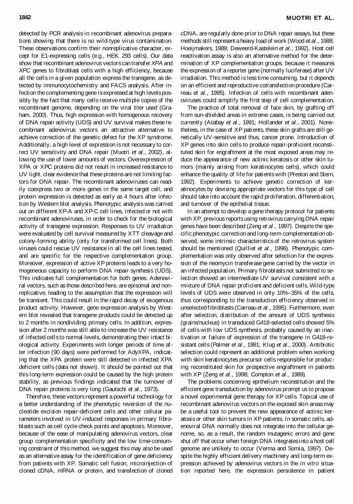
Because of the episomal and transient behavior of recombi-nant adenovirus, we were interested in knowing how long thisviral correction of DNA repair defects could be maintained.Transgene expression in nonreplicative (confluent quiescentcells) infected primary XP cells was followed by Western blotanalysis, as well as for the biologic efficiency of the correction.As shown in Figure 8, both XPA and XPC proteins are detectedup to 2 months after infection, with only a slight decrease (ifany) in protein expression. In order to check for the biologicactivity of these proteins, confluent XP-A cells, infected 2months before with the specific recombinant adenovirus wereexposed to a single dose of UV light (5 J/m2) and kept in cul-ture until analysis. Quantification of the UV-killing effect onuntransduced and transduced cells is shown in Figure 8C. Theresults clearly demonstrate that a long-term restoration of UV-survival was achieved (up to 2 months after infection).
DISCUSSION
In the present study, the efficiency to correct DNA repair-deficient cells belonging to XP-A and XP-C groups by recom-binant adenovirus vector mediated gene transfer was investi-gated. The features and complementation groups of the cell linesemployed had been characterized before, and include SV40-transformed and primary diploid skin fibroblasts (Eveno et al.,1995). Adenovirus vectors infect a broad range of cells, inde-pendent of the cell cycle phase; thus, these vectors can be usedto transduce exogenous genes into a variety of primary andtransformed cell lines. In this work, the efficiency of transduc-tion and the biological activity of the XPA and XPC genes inSV40-transformed as well as primary fibroblasts were shownto be effective in the entire cell population infected with re-combinant viruses. In fact, adenovirus carrying the denV genefrom bacteriophage T4, which encodes CPD-specific endonu-clease, has been already examined for the ability to restore DNArepair in XP fibroblasts. Although an increased rate of CPD re-pair was observed, there was only partial UV sensitivity re-covery in XP groups A, C, D and no effect in XPG cells (Col-icos et al., 1991; Francis et al., 2000).
The recombinant viruses described here show the absence ofcytopathic effect in all cell lines used and the E1 gene was not
XP COMPLEMENTATION BY ADENOVIRUS VECTORS 1841
FIG. 8. Long-term complementation of NER deficiency by XP adenovirus recombinant vectors. A: Western blot detection ofXPA and EGFP proteins expression in quiescent phase XP-A cells as a function of time after infection with AdyXPA. B: West-ern blot detection of XPC protein expression in quiescent phase XP-C cells as a function of time after infection with AdXPC(see Materials and Methods). C: Quiescent phase XP456 XP-A cells were infected 2 months before with AdyXPA (10 multi-plicity of infection [MOI]), UV-irradiated with a single dose of 5 J/m2 and stained 2 weeks later. UV survival analyzed by XTTcleavage is shown in the histogram.
detected by PCR analysis in recombinant adenovirus prepara-tions showing that there is no wild-type virus contamination.These observations confirm their nonreplicative character, ex-cept for E1-expressing cells (e.g., HEK 293 cells). Our datashow that recombinant adenovirus vectors can transfer XPA andXPC genes to fibroblast cells with a high efficiency, becauseall the cells in a given population express the transgene, as de-tected by immunocytochemistry and FACS analysis. After in-fection the complementing gene is expressed at high levels pos-sibly by the fact that many cells receive multiple copies of therecombinant genome, depending on the viral titer used (Gra-ham, 2000). Thus, high expression with homogenous recoveryof DNA repair activity (UDS) and UV survival makes these re-combinant adenovirus vectors an attractive alternative toachieve correction of the genetic defect for the XP syndrome.Additionally, a high level of expression is not necessary to cor-rect UV sensitivity and DNA repair (Muotri et al., 2002), al-lowing the use of lower amounts of vectors. Overexpression ofXPA or XPC proteins did not result in increased resistance toUV light, clear evidence that these proteins are not limiting fac-tors for DNA repair. The recombinant adenoviruses can read-ily coexpress two or more genes in the same target cell, andprotein expression is detected as early as 4 hours after infec-tion by Western blot analysis. Phenotypic analysis was carriedout on different XP-A and XP-C cell lines, infected or not withrecombinant adenoviruses, in order to check for the biologicalactivity of transgene expression. Responses to UV irradiationwere evaluated by cell survival measured by XTT cleavage andcolony-forming ability (only for transformed cell lines). Bothviruses could rescue UV resistance in all the cell lines tested,and are specific for the respective complementation group.Moreover, expression of active XP proteins leads to a very ho-mogeneous capacity to perform DNA repair synthesis (UDS).This indicates full complementation for both genes. Adenovi-ral vectors, such as those described here, are episomal and non-replicative, leading to the assumption that the expression willbe transient. This could result in the rapid decay of exogenousproduct activity. However, gene expression analysis by West-ern blot revealed that transgene products could be detected upto 2 months in nondividing primary cells. In addition, expres-sion after 2 months was still able to increase the UV resistanceof infected cells to normal levels, demonstrating their intact bi-ological activity. Experiments with longer periods of time af-ter infection (90 days) were performed for AdyXPA, indicat-ing that the XPA protein were still detected in infected XPAdeficient cells (data not shown). It should be pointed out thatthis long-term expression could be caused by the high proteinstability, as previous findings indicated that the turnover ofDNA repair proteins is very long (Gautschi et al., 1973).
Therefore, these vectors represent a powerful technology fora better understanding of the phenotypic reversion of the nu-cleotide excision repair-deficient cells and other cellular pa-rameters involved in UV-induced responses in primary fibro-blasts such as cell cycle check points and apoptosis. Moreover,because of the ease of manipulating adenovirus vectors, cleargroup complementation specificity and the low time-consum-ing constraint of this method, we suggest this may also be usedas an alternative assay for the identification of gene deficiencyfrom patients with XP. Somatic cell fusion, microinjection ofcloned cDNA, mRNA or protein, and transfection of cloned
cDNA, are regularly done prior to DNA repair assays, but thesemethods still represent a heavy load of work (Wood et al., 1988;Hoeijmakers, 1989; Deweerd-Kastelein et al., 1992). Host cellreactivation assay is also an alternative method for the deter-mination of XP complementation groups, because it measuresthe expression of a reporter gene (normally luciferase) after UVirradiation. This method is less time consuming, but it dependson an efficient and reproductive cotransfection procedure (Car-reau et al., 1995). Infection of cells with recombinant aden-oviruses could simplify the first step of cell complementation.
The practice of total removal of face skin, by grafting offfrom sun-shielded areas in extreme cases, is being carried outcurrently (Atabay et al., 1991; Hollander et al., 2001). None-theless, in the case of XP patients, these skin grafts are still ge-netically UV-sensitive and thus, cancer prone. Introduction ofXP genes into skin cells to produce repair-proficient reconsti-tuted skin for engraftment at the most exposed areas may re-duce the appearance of new actinic keratosis or other skin tu-mors (mainly arising from keratinocytes cells), which couldenhance the quality of life for patients with (Preston and Stern,1992). Experiments to achieve genetic correction of ker-atinocytes by devising appropriate vectors for this type of cellshould take into account the rapid proliferation, differentiation,and turnover of the epithelial tissue.
In an attempt to develop a gene therapy protocol for patientswith XP, previous reports using retrovirus carrying DNA repairgenes have been described (Zeng et al., 1997). Despite the spe-cific phenotypic correction and long-term complementation ob-served, some intrinsic characteristics of the retrovirus systemshould be mentioned (Quilliet et al., 1996). Phenotypic com-plementation was only observed after selection for the expres-sion of the neomycin transferase gene carried by the vector inan infected population. Primary fibroblasts not submitted to se-lection showed an intermediate UV survival consistent with amixture of DNA repair proficient and deficient cells. Wild-typelevels of UDS were observed in only 10%–35% of the cells,thus corresponding to the transduction efficiency observed inunselected fibroblasts (Carreau et al., 1995). Furthermore, evenafter selection, distribution of the amount of UDS synthesis(grains/nucleus) in transduced G418-selected cells showed 5%of cells with low UDS synthesis, probably caused by an inac-tivation or failure of expression of the transgene in G418-re-sistant cells (Palmer et al., 1991; Klug et al., 2000). Antibioticselection could represent an additional problem when workingwith skin keratinocytes precursor cells responsible for produc-ing reconstituted skin for prospective engraftment in patientswith XP (Zeng et al., 1998; Compton et al., 1989).
The problems concerning epithelium reconstitution and theefficient gene transduction by adenovirus prompt us to proposea novel experimental gene therapy for XP cells. Topical use ofrecombinant adenovirus vectors on the exposed skin areas maybe a useful tool to prevent the new appearance of actinic ker-atosis or other skin tumors in XP patients. In somatic cells, ad-enoviral DNA normally does not integrate into the cellular ge-nome, so, as a result, the random mutagenic errors and geneshut off that occur when foreign DNA integrates into a host cellgenome are unlikely to occur (Verma and Somia, 1997). De-spite the highly efficient delivery machinery and long-term ex-pression achieved by adenovirus vectors in the in vitro situa-tion reported here, the expression persistence in patient
MUOTRI ET AL.1842

keratinocytes should be evaluated periodically for the recoveryof cellular UV resistance as a first step in this strategy. Indi-viduals belonging to XP-A and XP-C groups are the most rep-resentative in the XP patient population (27% and 26%, re-spectively) (Cleaver and Kraemer, 1995). Moreover, unlikemost patients with XP, XP-C individuals suffer only from skintumors (without any neurologic clinical features detected) and,thus, have been considered the most probable candidates to en-visage an experimental gene therapy protocol targeting the skin.Of course, the well-known immunologic problem characteris-tic of adenovirus should be taken into account when workingin vivo, but there is a hope that in the engraftment procedure,these problems might be minimized (Yeh and Perricaudet,1997; Marchetto et al., 2001). The value of the adenovirus sys-tem to gene therapy relies on careful studies of the viral pro-teins responsible for immunologic problems in humans. The useof helper-dependent adenovirus, deleted in almost all viralgenes, may represent the benefits concerning the next genera-tion of adenovirus vectors (Amalfitano, 1999; Ventura, 2000).
XP knockout mice have been described in scientific litera-ture (revised in De Boer and Hoeijmakers, 1999). Although nei-ther strain shows complete phenotypic equivalency to humanXP patients, these strains exhibit reduced NER, increased sus-ceptibility to UV and chemical-carcinogen–induced skin car-cinogenesis, representing a valuable model for cell comple-mentation examination (Nakane et al., 1995; Sands et al., 1995;Giese et al., 1999). Because of the high homology between hu-man and mouse cDNAs, the recombinant viruses AdyXPA andAdXPC should be able to restore the DNA repair in XPA2 /2
and XPC2/2 knockout mice, as a first step to show the viabil-ity of XP gene therapy.
ACKNOWLEDGMENTS
This work was supported by the Fundação de Amparo àPesquisa do Estado de São Paulo–FAPESP (proc. # 98/11119-7, São Paulo, Brazil), Conselho Nacional de DesenvolvimentoCientífico e Tecnológico (CNPq, Brasília, Brazil), CAPES-COFECUB (Brazil, France), and National Institutes of Health(R24 DK58739, USA) (T.A.L.). M.C.N.M. and A.R.M. havefellowships from FAPESP, respectively. We wish to thank Dr.Hoeijmakers for providing anti-XPC antibody (Centre for Bio-medical Genetics, Erasmus University, The Netherlands).
REFERENCES
AMALFITANO, A. (1999). Next-generation adenoviral vectors: Newand improved. Gene Ther. 6, 1643–1645.
ATABAY, K., CELEBI, C., CENETOGLU, S., BARAN, N.K., andKIYMAZ, Z. (1991). Facial resurfacing in xeroderma pigmentosumwith monoblock full-thickness skin graft. Plast. Reconstr. Surg. 87,1121–1125.
BENIHOUD, K., YEH, P., and PERRICAUDET, M. (1999). Adeno-virus vectors for gene delivery. Curr. Opin. Biotechnol. 5, 440–447.
CARREAU, M., EVENO, E., QUILLIET, X., CHEVALIER-LA-GENTE, O., BENOIT, A., TANGANELLI, B., STEFANINI, M.,VERMEULEN, W., HOEIJMAKERS, J.H., and SARASIN, A.(1995). Development of a new easy complementation assay for DNA
repair deficient human syndromes using cloned repair genes. Car-cinogenesis 16, 1003–1009.
CLEAVER, J.E. (1968). Defective repair replication of DNA in Xero-derma pigmentosum. Nature 218, 652–654.
CLEAVER, J.E., and KRAEMER, K.H. (1995). Xeroderma pigmen-tosum and Cockayne syndrome. In: The Metabolic and MolecularBasis of Inherited Disease, 7th ed. C.R. Scriver, A.L. Beaudet, W.S.Sly, D. Valle, eds.. (McGraw Hill, New York) pp. 4393–4419.
CLEAVER, J.E., and THOMAS, G.H. (1981). Measurement of un-scheduled synthesis by autoradiography. In DNA Repair: A Labora-tory Manual of Research Procedures. E.C. Friedberg and P.C.Hanawalt, eds. (Marcel Dekker, Inc., New York) pp. 227–287.
COLICOS M.A., HAJ-AHMAD, Y., VALERIE, K., HENDERSON,E.E., and RAINBOW, A.J. (1991). Construction of a recombinantadenovirus containing the denV gene from bacteriophage T4 whichcan partially restore the DNA repair deficiency in xeroderma pig-mentosum fibroblasts. Carcinogenesis 2, 249–255.
COMPTON, C.C., GILL, J.M., BRADFORD, D.A., REGAUER, S.,GALLICO, G.G., and O’CONNOR, N.E. (1989). Skin regeneratedfrom cultured epithelial autografts on full-thickness burn woundsfrom 6 days to 5 years after grafting. A light, electron microscopicand immunohistochemical study. Lab. Invest. 60, 600–612.
COX, S.E., ROBERTS, L.J., and BERGSTRESSER, P.R. (1993). Pre-vention of skin cancer in xeroderma pigmentosum: The physician asadvocate. J. Am. Acad. Dermatol. 29, 1045–1046.
DAVIS, B.E., KOH, H.K., ROHRER, T.E., GONZALEZ, E., andCLEAVER, J.E. (1994). Sunlight avoidance and cancer preventionin xeroderma pigmentosum. Arch. Dermatol. 130, 806–808.
DE BOER, J., and HOEIJMAKERS, J.H.J. (2000). Nucleotide excisionrepair and human syndromes. Carcinogenesis 21, 453–460.
DE BOER, J., and HOEIJMAKERS, J.H.J. (1999). Cancer from theoutside, aging from the inside: Mouse models to study the conse-quences of defective nucleotide excision repair. Biochimie 81,127–138.
DE LAAT, W., JASPERS, N.G.J., and HOEIJMAKERS, J.H.J. (1999).Molecular mechanism of nucleotide excision repair. Genes Dev. 13,768–785.
DE VRIES, A., VAN OOSTROM, C.T.M., HOFHUIS, F.M.A., DOR-TANST, P.M., BERG, R.J., GRUIJL, F.R., WESTER, P.W.,KREIJL, C.F.V., CAPEL, P.J.A., STEEG, H.V., and VERBEEK, S.J.(1995). Increased susceptibility to ultraviolet-B and carcinogens ofmice lacking the DNA excision repair gene XPA. Nature 377,169–173.
DEWEERD-KASTELEIN, E.A., KEIJZER, W., and BOOTSMA, D.(1992). Genetic heterogeneity of xeroderma pigmentosum demon-strated by somatic cell hybridization. Nature 238, 80–83.
EVENO, E., QUILLIET, X., CHEVALLIER-LAGENTE, O., ROZA,L., EKER, A.P.M., KLEIJER, W.J., NIKAIDO, O., STEFANINI,M., HOEIJMAKERS, J.H.J., BOOTSMA, D., CLEAVER, J.E.,SARASIN, A., and MEZZINA, M. (1995). Different removal ofultraviolet photoproducts in genetically related xeroderma pig-mentosum and trichothiodystrophy diseases. Cancer Res. 55, 4325–4332.
FRANCIS, M.A., BAGGA, P., ATHWAL, R., and RAINBOW A.J.(2000). Partial complementation of the DNA repair defects in cellsfrom xeroderma pigmentosum groups A, C, D and F but not G bythe denV gene from bacteriophage T4. Photochem. Photobiol. 3,365–373.
GAUTSCHI, J.R., YOUNG, B.R., CLEAVER J.E. (1973). Repair ofdamage DNA in the absence of protein synthesis in mammalian cells.Exp. Cell Res. 76, 87–94.
GIESE, H., DOLLE, M.E., HEZEL, A., and VAN STEEG, H.V. (1999).Accelerated accumulation of somatic mutations in mice deficient inthe nucleotide excision repair gene XPA. Oncogene 18, 1257–1260.
GRAHAM, F.L. (2000). Adenovirus vectors for high-efficiency genetransfer into mammalian cells. Immunol. Today 21, 426–428.
XP COMPLEMENTATION BY ADENOVIRUS VECTORS 1843

GRAHAM, F.L., and PREVEC, L. (1991). Manipulation of Adenovi-rus Vectors. In Methods in Molecular Biology. Volume 7: GeneTransfer and Expression Protocols. E.J., Murray, ed. (The HumanaPress Inc., Clifton, NJ) pp. 109–128.
HOEIJMAKERS, J.H.J. (1989). Use of microneedle injection to studyDNA repair in mammalian cells. In DNA Repair: A Laboratory Man-ual of Research Procedures. E.C. Friedberg and P.C. Hanawalt, eds.(Marcel Dekker, Inc., New York) pp. 133–150.
HOLLANDER, D.A., SORANZO, C., FALK, S., and WINDOLF, J.(2001). Extensive traumatic soft tissue loss: Reconstruction in se-verely injured patients using cultured hyaluronan-based three-dimensional dermal and epidermal autographs. J. Trauma 50,1125–1136.
HORIKI, S., MIYAUCHI-HASIMOTO, H., TANAKA, K., NIKAIDO,O., and HORIO, T. (2001). Protective effects of sunscreening agentson photocarcinogenesis, photoaging, and DNA damage in XPA geneknockout mice. Arch. Dermatol. Res. 292, 511–518.
KLUG, C.A., CHESHIER, S., and WEISSMAN, I.L. (2000). Inactiva-tion of a GFP retrovirus occurs at multiple levels in long-term re-populating stem cells and their differentiated progeny. Blood 96,894–901.
KRAEMER, K.H., DIGIOVANNA, J.J., MOSHELL, A.N., TARONE,R.E., and PECK, G.L. (1998). Prevention of skin cancer in xero-derma pigmentosum with the use of oral isoretinoin. N. Engl. J. Med.318, 1633–1637.
KRAEMER, K.H., LEE, M.M., ANDREWS, A.D., and LAMBERT,W.C. (1994). The role of sunlight and DNA repair in melanoma andnonmelanoma skin cancer. The xeroderma pigmentosum paradigm.Arch. Dermatol. 130, 1018–1021.
KRAEMER, K.H., LEE, M.M., and SCOTTO, J. (1997). Xerodermapigmentosum. Cutaneous, ocular, and neurological abnormalities in830 published cases. Arch. Dermatol. 123, 241–250.
LOTFI, C.F., and ARMELIN, H.A. (2001). cfos and cjun antisenseoligonucleotides block mitogenesis triggered by fibroblast growthfactor-2 and ACTH in mouse Y1 adrenocortical cells. J. Endocrinol .168, 381–389.
MARCHETTO, M.C.N., MUOTRI, A.R., MAGALHÃES, G.S.,ZERBINI, L.F.C., LIBERMANN, T., VENTURA, A.M., andMENCK, C.F.M. (2001). The EGFP recombinant adenovirus: an ex-ample of efficient gene delivery and expression in human cells. VirusRev. Res. 6, 23–33.
MASUTANI, C., KUSUMOTO, R., YAMADA, A., DOHMAE, N.,YOKOI, M., YUASA, M., ARAKI, M., IWAI, S., TAKIO, K., andHANAOKA, R. (1999). The XPV (xeroderma pigmentosum variant)gene encodes human DNA polymerase eta. Nature 399, 700–704.
MIZUGUCHI, H., and KAY, M.A. (1998). Efficient construction of arecombinant adenovirus vector by an improved in vitro ligationmethod. Hum. Gene Ther. 9, 2577–2583.
MORIWAKI, S., and KRAEMER, K.H. (2001). Xeroderma pigmen-tosum: bridging a gap between clinic and laboratory. Photoderma-tol. Photoimmunol. Photomed. 17, 47–54.
MUOTRI, A.R., MARCHETTO, M.C.N., SUZUKI, M.F., OKAZAKI,K., LOTFI, C.F.P., BRUMATTI, G., AMARANTES-MENDES,G.P., and MENCK, C.F.M. (2002). Low amount of the DNA repairXPA protein are sufficient to recover UV-resistance. Carcinogene-sis 23, 1039–1046.
NAKANE, H., TAKEUCHI, S., YUBA, S., SAIJO, M., NAKATSU,Y., MURAI, H., NAKATSURU, Y., ISHIKAWA, T., HIROTA, S.,KITAMURA, Y., et al. (1995). High incidence of ultraviolet-B- orchemical-carcinogen-induced skin tumors in mice lacking the xero-derma pigmentosum group A gene. Nature 377, 165–168.
NYBERG-HOFFMAN, C., SHABRAM, P., LI, W., GIROUX, D, andAGUILAR-CORDOVA, E. (1997). Sensitivity and reproducibilityin adenoviral infectious titer determination. Nat. Med. 3, 808–811.
PALMER, T.D., ROSMAN, G.J., OSBORNE, W.R.A., and MILLERA.D. (1991). Genetically modified skin fibroblasts persist long after
transplantation but gradually inactive introduced genes. Proc. Natl.Acad. Sci. U.S.A. 88, 1330–1334.
PRESTON, D., and STERN, R.S. (1992). Nonmelanoma cancers of theskin. N. Engl. J. Med. 327, 1649–1662.
QUILLIET, X., CHEVALLIER-LAGENTE, O., EVENO, E., STOJ-KOVIC, T., DESTEE, A., SARASIN, A., and MEZZINA, M. (1996).Long-term complementation of DNA repair deficient human primaryfibroblasts by retroviral transduction of the XPD gene. Mutat. Res.364, 161–169.
ROBBINS, J.H., BRUMBACK, R.A., MENDIONES, M., BARRET,S.F., CARL, J.R., CHO, S., DENCKLA, M.B., GANGES, M.B.,GERBER, L.H., GUTHRIE, R.A., MEER J., MOSHELL A.N.,POLINSKY R.J., RAVIN P.D., SONIES B.C., and TARONE R.E.(1991). Neurological disease in xeroderma pigmentosum. Docu-mentation of a late onset type of the juvenile onset form. Brain 114,1335–1361.
SAMBROOK, J., and RUSSEL, D.W. (2001). Molecular Cloning. ALaboratory Manual. 3rd ed, Vol. 1–3. (Cold Spring Harbour Labo-ratory Press. Cold Spring Harbour, NY).
SANDS, A.T., ABUIN, A., SANCHEZ, A., CONTI, C.J., andBRADLEY A. (1995). High susceptibility to ultraviolet-induced car-cinogenesis in mice lacking XPC. Nature 14, 162–165.
TAKAGI, M., NISHIOKA, M., KAKIHARA, H., KITABAYASHI,M., INOUE, H., KAWAKAMI, B., and OKA, M. (1997). Charac-terization of DNA polymerase from Pyrococcus sp. strain KOD1 andits application to PCR. Appl. Environ. Microbiol. 63, 4505–4510.
VENTURA, A.M. (2000). Adenovirus vectors and their application ingene therapy. Virus Rev. Res. 5, 7–12.
VERMA, I.M., and SOMIA, N. (1997). Gene therapy: Promises, prob-lems and prospects. Nature 389, 239–242.
VOLKER, M., MONÉ, M.J., KARMAKAR, P., VAN HOFFEN, A.,SCHUL, W., VERMEULEN, W., HOEIJMAKERS, J.H.J., VANDRIEL, R., VAN ZEELAND, A.A., and MULLENDERS, L.H.F.(2001). Sequential assembly of the nucleotide excision repair factorsin vivo. Mol. Cell 8, 213–224.
WOOD, R.D., ROBINS, P., and LINDAHL, T. (1988). Complementa-tion of the xeroderma pigmentosum DNA repair defect in cell-freeextracts. Cell 54, 97–106.
YEH, P., and PERRICAUDET, M. (1997). Advances in adenoviral vec-tors: From genetic engineering to their biology. FASEB J. 8,615–623.
ZENG, L., QUILLIET, X., CHEVALIER-LAGENTE, O., EVENO, E.,SARASIN, A., and MEZZINA, M. (1997). Retrovirus-mediated genetransfer corrects DNA repair defect of xeroderma pigmentosum cellsof groups A, B and C. Gene Ther. 4, 1077–1084.
ZENG, L., SARASIN, A., and MEZZINA, M. (1998). Retrovirus-me-diated DNA repair gene transfer into xeroderma pigmentosum cells:Perspectives for a gene therapy. Cell Biol. Toxicol. 14, 105–110.
Address reprint requests to:Carlos Frederico Martins Menck
Depto. de MicrobiologiaInstituto de Ciências Biomédicas
Universidade de São PauloAv. Prof. Lineu Prestes
1374, São PauloSP, 05508-900
Brazil
E-mail: [email protected]
Received for publication March 25, 2002; accepted after revi-sion September 6, 2002.
Published online: September 23, 2002.
MUOTRI ET AL.1844


