Embed Size (px)
Citation preview
Histochemistry (1993) 100:53-64 Histochemistry © Springer-Verlag 1993
Connexin43 in rat pituitary: localization at pituicyte and stellate cell gap junctions and within gonadotrophs T. Yamamoto 1, M.Z. Hossain 2, E.L. Hertzberg 3, H. Uemura 1, L.J. Murphy 2, J.I. Nagy 2
1 Biological Laboratory, Kanagawa Dental College, Yokosuka, Japan z Department of Physiology, Faculty of Medicine, University of Manitoba, 770 Bannatyne Ave, Winnipeg, Manitoba, Canada R3E OW3 3 Departments of Neuroscience, Anatomy and Structural Biology, Albert Einstein College of Medicine, Bronx, New York 10461, USA
Accepted: 21 April 1993
Abstract. Immunohistochemical methods were employed to investigate the cellular and ultrastructural localization of the gap junction protein connexin43 (Cx43) in rat pitu- itary. Western blots of pituitary homogenates probed with anti-Cx43 antibodies showed the presence of Cx43 in both anterior and posterior pituitary lobes. By light microscopy (LM), Cx43-immunoreactive (Cx43-IR) puncta were found in all areas of the posterior lobe, but at greater concentrations in peripheral regions of this structure. By electron microscopy (EM), immunogold la- belling for Cx43 was seen at gap junctions between thin cytoplasmic processes of pituicytes. No immunoreactivi- ty was detected in the intermediate lobe. The anterior lobe contained puncta similar to but more sparsely scat- tered than those in the posterior lobe, and by EM analy- sis these were demonstrated to correspond to labelled gap junctions between stellate cells. In addition, anti- Cx43 antibodies produced intracellular labelling in a small percentage of endocrine cells, which were distribut- ed throughout the anterior lobe and determined by dou- ble immunostaining methods to be cells containing luteinizing hormone. By EM, labelling within these cells was associated with predominantly large secretory gran- ules and other loosely organized organelles. The results indicate that gap junctions in the pituitary are composed of Cx43 and that this or a related protein may have a novel intracellular function within gonadotrophs.
Introduction
Gap junctions allow intercellular communication via movement of small molecules and/or ions across trans- cellular channels that form at closely apposed plasma membranes of adjacent cells. Investigations of the role of this mode of communication in processes such as the coordination of cellular metabolic activity and transfer of regulatory signals between gap junctionally
Correspondence to: J.I. Nagy
coupled cells (Bennett and Goodenough 1978; Hooper and Subak-Sharpe 1981) has been facilitated by the iden- tification of the structural proteins of gap junctions termed connexins (Beyer et al. 1987). In rat tissues, near- ly a dozen different connexins having various degrees of sequence homology have been found and these are distinguished by a suffix referring to connexin molecular weight as predicted from the corresponding cDNAs (Beyer et al. 1990; Bennett et al. 1991). These connexins are differentially expressed in a variety of tissues and this is thought to reflect cell specific regulation of gap junctional coupling and functional demands for gap junctions in different cell types. Connexin43 (Cx43), first identified as a component of cardiac gap junctions, is among the most widely expressed connexins based on information available to date (Beyer et al. 1987; Luke et al. 1989; Laird and Revel 1990; Yancey et al. 1992; Larson et al. 1990; Beyer et al. 1989). With the discovery that there exists a rather extended family of connexins, determination of the connexin(s) expressed in the partic- ular cell types under investigation and confirmation of its localization at gap junctions in these cells is especially useful. This can be achieved with antibodies against non- homologous sequences of connexins, especially those lo- cated at the highly divergent carboxy-terminal regions which extend into the cytoplasm from a contiguous transmembrane domain.
In attempts to identify the connexins that mediate gap junctional communication in the central nervous system (CNS), we and others have used a variety of experimental approaches. It has been found that Cx43 is expressed in astrocytes and in various cell types lining the brain surfaces, that connexin26 is expressed in addi- tion by leptomeningial cells and that connexin32 (Cx32) is expressed by oligodendrocytes (Dermietzel et al. 1989; Micevych and Abelson 1991; Yamamoto et al. 1990a, b, 1992b; Nagy et al. 1992). While it is reasonably cer- tain that these results indicate a legitimate gap junctional role of each of these connexins in the respective cell type, there have been suggestions that gap junction pro- teins may have other than the traditionally accepted

54
funct ions (Beyer and Steinberg 1991; see also the 'D i s - cuss ion ' ) . This n o t i o n is consis tent with, for example, our immunoh i s tochemica l results demons t r a t i ng at the u l t ras t ruc tura l level tha t different an t ibodies agains t Cx32 recognize in t racel lu lar s tructures termed subsur- face cisterns which, at least in the m o t o n e u r o n s we exam- ined, display a morpholog ica l s imilari ty to gap j unc t ions (Yamamoto et al. 1990c, 1991). Such observat ions to- gether with proposals of novel roles for ectopic gap junc - t ions wa r r an t analysis and cons idera t ion of cases where k n o w n connexins or connexin- re la ted prote ins are found to have unexpected cellular d is t r ibut ions .
In our con t inu ing studies of Cx43, i m m u n o h i s t o - chemical approaches have been used to d o c u m e n t the local izat ion of this gap j u n c t i o n pro te in in the anter ior and poster ior p i tu i ta ry lobes of rat. G a p junc t ions be- tween var ious cell types in these structures have been previously detected by s t anda rd t ransmiss ion electron microscopy (Horva th et al. 1977; Soji and Herber t 1989; Soji et al. J990, 1991). We repor t tha t pi tuicytes in the poster ior lobe, like their p resumed astroglial counter - par ts in bra in , and stellate cells in the anter ior lobe, fo rm gap junc t ions composed of Cx43. In addi t ion , we f ind that an t ibodies agains t Cx43 produce dist inct in t ra- cellular immuno labe l l i ng in a par t icu lar class of hor- mone secretory cells identif ied as gonado t rophs .
M a t e r i a l s and m e t h o d s
Antibodies
The present studies were conducted with an anti-Cx43 antibody that was generated in rabbit and subsequently affinity-purified. Animals were immunized with a peptide corresponding to the ami- no acid sequence 346-363 in the carboxy-terminal region of Cx43 (Beyer et al. 1989). For affinity purification, a synthetic peptide to which a cys was added at the amino terminus, was coupled to a Beckman Ultraffinity-EP column. Serum was then passed over the column and bound antibodies eluted with 0.2 M glycine, pH 2.1. Eluate was immediately neutralized with I M Tris base and dialyzed overnight against phosphate-buffered saline, pH 7.4. In addition, some pituitary sections were processed with an anti- body against a peptide corresponding to the amino acid sequence 241-260 of Cx43. The specificity of these antibodies was evaluated by Western blots of heart and brain homogenates and abolition of immunolabelling in sections of these tissues after preadsorption of antibodies with the appropriate peptide, has been previously demonstrated (Yamamoto et al. 1990a, b, 1992b; Nagy et al. 1992). Antibody against rat growth hormone (rGH) produced in monkey and antibodies against the luteinizing hormone fl-chain (LH), pro- lactin, and adrenocorticotropic hormone (ACTH), all produced in rabbit, were provided through the National Institute of Arthritis, Diabetes and Digestive and Kidney Diseases (NIADDK) National Hormone and Pituitary Programme, The specificity of these anti- bodies has been either previously reported (Yamamoto et al. 1992 a) or established at NIADDK.
Western blots Frozen tissue samples were homogenized in I mM NaHCO3 (pH 8.0) with 1 mM phenylmethylsulphonyl fluoride (PMSF) and homogenate protein concentrations were determined by the Bio- Rad protein microassay kit with bovine serum albumin (BSA) as standard. Aliquots of homogenate containing 10 gg total protein were mixed with equal volumes of 2 x samPle buffer containing 10% (final concentration) fi-mercaptoethanol as a reducing agent prepared as previously described (Laemmli 1970). Samples were vortexed, heated in boiling water for 2 min and loaded onto 10%
SDS-polyacrylamide gels for electrophoresis. Electroblotting onto nitrocellulose membranes (Micron Separations Westboro, Mass., USA) was performed in Towbin buffer (Towbin et al. 1979) con- taining 0.5% SDS. Following transfer, membranes were blocked overnight at 4 ° C in freshly prepared 3% BSA in TBS-T (20 mM Tris, 137 mM NaC1, pH 7.6 with 0.2% Tween-20) and then incu- bated for 1.5 h with an affinity purified rabbit polyclonal anti-Cx43 antibody (directed against amino acids 346-363) diluted 1:10000 in blocking solution as above. The membranes were then washed several times in TBS-T and incubated with a horseradish peroxi- dase-conjugated secondary antibody (Amersham) for 1 h. After several washes with TBS-T, peroxidase activity representing levels of Cx43 was detected by an enhanced chemiluminescence kit (Amersham Oakuirle, Ont., Canada).
Tissue preparation
Seven male Spraue-Dawley rats weighing 280-350 g were deeply anesthetized with chloral hydrate and perfused transcardially with 75 ml of ice-cold 0.9% saline containing 50 mM sodium phosphate buffer (PB; pH 7.4), 0.1% sodium nitrate and heparin (1 Unit/ml) followed by 200 ml of 4% paraformaldehyde in 0.1 M PB (pH 7.4) and then by 200 ml of 4% paraformaldehyde in 50 mM borate buffer (pH 9.0). Pituitaries were removed and post-fixed in the borate-buffered fixative for 16 h. After cryoprotection for 1 to 2 days in 50 mM PB containing 25% sucrose and 10% glycerol, the pituitaries were sectioned at a thickness of 10 or 20 ~tm on a sliding microtome. Sections were rinsed for 24 h in PB containing 0.9% saline and 0.3% Triton X-100 (PBST) then processed by peroxidase-antiperoxidase (PAP), immunofluorescence or electron microscopic (EM) immunohistochemical methods.
Immunohistochemistry
For labelling by the immunoperoxidase method, pituitary sections were incubated with primary antibody for 48 h at 4 ° C. The anti- body dilutions in PBST containing 1% BSA were as follows : affini- ty-purified rabbit anti-Cx43 diluted 1:2000; anti-LH diluted 1 : 2000; anti-prolactin diluted i : 10 000; and anti-ACTH diluted 1 : 2000. After a wash in PBST for 1 h, the sections were incubated at room temperature for 2 h with goat anti-rabbit IgG (Sternberger- Meyer, Baltimore, Md., USA) diluted 1:40 in PBST. The sections were again washed for 1 h in PBST followed by incubation at room temperature for 2 h with rabbit PAP (Sternberger-Meyer) diluted 1 : 100 in PBST. The sections were then washed for 30 rain in PBST followed by a further wash for 30 min in 50 mM Tr&-HC1 buffer (pH 7.4) and then reacted for 5-10 rain in 50 mM Tr&-HC1 buffer (pH 7.4) containing 0.02% 3,3'-diaminobenzidine hydro- chloride (DAB) and 0.005% hydrogen peroxide.
For co-localization analysis of Cx43-immunoreactivity in var- ious cell types containing different pituitary hormones, either adja- cent sections or double immunofluorescence methods were used depending on the availability of primary antibodies from different species. For the analysis of adjacent sections, pituitaries were dehy- drated and wax-embedded according to conventional procedures. Alternate consecutive sections cut at a thickness of 3 gm on a sliding microtome were mounted onto glass slides and sections were separately processed by the PAP protocol described above such that one of a pair of adjacent sections was incubated with anti-Cx43 antibody and the other with an anti-pituitary hormone antibody.
Double immunofluorescence labelling in the same section was conducted in the case of co-localization analysis with anti-rGH antibody raised in monkey. Free-floating sections were incubated with anti-Cx43 antibody as above, washed in PBST for 1 h at room temperature and then incubated for 1.5 h at room tempera- ture with Texas Red-conjugated donkey anti-rabbit IgG (Amer- sham) diluted 1 : 100 in PBST. After a I h wash they were further incubated with anti-rGH antibody as above, washed for 1 h in PBST and then incubated for 1.5 h at room temperature with fluo- rescein isothiocyanate (FITC)-conjugated swine anti-monkey IgG (Cappel Scarborough, Ontario, Canada) diluted 1:20 in PBST.

55
The sections were then washed, mounted onto gelatin-coated slides and coverslipped with anti-fade medium (Valnes and Brandtzaeg 1985).
In order to obtain additional confirmatory information on the extent to which Cx43-immunoreactivity was localized in gonado- trophs, some sections were processed sequentially first with anti- Cx43 antibody by the PAP method described above and then with anti-LH antibody by immunofluorescenee. The free-floating sec- tions showing Cx43-IR cells by DAB deposition were incubated with anti-LH antibody as above, washed, and then incubated for 1.5 h at room temperature with FITC-conjugated donkey anti-rab- bit IgG (Amersham) diluted 1:100 in PBST. Although both prima- ry antibodies in this case were from the same species, the object here was to observe labelled cells in the same rather than adjacent sections where portions of an individual cell may not be repre- sented. Thus, while DAB deposition where it occurs simultaneously with immunofluorescence tends to obscure the latter, this at least allows visualization of the degree to which fluorescent cells (LH- labelled) lack DAB reaction product (Cx43-1abelled).
Control sections were processed with omission of anti-Cx43 antibody or with this antibody preabsorbed with the synthetic pep- tide (5 lag/ml) against which it was directed. In double labelling procedures, controls for possible cross reactions of primary anti- bodies with each other or with inappropriate secondaries and for possible cross reactions of secondaries with each other were con- ducted as described in our previous triple immunofluorescence la- belling studies (Staines et al. 1988; Yamamoto et al. 1992a). Obser- vations were performed with a Leitz Dialux 20 microscope equipped with N2 and L3 Ploemopak filter cubes for visualization of Texas Red and FITC, respectively.
A
1 2 3 4 B
-,'-43 "~'41
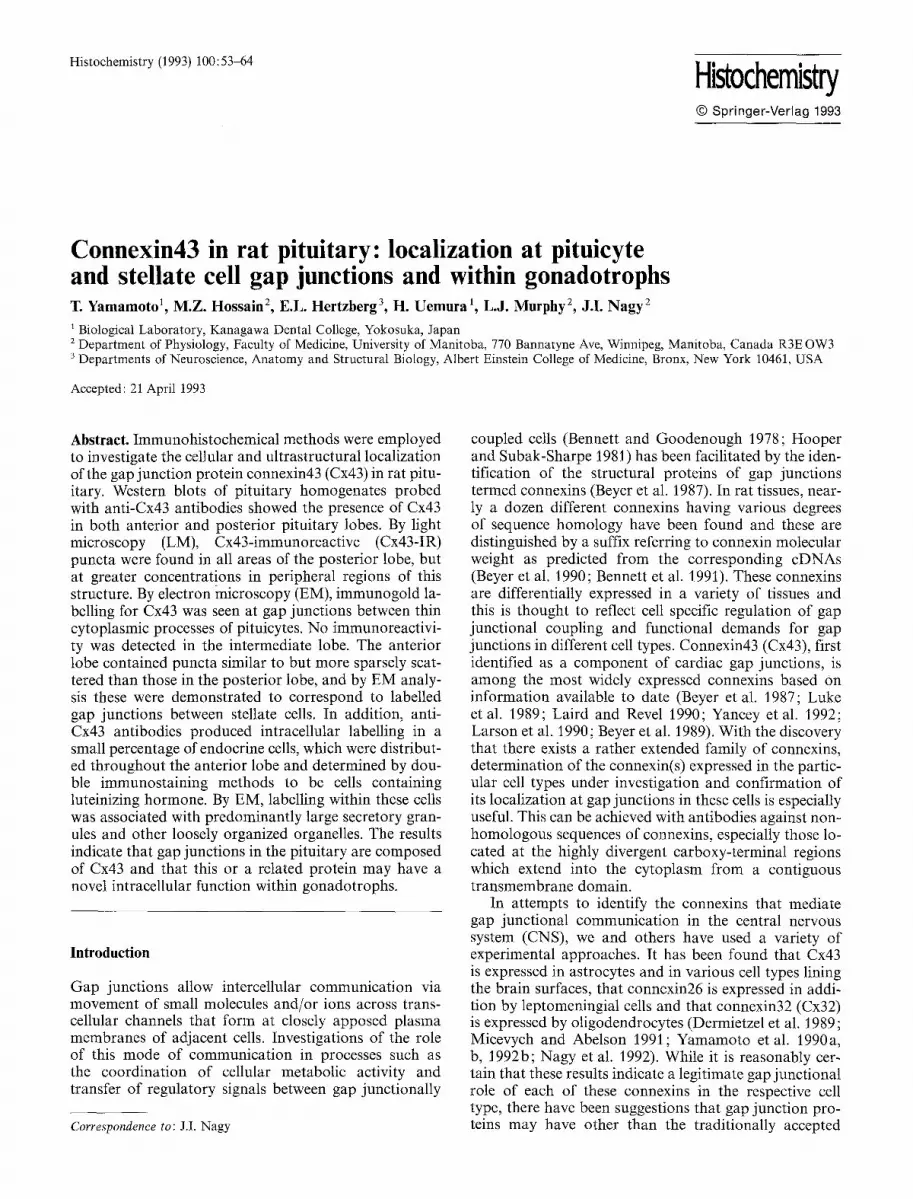
Fig. 1 h, B. Connexin43 (Cx43) protein levels in anterior and poste- rior pituitary as demonstrated by Western blots. An aliquot of 10 lag of total protein from tissue homogentates was loaded onto each lane of SDS polyacrylamide gels. A Exposure (20 s) of a mem- brane showing anterior pituitary (lane 1), posterior pituitary (lane 2), heart (lane 3) and brain (lane 4). B Exposure (2 rain) of lane 1. Positions of Cx43 bands are indicated by arrows with approxi- mate molecular weights in kDa
Immuno-electron microscopy
For immuno-electron microscopy, some sections were processed with Cx43 antibody by the PAP method as described above, except that all incubation and washing steps were conducted without the inclusion of detergent. After the DAB reaction step, sections were washed, post-fixed for I h in 2% osmium tetroxide diluted in 0.1 M PB and embedded in Jembed (J.B. EM Services, Dorval, Quebec, Canada). Alternatively, after incubation with anti-Cx43 antibody some sections were processed by immunogold methods. These were incubated for 3 h at room temperature with 1 nm colloidal gold conjugated to anti-rabbit [gG (Amersham) diluted 1 : 20 in 50 mM Tris-HC1 buffer (pH 7.4) containing 1% BSA. The sections were then washed in this dilution buffer, immersed in silver intensifica- tion medium for 1 rain as previously described (Lah et al. 1990), post-fixed in 2% osmium tetroxide diluted in 0.1 M PB, and em- bedded in Jembed. Sections were inspected by light microscopy and selected areas were trimmed and glued onto resin blocks. U1- trathin sections (800 A thick) were collected on mesh grids, coun- terstained with lead citrate for i rain and examined with a Phillip 202 electron microscope.
Results
Western blots
Weste rn b lo ts o f an te r io r and pos t e r i o r p i t u i t a ry h o m o g - enates ( p robed wi th ant i -Cx43 a m i n o acids 346-363) in- d i ca ted the presence o f Cx43 in b o t h o f these tissues (Fig. 1). A n ini t ia l shor t exposure (20 s) o f f i lm to the t ransfer m e m b r a n e was sufficient to d e m o n s t r a t e the presence o f b a n d s c o r r e s p o n d i n g to Cx43 in the pos ter i - or p i t u i t a ry (Fig. i a, lane 2), b u t no i m m u n o r e a c t i v e Cx43 b a n d s with this same exposure were seen in anter i - or p i t u i t a ry (Fig. 1 a, lane 1). However , a longer exposure (2 min) o f the same m e m b r a n e revealed a n t i b o d y recog- n i t ion o f Cx43 p ro t e in also in the an te r io r p i t u i t a ry
(Fig. 1 b). I t has been shown tha t d i f ferent levels o f p h o s p h o r y l a t e d and n o n - p h o s p h o r y l a t e d forms o f Cx43 are p resen t in d i f ferent t issues a n d tha t the s ta te o f its p h o s p h o r y l a t i o n results in mul t ip le Cx43 b a n d s in SDS- gels (Kad le et al. 1991).
C o m p a r i s o n wi th the Cx43 forms obse rved in well s tud ied t issues such as hea r t (Fig. 1 a, lane 3) and b ra in (Fig. 1 a, lane 4) shows tha t Cx43 in an te r io r p i t u i t a ry is equa l ly d i s t r ibu ted in three fo rms o f differ ing a p p a r e n t mo lecu l a r weight , namely , a 4 1 - k D a and a d o u b l e t o f bands c o r r e s p o n d i n g to two forms at a p p r o x i m a t e l y 43 kDa . In con t ras t , the 4 3 - k D a d o u b l e t is the p r e d o m i - nan t fo rm of Cx43 in pos t e r i o r p i t u i t a ry and thus resem- bles the p a t t e r n obse rved in hear t . However , the relat ive intensi t ies o f the b a n d s in this doub l e t differs f rom tha t in hea r t suggest ing d i f ferent Cx43 p h o s p h o r y l a t i o n p ro - files. The d o u b l e t in hea r t is s l ight ly d i s to r t ed relat ive to tha t in p i t u i t a ry and bra in , which m a y be due to the presence o f an a b u n d a n t p ro t e in in hea r t mig ra t ing jus t above the double t . A l ight b a n d mig ra t ing at a b o u t 45 k D a is also de tec ted in pos t e r i o r p i tu i t a ry , hear t , and bra in , This m a y c o r r e s p o n d to an as ye t p o o r l y under - s tood , t r ans ien t ly expressed i so fo rm of Cx43 which has been obse rved in m a m m a l i a n f ib rob las t s (Crow et al. 1990).
Light microscopy
In sect ions p rocessed by ei ther i m m u n o p e r o x i d a s e or immunof luo rescence , Cx43 - immunoreac t i v i t y wi th ant i - b o d y d i rec ted aga ins t a m i n o acids 346-363 in the Cx43 sequence (ant i -Cx43, aa346-363) was seen in b o t h the pos t e r io r and an te r io r p i t u i t a ry lobes. Howeve r no

56
Fig. 2A-D. Photomicrographs showing differential patterns of Cx43-immunoreactivity (IR) in rat pituitary. A Low magnification showing Cx43-IR cells in the anterior lobe (AL), punctate immuno- staining in the posterior lobe (PL), and the absence of immunola- belling in the intermediate lobe (IL). B Higher magnification of the posterior lobe showing a greater density of punctate immuno- staining at the peripheral edges of this structure. C Magnification
of the anterior lobe showing Cx43-IR puncta (large arrows) and the granular appearance of immunolabelling in Cx43-IR cells. Note also the range of immunostaining density in intensely (small arrows) and weakly (arrowheads) stained cells. D Section showing the ab- sence of Cx43-immunoreactivity in the anterior lobe after incuba- tion with affinity-purified antibody preabsorbed with synthetic peptide. A x75;B x200;C x300;D ×J50
Cx43-IR was seen in the intermediate lobe where failure to detect the protein does not necessarily exclude possi- bly very low levels o f expression (Fig. 2). Throughout the posterior lobe (Fig. 2A, B), immunoreact ivi ty was distributed uniformly except at peripheral regions where the immunostaining density was greater within a dis- tance of about 25-75 gm bordering the intermediate lobe. Immunoreact ive structures consisted exclusively of puncta ranging f rom 0.2 to 0.5 gm in size. In the anterior lobe (Fig. 2A, C), immunoreact ivi ty was localized to sparsely scattered puncta similar to those seen in the posterior lobe. In addition, the anterior lobe contained Cx43-immunoreactive (Cx43-IR) cells that were dis- persed throughout but slightly more concentrated at its border with the intermediate lobe. These cells were round or oval, ranged f rom 10 to 20 gm in diameter and occurred in isolation or were grouped together around blood vessels. Cx43-IR cells exhibited wide vari- ation in immunostaining density and this was a consitent observation in sections f rom different animals. Immuno-
reactivity within cells had a granular appearance and some cells were filled with immunoposi t ive granules while others contained moderate numbers or only a few. Immunoreact ive puncta were difficult to distinguish f rom immunolabeled granules when the former were near immunoposi t ive cells or when the latter were sparse and the outline of cells in which they were contained was not clearly evident.
The immunostaining patterns described above were seen in pituitary sections f rom each of the animals exam- ined. The same patterns, except with higher background (not shown), were seen with a less sensitive rabbit anti- body directed against a peptide corresponding to amino acids 246-260 in the Cx43 sequence (anti-Cx43, aa246- 260). No immunostaining was seen with preimmune se- rum taken f rom rabbits producing these antibodies. In addition, no immunoreact ivi ty was detected in either the posterior or anterior (Fig. 2D) lobes after preabsorpt ion of anti-Cx43 (aa346-363) ant ibody with the correspond- ing peptide.

57
Fig. 3A-D. Photomicrographs showing the localization of Cx43- immunoreactivity (A, C) in gonadotrophs immunolabelled for lu- teinizing hormone (LH; B, D). A, B Adjacent sections showing that most Cx43-IR cells (arrows in A) are also LH-immunopositive (arrows in B). C, D The same section immunostained for Cx43
by the peroxidase-antiperoxidase (PAP) method (C) and then by immunofluorescence for LH (D). Some weak LH-immunofluores- cence is seen in intensely labelled Cx43-IR cells (arrows), but not all LH-positive cells exhibit Cx43-immunoreactivity (arrowheads). A,B x310;C,D x390
Co-localization analysis
The above observation regarding the presence of Cx43- IR cells in the anterior pituitary prompted investigations concerning whether these cells corresponded to any par- ticular cell type that comprise the well established subpo- pulations of hormone-containing secretory cells. In pairs of adjacent sections processed with anti-Cx43 antibody (Fig. 3 A) and anti-LH antibody (Fig. 3 B), it was found that all Cx43-IR cells whose counterparts could be iden- tiffed in the adjacent section were also immunopositive for LH. However, it appeared that not all LH-immuno- reactive (LH-IR) cells exhibited Cx43-immunoreactivity. This finding was confirmed in double-labelled sections where it was found that some cells exhibiting intense immunofluorescence for LH (Fig. 3 D) lacked and there- fore were not obscured by immunoperoxidase labelling for Cx43 (Fig. 3C). It was estimated that about 15% of the LH- IR cells were devoid of Cx43 and these tended
to be larger than cells displaying both LH- and Cx43- immunoreactivity.
Despite these results showing the presence of Cx43 exclusively in gonadotrophs, further analyses were con- ducted to exclude the possible localization of Cx43 in other cell types. In pituitary sections double-stained by immunofluorescence with anti-Cx43 (Fig. 4A) and anti- r G H (Fig. 4 B) antibodies, no Cx43-IR cells were found to contain r G H and conversely no rGH-containing cells were seen to exhibit Cx43-immunoreactivity. In pairs of adjacent sections taken for immunoperoxidase label- ling with anti-Cx43 (Fig. 4C) and anti-prolactin (Fig. 4 D) antibodies, Cx43-IR cells were devoid of pro- lactin and cells containing prolactin showed an absence of Cx43-immunoreactivity. Similarly, in adjacent sec- tions taken for comparative analysis of Cx43-IR and ACTH-containing cells, no cells were found to be im- munopositive for both Cx43 and ACTH (not shown).

58
.... ' a ,
Fig. 4 A-D. Photomicrographs showing the absence of Cx43-immu- noreactivity (A, C) in somatotrophs (B) and mammotrophs (D). A, B The same section processed by immunofluorescence to show Cx43-IR cells (A) and rat growth hormone (rGH)-IR (B). None of the Cx43-IR cells are positive for rGH (arrows) and none of the rGH-positive cells exhibit Cx43-immunoreactivity (arrow-
heads). C, D Adjacent sections processed by the PAP method to show Cx43-IR cells (C) and prolactin-IR cells (D). None of the Cx43-IR cells are positive for prolactin (arrows) and none of the prolactin-positive cells display Cx43-immunoreactivity (arrow- heads). A, B x 430; C, D x 350
Electron microscopy
In pituitary sections processed by the immunoperoxidase method (Fig. 5), Cx43-IR cells in the anterior lobe had extensive endoplasmic reticulum (ER) and were found to contain many small (120-200 nm) and a few large (300450 nm) secretory granules (Fig. 5A). Immunola- belling in these cells was heterogeneously distributed such that large patches of labelled and unlabelled area were evident and no immunoreactivity was observed in the nucleus. Dense DAB reaction product was seen on and within some, but not all, small and large secretory ganules and on parts of ER membranes that were in close apposition to these granules. In addition, reaction product was associated with round organelles scattered among secretory granules. These organelles had a diame-
ter of about 300 nm and were composed of small gran- ules (20 nm) which sometimes appeared to be arrayed in concentric or spiral rings (Fig. 5 B).
Cx43-IR cells in anterior pituitary sections processed by intensified immunogold procedures exhibited charac- teristics (Fig. 6A) similar to those seen in sections pro- cessed by the PAP method. Immunogold particles in these cells were seen in dense clusters corresponding roughly to the size of large secretory granules (Fig. 6A, B). These clusters were sometimes seen to be surrounded by unit membranes and occupied an area that was slight- ly more electron dense than the surrounding cytoplasm (Fig. 6 C). In addition, immunogold labelling was often seen around and in association with the limiting mem- brane of large secretory granules and only occasionally on the peripheral regions of small secretory granules

59
Fig. 5A, B. Electron micrographs of a Cx43-IR cell in a section of the anterior pituitary lobe processed by the PAP method. A Low magnification showing a heterogeneous distribution of DAB deposition (arrows) within the cell cytoplasm. B Higher magnifica- tion showing that intensely labelled area in A are composed of
clusters of small immunoreactive granules (large arrows), immuno- reactive secretory granules (arrowheads in A and B) and immunola- belled intracellular membranes (small arrows). A x 8900; B x 33700
(Fig. 6 B, D). In contrast to observations of immunoper- oxidase reacted sections, the interior of secretory gran- ules were free of gold particles. This difference may re- flect false-positive labelling with the PAP method where DAB may have diffused into the granule from its site of generation at the granule surface, or false-negative labelling with the immunogold method due to failure of antibody penetration into the granule. In a rare case, immunolabelling was seen around a granule located very close to the plasma membrane (Fig. 6D); immunogold particles were distributed both around the intracellular portion of the granule and at its abutment with the plas- ma membrane. Within the cytoplasm, closely apposed membranes exhibiting a 2 nm inter-membrane space were frequently seen near clusters of immuno-gold parti- cles (Fig. 6C), as well as near immunogold deposits on secretory granules (Fig. 6E); one of the apposing mem- branes corresponded to the limiting membrane of the secretory granule, while the identity or source of the other was not discernable.
Immunogold labelling for Cx43 was also seen at gap junctions in both the anterior and posterior pituitary lobes. In the anterior lobe (Fig. 6 F, G), immunolabelled junctions were observed at closely apposed plasma mem- branes of stellate cells and gold particles were evident intracellularly on both sides of the junctional membrane. In the posterior lobe (Fig. 7), immunolabelled gap junc- tions were found exclusively between thin processes of pituicytes which exhibited scanty cytoplasm and sur- rounded neuronal elements (Fig. 7 A, B). These junctions were more numerous at peripheral edges of the posterior lobe where there was a greater concentration of blood capillaries and intermingled pituicyte processes. Immun- ogold particles were equally distributed on both sides of the junctional membranes and the electron density
of the cytoplasm within each of the pituicyte processes forming these linear junctions was similar (Fig. 7 E). Oc- casionally, labelling was seen densely clustered around annular gap junctions that had diameters of about 0.3- 0.4 gm (Fig. 7 B, C). The inter-membrane space at these annular junctions ranged from 2 to 5 nm (Fig. 7 D) and immunogold particles were associated only with the out- er surface of these profiles. The electron density of the cytoplasm outside annular junctions was generally high- er than that of their interior.
Discussion
The present results confirm previous reports that have shown the existence of gap junctions between pituicytes in the posterior pituitary lobe and between stellate cells in the anterior lobe (Soji and Herbert 1889; Soji et al. 1990, 1991) and extend the list of tissues in which cells form intercellular communicating channels via Cx43. In the posterior lobe, pituicytes are thought to be related to and function similarly to astrocytes in brain (Wein- rauder and Zareba-Kowalska 1984; Hatton 1988). Thus by analogy with their astrocyte counterparts (Walz and Hertz 1983; Walz 1989), it has been proposed that pitui- cytes contribute to the maintenance of a normal meta- bolic environment which includes the redistribution of extracellular potassium ions around active nerve termi- nals (Cazalis et al. 1987; Hatton 1988). This view is con- sistent with our previous observations of numerous Cx43-IR gap junctions associated with astrocytic end- feet on blood vessels (Yamamoto et al. 1990b) and also
' with the present results. Elevated levels of Cx43-IR gap junctions were demonstrated in peripheral posterior pi- tuitary regions where enhanced intercellular exchange

60

61
Fig. 7 A-E. Electron micrographs of Cx43-IR structures in the pos- terior pituitary lobe as demonstrated by immunogold labelling with intensified 1 nm colloidal gold. A Low magnification showing im- munogold-labelled gap junctions (arrows) in an area near the inter- mediate lobe which is off the field of view to the left. Note the greater number of labelled junctions in the area to the left. B Micro- graph showing linear and annular immunolabelled gap junctions (arrows). Intermediate junctions (arrowheads) are devoid of label-
ring. C, D Higher magnifications of the annular gap junction (C) and the annular gap junctional membrane (D) shown in B. Note the absence of immunogold labelling With the annular junction (C) and the ~ 4 nm extracellular space between the annular junc- tional membranes (arrows in D). E Higher magnification of the linear gap junction shown in B. Note the presence of labelling on both sides of the junctional membranes. A x3700; B x 17300; C x83900; D x 307500; E x 83000
Fig. 6A-G. Electron micrographs of Cx43-IR structures in the an- terior pituitary lobe as demonstrated by immunogold labelling with intensified 1 nm colloidal gold: A, B Low and medium magnifica- tions showing an uneven distribution (arrows in A) of immunogold labelling within a cell. Note the absence of gold particles in what is presumed to be an adrenocorticotropic cell (asterisk in A) based on the paucity of secretory granules and the scanty cytoplasm. Immunogold labelling is seen in densely packed clusters (arrows in B) and around large secretory granules (arrowheads in B). C Higher magnification of labelling within a densely packed cluster. Note the moderate electron density within the area of immunogold labelling compared with the surrounding lighter density and the
close apposition of intracellular membranes near this area (arrows). D Deposits of immunogold particles around a small secretory gran- ule located near the plasma membrane. One of the deposits spans the region between the granule and the plasma membrane (arrow- head). Cytoplasmic membranes of adjacent cells are indicated by arrows. E Immunogold particles associated with the membranes (arrows) of two small secretory granules (SG) in close proximity. F Low magnification showing two immunogold labelled gap junc- tions (arrows) between stellate cells (SC). G Higher magnification of one of the gap junctions shown in F. A x 8700; B x 17000; C, D x 83000; E x230000; F x 18300; G x 166800

62
of ions or metabolites via gap junctions may facilitate transport into the rich supply of blood capillaries found at these same peripheral regions. Since such homeostatic processes are probably mediated in part by gap junctions between pituicytes, the identification of Cx43 expression by these cells should facilitate investigations of the mech- anisms, postulated to occur in brain (Yamamoto et al. 1990b, 1992b; Vukelic et al. 1991), whereby activity of neuronal elements influence the formation and disposi- tion of gap junctions between surrounding support cells. Such mechanisms may be operative in, for example, situ- ations where pituicyte processes undergo physical remo- delling around neuronal elements in response to chang- ing demands for hormone secretion by neurohypophy- sial nerve terminals (Hatton 1988).
Annular gap junctions having the appearance ob- served here in pituicytes have been found in a wide vari- ety of tissues (Larsen 1977; Mazet et al. 1985). As de- scribed at intercalated discs in heart (Forbes and Spere- lakis 1985), such structures may represent a transverse view of an evagination of a thin process from one pitui- cyte into a larger process of another where the entire evagination at the plane of section is composed of junc- tional membrane. Alternatively, they may represent sec- tions through spheres of gap junctional membranes con- tained entirely within the cell cytoplasm. The existence of such spheres in some cell types has been established by serial section analysis (Larsen 1977). We favour the latter possibility for two reasons. Firstly, annular junc- tions in pituicytes are small, being about the size ex- pected if they originated from internalized gap junctional plasma membranes. Secondly, their low internal electron density suggests a composition different from the cyto- plasmic compartment of pituicytes. Although the pur- pose served by internalized annular junctional mem- branes is unclear, they may be involved in pathways of gap junction assembly and/or degradation.
An interesting observation was the absence of im- munogold labelling for Cx43 within annular gap junc- tions. This asymmetric labelling may be due to the pres- ence of a connexin other than Cx43 on the internal mem- brane of annular junctions. This is unlikely if annular junctions arise from linear plasma membrane junctions which show robust symmetric immunogold labelling. Al- ternatively, it may be due to failure of antibody penetra- tion into these structures, which would again be consis- tent with the possibility that annular junctions are closed spherical vesicles composed of gap junctional mem- branes. An additional possibility is that in the interior of the annular gap junction profiles, the carboxy-termi- nal region of Cx43 against which the antibody was di- rected may have been degraded. Similar assymetric label- ling in some annular junctional profiles has been seen in ultrastructural studies of leptomeningeal cells (E.L. Hertzberg, unpublished observations).
The presence of intracellular Cx43-immunoreactivity in hormone secretory cells of the anterior pituitary lobe is peculiar in several respects. Firstly, in all of the periph- eral and CNS tissues we have investigated by light and electron microscopic immunohistochemical methods, immunolabelling for Cx43 is only detected at or in very
close proximity to gap junctions and is rarely found dis- tributed elsewhere in the cell cytoplasm. This restricted cellular localization gives rise to punctate immunolabell- ing patterns which we have previously described in brain and are most evident (this study) in the posterior lobe. Secondly, Cx43-immunoreactivity was restricted to a large proportion of one cell type, namely, gonadotrophs. Thirdly, although structures resembling gap junctions have been described between gonadotrophs and mam- motrophs (Horvath et al. 1977), gap junctions between these cells have not been detected in more recent studies (Soji and Herbert 1989; Soji et al. 1990, 1991), nor were they found in the present study. Fourthly, Cx43-immun- ogold labelling within gonadotrophs was localized at cir- cular clusters of organelles and the external surface of mostly large secretory granules.
The present Western blot data clearly demonstrate antibody detection of Cx43 in homogenates of both an- terior and posterior pituitary and show that this protein is differentially expressed in these two structures, being significantly higher in the latter. The difference in the predominant forms of Cx43 between anterior and poste- rior pituitary indicates that, besides the degree of expres- sion, Cx43 is possibly differentially phosphorylated in these tissues, though the significance of this finding is presently unclear. It is difficult to relate these quantita- tive differences on blots to relative differences in immu- nohistochemical staining densities especially since the staining pattern in the anterior lobe was intracellular while that in the posterior lobe was punctate. Neverthe- less the lower levels of Cx43 in the anterior lobe as seen on Western blots are consistent with the presence of Cx43-immunoreactivity exclusively in gonadotrophs, which represent only 8-9% of the secretory cells in this structure (Childs et al. 1982).
Antibody specificity is indicated by the following points. (1) Both punctate and intracellular patterns of staining were seen with two different antibodies directed against different regions of Cx43. (2) The data presented here were obtained with antibody that had been affinity- purified. (3) All immunohistochemical staining was abol- ished after antibody preadsorption with the appropriate synthetic peptide. (4) No staining was seen with preim- mune serum. (5) The antibodies used here have been shown to produce specific labelling of gap junctions in heart as well as brain (Yamamoto et al. 1990a, b; Nagy et al. 1992). While all of these points suggest antibody detection of Cx43 or a related connexin in these cells, the identity of the protein recognized remains to be es- tablished.
The demonstration that Cx43-IR cells in the anterior lobe are gonadotrophs is indicated by their characteristic fine structural features (Kurosumi 1968) and their hor- mone profile as observed here by immunohistochemical detection of LH. Gonadotrophs contain secretory gran- ules that fall into two distinct size classes and they secrete in fact two different hormones, namely, LH and follicle stimulating hormone (FSH; Childs et al. 1982; Dacheux 1984; Inoue and Kurosumi 1984; Watanabe 1986; Wa- tanabe et al. 1991; Uchiyama et al. 1991). It has been established that FSH is stored mainly in large granules,

63
while L H is s tored in small granules. These findings to- gether with the present results indicate a relatively selec- tive associat ion o f Cx43- immunoreact iv i ty with F S H - conta in ing large secretory granules. These ho rmones are conta ined in different granules and are well k n o w n to be secreted differentially under various physiological condit ions. Therefore Cx43 or a connexin-related pro- tein m a y either be involved in intracellular pa thways governing the fo rma t ion and segregation o f F S H gran- ules and L H granules, or m a y part icipate in secretion mechanisms specific to F S H granules thereby providing a means for the secretion o f F S H independent ly o f LH. Both o f these p rocesses involve m e m b r a n e - m e m b r a n e interactions and, in particular, m e m b r a n e fusion evens.
In this context it is no tewor thy that there has been considerable speculat ion and some evidence that exocy- tosis arising f rom the regulated secretion pa thway (Kelly 1985) in certain cell types m a y occur via initial fo rmat ion of a fusion pore similar to gap junct ional channels (Breckenridge and Almers 1987a, b; Lemos et al. 1989, 1991 ; Z immerberg et al. 1985; Spruce et al. 1990; Chow et al. 1992; Lee et al, 1992). However , if a connexin-like protein were involved in this process, it would require an or ientat ion in the granule m e m b r a n e similar to tha t o f connexins in the p lasma membrane , namely, the adhe- sive domain facing the cytoplasm and the carboxy-termi- nal region located within the granule. An opposi te orien- ta t ion is suggested by our finding o f i m m u n o g o l d label- ling on the surface o f secretory granules with carboxy- terminal directed anti-Cx43 antibodies. On the other hand, fusion of these vesicles with the p lasma membrane would add Cx43 in an or ientat ion identical to that ob- served in gap junctions. Fur ther identification o f the granule prote in recognized by these antibodies is re- quired before the possible role o f a granule connexin can be considered.
Acknowledgements. The authors thank A. Ochalski for excellent technical assistance and Dr. A.F. Parlow, NIADDK, the National Pituitary Programme and the University of Maryland School of Medicine for generous supplies of antibodies against rGH, prolac- tin, ACTH and LH. This work was supported by grants from the Canadian Medical Research Council (MRC) and the University of Manitoba Faculty Fund to J.I.N. and L.J.M. and by a grant (GM 30667) from the National Institute of Health and an Irma T. Hirschl Career Scientist Award to E,L.H.J.I.N. and L.J.M. are recipients of MRC Scientist Awards.
References
Bennett MVL, Goodenough DA (1978) Gap junctions, electrotonic coupling, and intercellular communication. Neurosci Res Prog Bull 16:373~485
Bennett MVL, Barrio LC, Bargiello TC, Spray DC, Hertzberg EL, Saez JC (1991) Gap junctions: new tools, new answers, new questions. Neuron 6:305-320
Beyer EC, Steinberg TH (1991) Evidence that the gap junction protein connexin43 is the ATP induced pore of mouse macro- phages. J Biol Chem 277 : 7971-7974
Beyer EC, Paul DL, Goodenough DA (1987) Connexin43: a pro- tein from rat heart homologous to a gap junction protein from liver. J Cell Biol 105:2621-2629
Beyer EC, Kistler J, Paul DL, Goodenough DA (1989) Antisera
directed against connexin43 peptides react with a 43-kD protein localized to gap junctions in myocardium and other tissues. J Cell Biol 108:595-605
Beyer EC, Paul DL, Goodenough DA (1990) Connexin family of gap junction proteins. J Membr Biol 116:187-194
Breckenridge LJ, Almers W (1987a) Currents through the fusion pore that forms during exocytosis of a secretory vesicle. Nature 329:814-817
Breckenridge LJ, Almers W (1987b) Final steps in exocytosis ob- served in a cell with giant secretory granules. Proc Natl Acad Sci USA 84:1945-1949
Cazalis M, Dayanithi G, Nordmann JJ (1987) Hormone release from isolated nerve endings of the rat neurohypophysis. J Phys- iol 390 : 55-70
Childs (Moriarty) GV, Ellison DG, Lorenzen JR, Collins T J, Schwartz NB (1982) Immunocytochemical studies of gonado- tropin storage in developing castration cells. Endocrinology 111:1318-1328
Chow RH, yon Ruden L, Neher E (1992) Delay in vesicle fusion revealed by electrochemical monitoring of single secretory events in adrenal chromaffin cells. Nature 356:60-63
Crow DS, Beyer EC, Paul DL, Kobe SS, Lau AF (1990) Phosphor- ylation of connexin43 gap junction protein in uninfected and rous sarcoma virus transformed mammalian fibroblasts. Mot Cell Biol 10:1754-1763
Dacheux F (1984) Subcellular localization of gonadotropic hor- mones in pituitary cells of the castrated pig with the use of pre- and post-embedding immunocytochemical methods. Cell Tissue Res 236:153-160
Dermietzel R, Traub V, Hwong TK, Beyer E, Bennett MVL, Spray DC, Willecke K (1989) Differential expression of three gap junction proteins in developing and mature brain tissues. Proc Natl Acad Sci USA 86:10148-10152
Forbes MS, Sperelakis N (1985) Intercalated discs of mammalian heart: a review of structure and function. Tissue Cell 17:605- 648
Hatton GI (1988) Pituicytes, glia and control of terminal secretion. J Exp Biol 139:67-79
Hooper ML, Subak-Sharpe JH (1981) Metabolic cooperation be- tween ceils. Int Rev Cytol 69:45-104
Horvath E, Kovacs K, Ezrin C (1977) Junctional contract between lactotrophs and gonadotrophs in the rat pituitary. IRCS Med Sci 5:511
lnoue K, Kurosumi K (1984) Ultrastructural immunocytochemical localization of LH and FSH in the pituitary of the untreated male rat. Cell Tissue Res 235:77-83
Kadle R, Zhang ,IT, Nicholson B,I (1991) Tissue-specific distribu- tion of differentially phosphorylated forms of Cx43. Mol Cell Biol 11:363 369
Kelly RB (1985) Pathways of protein secretion in eukaryotes. Sci- ence 230: 25-32
Kurosumi K (1968) Functional classification of cell types of the anterior pituitary gland accomplished by electron microscopy. Arch Histol Jpn 29:329-362
Laemmli UK (1970) Cleavage of structural proteins during the assembly of the head of bacteriophage T4. Nature 227 : 68(~685
Lah J,i, Hayes DM, Burry RW (1990) A neutral pH silver develop- ment method for the visualization of 1-nanometer gold particles in pre-embedding electron microscopic immunocytochemistry. J Histochem Cytochem 38 : 503-508
Laird DW, Revel J-P (1990) Biochemical and immunochemical analysis of the arrangement of connexin43 in rat heart gap junction membranes. ,i Cell Sci 97:109-117
Larsen WJ (1977) Structural diversity of gap junctions. A review. Tissue Cell 3 : 373-394
Larson DM, Haudenschild CC, Beyer EC (1990) Gap junction messenger RNA expression by vascular wall cells. Circ Res 66:1074-1080
Lee C,I, Dayanithi G, Nordmann J ' i , Lemos JR (1992) Possible role during exocytosis of a Ca 2 +-activated channel in neurohy- pophysial granules. Neuron 8 : 335-342

64
Lemos JR, Ocorr KA, Nordmann JJ (1989) Possible role for ionic channels in neurosecretory granules of the rat neurohypophysis. In: Oxford G, Armstrong C (eds) Secretion and its control. Rockefeller University Press, New York, pp 333 347
Lemos JR, Lee OC, Ocorr KA, Dayanithi G, Nordmann JJ (1991) Possible role for neurosecretory granule channel that resembles gap junctions. Ann NY Acad Sci 635:480-482
Luke RA, Beyer EC, Hoyt RH, Saffitz JE (1989) Quantitative analysis of intercellular connections by irnmunohistochemistry of the cardiac gap junction protein connexin43. Circ Res 65:1450-1457
Mazet F, Wittenberg BA, Spray DC (1985) Fate of intercellular junctions in isolated adult rat cardiac cells. Circ Res 56:195-204
Micevych PE, Abelson L (1991) Distribution of mRNAs coding for liver and heart gap junction proteins in the rat central ner- vous system. J Comp Neurol 305:96-118
Nagy JI, Yamamoto T, Sawchuk MA, Nance DM, Hertzberg EL (1992) Quantitative immunohistochemical and biochemical cor- relates of connexin43 localization in rat brain. Glia 51 : 1-9
Soji T, Herbert DC (1989) Intercellular communication between rat anterior pituitary cells. Anat Rec 224:523-533
Soji T, Yashiro T, Herbert DC (1990) Intercellular communication within the rat anterior pituitary gland. I. Postnatal development and periodic changes after injection of luteinizing hormone- releasing hormone (LHRH) or testosterone. Anat Rec 226: 337- 341
Soji T, Nishizono H, Yashiro T, Herbert DC (1991) Intercellular communication within the rat anterior pituitary gland. III. Postnatal development and periodic changes of cell-to-cell com- munications in female rats. Anat Rec 231:351-357
Spruce AE, Breckenbridge LJ, Lee AK, Almers W (1990) Proper- ties of the fusion pore that forms during exocytosis of a mast cell secretory vesicle. Neuron 4:643-654
Staines WA, Meister B, Melander T, Nagy JI, Hokfelt T (1988) Three-color immunofluorescence histochemistry allowing triple labelling within a single section. J Histochem Cytochem 36 : 145-151
Towbin J, Staehelin T, Gordon J (1979) Electrophoretic transfer of proteins fl'om polyacrylamide gels to nitrocellulose sheets: procedure and some applications. Proc Natl Acad Sci USA 76: 4350-4354
Uchiyama Y, Nakajima M, Watanabe T, Waguri S, Sato N, Yama- moto M, Hashizume Y, Kominami E (1991) Immunocytochem- ical localization of cathepsin B in rat anterior pituitary endo- crine cells, with special reference to its co-localization with renin and prorenin in gonadotrophs. J Histochem Cytochem 39:1199-1205
Valnes K, Brandtzaeg P (1985) Retardation of immunofluorescence fading during microscopy. J Histochem Cytochem 33 : 755-761
Vukelic JI, Yamamoto T, Hertzberg EL, Nagy JI (1991) Depletion of connexin43 immunoreactivity in astrocytes after kainic acid- induced lesions in rat brain. Neurosci Lett 130:120-124
Walz W (1989) Role of glial ceils in the regulation of the brain ion microenvironment. Prog Neurobiol 33 : 30%333
Walz W, Hertz L (1983) Functional interactions between neurons and astrocytes. II. Potassium homeostasis at the cellular level. Prog Neurobiol 20:/33-183
Watanabe YG (1986) Sex differences in pituitary LH storage and release in LHRH-stimulated pubertal rats. A correlative immu- nohistochemical and radioimmunoassay study. Cell Tissue Res 243:509-515
Watanabe T, Uchiyama Y, Grube D (1991) Topology of chromo- granin A and secretogranin II in the rat anterior pituitary: potential marker proteins for distinct secretory pathways in gonadotrophs. Histochemistry 96: 285-293
Weinrauder H, Zareba-Kowalska A (1984) Glial fibrillary acidic protein and differentiation of neonatal rat pituicytes in vitro. Cell Tissue Res 238:191-195
Yamamoto T, Ochalski A, Hertzberg EL, Nagy JI (1990a) LM and EM immunolocalization of the gap junctional protein con- nexin43 in rat brain. Brain Res 508:313-319
Yamamoto T, Ochalski A, Hertzberg EL, Nagy JI (1990b) On the organization of astrocytic gap junctions in rat brain as sug- gested by LM and EM immunohistochemistry of connexin43 expression. J Comp Neurol 302:853-883
Yamamoto T, Hertzberg EL, Nagy JI (1990c) Epitopes of gap junctional proteins localized in neuronal subsurface cisterns. Brain Res 527:135-139
Yamamoto T, Hertzberg EL, Nagy JI (1991) Subsurface cisterns in c~-motoneurons of the rat and cat: Immunohistochemical detection with antibodies against connexin32. Synapse 8:119- 136
Yamamoto T, Katsumata N, Tachibana K, Friesen HG, Nagy JI (1992 a) Distribution of a novel peptide in the anterior pituitary, gastric pyloric gland and pancreatic islets of rat. J Histochem Cytochem 40: 221-229
Yamamoto T, Vukelic J, Hertzberg EL, Nagy JI (1992b) Differen- tial anatomical and cellular patterns of connexin43 expression during postnatal development of rat brain. Dev Brain Res 66:165-180
Yancey SB, Biswal S, Revel J-P (1992) Spatial and temporal pat- terns of distribution of the gap junction protein connexin43 during mouse gastrulation and organogenesis. Development 114:203-212
Zimmerberg J, Curran M, Cohen FS, Brodwick M (1985) Simulta- neous electrical and optical measurements show that membrane fusion precedes secretory granule swelling during exocytosis of beige mouse mast cells. Proc Natl Acad Sci USA 84:1585-1589


