Embed Size (px)
Citation preview

Spectroscopy 18 (2004) 363–373 363IOS Press
Continuous assays of glutaminyl cyclase:from development to application
Stephan Schilling and Hans-Ulrich Demuth ∗
Probiodrug AG, Weinbergweg 22, 06120 Halle/Saale, Germany
Abstract. Glutaminyl cyclase (QC, EC 2.3.2.5) catalyses the formation of pyroglutamyl residues from glutamine at the N-terminus of peptides and proteins. In previously applied assays, QC activity was determined by either analysing the productsformed using HPLC coupled with photometric or fluorometric detection, radioimmunoassay, or by detecting the release of am-monia spectrophotometrically. Although these methods are sensitive, they are all discontinuous and therefore time-consumingand laborious. To conduct a detailed kinetic investigation of QC catalysis, we developed coupled continuous assays suitable formicroplates which allow now convenient determination of QC activity. The methods either use pyroglutamyl aminopeptidase orglutamate dehydrogenase as auxiliary enzymes, which results in the liberation of chromophores or fluorophores such as pNA,AMC, βNA or in the conversion of the chromophore NADH/H+ into NAD+, respectively.
The assays were applied in various enzyme isolation and characterisation studies, using crude protein solutions as well aspurified enzyme in pH-dependence, substrate and inhibitor specificity investigations. Depending on the respective analyticaltask, both assays complement each other. Therefore, different enzymatic properties could be explored in more detail. Since theemployed strategy of assay development could be of interest also for the analysis of other enzymes, the methods are describedhere in a comprehensive manner.
1. Introduction
Several peptides and proteins contain pyroglutamic acid at their N-terminus. Initially, pyroglutamylformation was assumed to result from a spontaneous cyclisation reaction of a N-terminal glutamineor glutamic acid residue. However, specific enzymatic conversion of glutamine by glutaminyl cyclase(EC 2.3.2.5) has been discovered in plant and animal tissues [1–4].
Although a QC was first explored in papaya latex, its physiological function in the plant is still enig-matic. More recently, however, QCs were also identified in several other plant species, suggesting ageneral physiological significance of this protein [5]. In contrast to plant QC, several physiological sub-strates and products of mammalian QC activities could be identified. Pyroglutamic acid is present, forinstance, in the hormones thyrotropin releasing hormone (TRH), gonadoliberin (GnRH) and gastrin,neurotensin and chemokines of the monocyte chemotactic protein (MCP) family. The formation of thisN-terminal 5-oxoproline residue has shown to cause the bioactive structure of the hormones and to im-prove their stability towards N-terminal proteolysis [6–8]. Interestingly, plant and animal QCs seemto be very similar at a first glance. Both enzyme forms are expressed via the secretory pathway, carrycarbohydrates and are monomeric proteins with similar molecular masses of 33–40 kDa [5,9,10]. Fur-thermore, all QCs are strictly specific for L-glutamine in the N-terminal position of the substrates and
*Corresponding author: H.-U. Demuth, Probiodrug AG, Weinbergweg 22, 06120 Halle, Germany. Tel.: +49 345 5559900;Fax: +49 345 5559901; E-mail: [email protected].
0712-4813/04/$17.00 2004 – IOS Press and the authors. All rights reserved

364 S. Schilling and H.-U. Demuth / Continuous assays of glutaminyl cyclase
their kinetic behaviour was found to obey the Michaelis–Menten equation [10–12]. The primary andsecondary structures of the QCs from C. papaya and that of the highly conserved QC from mammals,however, did not reveal homology [5,13,14]. Due to this apparent divergence, a detailed comparison ofthe catalytic properties of the QC forms could be helpful for deepening the understanding of pyroglu-tamyl formation and to identify, whether the different QC forms catalyse the pyroglutamyl formation bythe same mechanism.
A detailed enzymatic characterisation of QC catalysis and inhibition, however, was hampered by thelack of handy assays. In previously applied methods, QC activity was determined by either analysingthe products formed using HPLC linked to photometric or fluorimetric detection [11,10] or radioim-munoassay [3,15] using antibodies directed against TRH, or by detecting the release of ammonia spec-trophotometrically in a coupled enzymatic assay [16]. Although the methods are sensitive, they are alldiscontinuous and therefore time-consuming and laborious. Furthermore, some of these methods can beonly applied for one certain substrate, thus hampering detailed substrate specificity studies. In anotherapproach, the change in absorbance occurring due to the formation of an intramolecular amid bond dur-ing N-terminal pyroglutamyl formation by QC is detected [17]. Although this assay allows a continuousdata monitoring, the observed changes in absorbance are very small, making the assay insensitive. Fur-thermore, due to measuring the absorbance change at 220 nm, at the wavelength characteristic for then → π∗ electron transition of peptide bonds, enzyme activity in crude samples cannot be determinedbecause of the huge background. Accordingly, also the high initial absorbance of large peptides hindersthe determination of catalytic parameters for such QC substrates.
Due to these disadvantages, the development of new assays was triggered that allow (a) the conve-nient and fast determination of QC activity, making it suitable during protein purification and charac-terisation and (b) to easily determine the specificity of QCs for an assortment of substrates of differentsize and structure. This flexibility was obtained by developing coupled continuous assays that utilisedifferent auxiliary enzymes. The inability of detecting the intramolecular amid transferase reaction inbuffered systems could be compensated by well-observable coupled reactions, enabling detailed QC-characterisation studies such as substrate and inhibitor specificity, influence of ionic strength and pH-dependence of the kinetic parameters.
2. Coupled enzymatic assays – theoretical considerations
Enzyme catalysed reactions are most often analysed using spectrophotometric or fluorimetric detec-tion, since the detectors, i.e. the photometers or fluorimeters, are relatively inexpensive and present innearly all life science laboratories. However, many enzyme catalysed reactions cannot be monitoreddirectly, since substrate conversion does not result in a change of the absorbance or fluorescence charac-teristics, as for instance the case in kinase-, phosphatase- or many transferase-catalysed reactions. There-fore, coupled enzymatic assays were established, using an auxiliary reaction that results in a change inabsorbance or fluorescence. In the coupled reaction, one of the products of the reaction that should beanalysed is consumed. The simplest case, which is also valid for the assays described below, can berepresented schematically
Ak1−−−−−−−→
primary enzymeB
k2−−−−−−−→auxiliary enzyme
C.
For a reliable assay, the following assumptions have to be fulfilled [18]: (a) k1 represents a zero-orderrate constant, i.e., the concentration of A does practically not change during the observed reaction time,

S. Schilling and H.-U. Demuth / Continuous assays of glutaminyl cyclase 365
(b) the second reaction is irreversible, and (c) k2 is a rate constant of first order, which requires thatthe concentration of B is always much lower than the Michaelis constant of the auxiliary enzyme forB ([B] � KB). Based on these assumptions, the rate equation focussed on formation and consumptionof B, which is the prerequisite of the observed spectroscopic changes, is
d[B]dt
= k1 − k2[B] (1)
which provides after integration
[B] =k1
k2
(1 − e−k2t
). (2)
As can be seen from this equation, if time runs to infinity (t → ∞), a constant concentration of B isreached, the so-called steady state concentration [Bss], characterised by a linear progress of the forma-tion of C. In a practical view, progress curves are usually indistinguishable from linearity, if 95% ofthe intermediate steady-state concentration is reached, which is sufficient for providing reliable results.The time to reach this state is characterised by a “lag phase”, the progress curve shows an exponentialincrease. After rearrangement of Eq. (2), substitution of −Vmax2t/KB for −k2t and [Bss] for k1/k2, thefollowing equation is obtained, which offers the opportunity to calculate the time until reaching assayconditions that provide progress curves indistinguishable from linearity
Vmax2 =−KB ln(1 − [B]/[Bss])
t∗. (3)
In this equation, t∗ denotes the time to reach a certain fraction of [Bss] (i.e., [B]/[Bss]), which is depen-dent on the concentration and specificity of the auxiliary enzyme for its substrate, indicated by Vmax2/KB.Therefore, with knowledge of the specificity of the respective auxiliary enzyme, one can calculate theamount of protein required to obtain a reliable assay, without consuming excessive protein quantities. Asfollows, Eq. (3) was applied for the development of two different continuous assays for determinationof QC activity.
3. Coupling QC to pyroglutamyl aminopeptidase (pGAP) catalysis – development
Coupling the cyclising activity of QC to a peptidase was accomplished by use of dipeptide surrogatesthat are prone to cleavage after conversion by QC. Accordingly, potential assay substrates possessingN-terminal glutamine are Gln-pNA, Gln-βNA or Gln-AMC. After cyclisation by QC, the respectiveintermediates pGlu-pNA, pGlu-βNA or pGlu-AMC are hydrolysed by pGAP, liberating a chromophoricor fluorogenic group. Since the spectrophores are liberated in equimolar amounts to the glutaminyl-substrate converted, QC activity can be calculated from standard curves. The reactions are exemplifiedfor the turnover of Gln-βNA in Fig. 1.
For assay development, the bacterial pyroglutamyl aminopeptidase from Bacillus amyloliquefacienswas chosen. This well-characterised cysteine protease shows a broad substrate specificity, suitable sta-bility and is commercially available. With regard to specificity, the potential intermediates in QC assayhave shown to be among the best substrates of this enzyme [19–22]. The time to reach virtual steady

366 S. Schilling and H.-U. Demuth / Continuous assays of glutaminyl cyclase
Fig. 1. Representation of the coupled QC assay using Gln-βNA as substrate. In the initial reaction, Gln-βNA is converted byQC into pGlu-βNA. Subsequently, the intermediate is cleaved by the abundant pyroglutamyl aminopeptidase into pyroglutamicacid and the fluorophore 2-naphthylamine, resulting in an increase in the observed fluorescence.
Table 1
Times to reach virtual steady state conditions in QC assay by coupling to pyroglutamyl aminopep-tidase (1 U/ml), calculated according to Eq. (3). The kinetic data for pGAP were obtained fromreferences [20] and [21] a
Substrate KB Vmax2 Time to reach(mM) (µmol mg−1 min−1) 95% [Bss] in QC assay at 1 U/ml
pGlu-pNA 0.69 0.56 2 minpGlu-AMC 0.33 5.7 6 spGlu-βNA 0.13 20.0 <1 s
aThe unit definition refers to pGlu-pNA. One unit of pGAP is defined as 1 µmol substrate convertedper min under the described conditions.
state conditions in the QC assay, if 1 U/ml auxiliary enzyme is applied, were calculated according toEq. (3) using the available specificity data of pGAP (Table 1). Due to the relatively low specificity ofpGAP towards the intermediate pGlu-pNA, the time until observation of linear progress curves is ap-proximately 2 min for the chromophoric substrate Gln-pNA. In contrast, when using Gln-βNA as QCsubstrate, virtual steady state conditions are observed within one second after initiation of the reactioncaused by the high specificity of pGAP for the intermediate pGlu-βNA.
In fact, linear progress curves were observed according to such calculation. They are exemplifiedfor Gln-βNA and Gln-AMC in Fig. 2. For all substrates, there was a linear relationship between theQC concentration and the observed rate, indicating the linear dependence of the assay on conversionof the QC substrate (not shown). Finally, the assay could be applied for recombinant human or mouseand purified papaya QC. The now possible characterisation studies enabled the comparison of the QCforms concerning differences and similarities of their catalysis. Due to the shorter lag times observedwith the fluorogenic substrates (Table 1), the assays using Gln-AMC or Gln-βNA provide a higherflexibility. Small alterations in the activity of the auxiliary enzyme do not affect the assay, because theauxiliary enzyme activity is still excessive to provide reliable results, i.e., the “lag times” are alwaysshorter than the time required for starting the reaction and mixing of the samples. Therefore, most of thecharacterising studies shown below were carried out using the fluorogenic substrates.

S. Schilling and H.-U. Demuth / Continuous assays of glutaminyl cyclase 367
Fig. 2. Progress curves of the conversion of Gln-βNA and Gln-AMC by QC, investigated by coupling the reaction to pGAPcatalysis. According to a calculation (Eq. (3)), linear progress curves were observed directly after initiation of the reaction.Assays were carried out in 0.05 M Tris/HCl, pH 8.0 at 30◦C. The substrate and QC concentrations were 0.25 mM and 0.9 nM,respectively.
4. Coupling QC to pyroglutamyl aminopeptidase (pGAP) catalysis – application
Applications of spectroscopic enzyme assays range from the identification of enzyme activity in tis-sues, quantification of enzymatic activity during protein purification, protein characterisation in terms ofpH-dependence of catalysis, substrate specificity and for inhibitor screening. Recently, we applied theassay during purification of papaya QC [23]. Although the continuous data monitoring already acceler-ated the enzyme determination during the purification procedure, its application is much more importantduring characterisation studies, since many assay reactions have to be performed, thus favouring thecontinuous assays. Moreover, only the analysis of kinetic parameters investigating a wide substrate con-centration range makes it possible to detect differences in kinetic mechanisms or models.
Hence, the plots of substrate concentration versus the respective velocities obtained for Gln-AMCand Gln-βNA follow different kinetic laws. Whereas the kinetic data for Gln-AMC readily resembledMichaelis–Menten kinetics in the concentration range limited by substrate solubility, Gln-βNA showeddiscernible substrate inhibition (Fig. 3). Interestingly, papaya QC showed a higher specificity for Gln-βNA, but was inhibited by the substrate with similar potency. To our knowledge, Gln-βNA is the onlyQC substrate showing deviations from Michaelis–Menten-kinetics, which could be indicative for a sim-ilar catalytic action of both enzymes.
Subsequently, the pH-dependence of the catalytic parameters kcat and KM for conversion of Gln-AMC by human QC was investigated. The pH-range used was restricted to 5.5–8.5, due to the limitedstability of QC and the auxiliary enzyme at more basic and acidic pH. Similar experiments were alreadyperformed with papaya QC using Gln-OtBu as substrate [17], revealing that the catalytic activity dependsonly on changes of substrate binding. Apparently, the substrate having a protonated amino group was notbound by the active site. The rate constant kcat did not change in the investigated pH-range. Also humanQC catalysis exhibited only a dependence in terms of substrate binding, reflected by a pH-dependentchange of KM, and a pH-independent kcat (Fig. 4). Fitting of the pH-dependent kinetic data of KM to anequation that accounts for two dissociating groups revealed a pKa-value that is very close to the pKa ofthe substrate amino group and a second pKa, probably representing a dissociating group of the enzyme.Thus, human and papaya QC bind only N-terminally unprotonated substrate molecules in a catalytically

368 S. Schilling and H.-U. Demuth / Continuous assays of glutaminyl cyclase
Fig. 3. Dependence of the conversion-rate of Gln-βNA from the substrate concentration, determined for human (!) and papayaQC (1). The resulting graphs were obtained by fitting the data to the general equation of substrate inhibition. Human QC(KM = 70 ± 3 µM, kcat = 21 ± 1 s−1, Ki = 1.21 ± 0.07 mM) showed a reduced specificity compared to papaya QC(KM = 36 ± 2 µM, kcat = 49 ± 1 s−1, Ki = 1.14 ± 0.05 mM), but the catalysis was inhibited to similar extends by thesubstrate. Reactions were carried out in 0.05 M Tris/HCl, pH 8.0 (human QC) or 0.05 M Tricine/NaOH, pH 8.0 (papaya QC)at 30◦C.
Fig. 4. The pH-dependence of Gln-AMC conversion by human QC. At varying pH-values, the kinetic parameters KM and kcatwere determined, and the logarithmic values plotted. Whereas the kcat-value (1) was independent from pH, the KM-values (")increased in the acidic and basic pH-region. Fitting the data to an equation that accounts for two dissociating groups resultedin pKa-values of 6.81 ± 0.04 and 8.6 ± 0.1. The former value is in good agreement with the pKa of the substrate whereas thelatter probably reflects a dissociating group of the enzyme. Reactions were carried out in a buffer system providing a constantionic strength over the entire pH-range, consisting of 0.06 M acetic acid, 0.06 M Mes and 0.12 M Tris [25] at 30◦C.
productive manner, indicating some general similarity in the catalytic mechanism, in spite of a lack ofstructural homology.
Differences, however, where observed in the binding of inhibitory compounds. Whereas papaya QCwas inhibited by peptides bearing N-terminal proline [12], human QC was not. We found, however, com-petitive inhibition of human QC by peptides bearing N-terminal γ-glutamyl-hydrazide residues (Fig. 5).Furthermore, human QC was also inhibited by imidazole derivatives which contrasts with plant QC(not shown). These results suggest differences of plant and human QC concerning substrate conversion,apparently due to differences in the substrate and inhibitor recognition modes [26].
S. Schilling and H.-U. Demuth / Continuous assays of glutaminyl cyclase 369
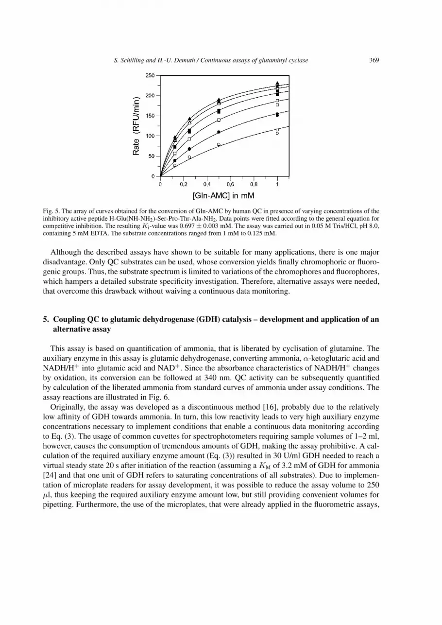
Fig. 5. The array of curves obtained for the conversion of Gln-AMC by human QC in presence of varying concentrations of theinhibitory active peptide H-Glu(NH-NH2)-Ser-Pro-Thr-Ala-NH2. Data points were fitted according to the general equation forcompetitive inhibition. The resulting Ki-value was 0.697 ± 0.003 mM. The assay was carried out in 0.05 M Tris/HCl, pH 8.0,containing 5 mM EDTA. The substrate concentrations ranged from 1 mM to 0.125 mM.
Although the described assays have shown to be suitable for many applications, there is one majordisadvantage. Only QC substrates can be used, whose conversion yields finally chromophoric or fluoro-genic groups. Thus, the substrate spectrum is limited to variations of the chromophores and fluorophores,which hampers a detailed substrate specificity investigation. Therefore, alternative assays were needed,that overcome this drawback without waiving a continuous data monitoring.
5. Coupling QC to glutamic dehydrogenase (GDH) catalysis – development and application of analternative assay
This assay is based on quantification of ammonia, that is liberated by cyclisation of glutamine. Theauxiliary enzyme in this assay is glutamic dehydrogenase, converting ammonia, α-ketoglutaric acid andNADH/H+ into glutamic acid and NAD+. Since the absorbance characteristics of NADH/H+ changesby oxidation, its conversion can be followed at 340 nm. QC activity can be subsequently quantifiedby calculation of the liberated ammonia from standard curves of ammonia under assay conditions. Theassay reactions are illustrated in Fig. 6.
Originally, the assay was developed as a discontinuous method [16], probably due to the relativelylow affinity of GDH towards ammonia. In turn, this low reactivity leads to very high auxiliary enzymeconcentrations necessary to implement conditions that enable a continuous data monitoring accordingto Eq. (3). The usage of common cuvettes for spectrophotometers requiring sample volumes of 1–2 ml,however, causes the consumption of tremendous amounts of GDH, making the assay prohibitive. A cal-culation of the required auxiliary enzyme amount (Eq. (3)) resulted in 30 U/ml GDH needed to reach avirtual steady state 20 s after initiation of the reaction (assuming a KM of 3.2 mM of GDH for ammonia[24] and that one unit of GDH refers to saturating concentrations of all substrates). Due to implemen-tation of microplate readers for assay development, it was possible to reduce the assay volume to 250µl, thus keeping the required auxiliary enzyme amount low, but still providing convenient volumes forpipetting. Furthermore, the use of the microplates, that were already applied in the fluorometric assays,

370 S. Schilling and H.-U. Demuth / Continuous assays of glutaminyl cyclase
Fig. 6. Representation of the QC-assay using GDH as auxiliary enzyme and an N -terminal glutaminyl peptide as substrate.In the initial reaction, the respective pyroglutamyl peptide and ammonia are formed. Subsequently, ammonia, α-ketoglutaricacid and NADH/H+ are converted into glutamic acid and NAD+ catalysed by GDH. The consumption of NADH/H+ can beobserved at 340 nm.
Fig. 7. Linear dependence of the initial rate of conversion on concentration of human QC using GDH as auxiliary enzyme. Theinset shows two progress curves, in the sample containing human QC (12 nM), a linear decrease of absorbance was observed.Without added QC, the decrease in absorbance was negligible. Reactions were carried out in 0.05 M Tris/HCl pH 8.0, containing5 mM EDTA.
enable a fast determination of QC activity in many samples at the same time, thus accelerating the deter-minations enormously. Finally, due to the detection of ammonia that is liberated from the QC substratesthe assay can be implemented for a fast examination of a variety of glutaminyl-peptides.
In fact, linear progress curves were obtained according to the predicted conditions, and most impor-tantly there was a linear relationship between QC concentration and initial velocity, indicating that theassay provides reliable results (Fig. 7). Subsequently, a detailed substrate specificity investigation wasperformed using about 40 newly synthesised substrates [27], showing that the assay is applicable in-

S. Schilling and H.-U. Demuth / Continuous assays of glutaminyl cyclase 371
Fig. 8. The influence of ionic strength on the specificity constant kcat/KM for conversion of various substrates by human andpapaya QC. For most peptides, there was little effect of changes in ionic strength detected. However, human and papaya QCspecificity towards positively charged peptides increased significantly by addition of 0.5 M KCl. Without additional salt added,the ionic strength was 0.029 M, corresponding to a 0.05 M Tris- or Tricine buffer.
dependently from changes of substrate amino acid composition and peptide size. Moreover, since theauxiliary enzyme was not influenced significantly by potassium chloride concentrations up to 0.5 M, themethod was also implemented to investigate the influence of ionic strength on conversion of differentsubstrates. Interestingly, the observed changes of the specificity constant kcat/KM were substrate depen-dent (Fig. 8). Neither papaya QC, nor human QC revealed altered activity by increasing ionic strengthtowards peptides with uncharged backbone and residues. Both enzymes, however, displayed a significantincrease in activity towards peptides comprising positively charged amino acid residues. Thus, besidesthe similar pH-dependence of catalysis for both enzymes, also the behaviour of the different QC-formsin environments with differing dielectric constants is similar, which could be also indicative for analo-gous catalytic mechanisms. Finally, this conclusion was also corroborated by the ability of both, humanand papaya QC, to cyclise N-terminal β-homoglutaminyl residues with the same catalytic efficiency (notshown).
6. Conclusions
The application of theoretical deductions [18] facilitated the development of the first coupled enzy-matic assays for glutaminyl cyclase (QC) activity. Due to the use of different enzymatic reactions forcoupling to QC catalysis, many characterisation studies could be performed including pH-dependence,inhibitory and substrate specificity. In this regard, the general differences in the coupling strategy, i.e.,the consumption of the QC products, either the pyroglutamyl peptide by pGAP or the liberated ammo-nia by GDH, led to a compensation of respective disadvantages of both assays. For instance, traces ofammonia in a sample hamper the QC determination in the GDH-coupled assay, but show no effect inthe assays using pGAP, resulting in the preferred usage of the latter enzyme for assays during enzymepurification. When investigating different peptide substrates, however, only the coupling to ammoniaproduction provided satisfying results since a large substrate spectrum that can be investigated. Thus,the use of the different assay coupling strategies enabled the convenient determination of QC activity indifferent fields of protein characterisation.
372 S. Schilling and H.-U. Demuth / Continuous assays of glutaminyl cyclase
Finally, the demonstrated strategy to develop continuous assay techniques could also be used to modifydiscontinuous assays for other enzymes or to develop new ways for their catalytic characterisation byimplementing different coupling strategies.
Acknowledgements
We thank K. Zunkel, H. Cynis and J. Zwanzig for valuable technical assistance.
References
[1] M. Messer, Enzymatic cyclization of L-glutamine and L-glutaminyl peptides, Nature 4874 (1963), 1299.[2] M. Messer and M. Ottesen, Isolation and properties of glutamine cyclotransferase of dried papaya latex, Biochim. Biophys.
Acta 92 (1964), 409–411.[3] W.H.J. Busby, G.E. Quackenbush, J. Humm, W.W. Youngblood and J.S. Kizer, An enzyme(s) that converts glutaminyl-
peptides into pyroglutamyl-peptides. Presence in pituitary, brain, adrenal medulla, and lymphocytes, J. Biol. Chem. 262(1987), 8532–8536.
[4] W.H. Fischer and J. Spiess, Identification of a mammalian glutaminyl cyclase converting glutaminyl into pyroglutamylpeptides, Proc. Natl. Acad. Sci. USA 84 (1987), 3628–3632.
[5] S.W. Dahl, C. Slaughter, C. Lauritzen, R.C. Bateman, Jr., I. Connerton and J. Pedersen, Carica papaya glutamine cyclo-transferase belongs to a novel plant enzyme subfamily: cloning and characterization of the recombinant enzyme, ProteinExpr. Purif. 20 (2000), 27–36.
[6] E. Van Coillie, P. Proost, I. Van Aelst, S. Struyf, M. Polfliet, I. De Meester, D.J. Harvey, J. Van Damme and G. Opdenakker,Functional comparison of two human monocyte chemotactic protein-2 isoforms, role of the amino-terminal pyroglutamicacid and processing by CD26/dipeptidyl peptidase IV, Biochemistry 37 (1998), 12672–12680.
[7] G.N. Abraham and D.N. Podell, Pyroglutamic acid. Non-metabolic formation, function in proteins and peptides, andcharacteristics of the enzymes effecting its removal, Mol. Cell. Biochem. 38 (Spec. No) (1981), 181–190.
[8] S.A. Hinke, J.A. Pospisilik, H.-U. Demuth, S. Manhart, K. Kühn-Wache, T. Hoffmann, E. Nishimura, R.A. Pedersen andC.H.S. McIntosh, Dipeptidyl peptidase IV (DPIV/CD26) degradation of Glucagon, J. Biol. Chem. 275 (2000), 3827–3834.
[9] S. Zerhouni, A. Amrani, M. Nijs, N. Smolders, M. Azarkan, J. Vincentelli and Y. Looze, Purification and characterizationof papaya glutamine cyclotransferase, a plant enzyme highly resistant to chemical, acid and thermal denaturation, Biochim.Biophys. Acta 1387 (1998), 275–290.
[10] T. Pohl, M. Zimmer, K. Mugele and J. Spiess, Primary structure and functional expression of a glutaminyl cyclase, Proc.Natl. Acad. Sci. USA 88 (1991), 10059–10063.
[11] A.P. Consalvo, S.D. Young, B.N. Jones and P.P. Tamburini, A rapid fluorometric assay for N-terminal glutaminyl cyclaseactivity using high-performance liquid chromatography, Anal. Biochem. 175 (1988), 131–138.
[12] M.Y. Gololobov, W. Wang and R.C.J. Bateman, Substrate and inhibitor specificity of glutamine cyclotransferase (QC),Biol. Chem. Hoppe Seyler 377 (1996), 395–398.
[13] K.A. Oberg, J.M. Ruysschaert, M. Azarkan, N. Smolders, S. Zerhouni, R. Wintjens, A. Amrani and Y. Looze, Papayaglutamine cyclase, a plant enzyme highly resistant to proteolysis, adopts an all-beta conformation, Eur. J. Biochem. 258(1998), 214–222.
[14] S. Schilling, T. Hoffmann, F. Rosche, S. Manhart, C. Wasternack and H.U. Demuth, Heterologous expression and charac-terization of human glutaminyl cyclase: evidence for a disulfide bond with importance for catalytic activity, Biochemistry41 (2002), 10849–10857.
[15] J.B. Koger, J. Humm and J.S. Kizer, Assay of glutaminylpeptide cyclase, Methods Enzymol. 168 (1989), 358–365.[16] R.C.J. Bateman, A spectrophotometric assay for glutaminyl-peptide cyclizing enzymes, J. Neurosci. Methods 30 (1989),
23–28.[17] M.Y. Gololobov, I. Song, W. Wang and R.C.J. Bateman, Steady-state kinetics of glutamine cyclotransferase, Arch.
Biochem. Biophys. 309 (1994), 300–307.[18] W.R. McClure, A kinetic analysis of coupled enzyme assays, Biochemistry 8 (1969), 2782–2786.[19] K. Fujiwara, R. Kobayashi and D. Tsuru, The substrate specificity of pyrrolidone carboxylyl peptidase from Bacillus
amyloliquefaciens, Biochim. Biophys. Acta 570 (1979), 140–148.[20] K. Fujiwara and D. Tsuru, New chromogenic and fluorogenic substrates for pyrrolidonyl peptidase, J. Biochem. (Tokyo)
83 (1978), 1145–1149.

S. Schilling and H.-U. Demuth / Continuous assays of glutaminyl cyclase 373
[21] D. Tsuru, K. Fujiwara and K. Kado, Purification and characterization of L-pyrrolidonecarboxylate peptidase from Bacillusamyloiliquefaciens, J. Biochem. (Tokyo) 84 (1978), 467–476.
[22] P.M. Cummins and B. O’Connor, Pyroglutamyl peptidase: an overview of the three known enzymatic forms, Biochim.Biophys. Acta 1429 (1998), 1–17.
[23] S. Schilling, T. Hoffmann, M. Wermann, U. Heiser, C. Wasternack and H.-U. Demuth, Continuous spectrometric assaysfor glutaminyl cyclase activity, Anal. Biochem. 303 (2002), 49–56.
[24] C. Frieden, Glutamic dehydrogenase. III. The order of substrate addition in the enzymatic reaction, J. Biol. Chem. 234(1959), 2891–2896.
[25] K.J. Ellis and J.F. Morrison, Buffers of constant ionic strength for studying pH-dependent processes, Methods Enzymol.87 (1982), 405–426.
[26] S. Schilling, S. Manhart, T. Hoffmann, H.-H. Ludwig, C. Wasternack and H.-U. Demuth, Substrate specificity of gluta-minyl cyclases from plants and animals, Biol. Chem. 384 (2003), 1583–1592.
[27] S. Schilling, A.J. Niestroj, J.-U. Rahfeld T. Hoffmann, M. Wermann, K. Zunkel, C. Wasternack and H.-U. Demuth, Potentand specific inhibition of human glutaminyl cyclase by imidazole derivatives reveals it as metalloenzyme, J. Biol. Chem.278 (2003), 49773–49779.


