Embed Size (px)
Citation preview

M A J O R A R T I C L E
Glutaminyl Cyclases as Novel Targets for theTreatment of Septic Arthritis
Annelie Hellvard,1,2 Katarzyna Maresz,3 Stephan Schilling,4 Sigrid Graubner,5 Ulrich Heiser,4 Roland Jonsson,1
Holger Cynis,4 Hans-Ulrich Demuth,4,5 Jan Potempa,3,6 and Piotr Mydel1,2
1Broegelmann Research Laboratory, The Gade Institute, University of Bergen, Norway; 2Department of Rheumatology and Inflammation Research,Sahlgrenska Academy, University of Gothenburg, Sweden; 3Department of Microbiology, Faculty of Biochemistry, Biophysics, and Biotechnology,Jagiellonian University, Krakow, Poland; 4Probiodrug, Halle/Saale, and 5Ingenium Pharmaceuticals, Martinsried, Germany; and 6Center for Oral Healthand Systemic Diseases, University of Louisville School of Dentistry, Kentucky
Background. Septic arthritis is a severe and rapidly debilitating disease mainly caused by Staphylococcusaureus. Here, we assess the antiarthritic efficiency of glutaminyl cyclase (QC) inhibitors.
Methods. Mice were inoculated with an arthritogenic amount of S. aureus intravenously or by local administra-tion into the knee joint. Animals were treated with QC inhibitors (PBD155 and PQ529) via chow during the experi-ment. QC and isoQC knockout mice were also analyzed for arthritis symptoms after local administration of bacteria.
Results. Both QC inhibitors significantly delayed the onset of clinical signs of arthritis, and inhibitors significantlydecreased weight loss in treated animals. Following intraarticular injection of S. aureus, PBD155-treated mice hadlower levels of synovitis and bone erosion, as well as less myeloperoxidase in synovial tissue. Fluorescence-activatedcell sorter analysis revealed that PBD155 treatment affected the expression pattern of adhesion molecules, preventingthe upregulation of cells expressing CD11b/CD18.
Conclusion. The compounds investigated here represent a novel class of small molecular antiarthritic inhibitors.In our studies, they exerted strong antiinflammatory actions, and therefore they might be suited for disease-modifyingtreatment of infectious arthritis.
Keywords. Glutaminyl cyclases; septic arthritis; posttranslational modifications; neutrophils.
Staphylococcus aureus–induced septic arthritis is amedical emergency because of its potential for causingmortality and rapid joint destruction, particularly inpatients with rheumatoid arthritis or immunocompro-mised status [1]. To study the pathogenesis of thisdisease, we used a mouse model of S. aureus–inducedarthritis in which intravenous administration of S.aureus leads to clinical and histological signs of arthri-tis within 48 hours [2]: neutrophils and activated mac-rophages infiltrate the synovium within 1 day, andT cells follow within a few days.
The extravasation of circulating neutrophils into in-flamed tissues, with the subsequent release of proteasesand production of reactive oxygen species, is an essen-tial component of the innate immune response [3].Neutrophils are also heavily involved in the recruitmentof monocytes by releasing chemokines [4]. Althoughneutrophils are important effector cells for the elimina-tion of invading pathogens, they can also cause collater-al damage to surrounding tissue [5]. Indeed, cartilagedestruction caused by neutrophil accumulation is aprominent feature of septic arthritis. Neutrophil-mediated tissue damage has been attributed to a varietyof proteolytic enzymes released by these cells, particu-larly elastase and cathepsin G [6, 7]. In addition, neu-trophils produce numerous inflammatory cytokines,including interleukin 8 and tumor necrosis factor α,that significantly contribute to the destructive cycle [8].Therefore, the influx of neutrophils into joints repre-sents an attractive target for the development of newtherapeutic strategies for septic arthritis.
Received 17 February 2012; accepted 20 June 2012; electronically published 29November 2012.
Correspondence: Piotr Mydel, M.D, PhD, Broegelmann Research Laboratory,University of Bergen, The Gade Institute, Laboratory Bldg, 5th Fl, Bergen, Horda-land 5021, Norway ([email protected]).
The Journal of Infectious Diseases 2013;207:768–77© The Author 2012. Published by Oxford University Press on behalf of the InfectiousDiseases Society of America. All rights reserved. For Permissions, please e-mail:[email protected]: 10.1093/infdis/jis729
768 • JID 2013:207 (1 March) • Hellvard et al
at Ernst M
ayr Library of the M
useum C
omp Z
oology, Harvard U
niversity on May 2, 2014
http://jid.oxfordjournals.org/D
ownloaded from

The migration of circulating neutrophils and monocytes tothe site of inflammation is tightly controlled by their interac-tion with the vascular endothelium [9]. Two β2 integrinsexpressed on the cell surface of leukocytes, lymphocyte func-tion-associated antigen 1 (CD11a/CD18) and Mac-1 (CD11b/CD18), and their counterpart expressed on endothelial cells,intercellular adhesion molecule 1, are crucial for neutrophiladhesion and migration [10, 11]. Furthermore, E-selectin andP-selectin, expressed mainly on the endothelium, togetherwith L-selectin, expressed on the surface of neutrophils, areabundant at sites of inflammation and are also important incontrolling migration across the endothelium [12].
We recently reported that the isoenzyme of glutaminylcyclase (isoQC) has an important role in monocyte infiltrationunder inflammatory conditions by mediating the posttransla-tional modification of monocyte chemoattractant protein 1(MCP-1/CCL2) [13]. This might be relevant to septic arthritisbecause the switch from neutrophil to monocyte recruitmentis regulated by CCL2 [14].
CCL2 is one of the major chemokines produced by activatedneutrophils and is crucial for monocyte migration to the site ofinflammation and for the switch from acute to chronic inflamma-tion [15]. CCL2 can exert a direct effect on neutrophil recruit-ment in vitro [16], and administration of CCL2 in the presence ofendotoxins induces strong migration of neutrophils to the site ofinflammation [17]. Similarly, neutralization of CCL2 causes a de-crease in neutrophil influx in a septic peritonitis model througheffects on the neutrophil chemoattractant leukotriene B4 [18].
The activity and stability of CCL2 are dependent on post-translational conversion of its N-terminal glutamine into py-roglutamate [13, 19]. This conversion is catalyzed by QCs andis required for the function of several proteins [20, 21]. QC isinvolved in the pathology of diseases such as Alzheimer’sdisease [22], melanoma [23], osteoporosis [24], and rheuma-toid arthritis [25]. Recently, an isoenzyme of QC was discov-ered in both humans and mice. This isoenzyme possessesnearly identical substrate specificity but differs in subcellularlocalization, with QC being secreted from cells and isoQCbeing retained in the Golgi apparatus [13, 26, 27].
To balance the inflammatory response and protect the hostfrom pathogens while avoiding extensive tissue damage from ex-cessive activation of the immune system, all cells, chemokines,and other components of the immune system have to work inconcert. In the present study, we evaluated the effect of 2 inhibi-tors of QC/isoQC, PBD155 and PQ529, on inflammatory disease,using an animal model of S. aureus–induced septic arthritis.
METHODS
MiceNMRI mice were obtained from Charles River Laboratories.Ten QC knockout mice [28] with the genetic background of
99.2% C57Bl/6 and 0.8% 129Sv, 10 isoQC knockout mice [13]produced in the C3J strain, and 20 wild-type littermates (10C3J mice and 10 C57Bl/6/129Sv mice) were used.
Treatment With QC/isoQC InhibitorsIsoform-nonspecific QC/isoQC inhibitors PQ529 [13] andPBD155 (Probiodrug) were used. Standard laboratory chowsupplemented with PQ529 or PBD155 was generated by Ssniffat a concentration of 6.8 g/kg. Mice were pretreated with sup-plemented chow 3 days before induction of septic arthritis.Ethics approval was obtained from the Animal Research Com-mittee of University of Gothenburg.
Bacterial Strain and Induction of Septic ArthritisStaphylococcus aureus strain LS-1, originating from a joint of aspontaneously arthritic NZB/W mouse, was used [29]. Thebacteria were diluted in phosphate-buffered saline (PBS), andmice received intravenous inoculation of 107 colony-formingunits (CFU) on day 0.
Determination of Bacterial GrowthGrowth of staphylococci in the kidneys was determined 3 and14 days after intravenous injection of S. aureus. Kidneys wereremoved aseptically, homogenized in PBS, and plated onblood agar plates. After incubation for 24 hours at 37°C, thenumber of CFU per animal was calculated.
Induction and Registration of Olive Oil–Induced InflammationInflammation was induced by intradermal injection of 30 μLof olive oil (Apoteksbolaget) in the hind footpad. Footpadswelling was measured 26 hours after injection, using anOditest spring caliper. The intensity of the olive oil–inducedinflammation was expressed as [(footpad thickness at 26hours) – (footpad thickness at 0 hours)] × 10–3 cm.
Clinical Evaluation of ArthritisClinical scoring of arthritis of the paws was assessed everysecond day through the following scoring system: 0, no sign ofinflammation; 1, mild swelling and/or erythema; 2, moderateswelling and erythema; and 3, marked swelling and erythema.Swelling and erythema of a toe or finger was scored as 0.5.Clinical score was calculated by adding scores for all 4 paws,resulting in a maximum score of 12 per mouse. In addition,mice weights were monitored.
Histological EvaluationMice were sacrificed on day 3 and 14, and paws with elbowsand knees were removed for histological assessment. Followingdecalcification with Parengy (Histolab), samples were embed-ded in paraffin, cut, and stained with hematoxylin-eosin, andsections were evaluated by a blinded examiner for synovitisand erosion of bone and/or cartilage. Synovitis was defined asa membrane thickness of >2 cell layers. A mean score of 12
Glutaminyl Cyclase Inhibitors in Septic Arthritis • JID 2013:207 (1 March) • 769
at Ernst M
ayr Library of the M
useum C
omp Z
oology, Harvard U
niversity on May 2, 2014
http://jid.oxfordjournals.org/D
ownloaded from

joints for all inspected animals was calculated, giving amaximum arthritis index of 3 per animal. The following histo-logical scoring system of synovitis and bone erosion was used:1, mild; 2, moderate; and 3, severe [30].
Intraarticular Challenge of S. aureusTreated mice and knockout mice were intraarticularly inocu-lated with 104 CFU of S. aureus and sacrificed 3 days later.Injected knees were obtained for histological evaluation, aspreviously described.
Determination of Synovial Myeloperoxidase ContentThe synovial membrane was dispersed into a single-cell suspen-sion, and cells were lysed for 1 hour at room temperature in 20µL of lysis buffer containing 0.2% cetrimonium bromide (Sigma-Aldrich) and 0.2% bovine serum albumin (Sigma-Aldrich) inPBS. The peroxidase substrate 1,2-phenylenediamine dihydro-chloride (Dako) was dissolved according to the manufacturer’sinstructions and mixed with H2O2 immediately before use. Atotal of 40 μL of peroxidase substrate was added to the samples,and the samples were incubated for 1 hour at room temperature.The absorbance was measured at 450 nm on a Spectra Max340PC (Molecular Devices).
Flow Cytometric AnalysisBlood was obtained from 14 untreated or PBD155-treatedmice 3 days after intraarticular inoculation of 104 CFU of S.aureus and compared with blood from 12 noninfected mice.Whole blood was subjected to isotonic lysis for elimination oferythrocytes. Cells were pelleted in a 96-well plate and blockedwith Fc-block (BD Pharmingen). Cells were stained witheFluor 450–conjugated anti-CD11b (clone M1/70, BD Biosci-ences), fluorescein isothiocyanate (FITC)–conjugated CD18(clone M18/2, eBioscience), and peridinin-chlorophyll-proteincomplex–conjugated anti-CD62L (clone MEL-14, BioLegend)for 30 minutes at 4°C. Cells were collected using FACSCantoII(BD-Bioscience) equipped with FACSDiva software. Analysiswas performed using FlowJo software (Tree Star), and Fluoro-chrome Minus One was used for determination of positivepopulations and gating when needed.
Phagocytosis AssaySamples of whole blood from 5 healthy NMRI mice treated for3 days with inhibitor and samples from 4 untreated controlswere evaluated by flow cytometry for phagocytosis of FITC-labeledE. coli,usingPhagotest (OrpegenPharma).Measurementwas performed according to the manufacturer’s instructions.Cells were collected using FACSCantoII with FACSDiva soft-ware. Analysis was performed using FlowJo software.
Statistical AnalysisDifferences between 2 groups were tested for statistical signifi-cance by means of the Mann–Whitney U test. Kaplan-Meier
plots were prepared, and the log-rank test was used for com-paring the curves of arthritis frequency. Weight curves wereanalyzed using 2-way analysis of variance with the Bonferroniposttest. All statistical analyses were performed using Graph-Pad Prism, version 5.0d for Mac (GraphPad), and a P value of<.05 was considered statistically significant.
RESULTS
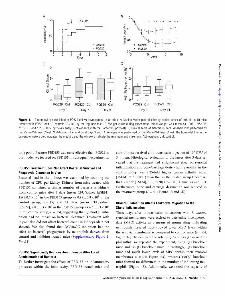
QC/isoQC Inhibitors Delay the Onset and Progression ofSeptic ArthritisTo investigate the role of QC/isoQC inhibition on the devel-opment of septic arthritis, mice were intravenously inoculatedwith S. aureus LS-1 and provided with chow impregnated withthe inhibitors PQ529 (for 10 mice) and PBD155 (for 10 mice)for the duration of the experiments. Treatment with PQ529resulted in a significant delay in the development of arthritis;at day 3, 90% of control mice displayed arthritis as comparedto 50% of treated mice (P = .01; Figure 1A). Considerableweight loss occurs during septic arthritis because of inflamma-tion. Mice treated with PQ529 showed a significantly lowerrate of weight loss and recovered more rapidly (Figure 1B).Despite the effect of PQ529 on arthritis development andweight loss, it had no effect on the clinical scoring of joints(Figure 1C). Histological scoring of sectioned joints and pawsat end point confirmed the clinical observations, with no dif-ferences between PQ529-treated mice and controls in bothshort-term and long-term experiments (Figure 1D).
PBD155 was more effective than PQ529 in delaying theprogression of septic arthritis. At day 3 after inoculation, only40% of treated mice displayed clinical signs of arthritis,whereas 90% of control mice had arthritis (P = .01; Figure. 2A).Mice treated with PBD155 also had significantly less severearthritis as compared to control mice. The clinical score of thecontrol group increased rapidly until day 9, when it reached amean of 5.1 (combined score, 40.5), compared with a mean of1.9 (combined score, 19.0) in the treated group (P = .01;Figure 2C). All mice experienced a decrease in body weightduring the development of septic arthritis. However, unlikethe control group, mice treated with PBD155 started to gainweight after 5 days (Figure 2B).
Histological evaluation for synovitis and erosions was per-formed at the early phase (day 3, for 20 mice) and late phase(day 14, for 20 mice) of inflammation. Morphological exami-nation confirmed that the inhibition of QC/isoQC withPBD155 alleviated arthritis. Cell infiltration at both timepoints was significantly reduced by PBD155 treatment at day 3(mean arthritis index [±SEM], 0.55 ± 0.04) and at day 14(mean arthritis index [±SEM], 0.72 ± 0.08) as compared to thecontrol group (mean arthritis index [±SEM], 1.01 ± 0.17 and1.18 ± 0.15 at days 3 and 14, respectively; Figure 2E and 2F).The extent of erosion did not differ between the groups at any
770 • JID 2013:207 (1 March) • Hellvard et al
at Ernst M
ayr Library of the M
useum C
omp Z
oology, Harvard U
niversity on May 2, 2014
http://jid.oxfordjournals.org/D
ownloaded from
time point. Because PBD155 was more effective than PQ529 inour model, we focused on PBD155 in subsequent experiments.
PBD155 Treatment Does Not Affect Bacterial Survival andPhagocytic Clearance In VivoBacterial load in the kidneys was examined by counting thenumber of CFU per kidney. Kidneys from mice treated withPBD155 contained a similar number of bacteria as kidneysfrom control mice after 3 days (mean CFU/kidney [±SEM],1.6 ± 0.7 × 10⁷ in the PBD155 group vs 0.98 ± 0.8 × 10⁷ in thecontrol group; P = .15) and 14 days (mean CFU/kidney[±SEM], 7.8 ± 0.3 × 10⁷ in the PBD155 group vs 4.3 ± 0.3 × 10⁷
in the control group; P = .15), suggesting that QC/isoQC inhi-bition had no impact on bacterial clearance. Treatment withPQ529 also did not affect bacterial count in kidneys (data notshown). We also found that QC/isoQC inhibition had noeffect on bacterial phagocytosis by neutrophils derived fromcontrol and inhibitor-treated mice (Supplementary Figure 1;P = .11).
PBD155 Significantly Reduces Joint Damage After LocalAdministration of BacteriaTo further investigate the effects of PBD155 on inflammatoryprocesses within the joint cavity, PBD155-treated mice and
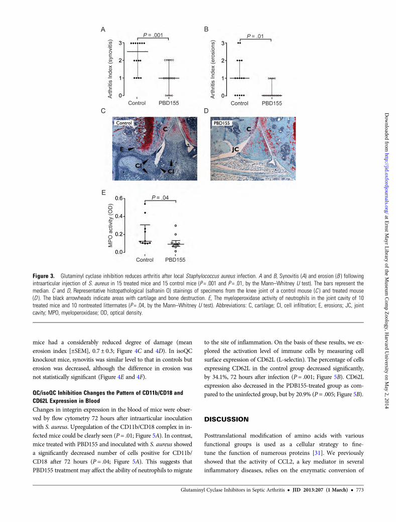
control mice received an intraarticular injection of 104 CFU ofS. aureus. Histological evaluation of the knees after 3 days re-vealed that the treatment had a significant effect on synovialinflammation and bone/cartilage destruction. Synovitis in thecontrol group was 2.25-fold higher (mean arthritis index[±SEM], 2.25 ± 0.21) than that in the treated group (mean ar-thritis index [±SEM], 1.0 ± 0.20) (P = .001; Figure 3A and 3C).Furthermore, bone and cartilage destruction was reduced inthe treatment group (P = .01; Figure 3B and 3D).
QC/isoQC Inhibition Affects Leukocyte Migration to theSite of InflammationThree days after intraarticular inoculation with S. aureus,synovial membranes were excised to determine myeloperoxi-dase (MPO) activity as a means of enumerating infiltratingneutrophils. Treated mice showed lower MPO levels withinthe synovial membrane as compared to control mice (P = .04;Figure 3E). To delineate the role of QC and isoQC in neutro-phil influx, we repeated the experiment, using QC knockoutmice and isoQC knockout mice. Interestingly, QC knockoutmice had much lower levels of MPO within their synovialmembranes (P = .04; Figure 4A), whereas isoQC knockoutmice showed no differences in the number of infiltrating neu-trophils (Figure 4B). Additionally, we tested the capacity of
Figure 1. Glutaminyl cyclase inhibitor PQ529 delays development of arthritis. A, Kaplan-Meier plots displaying clinical onset of arthritis in 10 micetreated with PQ529 and 10 controls (P = .01, by the log-rank test). B, Weight curve during experiment. Initial weight was taken as 100% (*P < .05,**P < .01, and ***P < .005, by 2-way analysis of variance with the Bonferroni posttest). C, Clinical score of arthritis in mice. Analysis was performed bythe Mann–Whitney U test. D, Articular inflammation at days 3 and 14. Analysis was performed by the Mann–Whitney U test. The horizontal line in thebox-and-whiskers plot indicates the median, and the whiskers indicate the minimum and maximum. Abbreviation: Ctrl, control.
Glutaminyl Cyclase Inhibitors in Septic Arthritis • JID 2013:207 (1 March) • 771
at Ernst M
ayr Library of the M
useum C
omp Z
oology, Harvard U
niversity on May 2, 2014
http://jid.oxfordjournals.org/D
ownloaded from

inflammatory cells to migrate to the site of inflammation,using a paw edema assay. This assay revealed that PBD155 de-creased cell influx at 26 hours after injection, compared withcontrol mice (P = .02; Supplementary Figure 2).
Inhibition of Both Isoforms Is Required to PreventJoint DamageTo explore the importance of both isoenzymes in protectionagainst inflammatory joint damage after bacterial inoculation,QC knockout mice, isoQC knockout mice, and wild-type micereceived intraarticular inoculations of 1 × 10⁴ CFU of S. aureus.
Three days after S. aureus inoculation, there was a histologicaltrend toward decreased synovitis and bone/cartilage destructionin the QC knockout mice and isoQC knockout mice, but thedifference was not significant. This outcome contrasted with thesignificant difference in synovitis and bone/cartilage destructionobtained with QC/isoQC inhibition. However, in the QC knock-out group, synovitis was present in 60% of mice (mean arthritisindex [±SEM], 1.1 ± 0.38), compared with 100% of wild-typecontrols (mean arthritis index [±SEM], 1.9 ± 0.28). In addition,3 of 10 wild-type mice had high bone/cartilage erosion scores(mean erosion index [±SEM], 1.3 ± 0.42), whereas QC knockout
Figure 2. Glutaminyl cyclase inhibitor (PBD155) delays the onset and ameliorates the progression of septic arthritis. A, Kaplan–Meyer plot of onsetof arthritis in 10 PBD155-treated mice and 10 controls (P = .01, by the log-rank test). B, Weight curve during the experiment with PBD155 (**P < .01 and***P < .005, by 2-way analysis of variance with the Bonferroni posttest). C, Clinical scoring during the experiment (P = .01, by the Mann–WhitneyU test). D, Articular inflammation at days 3 and 14 (P = .04 and P = .02, by the Mann–Whitney U test, on days 3 and 14, respectively). E and F, Represen-tative histological changes in the front paw joints of PBD155-treated animals and nontreated controls (original magnification ×50). The horizontal line inthe box-and-whiskers plot indicates the median, and the whiskers indicate the minimum and maximum. Abbreviations: CI, cell infiltration; Ctrl, control; E,erosions.
772 • JID 2013:207 (1 March) • Hellvard et al
at Ernst M
ayr Library of the M
useum C
omp Z
oology, Harvard U
niversity on May 2, 2014
http://jid.oxfordjournals.org/D
ownloaded from
mice had a considerably reduced degree of damage (meanerosion index [±SEM], 0.7 ± 0.3; Figure 4C and 4D). In isoQCknockout mice, synovitis was similar level to that in controls buterosion was decreased, although the difference in erosion wasnot statistically significant (Figure 4E and 4F).
QC/isoQC Inhibition Changes the Pattern of CD11b/CD18 andCD62L Expression in BloodChanges in integrin expression in the blood of mice were obser-ved by flow cytometry 72 hours after intraarticular inoculationwith S. aureus. Upregulation of the CD11b/CD18 complex in in-fected mice could be clearly seen (P = .01; Figure 5A). In contrast,mice treated with PBD155 and inoculated with S. aureus showeda significantly decreased number of cells positive for CD11b/CD18 after 72 hours (P = .04; Figure 5A). This suggests thatPBD155 treatment may affect the ability of neutrophils to migrate
to the site of inflammation. On the basis of these results, we ex-plored the activation level of immune cells by measuring cellsurface expression of CD62L (L-selectin). The percentage of cellsexpressing CD62L in the control group decreased significantly,by 34.1%, 72 hours after infection (P = .001; Figure 5B). CD62Lexpression also decreased in the PDB155-treated group as com-pared to the uninfected group, but by 20.9% (P = .005; Figure 5B).
DISCUSSION
Posttranslational modification of amino acids with variousfunctional groups is used as a cellular strategy to fine-tune the function of numerous proteins [31]. We previouslyshowed that the activity of CCL2, a key mediator in severalinflammatory diseases, relies on the enzymatic conversion of
Figure 3. Glutaminyl cyclase inhibition reduces arthritis after local Staphylococcus aureus infection. A and B, Synovitis (A) and erosion (B ) followingintraarticular injection of S. aureus in 15 treated mice and 15 control mice (P = .001 and P = .01, by the Mann–Whitney U test). The bars represent themedian. C and D, Representative histopathological (safranin O) stainings of specimens from the knee joint of a control mouse (C ) and treated mouse(D ). The black arrowheads indicate areas with cartilage and bone destruction. E, The myeloperoxidase activity of neutrophils in the joint cavity of 10treated mice and 10 nontreated littermates (P = .04, by the Mann–Whitney U test). Abbreviations: C, cartilage; CI, cell infiltration; E, erosions; JC, jointcavity; MPO, myeloperoxidase; OD, optical density.
Glutaminyl Cyclase Inhibitors in Septic Arthritis • JID 2013:207 (1 March) • 773
at Ernst M
ayr Library of the M
useum C
omp Z
oology, Harvard U
niversity on May 2, 2014
http://jid.oxfordjournals.org/D
ownloaded from

the N-terminal glutamine into pyroglutamate in a reactioncatalyzed by iso-glutaminyl cyclase [13]. The formation ofN-terminal pyroglutamate modulates bioactivity but also pro-tects proteins and peptides against degradation by aminopepti-dases [32]. Here, we used oral inhibitors (PQ529 and PBD155)to show the importance of QC/isoQC for disease development,neutrophil migration to the inflammation site, and synovitisand bone/cartilage erosion in hematogenously induced S.aureus arthritis.
In S. aureus arthritis, mice treated with PQ529 developedclinical signs of arthritis significantly later than control mice.PQ529-treated mice also had less weight loss during the acutephase of the disease and gained weight faster during recovery.PBD155, a more optimized compound than PQ529, was alsoeffective, leading to a significant delay in arthritis development,reduced weight loss, and a lower clinical score. Histological
assessments were consistent with clinical observations: treatmentwith PBD155 led to a significant reduction in synovitis at days 3and 14.
The peak bacterial burden after intravenous S. aureus inoc-ulation typically occurs in blood within the first 3 days and inkidneys within 1 week [33]. Bacterial clearance is dependenton phagocytosis by neutrophils [34], which is the primary leu-kocyte population in the joint lesions [2]. However, activatedneutrophils also serve as the primary cells responsible fortissue damage by releasing damaging proteinases and proin-flammatory cytokines [35]. NADPH oxidase–derived superox-ide discharged from infiltrating neutrophils also contributes tothe degradation of the joint surface [36].
We detected lower MPO activity, a measure of neutrophilnumbers, in QC inhibitor–treated mice, which suggests reducedinfiltration of neutrophils. This contrasts with previous results
Figure 4. Knockout (KO) of a single glutaminyl cyclase (QC) isoform does not protect against septic arthritis. Ten homozygous QC KO mice and 10isoform QC (isoQC) KO mice were intraarticulary inoculated with 1 × 10⁴ Staphylococcus aureus. Knee joints and synovial membranes were excised onday 3 for histological evaluation and assessment of myeloperoxidase (MPO) activity in the joints. A, Levels of MPO activity in 6 QC KO mice and 7 wild-type mice (P = .04, by the Mann–Whitney U test). B, MPO activity in synovia of 9 isoQC KO mice and 9 wild-type littermates. C and D, Histologicalevaluation of lymphocyte infiltration (C ) and erosions (D ) in the joints of 10 QC KO mice and 10 wild-type controls. E and F, Evaluation of synovitis (E )and erosions (F ) in 10 isoQC KO mice and 10 wild-type mice. Abbreviation: OD, optical density.
774 • JID 2013:207 (1 March) • Hellvard et al
at Ernst M
ayr Library of the M
useum C
omp Z
oology, Harvard U
niversity on May 2, 2014
http://jid.oxfordjournals.org/D
ownloaded from

showing that granulocyte infiltration in thioglycollate-inducedperitonitis and lipopolysaccharide-induced lung inflammationwas not affected in QC and isoQC knockout mice [13]. QCand isoQC exhibit nearly identical substrate specificity and aredifferentiated only by their subcellular localization: QC is se-creted to the extracellular milieu, and isoQC remains in theGolgi apparatus [26]. Because the compounds used in thisstudy inhibit both isoenzymes, we used QC and isoQC knock-out mice to determine whether the beneficial effects of treat-ment depend on the inhibition of a certain isoform orwhether both isoforms need to be blocked for the therapeuticeffect.
In QC-deficient mice and isoQC-deficient mice, there was atrend toward a lower arthritis score following local inoculationof bacteria, compared with the score for wild-type mice.However, only in the QC knockout mice were MPO levels sig-nificantly lower than those in the other infected groups.Under septic conditions, CCL2 regulates neutrophil recruit-ment via direct chemotactic activity and via modulatoryeffects on CCL8 and leukotriene B4 [37]. In addition, bonemarrow neutrophils express fully functional CCR2, which sug-gests a role for CC chemokines in the differentiation and reg-ulation of neutrophil functions [16].
The data presented here provide evidence that QC/isoQCmight play a role in leukocyte infiltration under certain experi-mental settings, such as septic arthritis. Clearly, CCL2 is notthe sole inflammatory effector molecule affected by QC/isoQCinhibition. Apart from known targets such as CCL-2, -7, -8,and -13, there might be other unknown targets for QC/isoQCthat may affect the inflammatory response in this experimentalsetting. These studies of QC/isoQC knockout mice provideclear evidence that the inhibition of both isoforms is requiredto alleviate septic arthritis in the intraarticular injection model.
FACS analysis suggests that QC inhibition affected integrinexpression. Activation of circulating neutrophils results inrapid upregulation of CD11b/CD18 expression, which allowsneutrophil immobilization and subsequent transmigration tothe site of inflammation [38]. Upregulation of CD11b/CD18was observed in the control group after S. aureus administra-tion into the joint but not in the group treated with PBD155.Treatment with PBD155 by itself caused a higher extent ofCD11b/CD18 expression before infection, followed by a reduc-tion during infection. Although the cause of this increased ex-pression in healthy mice is unknown, it is clear that theinfiltration of leukocytes at site of inflammation is reduced.Downregulation of CD11b/CD18 could result in decreased af-finity between neutrophils and the endothelium, which couldexplain the reduced neutrophil transmigration to the site ofinflammation in PBD155-treated mice. The observed downre-gulation of CD62L ligand may also have contributed to theimpaired migration to the inflamed tissue in PBD155-treatedmice. Leukocytes constitutively express CD62L, which plays apivotal role in attachment to the endothelium at the site ofinflammation [39]. In response to cell activation by variousstimuli, including lipoteichoic acid, a major component ofS. aureus cell wall [40], and lipopolysaccharide [41], the ex-pression of CD62L is downregulated [42]. We observed down-regulation of CD62L in the treated group, indicating thatL-selectin is not responsible for the reduced infiltration injoint.
To summarize, we have shown that treatment of mice withspecific QC/isoQC inhibitors alleviates the development andprogression of experimental septic arthritis. Collectively, weprovide compelling evidence that the treatment decreases cellinfiltration into the synovium and reduces synovitis and bone/cartilage destruction. Although not all substrates targeted by
Figure 5. Treatment with glutaminyl cyclase inhibitor (PBD155) changes the pattern of integrin expression. NMRI mice were inoculated with 1 × 10⁴
Staphylococcus aureus in the joint cavity, and on day 3 the expression of integrins was evaluated through flow cytometric analysis. A, CD11b/CD18expression on cells circulating in the blood of control mice (P = .01), and treated mice (P = .04). B, Expression of CD62L in the blood of the control group(P = .001) and in infected mice treated with PBD155 (P = .005). The horizontal line in the box-and-whiskers plot indicates the median, and the whiskersindicate the minimum and maximum. Differences between groups were analyzed using the Mann–Whitney U test.
Glutaminyl Cyclase Inhibitors in Septic Arthritis • JID 2013:207 (1 March) • 775
at Ernst M
ayr Library of the M
useum C
omp Z
oology, Harvard U
niversity on May 2, 2014
http://jid.oxfordjournals.org/D
ownloaded from

QC inhibitors are known, we suggest that the therapeuticeffect is at least partially based on the impaired ability of neu-trophils to massively migrate to the site of inflammation andinflict major damage. However, even though we see a de-creased capacity of neutrophils to migrate into the inflamedsynovial tissue, our phagocytosis experiments proved poly-morphonuclear cells to be fully capable of clearing bacteriaand preventing dissemination of the infection. Because QC/isoQC inhibitors are approaching the regulatory stage for thetreatment of various inflammatory disorders, this study pro-vides novel insights into their mechanism of action and opensdoors to the development of new combination therapies.
Supplementary Data
Supplementary materials are available at The Journal of Infectious Diseasesonline (http://jid.oxfordjournals.org/). Supplementary materials consist ofdata provided by the author that are published to benefit the reader. Theposted materials are not copyedited. The contents of all supplementarydata are the sole responsibility of the authors. Questions or messagesregarding errors should be addressed to the author.
Notes
Financial support. This work was supported by the Medical Society ofGöteborg; the Swedish Society of Medicine, Tore Nilsson Foundation; theSwedish Association Against Rheumatism; the Nanna Swartz Foundation;the Rune and Ulla Amlövs Trust; and the European Commission FP7(Gums & Joints no. 261460) and Marie Curie ITN (RAPID no.290246). K. M. and J. P. are supported by a Marie Curie ReintegrationGrant (PIRG03-GA-2008-230850JP), the National Institutes of Health(grant DE 09761), the National Science Center (2011/01/B/NZ6/00268,Kraków, Poland), and the Foundation for Polish Science (TEAM projectDPS/424-329/10). The Faculty of Biochemistry, Biophysics and Biotech-nology of the Jagiellonian University is a beneficiary of structural fundsfrom the European Union (POIG.02.01.00-12-064/08).Potential conflicts of interest. S. S., U. H., and H. C. are former or
present employees of Probiodrug. S. G. is employee of IngeniumPharmaceuticals. H. U. D. is chief science officer of Probiodrug and man-aging director of Ingenium Pharmaceuticals, a daughter company of Pro-biodrug, and holds stock in the Probiodrug group. All other authorsreport no potential conflicts.All authors have submitted the ICMJE Form for Disclosure of Potential
Conflicts of Interest. Conflicts that the editors consider relevant to thecontent of the manuscript have been disclosed.
References
1. Koopman WJ, Moreland LW. Arthritis and allied conditions: a text-book of rheumatology. 15th ed. Philadelphia: Lippincott Williams &Wilkins, 2005:2577–92.
2. Bremell T, Abdelnour A, Tarkowski A. Histopathological and serolog-ical progression of experimental Staphylococcus aureus arthritis. InfectImmun 1992; 60:2976–85.
3. Faurschou M, Borregaard N. Neutrophil granules and secretory vesi-cles in inflammation. Microbes Infect 2003; 5:1317–27.
4. Soehnlein O, Zernecke A, Eriksson EE, et al. Neutrophil secretionproducts pave the way for inflammatory monocytes. Blood 2008;112:1461–71.
5. Edwards SW, Hallett MB. Seeing the wood for the trees: the forgottenrole of neutrophils in rheumatoid arthritis. Immunol Today 1997;18:320–4.
6. Velvart M, Fehr K. Degradation in vivo of articular cartilage in rheu-matoid arthritis and juvenile chronic arthritis by cathepsin G and elas-tase from polymorphonuclear leukocytes. Rheumatol Int 1987;7:195–202.
7. Hilbert N, Schiller J, Arnhold J, Arnold K. Cartilage degradation bystimulated human neutrophils: elastase is mainly responsible for carti-lage damage. Bioorg Chem 2002; 30:119–32.
8. Cassatella MA. The production of cytokines by polymorphonuclearneutrophils. Immunol Today 1995; 16:21–6.
9. Henderson RB, Hobbs JA, Mathies M, Hogg N. Rapid recruitment ofinflammatory monocytes is independent of neutrophil migration.Blood 2003; 102:328–35.
10. Henderson RB, Lim LH, Tessier PA, et al. The use of lymphocytefunction-associated antigen (LFA)-1-deficient mice to determine therole of LFA-1, Mac-1, and alpha4 integrin in the inflammatory re-sponse of neutrophils. J Exp Med 2001; 194:219–26.
11. Sumagin R, Prizant H, Lomakina E, Waugh RE, Sarelius IH. LFA-1and Mac-1 define characteristically different intralumenal crawlingand emigration patterns for monocytes and neutrophils in situ. JImmunol 2010; 185:7057–66.
12. Albelda SM, Smith CW, Ward PA. Adhesion molecules and inflam-matory injury. FASEB J 1994; 8:504–12.
13. Cynis H, Hoffmann T, Friedrich D, et al. The isoenzyme of glutaminylcyclase is an important regulator of monocyte infiltration under in-flammatory conditions. EMBO Mol Med 2011; 9:545–58.
14. Kaplanski G, Marin V, Montero-Julian F, Mantovani A, Farnarier C.IL-6: a regulator of the transition from neutrophil to monocyte re-cruitment during inflammation. Trends Immunol 2003; 24:25–9.
15. Yamashiro S, Kamohara H, Yoshimura T. MCP-1 is selectively ex-pressed in the late phase by cytokine-stimulated human neutrophils:TNF-alpha plays a role in maximal MCP-1 mRNA expression. JLeukoc Biol 1999; 65:671–9.
16. Iida S, Kohro T, Kodama T, Nagata S, Fukunaga R. Identification ofCCR2, flotillin, and gp49B genes as new G-CSF targets during neutro-philic differentiation. J Leukoc Biol 2005; 78:481–90.
17. Maus U, Huwe J, Maus R, Seeger W, Lohmeyer J. Alveolar JE/MCP-1and endotoxin synergize to provoke lung cytokine upregulation, se-quential neutrophil and monocyte influx, and vascular leakage inmice. Am J Respir Crit Care Med 2001; 164:406–11.
18. Matsukawa A, Hogaboam CM, Lukacs NW, Lincoln PM, Strieter RM,Kunkel SL. Endogenous monocyte chemoattractant protein-1(MCP-1) protects mice in a model of acute septic peritonitis: cross-talk between MCP-1 and leukotriene B4. J Immunol 1999; 163:6148–54.
19. Van Coillie E, Proost P, Van Aelst I, et al. Functional comparison oftwo human monocyte chemotactic protein-2 isoforms, role of theamino-terminal pyroglutamic acid and processing by CD26/dipeptidylpeptidase IV. Biochemistry 1998; 37:12672–80.
20. Fischer WH, Spiess J. Identification of a mammalian glutaminylcyclase converting glutaminyl into pyroglutamyl peptides. Proc NatlAcad Sci U S A 1987; 84:3628–32.
21. Busby, WH Jr, Quackenbush GE, Humm J, Youngblood WW, KizerJS. An enzyme(s) that converts glutaminyl-peptides into pyroglutam-yl-peptides. Presence in pituitary, brain, adrenal medulla, and lympho-cytes. J Biol Chem 1987; 262:8532–6.
22. Schilling S, Zeitschel U, Hoffmann T, et al. Glutaminyl cyclase inhibi-tion attenuates pyroglutamate Abeta and Alzheimer’s disease-like pa-thology. Nat Med 2008; 14:1106–11.
23. Muthusamy V, Duraisamy S, Bradbury CM, et al. Epigenetic silencingof novel tumor suppressors in malignant melanoma. Cancer Res 2006;66:11187–93.
24. Ezura Y, Kajita M, Ishida R, et al. Association of multiple nucleotidevariations in the pituitary glutaminyl cyclase gene (QPCT) with lowradial BMD in adult women. J Bone Miner Res 2004; 19:1296–301.
25. Batliwalla FM, Baechler EC, Xiao X, et al. Peripheral blood geneexpression profiling in rheumatoid arthritis. Genes Immun 2005; 6:388–97.
776 • JID 2013:207 (1 March) • Hellvard et al
at Ernst M
ayr Library of the M
useum C
omp Z
oology, Harvard U
niversity on May 2, 2014
http://jid.oxfordjournals.org/D
ownloaded from
26. Cynis H, Rahfeld JU, Stephan A, et al. Isolation of an isoenzyme of humanglutaminyl cyclase: retention in the Golgi complex suggests involvementin the protein maturation machinery. J Mol Biol 2008; 379:966–80.
27. Stephan A, Wermann M, von Bohlen A, et al. Mammalian glutaminylcyclases and their isoenzymes have identical enzymatic characteristics.FEBS J 2009; 276:6522–36.
28. Schilling S, Kohlmann S, Bauscher C, et al. Glutaminyl cyclase knock-out mice exhibit slight hypothyroidism but no hypogonadism: impli-cations for enzyme function and drug development. J Biol Chem2011; 286:14199–208.
29. Bremell T, Lange S, Svensson L, et al. Outbreak of spontaneous staphylo-coccal arthritis and osteitis in mice. Arthritis Rheum 1990; 33:1739–44.
30. Verdrengh M, Jonsson IM, Holmdahl R, Tarkowski A. Genistein as ananti-inflammatory agent. Inflamm Res 2003; 52:341–6.
31. Anderton SM. Post-translational modifications of self antigens: impli-cations for autoimmunity. Curr Opin Immunol 2004; 16:753–8.
32. Rink R, Arkema-Meter A, Baudoin I, et al. To protect peptide pharma-ceuticals against peptidases. J Pharmacol Toxicol Methods 2010; 61:210–8.
33. Verba V, Tarkowski A. Participation of V beta 4(+)-, V beta 7(+)-,and V beta 11(+)-T lymphocytes in haematogenously acquired Staph-ylococcus aureus nephritis. Scand J Immunol 1996; 44:261–6.
34. Verdrengh M, Tarkowski A. Role of neutrophils in experimental septi-cemia and septic arthritis induced by Staphylococcus aureus. InfectImmun 1997; 65:2517–21.
35. Pham CT. Neutrophil serine proteases: specific regulators of inflam-mation. Nat Rev Immunol 2006; 6:541–50.
36. Tiku ML, Shah R, Allison GT. Evidence linking chondrocyte lipid per-oxidation to cartilage matrix protein degradation. Possible role in car-tilage aging and the pathogenesis of osteoarthritis. J Biol Chem 2000;275:20069–76.
37. Balamayooran G, Batra S, Balamayooran T, Cai S, Jeyaseelan S. Mono-cyte chemoattractant protein 1 regulates pulmonary host defense vianeutrophil recruitment during Escherichia coli infection. InfectImmun 2011; 79:2567–77.
38. Sheikh S, Nash GB. Continuous activation and deactivation of integrinCD11b/CD18 during de novo expression enables rolling neutrophilsto immobilize on platelets. Blood 1996; 87:5040–50.
39. Griffin JD, Spertini O, Ernst TJ, et al. Granulocyte-macrophagecolony-stimulating factor and other cytokines regulate surface expres-sion of the leukocyte adhesion molecule-1 on human neutrophils,monocytes, and their precursors. J Immunol 1990; 145:576–84.
40. Lotz S, Aga E, Wilde I, et al. Highly purified lipoteichoic acid activatesneutrophil granulocytes and delays their spontaneous apoptosis viaCD14 and TLR2. J Leukoc Biol 2004; 75:467–77.
41. Wilson ME. Effects of bacterial endotoxins on neutrophil function.Rev Infect Dis 1985; 7:404–18.
42. Kishimoto TK, Jutila MA, Berg EL, Butcher EC. Neutrophil Mac-1and MEL-14 adhesion proteins inversely regulated by chemotacticfactors. Science 1989; 245:1238–41.
Glutaminyl Cyclase Inhibitors in Septic Arthritis • JID 2013:207 (1 March) • 777
at Ernst M
ayr Library of the M
useum C
omp Z
oology, Harvard U
niversity on May 2, 2014
http://jid.oxfordjournals.org/D
ownloaded from


