Embed Size (px)
Citation preview
Biochemical and Biophysical Research Communications 406 (2011) 36–41
Contents lists available at ScienceDirect
Biochemical and Biophysical Research Communications
journal homepage: www.elsevier .com/locate /ybbrc
Correlation of C4ST-1 and ChGn-2 expression with chondroitin sulfate chainelongation in atherosclerosis
Vita Yanti Anggraeni a, Noriaki Emoto a,b,⇑, Keiko Yagi b, Dyah Samti Mayasari a, Kazuhiko Nakayama b,Tomomi Izumikawa c, Hiroshi Kitagawa c, Ken-ichi Hirata a
a Division of Cardiovascular Medicine, Department of Internal Medicine, Kobe University Graduate School of Medicine, Kobe, Japanb Department of Clinical Pharmacy, Kobe Pharmaceutical University, Kobe, Japanc Department of Biochemistry, Kobe Pharmaceutical University, Kobe, Japan
a r t i c l e i n f o
Article history:Received 19 January 2011Available online 1 February 2011
Keywords:AtherosclerosisProteoglycansGlycosaminoglycansChondroitin sulfateChondroitin 4-O-sulfotransferase-1Chondroitin N-acetylgalactosaminyltransferase-2
0006-291X/$ - see front matter � 2011 Elsevier Inc. Adoi:10.1016/j.bbrc.2011.01.096
⇑ Corresponding author at: Division of CardiovascuInternal Medicine, Kobe University Graduate SchooKusunoki-cho, Chuo-ku, Kobe 650-0017, Japan. Fax: +
E-mail address: [email protected] (N. Emo
a b s t r a c t
Subendothelial retention of lipoproteins by proteoglycans (PGs) is the initiating event in atherosclerosis. Theelongation of chondroitin sulfate (CS) chains is associated with increased low-density lipoprotein (LDL)binding and progression of atherosclerosis. Recently, it has been shown that 2 Golgi enzymes, chondroitin4-O-sulfotransferase-1 (C4ST-1) and chondroitin N-acetylgalactosaminyltransferase-2 (ChGn-2), play acritical role in CS chain elongation. However, the roles of C4ST-1 and ChGn-2 during the progression of ath-erosclerosis are not known. The aim of this study was to analyze the expression of C4ST-1 and ChGn-2 in ath-erosclerotic lesions in vivo and determine whether their expression correlated with CS chain elongation.
Low-density lipoprotein receptor knockout (LDLr KO) mice were fed a western diet for 2, 4, and 8 weeks tostimulate development of atherosclerosis. The binding of LDL and CS PG in this mouse model was confirmedby chondroitinase ABC (ChABC) digestion and apolipoprotein B (apo B) staining. Gel filtration analysisrevealed that the CS chains began to elongate as early as 2 weeks after beginning a western diet and contin-ued as the atherosclerosis progressed. Furthermore, quantitative real-time polymerase chain reaction(qRT-PCR) showed that the mRNA levels of C4ST-1 and ChGn-2 increased after 8 weeks of this diet. Incontrast, the mRNA levels of their homologs, C4ST-2 and ChGn-1, were unchanged. In addition, immunohis-tochemical analysis demonstrated that the expression of C4ST-1 and ChGn-2 appeared to have similarsite-specific patterns and coincided with biglycan expression at the aortic root.
Our results suggested that C4ST-1 and ChGn-2 may be involved in the elongation of CS chains in the arterialwall during the progression of atherosclerosis. Therefore, modulating their expression and activity might bea novel therapeutic strategy for atherosclerosis.
� 2011 Elsevier Inc. All rights reserved.
1. Introduction
The ‘‘response to retention’’ hypothesis of atherosclerosis [1,2]proposes that the key initiating step in atherogenesis is subendo-thelial retention of atherogenic lipoproteins, such as low-densitylipoprotein (LDL), by extracellular matrix (ECM) molecules, partic-ularly chondroitin sulfate (CS)/dermatan sulfate (DS) proteogly-cans (PGs). Lipoproteins bind to CS/DS glycosaminoglycan (GAG)chains on PGs [3–5]. Biglycan is a common type of CS/DS PG thatis colocalized with apolipoprotein (Apo) B in early and advancedhuman atherosclerotic coronary arteries [6]. Many in vitro studiessuggest that CS chains on PGs are essential for PGs to bind lipopro-teins. In addition, several other atherogenic factors, such as trans-forming growth factor-b (TGF-b), platelet-derived growth factor
ll rights reserved.
lar Medicine, Department ofl of Medicine, Kobe, 7-5-181 78 3825859.
to).
(PDGF), and thrombin, stimulate the elongation of CS chains, whichincreases LDL binding [7–9]. Thus, CS chain elongation may be atherapeutic target for the prevention of atherosclerosis [10,11].
Several glycosyltransferases and sulfotransferases are involvedin the biosynthesis of CS chains [12]. However, the precise mecha-nism of the elongation of CS chains is not known. Recently, Izumik-awa et al. demonstrated that chondroitin 4-O-sulphotransferase-1(C4ST-1) and chondroitin N-acetylgalactosaminyltransferase-2(ChGn-2) regulate the CS chain length and amount of CS of PGsin vitro [13]. They also showed that chondroitin polymerizing fac-tor (ChPF) exhibits polymerization activity only when it co-ex-pressed with any of these chondroitin synthase (ChSy) enzymes,ChSy-1, ChSy-2, or ChSy-3 [14–16]. However, the expression androle of C4ST-1 and ChGn-2 enzymes in atherosclerosis developmentin vivo have not been studied yet.
Here, we analyzed the expression of C4ST-1 and ChGn-2 duringthe progression of atherosclerosis in vivo and determined whethertheir expression correlated with CS chain elongation.
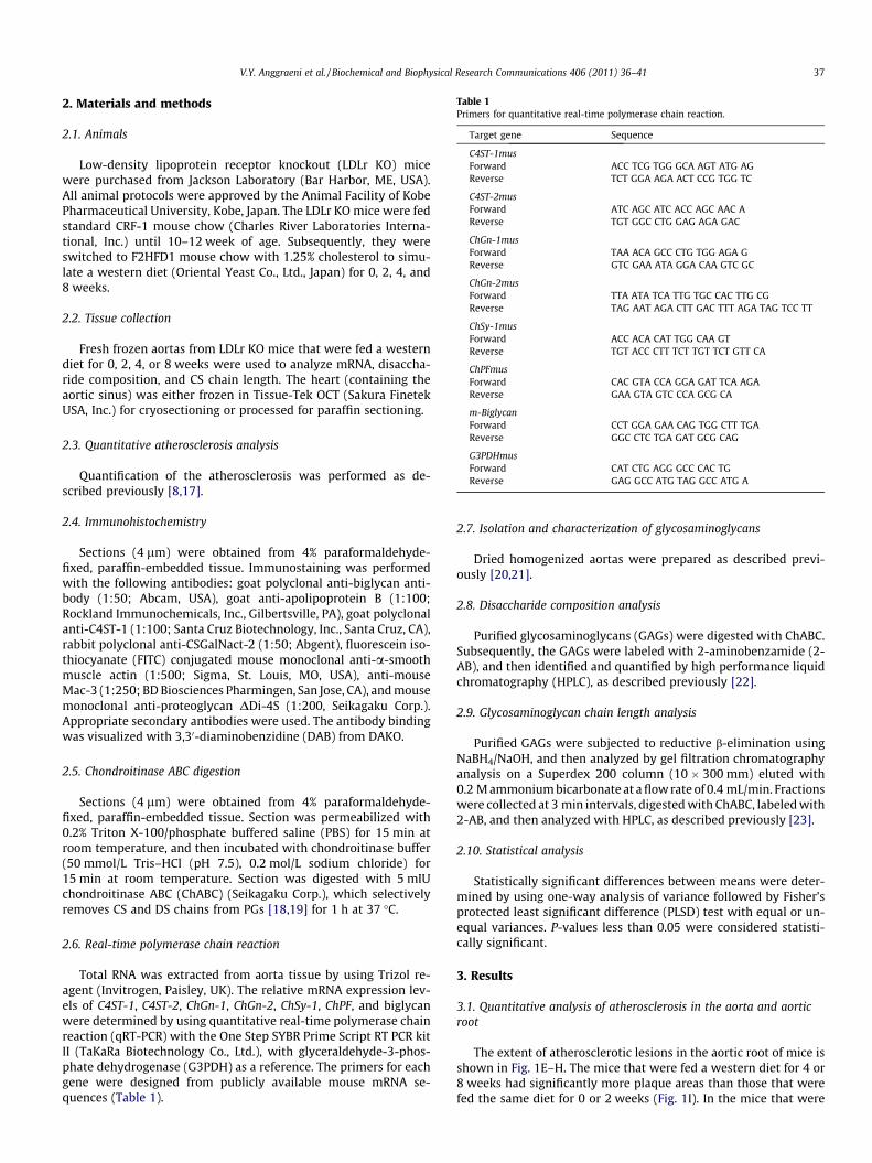
Table 1Primers for quantitative real-time polymerase chain reaction.
Target gene Sequence
C4ST-1musForward ACC TCG TGG GCA AGT ATG AGReverse TCT GGA AGA ACT CCG TGG TC
C4ST-2musForward ATC AGC ATC ACC AGC AAC AReverse TGT GGC CTG GAG AGA GAC
ChGn-1musForward TAA ACA GCC CTG TGG AGA GReverse GTC GAA ATA GGA CAA GTC GC
ChGn-2musForward TTA ATA TCA TTG TGC CAC TTG CGReverse TAG AAT AGA CTT GAC TTT AGA TAG TCC TT
ChSy-1musForward ACC ACA CAT TGG CAA GTReverse TGT ACC CTT TCT TGT TCT GTT CA
ChPFmusForward CAC GTA CCA GGA GAT TCA AGAReverse GAA GTA GTC CCA GCG CA
m-BiglycanForward CCT GGA GAA CAG TGG CTT TGAReverse GGC CTC TGA GAT GCG CAG
G3PDHmusForward CAT CTG AGG GCC CAC TGReverse GAG GCC ATG TAG GCC ATG A
V.Y. Anggraeni et al. / Biochemical and Biophysical Research Communications 406 (2011) 36–41 37
2. Materials and methods
2.1. Animals
Low-density lipoprotein receptor knockout (LDLr KO) micewere purchased from Jackson Laboratory (Bar Harbor, ME, USA).All animal protocols were approved by the Animal Facility of KobePharmaceutical University, Kobe, Japan. The LDLr KO mice were fedstandard CRF-1 mouse chow (Charles River Laboratories Interna-tional, Inc.) until 10–12 week of age. Subsequently, they wereswitched to F2HFD1 mouse chow with 1.25% cholesterol to simu-late a western diet (Oriental Yeast Co., Ltd., Japan) for 0, 2, 4, and8 weeks.
2.2. Tissue collection
Fresh frozen aortas from LDLr KO mice that were fed a westerndiet for 0, 2, 4, or 8 weeks were used to analyze mRNA, disaccha-ride composition, and CS chain length. The heart (containing theaortic sinus) was either frozen in Tissue-Tek OCT (Sakura FinetekUSA, Inc.) for cryosectioning or processed for paraffin sectioning.
2.3. Quantitative atherosclerosis analysis
Quantification of the atherosclerosis was performed as de-scribed previously [8,17].
2.4. Immunohistochemistry
Sections (4 lm) were obtained from 4% paraformaldehyde-fixed, paraffin-embedded tissue. Immunostaining was performedwith the following antibodies: goat polyclonal anti-biglycan anti-body (1:50; Abcam, USA), goat anti-apolipoprotein B (1:100;Rockland Immunochemicals, Inc., Gilbertsville, PA), goat polyclonalanti-C4ST-1 (1:100; Santa Cruz Biotechnology, Inc., Santa Cruz, CA),rabbit polyclonal anti-CSGalNact-2 (1:50; Abgent), fluorescein iso-thiocyanate (FITC) conjugated mouse monoclonal anti-a-smoothmuscle actin (1:500; Sigma, St. Louis, MO, USA), anti-mouseMac-3 (1:250; BD Biosciences Pharmingen, San Jose, CA), and mousemonoclonal anti-proteoglycan DDi-4S (1:200, Seikagaku Corp.).Appropriate secondary antibodies were used. The antibody bindingwas visualized with 3,30-diaminobenzidine (DAB) from DAKO.
2.5. Chondroitinase ABC digestion
Sections (4 lm) were obtained from 4% paraformaldehyde-fixed, paraffin-embedded tissue. Section was permeabilized with0.2% Triton X-100/phosphate buffered saline (PBS) for 15 min atroom temperature, and then incubated with chondroitinase buffer(50 mmol/L Tris–HCl (pH 7.5), 0.2 mol/L sodium chloride) for15 min at room temperature. Section was digested with 5 mIUchondroitinase ABC (ChABC) (Seikagaku Corp.), which selectivelyremoves CS and DS chains from PGs [18,19] for 1 h at 37 �C.
2.6. Real-time polymerase chain reaction
Total RNA was extracted from aorta tissue by using Trizol re-agent (Invitrogen, Paisley, UK). The relative mRNA expression lev-els of C4ST-1, C4ST-2, ChGn-1, ChGn-2, ChSy-1, ChPF, and biglycanwere determined by using quantitative real-time polymerase chainreaction (qRT-PCR) with the One Step SYBR Prime Script RT PCR kitII (TaKaRa Biotechnology Co., Ltd.), with glyceraldehyde-3-phos-phate dehydrogenase (G3PDH) as a reference. The primers for eachgene were designed from publicly available mouse mRNA se-quences (Table 1).
2.7. Isolation and characterization of glycosaminoglycans
Dried homogenized aortas were prepared as described previ-ously [20,21].
2.8. Disaccharide composition analysis
Purified glycosaminoglycans (GAGs) were digested with ChABC.Subsequently, the GAGs were labeled with 2-aminobenzamide (2-AB), and then identified and quantified by high performance liquidchromatography (HPLC), as described previously [22].
2.9. Glycosaminoglycan chain length analysis
Purified GAGs were subjected to reductive b-elimination usingNaBH4/NaOH, and then analyzed by gel filtration chromatographyanalysis on a Superdex 200 column (10 � 300 mm) eluted with0.2 M ammonium bicarbonate at a flow rate of 0.4 mL/min. Fractionswere collected at 3 min intervals, digested with ChABC, labeled with2-AB, and then analyzed with HPLC, as described previously [23].
2.10. Statistical analysis
Statistically significant differences between means were deter-mined by using one-way analysis of variance followed by Fisher’sprotected least significant difference (PLSD) test with equal or un-equal variances. P-values less than 0.05 were considered statisti-cally significant.
3. Results
3.1. Quantitative analysis of atherosclerosis in the aorta and aorticroot
The extent of atherosclerotic lesions in the aortic root of mice isshown in Fig. 1E–H. The mice that were fed a western diet for 4 or8 weeks had significantly more plaque areas than those that werefed the same diet for 0 or 2 weeks (Fig. 1I). In the mice that were
Aor
tic r
oot
plaq
ue a
rea
(%)
0
5
10
15
20
25
*
**
I
0
1
2
0w 2w 4w 8w
0w 2w 4w 8w
*
Big
lyca
n/G
3PD
H
V
0 w 2 w 4 w 8 wO
il R
ed O
En
Fac
e A
ort
a
A B C D
E F G H
Big
lyca
n-S
MA
Mac
-3
J K L M
N O P Q
R S T 250 mU
Fig. 1. (A–I) Quantification of atherosclerotic plaque areas. (A–D) Lipid deposition in the aorta as shown by Oil Red O staining on the en face aorta of low-density lipoproteinreceptor (LDLr) knockout (KO) mice after consuming a western diet for 0, 2, 4, or 8 weeks. (E–H) Oil Red O staining of the aortic sinus of LDLr KO mice after consuming a westerndiet for 0, 2, 4, and 8 weeks. (I) Plaque areas at the aortic root were determined by using Image J software. Data are expressed as mean (SE) (n = 6). ⁄p < 0.0001 versus 0, 2, and4 weeks of consuming a western diet. ⁄⁄p < 0.005 versus 0, 2, and 8 weeks of consuming a western diet. (J–M) Biglycan expression during progression of atherosclerosis.Representative images of immunostained biglycan (J–M), a-smooth muscle actin (a-SMA) (N–Q), and Mac-3 (R–U) in the aortic sinus of LDLr KO mice after consuming awestern diet for 0, 2, 4, or 8 weeks, respectively. (V) mRNA expression of biglycan in the aorta of LDLr KO mice after consuming a western diet for 0, 2, 4, or 8 weeks.
38 V.Y. Anggraeni et al. / Biochemical and Biophysical Research Communications 406 (2011) 36–41
fed a western diet for 0, 2, or 4 weeks, the samples could not be fur-ther analyzed by en face aorta analysis because there were veryfew lesions (Fig. 1A–D).
3.2. Biglycan expression during atherosclerosis progression
Immunohistochemistry showed that biglycan expressed at theaortic root at the beginning of the experimental period and in-creased as atherosclerosis progressed (Fig. 1J–M). Biglycan immu-nostaining also was expressed in the neointima and media. Inaddition, the accumulation of biglycan immunostaining coincidedwith the expression of a-SMA (Fig. 1N–Q) and Mac-3 immuno-staining. After 8 weeks of consuming a western diet, the mRNAexpression level of biglycan in mice were significantly higher thanthose that had consumed the same diet for 0, 2, or 4 weeks(Fig. 1V).
3.3. Disaccharide composition and glycosaminoglycan chain lengthanalysis of proteoglycans
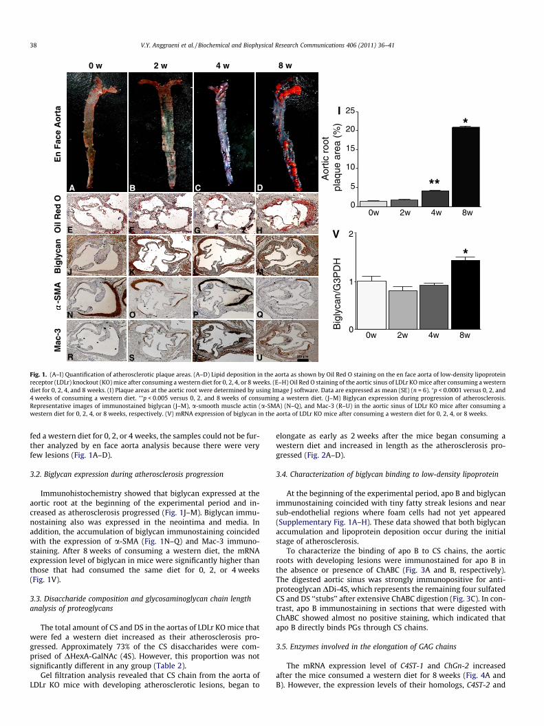
The total amount of CS and DS in the aortas of LDLr KO mice thatwere fed a western diet increased as their atherosclerosis pro-gressed. Approximately 73% of the CS disaccharides were com-prised of DHexA-GalNAc (4S). However, this proportion was notsignificantly different in any group (Table 2).
Gel filtration analysis revealed that CS chain from the aorta ofLDLr KO mice with developing atherosclerotic lesions, began to
elongate as early as 2 weeks after the mice began consuming awestern diet and increased in length as the atherosclerosis pro-gressed (Fig. 2A–D).
3.4. Characterization of biglycan binding to low-density lipoprotein
At the beginning of the experimental period, apo B and biglycanimmunostaining coincided with tiny fatty streak lesions and nearsub-endothelial regions where foam cells had not yet appeared(Supplementary Fig. 1A–H). These data showed that both biglycanaccumulation and lipoprotein deposition occur during the initialstage of atherosclerosis.
To characterize the binding of apo B to CS chains, the aorticroots with developing lesions were immunostained for apo B inthe absence or presence of ChABC (Fig. 3A and B, respectively).The digested aortic sinus was strongly immunopositive for anti-proteoglycan DDi-4S, which represents the remaining four sulfatedCS and DS ‘‘stubs’’ after extensive ChABC digestion (Fig. 3C). In con-trast, apo B immunostaining in sections that were digested withChABC showed almost no positive staining, which indicated thatapo B directly binds PGs through CS chains.
3.5. Enzymes involved in the elongation of GAG chains
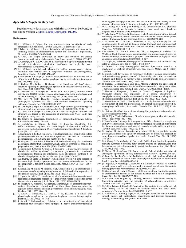
The mRNA expression level of C4ST-1 and ChGn-2 increasedafter the mice consumed a western diet for 8 weeks (Fig. 4A andB). However, the expression levels of their homologs, C4ST-2 and
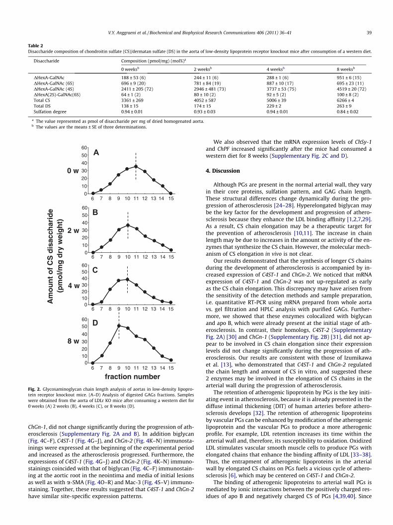
Table 2Disaccharide composition of chondroitin sulfate (CS)/dermatan sulfate (DS) in the aorta of low-density lipoprotein receptor knockout mice after consumption of a western diet.
Disaccharide Composition (pmol/mg) (mol%)a
0 weeksb 2 weeksb 4 weeksb 8 weeksb
DHexA-GalNAc 188 ± 53 (6) 244 ± 11 (6) 288 ± 1 (6) 951 ± 6 (15)DHexA-GalNAc (6S) 696 ± 9 (20) 781 ± 84 (19) 887 ± 10 (17) 695 ± 23 (11)DHexA-GalNAc (4S) 2411 ± 205 (72) 2946 ± 481 (73) 3737 ± 53 (75) 4519 ± 20 (72)DHexA(2S)-GalNAc(6S) 64 ± 1 (2) 80 ± 10 (2) 92 ± 5 (2) 100 ± 8 (2)Total CS 3361 ± 269 4052 ± 587 5006 ± 39 6266 ± 4Total DS 138 ± 15 174 ± 15 229 ± 2 263 ± 9Sulfation degree 0.94 ± 0.01 0.93 ± 0.03 0.94 ± 0.01 0.84 ± 0.02
a The value represented as pmol of disaccharide per mg of dried homogenated aorta.b The values are the means ± SE of three determinations.
fraction number
Am
ou
nt
of
CS
dis
acch
arid
e(p
mo
l/mg
dry
wei
gh
t)
01020
30
40
5060
6 7 8 9 10 11 12 13 14 15
D
8 w
0102030405060
6 7 8 9 10 11 12 13 14 15
C
4 w
2 w
0
10
20
30
40
50
60
6 7 8 9 10 11 12 13 14 15
B
0
10
20
30
40
50
60
6 7 8 9 10 11 12 13 14 15
A
0 w
Fig. 2. Glycosaminoglycan chain length analysis of aortas in low-density lipopro-tein receptor knockout mice. (A–D) Analysis of digested GAGs fractions. Sampleswere obtained from the aorta of LDLr KO mice after consuming a western diet for0 weeks (A) 2 weeks (B), 4 weeks (C), or 8 weeks (D).
V.Y. Anggraeni et al. / Biochemical and Biophysical Research Communications 406 (2011) 36–41 39
ChGn-1, did not change significantly during the progression of ath-erosclerosis (Supplementary Fig. 2A and B). In addition biglycan(Fig. 4C–F), C4ST-1 (Fig. 4G–J), and ChGn-2 (Fig. 4K–N) immunosta-inings were expressed at the beginning of the experimental periodand increased as the atherosclerosis progressed. Furthermore, theexpressions of C4ST-1 (Fig. 4G–J) and ChGn-2 (Fig. 4K–N) immuno-stainings coincided with that of biglycan (Fig. 4C–F) immunostain-ing at the aortic root in the neointima and media of initial lesionsas well as with a-SMA (Fig. 4O–R) and Mac-3 (Fig. 4S–V) immuno-staining. Together, these results suggested that C4ST-1 and ChGn-2have similar site-specific expression patterns.
We also observed that the mRNA expression levels of ChSy-1and ChPF increased significantly after the mice had consumed awestern diet for 8 weeks (Supplementary Fig. 2C and D).
4. Discussion
Although PGs are present in the normal arterial wall, they varyin their core proteins, sulfation pattern, and GAG chain length.These structural differences change dynamically during the pro-gression of atherosclerosis [24–28]. Hyperelongated biglycan maybe the key factor for the development and progression of athero-sclerosis because they enhance the LDL binding affinity [1,2,7,29].As a result, CS chain elongation may be a therapeutic target forthe prevention of atherosclerosis [10,11]. The increase in chainlength may be due to increases in the amount or activity of the en-zymes that synthesize the CS chain. However, the molecular mech-anism of CS elongation in vivo is not clear.
Our results demonstrated that the synthesis of longer CS chainsduring the development of atherosclerosis is accompanied by in-creased expression of C4ST-1 and ChGn-2. We noticed that mRNAexpression of C4ST-1 and ChGn-2 was not up-regulated as earlyas the CS chain elongation. This discrepancy may have arisen fromthe sensitivity of the detection methods and sample preparation,i.e. quantitative RT-PCR using mRNA prepared from whole aortavs. gel filtration and HPLC analysis with purified GAGs. Further-more, we showed that these enzymes colocalized with biglycanand apo B, which were already present at the initial stage of ath-erosclerosis. In contrast, their homologs, C4ST-2 (SupplementaryFig. 2A) [30] and ChGn-1 (Supplementary Fig. 2B) [31], did not ap-pear to be involved in CS chain elongation since their expressionlevels did not change significantly during the progression of ath-erosclerosis. Our results are consistent with those of Izumikawaet al. [13], who demonstrated that C4ST-1 and ChGn-2 regulatedthe chain length and amount of CS in vitro, and suggested these2 enzymes may be involved in the elongation of CS chains in thearterial wall during the progression of atherosclerosis.
The retention of atherogenic lipoprotein by PGs is the key initi-ating event in atherosclerosis, because it is already presented in thediffuse intimal thickening (DIT) of human arteries before athero-sclerosis develops [32]. The retention of atherogenic lipoproteinsby vascular PGs can be enhanced by modification of the atherogeniclipoprotein and the vascular PGs to produce a more atherogenicprofile. For example, LDL retention increases its time within thearterial wall and, therefore, its susceptibility to oxidation. OxidizedLDL stimulates vascular smooth muscle cells to produce PGs withelongated chains that enhance the binding affinity of LDL [33–38].Thus, the entrapment of atherogenic lipoproteins in the arterialwall by elongated CS chains on PGs fuels a vicious cycle of athero-sclerosis [6], which may be centered on C4ST-1 and ChGn-2.
The binding of atherogenic lipoproteins to arterial wall PGs ismediated by ionic interactions between the positively charged res-idues of apo B and negatively charged CS of PGs [4,39,40]. Since
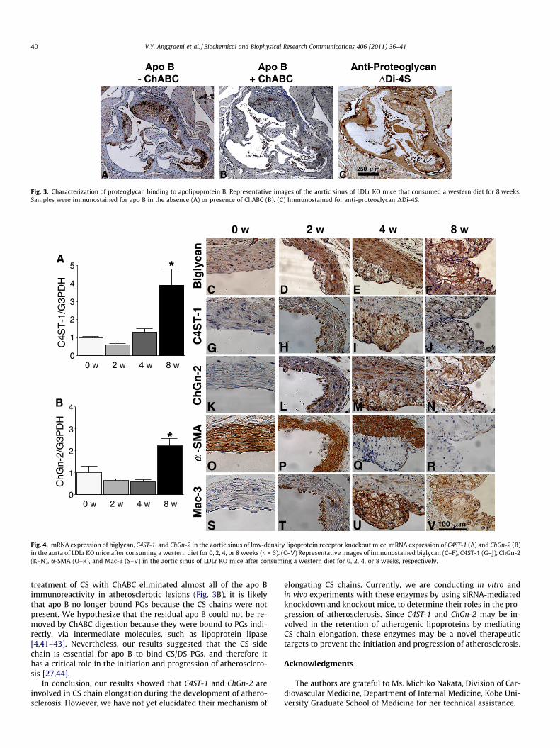
Fig. 3. Characterization of proteoglycan binding to apolipoprotein B. Representative images of the aortic sinus of LDLr KO mice that consumed a western diet for 8 weeks.Samples were immunostained for apo B in the absence (A) or presence of ChABC (B). (C) Immunostained for anti-proteoglycan DDi-4S.
0 w 2 w 4 w 8 w
0
1
2
3
4
5
0 w 2 w 4 w 8 w
C4S
T-1
/G3P
DH
*A
0
1
2
3
4
0 w 2 w 4 w 8 w
ChG
n-2/
G3P
DH
*
B
C4S
T-1
Ch
Gn
-2-S
MA
Mac
-3
H I
L M
P Q
T U
G J
K N
RO
S
C D E FBig
lyca
n
V 100 m
Fig. 4. mRNA expression of biglycan, C4ST-1, and ChGn-2 in the aortic sinus of low-density lipoprotein receptor knockout mice. mRNA expression of C4ST-1 (A) and ChGn-2 (B)in the aorta of LDLr KO mice after consuming a western diet for 0, 2, 4, or 8 weeks (n = 6). (C–V) Representative images of immunostained biglycan (C–F), C4ST-1 (G–J), ChGn-2(K–N), a-SMA (O–R), and Mac-3 (S–V) in the aortic sinus of LDLr KO mice after consuming a western diet for 0, 2, 4, or 8 weeks, respectively.
40 V.Y. Anggraeni et al. / Biochemical and Biophysical Research Communications 406 (2011) 36–41
treatment of CS with ChABC eliminated almost all of the apo Bimmunoreactivity in atherosclerotic lesions (Fig. 3B), it is likelythat apo B no longer bound PGs because the CS chains were notpresent. We hypothesize that the residual apo B could not be re-moved by ChABC digestion because they were bound to PGs indi-rectly, via intermediate molecules, such as lipoprotein lipase[4,41–43]. Nevertheless, our results suggested that the CS sidechain is essential for apo B to bind CS/DS PGs, and therefore ithas a critical role in the initiation and progression of atherosclero-sis [27,44].
In conclusion, our results showed that C4ST-1 and ChGn-2 areinvolved in CS chain elongation during the development of athero-sclerosis. However, we have not yet elucidated their mechanism of
elongating CS chains. Currently, we are conducting in vitro andin vivo experiments with these enzymes by using siRNA-mediatedknockdown and knockout mice, to determine their roles in the pro-gression of atherosclerosis. Since C4ST-1 and ChGn-2 may be in-volved in the retention of atherogenic lipoproteins by mediatingCS chain elongation, these enzymes may be a novel therapeutictargets to prevent the initiation and progression of atherosclerosis.
Acknowledgments
The authors are grateful to Ms. Michiko Nakata, Division of Car-diovascular Medicine, Department of Internal Medicine, Kobe Uni-versity Graduate School of Medicine for her technical assistance.
V.Y. Anggraeni et al. / Biochemical and Biophysical Research Communications 406 (2011) 36–41 41
Appendix A. Supplementary data
Supplementary data associated with this article can be found, inthe online version, at doi:10.1016/j.bbrc.2011.01.096.
References
[1] K.J. Williams, I. Tabas, The response-to-retention hypothesis of earlyatherogenesis, Arterioscler. Thromb. Vasc. Biol. 15 (1995) 551–561.
[2] I. Tabas, K.J. Williams, J. Boren, Subendothelial lipoprotein retention as theinitiating process in atherosclerosis: update and therapeutic implications,Circulation 116 (2007) 1832–1844.
[3] A. Chait, T.N. Wight, Interaction of native and modified low-densitylipoproteins with extracellular matrix, Curr. Opin. Lipidol. 11 (2000) 457–463.
[4] C. Germán, H.-C. Eva, W. Olov, et al., Association of apo B lipoproteins witharterial proteoglycans: pathological significance and molecular basis,Atherosclerosis 139 (1998) 205–222.
[5] K.J. Williams, Arterial wall chondroitin sulfate proteoglycans: diversemolecules with distinct roles in lipoprotein retention and atherogenesis,Curr. Opin. Lipidol. 12 (2001) 477–487.
[6] Y. Nakashima, T.N. Wight, K. Sueishi, Early atherosclerosis in humans: role ofdiffuse intimal thickening and extracellular matrix proteoglycans, Cardiovasc.Res. 79 (2008) 14–23.
[7] H. Dadlani, M.L. Ballinger, N. Osman, et al., Smad and p38 MAP kinase-mediated signaling of proteoglycan synthesis in vascular smooth muscle, J.Biol. Chem. 283 (2008) 7844–7852.
[8] R. Getachew, M.L. Ballinger, M.L. Burch, et al., PDGF {beta}-receptor kinaseactivity and ERK1/2 mediate glycosaminoglycan elongation on biglycan andincreases binding to LDL, Endocrinology 151 (2010) 4356–4367.
[9] M.E. Ivey, P.J. Little, Thrombin regulates vascular smooth muscle cellproteoglycan synthesis via PAR-1 and multiple downstream signallingpathways, Thromb. Res. 123 (2008) 288–297.
[10] M.L. Ballinger, J. Nigro, K.V. Frontanilla, et al., Regulation of glycosaminoglycanstructure and atherogenesis, Cell. Mol. Life Sci. 61 (2004) 1296–1306.
[11] P.J. Little, M.L. Ballinger, N. Osman, Vascular wall proteoglycan synthesis andstructure as a target for the prevention of atherosclerosis, Vasc. Health RiskManage. 3 (2007) 117–124.
[12] J.E. Silbert, G. Sugumaran, Biosynthesis of chondroitin/dermatan sulfate,IUBMB Life 54 (2002) 177–186.
[13] T. Izumikawa, Y. Okuura, T. Koike, H. Kitagawa, Chondrotin 4-O-sulfotransferase-1 regulates the chain length of chondroitin sulfate incooperation with chondroitin N-acetylgalactosaminyltransferase-2, Biochem.J. 434 (2011) 321–331.
[14] T. Izumikawa, T. Koike, S. Shiozawa, et al., Identification of chondroitin sulfateglucuronyltransferase as chondroitin synthase-3 involved in chondroitinpolymerization, J. Biol. Chem. 283 (2008) 11396–11406.
[15] H. Kitagawa, T. Izumikawa, T. Uyama, et al., Molecular cloning of a chondroitinpolymerizing factor that cooperates with chondroitin synthase for chondroitinpolymerization, J. Biol. Chem. 278 (2003) 23666–23671.
[16] T. Izumikawa, T. Uyama, Y. Okuura, K. Sugahara, H. Kitagawa, Involvement ofchondroitin sulfate synthase-3 (chondroitin synthase-2) in chondroitinpolymerization through its interaction with chondroitin synthase-1 orchondroitin-polymerizing factor, Biochem. J. 403 (2007) 545–552.
[17] A.S. Plump, C.J. Scott, J.L. Breslow, Human apolipoprotein A-I gene expressionincreases high density lipoprotein and suppresses atherosclerosis in theapolipoprotein E-deficient mouse, Proc. Natl. Acad. Sci. USA 91 (1994) 9607–9611.
[18] S. Nadanaka, M. Ishida, M. Ikegami, et al., Chondroitin 4-O-sulfotransferase-1modulates Wnt-3a signaling through control of E disaccharide expression ofchondroitin sulfate, J. Biol. Chem. 283 (2008) 27333–27343.
[19] T. Uyama, M. Ishida, T. Izumikawa, et al., Chondroitin 4-O-sulfotransferase-1regulates E disaccharide expression of chondroitin sulfate required for herpessimplex virus infectivity, J. Biol. Chem. 281 (2006) 38668–38674.
[20] H. Kitagawa, A. Kinoshita, K. Sugahara, Microanalysis of glycosaminoglycan-derived disaccharides labeled with the fluorophore 2-aminoacridone bycapillary electrophoresis and high-performance liquid chromatography, Anal.Biochem. 232 (1995) 114–121.
[21] T. Koike, T. Izumikawa, J.I. Tamura, et al., FAM20B is a kinase thatphosphorylates xylose in the glycosaminoglycan protein linkage region,Biochem. J. 421 (2009) 157–162.
[22] J. Sorrell, F. Mahmoodian, I. Schafer, et al., Identification of monoclonalantibodies that recognize novel epitopes in native chondroitin/dermatan
sulfate glycosaminoglycan chains: their use in mapping functionally distinctdomains of human skin, J. Histochem. Cytochem. 38 (1990) 393–402.
[23] W.-C. Huang, W.-C. Kuo, J.-H. Cherng, et al., Chondroitinase ABC promotesaxonal re-growth and behavior recovery in spinal cord injury, Biochem.Biophys. Res. Commun. 349 (2006) 963–968.
[24] Y. Nakashima, Y.-X. Chen, N. Kinukawa, et al., Distributions of diffuse intimalthickening in human arteries: preferential expression in atherosclerosis-pronearteries from an early age, Virchows Arch. 441 (2002) 279–288.
[25] S. Yla-Herttuala, T. Solakivi, J. Hirvonen, et al., Glycosaminoglycans andapolipoproteins B and A-I in human aortas. Chemical and immunologicalanalysis of lesion-free aortas from children and adults, Arterioscler. Thromb.Vasc. Biol. 7 (1987) 333–340.
[26] K.D. O’Brien, K.L. Olin, C.E. Alpers, W. Chiu, M. Ferguson, K. Hudkins, T.N.Wight, A. Chait, Comparison of apolipoprotein and proteoglycan deposits inhuman coronary atherosclerotic plaques: colocalization of biglycan withapolipoproteins, Circulation 98 (1998) 519–527.
[27] T.N. Wight, M.J. Merrilees, Proteoglycans in atherosclerosis and restenosis: keyroles for versican, Circ. Res. 94 (2004) 1158–1167.
[28] L. Tannock, V. King, Proteoglycan mediated lipoprotein retention: amechanism of diabetic atherosclerosis, Rev. Endocr. Metab. Dis. 9 (2008)289–300.
[29] E. Schonherr, H. Jarvelainen, M. Kinsella, et al., Platelet-derived growth factorand transforming growth factor-ft differentially affect the synthesis ofbiglycan and decorin by monkey arterial smooth muscle cells, Arterioscler.Thromb. Vasc. Biol. 1995 (1993) 1026–1036.
[30] N. Hiraoka, H. Nakagawa, E. Ong, et al., Molecular cloning and expression oftwo distinct human chondroitin 4-O-sulfotransferases that belong to the HNK-1 sulfotransferase gene family, J. Biol. Chem. 275 (2000) 20188–20196.
[31] T. Uyama, H. Kitagawa, J. Tanaka, J.-i. Tamura, T. Ogawa, K. Sugahara,Molecular cloning and expression of a second chondroitin N-acetylgalactosaminyltransferase involved in the initiation and elongation ofchondroitin/dermatan sulfate, J. Biol. Chem. 278 (2003) 3072–3078.
[32] Y. Nakashima, H. Fujii, S. Sumiyoshi, et al., Early human atherosclerosis:accumulation of lipid and proteoglycans in intimal thickenings followed bymacrophage infiltration, Arterioscler. Thromb. Vasc. Biol. 27 (2007) 1159–1165.
[33] M. Aviram, Modified forms of low density lipoprotein and atherosclerosis,Atherosclerosis 98 (1993) 1–9.
[34] H.F. Hoff, J.A. O’Neil, Oxidation of LDL: role in atherogenesis, Klin. Wochenschr.69 (21–23) (1991) 1032–1038.
[35] E. Hurt-Camejo, G. Camejo, B. Rosengren, et al., Effect of arterial proteoglycansand glycosaminoglycans on low density lipoprotein oxidation and its uptakeby human macrophages and arterial smooth muscle cells, Arterioscler.Thromb. Vasc. Biol. 12 (5) (1992) 569–583.
[36] M. Kaplan, M. Aviram, Retention of oxidized LDL by extracellular matrixproteoglycans leads to its uptake by macrophages: an alternative approach tostudy lipoproteins cellular uptake, Arterioscler. Thromb. Vasc. Biol. 21 (2001)386–393.
[37] M.Y. Chang, S. Potter-Perigo, C. Tsoi, et al., Oxidized low density lipoproteinsregulate synthesis of monkey aortic smooth muscle cell proteoglycans thathave enhanced native low density lipoprotein binding properties, J. Biol. Chem.275 (2000) 4766–4773.
[38] K. Skalen, M. Gustafsson, E.K. Rydberg, et al., Subendothelial retention ofatherogenic lipoproteins in early atherosclerosis, Nature 417 (2002) 750–754.
[39] C. Bancells, S.n. BenÃ-tez, M. Jauhiainen, et al., High binding affinity ofelectronegative LDL to human aortic proteoglycans depends on its aggregationlevel, J. Lipid Res. 50 (2009) 446–455.
[40] J.E. Figueroa, P. Vijayagopal, Angiotensin II stimulates synthesis of vascularsmooth muscle cell proteoglycans with enhanced low density lipoproteinbinding properties, Atherosclerosis 162 (2002) 261–268.
[41] M. Gustafsson, M. Levin, K. Skalen, et al., Retention of low-density lipoproteinin atherosclerotic lesions of the mouse: evidence for a role of lipoproteinlipase, Circ. Res. 101 (2007) 777–783.
[42] M.F. Khalil, W.D. Wagner, I.J. Goldberg, Molecular interactions leading tolipoprotein retention and the initiation of atherosclerosis, Arterioscler.Thromb. Vasc. Biol. 24 (2004) 2211–2218.
[43] M.O. Pentikainen, R. Oksjoki, K. Oorni, et al., Lipoprotein lipase in the arterialwall: linking LDL to the arterial extracellular matrix and much more,Arterioscler. Thromb. Vasc. Biol. 22 (2002) 211–217.
[44] K. Hashimura, K. Sudhir, J. Nigro, et al., Androgens stimulate human vascularsmooth muscle cell proteoglycan biosynthesis and increase lipoproteinbinding, Endocrinology 146 (2005) 2085–2090.


