Embed Size (px)
Citation preview

Graduate Theses and Dissertations Iowa State University Capstones, Theses andDissertations
2015
Cross-protection in Fostera™ PRRS vaccinatednursery swine against contemporary, heterologousporcine reproductive and respiratory syndromevirus (PRRSV) field isolates from different lineagesDrew Robert MagstadtIowa State University
Follow this and additional works at: https://lib.dr.iastate.edu/etd
Part of the Allergy and Immunology Commons, Immunology and Infectious Disease Commons,Medical Immunology Commons, and the Virology Commons
This Thesis is brought to you for free and open access by the Iowa State University Capstones, Theses and Dissertations at Iowa State University DigitalRepository. It has been accepted for inclusion in Graduate Theses and Dissertations by an authorized administrator of Iowa State University DigitalRepository. For more information, please contact [email protected].
Recommended CitationMagstadt, Drew Robert, "Cross-protection in Fostera™ PRRS vaccinated nursery swine against contemporary, heterologous porcinereproductive and respiratory syndrome virus (PRRSV) field isolates from different lineages" (2015). Graduate Theses and Dissertations.14501.https://lib.dr.iastate.edu/etd/14501

Cross-protection in Fostera™ PRRS vaccinated nursery swine against
contemporary, heterologous porcine reproductive and respiratory syndrome virus
(PRRSV) field isolates from different lineages
by
Drew Robert Magstadt
A thesis submitted to the graduate faculty
in partial fulfillment of the requirements for the degree of
MASTER OF SCIENCE
Major: Veterinary Microbiology
Program of Study Committee:
Phillip Gauger, Major Professor
Jesse Hostetter
Jianqiang Zhang
Iowa State University
Ames, Iowa
2015
Copyright © Drew Robert Magstadt. 2015. All rights reserved.
ii
TABLE OF CONTENTS
Page
LIST OF TABLES……………………………………………………………………. iv
LIST OF FIGURES…………………………………………………………………... v
ACKNOWLEDGEMENTS………………………………………………………….. vi
ABSTRACT………………………………………………………………………….. vii
CHAPTER 1. INTRODUCTION……………………………………………………. 1
Background…………………………………………………………………... 1
Objective……………………………………………………………………... 1
Thesis Organization………………………………………………………….. 1
CHAPTER 2. LITERATURE REVIEW……………………………………………... 3
Introduction…………………………………………………………………... 3
PRRS Virus and Pathogenesis………………………………………………... 4
Viral Classification and Diversity……………………………………………. 7
Immune Response to Infection……………………………………………….. 10
Diagnostic Testing……………………………………………………………. 12
Control Strategies…………………………………………………………….. 14
Vaccination…………………………………………………………………… 15
Summary of PRRSV Vaccine Challenge Studies……………………………. 18
Conclusion……………………………………………………………………. 24
References……………………………………………………………………. 25
CHAPTER 3. EVALUATION OF CROSS-PROTECTION IN FOSTERA™
PRRS VACCINATED NURSERY SWINE CHALLENGED WITH A
CONTEMPORARY, HETEROLOGOUS LINEAGE 9 PRRSV……………………. 37
Abstract………………………………………………………………………. 37
Introduction…………………………………………………………………... 38
Materials and Methods……………………………………………………….. 40
Results………………………………………………………………………... 46
Discussion……………………………………………………………………. 49
Acknowledgements ………………………………………………………….. 54
References……………………………………………………………………. 54
CHAPTER 4. EFFICACY OF FOSTERA™ PRRS VACCINE AGAINST A
CONTEMPORARY, HETEROLOGOUS LINEAGE 1 PORCINE
REPRODUCTIVE AND RESPIRATORY SYNDROME VIRUS
CHALLENGE IN NURSERY PIGS…………………………………………………. 67
Abstract………………………………………………………………………. 67
Introduction…………………………………………………………………... 68

iii
Materials and Methods……………………………………………………….. 70
Results………………………………………………………………………... 76
Discussion……………………………………………………………………. 80
Acknowledgements…………………………………………………………... 84
References……………………………………………………………………. 84
CHAPTER 5. CONCLUSION……………………………………………………….. 94

iv
LIST OF TABLES
Page
CHAPTER 3: EVALUATION OF CROSS-PROTECTION IN FOSTERA™
PRRSV VACCINATED NURSERY SWINE CHALLENGED WITH A
CONTEMPORARY, HETEROLOGOUS LINEAGE 9 PRRSV
Table 1. Experimental design………………………………………………… 59
Table 2. Nucleotide sequences of the forward and reverse primers and
individual probes for 12-39404A challenge PRRSV and Fostera™
PRRS vaccine viruses used in the real time RT-PCR for absolute
quantification of PRRSV genomic copies……………………………. 60
Table 3. Mean microscopic pneumonia, microscopic lung lesion scores
and quantitative RT-PCR values in lung, BALF and tonsil………….. 61
Table 4. Mean PRRSV 12-39404A log10 genomic copies/ml for serum
and pen-based oral fluid samples…………………………………….. 62
Table 5. Average daily gain during the post-vaccination and post-challenge
phase………………………………………………………………….. 63
CHAPTER 4: EFFICACY OF FOSTERA™ PRRSV VACCINE AGAINST A
CONTEMPORARY, HETEROLOGOUS LINEAGE 1 PORCINE
REPRODUCTIVE AND RESPIRATORY SYNDROME VIRUS CHALLENGE
IN NURSERY PIGS
Table 1. Experimental design………………………………………………… 87
Table 2. Sequences of the forward and reverse primers and individual probe
for 14-4099 challenge PRRS virus used in the real time RT-PCR for
absolute quantification of PRRSV genomic copies………………….. 88
Table 3. Mean macroscopic pneumonia, microscopic lung lesion scores and
quantitative RT-PCR values in lung, BALF and tonsil………………. 89
Table 4. Mean PRRSV 14-4099 log10 genomic copies/ml for serum and
pen-based oral fluid samples…………………………………………. 90
Table 5. Average daily gain during the post-vaccination and post-challenge
phase………………………………………………………………….. 91
v
LIST OF FIGURES
Page
CHAPTER 3: EVALUATION OF CROSS-PROTECTION IN FOSTERA™
PRRSV VACCINATED NURSERY SWINE CHALLENGED WITH A
CONTEMPORARY, HETEROLOGOUS LINEAGE 9 PRRSV
Figure 1. Mean anti-PRRSV ELISA antibody S/P ratios in serum…………... 64
Figure 2. Macroscopic and microscopic lung lesions observed at necropsy…. 65
Figure 3. Phylogenetic analysis showing lineage classification and
diversity, based on ORF 5 sequences, between historical reference
PRRSV strains, several PRRSV strains used in vaccines, Fostera™
PRRS vaccine strain, and the PRRSV strain (12-39404) selected
for challenge………………………………………………………….. 66
CHAPTER 4: EFFICACY OF FOSTERA™ PRRSV VACCINE AGAINST A
CONTEMPORARY, HETEROLOGOUS LINEAGE 1 PORCINE
REPRODUCTIVE AND RESPIRATORY SYNDROME VIRUS CHALLENGE
IN NURSERY PIGS
Figure 1. Mean anti-PRRSV ELISA antibody S/P ratios in serum…………... 92
Figure 2. Macroscopic and microscopic lung lesions and
immunohistochemistry staining of lung observed at necropsy………. 93

vi
ACKNOWLEDGEMENTS
I would like to thank my major professor, Dr. Phillip Gauger, along with Drs.
Jesse Hostetter and Jianqiang Zhang, for serving on my committee and for all of their
support and guidance throughout my research.
I would like to thank the faculty and staff at the Iowa State University Diagnostic
Laboratory for their help in sample testing, as well as the animal caretakers from Iowa
State University Laboratory Animal Resources for their help in ensuring the success of
the research.
I would also like to thank my wife, Tiffany, for her love, support, encouragement,
and patience.

vii
ABSTRACT
Porcine reproductive and respiratory syndrome virus (PRRSV) emerged
worldwide approximately 25 years ago, and continues to be the most costly disease of
modern swine production. The genetic diversity of PRRSV is one of the major hurdles
encountered in attempts to reduce the effects of the virus through vaccines that provide
broad cross-protection. The objective of this research is to examine the efficacy of a
recently approved vaccine, Fostera™ PRRS, in growing pigs against separate challenges
with heterologous PRRSV strains belonging to lineage 9 and lineage 1. The selected
challenge viruses were isolated from recent PRRSV field infections in swine that
originated in Iowa. The results of both studies indicate vaccine-induced immunity confers
partial protection against the effects of heterologous PRRSV infection, as measured by
reduced quantities of virus in serum, oral fluids and bronchoalveolar lavage fluid, and an
increase in average daily gain in vaccinated pigs compared to non-vaccinated pigs
challenged with the same virus. The two studies resulted in differing levels of protection
as measured by severity of lung lesions and quantities of virus in tissues; these
differences may be due to several factors, including variations in study design or in the
degree of heterogeneity of the challenge viruses. The results show that Fostera™ PRRS
vaccine can provide growing pigs with partial protection against heterologous challenge
with currently circulating PRRSV. To improve prevention and control efforts, further
investigations are needed into the cross-protection elicited by commercial PRRSV
vaccines against contemporary, heterologous virus strains.
1
CHAPTER 1: INTRODUCTION
Background
Porcine reproductive and respiratory syndrome virus (PRRSV) is the most costly
pathogen currently affecting the swine industry. Vaccines are commonly used in various
programs designed to mitigate the impact of disease in growing pigs. Due to the genetic
diversity of PRRSV, vaccines do not result in complete protection against challenge with
heterologous viruses. The effectiveness of PRRSV vaccines can be evaluated by several
methods, including increased average daily gain, decreased lung lesions, and decreased
viral replication within the animal. In order to make significant progress towards
decreasing the impact of PRRSV infection in swine, approved vaccines should elicit
significant cross-protective immunity that prevents infection with and transmission of
PRRSV strains representative of those present in current production settings.
Objective
The objective of this research was to determine the efficacy of a recently
approved commercial modified live PRRSV vaccine, Fostera™ PRRS, against challenge
with two field strains of considerable genetic diversity compared to the strain represented
in the vaccine and belonging to different PRRSV lineages.
Thesis Organization
This thesis consists of an introduction, literature review, two original research
chapters that will be submitted for publication, and a conclusion. Chapter 2 serves as a
literature review of several topics regarding PRRSV that are pertinent to the study design

2
of research trials presented in later chapters. Topics include PRRSV emergence,
importance to the industry, viral structure and pathogenesis, immune response, control
strategies, vaccination, and a summary of published PRRSV challenge studies.
Chapters 3 and 4 present original research regarding the efficacy of a commercial
PRRSV vaccine, Fostera™ PRRS. The author’s role in each study included development
of study design, the growth, selection and titration of viral isolates, execution of the
animal studies, sample collection and necropsy of experimental animals and
interpretation of results. Chapters are presented as prepared for publication. Chapter 5
consists of a short discussion of results and conclusions.

3
CHAPTER 2: LITERATURE REVIEW
Introduction
In the late 1980’s and early 1990’s outbreaks of infertility and respiratory disease
of unknown origin were described in the swine populations of North America, Europe,
and Asia. After being referred to by several different names (such as swine infertility and
respiratory syndrome, porcine epidemic abortion and respiratory syndrome, and mystery
swine disease), a consensus was reached that the clinical description would be known as
porcine reproductive and respiratory syndrome (PRRS). A virus associated with the
disease was first isolated in Europe in 1991 and designated as the Lelystad virus,1 with a
North American strain isolated shortly thereafter; collectively, the new pathogen became
known as porcine reproductive and respiratory syndrome virus (PRRSV). A retrospective
study found anti-PRRSV antibody positive serum in Canada as early as 1979.2 This is the
earliest seropositive report of PRRSV, suggesting the virus was present in the swine
population for several years before reports of clinical disease; however, PRRSV was not
detected in the seropositive samples.
As the name suggests, the clinical disease caused by PRRSV includes both
reproductive and respiratory manifestations. The reproductive component of the disease
is characterized by increased abortions in sows and decreased fertility in both sows and
boars; the respiratory component of PRRSV infection is characterized by interstitial
pneumonia and dyspnea in pigs of any age.3,4
The severity of clinical signs apparent
during PRRSV infection is often dependent upon immune status of the animal, the strain
of the virus,5,6
and the presence of viral co-infections (such as swine influenza) or

4
secondary bacterial infections (such as Haemophilus parasuis, Pasteurella multocida,
and Streptococcus suis).4 Despite the vast amount of research into the dynamics of
PRRSV infection, other unidentified factors may play a role in disease severity.
A study in 2005 estimated that clinical disease attributable to PRRSV infection
costs the United States (US) swine industry approximately $560 million annually due to
increased mortality, decreased reproductive performance, and reduced feed efficiency;
88% of these costs were attributed to effects of the virus in growing pigs.7 Using year
2000 survey results from the National Animal Health Monitoring System (NAHMS), the
researchers found that nearly 45% of breeding females were in PRRSV-positive herds.
The study did not take into account the cost of vaccination, biosecurity measures,
monitoring of endemic herds, or the effect of subclinical disease. A more recent study in
2012 estimated the cost of lost productivity due to PRRSV at $664 million, with growing
pigs accounting for 55% of the total economic loss.8 The researchers used a survey of
swine veterinarians to gather data on incidence of PRRSV as well as additional
production costs that were not evaluated in the previous study. Based on the survey
results, they estimated an additional cost of $477 million to US swine producers in
animal health, biosecurity, and outbreak related expenses attributed to PRRSV.
PRRS Virus and Pathogenesis
Porcine reproductive and respiratory syndrome virus is a positive sense, single-
stranded RNA virus belonging to the order Nidovirales, the family Arteriviridae, and the
genus Arterivirus,9 which also includes equine arteritis virus, lactate dehydrogenase
elevating virus of mice, and simian hemorrhagic fever virus.10
The PRRSV genome is

5
approximately 15 kb, and nine open reading frames (ORF) have been identified.11
ORF1a
and ORF1b make up a large portion of the genome and encode for various non-structural
proteins (NSP) involved with viral replication and subgenomic transcription, including
RNA-dependent RNA polymerase, while ORFs 2a, 2b, 3 and 4 encode for minor
structural proteins found in the viral envelope.11
Minor structural proteins may play a role
in determining viral tropism through interaction with target cell receptors.
The major structural and envelope proteins of PRRSV are encoded by ORFs 5
through 7.12
The first major envelope protein, GP5, is encoded by ORF5 and is the most
highly variable protein of PRRSV.13
Variability in GP5 may contribute to the lack of
immune cross protection between virus isolates,14
and the results of several studies
suggest that antibodies against GP5 were the most effective at virus neutralization.15,16,17
A second major envelope protein, the M protein, is encoded by ORF610
and is highly
conserved across PRRSV strains.18
Research suggests that GP5 and M protein may be
involved in viral attachment to and internalization by macrophages,19,20,21
and research
has shown both proteins are vital components of the envelope; deletion of either ORF5 or
ORF6 results in a failure of viral reproduction.22
The nucleocapsid (N) protein is encoded
by ORF7.11
The N protein is the most immunogenic protein of PRRSV, with anti-N
protein antibodies appearing as soon as 5 days after infection; however, these antibodies
do not play a role in virus neutralization.17,23
Within the host PRRSV displays a predilection for monocyte derived cells,
particularly those in the lung and lymphoid tissues. The virus displays a particular
tropism for pulmonary alveolar macrophages,24
both in vitro and in vivo. In addition,
several cell lines derived from monkey kidney cells have been used to grow and study

6
PRRSV.25,26
A cell surface molecule, heparan sulfate, has been shown to interact with the
M/GP5 proteins on the PRRSV envelope.19
This molecule is found on many cells and
although not necessary for viral infection, it is thought to loosely adhere to and
concentrate the virus on the cell suface.27
The macrophage-restricted protein sialoadhesin
has been shown to be necessary for virus attachment and internalization, through the
binding of the M/GP5 proteins.28,29,30
Release of the virus within the cell and viral
uncoating has been shown to involve a transmembrane protein, CD163.31,32
Once inside
susceptible macrophages, several proteases are also involved in uncoating the virus.33
Infection with PRRSV does not only affect macrophages, but can also induce apoptosis
of nearby uninfected cells.34
Infection also impairs macrophage function, which reduces
basic immune physiologic mechanisms such as phagocytosis,35,36
and can also induce
apoptosis of infected cells.37
Direct contact with infected tissues or fluids from swine and aerosolization of the
virus are the most common routes of PRRSV transmission. After initial infection and
replication, viral particles are released from the cell and can readily spread throughout the
body via the bloodstream. Viral replication markedly increases the amount of infectious
virus, and replication persists in macrophages of the lung, tonsil, and lymphoid
organs.38,39
Although viremia is typically resolved after 28 days, viral RNA has been
detected in serum up to 251 days post infection,24,40
and in congenitally infected pigs for
up to 228 days.41
Persistence may be due to immune modulation through altered cytokine
expression,42
ineffective cell-mediated immune response,23
shielding of virus and viral
proteins within endosomes,43
and/or antibody-mediated enhancement.44
Once infection is

7
established the virus is shed in most bodily fluids/secretions: virus has been isolated from
serum, semen, saliva, feces, urine, and nasal secretions.4
Clinical signs in PRRSV-infected growing pigs include fever, anorexia, dyspnea,
and lethargy, and are usually more severe in younger pigs.4 Gross lesions typically
include a mottled-tan, non-collapsing lung with markedly enlarged lymph nodes
throughout the body.5,45
Microscopic lung lesions include lymphocytic and macrophagic
interstitial pneumonia commonly accompanied by type 2 pneumocyte hypertrophy,
necrotic macrophages, and cellular debris accumulations within alveoli.46
Infection in
pregnant sows is characterized by an increase in abortions, stillbirths, mummified fetuses,
and infertility.47
Pathologic lesions reported in sows or piglets from outbreaks of
reproductive failure are inconsistent, non-specific, and/or absent. Clinical signs, gross and
microscopic lesions can vary due to differences in the virulence of the PRRSV strains.5,6
Viral Classification and Diversity
European PRRSV strains are also known as type 1 PRRSV, with the Lelystad
virus as the prototype strain. North American strains are known as type 2 PRRSV, with
the prototype strain VR-2332. Despite disease emergence at nearly the same time, there is
considerable genetic diversity both between and within the two genotypes. Genetic
sequence homology between type 1 and type 2 PRRSV in various regions of the genome
is 55-79%.13,48,49,50
While diversity between the two types varies, it is common for strains
within each genotype to demonstrate only 80-85% homology at ORF5. There are also
significant differences in antigenicity between genotypes.51
The large disparity in genetic
and antigenic viral characteristics between genotypes suggests that the original virus

8
species underwent an initial divergence event, with each genotype acquiring considerable
genetic diversity while being isolated to their respective continents and swine
populations.52
Due to the genetic, antigenic, and pathogenic variability within genotypes several
other classification methods have been developed in an attempt to more closely group
similar PRRSV strains. One early method of virus classification, that was initially
developed to differentiate vaccine strains from field strains, involves analysis of the
restriction fragment length polymorphism (RFLP) of each virus strain.53
Determination of
the RFLP patterns involves enzymatic digestion of an amplified ORF5 fragment by a set
of three restriction enzymes. Digestion with each enzyme can result in several possible
patterns; the cut pattern for each enzyme is given a number, and a three-number RFLP
code is assigned to the virus (for example, RFLP 1-7-4).
While this method is fairly convenient, it does not represent an accurate
comparison of viral genotypes. The RFLP code is essentially a description of the ORF5
sequence, which is highly variable and has little predictive value regarding the
phenotype, antigenicity, or pathogenicity of the virus. Furthermore, enzymatic digestion
of ORF5 relies on specific genome sequences, which can change as the virus mutates
during replication within pigs.54
Using PRRSV strain VR-2332, researchers performed 13
serial passages of virus in pigs and analyzed the RFLP patterns of the resulting isolates.55
Twenty percent of the recovered viruses had a different RFLP code than the initial
challenge virus, despite a change to the ORF5 sequence of only 0.5-1.45%. The findings
suggest that closely related viruses based on sequencing may have different RFLP codes.
While the different enzymatic digestion patterns were initially few in number, an increase

9
in testing along with ongoing genetic mutation has led to an increase in the number of
digestion patterns and the number of distinct RFLP codes.52
In response to the increasing number of available ORF5 genomic sequences and
in an attempt to group similar viruses more accurately, a system was developed that
classifies type 2 PRRSV strains based on their ORF5 sequence.56
Researchers used over
8,000 ORF5 sequences available from GenBank to create nine distinct phylogenetic
PRRSV lineages. This classification scheme resulted in groups of virus strains that were
less than 11% diverse within lineages. Lineages 3 and 4 represent a small number of
PRRSV sequences, all of which were found in Asia. Lineages 2, 6 and 7 are also
relatively small and mainly contain virus isolates from the US.
The vast majority (>85%) of PRRSV isolates cluster in one of the four remaining
lineages, each containing virus strains from around the world. Lineage 5 has relatively
low intralineage diversity, and contains the historical isolate VR-2332 (the type 2
prototype strain). Lineage 8 contains the historical isolates SDSU-73 and JA-142.
Lineages 1 and 9 contain the largest number of isolates, and also the highest intralineage
diversity of the four large lineages. Lineage 1 contains the historical isolate MN-184.56
There are several mechanisms that may explain the vast amount of genetic
diversity present in PRRSV. The primary culprit is thought to be error prone RNA
polymerase and the lack of a corrective mechanism to ensure genomic integrity during
replication. The rate of nucleotide substitution in PRRSV is the highest of all RNA
viruses.57
Genetic recombination has been shown to occur, and may play a significant
role in viral diversity.58,59
Host induced mutation using cytidine deaminase enzymes has
also been considered a mechanism capable of driving genetic diversity in RNA viruses.60

10
Immune Response to Infection
While infection with PRRSV eventually induces protective immunity against the
challenge strain, development of immune protection is slow and cross-protection between
genetically diverse strains is generally incomplete and highly variable. Evidence suggests
this may be due to a weak innate immune response by the target cell of PRRSV, the
pulmonary alveolar macrophage. Viruses typically elicit the production of interferon after
infection of a host cell, which increases the production of cytokines that work collectively
to suppress viral protein synthesis and replication.61
Significantly increased cytokine
production, such as interferon-alpha and tumor necrosis factor-alpha, is a not a feature of
early PRRSV infection,42,62,63
and different field isolates have been found to have
differing abilities to induce cytokine production.64
Low amounts of interferon production
may also lead to a lack of natural killer cell activation.65
Decreased to absent cytokine
production early in infection may play a part in the delayed humoral immune response as
well as the prolonged course of infection. Several studies have shown an eventual
elevation in various cytokine levels (interferon-gamma, tumor necrosis factor, and
interleukin-10) within the lung or BALF collected from infected pigs, but the increase
was not apparent until 7-10 days after infection.66,67,68
The humoral immune response to PRRSV can be detected within one week of
infection. IgM antibodies to PRRSV are generated within 5-7 days of infection, with peak
IgM levels observed 14-21 days post inoculation.69
Anti-PRRSV IgG antibodies can be
detected by 7-10 days post infection, peak at 14-28 days post inoculation,17
and persist
for up to 10 months.70
Antibodies specific for individual viral proteins are produced but
not in a consistent timeframe; in one study, IgM to NSP-2 and N-protein peaked at 7 days

11
post infection with an IgG peak around 35 days, while peak levels of IgM to GP5 were
not detected until 3-4weeks post infection.71
The IgG response to GP5 was also delayed,
being first detected at 21 days and peaking at 35 days post infection, one to two weeks
after the resolution of viremia. While serum neutralizing antibodies to GP5, GP4, and M
protein have been reported, antibodies to GP5 are considered most effective at
neutralizing virus.17,72
The role of neutralizing antibody remains unclear; studies have
detected neutralizing antibodies along with concurrent viremia.3,73
In addition, several
antibody kinetics studies have failed to detect neutralizing antibodies in PRRSV
inoculated pigs.69,74
While antibody production towards infectious agents is generally regarded as
helpful, antibody-dependent enhancement of PRRSV infection may occur. Macrophages
display receptors that bind antigen-antibody complexes for phagocytosis. Low levels of
anti-PRRSV antibody may enhance the association of viral particles with pulmonary
alveolar macrophages, leading to increased viral uptake.44
An in vitro study showed that
serum containing antibodies to PRRSV actually enhanced infection of alveolar
macrophages.75
The T-cell response to PRRSV infection is first apparent around 4 weeks after
infection where studies have demonstrated an increase in cytotoxic T-cells in the lung76,77
and an increase in helper T-cells in the blood at that time.78
However, this is a delayed
and relatively weak response compared to the cell-mediated immune response induced by
other viruses.79
In addition, PRRSV infection results in a down-regulation of major
histocompatibility complex expression on dendritic cells, negatively affecting the
efficiency of antigen presentation.80
Protective immunity to homologous PRRSV is

12
predicated on memory cell induction. Large numbers of memory B cells are produced
and can be found in many lymphoid tissues, particularly the spleen, tonsil, and sternal
lymph node.71
Re-challenge may result in significant protection without an anamnestic
antibody response, despite the abundance of memory B cells.71,81
The immunity produced
by PRRSV infection is of long duration.82
However, viral persistence within the animal
may exceed the life span of commercial pigs.41
Protective immunity to heterologous PRRSV strains is highly variable due in part
to the antigenic diversity of the virus strains. Exposure to one PRRSV strain does not
usually confer complete protective immunity against another. In a multi-strain challenge
study, only heterologous strains were detected in pigs after challenge; attenuated
vaccination only provided complete protection against homologous strains.83
However,
research into cross protection has found variable levels of partial protection between virus
isolates, characterized by a reduction in clinical signs, decreased pathologic lung lesions,
lower levels of viremia and/or increased average daily gain.
Diagnostic Testing
Due to highly variable and non-specific clinical signs, using clinical parameters
alone to diagnose PRRSV infection is potentially inaccurate, biased, and unreliable.
Gross and histologic changes caused by respiratory PRRSV infection are also non-
specific, and may be complicated by the presence of viral co-infections or secondary
bacterial infections. Additional diagnostic testing is necessary to confirm PRRSV
infection; several methods have been developed and are routinely used today. Timely and

13
accurate diagnosis is critical to the swine industry in order to limit the spread and control
outbreaks of PRRSV.
Most diagnostics are centered on the detection of either anti-PRRSV antibodies or
detection of the virus itself. Antibodies to PRRSV are readily detected in serum and oral
fluids by several methods, including serum neutralization, fluorescent focus
neutralization, and enzyme-linked immunosorbent assay (ELISA).84,85
Antibody testing
of serum merely detect animals that have been exposed to and induced an immune
response to PRRSV. They are commonly used to monitor herds that produce PRRSV-
negative breeding animals. Immunohistochemistry (IHC) staining of formalin-fixed
tissue uses antibody to detect viral antigen within tissue,86
which has the added benefit of
associating the virus with characteristic microscopic lesions.
Virus isolation was initially the only way of detecting PRRSV, and has been
shown to be possible using many types of tissue infected with PRRSV including serum,
lung, and semen. One drawback of virus isolation is that it requires live PRRSV; if the
virus present within the sample is not viable, the test will be falsely negative. With the
advent of polymerase chain reaction (PCR) testing, both viable and non-viable PRRSV
can be detected through amplification of viral specific RNA. One benefit of PCR is its
high sensitivity and specificity, and various samples can be routinely tested; viral RNA is
commonly detected in lung tissue, organs of the immune system (tonsil and lymph
nodes), serum, oral fluids, and semen from infected pigs.40,87,88,89
While PCR verifies the
presence of viral RNA, the test does not discriminate between viable and non-viable
PRRSV; it is simply a test for the presence or absence of PRRSV genetic material.

14
Sequencing of the whole viral genome as well as selected ORF segments has
allowed for detailed genetic comparison and sorting of isolates into various classification
schemes. Sequencing of ORF5 is the most common, and is the basis for classifying virus
strains into lineages. Genome sequencing can also be used to determine if virus isolates
are wild-type or vaccine-type strains, and may be used as an epidemiological tool to trace
the spread and mutation rate of PRRSV within an endemic area.
Control Strategies
Due to the economic cost of PRRSV infection, considerable amounts of time and
effort have been devoted to the development of various methods for the control and/or
elimination of PRRSV. Prevention, control, and elimination of PRRSV depends on many
factors, including the type of production system, management practices, flow of animals,
exposure status of the site and the incoming animals, on-farm biosecurity, and goals of
the operation. Vaccination against PRRSV is one component commonly used in several
strategies aimed at either the control or elimination of PRRSV.
Control of PRRSV within an endemic herd begins with stabilizing the herd
exposure to PRRSV. An endemic herd with widespread immunity to the resident PRRSV
strain is more likely to avoid production losses. After the entire population is protected,
preventing the introduction of a novel strain is of utmost importance. A phylogenetic
study of field isolates found that the most common source of new virus in a PRRSV-
positive herd was from the introduction of replacement animals carrying a new PRRSV
strain.90
Prevention of PRRSV introduction through the use of PRRSV-negative semen is
crucial; currently most boar studs have strict testing protocols in place to ensure semen is
15
PRRSV-negative. Another method for controlling PRRSV-positive herds is to
acclimatize gilts before they are bred and introduced into the primary herd. This method
relies on exposing gilts to the strain of PRRSV currently circulating at their eventual
breeding farm during the growing phase, in order to stimulate a protective immune
response that will help prevent infection, virus transmission, and reproductive failure
upon introduction into the breeding herd.
Elimination of PRRSV from infected herds can be costly, but has been
accomplished through several methods.91
One method includes serologic testing and
removal of all seropositive animals.92
Another involves the entire depopulation of a farm,
disinfection of all facilities, and repopulation with PRRSV-negative pigs. Both of these
methods result in the early removal of animals and loss of future production. A
potentially less expensive method involves ceasing the introduction of new females for
six months. Herd closure results in the exposure of all breeding females to the resident
PRRSV strain and allows time for the development of protective immunity and eventual
elimination of the virus. The lack of naïve, susceptible animals should result in a drastic
reduction or elimination of the virus. Elimination methods rely on strict biosecurity to
protect against the introduction of novel strains. The effectiveness of PRRSV elimination
may be enhanced or adversely affected by many factors within an integrated production
system.
Vaccination
Various vaccination protocols have been developed in an attempt to improve
immune protection against infection and clinical disease. While vaccination routinely

16
elicits some level of humoral and cell mediated immune response (as measured by
neutralizing antibody and detection of interferon-gamma secreting cells),93
the
effectiveness of vaccination at reducing the severity of or preventing clinical disease is
highly variable. This variability is due in large part to genetic diversity between the
vaccine and wild-type viruses.
Inactivated PRRSV vaccines stimulate a very weak cell mediated immune
response in naïve pigs,94,95
and have been shown to provide little to no protective
immunity.96
They can be used as a booster to modified live virus (MLV) vaccination or
intentional exposure, but are generally regarded as ineffective.97
Currently, there are no
licensed commercial inactivated PRRSV vaccines in the US, but there are several
available worldwide. While inactivated vaccines have limited practical value on their
own, they are considered safe with almost no chance of reversion to virulence. Recent
research into nanoparticle-bound inactivated PRRSV has shown an increased efficacy
based on the rate of virus clearance from serum and altered cytokine expression.98
In contrast, MLV vaccines consistently elicit an immune response, including both
humoral and cell mediated immunity, which may result in protection against clinical
disease.14
Use of MLV vaccines has been shown to result in strain specific protective
immunity,83
reduced clinical disease,99,100
and decreased viral shedding.101
However,
since the vaccine uses live virus the potential for reversion to virulence exists and does
occur.102
MLV vaccines have been shown to induce protective immunity against
homologous challenge, but complete cross protection against heterologous strains has yet
to be demonstrated. Differences in the vaccine strain used in various commercial

17
products along with diversity in regional circulating PRRSV strains may also contribute
to the variability in effectiveness of MLV vaccination.
Several commercial MLV vaccines are currently available in the US. Ingelvac®
PRRS MLV (Boehringer Ingelheim Vetmedica, Inc., St. Joeseph, MO) became the first
approved PRRSV vaccine in 1994, and is labeled for use in healthy swine as an aid in the
reduction of disease associated with both the reproductive and respiratory forms of
PRRSV. The parental strain of the vaccine is the North American prototype PRRSV
strain, VR2332. Both the vaccine and the parental strain belong to lineage 5.
Ingelvac® PRRS ATP (Boehringer Ingelheim Vetmedica, Inc., St. Joeseph, MO)
is labeled for use as an aid in the reduction of disease associated with both forms of
PRRSV, but is only recommended for use in PRRSV positive herds. It is a type 2 PRRSV
vaccine belonging to lineage 8, along with its parental strain JA142. The label of both
BIVI products claims a 4 month duration of immunity, based on internal research.
Prime Pac™ PRRS+ (Merck Animal Health, Summit, NJ) was approved in 2014
and is labeled for use as an aid in the reduction of clinical signs or reproductive disease
and respiratory disease due to PRRSV. The vaccine is based on a field isolate, and the
product label claims a 4 month duration of immunity, based on internal research.
Fostera™ PRRS (Zoetis Animal Health US, Florham Park, NJ) is an MLV
vaccine recently approved and currently licensed in the US and Canada. It was originally
labeled for use in healthy swine three weeks of age or older in PRRSV-positive herds, or
in herds deemed at risk of exposure, as an aid in preventing clinical respiratory disease
caused by type 2 PRRSV. It was recently licensed for use in one-day old swine, as well
as sows and gilts as an aid in preventing reproductive disease. Fostera™ PRRS is the first

18
PRRSV vaccine to earn the claim “aid in preventing” respiratory disease. The company
claims a 6 month duration of immunity, based on internal research. The vaccine is based
on the type 2 PRRSV strain P129, which belongs to lineage 8.
Several novel approaches to vaccine development have recently been attempted.
The effects of various adjuvant additions to a commercial MLV vaccine showed that IL-
12 enhanced cell mediated immunity but did not reduce clinical signs.103
Vectored
vaccines using pseudorabies,104
adenovirus,105
and transmissible gastroenteritis virus106
to
express PRRSV proteins have been developed. Engineered chimeric vaccines combining
a commercial MLV vaccine and a wild-type isolate have been investigated.107
Several
other vaccine platforms are under investigation, but unfortunately have yet to show
improved efficacy over commercially available MLV vaccines.
Summary of PRRSV Vaccine Challenge Studies
The efficacy of PRRSV vaccines and the level of cross protection they confer
have been studied ever since the virus was first isolated. The results of these studies can
be very difficult to interpret, for several reasons. Vaccine selection and administration
protocols, animal selection and housing, and the selection and measurement of various
parameters involved in immune protection can be highly variable between studies.
Challenge virus selection can vary depending on the research group. In an attempt to
eliminate this discrepancy and allow comparison between research trials, many studies
use historic isolates. Due to the vast amount of antigenic drift, historic isolates may be
considerably different genetically and phenotypically from currently circulating field
strains, rendering the results from these challenge studies of questionable significance

19
regarding vaccine performance against contemporary strains. In addition, many studies
have looked at vaccine induced protection against homologous challenge without
evaluating protection against heterologous strains of virus.
Early vaccine research focused on viral shedding and the prevention of
reproductive failure due to PRRSV. In one study, sows given an inactivated PRRSV
vaccine and challenged with the homologous type 1 PRRSV strain delivered higher
numbers of live, healthy piglets compared to unvaccinated sows.108
Another study found
that boars vaccinated with an MLV vaccine had lower levels of viremia and shed less
virus in semen than boars vaccinated with an inactivated vaccine.109
The inactivated
vaccine did not decrease the level or duration of viremia or the shedding of virus in
semen when compared to the non-vaccinated control group. Another study evaluated the
efficacy of MLV vaccination of boars against PRRSV challenge with the type 2
prototype strain VR-2332.110
Vaccination reduced or eliminated the shedding of
challenge virus in 4 out of 5 boars; however, semen quality was negatively affected both
after vaccination and challenge.
Heterologous challenge studies using MLV vaccines that assess the effects of
vaccination on respiratory disease have been reported. In one such study, two PRRSV
field isolates from Japan were used to assess cross protection of a commercially available
vaccine, Ingelvac PRRS MLV.111
One challenge virus was 94.0% similar at ORF5 to the
vaccine strain, and vaccination resulted in reduced virus titers in lung, lymph node, and
bronchoalveolar lavage fluid (BALF). A significant reduction in the duration of viremia
and magnitude of gross lung lesions was also reported. The second challenge isolate was
87.5% similar at ORF5, and vaccination had no significant effect on lung lesions or viral

20
titers in tissues or serum. However, these trials used field isolates from 2006, and
compared data from two-to-four pigs per group in each study.
Cross-protection induced by MLV vaccines has been studied using a wild-type
Lelystad-like PRRSV challenge strain (98% ORF 5 homology to the vaccine strain) and a
wild-type Italian field challenge isolate from 2001 (84% ORF5 homology).112
Pigs
vaccinated with an MLV Lelystad-based vaccine were completely protected against
challenge with the Lelystad-like virus at 49 days post vaccination, based on a lack of
virus detection in serum and BALF. Those challenged with the Italian field isolate were
partially protected based on lower virus titers in BALF and serum compared to
unvaccinated pigs. Severity of lung lesions and virus levels in tissue were not assessed.
These findings, along with those from the Japanese study, suggest that increasing
diversity in the PRRSV genomic sequence at ORF5, when compared to the vaccine
strain, may affect vaccine efficacy in regards to cross-protection.
Another recent study evaluated the efficacy of Ingelvac PRRSV MLV against
both homologous (using the type 2 prototype strain VR-2332) and heterologous challenge
(using a field isolate from Kansas).113
The study reported a complete absence of viremia
in the homologous challenge group at 7 days post challenge (dpc) along with
significantly reduced lung lesions at necropsy; however, there was no significant decrease
in viremia in the heterologous challenge group until 14 dpc. Pigs in the homologous
challenge group also had significantly reduced lung lesions compared to the heterologous
challenge group. While the study provides evidence for vaccine-induced protective
immunity to homologous challenge, the level of protection was based on decreased lung
scores despite the lack of negative controls for comparison. In addition, the heterologous

21
challenge virus used in the study was isolated in 2006 and data regarding ORF5 genomic
sequence homology to the vaccine strain was not provided in the publication.
Another study looked at the benefit of vaccination using Ingelvac PRRSV MLV
in a herd endemically infected with a homologous strain of the virus and the response to
heterologous challenge after different vaccination schedules.99
Historic PRRSV isolates
were used as both the homologous (VR-2332) and heterologous (MN-184) challenge
strains. Pigs were intentionally exposed to the homologous challenge virus, and then
vaccinated according to various schedules. At 97 day after initial infection, pigs were
challenged with the heterologous virus. The study found that exposure and vaccination
reduced clinical signs and improved weight gain compared to unvaccinated pigs, but did
not prevent infection with heterologous challenge virus. An assessment of lung lesions
was not included in determining the level of protection. The study may be of limited
value to field situations, as it would be uncommon to have a herd infected with a PRRSV
that is homologous to the strain used in the vaccine.
A similar study investigated the benefit of Ingelvac PRRSV MLV in a herd
endemically infected with a heterologous strain of the virus, along with the response to
heterologous challenge.100
A PRRSV field isolate was used to intentionally expose pigs,
who were subsequently vaccinated according to various schedules. Ninety-seven days
after initial infection with the endemic PRRSV, pigs were challenged with the historical
isolate MN-184. Therapeutic vaccination through any of the three experimental protocols
did not significantly reduce the viral load in tissue. However, the study found that
previous PRRSV infection and vaccination resulted in partial protection against the
heterologous MN-184 challenge, determined by a reduction in clinical signs, decreased

22
level of viremia, and improved growth performance. Evaluation of lung lesions and
ORF5 sequence homology were not reported. The results suggest that a combination of
previous exposure and vaccination to PRRSV can elicit partial protection against
heterologous challenge.
Intranasal delivery of a commercially available MLV vaccine has been assessed
with subsequent heterologous challenge.114
The study found that intranasal vaccination of
pigs with Ingelvac PRRSV MLV showed a reduction in clinical disease, decreased gross
lung lesions, and a lower level of viremia at 15 days post inoculation (dpi), along with
increased weight gain after heterologous challenge compared to unvaccinated pigs.
However, the level of viremia was not significantly different at 30 or 60 dpi compared to
non-vaccinated controls, and the historical isolate MN-184 was used as the challenge
virus in this study.
The efficacy of experimental inactivated vaccines has recently been evaluated. In
two separate studies, pigs administered an experimental homologous inactivated vaccine
or a commercial attenuated vaccine demonstrated a significantly shortened viremic phase
following challenge, while a heterologous inactivated and commercial inactivated
vaccines had no effect on viremia.115
While the results show some degree of partial
protection against homologous challenge, the study reinforces previous study findings of
the limited efficacy of inactivated vaccines compared to MLV vaccines at eliciting
homologous protection, along with the complete lack of heterologous cross-protection
induced by inactivated vaccines.
Another study evaluated the protective immune response in gilts elicited by
vaccination with a commercial inactivated PRRSV vaccine based on a Lelystad-like

23
virus.116
Two doses of vaccine were administered, and gilts were inoculated at 90 days
gestation with an Italian field strain of PRRSV (no ORF5 percent homology was
reported). Researchers found no significant difference in reproductive performance
between vaccinated and unvaccinated gilts. In addition, the vaccine failed to prevent
clinical signs and viremia; however, preweaning mortality was reduced, suggesting that
even though the immune response produced through vaccination did not prevent
infection, it may have conferred some protective benefit to the piglets. The authors
suggest that inactivated vaccines could be used to prime the immune system to improve
the response to a more virulent immunization/challenge later in life.
Since PRRSV genotypes are not confined to their respective continents and can be
found together in mixed infections, a recent study in Asia evaluated potential cross-
genotype protection of a commercially available type 1 PRRSV vaccine against a highly
virulent type 2 field isolate.117
Results of the study indicate partial protection, based on a
reduction in clinical signs and an increase in weight gain in vaccinated pigs compared to
unvaccinated pigs. No differences in viremia were found and vaccination did not induce
neutralizing antibodies to the challenge strain, suggesting that cell-mediated immunity
may play an important role in the partially protective effect MLV vaccination has on
heterologous challenge using type 1 and type 2 PRRSV vaccination and challenge
models.
Cell-mediated immunity was also reported as being responsible for cross
protection in another study,118
where researchers used a commercially available MLV
vaccine and a heterologous field isolate that was only 84% homologous to the vaccine
strain at ORF5. Vaccinated pigs showed significantly reduced clinical signs and increased

24
weight gain, but failed to show a difference in viremia when compared to controls. The
study did not investigate differences in lung lesions or viral tissue distribution. In
addition, despite only testing the vaccine against a single virus, the authors reported that
their results supported a previous study119
that suggested genetic differences or
similarities between the challenge and vaccine viruses in the ORF5 sequence are not
predictive of potential cross-protective immunity.
A recent field trial investigated various clinical disease and production parameters
in Fostera™ PRRS vaccinated pigs compared to non-vaccinated pigs on three separate
farms.120
Field isolates recovered from the three farms were determined to range from
85.4-92.2% ORF5 homology; two of the isolates belonged to lineage 5 while the other
was a lineage 1 PRRSV. Results showed vaccinated pigs had significantly lower days to
market, higher average daily weight gain, decreased mortality, and reduced microscopic
lung lesions. However, the farms in the study were selected simply due to the likelihood
of PRRSV infection, the level of viremia was not significantly different between
vaccinated and unvaccinated pigs, and no direct challenge of pigs with a known viral
dose occurred.
Conclusion
Due to the differences in PRRSV strains, the virus’s propensity for mutation, and
the specificity of the immune response, heterologous challenge studies under controlled
conditions would provide the best representation of the degree of cross protective
immunity elicited by vaccination. Challenging vaccinated pigs with virus isolates
currently circulating in swine would provide a better representation of potential vaccine

25
efficacy in commercial swine production than using historical isolates. In addition,
research studies that challenge the same vaccine against several PRRSV of variable
diversity may suggest ORF5 homology could be a useful tool in predicting the magnitude
of potential cross-protection. To the knowledge of the author, there has been no
heterologous challenge study reported that used a contemporary field isolate and
demonstrated a reduction in gross and microscopic lesions, lower level of viremia, and an
increase in average daily gain in vaccinated pigs.
References
1. Terpstra C, Wensvoort G, Pol JM Experimental reproduction of porcine epidemic
abortion and respiratory syndrome (mystery swine disease) by infection with
Lelystad virus: Koch's postulates fulfilled. Vet Q 13:131-6, 1991.
2. Carman S, Sanford SE, Dea S Assessment of seropositivity to porcine
reproductive and respiratory syndrome (PRRS) virus in swine herds in Ontario--
1978 to 1982. Can Vet J 36:776-7, 1995.
3. Rossow KD, Bautista EM, Goyal SM, Molitor TW, Murtaugh MP, Morrison RB,
et al. Experimental porcine reproductive and respiratory syndrome virus infection
in one-, four-, and 10-week-old pigs. J Vet Diagn Invest 6:3-12, 1994.
4. Rossow KD Porcine reproductive and respiratory syndrome. Vet Pathol 35:1-20,
1998.
5. Halbur PG, Paul PS, Frey ML, Landgraf J, Eernisse K, Meng XJ, et al.
Comparison of the pathogenicity of two US porcine reproductive and respiratory
syndrome virus isolates with that of the Lelystad virus. Vet Pathol 32:648-60,
1995.
6. Halbur PG, Paul PS, Meng XJ, Lum MA, Andrews JJ, Rathje JA Comparative
pathogenicity of nine US porcine reproductive and respiratory syndrome virus
(PRRSV) isolates in a five-week-old cesarean-derived, colostrum-deprived pig
model. J Vet Diagn Invest 8:11-20, 1996.
7. Neumann EJ, Kliebenstein JB, Johnson CD, Mabry JW, Bush EJ, Seitzinger AH,
et al. Assessment of the economic impact of porcine reproductive and respiratory
syndrome on swine production in the United States. J Am Vet Med Assoc
227:385-92, 2005.

26
8. Holtkamp DJ, Kliebenstein JB, Neumann EJ, Zimmerman JJ, Rotto HF, Yoder
TK, et al. Assessment of the economic impact of porcine reproductive and
respiratory syndrome virus on United States pork producers. J Swine Health and
Production 21:72-84, 2013.
9. Cavanagh D Nidovirales: a new order comprising Coronaviridae and
Arteriviridae. Arch Virol 142:629-33, 1997.
10. Plagemann PG, Moennig V Lactate dehydrogenase-elevating virus, equine
arteritis virus, and simian hemorrhagic fever virus: a new group of positive-strand
RNA viruses. Adv Virus Res 41:99-192, 1992.
11. Dokland T The structural biology of PRRSV. Virus Res 154:86-97, 2010.
12. Dea S, Gagnon CA, Mardassi H, Pirzadeh B, Rogan D Current knowledge on the
structural proteins of porcine reproductive and respiratory syndrome (PRRS)
virus: comparison of the North American and European isolates. Arch Virol
145:659-88, 2000.
13. Murtaugh MP, Elam MR, Kakach LT Comparison of the structural protein coding
sequences of the VR-2332 and Lelystad virus strains of the PRRS virus. Arch
Virol 140:1451-60, 1995.
14. Meng XJ Heterogeneity of porcine reproductive and respiratory syndrome virus:
implications for current vaccine efficacy and future vaccine development. Vet
Microbiol 74:309-29, 2000.
15. Gonin P, Pirzadeh B, Gagnon CA, Dea S Seroneutralization of porcine
reproductive and respiratory syndrome virus correlates with antibody response to
the GP5 major envelope glycoprotein. J Vet Diagn Invest 11:20-6, 1999.
16. Yang L, Frey ML, Yoon KJ, Zimmerman JJ, Platt KB Categorization of North
American porcine reproductive and respiratory syndrome viruses: epitopic
profiles of the N, M, GP5 and GP3 proteins and susceptibility to neutralization.
Arch Virol 145:1599-619, 2000.
17. Yoon KJ, Zimmerman JJ, Swenson SL, McGinley MJ, Eernisse KA, Brevik A, et
al. Characterization of the humoral immune response to porcine reproductive and
respiratory syndrome (PRRS) virus infection. J Vet Diagn Invest 7:305-12, 1995.
18. Meng XJ, Paul PS, Halbur PG, Lum MA Phylogenetic analyses of the putative M
(ORF 6) and N (ORF 7) genes of porcine reproductive and respiratory syndrome
virus (PRRSV): implication for the existence of two genotypes of PRRSV in the
U.S.A. and Europe. Arch Virol 140:745-55, 1995.
19. Delputte PL, Vanderheijden N, Nauwynck HJ, Pensaert MB Involvement of the
matrix protein in attachment of porcine reproductive and respiratory syndrome

27
virus to a heparinlike receptor on porcine alveolar macrophages. J Virol 76:4312-
20, 2002.
20. Delputte PL, Nauwynck HJ Porcine arterivirus infection of alveolar macrophages
is mediated by sialic acid on the virus. J Virol 78:8094-101, 2004.
21. Delputte PL, Costers S, Nauwynck HJ Analysis of porcine reproductive and
respiratory syndrome virus attachment and internalization: distinctive roles for
heparan sulphate and sialoadhesin. J Gen Virol 86:1441-5, 2005.
22. Wissink EH, Kroese MV, van Wijk HA, Rijsewijk FA, Meulenberg JJ, Rottier PJ
Envelope protein requirements for the assembly of infectious virions of porcine
reproductive and respiratory syndrome virus. J Virol 79:12495-506, 2005.
23. Murtaugh MP, Xiao Z, Zuckermann F Immunological responses of swine to
porcine reproductive and respiratory syndrome virus infection. Viral Immunol
15:533-47, 2002.
24. Duan X, Nauwynck HJ, Pensaert MB Virus quantification and identification of
cellular targets in the lungs and lymphoid tissues of pigs at different time intervals
after inoculation with porcine reproductive and respiratory syndrome virus
(PRRSV). Vet Microbiol 56:9-19, 1997.
25. Kim HS, Kwang J, Yoon IJ, Joo HS, Frey ML Enhanced replication of porcine
reproductive and respiratory syndrome (PRRS) virus in a homogeneous
subpopulation of MA-104 cell line. Arch Virol 133:477-83, 1993.
26. Mengeling WL, Lager KM, Vorwald AC Diagnosis of porcine reproductive and
respiratory syndrome. J Vet Diagn Invest 7:3-16, 1995.
27. Van BW, Delputte PL, Van GH, Misinzo G, Vanderheijden N, Duan X, et al.
Porcine reproductive and respiratory syndrome virus entry into the porcine
macrophage. J Gen Virol 91:1659-67, 2010.
28. Duan X, Nauwynck HJ, Favoreel H, Pensaert MB Porcine reproductive and
respiratory syndrome virus infection of alveolar macrophages can be blocked by
monoclonal antibodies against cell surface antigens. Adv Exp Med Biol 440:81-8,
1998.
29. Duan X, Nauwynck HJ, Favoreel HW, Pensaert MB Identification of a putative
receptor for porcine reproductive and respiratory syndrome virus on porcine
alveolar macrophages. J Virol 72:4520-3, 1998.
30. Vanderheijden N, Delputte PL, Favoreel HW, Vandekerckhove J, Van DJ, van
Woensel PA, et al. Involvement of sialoadhesin in entry of porcine reproductive
and respiratory syndrome virus into porcine alveolar macrophages. J Virol
77:8207-15, 2003.

28
31. Calvert JG, Slade DE, Shields SL, Jolie R, Mannan RM, Ankenbauer RG, et al.
CD163 expression confers susceptibility to porcine reproductive and respiratory
syndrome viruses. J Virol 81:7371-9, 2007.
32. Van GH, Van BW, Delputte PL, Nauwynck HJ Sialoadhesin and CD163 join
forces during entry of the porcine reproductive and respiratory syndrome virus. J
Gen Virol 89:2943-53, 2008.
33. Misinzo GM, Delputte PL, Nauwynck HJ Involvement of proteases in porcine
reproductive and respiratory syndrome virus uncoating upon internalization in
primary macrophages. Vet Res 39:55, 2008.
34. Miller LC, Fox JM Apoptosis and porcine reproductive and respiratory syndrome
virus. Vet Immunol Immunopathol 102:131-42, 2004.
35. De Baere MI, Van GH, Delputte PL, Nauwynck HJ Interaction of the European
genotype porcine reproductive and respiratory syndrome virus (PRRSV) with
sialoadhesin (CD169/Siglec-1) inhibits alveolar macrophage phagocytosis. Vet
Res 43:47, 2012.
36. Thanawongnuwech R, Young TF, Thacker BJ, Thacker EL Differential
production of proinflammatory cytokines: in vitro PRRSV and Mycoplasma
hyopneumoniae co-infection model. Vet Immunol Immunopathol 79:115-27,
2001.
37. Costers S, Lefebvre DJ, Delputte PL, Nauwynck HJ Porcine reproductive and
respiratory syndrome virus modulates apoptosis during replication in alveolar
macrophages. Arch Virol 153:1453-65, 2008.
38. Lamontagne L, Page C, Larochelle R, Magar R Porcine reproductive and
respiratory syndrome virus persistence in blood, spleen, lymph nodes, and tonsils
of experimentally infected pigs depends on the level of CD8high T cells. Viral
Immunol 16:395-406, 2003.
39. Xiao Z, Batista L, Dee S, Halbur P, Murtaugh MP The level of virus-specific T-
cell and macrophage recruitment in porcine reproductive and respiratory
syndrome virus infection in pigs is independent of virus load. J Virol 78:5923-33,
2004.
40. Wills RW, Doster AR, Galeota JA, Sur JH, Osorio FA Duration of infection and
proportion of pigs persistently infected with porcine reproductive and respiratory
syndrome virus. J Clin Microbiol 41:58-62, 2003.
41. Rowland RR, Lawson S, Rossow K, Benfield DA Lymphoid tissue tropism of
porcine reproductive and respiratory syndrome virus replication during persistent
infection of pigs originally exposed to virus in utero. Vet Microbiol 96:219-35,
2003.

29
42. Van RK, Labarque G, Nauwynck H, Pensaert M Differential production of
proinflammatory cytokines in the pig lung during different respiratory virus
infections: correlations with pathogenicity. Res Vet Sci 67:47-52, 1999.
43. Costers S, Delputte PL, Nauwynck HJ Porcine reproductive and respiratory
syndrome virus-infected alveolar macrophages contain no detectable levels of
viral proteins in their plasma membrane and are protected against antibody-
dependent, complement-mediated cell lysis. J Gen Virol 87:2341-51, 2006.
44. Yoon KJ, Wu LL, Zimmerman JJ, Platt KB Field isolates of porcine reproductive
and respiratory syndrome virus (PRRSV) vary in their susceptibility to antibody
dependent enhancement (ADE) of infection. Vet Microbiol 55:277-87, 1997.
45. Done SH, Paton DJ Porcine reproductive and respiratory syndrome: clinical
disease, pathology and immunosuppression. Vet Rec 136:32-5, 1995.
46. Rossow KD, Collins JE, Goyal SM, Nelson EA, Christopher-Hennings J,
Benfield DA Pathogenesis of porcine reproductive and respiratory syndrome virus
infection in gnotobiotic pigs. Vet Pathol 32:361-73, 1995.
47. Christianson WT, Collins JE, Benfield DA, Harris L, Gorcyca DE, Chladek DW,
et al. Experimental reproduction of swine infertility and respiratory syndrome in
pregnant sows. Am J Vet Res 53:485-8, 1992.
48. Forsberg R Divergence time of porcine reproductive and respiratory syndrome
virus subtypes. Mol Biol Evol 22:2131-4, 2005.
49. Kapur V, Elam MR, Pawlovich TM, Murtaugh MP Genetic variation in porcine
reproductive and respiratory syndrome virus isolates in the midwestern United
States. J Gen Virol 77 ( Pt 6):1271-6, 1996.
50. Nelsen CJ, Murtaugh MP, Faaberg KS Porcine reproductive and respiratory
syndrome virus comparison: divergent evolution on two continents. J Virol
73:270-80, 1999.
51. Wensvoort G, De Kluyver EP, Luijtze EA, den BA, Harris L, Collins JE, et al.
Antigenic comparison of Lelystad virus and swine infertility and respiratory
syndrome (SIRS) virus. J Vet Diagn Invest 4:134-8, 1992.
52. Murtaugh MP, Stadejek T, Abrahante JE, Lam TT, Leung FC The ever-expanding
diversity of porcine reproductive and respiratory syndrome virus. Virus Res
154:18-30, 2010.
53. Wesley RD, Mengeling WL, Lager KM, Clouser DF, Landgraf JG, Frey ML
Differentiation of a porcine reproductive and respiratory syndrome virus vaccine
strain from North American field strains by restriction fragment length
polymorphism analysis of ORF 5. J Vet Diagn Invest 10:140-4, 1998.

30
54. Wesley RD, Mengeling WL, Lager KM, Vorwald AC, Roof MB Evidence for
divergence of restriction fragment length polymorphism patterns following in
vivo replication of porcine reproductive and respiratory syndrome virus. Am J Vet
Res 60:463-7, 1999.
55. Cha SH, Chang CC, Yoon KJ Instability of the restriction fragment length
polymorphism pattern of open reading frame 5 of porcine reproductive and
respiratory syndrome virus during sequential pig-to-pig passages. J Clin
Microbiol 42:4462-7, 2004.
56. Shi M, Lam TT, Hon CC, Murtaugh MP, Davies PR, Hui RK, et al. Phylogeny-
based evolutionary, demographical, and geographical dissection of North
American type 2 porcine reproductive and respiratory syndrome viruses. J Virol
84:8700-11, 2010.
57. Hanada K, Suzuki Y, Nakane T, Hirose O, Gojobori T The origin and evolution
of porcine reproductive and respiratory syndrome viruses. Mol Biol Evol
22:1024-31, 2005.
58. van Vugt JJ, Storgaard T, Oleksiewicz MB, Botner A High frequency RNA
recombination in porcine reproductive and respiratory syndrome virus occurs
preferentially between parental sequences with high similarity. J Gen Virol
82:2615-20, 2001.
59. Yuan S, Nelsen CJ, Murtaugh MP, Schmitt BJ, Faaberg KS Recombination
between North American strains of porcine reproductive and respiratory
syndrome virus. Virus Res 61:87-98, 1999.
60. Franca R, Spadari S, Maga G APOBEC deaminases as cellular antiviral factors: a
novel natural host defense mechanism. Med Sci Monit 12:RA92-RA98, 2006.
61. Pfeffer LM, Dinarello CA, Herberman RB, Williams BR, Borden EC, Bordens R,
et al. Biological properties of recombinant alpha-interferons: 40th anniversary of
the discovery of interferons. Cancer Res 58:2489-99, 1998.
62. Albina E, Carrat C, Charley B Interferon-alpha response to swine arterivirus
(PoAV), the porcine reproductive and respiratory syndrome virus. J Interferon
Cytokine Res 18:485-90, 1998.
63. Buddaert W, Van RK, Pensaert M In vivo and in vitro interferon (IFN) studies
with the porcine reproductive and respiratory syndrome virus (PRRSV). Adv Exp
Med Biol 440:461-7, 1998.
64. Lee SM, Schommer SK, Kleiboeker SB Porcine reproductive and respiratory
syndrome virus field isolates differ in in vitro interferon phenotypes. Vet
Immunol Immunopathol 102:217-31, 2004.
31
65. Tay CH, Szomolanyi-Tsuda E, Welsh RM Control of infections by NK cells. Curr
Top Microbiol Immunol 230:193-220, 1998.
66. Choi C, Cho WS, Kim B, Chae C Expression of Interferon-gamma and tumour
necrosis factor-alpha in pigs experimentally infected with Porcine Reproductive
and Respiratory Syndrome Virus (PRRSV). J Comp Pathol 127:106-13, 2002.
67. Suradhat S, Thanawongnuwech R Upregulation of interleukin-10 gene expression
in the leukocytes of pigs infected with porcine reproductive and respiratory
syndrome virus. J Gen Virol 84:2755-60, 2003.
68. Thanawongnuwech R, Rungsipipat A, Disatian S, Saiyasombat R, Napakanaporn
S, Halbur PG Immunohistochemical staining of IFN-gamma positive cells in
porcine reproductive and respiratory syndrome virus-infected lungs. Vet Immunol
Immunopathol 91:73-7, 2003.
69. Loemba HD, Mounir S, Mardassi H, Archambault D, Dea S Kinetics of humoral
immune response to the major structural proteins of the porcine reproductive and
respiratory syndrome virus. Arch Virol 141:751-61, 1996.
70. Nielsen J, Botner A Hematological and immunological parameters of 4 1/2-month
old pigs infected with PRRS virus. Vet Microbiol 55:289-94, 1997.
71. Mulupuri P, Zimmerman JJ, Hermann J, Johnson CR, Cano JP, Yu W, et al.
Antigen-specific B-cell responses to porcine reproductive and respiratory
syndrome virus infection. J Virol 82:358-70, 2008.
72. Weiland E, Wieczorek-Krohmer M, Kohl D, Conzelmann KK, Weiland F
Monoclonal antibodies to the GP5 of porcine reproductive and respiratory
syndrome virus are more effective in virus neutralization than monoclonal
antibodies to the GP4. Vet Microbiol 66:171-86, 1999.
73. Wills RW, Zimmerman JJ, Yoon KJ, Swenson SL, McGinley MJ, Hill HT, et al.
Porcine reproductive and respiratory syndrome virus: a persistent infection. Vet
Microbiol 55:231-40, 1997.
74. Nelson EA, Christopher-Hennings J, Benfield DA Serum immune responses to
the proteins of porcine reproductive and respiratory syndrome (PRRS) virus. J Vet
Diagn Invest 6:410-5, 1994.
75. Yoon KJ, Wu LL, Zimmerman JJ, Hill HT, Platt KB Antibody-dependent
enhancement (ADE) of porcine reproductive and respiratory syndrome virus
(PRRSV) infection in pigs. Viral Immunol 9:51-63, 1996.
76. Samsom JN, de Bruin TG, Voermans JJ, Meulenberg JJ, Pol JM, Bianchi AT
Changes of leukocyte phenotype and function in the broncho-alveolar lavage fluid
of pigs infected with porcine reproductive and respiratory syndrome virus: a role
for CD8(+) cells. J Gen Virol 81:497-505, 2000.
32
77. Shimizu M, Yamada S, Kawashima K, Ohashi S, Shimizu S, Ogawa T Changes
of lymphocyte subpopulations in pigs infected with porcine reproductive and
respiratory syndrome (PRRS) virus. Vet Immunol Immunopathol 50:19-27, 1996.
78. Bautista EM, Molitor TW Cell-mediated immunity to porcine reproductive and
respiratory syndrome virus in swine. Viral Immunol 10:83-94, 1997.
79. Meier WA, Galeota J, Osorio FA, Husmann RJ, Schnitzlein WM, Zuckermann
FA Gradual development of the interferon-gamma response of swine to porcine
reproductive and respiratory syndrome virus infection or vaccination. Virology
309:18-31, 2003.
80. Wang X, Eaton M, Mayer M, Li H, He D, Nelson E, et al. Porcine reproductive
and respiratory syndrome virus productively infects monocyte-derived dendritic
cells and compromises their antigen-presenting ability. Arch Virol 152:289-303,
2007.
81. Foss DL, Zilliox MJ, Meier W, Zuckermann F, Murtaugh MP Adjuvant danger
signals increase the immune response to porcine reproductive and respiratory
syndrome virus. Viral Immunol 15:557-66, 2002.
82. Lager KM, Mengeling WL, Brockmeier SL Duration of homologous porcine
reproductive and respiratory syndrome virus immunity in pregnant swine. Vet
Microbiol 58:127-33, 1997.
83. Mengeling WL, Lager KM, Vorwald AC, Koehler KJ Strain specificity of the
immune response of pigs following vaccination with various strains of porcine
reproductive and respiratory syndrome virus. Vet Microbiol 93:13-24, 2003.
84. Albina E, Leforban Y, Baron T, Plana Duran JP, Vannier P An enzyme linked
immunosorbent assay (ELISA) for the detection of antibodies to the porcine
reproductive and respiratory syndrome (PRRS) virus. Ann Rech Vet 23:167-76,
1992.
85. Prickett JR, Zimmerman JJ The development of oral fluid-based diagnostics and
applications in veterinary medicine. Anim Health Res Rev 11:207-16, 2010.
86. Halbur PG, Andrews JJ, Huffman EL, Paul PS, Meng XJ, Niyo Y Development
of a streptavidin-biotin immunoperoxidase procedure for the detection of porcine
reproductive and respiratory syndrome virus antigen in porcine lung. J Vet Diagn
Invest 6:254-7, 1994.
87. Bierk MD, Dee SA, Rossow KD, Collins JE, Guedes MI, Pijoan C, et al.
Diagnostic investigation of chronic porcine reproductive and respiratory
syndrome virus in a breeding herd of pigs. Vet Rec 148:687-90, 2001.

33
88. Christopher-Hennings J, Nelson EA PCR analysis for the identification of porcine
reproductive and respiratory syndrome virus in boar semen. Methods Mol Biol
92:81-8, 1998.
89. Prickett J, Simer R, Christopher-Hennings J, Yoon KJ, Evans RB, Zimmerman JJ
Detection of Porcine reproductive and respiratory syndrome virus infection in
porcine oral fluid samples: a longitudinal study under experimental conditions. J
Vet Diagn Invest 20:156-63, 2008.
90. Pesente P, Rebonato V, Sandri G, Giovanardi D, Ruffoni LS, Torriani S
Phylogenetic analysis of ORF5 and ORF7 sequences of porcine reproductive and
respiratory syndrome virus (PRRSV) from PRRS-positive Italian farms: a
showcase for PRRSV epidemiology and its consequences on farm management.
Vet Microbiol 114:214-24, 2006.
91. Corzo CA, Mondaca E, Wayne S, Torremorell M, Dee S, Davies P, et al. Control
and elimination of porcine reproductive and respiratory syndrome virus. Virus
Res 154:185-92, 2010.
92. Dee SA, Bierk MD, Deen J, Molitor TW An evaluation of test and removal for
the elimination of porcine reproductive and respiratory syndrome virus from 5
swine farms. Can J Vet Res 65:22-7, 2001.
93. Diaz I, Gimeno M, Callen A, Pujols J, Lopez S, Charreyre C, et al. Comparison of
different vaccination schedules for sustaining the immune response against
porcine reproductive and respiratory syndrome virus. Vet J 197:438-44, 2013.
94. Bassaganya-Riera J, Thacker BJ, Yu S, Strait E, Wannemuehler MJ, Thacker EL
Impact of immunizations with porcine reproductive and respiratory syndrome
virus on lymphoproliferative recall responses of CD8+ T cells. Viral Immunol
17:25-37, 2004.
95. Piras F, Bollard S, Laval F, Joisel F, Reynaud G, Charreyre C, et al. Porcine
reproductive and respiratory syndrome (PRRS) virus-specific interferon-
gamma(+) T-cell responses after PRRS virus infection or vaccination with an
inactivated PRRS vaccine. Viral Immunol 18:381-9, 2005.
96. Zuckermann FA, Garcia EA, Luque ID, Christopher-Hennings J, Doster A, Brito
M, et al. Assessment of the efficacy of commercial porcine reproductive and
respiratory syndrome virus (PRRSV) vaccines based on measurement of serologic
response, frequency of gamma-IFN-producing cells and virological parameters of
protection upon challenge. Vet Microbiol 123:69-85, 2007.
97. Nilubol D, Platt KB, Halbur PG, Torremorell M, Harris DL The effect of a killed
porcine reproductive and respiratory syndrome virus (PRRSV) vaccine treatment
on virus shedding in previously PRRSV infected pigs. Vet Microbiol 102:11-8,
2004.

34
98. Binjawadagi B, Dwivedi V, Manickam C, Ouyang K, Wu Y, Lee LJ, et al.
Adjuvanted poly(lactic-co-glycolic) acid nanoparticle-entrapped inactivated
porcine reproductive and respiratory syndrome virus vaccine elicits cross-
protective immune response in pigs. Int J Nanomedicine 9:679-94, 2014.
99. Cano JP, Dee SA, Murtaugh MP, Trincado CA, Pijoan CB Effect of vaccination
with a modified-live porcine reproductive and respiratory syndrome virus vaccine
on dynamics of homologous viral infection in pigs. Am J Vet Res 68:565-71,
2007.
100. Cano JP, Dee SA, Murtaugh MP, Pijoan C Impact of a modified-live porcine
reproductive and respiratory syndrome virus vaccine intervention on a population
of pigs infected with a heterologous isolate. Vaccine 25:4382-91, 2007.
101. Linhares DC, Cano JP, Wetzell T, Nerem J, Torremorell M, Dee SA Effect of
modified-live porcine reproductive and respiratory syndrome virus (PRRSv)
vaccine on the shedding of wild-type virus from an infected population of
growing pigs. Vaccine 30:407-13, 2012.
102. Botner A, Strandbygaard B, Sorensen KJ, Have P, Madsen KG, Madsen ES, et al.
Appearance of acute PRRS-like symptoms in sow herds after vaccination with a
modified live PRRS vaccine. Vet Rec 141:497-9, 1997.
103. Charerntantanakul W, Platt R, Johnson W, Roof M, Vaughn E, Roth JA Immune
responses and protection by vaccine and various vaccine adjuvant candidates to
virulent porcine reproductive and respiratory syndrome virus. Vet Immunol
Immunopathol 109:99-115, 2006.
104. Qiu HJ, Tian ZJ, Tong GZ, Zhou YJ, Ni JQ, Luo YZ, et al. Protective immunity
induced by a recombinant pseudorabies virus expressing the GP5 of porcine
reproductive and respiratory syndrome virus in piglets. Vet Immunol
Immunopathol 106:309-19, 2005.
105. Jiang W, Jiang P, Wang X, Li Y, Du Y, Wang X Enhanced immune responses of
mice inoculated recombinant adenoviruses expressing GP5 by fusion with GP3
and/or GP4 of PRRS virus. Virus Res 136:50-7, 2008.
106. Cruz JL, Zuniga S, Becares M, Sola I, Ceriani JE, Juanola S, et al. Vectored
vaccines to protect against PRRSV. Virus Res 154:150-60, 2010.
107. Ellingson JS, Wang Y, Layton S, Ciacci-Zanella J, Roof MB, Faaberg KS
Vaccine efficacy of porcine reproductive and respiratory syndrome virus
chimeras. Vaccine 28:2679-86, 2010.
108. Plana-Duran J, Bastons M, Urniza A, Vayreda M, Vila X, Mane H Efficacy of an
inactivated vaccine for prevention of reproductive failure induced by porcine
reproductive and respiratory syndrome virus. Vet Microbiol 55:361-70, 1997.

35
109. Nielsen TL, Nielsen J, Have P, Baekbo P, Hoff-Jorgensen R, Botner A
Examination of virus shedding in semen from vaccinated and from previously
infected boars after experimental challenge with porcine reproductive and
respiratory syndrome virus. Vet Microbiol 54:101-12, 1997.
110. Christopher-Hennings J, Nelson EA, Nelson JK, Benfield DA Effects of a
modified-live virus vaccine against porcine reproductive and respiratory
syndrome in boars. Am J Vet Res 58:40-5, 1997.
111. Okuda Y, Kuroda M, Ono M, Chikata S, Shibata I Efficacy of vaccination with
porcine reproductive and respiratory syndrome virus following challenges with
field isolates in Japan. J Vet Med Sci 70:1017-25, 2008.
112. Labarque G, Reeth KV, Nauwynck H, Drexler C, Van GS, Pensaert M Impact of
genetic diversity of European-type porcine reproductive and respiratory syndrome
virus strains on vaccine efficacy. Vaccine 22:4183-90, 2004.
113. Li X, Galliher-Beckley A, Pappan L, Trible B, Kerrigan M, Beck A, et al.
Comparison of host immune responses to homologous and heterologous type II
porcine reproductive and respiratory syndrome virus (PRRSV) challenge in
vaccinated and unvaccinated pigs. Biomed Res Int 2014:416727, 2014.
114. Dwivedi V, Manickam C, Patterson R, Dodson K, Murtaugh M, Torrelles JB, et
al. Cross-protective immunity to porcine reproductive and respiratory syndrome
virus by intranasal delivery of a live virus vaccine with a potent adjuvant. Vaccine
29:4058-66, 2011.
115. Geldhof MF, Vanhee M, Van BW, Van DJ, Karniychuk UU, Nauwynck HJ
Comparison of the efficacy of autogenous inactivated Porcine Reproductive and
Respiratory Syndrome Virus (PRRSV) vaccines with that of commercial vaccines
against homologous and heterologous challenges. BMC Vet Res 8:182, 2012.
116. Scortti M, Prieto C, Alvarez E, Simarro I, Castro JM Failure of an inactivated
vaccine against porcine reproductive and respiratory syndrome to protect gilts
against a heterologous challenge with PRRSV. Vet Rec 161:809-13, 2007.
117. Roca M, Gimeno M, Bruguera S, Segales J, Diaz I, Galindo-Cardiel IJ, et al.
Effects of challenge with a virulent genotype II strain of porcine reproductive and
respiratory syndrome virus on piglets vaccinated with an attenuated genotype I
strain vaccine. Vet J 193:92-6, 2012.
118. Martelli P, Gozio S, Ferrari L, Rosina S, De AE, Quintavalla C, et al. Efficacy of
a modified live porcine reproductive and respiratory syndrome virus (PRRSV)
vaccine in pigs naturally exposed to a heterologous European (Italian cluster)
field strain: Clinical protection and cell-mediated immunity. Vaccine 27:3788-99,
2009.

36
119. Prieto C, Alvarez E, Martinez-Lobo FJ, Simarro I, Castro JM Similarity of
European porcine reproductive and respiratory syndrome virus strains to vaccine
strain is not necessarily predictive of the degree of protective immunity conferred.
Vet J 175:356-63, 2008.
120. Park C, Seo HW, Kang I, Jeong J, Choi K, Chae C A new modified live porcine
reproductive and respiratory syndrome vaccine improves growth performance in
pigs under field conditions. Clin Vaccine Immunol 21:1350-6, 2014.

37
CHAPTER 3: EVALUATION OF CROSS-PROTECTION IN FOSTERA™ PRRS
VACCINATED NURSERY SWINE CHALLENGED WITH A
CONTEMPORARY, HETEROLOGOUS LINEAGE 9 PORCINE
REPRODUCTIVE AND RESPIRATORYSYNDROME VIRUS
Prepared for submission for publication
Running title: Cross-protection in PRRSV vaccinated swine challenged with a lineage 9
virus.
Drew Magstadt,1 Jianqiang Zhang,
1 Jesse Hostetter,
2 Darin M. Madson,
1
Eric Burrough,1 Paulo H.E. Arruda,
1 Angela Pillatzki,
3 Karen Harmon,
1
Chong Wang,1,4
Phillip C. Gauger1#
1Department of Veterinary Diagnostic and Production Animal Medicine, College of
Veterinary Medicine, Iowa State University, Ames, IA 50011, USA 2Department of Veterinary Pathology, College of Veterinary Medicine,
Iowa State University, Ames, IA 50011, USA 3Veterinary and Biomedical Sciences, South Dakota State University,
Brookings, SD 57007, USA 4Department of Statistics, Iowa State University, Ames, IA, 50011, USA
#Corresponding author. Mailing address: 1575 Vet Med, Iowa State University, 1600
South 16th
Street, Ames, IA, 50011, USA. Phone: (515) 294-2437. Fax: (515) 294-3564.
E-mail: [email protected].
Abstract
Porcine reproductive and respiratory syndrome virus currently circulating in
swine demonstrates marked genetic diversity and forms multiple phylogenetic lineages.
Modified live PRRSV vaccines have been used in intervention strategies to provide
protection against infection and reduce the impact of clinical disease. Prevention and
control of the virus has become challenging due to antigenic differences observed in
contemporary PRRSV isolated from swine. The objective of this study was to evaluate
38
the efficacy of Fostera™ PRRS vaccine administered to weaned pigs against challenge
with a contemporary, heterologous lineage 9 PRRSV. The challenge virus 12-39404 was
isolated from nursery pigs in 2012 and shared 92% ORF5 nucleotide homology with the
Fostera™ PRRS vaccine virus. Magnitude of gross and microscopic lung lesions at 12
days post infection were significantly lower in pigs that received Fostera™ PRRS
compared to the non-vaccinated and challenged group. Viremia at 7 and 12 days post
infection was also significantly lower in pigs that received Fostera™ PRRS and challenge
virus was detected at significantly lower quantities in lung and bronchoalveolar lavage
fluid compared to non-vaccinated and challenged pigs at necropsy. Vaccinated pigs also
demonstrated improved average daily gain during the post-vaccination period concurrent
with mild respiratory clinical signs. These results suggest Fostera™ PRRS induced
partial cross-protection against a heterologous PRRSV recently isolated from swine and
may represent outcomes that could be expected in current field infections with a
contemporary lineage 9 PRRSV.
Introduction
Porcine reproductive and respiratory syndrome (PRRS), characterized by
reproductive failure or abortion in sows and respiratory disease in growing pigs, is caused
by PRRS virus (PRRSV) that was initially isolated in the United States (US) in 1990.1
PRRSV is a positive sense, enveloped, single-stranded RNA virus of the family
Arteriviridae in the order Nidovirales.2 The virus is ubiquitous in swine throughout the
world and has been estimated to cost the US swine industry approximately $664 million
annually.3,4,5
Effects in growing pigs account for 55% of these losses, mostly attributed to

39
increased mortality and elevated feed costs due to decreased feed efficiency.4 Infection
with PRRSV commonly results in interstitial pneumonia, but has also been shown to have
immunosuppressive effects leading to increased susceptibility to infection with other
swine pathogens.6
Two predominant PRRSV genotypes circulate in swine and are recognized as the
European type 1 (strain Lelystad) and the North American type 2 genotypes (strain VR-
2332).7,8
However, PRRSV currently circulating in US swine, regardless of genotype, are
genetically and antigenically diverse. The virus has rapidly evolved through genetic
mutation, recombination and redistribution throughout swine-producing
regions.5,9,10,11,12,13
Phylogenetic analysis has also classified PRRSV into nine lineages
and multiple sublineages representing immense differences in the genetic content and
potential antigenic properties of the virus.5 Lineage 1 PRRSV includes the RFLP pattern
1-8-4 that became prevalent in the early 2000’s. Lineage 8 and 9 PRRSV were
responsible for several field outbreaks involving abortion storms described in 1996 and
includes the NADC20 isolate.5,14
Currently, swine producers are encouraged to monitor
the genotype of PRRSV circulating in their herd as well as swine producing regions, to
detect the introduction of a new, antigenically distinct PRRSV.
Modified live and inactivated PRRSV vaccines have been used for the prevention
and control of the virus with the goal of reducing the number of susceptible animals in
the breeding herd, facilitating the production of negative piglets and preventing
respiratory disease and production losses in grow-finish swine.15
Inactivated PRRSV
vaccines are less efficacious compared to PRRS modified-live virus (MLV) products.16
Vaccination with PRRS MLV has demonstrated more effective control at reducing

40
clinical signs, decreasing lung lesions and viremia following challenge with homologous
PRRSV; however, protection against heterologous infection is often
inadequate.17,18,19,20,21
A recent report demonstrated Fostera™ PRRS vaccinated pigs had
increased average daily gain, decreased mortality and lower microscopic lung lesions
compared to non-vaccinated swine when pigs were naturally exposed to the virus.22
However, the study failed to show a significant decrease in viremia in vaccinated pigs
and did not evaluate similar parameters using a heterologous challenge model under
experimental conditions.
Experimental vaccine studies often use historical PRRSV isolates as challenge
virus that may not be representative of the genetically diverse viruses currently
circulating in the swine population.20,23,24
Additional vaccine/challenge studies are needed
using contemporary PRRSV that may represent potential cross-protection experienced
under field conditions. Therefore, a lineage 9 heterologous PRRSV isolate was selected
as the challenge inoculum from a field infection diagnosed in nursery pigs in 2012. The
objectives of this study were to assess the magnitude of cross-protection induced by
Fostera™ PRRS vaccine by comparing the level of viremia, gross and microscopic lung
lesions, and average daily gain in vaccinated and non-vaccinated growing pigs challenged
with a contemporary, heterologous lineage 9 PRRSV field isolate.
Materials and Methods
Animals
Sixty-two, four-week-old male, non-vaccinated weaned pigs were sourced from a
single commercial, cross-bred farrow-to-wean herd. Upon arrival, all pigs were tested via

41
PCR for PRRSV and porcine circovirus type 2 (PCV2) in serum and influenza A virus
(IAV) and Mycoplasma hyopneumoniae (MHP) in nasal swabs. Pigs were tested for the
presence of antibody to PRRSV, IAV, PCV2, and MHP in serum prior to the start of the
trial. All pigs were administered an intramuscular (IM) injection of ceftiofur crystalline
free acid (Excede®, Zoetis, Florham Park, NJ) per label instructions upon arrival to the
research facility and one day prior to challenge, and one IM injection of enrofloxacin
(Baytril®, Bayer Healthcare, LLC, Shawnee Mission, KS) one day prior to vaccination to
reduce potential mortality due to secondary bacterial infections common in swine.
PRRSV challenge virus
PRRSV isolate 12-39404A was recovered from serum collected from five-week-
old nursery pigs located in Northwest Iowa in 2012. PRRSV 12-39404A was amplified
by three passages on MARC-145 cells as previously described.25
Infected cells were
lysed by two freeze-thaw cycles after a PRRSV-specific cytopathic effect was observed.
Clarification by centrifugation at 2000 x g for 10 min to pellet cell debris was followed
by titration on MARC-145 cells, distribution of the virus in 5 ml aliquots and freezing at -
80°C until challenge.
Experimental design
Pigs were block randomized by weight into three groups: non-vaccinated/non-
challenged (NV/NC; n=21), non-vaccinated/challenged (NV/C; n=21), and
vaccinated/challenged (V/C; n=20). Groups were housed in the same facility, but
separated by room and ventilation system. Pigs in each room were fed a balanced diet ad
libitum based on weight and given free access to water. At 0 days post vaccination (dpv)

42
pigs in the V/C group received a 2 ml intramuscular (IM) dose of Fostera™ PRRS
vaccine per manufacturer’s instructions. Pigs in the NV/NC and NV/C groups received a
2 ml IM dose of sterile diluent. At 28 dpv (0 days post inoculation (dpi)), pigs in the
NV/C and V/C groups received 2 ml IM and 2 ml intranasal (IN) doses of challenge virus
at 1x10⁵ TCID50/ml. The NV/NC group received 2 ml IM and 2 ml IN doses of sterile
cell culture medium. Serum was collected on 0, 14, and 28 dpv, and 3, 7, and 12 dpi. Pen-
based oral fluids were collected on 0, 7, 14, 21, and 28 dpv, and 3, 7, and 12 dpi. Humane
euthanasia was performed with a lethal dose of pentobarbital (Fatal-Plus, Vortech
Pharmaceuticals, Ltd. Dearborn, MI) at 12 dpi. Fresh and formalin fixed samples
collected at necropsy included: lung, tonsil, tracheobronchial, mediastinal, iliac, and
inguinal lymph nodes. Five sections of formalin fixed lung were collected from the right
cranial, right caudal, and the accessory lung lobes of each pig after insufflation and
submersion for 15 minutes in 10% neutral buffered formalin. Broncho-alveolar lavage
fluid (BALF) was collected from the left lung lobes at necropsy. The experimental design
is described in Table 1 and was approved by the Iowa State University Institutional
Animal Care and Use Committee (protocol log #10-13-7659-S).
Clinical assessment
Body weights were recorded for each pig on arrival, prior to challenge (25 dpv),
and at necropsy (12 dpi). Weights were used for calculating average daily gain (ADG).
Rectal temperature was recorded from -1 dpi through 7 dpi, and again at 10 dpi.

43
Pathology
At necropsy, lungs were evaluated for the presence of macroscopic lesions typical
of PRRSV by a single veterinary pathologist blinded to the treatment groups. The percent
of the lung surface affected with pneumonia was subjectively estimated for each lung
lobe and the total percentage affected was calculated based on weighted proportions of
each lobe relative to the total lung volume as previously described.26
After 48 hrs fixation
in neutral buffered formalin, tissue sections were trimmed, processed, and embedded in
paraffin. Four micron thick sections were cut and stained with hematoxylin and eosin. All
five lung sections were scored individually for interstitial pneumonia by a single
veterinary pathologist blinded to the study, using the following scale: 0=normal, 1=mild
multifocal, 2=mild diffuse, 3=moderate multifocal, 4=moderate diffuse, 5=severe
multifocal, 6=severe diffuse. The scores were averaged to obtain an overall microscopic
lung lesion score for each pig.
Differential Quantitative PCR
All PCR assays were conducted at the Iowa State University Veterinary
Diagnostic Laboratory (ISU-VDL). Serum and oral fluid samples collected prior to
challenge were extracted using the MagMAX™ Viral RNA Isolation Kit (Life
Technologies, Carlsbad, CA) and a Kingfisher 96 instrument (Thermo Scientific,
Waltham, MA) according to manufacturer’s instructions. Real-time RT-PCR was
performed on nucleic acid extracts using the commercially available MagMAX™ NA
and EU PRRSV-specific PCR assay (Life Technologies Carlsbad, CA) to detect the

44
presence of Fostera™ PRRSV vaccine virus and ensure non-vaccinated pigs were virus
negative.
Post-challenge serum, oral fluids, BALF, lung and tonsil were analyzed by a
multiplex real-time RT-PCR with primers and probes designed at the ISU-VDL
specifically targeting the challenge or vaccine virus. Three grams of fresh lung and tonsil
were placed in 30 ml of MEM, processed by a tissue stomacher, and centrifuged to make
a 10% homogenate. Samples were extracted using the MagMAX™ Viral RNA Isolation
Kit (Life Technologies, Carlsbad, CA) and a Kingfisher 96 instrument (Thermo
Scientific, Waltham, MA) according to manufacturer’s instructions. Real-time RT-PCR
was performed on nucleic acid extracts using the commercially available MagMAX™
NA and EU PRRSV-specific PCR assay (Life Technologies Carlsbad, CA) and
substituting 400 nM of each forward and reverse primer and 200 nM of the probe specific
to the challenge or vaccine virus (Table 2). Serum, lung and BALF reactions included
12.5 μl of 2X RT-PCR buffer, 2.5 μl of 10X PRRSV primer probe mix, 1.25 μL of 20X
multiplex RT-PCR enzyme mix, and 0.75 μl of nuclease-free water. Each oral fluid
reaction used the same volume of reagents described for the serum with the exception of
2.5 μL of 20X multiplex RT-PCR enzyme mix and 0.5 μL of nuclease-free water. A final
volume of 25 μL consisting of 17 μL mastermix and 8 μL of RNA extract for the serum,
lung and BALF samples or 18 μL mastermix and 7 μL of RNA extract for the oral fluid
samples was placed in each well of a 96-well fast PCR plate (Life Technologies
Carlsbad, CA). Real-time RT-PCR was performed using an AB 7500 fast thermocycler
with the following cycling conditions: 1 cycle at 45°C for 10 min, 1 cycle at 95°C for 10
min, and 40 cycles of 97°C for 2 sec, 60°C for 40 sec. Amplification curves were

45
analyzed with commercial thermal cycler system software. Each PCR assay included
eight progressive 1:10 dilutions of a known copy number of 12-39404A and Fostera™
PRRS vaccine virus based on genomic copies per ml. Aliquots of each dilution were
stored at -80°C and used as the standard curve for quantitative RT-PCR.
Serology
All serology assays were conducted at the Iowa State University Veterinary
Diagnostic Laboratory (ISU-VDL) according to laboratory procedures. Serum from
individual pigs and oral fluid samples from each pen post-vaccination and post-challenge
were analyzed for the presence of anti-PRRSV antibody using the PRRSV X3 ELISA
(IDEXX, Westbrook, ME).
Statistical Analysis
Percent pneumonia, lung lesion histopathology scores, log10 transformed PRRSV
genomic copies in serum, lung, tonsil and BALF and PRRSV ELISA S/P ratios were
analyzed using the Wilcoxon rank-sum test with a P-value ≤ 0.05 considered significant
(SAS, SAS Institute, Cary, NC). Response variables shown to have a significant effect by
treatment group were subjected to pair-wise comparisons using the Kruskal-Wallis test.
Weight data was analyzed using analysis of variance (ANOVA). Response variables
shown to have a significant effect by treatment group were subjected to pair-wise
comparisons using the Tukey–Kramer test. A P-value ≤ 0.05 was considered significant.

46
Results
Pre- and post-screening Microbiological Assays
No extraneous viral or bacterial nucleic acid was detected in serum (PRRSV,
PCV2) or nasal swabs (IAV, MHP) from pigs prior to the start of the experiment. All pigs
were free of anti-PRRSV and -MHP antibodies prior to the start of the study. One of 62
pigs was positive for influenza A nucleoprotein antibody, and 27/62 pigs were positive
for anti-PCV2 antibodies prior to the start of the experiment. BALF collected from all
pigs at necropsy (12 dpi) tested negative for PCV2, IAV, and MHP nucleic acid.
Pre-challenge PRRSV ELISA and RT-PCR
All pigs in the V/C group were PRRSV ELISA antibody positive on 14 and 28
dpv with S/P ratios of 0.95 and 1.58, respectively (Figure 1). Fostera™ PRRS vaccine
virus was detected in serum by PCR in 95% (19/20) and 85% (17/20) of pigs at 14 and 28
dpv, respectively. Pen-based oral fluid samples collected from the V/C group were
positive for PRRSV ELISA antibody and Fostera™ PRRS vaccine virus at 7, 14, 21 and
28 dpv (data not shown). All pigs in the NV/NC and NV/C groups were PRRSV PCR
and PRRSV antibody negative in serum and oral fluids during the pre-challenge phase of
the study.
Clinical Disease
Pigs in the NV/NC group did not exhibit respiratory clinical signs for the duration
of the study. Clinical signs of respiratory disease were not observed in the NV/C or V/C
groups from 0 to 28 dpv. Pigs in the NV/C group became depressed, lethargic, and mildly
47
anorexic at 5 dpi, with a majority of pigs more severely affected at 7 dpi and clinical
signs persisting through 12 dpi. The V/C group demonstrated mild lethargy from 7-10
dpi, although anorexia was not clinically apparent. Significant trends were not observed
between the V/C and NV/C group mean body temperatures at any time point during the
study (data not shown).
Lung Pathology
Macroscopic lesions in the NV/C and V/C group were characterized by mild to
moderate enlargement of tracheobronchiolar lymph nodes and varying amounts of lung
surface affected by mottled-tan pneumonia that were more extensive in the NV/C pigs
(Figure 2). Mean percent pneumonia was significantly lower in the V/C group (mean
10.2%; range 0-40%) compared to the NV/C group (mean 16.9%; range 5-51%) at
necropsy (Table 3). The NV/NC group averaged 2.0% pneumonia (range 0-13%).
Microscopic lesions consisted of multifocal to diffuse, mild to severe lymphohistiocytic
interstitial pneumonia (Figure 2). Microscopic interstitial pneumonia scores were
significantly lower in the V/C group (mean 2.41; range 1.0-4.2) compared to the NV/C
group (mean 3.13; range 1.4-5.4) (Table 3). The NV/NC group mean score was 1.57
(range 0.2-2.8).
Post-challenge PRRSV Quantitative RT-PCR
PRRSV virus was not detected by RT-PCR in the NV/NC group at any time
during the study. Fostera™ PRRS virus was not detected by the differential, multiplex
RT-PCR in the NV/C or V/C groups throughout the post-challenge period. Challenge

48
virus was detected in serum from all pigs in the NV/C and V/C groups at 3, 7 and 12 dpi
(Table 4). Mean PRRSV log10 genomic copies/ml of serum from NV/C and V/C pigs was
not statistically different at 3 dpi. At 7 and 12 dpi, mean PRRSV log10 genomic copies/ml
were significantly lower in the V/C group compared to the NV/C pigs.
Oral fluid samples from NV/C and V/C groups were positive for challenge virus
at 3, 7, and 12 dpi (Table 4). Mean PRRSV log10 genomic copies/ml of oral fluid from
NV/C and V/C pigs were similar at 3 dpi, and demonstrated a lower trend in the V/C
group at 7 and 12 dpi.
PRRSV was not detected in tissues from the NV/NC group at necropsy. Mean
log10 genomic copies/ml of the challenge virus in lung homogenate and BALF were
significantly lower in the V/C pigs compared to the NV/C group (Table 3). In contrast,
the NV/C group had significantly lower amounts of challenge virus in tonsil homogenate
compared to the V/C group (Table 3).
Post-Challenge PRRSV ELISA Antibody
All pigs in the V/C group were positive for PRRSV ELISA antibody in serum at
3, 7, and 12 dpi with mean S/P ratios of 1.82, 2.05, and 2.20, respectively. PRRSV
ELISA antibody was also detected in pen-based oral fluid samples at 3, 7 and 12 dpi in
the V/C group (data not shown). Post-challenge, 0/21, 18/21, and 21/21 pigs in the NV/C
group were PRRSV ELISA antibody positive via serum at 3, 7, and 12 dpi, respectively
(Figure 1). The NV/C group was PRRSV antibody negative via oral fluid at 3 dpi, and
PRRSV ELISA antibody positive at 7 and 12 dpi (data not shown). All pigs in the

49
NV/NC group were PRRSV antibody negative via serum and oral fluids throughout the
challenge period.
Production Parameters
A statistical difference was not observed in mean average daily gain (ADG) between
NV/NC, NV/C and V/C pigs from vaccination until challenge. Post-challenge mean ADG
was significantly higher in the V/C group (0.71 kg/day) compared to the NV/C group
(0.58 kg/day) (Table 5). The post-challenge NV/C and V/C mean ADG was significantly
lower compared to the NV/NC group (0.84 kg/day).
Discussion
Porcine reproductive and respiratory syndrome virus has spread globally since its
emergence in the US and is the most economically significant pathogen affecting the
swine industry worldwide.4,27
Swine producers have implemented multiple programs to
prevent the introduction of PRRSV into negative herds or eliminate entry of a variant
virus into infected but PRRSV stable farms.28,29
One of the primary objectives of an
effective PRRSV control program at the herd level is to produce PRRSV negative piglets
at weaning.30
Procedures commonly used to achieve this outcome include gilt
acclimation,30
live virus inoculation or exposure,31
and implementation of inactivated or
modified live virus vaccines. Currently, MLV PRRSV vaccines have demonstrated more
consistent efficacy compared to inactivated products and provide adequate protection
against reinfection with homologous PRRSV24,32
or reduce viral shedding.33
However,
protection against challenge with a heterologous PRRSV may vary or be

50
incomplete.17,18,19,23,34,35,36
In addition, experimental studies often use historic PRRSV
isolates for challenge, such as VR-2332, that may not represent the diverse ecology of
PRRSV currently circulating in swine.18,32,33
The objective of the current study was to evaluate the efficacy of Fostera™
PRRS, a commercial MLV vaccine, against a heterologous lineage 9 virus considered
representative of contemporary PRRSV circulating in swine. A phylogenetic analysis
based on the ORF5 and using lineage reference strains identified several recent lineage 9
isolates at the ISU VDL.37
Isolate 12-39404 was chosen as the challenge virus after a
pilot study in weaned pigs confirmed it was a virulent strain that induced macroscopic
and microscopic lung lesions (data not shown). The 12-39404 challenge virus RFLP is 1-
4-2 and the ORF5 nucleotide homology is 92.5% with Fostera™ PRRS and 91.5%
homologous to the lineage 9 reference strain NADC20 (Figure 3). Whole genome
sequencing determined 12-39404 is 92.6% and 82.6% homologous to the Fostera™
PRRS whole genome nucleotide and amino acid sequences, respectively. The challenge
virus was considered sufficiently heterologous to the Fostera™ PRRS, which is based on
the virulent lineage 8 US PRRSV isolate P129.
Fostera™ PRRS conferred partial cross-protection against clinical disease and
infection in V/C pigs compared to the NV/C group that overall demonstrated more severe
clinical signs although mortality was not observed in either challenge group. Mean
percent pneumonia was 39.6% lower in the V/C group compared to the NV/C pigs
(10.2% vs. 16.9%, respectively) and microscopic lesion scores were significantly lower
in pigs that received Fostera™ PRRS vaccine. Challenge virus was detected at
significantly lower levels in the lung and BALF from vaccinated pigs suggesting a

51
reduction in viral replication may have been responsible for the decreased severity of
lung lesions compared to the NV/C group at necropsy. However, it is unknown why this
trend was reversed in the tonsil where the quantity of challenge virus was higher in V/C
pigs compared to the non-vaccinated group. The tonsil consists of lymphoid and
connective tissue that may be more difficult to uniformly homogenize compared to the
lung suggesting disproportionate quantities of the tissue may have falsely lowered the
level of challenge virus detected in some of the NV/C pigs. PRRSV persists longer in
tonsil and lymph nodes than serum and lung, up to 157 days under experimental
conditions.38,39
Challenged pigs in the current study were euthanized at 12 dpi to
maximize the severity of lung lesions, which may have limited the time available to allow
differences in PRRSV replication between the V/C and NV/C tonsil to be detected. These
data are consistent with previous reports that describe tonsil as a source of virus during
persistent infection.40,41,42
However, even in the presence of humoral immunity, PRRSV
asymptomatic infections have been shown to persist in tonsil and other lymphoid tissues,
which may suggest a reason for the lack of differences in virus quantities observed in the
tonsil from V/C and NV/C pigs in the current study.38
Although similar levels of 12-39404 were detected via qRT-PCR at 3 dpi in both
challenge groups, PRRSV viremia was significantly reduced at 7 and 12 dpi in the V/C
pigs. Pen-based oral fluid samples collected at 7 and 12 dpi also demonstrated lower
levels of challenge virus in pigs that received MLV vaccine. These data suggest
Fostera™ PRRSV vaccine reduced viremia in spite of the heterogeneity between vaccine
and challenge viruses. In contrast, previous PRRSV MLV vaccine and challenge studies
did not demonstrate significant reductions in viremia.22,35,43
The reason for the discrepant

52
results with the current study remains unknown although the previous studies were
conducted in naturally infected pigs where level of virus exposure may have varied
among challenged pigs and genetic or antigenic differences between vaccine and
challenge viruses may have varied between studies. PRRSV ELISA antibody was
detected in Fostera™ PRRS vaccinated pigs at the time of challenge suggesting the
immune response elicited by the vaccine contributed to a reduction in viremia.
Interestingly, PRRSV fluorescent focus neutralizing (FFN) assays with Fostera™ PRRS
antisera from pigs vaccinated longer than 42 dpi were conducted against the challenge
virus and determined negative (data not shown) indicating a neutralizing antibody
response specific to the 12-39404 PRRSV was not induced by Fostera™ PRRS or present
at the time of challenge. It is unknown which component of the immune response may
have contributed to partial protection or if biological factors contributed to the difference
in level of viremia. The properties that induce protection after infection with live virus
have not been fully elucidated.15
However, differences in level of viremia observed
between the Fostera™ PRRSV vaccinated and non-vaccinated pigs suggests the immune
response, humoral or cell-mediated, may have contributed to the partial-protection
observed in the current study.
Economic losses due to PRRSV infections in nursery and grow-finish swine are
attributed to poor production realized through decreased average daily gain, reduced feed
efficiency and increased mortality.4 Mortality was not observed in the current study;
however, pigs euthanized at 12 dpi may have precluded the time necessary to experience
death loss under the conditions of the study. In contrast, 12 dpi was sufficient to detect
significant differences in average daily gain (ADG) among the negative control and two

53
challenge groups. Although vaccinated pigs demonstrated lower ADG from vaccination
until challenge compared to the non-challenged pigs, differences were not significant.
Post-challenge, V/C pigs gained 18.3% more body weight per day compared to the NV/C
group. A previous study demonstrated similar production advantages in vaccinated pigs
after heterologous challenge compared to the non-vaccinated, challenged pigs in spite of
similar levels of viremia between the experimental groups.36
The significantly elevated
ADG recorded in the NV/NC pigs compared to the groups that received 12-39404
supports the pathogenicity of the challenge virus. However, the lower levels of virus
detected in the lung and reduced viremia detected in Fostera™ PRRS vaccinated pigs
may have improved pig performance compared to non-vaccinated pigs in spite of partial
cross-protection. At necropsy, clinical signs (depression and lethargy) in the V/C group
were resolving while the NV/C group appeared to be at peak severity. It is unknown if
extending the length of the challenge period would have resulted in even greater disparity
in ADG between the V/C and NV/C groups.
In summary, vaccination with Fostera™ PRRS resulted in partial cross-protection
against a contemporary, heterologous lineage 9 PRRSV isolated from a recent infection
in nursery pigs. While cross-protection was not complete, vaccination corresponded with
decreased levels of 12-39404 in serum and virus replication in lung and BALF, reduced
gross and microscopic lung lesions and increased ADG compared to challenged pigs that
did not receive vaccination. Future studies are needed to evaluate cross-protection against
PRRSV from different lineages that demonstrate greater heterogeneity or antigenic
variation than what has been described in the current study, using contemporary viruses
that represent potential field infections. Lengthening the challenge period to gain further

54
insight into the production benefits of Fostera™ PRRS vaccination may provide
additional useful information. Prevention and control of PRRSV continues to challenge
the swine industry and additional challenge studies using commercially available
vaccines and contemporary isolates are necessary.
Acknowledgements
We would like to thank Iowa State University Veterinary Diagnostic Laboratory
staff for their assistance with sample testing and Lab Animal Resources staff for their
assistance with animal care.
References
1. Collins JE, Benfield DA, Christianson WT, Harris L, Hennings JC, Shaw DP, et
al. Isolation of swine infertility and respiratory syndrome virus (isolate ATCC
VR-2332) in North America and experimental reproduction of the disease in
gnotobiotic pigs. J Vet Diagn Invest 4:117-26, 1992.
2. Cavanagh D Nidovirales: a new order comprising Coronaviridae and
Arteriviridae. Arch Virol 142:629-33, 1997.
3. Cho JG, Dee SA Porcine reproductive and respiratory syndrome virus.
Theriogenology 66:655-62, 2006.
4. Holtkamp D, Kliebenstein J, Zimmerman J, Neumann E, Rotto H, Yoder T, et al.
Economic Impact of Porcine reproductive and Respiratory Syndrome Virus on
U.S. Pork Producers. Iowa State University Animal Industry Report ASLeaflet
R2671: 2012.
5. Shi M, Lam TT, Hon CC, Hui RK, Faaberg KS, Wennblom T, et al. Molecular
epidemiology of PRRSV: a phylogenetic perspective. Virus Res 154:7-17, 2010.
6. Rossow KD Porcine reproductive and respiratory syndrome. Vet Pathol 35:1-20,
1998.
7. Allende R, Lewis TL, Lu Z, Rock DL, Kutish GF, Ali A, et al. North American
and European porcine reproductive and respiratory syndrome viruses differ in
non-structural protein coding regions. J Gen Virol 80 ( Pt 2):307-15, 1999.

55
8. Nelsen CJ, Murtaugh MP, Faaberg KS Porcine reproductive and respiratory
syndrome virus comparison: divergent evolution on two continents. J Virol
73:270-80, 1999.
9. Hanada K, Suzuki Y, Nakane T, Hirose O, Gojobori T The origin and evolution
of porcine reproductive and respiratory syndrome viruses. Mol Biol Evol
22:1024-31, 2005.
10. Murtaugh MP, Yuan S, Faaberg KS Appearance of novel PRRSV isolates by
recombination in the natural environment. Adv Exp Med Biol 494:31-6, 2001.
11. Yuan S, Nelsen CJ, Murtaugh MP, Schmitt BJ, Faaberg KS Recombination
between North American strains of porcine reproductive and respiratory
syndrome virus. Virus Res 61:87-98, 1999.
12. Yuan S, Murtaugh MP, Faaberg KS Heteroclite subgenomic RNAs are produced
in porcine reproductive and respiratory syndrome virus infection. Virology
275:158-69, 2000.
13. Yuan S, Murtaugh MP, Schumann FA, Mickelson D, Faaberg KS
Characterization of heteroclite subgenomic RNAs associated with PRRSV
infection. Virus Res 105:75-87, 2004.
14. Bush E, Corso JB, Zimmerman J, Swenson S, Pyburn D, Burkgren T Update on
teh acute PRRS investigative study. Swine Health Prod 7:179-80, 1999.
15. Zimmerman, J. J., D. A. Benfield, S. A. Dee, M. P. Murtaugh, T. Stadejek, G. W.
Stevenson, and M. Torremorell. 2012. Porcine Reproductive and Respiratory
Syndrome Virus (Porcine Arterivirus), p. 461-486. In J. J. Zimmerman, L. A.
Karriker, A. Ramirez, K. J. Schwartz, and G. A. Stevenson (eds.), Diseases of
Swine. Wiley-Blackwell, Ames.
16. Zimmerman J, Benfield DA, Murtaugh MP, Osorio FA, Stevenson GW, and
Torremorell M. 2006. Porcine reproductive and respiratory syndrome virus
(Porcine Arterivirus), p. 387-418. In Straw BE, Zimmerman J, D'Allaire S, and
Taylor D (eds.), Diseases of Swine. Blackwell Publishing.
17. Charerntantanakul W, Platt R, Johnson W, Roof M, Vaughn E, Roth JA Immune
responses and protection by vaccine and various vaccine adjuvant candidates to
virulent porcine reproductive and respiratory syndrome virus. Vet Immunol
Immunopathol 109:99-115, 2006.
18. Foss DL, Zilliox MJ, Meier W, Zuckermann F, Murtaugh MP Adjuvant danger
signals increase the immune response to porcine reproductive and respiratory
syndrome virus. Viral Immunol 15:557-66, 2002.
19. Geldhof MF, Vanhee M, Van BW, Van DJ, Karniychuk UU, Nauwynck HJ
Comparison of the efficacy of autogenous inactivated Porcine Reproductive and

56
Respiratory Syndrome Virus (PRRSV) vaccines with that of commercial vaccines
against homologous and heterologous challenges. BMC Vet Res 8:182, 2012.
20. Labarque G, Van GS, Van RK, Nauwynck H, Pensaert M Respiratory tract
protection upon challenge of pigs vaccinated with attenuated porcine reproductive
and respiratory syndrome virus vaccines. Vet Microbiol 95:187-97, 2003.
21. van Woensel PA, Liefkens K, Demaret S European serotype PRRSV vaccine
protects against European serotype challenge whereas an American serotype
vaccine does not. Adv Exp Med Biol 440:713-8, 1998.
22. Park C, Seo HW, Kang I, Jeong J, Choi K, Chae C A new modified live porcine
reproductive and respiratory syndrome vaccine improves growth performance in
pigs under field conditions. Clin Vaccine Immunol 21:1350-6, 2014.
23. Cano JP, Dee SA, Murtaugh MP, Pijoan C Impact of a modified-live porcine
reproductive and respiratory syndrome virus vaccine intervention on a population
of pigs infected with a heterologous isolate. Vaccine 25:4382-91, 2007.
24. Cano JP, Dee SA, Murtaugh MP, Trincado CA, Pijoan CB Effect of vaccination
with a modified-live porcine reproductive and respiratory syndrome virus vaccine
on dynamics of homologous viral infection in pigs. Am J Vet Res 68:565-71,
2007.
25. Gauger PC, Faaberg KS, Guo B, Kappes MA, Opriessnig T Genetic and
phenotypic characterization of a 2006 United States porcine reproductive and
respiratory virus isolate associated with high morbidity and mortality in the field.
Virus Res 163:98-107, 2012.
26. Halbur PG, Paul PS, Frey ML, Landgraf J, Eernisse K, Meng XJ, et al.
Comparison of the pathogenicity of two US porcine reproductive and respiratory
syndrome virus isolates with that of the Lelystad virus. Vet Pathol 32:648-60,
1995.
27. Chand RJ, Trible BR, Rowland RR Pathogenesis of porcine reproductive and
respiratory syndrome virus. Curr Opin Virol 2:256-63, 2012.
28. Dee SA, Torremorell M, Rossow K, Mahlum C, Otake S, Faaberg K
Identification of genetically diverse sequences (ORF 5) of porcine reproductive
and respiratory syndrome virus in a swine herd. Can J Vet Res 65:254-60, 2001.
29. Pitkin A, Deen J, Dee S Further assessment of fomites and personnel as vehicles
for the mechanical transport and transmission of porcine reproductive and
respiratory syndrome virus. Can J Vet Res 73:298-302, 2009.
30. Corzo CA, Mondaca E, Wayne S, Torremorell M, Dee S, Davies P, et al. Control
and elimination of porcine reproductive and respiratory syndrome virus. Virus
Res 154:185-92, 2010.

57
31. Batista L, Pijoan C, Torremorell M Experimental injectin of gilts with porcine
reproductive and respiratory syndrome virus (PRRSV) during aclimatization.
Journal of Swine Health and Production 10:147-50, 2002.
32. Zuckermann FA, Garcia EA, Luque ID, Christopher-Hennings J, Doster A, Brito
M, et al. Assessment of the efficacy of commercial porcine reproductive and
respiratory syndrome virus (PRRSV) vaccines based on measurement of serologic
response, frequency of gamma-IFN-producing cells and virological parameters of
protection upon challenge. Vet Microbiol 123:69-85, 2007.
33. Cano JP, Dee SA, Trincado C, Murtaugh MP, Pijoan C Effect of a modified-live
porcine reproductive and respiratory syndrome virus vaccine on the dynamics of
homologous viral infection in pig populations. Am J Vet Res 68:565-71, 2007.
34. Martelli P, Cordioli P, Alborali LG, Gozio S, De AE, Ferrari L, et al. Protection
and immune response in pigs intradermally vaccinated against porcine
reproductive and respiratory syndrome (PRRS) and subsequently exposed to a
heterologous European (Italian cluster) field strain. Vaccine 25:3400-8, 2007.
35. Martelli P, Gozio S, Ferrari L, Rosina S, De AE, Quintavalla C, et al. Efficacy of
a modified live porcine reproductive and respiratory syndrome virus (PRRSV)
vaccine in pigs naturally exposed to a heterologous European (Italian cluster)
field strain: Clinical protection and cell-mediated immunity. Vaccine 27:3788-99,
2009.
36. Okuda Y, Kuroda M, Ono M, Chikata S, Shibata I Efficacy of vaccination with
porcine reproductive and respiratory syndrome virus following challenges with
field isolates in Japan. J Vet Med Sci 70:1017-25, 2008.
37. Shi M, Lam TT, Hon CC, Murtaugh MP, Davies PR, Hui RK, et al. Phylogeny-
based evolutionary, demographical, and geographical dissection of North
American type 2 porcine reproductive and respiratory syndrome viruses. J Virol
84:8700-11, 2010.
38. Rowland RR, Lawson S, Rossow K, Benfield DA Lymphoid tissue tropism of
porcine reproductive and respiratory syndrome virus replication during persistent
infection of pigs originally exposed to virus in utero. Vet Microbiol 96:219-35,
2003.
39. Wills RW, Doster AR, Galeota JA, Sur JH, Osorio FA Duration of infection and
proportion of pigs persistently infected with porcine reproductive and respiratory
syndrome virus. J Clin Microbiol 41:58-62, 2003.
40. Allende R, Laegreid WW, Kutish GF, Galeota JA, Wills RW, Osorio FA Porcine
reproductive and respiratory syndrome virus: description of persistence in
individual pigs upon experimental infection. J Virol 74:10834-7, 2000.

58
41. Horter DC, Pogranichniy RM, Chang CC, Evans RB, Yoon KJ, Zimmerman JJ
Characterization of the carrier state in porcine reproductive and respiratory
syndrome virus infection. Vet Microbiol 86:213-28, 2002.
42. Wills RW, Zimmerman JJ, Yoon KJ, Swenson SL, McGinley MJ, Hill HT, et al.
Porcine reproductive and respiratory syndrome virus: a persistent infection. Vet
Microbiol 55:231-40, 1997.
43. Mavromatis I, Kritas SK, Alexopoulos C, Tsinas A, Kyriakis SC Field evaluation
of a live vaccine against porcine reproductive and respiratory syndrome in
fattening pigs. Zentralbl Veterinarmed B 46:603-12, 1999.

59
Table 1. Experimental design
Days post vaccination: dpv Days post inoculation: dpi
Group N Age Vaccine Age Challenge 0 7 14 21 0 3 7 12
NV/NC 21 4 wks Non-vaccinated 8 wks Non-challenged S, NS, OF OF S, OF OF S, OF S, OF S, OF S, OF
Necropsy
NV/C 21 4 wks Non-vaccinated 8 wks ISU-12-39404 S, NS, OF OF S, OF OF S, OF S, OF S, OF S, OF
Necropsy
V/C 20 4 wks Fostera PRRSV 8 wks ISU-12-39404 S, NS, OF OF S, OF OF S, OF S, OF S, OF S, OF
Necropsy
NV/NC: non-vaccinated/non-challenged; NV/C: non-vaccinated/challenged; V/C: vaccinated/ challenged; S: serum; NS: nasal swab;
OF: oral fluid; BALF: bronchoalveolar lavage fluid

60
Table 2. Nucleotide sequences of the forward and reverse primers and individual probes for 12-39404A challenge PRRSV and
Fostera™ PRRS vaccine viruses used in the real time RT-PCR for absolute quantification of PRRSV genomic copies.
Name Sequence
PRRSV (forward primer) 5’-GTGCTCTGGCTGCGTTGA-3’
PRRSV (reverse primer) 5’-CGCCAGGACATGCAGTTCT-3’
12-39404A PRRSV (MGB probe) 5’-VIC-TTGCTTCATCATCAGGTTT-3’
Fostera™ PRRS Vaccine (MGB probe) 5’-FAM-TTGCTTCGTTATTAGGCTTG-3’

61
Table 3. Mean macroscopic pneumonia, microscopic lung lesion scores and quantitative RT-PCR values for challenge virus in lung,
BALF and tonsil collected from NV/NC, NV/C and V/C pigs at 12 dpi ± standard error of the mean. Different letters within a column
represent a statistical difference at P< 0.05.
Group
Lung Lesions Quantitative PCR (Mean log 10 genomic copies/ml±SE)
Macroscopic
percent pneumonia
(Mean±SE)
Microscopic lung
scores
(Mean±SE)
Lung BALF Tonsil
NV/NC 2.0 ± 0.1a 1.57 ± 0.20
a 0.00 ± 0.00
a 0.00 ± 0.00
a 0.00 ± 0.00
a
NV/C 16.9 ± 2.3b 3.13 ± 0.24
b 6.93 ± 0.08
b 7.23 ± 0.09
b 6.71 ± 0.08
b
V/C 10.2 ± 2.4c 2.41 ± 0.20
c 5.71 ± 0.19
b 6.56 ± 0.19
c 7.04 ± 0.09
c
NV/NC, non-vaccinated-non-challenged; NV/C, Non-vaccinated-challenged; V/C, vaccinated-challenged

62
Table 4. Mean PRRSV 12-39404A log10 genomic copies/ml ± standard error of the mean for serum and pen-based oral fluid samples from
NV/NC, NV/C and V/C pigs collected at 0, 3, 7 and 12 days post-inoculation (dpi). Different letters within a column are statistically
different at P< 0.05.
Serum (Mean log 10 genomic copies/ml±SE) Oral Fluid (Mean log 10 genomic copies/ml±SE)
Group 3 dpi 7 dpi 12 dpi 3 dpi 7 dpi 12 dpi
NV/NC 0.00 ± 0.00a 0.00 ± 0.00
a 0.00 ± 0.00
a 0.00 0.00 0.00
NV/C 6.30 ± 0.12b 6.93 ± 0.09
b 6.19 ± 0.15
b 4.91 5.23 4.95
V/C 6.10 ± 0.14b 5.61 ± 0.19
c 3.87 ± 0.26
c 5.08 4.69 3.14
NV/NC, non-vaccinated-non-challenged; NV/C, Non-vaccinated-challenged; V/C, vaccinated-challenged

63
Table 5. Average daily gain reported in kilograms during the post-vaccination and post-challenge
phase of the study in NV/NC, NV/C and V/C groups ± standard error of the mean. Different
letters within a column are statistically different at P < 0.05.
Average Daily Gain in Kilograms
Group Post-Vaccination
(-3 to 25dpv)
Post-Challenge
(25dpv to 12dpi)
NV/NC 0.49 ± 0.02a 0.84 ± 0.02
a
NV/C 0.50 ± 0.01a 0.58 ± 0.03
b
V/C 0.46 ± 0.02a 0.71 ± 0.03
c
NV/NC, non-vaccinated-non-challenged; NV/C, Non-vaccinated-challenged; V/C, vaccinated-
challenged

64
Figure 1. Mean anti-PRRSV ELISA antibody S/P ratios in serum from NV/NC, NV/C and V/C
pigs at 0, 14 and 28 days post vaccination (dpv) and 3, 7 and 12 days post inoculation (dpi). Error
bars represent standard error of the mean.
0.00
0.50
1.00
1.50
2.00
2.50
0 dpv 14 dpv 28 dpv/0 dpi 3 dpi 7 dpi 12 dpi
S/P
Rat
io
NV/NC NV/C V/C

65
Figure 2. Macroscopic and microscopic lung lesions observed at necropsy in (A) NV/NC, (B)
NV/C and (B) V/C pigs challenged with placebo or 12-39404 PRRSV.
A
B
C

66
Figure 3. Phylogenetic analysis showing lineage classification and diversity, based on ORF 5
sequences, between historical reference PRRSV strains, several PRRSV strains used in vaccines,
Fostera™ PRRS vaccine strain, and the PRRSV strain (12-39404) selected for challenge.

67
CHAPTER 4: EFFICACY OF FOSTERA™ PRRS VACCINE AGAINST A
CONTEMPORARY, HETEROLOGOUS LINEAGE 1 PORCINE
REPRODUCTIVE AND RESPIRATORY SYNDROME VIRUS
CHALLENGE IN NURSERY PIGS
Prepared for submission for publication
Running title: Cross-protection in PRRSV vaccinated swine challenged with a lineage 1
virus.
Drew Magstadt,1 Jianqiang Zhang,
1 Jesse Hostetter,
2 Darin M. Madson,
1 Eric Burrough,
1
Rachel Derscheid,1 Karen Harmon,
1 Chong Wang,
1,3 Phillip C. Gauger
1#
1Department of Veterinary Diagnostic and Production Animal Medicine, College of
Veterinary Medicine, Iowa State University, Ames, IA 50011, USA 2Department of Veterinary Pathology, College of Veterinary Medicine,
Iowa State University, Ames, IA 50011, USA 3Department of Statistics, Iowa State University, Ames, IA, 50011, USA
#Corresponding author. Mailing address: 1575 Vet Med, Iowa State University, 1600
South 16th
Street, Ames, IA, 50011, USA. Phone: (515) 294-2437. Fax: (515) 294-3564.
E-mail: [email protected].
Abstract
Porcine reproductive and respiratory syndrome virus is a diverse, ever-changing
RNA virus that causes severe economic loss to the swine industry. Vaccination of pigs
with modified live PRRSV vaccines have been used to control the impact of strains
circulating within a herd, but have demonstrated variable protection against the
introduction of antigenically diverse PRRSV. The objective of this study was to evaluate
the efficacy of a commercial MLV PRRSV vaccine, Fostera™ PRRS, against challenge
with a contemporary, heterologous lineage 1 virus. The challenge virus was isolated from
serum collected from nursery pigs in Iowa in 2014, and shared 84.4% ORF5 nucleotide

68
homology with the vaccine strain. Viremia was significantly lower in vaccinated and
challenged pigs at 3, 7, and 12 dpi, and average daily gain during the post-challenge
period was significantly higher compared to the non-vaccinated and challenged pigs.
Significantly lower levels of challenge virus were detected in bronchoalveolar lavage
fluid from vaccinated pigs at necropsy. However, gross and microscopic lung lesion
scores between vaccinated and non-vaccinated pigs were not significantly different.
These data suggest that vaccination with Fostera™ PRRS elicited partial protection
against a recently isolated, heterologous PRRSV lineage 1 virus demonstrated by the
improved average daily gain and reduction in viremia in growing pigs challenged 28 days
after vaccination.
Introduction
Porcine reproductive and respiratory syndrome virus (PRRSV) is an enveloped,
single-stranded RNA virus belonging to the Order Nidovirales and the family
Arteriviridae.1 Clinical disease results in reproductive failure and abortion in sows and
respiratory disease in growing pigs; the clinical disease is commonly referred to as
porcine reproductive and respiratory syndrome (PRRS). The classic pathologic lesion
caused by viral infection in growing pigs is interstitial pneumonia; however, studies have
shown that PRRSV infection can also result in increased susceptibility to co-infections
with other viral and bacterial pathogens.2 Recently, PRRSV was estimated to cost the
United States (US) swine industry approximately $664 million each year, currently
making PRRS the most economically important disease to the US swine industry.3 The

69
economic effects of viral infection in growing pigs are the result of decreased feed
efficiency and increased mortality.3
There are two recognized genotypes of PRRSV; European type 1 (reference strain
Lelystad) and North American type 2 (reference strain VR-2332) and viruses in both
genotypes display considerable genetic diversity. Type 2 PRRSV strains have been
further classified into nine separate lineages based on phylogenetic analysis.4 This
classification system groups genetically similar PRRSV together, and as a result, the open
reading frame 5 (ORF5) sequences within each virus lineage demonstrate no more than
11% nucleotide diverstiy.4 The vast majority of PRRSV sequences fall into only 4
lineages (1, 5, 8 and 9). Classification of PRRSV strains is a helpful tool in
epidemiological studies and may aid attempts to control disease, due to their high degree
of genetic and antigenic diversity and the lack of cross-protection between strains elicited
after natural infection. High rates of genetic mutation and recombination continue to
allow PRRSV to evolve rapidly, and pose a challenge to the development of cross-
protective vaccines.4,5,6,7
Both inactivated and modified live virus (MLV) PRRSV vaccines have been
developed with the intent to stimulate broad cross-protection. Inactivated vaccines have
been shown to be ineffective when used alone, but may potentially induce an anamnestic
response after previous vaccination or natural infection. Modified live virus PRRS
vaccines have been proven more efficacious at eliciting an immune response, and have
demonstrated adequate protection against homologous challenge. Studies evaluating
PRRSV MLV vaccines against heterologous challenge have found varying levels of
partial protection based on a reduction in clinical signs and/or pathologic lung
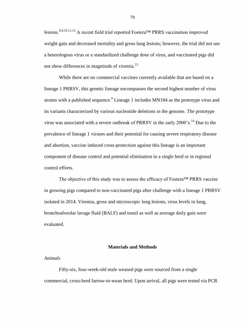
70
lesions.8,9,10,11,12
A recent field trial reported Fostera™ PRRS vaccination improved
weight gain and decreased mortality and gross lung lesions; however, the trial did not use
a heterologous virus or a standardized challenge dose of virus, and vaccinated pigs did
not show differences in magnitude of viremia.13
While there are no commercial vaccines currently available that are based on a
lineage 1 PRRSV, this genetic lineage encompasses the second highest number of virus
strains with a published sequence.4 Lineage 1 includes MN184 as the prototype virus and
its variants characterized by various nucleotide deletions in the genome. The prototype
virus was associated with a severe outbreak of PRRSV in the early 2000’s.14
Due to the
prevalence of lineage 1 viruses and their potential for causing severe respiratory disease
and abortion, vaccine induced cross-protection against this lineage is an important
component of disease control and potential elimination in a single herd or in regional
control efforts.
The objective of this study was to assess the efficacy of Fostera™ PRRS vaccine
in growing pigs compared to non-vaccinated pigs after challenge with a lineage 1 PRRSV
isolated in 2014. Viremia, gross and microscopic lung lesions, virus levels in lung,
bronchoalveolar lavage fluid (BALF) and tonsil as well as average daily gain were
evaluated.
Materials and Methods
Animals
Fifty-six, four-week-old male weaned pigs were sourced from a single
commercial, cross-bred farrow-to-wean herd. Upon arrival, all pigs were tested via PCR

71
for PRRSV and porcine circovirus type 2 (PCV2) in serum and influenza A virus (IAV)
and Mycoplasma hyopneumoniae (MHP) in nasal swabs. Pigs were tested at the Iowa
State Univeristy Veterinary Diagnostic Laboratory for the presence of antibody to
PRRSV, IAV, PCV2, and MHP in serum prior to the start of the trial and pigs were
administered no vaccines prior to arrival. All pigs were administered an intramuscular
(IM) injection of ceftiofur crystalline free acid (Excede®, Zoetis, Florham Park, NJ) per
label instructions upon arrival to the research facility and one day prior to challenge, and
one IM injection of enrofloxacin (Baytril®, Bayer Healthcare, LLC, Shawnee Mission,
KS) one day prior to vaccination to control potential secondary bacterial infections.
PRRSV challenge virus
PRRSV isolate 14-4099 was recovered from serum collected from five-week-old
nursery pigs located in Northwest Iowa in 2014 with clinical signs that included
coughing, malaise and respiratory distress. PRRSV 14-4099 was amplified by three
passages on MARC-145 cells as previously described.15
Infected cells were lysed by two
freeze-thaw cycles after a PRRSV-specific cytopathic effect was observed. Clarification
by centrifugation at 2000 x g for 10 min to pellet cell debris was followed by titration on
MARC-145 cells, distribution of the virus in 5 ml aliquots and freezing at -80°C until
challenge.
Experimental design
Pigs were block randomized by weight into three groups: non-vaccinated/non-
challenge (NV/NC; n=9), non-vaccinated/challenged (NV/C; n=24), and
vaccinated/challenged (V/C; n=23). Groups were housed in the same facility, but

72
separated by room and ventilation system prior to challenge. Each group was divided into
pens of two pigs, with two pens containing only one pig. Pigs in each room were fed a
balanced diet ad libitum based on weight and given free access to water. At 0 days post
vaccination (dpv) pigs in the V/C group received a 2 ml intramuscular (IM) dose of
Fostera™ PRRS vaccine per manufacturer’s instructions. Pigs in the NV/NC and NV/C
groups received a 2 ml IM dose of sterile diluent. At 28 dpv (0 days post inoculation
(dpi)) pigs in the NV/C and V/C groups received 2 ml IM and 2 ml intranasal (IN) doses
of challenge virus at 1x10⁵ TCID50/ml. The NV/NC group received 2 ml IM and 2 ml IN
doses of sterile cell culture medium. In addition, pigs were co-mingled at challenge by
randomly selecting one-half of the pigs in the NV/C and V/C groups, respectively, and
moving them into the corresponding pens in the opposite rooms. Serum was collected
from individual pigs at 0, 14, and 28 dpv, and 3, 7, and 12 dpi. Oral fluid samples were
collected from each pen at 0, 7, 14, 21, and 28 dpv, and 3, 7, and 12 dpi. Humane
euthanasia was performed with a lethal dose of pentobarbital (Fatal-Plus, Vortech
Pharmaceuticals, Ltd. Dearborn, MI) at necropsy (12 dpi). Fresh and formalin fixed tissue
samples collected at necropsy included: lung, tonsil, tracheobronchial, mediastinal, iliac,
and inguinal lymph nodes. Five sections of formalin fixed lung were collected from the
right cranial, right caudal, and accessory lung lobes of each pig after insufflation with and
submersion in 10% neutral buffered formalin for 15 minutes for further fixation.
Broncho-alveolar lavage fluid was collected from the left lung lobes at necropsy. The
experimental design is described in Table 1 and was approved by the Iowa State
University Institutional Animal Care and Use Committee (protocol log #10-13-7659-S).

73
Clinical assessment
Body weights were recorded for each pig on arrival, prior to challenge (26 dpv),
and at necropsy (12 dpi). Weights were used for calculating average daily gain (ADG).
Rectal temperature was recorded from -1 dpi through 7 dpi, and at 10 dpi.
Pathology
At necropsy, lungs were evaluated for the presence of macroscopic lesions typical
of PRRSV by a single veterinary pathologist blinded to the treatment groups. The percent
of the lung surface affected with pneumonia was subjectively estimated for each lung
lobe and the total percentage affected was calculated based on weighted proportions of
each lobe relative to the total lung volume as previously described.16
After 48 hrs of
fixation in neutral buffered formalin, tissue sections were trimmed, processed, and
embedded in paraffin. Four micron thick sections were cut and stained with hematoxylin
and eosin. All five lung sections were scored individually for interstitial pneumonia by a
single veterinary pathologist blinded to the study, using the following scale: 0=normal,
1=mild multifocal, 2=mild diffuse, 3=moderate multifocal, 4=moderate diffuse, 5=severe
multifocal, 6=severe diffuse. The scores designated for each section were averaged to
obtain an overall microscopic lung lesion score for each pig.
Post-Challenge Quantitative PCR
All PCR assays were conducted at the Iowa State University Veterinary
Diagnostic Laboratory (ISU-VDL). Serum and oral fluid samples collected prior to
challenge were extracted using the MagMAX™ Viral RNA Isolation Kit (Life

74
Technologies, Carlsbad, CA) and a Kingfisher 96 instrument (Thermo Scientific,
Waltham, MA) according to manufacturer’s instructions. Real-time RT-PCR was
performed on nucleic acid extracts using the commercially available MagMAX™ NA
and EU PRRSV-specific PCR assay (Life Technologies Carlsbad, CA) to detect the
presence of Fostera™ PRRSV vaccine virus and ensure non-vaccinated pigs were virus
negative.
Post-challenge serum, oral fluids, BALF, lung and tonsil were analyzed by a real-
time RT-PCR with primers and probes designed at the ISU-VDL specifically targeting
ORF5 of the challenge virus. Three grams of fresh lung and tonsil were placed in 30 ml
of MEM, processed by a tissue stomacher, and centrifuged to make a 10% homogenate.
Samples were extracted using the MagMAX™ Viral RNA Isolation Kit (Life
Technologies, Carlsbad, CA) and a Kingfisher 96 instrument (Thermo Scientific,
Waltham, MA) according to manufacturer’s instructions. Real-time RT-PCR was
performed on nucleic acid extracts using the commercially available MagMAX™ NA
and EU PRRSV-specific PCR assay (Life Technologies Carlsbad, CA) and substituting
400 nM of each forward and reverse primer and 200 nM of the probe specific to the
challenge virus (Table 2). Serum, lung and BALF reactions included 12.5 μl of 2X RT-
PCR buffer, 2.5 μl of 10X PRRSV primer probe mix, 1.25 μL of 20X multiplex RT-PCR
enzyme mix, and 0.75 μl of nuclease-free water. Each oral fluid reaction used the same
volume of reagents described for the serum with the exception of 2.5 μL of 20X
multiplex RT-PCR enzyme mix and 0.5 μL of nuclease-free water. A final volume of 25
μL consisting of 17 μL mastermix and 8 μL of RNA extract for the serum, lung and
BALF samples or 18 μL mastermix and 7 μL of RNA extract for the oral fluid samples

75
was placed in each well of a 96-well fast PCR plate (Life Technologies Carlsbad, CA).
Real-time RT-PCR was performed using an AB 7500 fast thermocycler with the
following cycling conditions: 1 cycle at 45°C for 10 min, 1 cycle at 95°C for 10 min, and
40 cycles of 97°C for 2 sec, 60°C for 40 sec. Amplification curves were analyzed with
commercial thermal cycler system software. Each PCR assay included eight progressive
1:10 dilutions of a known copy number of 14-4099 challenge virus based on genomic
copies per ml. Aliquots of each dilution were stored at -80°C and used as the standard
curve for quantitative RT-PCR.
Serology
All antibody assays were conducted at the Iowa State University Veterinary
Diagnostic Laboratory (ISU-VDL) according to laboratory procedures. Serum from
individual pigs and oral fluid samples from each pen post-vaccination and post-challenge
were analyzed for the presence of anti-PRRSV antibody using the PRRSV X3 ELISA
(IDEXX, Westbrook, ME).
Immunohistochemistry Staining
Immunohistochemistry (IHC) staining for the detection of PRRSV-specific
antigen was performed on lung sections at the ISU-VDL as previously described.17
Sections were scored for magnitude of immunoreactive signals by a single veterinary
pathologist blinded to the study, using the following scale: 0=normal, 1=minimal,
2=mild, 3=moderate. Scores were averaged to obtain an overall treatment group mean for
descriptive statistics.

76
Statistical Analysis
A statistical analysis was performed between the vaccinated/challenged and non-
vaccinated/challenged groups. Descriptive summary statistics were performed for the
non-vaccinated/non-challenged group. Percent lung lesions were transformed by the
arcsine-square root transformation and were analyzed by a general linear mixed model
approach. Microscopic lung lesions, virus levels in lung tissue, BALF, tonsil, oral fluids
and serum and average daily gain were analyzed using the same statistical model. The
SAS Proc Mixed procedure (SAS, Cary NC) included the fixed effect of treatment and
the random effects of room, block-within-room and treatment-by-block-within-room.
Treatment least squares mean (LS Mean) were calculated for each group. Pair-wise
comparisons of LS Means between the two groups were performed by the two-sided
Student’s t-test at the 5% level of significance. A p-value < 0.05 was considered
significant.
Results
Pre- and Post-screening Microbiological Assays
No extraneous viral or bacterial nucleic acids were detected in serum (PRRSV,
PCV2) or nasal swabs (IAV, MHP) from pigs prior to the start of the experiment. All pigs
were free of anti-PRRSV and -MHP antibodies as well as influenza A nucleoprotein
antibody prior to the start of the study. Anti-PCV2 antibody was detected in 54/56 pigs
prior to the start of the experiment. BALF collected from all pigs at necropsy (12 dpi)
tested negative for PCV2, IAV, and MHP nucleic acid.

77
Pre-challenge PRRSV ELISA and RT-PCR
Twenty-one of 23 and 23/23 pigs in the V/C group were PRRSV ELISA antibody
positive at 14 and 28 dpv, respectively. Fostera™ PRRS vaccine virus was detected in
serum by PCR in 87% (20/23) and 100% (23/23) of pigs at 14 and 28 dpv, respectively.
All pigs in the NV/NC and NV/C groups were PRRSV PCR and PRRSV antibody
negative in serum during the pre-challenge phase of the study.
Clinical Disease
Pigs in the NV/NC group did not exhibit clinical signs of respiratory disease for
the duration of the study. Clinical signs of respiratory disease were not observed in the
NV/C or V/C groups from 0 to 28 dpv. Approximately 75% of pigs in the NV/C group
became depressed, lethargic, and moderately anorexic at 5 dpi; peak severity of clinical
disease was apparent at 10 dpi characterized by moderate dyspnea, lethargy and marked
anorexia. Approximately 50% of the V/C pigs displayed mild lethargy and anorexia from
5-12 dpi. Significant differences were not observed between the V/C and NV/C group
mean body temperatures at any time point during the study (data not shown).
Lung Pathology
Macroscopic lesions in the NV/C and V/C group were characterized by moderate
to marked enlargement of tracheobronchiolar lymph nodes and variable percentage of
lung surface affected by mottled-tan pneumonia lesions consistent with PRRSV infection.
Mean percent pneumonia was decreased in the V/C group (mean 12.63%; range 0-50%)
compared to the NV/C group (mean 17.21%; range 2-58%); however, the difference in

78
gross lung lesions was not statistically significant (Table 3). The NV/NC group mean was
1.5% (range 0-12%).
Microscopic lung lesions consisted of multifocal to diffuse, mild to severe
lymphohistiocytic interstitial pneumonia. Microscopic interstitial pneumonia scores were
minimally decreased in the V/C group (mean 2.47; range 1.0-4.6) compared to the NV/C
group (mean 2.57; range 0.8-4.6); however, the difference in microscopic lung lesions
was not statistically significant. The NV/NC mean microscopic lung lesion score was
0.78 (range 0.4-1.0).
Post-challenge PRRSV Quantitative RT-PCR
PRRSV virus was not detected by RT-PCR in serum or oral fluid from the
NV/NC group at any time during the study. PRRS challenge virus was detected in serum
from all pigs in the NV/C and V/C groups at 3, 7 and 12 dpi. Mean PRRSV log10
genomic copies/ml of serum from V/C pigs was significantly lower compared to NV/C
pigs at 3, 7, and 12 dpi (Table 4).
All pen-based oral fluid samples from NV/C and V/C groups were positive for
PRRS challenge virus at 3, 7, and 12 dpi. Mean PRRSV log10 genomic copies/ml of oral
fluid from V/C pigs was decreased compared to oral fluid from NV/C pigs at 3 and 7 dpi;
however, the difference between groups was not statistically significant. Mean PRRSV
log10 genomic copies/ml of oral fluid was significantly lower in V/C pigs at 12 dpi
compared to NV/C pigs (Table 4).
PRRSV was not detected in tissues collected from the NV/NC group at necropsy.
Mean log10 genomic copies/ml of the challenge virus in BALF was significantly lower in

79
the V/C pigs compared to the NV/C group (Table 3). Mean log10 genomic copies/ml of
the challenge virus in lung homogenate was lower in V/C pigs compared to NV/C pigs
but not statistically significant. In contrast, the V/C group had higher amounts of
challenge virus in tonsil homogenate compared to the NV/C group; however, this
difference was not significant.
Post-Challenge PRRSV ELISA Antibody
All pigs in the NV/NC group were PRRSV antibody negative via serum and oral
fluids throughout the challenge period. All pigs in the V/C group were positive for
PRRSV ELISA antibody in serum at 3 and 7 dpi with mean S/P ratios of 1.13 and 1.28,
respectively (Figure 1). At 12 dpi, 21/23 pigs in the V/C group were positive for serum
antibody to PRRSV, with a mean S/P ratio of 1.23. PRRSV ELISA antibody was also
detected in pen-based oral fluid samples at 3, 7 and 12 dpi in the V/C group, with mean
S/P ratios of 2.48, 4.01, and 4.41, respectively (data not shown). Post-challenge, 0/24,
21/24, and 24/24 pigs in the NV/C group were PRRSV ELISA antibody positive via
serum at 3, 7, and 12 dpi, respectively. The NV/C group was PRRSV ELISA antibody
negative via oral fluid at 3 dpi, and PRRSV ELISA antibody positive at 7 and 12 dpi,
with mean S/P ratios of 0.97 and 4.21, respectively (data not shown).
Production Parameters
A statistical difference was not observed in mean average daily gain (ADG)
between NV/C and V/C pigs from vaccination until challenge. Post-challenge mean ADG

80
was significantly higher in the V/C group (0.44 kg/day) compared to the NV/C group
(0.32 kg/day) (Table 5). The post-challenge ADG in the NV/NC group was 0.83 kg/day.
Immunohistochemistry
Lung sections from all non-challenged control pigs were negative for PRRSV-
antigen via IHC scoring. Mean lung section IHC scores for vaccinated and challenged
pigs were 0.96 (range 0-3), while non-vaccinated and challenged pigs had a mean IHC
score of 0.75 (range 0-3).
Discussion
Since its emergence in the late 1980’s, PRRSV has become the most
economically important disease of the swine industry. Vaccination against the virus may
be utilized in various control programs to prevent the introduction of a new strain or aid
in the production of PRRSV negative pigs at weaning. MLV vaccines have demonstrated
more effective control compared to inactivated vaccines when challenged with a
homologous PRRSV; however, MLV vaccines have only demonstrated the ability to
induce partial protective immunity against heterologous challenge.11
PRRSV vaccine
challenge studies have typically used prototype isolates as the cell-culture challenge
virus. Due to the large amount of genetic and antigenic diversity in PRRSV isolates,
historical prototype viruses, such as VR2332 and MN-184, may not represent the
genotypic or phenotypic characteristics of current strains circulating in swine that
vaccine-induced immunity must protect against.

81
The objective of the current study was to evaluate the efficacy of a commercial
MLV vaccine, Fostera™ PRRS, against a heterologous lineage 1 PRRSV recently
isolated from nursery pigs in Iowa in 2014. Lineage 1 viruses are currently the most
common PRRSV circulating in the US swine population demonstrating the greatest
sequence divergence among field viruses.18,19
Several recent lineage 1 virus isolates were
identified from case submissions at the ISU VDL. Isolate 14-4099 was selected as the
challenge virus after an initial pilot study confirmed its virulence in swine. Results of the
current study suggest Fostera™ PRRS vaccine administered four weeks prior to
challenge elicited partial cross-protection against a heterologous lineage 1 challenge virus
that shares only 84.4% ORF5 nucleotide homology. Vaccinated pigs had significantly
lower levels of viremia at all sampling points, and vaccinated pigs had lower levels of
virus in oral fluids and BALF at 12 dpi. In spite of these differences in virus levels
between vaccinated and non-vaccinated pigs, gross and microscopic lung lesions (while
trending lower in vaccinated pigs) did not demonstrate significant differences between
the challenge groups. It is unknown why significant differences in lung lesions were
lacking between challenge groups, in spite of the differences in viremia. Previous studies
suggest increasing genetic diversity between challenge and vaccine viruses may affect
vaccine efficacy demonstrated by similar severity of lung lesions regardless of the
vaccine administered.12
PRRSV has a tropism for pulmonary alveolar macrophages
(PAMs), which are the target of viral replication in swine.20
Levels of challenge virus in
BALF in the current study suggest viral replication was decreasing at 12 dpi although it is
possible sufficient time was lacking for differences in viral clearance from lung and tonsil
to be detected as suggested by the similar magnitude of IHC signals observed in lung

82
sections from both challenge groups, regardless of vaccination. Severity of lung lesions
may also be influenced by the virulence of the challenge virus, dose, routes of
administration and differences in host immune response. Collectively, pigs administered
Fostera™ PRRSV demonstrated lower levels of virus in serum at all sample collections
suggesting partial efficacy imparted by the vaccine.
The impact of co-mingling pigs in the same room may have also affected the
magnitude of lung lesions observed in challenged pigs regardless of vaccination status.
Research has shown that vaccinated boars shed lower amounts of virus in semen than
non-vaccinated boars.21
Studies in growing pigs have shown that therapeutic vaccination
can decrease the amount of virus shed in herds endemically infected with homologous8 or
heterologous PRRSV strains.22
In addition, experimental studies have reported the ability
of infected pigs to transmit PRRSV to sentinel pigs via aerosols over short distances.23
These studies suggest that co-mingling pigs could effectively expose vaccinated pigs to a
greater dose of PRRSV throughout the post-challenge period (due to viral shedding from
non-vaccinated roommates) compared to being exposed to pigs administered a similar
treatment. While it is unclear what affect this may have on the dynamics of PRRSV
infection, an artificial increase in virus levels detected in lung and elevated lung lesions
are plausible outcomes.
In this study, each pig was given a standard dose of challenge virus. Oral fluids
from vaccinated pigs contained lower amounts of virus than oral fluids from non-
vaccinated pigs at 12 dpi, indicating that vaccination had resulted in lower quantities of
virus being shed. Nose-to-nose contact would allow direct exposure to oral fluids, while
aerosol transmission within each room also would be expected. Higher levels of

83
challenge virus shed from non-vaccinated pigs to vaccinated pigs may have contributed
to the lack of difference in lung lesions or level of viral clearance in lung tissue; however,
significantly lower levels of viremia and decreased PRRSV challenge virus in BALF
from the vaccinated pigs would appear to contradict this theory. While the differences in
serum and BALF virus quantities were still significant, the differences may have been
falsely reduced from expected levels if pigs had not been co-mingled in the same room.
Collectively, it could be argued that the co-mingling of vaccinated and non-vaccinated
pigs post-challenge may affect the results of several commonly assessed parameters of
immune protection.
Much of the losses attributed to PRRSV infection in growing pigs are due to
increased mortality, decreased ADG, and decreased feed efficiency. In this study, a
significant difference in average daily gain between vaccinated and non-vaccinated pigs
was apparent after only 12 days post challenge with a virulent PRRSV. This suggests that
vaccination with Fostera™ PRRS confers significant production benefits in the face of
heterologous challenge. Considering clinical severity was at its peak at the time of
necropsy in NV/C pigs, extending the post-challenge period may have further increased
the difference in ADG between V/C and NV/C groups. In addition, vaccination did not
depress ADG in the pre-challenge period, suggesting immunity was elicited without a
concurrent decrease in production. The improved weight gain may be partially explained
by the reduced viremia and reduced virus levels in BALF.
In summary, vaccination of nursery pigs with Fostera™ PRRS resulted in partial
cross-protection when challenged with a contemporary, heterologous lineage 1 PRRSV.
Vaccinated pigs showed decreased viremia and increased ADG compared to non-
84
vaccinated pigs; however, vaccination did not significantly decrease gross or microscopic
lung lesions. Further investigation into cross-protection elicited by MLV vaccines is
needed, along with research into the differences in cross-protection conferred by various
commercial MLV vaccines. The results of this study show that the impact of PRRSV on
growing pigs can be substantially mitigated through the use of MLV vaccines. In order to
make substantial progress regarding PRRS control, future vaccine development should
focus on the stimulation of broad heterologous cross-protection.
Acknowledgements
We would like to thank Iowa State University Veterinary Diagnostic Laboratory
staff for their assistance with sample testing and Lab Animal Resources staff for their
assistance with animal care.
References
1. Cavanagh D Nidovirales: a new order comprising Coronaviridae and Arteriviridae.
Arch Virol 142:629-33, 1997.
2. Rossow KD Porcine reproductive and respiratory syndrome. Vet Pathol 35:1-20,
1998.
3. Holtkamp DJ, Kliebenstein JB, Neumann EJ, Zimmerman JJ, Rotto HF, Yoder TK,
et al. Assessment of the economic impact of porcine reproductive and respiratory
syndrome virus on United States pork producers. J Swine Health and Production
21:72-84, 2013.
4. Shi M, Lam TT, Hon CC, Murtaugh MP, Davies PR, Hui RK, et al. Phylogeny-
based evolutionary, demographical, and geographical dissection of North American
type 2 porcine reproductive and respiratory syndrome viruses. J Virol 84:8700-11,
2010.
5. Hanada K, Suzuki Y, Nakane T, Hirose O, Gojobori T The origin and evolution of
porcine reproductive and respiratory syndrome viruses. Mol Biol Evol 22:1024-31,
2005.

85
6. Murtaugh MP, Yuan S, Faaberg KS Appearance of novel PRRSV isolates by
recombination in the natural environment. Adv Exp Med Biol 494:31-6, 2001.
7. Yuan S, Nelsen CJ, Murtaugh MP, Schmitt BJ, Faaberg KS Recombination
between North American strains of porcine reproductive and respiratory syndrome
virus. Virus Res 61:87-98, 1999.
8. Cano JP, Dee SA, Murtaugh MP, Trincado CA, Pijoan CB Effect of vaccination
with a modified-live porcine reproductive and respiratory syndrome virus vaccine
on dynamics of homologous viral infection in pigs. Am J Vet Res 68:565-71, 2007.
9. Geldhof MF, Vanhee M, Van BW, Van DJ, Karniychuk UU, Nauwynck HJ
Comparison of the efficacy of autogenous inactivated Porcine Reproductive and
Respiratory Syndrome Virus (PRRSV) vaccines with that of commercial vaccines
against homologous and heterologous challenges. BMC Vet Res 8:182, 2012.
10. Labarque G, Reeth KV, Nauwynck H, Drexler C, Van GS, Pensaert M Impact of
genetic diversity of European-type porcine reproductive and respiratory syndrome
virus strains on vaccine efficacy. Vaccine 22:4183-90, 2004.
11. Li X, Galliher-Beckley A, Pappan L, Trible B, Kerrigan M, Beck A, et al.
Comparison of host immune responses to homologous and heterologous type II
porcine reproductive and respiratory syndrome virus (PRRSV) challenge in
vaccinated and unvaccinated pigs. Biomed Res Int 2014:416727, 2014.
12. Okuda Y, Kuroda M, Ono M, Chikata S, Shibata I Efficacy of vaccination with
porcine reproductive and respiratory syndrome virus following challenges with field
isolates in Japan. J Vet Med Sci 70:1017-25, 2008.
13. Park C, Seo HW, Han K, Kang I, Chae C Evaluation of the efficacy of a new
modified live porcine reproductive and respiratory syndrome virus (PRRSV)
vaccine (Fostera PRRS) against heterologous PRRSV challenge. Vet Microbiol
172:432-42, 2014.
14. Han J, Wang Y, Faaberg KS Complete genome analysis of RFLP 184 isolates of
porcine reproductive and respiratory syndrome virus. Virus Res 122:175-82, 2006.
15. Gauger PC, Faaberg KS, Guo B, Kappes MA, Opriessnig T Genetic and phenotypic
characterization of a 2006 United States porcine reproductive and respiratory virus
isolate associated with high morbidity and mortality in the field. Virus Res 163:98-
107, 2012.
16. Halbur PG, Paul PS, Frey ML, Landgraf J, Eernisse K, Meng XJ, et al. Comparison
of the pathogenicity of two US porcine reproductive and respiratory syndrome virus
isolates with that of the Lelystad virus. Vet Pathol 32:648-60, 1995.
17. Halbur PG, Miller LD, Paul PS, Meng XJ, Huffman EL, Andrews JJ
Immunohistochemical identification of porcine reproductive and respiratory

86
syndrome virus (PRRSV) antigen in the heart and lymphoid system of three-week-
old colostrum-deprived pigs. Vet Pathol 32:200-4, 1995.
18. Brar MS, Shi M, Murtaugh MP, Leung FC Evolutionary Diversification of Type 2
Porcine Reproductive and Respiratory Syndrome Virus. J Gen Virol 2015.
19. Rovira A PRRS diagnostic trends: What changed with PRRS behavior? American
Association of Swine Veterinarians 509-11, 2015.
20. Duan X, Nauwynck HJ, Pensaert MB Virus quantification and identification of
cellular targets in the lungs and lymphoid tissues of pigs at different time intervals
after inoculation with porcine reproductive and respiratory syndrome virus
(PRRSV). Vet Microbiol 56:9-19, 1997.
21. Han K, Seo HW, Shin JH, Oh Y, Kang I, Park C, et al. Effect of the modified live
porcine reproductive and respiratory syndrome virus (PRRSV) vaccine on European
and North American PRRSV shedding in semen from infected boars. Clin Vaccine
Immunol 18:1600-7, 2011.
22. Cano JP, Dee SA, Murtaugh MP, Pijoan C Impact of a modified-live porcine
reproductive and respiratory syndrome virus vaccine intervention on a population of
pigs infected with a heterologous isolate. Vaccine 25:4382-91, 2007.
23. Torremorell M, Pijoan C, Janni K, Walker R, Joo HS Airborne transmission of
Actinobacillus pleuropneumoniae and porcine reproductive and respiratory
syndrome virus in nursery pigs. Am J Vet Res 58:828-32, 1997.
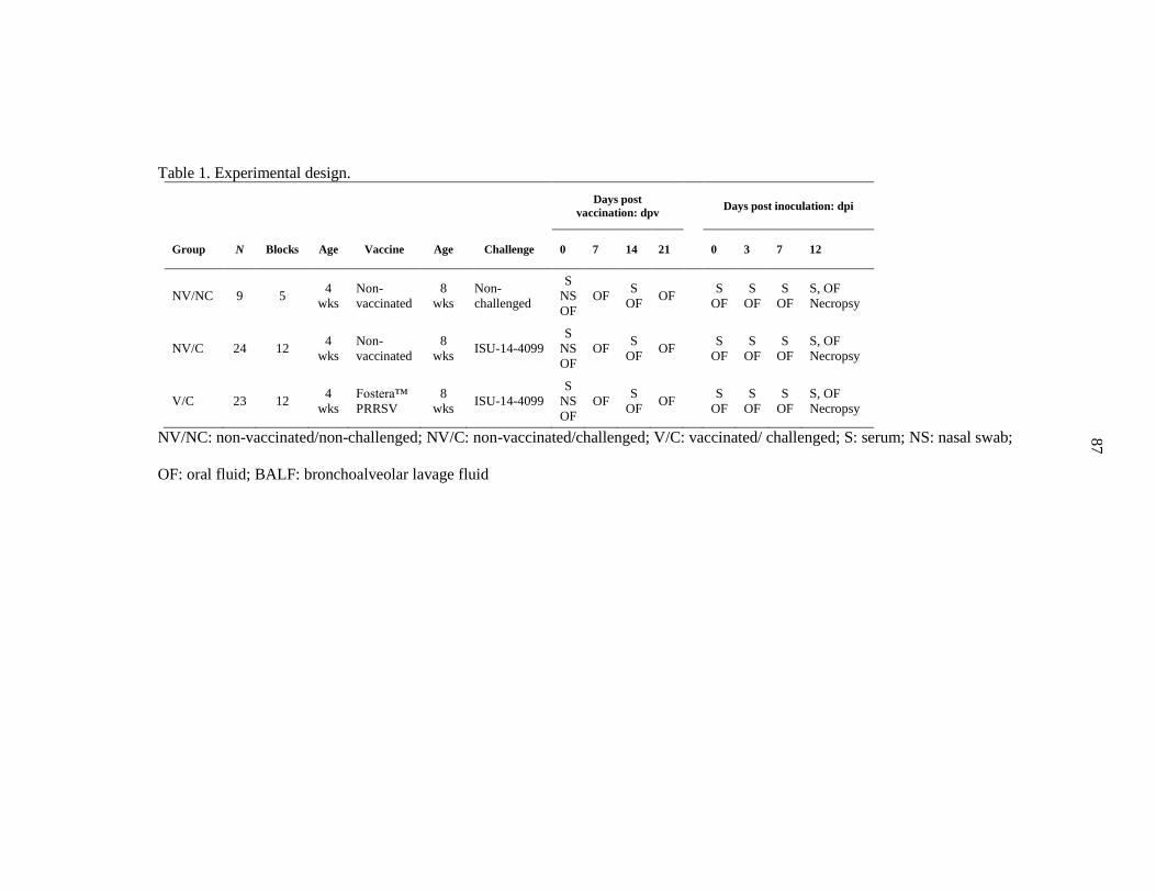
87
Table 1. Experimental design.
Days post
vaccination: dpv
Days post inoculation: dpi
Group N Blocks Age Vaccine Age Challenge 0 7 14 21
0 3 7 12
NV/NC 9 5 4
wks
Non-
vaccinated
8
wks
Non-
challenged
S
NS
OF
OF S
OF OF
S
OF
S
OF
S
OF
S, OF
Necropsy
NV/C 24 12 4
wks
Non-
vaccinated
8
wks ISU-14-4099
S
NS
OF
OF S
OF OF
S
OF
S
OF
S
OF
S, OF
Necropsy
V/C 23 12 4
wks
Fostera™
PRRSV
8
wks ISU-14-4099
S
NS
OF
OF S
OF OF
S
OF
S
OF
S
OF
S, OF
Necropsy
NV/NC: non-vaccinated/non-challenged; NV/C: non-vaccinated/challenged; V/C: vaccinated/ challenged; S: serum; NS: nasal swab;
OF: oral fluid; BALF: bronchoalveolar lavage fluid

88
Table 2. Sequences of the forward and reverse primers and individual probe for 14-4099 challenge PRRS virus used in the real
time RT-PCR for absolute quantification of PRRSV genomic copies.
Name Sequence
PRRSV (forward primer) 5’-ATATCTCCACGGGCGGTATG -3’
PRRSV (reverse primer) 5’-TTGTAAGCCTGATGGCAAAGC-3’
14-4099 PRRSV 5’-/56-FAM/CTGTCTGCG/ZEN/CTCTTGCCGCG/3IABkFQ/-3’

89
Table 3. Mean macroscopic pneumonia, microscopic lung lesion scores and quantitative RT-PCR values in lung, BALF and tonsil
collected from NV/NC, NV/C and V/C pigs at 12 dpi ± standard error of the mean. Different letters within a column represent a statistical
difference at P < 0.05.
Group
Lung Lesions Quantitative PCR (Mean log 10 genomic copies/ml±SE)
Macroscopic percent
pneumonia
(Mean±SE)
Microscopic lung
scores
(Mean±SE)
Lung BALF Tonsil
NV/NC 1.5 ± 0.7 0.78 ± 0.08 0.00 ± 0.00 0.00 ± 0.00 0.00 ± 0.00
NV/C 17.2 ± 3.2a 2.57 ± 0.26
a 6.21 ± 0.19
a 6.19 ± 0.21
a 6.71 ± 0.11
a
V/C 12.7 ± 2.1a 2.47 ± 0.26
a 6.04 ± 0.19
a 5.44 ± 0.21
b 6.87 ± 0.11
a
NV/NC, non-vaccinated-non-challenged; NV/C, Non-vaccinated-challenged; V/C, vaccinated-challenged
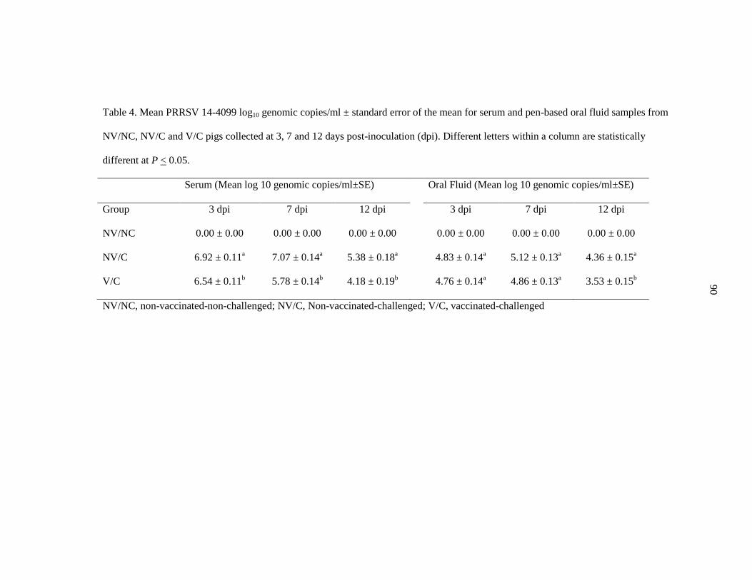
90
Table 4. Mean PRRSV 14-4099 log10 genomic copies/ml ± standard error of the mean for serum and pen-based oral fluid samples from
NV/NC, NV/C and V/C pigs collected at 3, 7 and 12 days post-inoculation (dpi). Different letters within a column are statistically
different at P < 0.05.
Serum (Mean log 10 genomic copies/ml±SE) Oral Fluid (Mean log 10 genomic copies/ml±SE)
Group 3 dpi 7 dpi 12 dpi 3 dpi 7 dpi 12 dpi
NV/NC 0.00 ± 0.00 0.00 ± 0.00 0.00 ± 0.00 0.00 ± 0.00 0.00 ± 0.00 0.00 ± 0.00
NV/C 6.92 ± 0.11a 7.07 ± 0.14
a 5.38 ± 0.18
a 4.83 ± 0.14
a 5.12 ± 0.13
a 4.36 ± 0.15
a
V/C 6.54 ± 0.11b 5.78 ± 0.14
b 4.18 ± 0.19
b 4.76 ± 0.14
a 4.86 ± 0.13
a 3.53 ± 0.15
b
NV/NC, non-vaccinated-non-challenged; NV/C, Non-vaccinated-challenged; V/C, vaccinated-challenged

91
Table 5. Average daily gain reported in kilograms during the post-vaccination and post-challenge
phase of the study in NV/NC, NV/C and V/C groups ± standard error of the mean. Different
letters within a column are statistically different at P< 0.05.
Average Daily Gain in Kilograms
Group Post-Vaccination
(-10 to 26dpv)
Post-Challenge
(26dpv to 12dpi)
NV/NC 0.441 ± 0.028 0.827 ± 0.026
NV/C 0.405 ± 0.014a 0.323 ± 0.027
a
V/C 0.405 ± 0.014a 0.441 ± 0.032
b
NV/NC, non-vaccinated-non-challenged; NV/C, Non-vaccinated-challenged; V/C, vaccinated-
challenged

92
Figure 1. Mean anti-PRRSV ELISA antibody S/P ratios in serum from NV/NC, NV/C and V/C
pigs at 0, 14 and 28 days post vaccination (dpv) and 3, 7 and 12 days post inoculation (dpi). Error
bars represent standard error of the mean.
0.00
0.25
0.50
0.75
1.00
1.25
1.50
0 dpv 14 dpv 28 dpv/0 dpi 3 dpi 7 dpi 12 dpi
NV/NC NV/C V/C
S/P
Rat
io

93
Figure 2. Macroscopic and microscopic lung lesions and immunohistochemistry staining of lung
observed at necropsy in (A) NV/NC, (B) NV/C and (B) V/C pigs challenged with placebo or 14-
4099 PRRSV.
A
C
B

94
CHAPTER 5: CONCLUSION
Reproductive failure, increased mortality, and reduced feed efficiency due to
porcine reproductive and respiratory syndrome virus (PRRSV) combine to make it the
mostly costly pathogen in modern swine production. Prevention and control of PRRSV is
difficult due to the vast genetic diversity present within currently circulating virus strains.
Protective immunity stimulated by natural infection or vaccination rarely results in
complete protection from infection; however, vaccination with modified live virus
(MLV) vaccines has been shown to result in partial protection, defined by an
improvement in weight gain, less severe lung lesions, and/or reduced viremia.
The advancements made in vaccine development may play a crucial role in
reducing the cost of disease, and someday may potentially result in PRRSV eradication.
New vaccines must prove to be more effective than current commercially available
vaccines, especially against relevant and contemporary field viruses. Most vaccine
challenge studies have used historic PRRSV isolates in order to allow some level of
comparison between results; however, historic isolates may be genetically and
phenotypically different from current field strains and virulence among strains even
within a PRRSV lineage may vary. A challenge model that uses currently circulating,
virulent PRRSV isolates would provide a much more accurate depiction of how a vaccine
could be expected to perform in current swine production settings.
With nine different lineages and thousands of available PRRSV isolates, the
options for selecting a suitable challenge virus are nearly endless. One goal of the current
research was to provide swine veterinarians and producers with information regarding a
recently approved vaccine, Fostera™ PRRS. To ensure the results were relevant, the

95
following criteria for virus selection were as follows: 1) the challenge virus must belong
to highly populated PRRSV lineage; 2) the challenge virus must be sufficiently
heterologous to adequately evaluate cross-protection; and 3) the challenge virus must
represent current circulating virus strains. Four of the nine lineage classifications have
more than 1,000 different PRRSV. Lineage 1 and lineage 9 are the two largest PRRSV
lineages; viruses within these lineages also display the most intralineage genetic diversity
at ORF5 of any of the large lineages.
In both studies presented in this thesis, the challenge PRRSV were chosen from a
group of recently isolated field isolates. Small pilot studies using three pigs in each
challenge group were used to examine the virulence of each virus and to ensure the
challenge model would result in gross lung lesions that were easy to observe. In addition
to proving the virulence of the challenge virus, several different parameters were
investigated, including microscopic lung lesions, average daily gain, and the amount of
virus detected in various fluids or tissues (serum, oral fluids, bronchoalveolar lavage
fluid, lung, and tonsil), to use as a measure of the degree of vaccine protection.
The results of the two studies presented here share several similarities. Average
daily gain of vaccinated pigs during the challenge period was greater than that of non-
vaccinated pigs in both studies, suggesting significant improvement in clinical disease
that may be correlated with increased feed intake and improved feed efficiency although
these parameters were not specifically measured during either experiment. Non-
challenged controls had better average daily gain than non-vaccinated challenged pigs,
which serves as a testament to the virulence of the challenge viruses; however, vaccinated
pigs gained less weight than non-challenged controls as well, suggesting a level of partial

96
protection but with considerable room for improvement. Viremia was improved at 3, 7
and 12 days post inoculation and fewer viral genomic copies were also present in BALF
at necropsy in both studies. Interestingly, both experiments detected higher levels of
PRRSV in the tonsil tissue of vaccinated pigs. This unexpected result may be due to
several factors, including processing difficulty due to the tough fibrous nature of the
tissue, or perhaps priming the immune system via vaccination leads to enhanced viral
uptake and replication in lymphoid tissue. PRRSV persists in lymphoid tissues of
infected pigs even after clearance of the virus from the blood. However, it remains
unknown why the vaccinated pigs demonstrated larger quantities of challenge virus in the
tonsil compared to non-vaccinated and challenged pigs in the current studies.
While there were some similarities between the challenge experiments described
in Chapter 2 and 3, there were also many differences between studies. The lineage 9
study demonstrated nearly a two log reduction in serum virus quantities in the vaccinated
pigs at 7 and 12 dpi and no significant difference prior to that time. Vaccinated pigs also
demonstrated lower levels of virus in lung tissue, along with significantly decreased gross
and microscopic lung lesions. Challenge with a lineage 1 virus did not result in a decrease
in lung lesions or virus load in the lung tissue, but did show a decrease in viremia at each
measured timepoint (3, 7 and 12 dpi). The reasons for the discrepancies between studies
are unknown; however, variation in study design (20 pigs per pen in the lineage 9 study
and 2 pigs per pen with 12 replicates in the lineage 1 study), pig housing, and challenge
virus along with differences in the individual pig’s susceptibility and response to PRRSV
infection may all play a role.

97
Along with random differences in the groups of pigs used in each study (although
sourced from the same farm), two major differences are likely to have contributed to the
variable results. First, the lineage 1 study used a challenge virus that was considerably
more genetically diverse than the lineage 9 challenge virus regarding their homology to
the vaccine PRRSV strain. Vaccines have been shown to be completely protective against
homologous viruses used in challenge studies, so it stands to reason that a vaccine may
offer a higher level of protection against a less genetically or antigenically diverse strain
compared to a more diverse strain. The lineage 9 challenge virus shares 92.5% ORF5
homology with the vaccine virus, while the lineage 1 challenge virus is only 84.4%
homologous at ORF5. This difference may account for the relatively higher degree of
protection seen in the lineage 9 challenge study based on the reduction in lung lesions
and reduced virus levels in lung tissue.
The second major difference involves the study design. The lineage 9 challenge
study housed vaccinated and non-vaccinated pigs in separate rooms for the duration of
the study (both pre-challenge and post-challenge periods). Pigs in each room were also
kept in one large pen. The lineage 1 challenge study housed vaccinated pigs and non-
vaccinated pigs in separate rooms for the pre-challenge period only, and pigs were
divided into pens of two in each room. On the day of challenge, one-half of the pigs were
moved into the opposite treatment room, in an attempt to eliminate potential room bias.
The design of the lineage 9 study eliminated any exposure to challenge virus
between pigs from different treatment (vaccination) groups, and more accurately reflects
what could be expected to occur in modern pig production when an entire group of pigs
are administered the same vaccine at the same time. It allowed us to compare the

98
differences between an entire room of vaccinated pigs and an entire room of non-
vaccinated pigs, to see if vaccination of a single room had any effect on post-challenge
production. Each pig was individually vaccinated and challenged; during the post-
challenge period, any additional virus contacted by the pig was due to the dynamics of
infection and subsequent shedding of pen-mates within a treatment group, either with or
without the potential benefit of vaccination. Room bias may have been present in the
lineage 9 study, but in a climate-controlled infectious disease research facility with
identical room dimensions, I believe the consequences to be insignificant and of
irrelevant impact.
The design of the lineage 1 study removed room bias by co-mingling an equal
number of pigs from each treatment group in each room, but in separate pens. Each pig
was vaccinated and challenged individually; however, pigs in different treatment groups
were allowed nose-to-nose contact. Therefore, at challenge, vaccinated pigs had direct
contact with non-vaccinated pigs exposing vaccinated pigs to higher levels of PRRSV
shedding into the environment compared to what would have been expected in a room
containing pigs with the same treatment (vaccination vs. no vaccination). Conversely,
with half of the pigs in each room vaccinated, non-vaccinated pigs came into contact with
lower levels of virus. The study design effectively changed the exposure dose of each pig
although it is unknown how that may have affected the effectiveness of vaccine-induced
immunity in regards to parameters such as lungs lesions and weight gain.
The results of the studies presented here suggest vaccination provided partial
protection as measured by a reduction in virus quantities in serum and increased weight
gain. Interestingly, this protection was seen in the absence of neutralizing antibodies.

99
Serum from Fostera™ PRRS-vaccinated sows was tested for the presence of neutralizing
antibodies toward 12-39404 (the challenge virus used in the lineage 9 challenge study)
and showed a complete lack of neutralization. Antibody neutralization of the lineage 1
challenge strain, 14-4099, was not directly tested; however, due to the large amount of
genetic diversity between this strain and the vaccine strain, neutralizing antibodies to a
strain with that degree of diversity would not be expected. These results suggest that
while neutralizing antibodies may be helpful (or even necessary) in preventing viremia,
they are not needed to provide partial protection as measured by a reduction in viremia
and improved production.
With the absence of complete protection from infection, the improvement in
average daily gain observed in both studies is the most impressive aspect of vaccine-
induced partial protection. The lineage 1 study ADG was particularly interesting, since no
difference in gross or microscopic lung lesions were detected by a subjective analysis but
based on expertise and published parameters. In a production setting with large numbers
of animals, small increases in efficiency can have a large impact on the economics of
disease. In addition to improved post challenge ADG, post vaccination average daily gain
was not significantly different in either study. In other words, there are no negative
effects on production due to vaccination with Fostera™ PRRS under the conditions of the
experiments. In production systems with a high-risk of PRRSV exposure, producers can
receive the benefit of partial protection without sacrificing production due to vaccination.
In order to eventually have any chance of eradicating PRRSV, science likely must
develop a vaccine that confers complete (or near-complete) protection against
heterologous infection. The rate of genetic change in field isolates will be a limiting

100
factor in the future effectiveness of current vaccines. More research is needed into highly
conserved epitopes and antibodies to those epitopes that may provide broad cross-
protection against current and future virus strains. There is a possibility that such epitopes
may not exist, and vaccine development may need to explore new alternatives, such as
nanoparticle or recombinant based vaccines.
In the meantime, more research is needed to determine the cross-protective
efficacy of other commercial vaccines against challenge with PRRSV strains that are
currently circulating in the world’s swine population. Improving our knowledge of
vaccine-induced immunity and its effect on various viruses can aid in the selection and
directed use of the most appropriate vaccine for each production setting. Genetic
sequencing of regional PRRSV strains may allow for widespread use of targeted
vaccination programs in an effort to reduce the impact of PRRSV. Sequencing can also
be useful in tracking the spread of the virus and identifying emerging routes of viral
dissemination. Research into the various aspects of PRRSV has uncovered much of the
mystery that initially surrounded the disease, but there is still much to learn.


