Embed Size (px)
Citation preview
CXCL10-Producing Mucosal CD4+ T Cells, NK Cells, and NKT CellsAre Associated with Chronic Colitis in IL-10−/− Mice, Which Can BeAbrogated by Anti-CXCL10 Antibody Inhibition
UDAI P. SINGH1, SHAILESH SINGH2, RAJESH SINGH2, YINGZI CONG3, DENNIS D. TAUB4,and JAMES W. LILLARD Jr1,2
1Department of Microbiology and Immunology, Morehouse School of Medicine, Atlanta, GA 30310
2Department of Microbiology and Immunology, University of Louisville, KY 40202
3Department of Medicine, Division of Gastroenterology, University of Alabama at Birmingham, AL 35294
4Laboratory of Immunology, National Institute of Aging, Gerontology Research Center, Baltimore, MD 21224
AbstractWe have shown previously that there is a temporal increase in the levels of CXCL10 and CXCR3mRNA during spontaneous murine colitis. We now show that CXCL10 is significantly expressed bymucosal CD4+ T cells, natural killer (NK) cells, and NKT cells, but not by dendritic cells (DCs),during chronic murine colitis. CXCL10 blockade alleviated chronic colitis and attenuated theassociated increase in serum amyloid A (SAA), interleukin-12 (IL-12)p40, tumor necrosis factor-α(TNF-α), interferon-γ (IFN-γ), IL-1α, and IL-1β Levels as well as in the number of CD4+ T, NKT,and NK cells that express CXCL10 and CXCR3, compared with groups treated with control antibody(Ab). After CXCL10 blockade, the number of CXCR3+ DCs in the mesenteric lymph nodes (MLNs)and Peyer’s patches (PPs) were increased to levels found before the onset of colitis. In contrast, thenumbers of splenic and intestinal lamina propria (LP) CXCR3+ DCs were reduced after anti-CXCL10 Ab treatment, compared with controls. Ex vivo antigen and CXCL10 stimulation of mucosalcells caused an increase in MHC class II, CD40, and CD86 as well as a decrease in CD30 ligand(CD30L) expression by DCs. This study provides insights into CXCL10 expression duringinflammatory bowel disease (IBD) and the cellular and molecular mechanisms of CXCL10-mediatedcolitis. Our data also support the premise that CXCL10 blockade can attenuate chronic colitis bypreventing the activation and recruitment of CXCR3+ leukocytes during IBD.
INTRODUCTIONThe etiology and pathogenesis of the two major forms of inflammatory bowel disease (IBD),Crohn’s disease (CD) and ulcerative colitis (UC), are poorly understood (Podolsky 2002). Itis widely held that human IBD is multifactorial and caused by immunologic, environmental,and genetic factors. It has been suggested that colitis may be the result of massive cellularinfiltrates and is associated with abnormalities in the immune system and normal gut flora(MacDonald and Pettersson 2000; Singh and others 2007a) or an overall autoimmunedysregulation/imbalance in T cells (Kuhn and others 1993; Mombaerts and others 1993;Hollander and others 1995).
Address reprint requests or correspondence to: James W. Lillard, Jr., University of Louisville, Brown Cancer Center, Department ofMicrobiology & Immunology, 580 S. Preston Street, Baxter II, Room 304C, Louisville, KY 40202, Tel: (502) 852-2174, Fax: (502)852-3842, E-mail: [email protected].
NIH Public AccessAuthor ManuscriptJ Interferon Cytokine Res. Author manuscript; available in PMC 2008 June 23.
Published in final edited form as:J Interferon Cytokine Res. 2008 January ; 28(1): 31–43.
NIH
-PA Author Manuscript
NIH
-PA Author Manuscript
NIH
-PA Author Manuscript
Under conventional housing conditions, interleukin-10−/− (IL-10−/−) mice developspontaneous colitis that has similarities to human CD. However, this murine model differsfrom human CD in that the colitis in IL-10−/− mice does not yield focal granulomatous ortransmural inflammation. There is a consensus that the mucosa of CD patients is dominatedby T cells producing inflammatory cytokines (Fiocchi 1998). We have shown that CXCL9,CXCL10, and CXCL11 are upregulated at the sites of murine colitis and clinical IBD (Singhand others 2003b, 2007b). It is well established that CD4+ Th1 cells mediate the chronicinflammation observed in the colons of IL-10−/− mice and that interferon-γ (IFN-γ) plays amajor role during the initial phase of inflammation. The primary cellular source of early IFN-γ production appears to be the natural killer (NK) cells, which have been shown to be involvedin the differentiation of naïve CD4+ T cells into Th1 cells (Romagnani 1992). To this end, thenumber of CD4+ CXCR3+ T cells in the intestinal lamina propria (LP) has been shown to behigher in IBD patients than in normal, healthy donors (Yuan and others 2001).
One of the ligands for CXCR3, CXCL10, is an immediate-early gene that is induced by IFN-γ and expressed by epithelium, fibroblasts, keratinocytes, NK cells, and monocytes (Luster andRavetch 1987; Farber 1997). Whereas we and others have shown that blocking CXCL10expression prevents the development of asymptomatic colitis (Singh and others 2003a; Hyunand others 2005), the present study demonstrates that CXCL10 inhibition also leads toremission of chronic colitis in IL-10−/− mice. The present study also shows that CD4+ T cells,NK cells, and NKT cells of the mucosa produce CXCL10, which correlates with highernumbers of CXCR3+ Th1 cells and CXCR3+ DCs as well as supports increases in MHC classII and costimulatory molecule expression by DCs.
MATERIALS AND METHODSAnimals
Female IL-10−/− mice, on a B6 background, age 8–12 weeks, were purchased from JacksonLaboratories (Bar Harbor, ME), and 4–5-month-old female New Zealand rabbits (Myrtle’sRabbitory, Thompson Station, TN) were used to generate anti-CXCL10 antibodies (Ab). Theanimals were housed and maintained in microisolator cages under conventional housingconditions. Experimental groups consisted of 5 mice, and each study was repeated three times.The guidelines proposed by the committee for the Care of Laboratory Animal ResourcesCommission of Life Sciences of the National Research Council were followed to minimizeanimal pain and distress.
Cell isolationSpleens and mesenteric lymph nodes (MLNs) from mice were mechanically dissociated, andred blood cells (RBCs) were disrupted with lysis buffer. The single-cell suspensions of spleenand MLN cells were passed through a sterile wire screen (Sigma, St. Louis, MO) and cellsfrom intestinal LP and Peyer’s patches (PPs) were isolated as described previously (Lillardand others 2001, 2003). Thereafter, lymphocytes were maintained in complete medium, whichconsisted of RPMI 1640 supplemented with 10 mL/L nonessential amino acids (Mediatech,Washington, DC), 1 mM sodium pyruvate (Sigma), 10 mM HEPES (Mediatech), 100 U/mLpenicillin, 100 μg/mL streptomycin, 40 μg/mL gentamicin (Elkins-Sinn, Inc., Cherry Hill, NJ),50 μM mercaptoethanol (Sigma), and 10% fetal bovine serum (FBS) (Atlanta Biologicals,Atlanta, GA).
Immunogens and AbsThe potential level of endotoxin contamination of immunogens was quantified by thechromogenic Limulus amebocyte lysate assay (Cape Cod, Inc., Falmouth, MA) to be < 5 EU/mg. Chicken egg ovalbumin (OVA) and bovine serum albumin (BSA) were purchased from
SINGH et al. Page 2
J Interferon Cytokine Res. Author manuscript; available in PMC 2008 June 23.
NIH
-PA Author Manuscript
NIH
-PA Author Manuscript
NIH
-PA Author Manuscript
Sigma. Rat antimouse CXCL10 monoclonal antibody (mAb) was obtained from R&D Systems(Minneapolis, MN). Rabbit antimouse CXCR3 Ab was purchased from Zymed LaboratoriesInc. (San Francisco, CA). Anti-CXCL10 and anti-CXCR3 Abs were conjugated toallophycocyanin (APC) (Prozyme, San Leandro, CA). The detailed methods of anti-CXCL10Ab generation, purification, and cross-reactivity determination for subsequent in vivoneutralization of CXCL10 were described previously (Singh and others 2003a). In brief,CXCL10 plus complete Freund’s or incomplete Freund’s adjuvants (Sigma) were used togenerate anti-CXCL10 Ab titers of ~1:2 × 10⁶ such that 10 μL anti-CXCL10 Ab neutralized20 ng CXCL10 by ELISA (R&D Systems) and chemotaxis assay (Chemicon, Temecula, CA)using CXCR3+ T cells. This Ab was titrated by direct ELISA, and no cross-reactivity wasdetected when tested against other CXCR3 ligands (CXCL9, CXCL11), chemokines (CCL2,CCL3, CCL4, CCL5, CCL7, CCL8, CCL11, CXCL6, CXCL8, and CXCL13), cytokines (IL-2,IL-4, IL-5, IL-6, IL-10, IL-12, and tumor necrosis factor-α [TNF-α]), CCR5, CXCR4, andCCR3 transfectants. The Th cell-derived cytokines (IL-1α, IL-1β, IL-2, IL-12p40, IFN-γ, andTNF-α) in serum were determined using a Beadlyte mouse multicytokine detection system kit(Bio-Rad, Hercules, CA). The serum amyloid A (SAA) level in mouse sera was determinedby ELISA (Biosource International, Camarillo, CA).
Flow cytometry analysisCells from the spleen, MLNs, PPs, and LP were freshly isolated, as described, for flowcytometry analysis. Cells were treated with Fc blocker for 15 min at 4°C, washed with FACSstaining buffer, and then stained with CY-conjugated, FITC-conjugated or PE-conjugated anti-CD3, anti-CD4, anti-CD8, anti-CD11b, CD11c, anti-NK1.1 Abs or with respective isotypecontrols (BD-PharMingen, San Diego CA) for 30 min with shaking. The cells were then washedtwo times with FACS staining buffer and resuspended in BD Cytofix/Cytoperm solution for20 min (BD-PharMingen). Next, the cells were washed two times with BD perm/wash solutionfor 10 min at 4°C. The brefeldin A-treated, fixed, and permeabilized cells were stained usingAPC-conjugated anti-CXCL10, anti-TNF-α, anti-IFN-γ, anti-IL-12p40, and/or anti-CXCR3Abs for 30 min at 4°C in the dark. The lymphocytes were washed with FACS staining buffer(phosphate-buffered saline [PBS] with 1% BSA) and analyzed by flow cytometry (BectonDickinson, Mountain View, CA).
Anti-CXCL10 Ab treatment of chronic colitisThe body weight of each mouse was monitored twice a week. We monitored SAA and IL-6levels every week and determined that the onset of asymptomatic colitis occurred when theselevels exceeded 200 μg/mL and 500 pg/mL, respectively. For the study of chronic colitis, SAAand IL-6 levels were allowed to reach 300 μg/mL and 5.0 ng/mL, respectively, with at least a10%–15% decrease from original body weight. Ten weeks after the onset of asymptomaticcolitis, mice received an intraperitoneal injection of 200 μL of the anti-CXCL10 Ab orpreimmune Ab solution every 3 days until they were killed (Fig. 1).
Cytokine, chemokine, and receptor mRNA expressionFor RT-PCR primer design, mouse mRNA sequences for TNF-α, IFN-γ, CXCL10, IL-12p40,and CXCR3 mRNAs as well as 18S rRNA were obtained from the NIH-NCBI genebank,accession numbers NM013693, K00083, M33266, M86671, AF045146, and X00686.1,respectively. Primers were designed using the Beacon 2.0 computer program to generate 100,98, 95, 102, 96, and 149 base pair (bp) fragments of TNF-α, IFN-γ, CXCL10, IL-12p40,CXCR3, and 18S rRNA, respectively. Thermodynamic analysis of the primers was conductedusing the following computer programs: Primer Premier and MIT Primer III (Boston, MA).The resulting primer sets were compared against the entire mouse genome to confirmspecificity and to ensure that they flanked mRNA splicing regions.
SINGH et al. Page 3
J Interferon Cytokine Res. Author manuscript; available in PMC 2008 June 23.
NIH
-PA Author Manuscript
NIH
-PA Author Manuscript
NIH
-PA Author Manuscript
Potential genomic DNA contamination was removed from these samples by treating them with10 U/μL RNase-free DNase (Invitrogen, San Diego, CA) for 15 min at 37°C. RNA was thenprecipitated and resuspended in RNA Secure (Ambion, Austin, TX). cDNA was generated byreverse transcribing 1.5 μg total RNA using Taqman reverse transcription reagents (AppliedBiosystems, Foster City, CA), according to the manufacturer’s protocols. Subsequently,cDNAs were amplified with specific cDNA primers (IDT, Coraville, IA) using SYBR GreenPCR master mix reagents (Applied Biosystems), according to the manufacturer’s protocol. Thenumber of mRNA copies in each tissue sample was evaluated by RT-PCR analysis using theBio-Rad Icycler and software.
CXCL10 and OVA stimulation of DO11.10 leukocytesMLN Th (CD3+ CD4+) cells and DCs (CD11b+ CD11cHi) were isolated from DO11.10 miceby FACS sorting. CD4+ T cells and DCs were subsequently added at a ratio of 5:1, respectively,at a density of 106 cells/mL in complete medium containing 0, 1, 10, 100, or 1000 ng/mLCXCL10. A class II-restricted OVA peptide containing amino acids 323–339 (1 mg/mL) wasused to indirectly activate DCs in a T cell-dependent fashion. After incubation for 2 days, thecells were stained with FITC-conjugated, PE-conjugated, Cy5-conjugated, and/or APC-conjugated rat antimouse I-Ab/I-Eb, CD80, CD86, CD30L, CD11b, and CD11c mAbs as wellas respective isotype controls (PharMingen) for 30 min with shaking. The lymphocytes werewashed with FACS buffer (PBS with 1% BSA), fixed in 2% paraformaldehyde in PBS, andanalyzed by flow cytometry (Becton Dickinson).
HistologyIntestinal tissues were preserved using Streck fixative (Streck Laboratories, LaVista, NE) andembedded in paraffin. Fixed tissues were sectioned at 6 μm and stained with hematoxylin andeosin (H&E) for microscopic examination. The histologic evaluation methods were performedas described previously (Singh and others 2003a).
StatisticsThe data are expressed as the mean ± standard error mean (SEM) and compared using a two-tailed paired Student’s t-test or an unpaired Mann-Whitney U-test. The results were analyzedusing the Statview II statistical program (Abacus Concepts, Inc., Berkeley, CA) and MicrosoftExcel (Microsoft, Seattle, WA) for Macintosh computers. Kolmogorov-Smirnov (K-S) two-sample test using CELL Quest Software (BD-PharMingen) for Macintosh computers was usedto compute the statistical significance between histograms. Results were consideredstatistically significant if p values were < 0.01.
RESULTSCharacteristics of chronic colitis progression in IL-10−/− mice
Chronic colitis in IL-10−/− mice corresponded with an increase in SAA levels (>300 μg/mL)(Fig. 1A) and with a 10%–15% reduction in the body weight of the mice compared with theirinitial body weight (Fig. 1B). CXCL10 blockade in mice with chronic colitis alleviated weightloss when compared with the weight loss experienced by IL-10−/− mice with chronic colitistreated with control Ab.
IFN-γ, IL-12p40, TNF-α, CXCL10, and CXCR3 gene expression after anti-CXCL10 Ab treatmentSignificant increases in the expression of TNF-α and IL-12p40 mRNA were noted in the MLNsand colons of IL-10−/− mice with chronic colitis compared with anti-CXCL10 Ab-treated mice(Fig. 2). CXCL10 mRNA expression by the colon and MLNs was also significantly elevatedduring chronic colitis in IL-10−/− mice treated with control Ab compared with anti-CXCL10
SINGH et al. Page 4
J Interferon Cytokine Res. Author manuscript; available in PMC 2008 June 23.
NIH
-PA Author Manuscript
NIH
-PA Author Manuscript
NIH
-PA Author Manuscript
Ab-treated mice. IFN-γ Levels were reduced in the MLNs of mice with severe colitis followinganti-CXCL10 Ab treatment compared with control Ab treatment; however, this Th1-associatedcytokine was below detectable levels in the colons of both groups. CXCR3 mRNA expressionwas significantly reduced in the colons of IL-10−/− mice with colitis after CXCL10 inhibition,but its level in MLNs was not diminished during the same treatment compared with controlAb-treated mice.
Anti-CXCL10 Ab ameliorates serum Th1 and inflammatory cytokine increases associatedwith chronic colitis
In confirmation of the RT-PCR analysis, anti-CXCL10 Ab treatment decreased IFN-γ andIL-12p40 serum levels in IL-10−/− mice with chronic colitis (Fig. 3). Serum IL-2, TNF-α,IL-1α, and IL-1β Levels also declined after CXCL10 blockade in IL-10−/− mice with chroniccolitis compared with the control Ab-treated mice. These data indicate that CXCL10 blockadecaused the reduction of SAA, IL-6, IL-12p40, IFN-γ, IL-2, TNF-α, IL-1α, and IL-1β serumlevels of the IL-10−/− mice with chronic colitis.
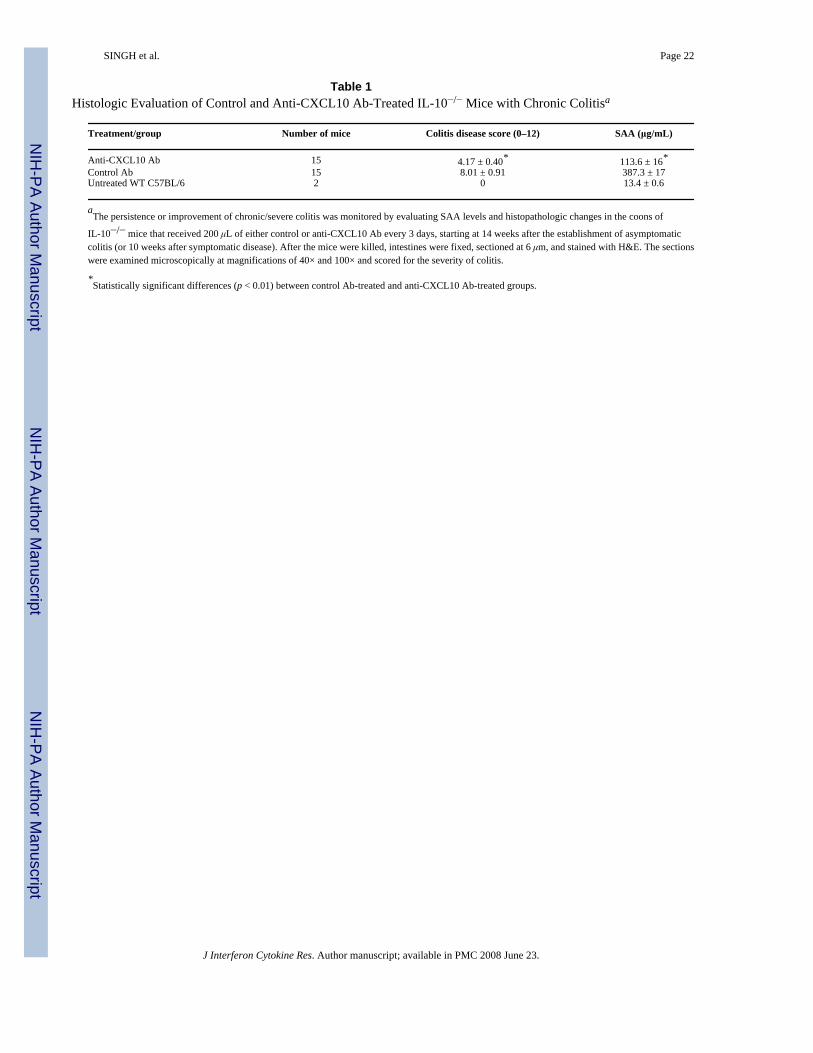
Changes in colitis severity following CXCL10 blockadeThe mice that received anti-CXCL10 Ab showed a significant reduction in intestinalinflammation. An increase in leukocyte infiltrates (Fig. 4A) and distortion of glandulararchitecture (Fig. 4B) were observed in the intestines during chronic colitis. Anti-CXCL10 Abreduced the lymphocyte infiltration and partially restored the glandular and goblet cellarchitecture (Fig. 4C), which also coincided with lengthening of intestinal crypts (Fig. 4D).The mean histologic scores of IL-10−/− mice with severe colitis that received control Ab weresignificantly higher than the scores of mice treated with anti-CXCL10 Ab (Table 1). Similarly,SAA levels correlated with the severity of colitis as determined by histologic analysis.Pathologic changes included leukocyte infiltrates in the LP of the colon of control Ab-treatedIL-10−/− mice, with the number of these infiltrates being reduced after CXCL10 blockade.Taken together, the results show a marked improvement in the characteristic intestinalinflammation associated with chronic colitis after CXCL10 blockade.
Characteristics of Th cells during chronic colitis and changes after CXCL10 inhibitionFlow cytometry analysis revealed that CD4+ T cells from the MLNs express CXCL10 duringchronic colitis. CXCL10 blockade in IL-10−/− mice with chronic colitis reduced the percentageof total CXCL10+ CD4+ and CXCR3+ CD4+ cells in the MLNs and the number of similar cellsin the LP (Fig. 5A). Moreover, the number of CD4+ T cell infiltrates expressing CXCL10 andCXCR3 in the spleen, MLNs, PPs, and LP of IL-10−/− mice with chronic colitis wassignificantly reduced following CXCL10 blockade compared with similar mice treated withcontrol Ab. We also observed a significant decline in the total number of splenic, MLN, PP,and LP CD4+ T cells expressing IFN-γ or TNF-α or both in mice treated with anti-CXCL10Ab compared with mice treated with control Ab (Fig. 5B).
CXCR3, TNF-α, IFN-γ, and CXCL10 expression by NK and NKT cellsThe mean fluorescence intensity of NK cells that expressed CXCL10 as well as CXCR3, IFN-γ, and TNF-α was elevated during chronic colitis, but this intensity of expression declined tonear background levels after anti-CXCL10 Ab treatment (data not shown). CXCL10 blockadealso reduced the number of splenic NK cells that expressed CXCL10, CXCR3, and IFN-γduring chronic colitis (Fig. 6A). The numbers of CXCL10+ and CXCR3+ NK cell populationsin MLN did not vary during chronic colitis compared with cell numbers from similar micebefore the onset of colitis. Following CXCL10 blockade, more NK cells than other cells fromthe MLN expressed IFN-γ. The number of TNF-α+-expressing NK cells was also elevatedduring severe/chronic colitis, but their number was significantly decreased following anti-
SINGH et al. Page 5
J Interferon Cytokine Res. Author manuscript; available in PMC 2008 June 23.
NIH
-PA Author Manuscript
NIH
-PA Author Manuscript
NIH
-PA Author Manuscript
CXCL10 Ab treatment. Similarly, the increase compared with normal controls in the numbersof CXCL10+, CXCR3+, IFN-γ+, and/or TNF-α+ NK cells from the PP was significantlydecreased after CXCL10 blockade.
In contrast to NK cells, the number of splenic NKT cells that expressed CXCL10, CXCR3, orIFN-γ was unchanged during chronic colitis, but CXCL10 blockade reduced the number ofsplenic TNF-α+ NKT cells that were increased during this disease (Fig. 6B). NKT cells fromMLN expressed TNF-α during CXCL10 blockade in IL-10−/− mice with chronic colitis. Oneof the most striking decreases in the number of these inflammatory cells occurred in PP-derivedCXCR3+ and TNF-α+ NKT cells following CXCL10 blockade.
Our data suggested that CD4+ T cells were a source of IFN-γ in the LP during severe colitis,and the numbers of IFN-γ+-expressing as well as TNF-α+ expressing NK and NKT cells wereelevated in the LP during severe colitis compared with the negative controls. CXCL10 blockadereduced the number of these cells that were isolated from this inflamed site. Although thenumber of CXCR3+ NK and NKT cells from the LP remained relatively unchanged, the numberof CXCL10+ NK cells from the LP was significantly reduced following CXCL10 blockade. Itis important to note that there were as many as 3-fold more CD4+ T cells and NKT cells thanNK cells in the LP during chronic colitis. Considered together, these results indicate thatCXCL10 blockade modulates the number of systemic and mucosal NKT and NK cellsexpressing CXCL10, CXCR3, TNF-α, and IFN-γ. Notably, NKT and CD4+ T cells appear tobe sources of IFN-γ and TNF-α, whereas NK and CD4+ T cells express more CXCL10 duringcolitis, which is abrogated by CXCL10 inhibition.
Alteration of CXCR3, TNF-α, IFN-γ, IL-12p40, and CXCL10 expression by DCsCXCR3 was differentially expressed by DCs from the MLN and LP during colitis (Fig. 7A).The intensity of CXCR3 expression by MLN DCs from mice with chronic colitis and anti-CXCL10 Ab-treated mice was significantly higher than that of similar cells from control Ab-treated mice with disease. In contrast, CXCR3 expression by LP DCs during chronic colitis,along with CXCL10 blockade, was significantly lower than that by similar cells from micereceiving control Ab during disease. CXCL10 blockade attenuated the number of CXCR3+
DCs in the LP and spleen of IL-10−/− mice with chronic colitis, while significantly increasingthe number of CXCR3+ DCs in MLNs and PPs, back to the normal observed in controls (Fig.7B). Mucosal DCs from IL-10−/− mice did not appear to express significant levels of CXCL10,but the numbers of CXCR3+ DCs in effector (LP) vs. inductive (MLN and PP) mucosal siteswere reciprocally modulated by CXCL10 blockade.
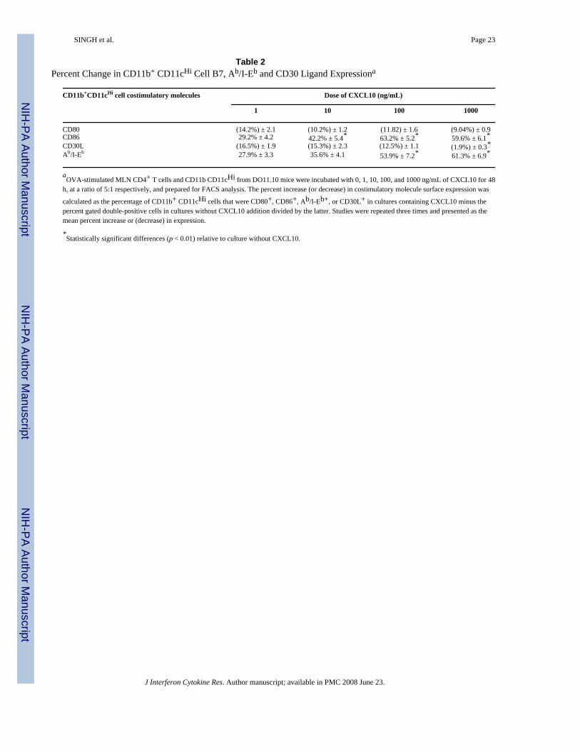
CXCL10 modulation of costimulatory molecule expression by DCsWe have shown previously that CXCR3 ligands positively modulate CD28 expression byantigen-stimulated CD4+ T cells (Singh and others 2003b). LP and immature DCs were inducedto rapid maturation and CD86 expression after entering the MLN (Huang and others 2000).To further establish the CXCL10-mediated effects on the regulation of DC costimulatorymolecules, we assessed the potential of CXCL10 to modulate the expression of Ab/I-Eb, CD80,CD86, and CD30L by DCs during class II-restricted antigen stimulation of MLN leukocytes.CXCL10 significantly increased the expression of class II (Ab/I-Eb) and CD86, but not CD80,by DCs during antigen stimulation in a dose-dependent fashion (Table 2 and Fig. 8). CXCL10also significantly downregulated the expression of CD30L by similar DCs, which supports thenotion that this CXCR3 ligand modulates DC activation or maturation or both.
SINGH et al. Page 6
J Interferon Cytokine Res. Author manuscript; available in PMC 2008 June 23.
NIH
-PA Author Manuscript
NIH
-PA Author Manuscript
NIH
-PA Author Manuscript
DISCUSSIONOur previous studies demonstrated that CXCL10 blockade abrogated spontaneous colitis inIL-10−/− mice (Singh and others 2003a), which is predominantly mediated by Th1-type αβ Tcell receptor (TCR+) and CXCR3+ cells (Singh and others 2003b). In confirmation, it wasshown recently that anti-CXCL10 Ab treatment can mitigate colitis in IL-10−/− mice bydecreasing the trafficking of Th1 cells (Hyun and others 2005). We hypothesized that blockingCXCL10-CXCR3 interactions alleviates chronic colitis in IL-10−/− mice by negativelymodulating Th1 cells as well as DCs, NK cells, and NKT cells. We show that anti-CXCL10Ab treatment resolves chronic colitis, corrects weight loss, and reduces local and systemiclevels of IL-1α, IL-1β, IL-6, SAA, IL-12p40, IFN-γ, and TNF-α in IL-10−/− mice with chroniccolitis. Our results suggest that both systemic and mucosal CD4+ T cells, NKT cells, and NKcells differentially express IFN-γ, TNF-α, CXCL10, or CXCR3, which can be mitigated byCXCL10 blockade. We also show that although DCs do not significantly express CXCL10,IFN-γ, and TNF-α during chronic colitis, CXCR3+ DCs are elevated in the spleen as well asin LP and are diminished at mucosal inductive sites (i.e., the PP and MLN) during severe colitis.It is also important to mention that there are significant difference between WT and IL-10−/−
mice. In previous studies by our laboratories and others, the numbers of CD4+ T cells weregreater in IL-10−/− mice than in WT B6 mice. Indeed, anti-CXCL10 Ab did not significantlyalter leukocyte populations in WT mice housed under germ-free conditions. However,IL-10−/− mice, but not WT B6 mice, housed under conventional housing undergo relativelyrapid immune dysregulation in part due to CXCL10-dependent mechanisms that are susceptibleto inhibition (Singh and others 2003a).
The association among CXCL10, CXCR3, and Th1-dependent immunity has been observedin several models of inflammatory diseases (Qin and others 1998). Several IBD models indicatethat CD4+ T cells play a major role in the induction of IBD, and much of the intestinal damageof this disease is a result of T cell-mediated injury (Elson and others 1996). To this end, wehave shown that the adoptive transfer of CXCR3+ CD4+ T cells results in colitis in TCR (β ×δ)−/− mice (Singh and others 2003b). Our results confirm that the number of CD4+ T cells inthe spleen, MLN, and PP represents the majority of lymphocytes that express inflammatorycytokines during colitis.
Along with TNF-α, sustained acute-phase responses are associated with both human IBD andmurine colitis (Berg and others 1996). SAA is elevated during CD (De Beer and others1982), activated macrophages increase IL-1α and IL-1β expression in patients with IBD(Casini-Raggi and others 1995), and IL-12, IL-23 (with IL-12p40 subunit), and IFN-γ playcritical roles in the induction and progression of colitis (Neurath and others 1995; Parronchiand others 1997). In the present study, we demonstrate that local and systemic TNF-α, IL-1α,IL-1β, IL-12p40, and IFN-γ expression is decreased by anti-CXCL10 Ab treatment inIL-10−/− mice with chronic colitis. It seems likely that NK cell interactions might serve toinitiate these factors to contribute to Th1-mediated colitis. We observed a decline in the numberof NK and NKT cells in the MLNs, PP, and LP lymphocytes after CXCL10 blockade comparedwith similar mice treated with control Ab. In addition, the expression of CXCL10, CXCR3,TNF-α, and IFN-γ by NK cells declined after CXCL10 treatment when compared with thecontrol Ab-treated mice. Hence, it is also possible that the Th1 imbalance in IL-10−/− mice ismodulated by NK cells during colitis.
In addition to LP CD4+ T cells and NK cells, we noted that NKT cells from the LP expressTNF-α. This NK cell subset represented the predominant leukocyte that expressed IFN-γ andCXCL10 during chronic colitis. However, NKT cells have been shown to contribute to thesuppression of Th1-mediated colitis (Wei and others 2005; Shibolet and others 2003;Menachem and others 2005). Future studies, possibly using NK cell depletion or CD1−/− ×
SINGH et al. Page 7
J Interferon Cytokine Res. Author manuscript; available in PMC 2008 June 23.
NIH
-PA Author Manuscript
NIH
-PA Author Manuscript
NIH
-PA Author Manuscript
IL-10−/− mice, will be needed to precisely and decisively demonstrate the role of NKT and NKcells in the initiation and maintenance of colitis.
Although we have shown that CXCR3+ CD4+ T, NK, and NKT cells are sources of CXCL10and inflammatory cytokines during chronic/severe colitis, it is also likely that other cells areequally responsible for the increase in inflammatory cytokines during colitis. CXCL10 hasbeen reported to be produced by other cell types, particularly epithelial cells (Sauty and others1999; Spurrell and otherse 2005) and neutrophils (Singh and others 2007a). Hence, the cellularmechanisms responsible for the differentiation of naïve CD4+ T cells, in this model, are stillin question, as neither mucosal inductive DCs (PP or MLN) nor effector DCs (LP) producedsignificant levels of IL-12. The heavily Th1-biased IL-10-deficient mouse model may not bethe best to determine the exact mechanism of Th1 polarization that occurs in CD or murinecolitis. Presumably, the IL-12p40+ DCs in the spleen and IL-12 and IFN-γ in the peripheryaided in Th1 differentiation.
Our current findings also indicate that CXCR3+ (and often TNF-α+) DCs predominantlypopulate the LP during chronic colitis. CXCL10 blockade reduced colitis severity and loweredthe number of CXCR3+ DCs in the spleen and LP and increased the number of DCs in MLNsand PPs to levels observed before the onset of disease. Depending on their activation state ormaturation, DCs may express CCR1, CCR2, CCR5, CCR6, CXCR2, and CXCR3 (Dieu andothers 1998; Sozzani and others 1998). Hence, the alteration in the number of DCs in mucosalinductive and effector sites during chronic colitis after CXCL10 blockade might be due to theupregulation of other chemokines and corresponding receptors that govern the trafficking ofDCs to lymphoid organs (e.g., MLNs and PPs). For example, CCL20 and CCR6 have beenshown to play a distinct role in PP DC subsets trafficking to MLNs (Iwasaki and Kelsall1999).
Whereas immature DCs can express CCL3 and CCL5, mature DCs selectively express CCL2,CCL5, and CXCL10 upon activation (Foti and others 1999). CCL5, like CXCL10, could alsoparticipate in Th1 cell recruitment via CCR5 interactions. In the present study, however, DCsmarginally expressed CXCL10 during chronic colitis. In a Mycobacterium aviumparatuberculosis model of colitis, we recently observed that DCs express significant levels ofCXCL11 (Singh and others 2007a), which would support CXCR3+ cell chemotaxis. Theprominent increase in CXCL10+ CXCR3+ T cells, NK cells, and NKT cells in conjunctionwith CXCR3+ DCs suggests that these cellular mechanisms are required for the initiation andmaintenance of severe/ chronic colitis.
We have shown that CXCL9, CXCL10, and CXCL11 significantly increase CD28 expressionby CD3ε-stimulated CD4+ T cells (Singh and others 2003b). Other chemokines, for example,CCL3, CCL4, and CCL5, also can upregulate CD86 expression (Lillard and others 2001; DelPrete and others 1995). In this study, CXCL10 downregulated CD30L expression by DCsduring OVA stimulation of DO11.10 MLN lymphocytes, which would simultaneously removean important signal for Th2 cell differentiation (Del Prete and others 1995). Further, CXCL10enhanced Ab/I-Eb expression by DCs in dose-dependent fashion. This coincides with reportsthat activated T cells could stimulate and mature APCs (Avice and others 1999; Andreae andothers 2002). We present a scenario in which TCR-Ab/I-Eb and CD28-CD86 interactions areenhanced and CD30-CD30L interactions are reduced by the abundance of CXCL10 duringcolitis, which would activate antigen-stimulated naïve T cells to develop into Th1 cells. Thesefindings illustrate possible mechanisms of CXCL10-mediated activation of DCs for subsequentexpansion and differentiation of CD4+ T cells that propagate colitis. Considered together, ourresults indicate that CXCR3 and CXCL10 interactions are physiologically and pathologicallyimportant for the regulation of colitis.
SINGH et al. Page 8
J Interferon Cytokine Res. Author manuscript; available in PMC 2008 June 23.
NIH
-PA Author Manuscript
NIH
-PA Author Manuscript
NIH
-PA Author Manuscript
Acknowledgements
The content of this paper benefited from many fruitful conversations with colleagues at the Morehouse School ofMedicine, the National Institutes on Aging, the University of Alabama at Birmingham, and the University of Louisville,as well as editing by Andrew Marsh. This work was supported in part by the Crohn’s & Colitis Foundation of America,National Institutes of Health grants RR03034, GM08248, MD000525, and AI57808, the Southeast Center forEmerging Biologic Threats, and the University of Louisville.
ReferencesAndreae S, Piras F, Burdin N, Triebel F. Maturation and activation of dendritic cells induced by
lymphocyte activation gene-3 (CD223). J Immunol 2002;168:3874–3880. [PubMed: 11937541]Avice MN, Sarfati M, Triebel F, Delespesse G, Demeure CE. Lymphocyte activation gene-3, a MHC
class II ligand expressed on activated T cells, stimulates TNF-alpha and IL-12 production bymonocytes and dendritic cells. J Immunol 1999;162:2748–2753. [PubMed: 10072520]
Berg DJ, Davidson N, Kuhn R, Muller W, Menon S, Holland G, Thompson-Snipes L, Leach MW,Rennick D. Enterocolitis and colon cancer in interleukin-10-deficient mice are associated with aberrantcytokine production and CD4(+) Th1-like responses. J Clin Invest 1996;98:1010–1020. [PubMed:8770874]
Casini-Raggi V, Kam L, Chong YJ, Fiocchi C, Pizarro TT, Cominelli F. Mucosal imbalance of IL-1 andIL-1 receptor antagonist in inflammatory bowel disease. A novel mechanism of chronic intestinalinflammation. J Immunol 1995;154:2434–2440. [PubMed: 7868909]
De Beer FC, Mallya RK, Fagan EA, Lanham JG, Hughes GR, Pepys MB. Serum amyloid-A proteinconcentration in inflammatory diseases and its relationship to the incidence of reactive systemicamyloidosis. Lancet 1982;2:231–234. [PubMed: 6124669]
Del Prete G, De Carli M, Almerigogna F, Daniel CK, D’Elios MM, Zancuoghi G, Vinante F, Pizzolo G,Romagnani S. Preferential expression of CD30 by human CD4+ T cells producing Th2-type cytokines.FASEB J 1995;9:81–86. [PubMed: 7821763]
Dieu MC, Vanbervliet B, Vicari A, Bridon JM, Oldham E, Ait-Yahia S, Briere F, Zlotnik A, LebecqueS, Caux C. Selective recruitment of immature and mature dendritic cells by distinct chemokinesexpressed in different anatomic sites. J Exp Med 1998;188:373–386. [PubMed: 9670049]
Elson CO, Beagley KW, Sharmanov AT, Fujihashi K, Kiyono H, Tennyson GS, Cong Y, Black CA,Ridwan BW, McGhee JR. Hapten-induced model of murine inflammatory bowel disease: mucosaimmune responses and protection by tolerance. J Immunol 1996;157:2174–2185. [PubMed: 8757344]
Farber JM. Mig and IP-10—CXC chemokines that target lymphocytes. J Leukoc Biol 1997;61:246–257.[PubMed: 9060447]
Fiocchi C. Inflammatory bowel disease: etiology and pathogenesis. Gastroenterology 1998;115:182–205.[PubMed: 9649475]
Foti M, Granucci F, Aggujaro D, Liboi E, Luini W, Minardi S, Mantovani A, Sozzani S, Ricciardi-Castagnoli P. Upon dendritic cell (DC) activation chemokines and chemokine receptor expressionare rapidly regulated for recruitment and maintenance of DC at the inflammatory site. Int Immunol1999;11:979–986. [PubMed: 10360972]
Hollander GA, Simpson SJ, Mizoguchi E, Nichogiannopoulou A, She J, Gutierrez-Ramos JC, Bhan AK,Burakoff SJ, Wang B, Terhorst C. Severe colitis in mice with aberrant thymic selection. Immunity1995;3:27–38. [PubMed: 7621076]
Huang FP, Platt N, Wykes M, Major JR, Powell TJ, Jenkins CD, MacPherson GG. A discretesubpopulation of dendritic cells transports apoptotic intestinal epithelial cells to T cell areas ofmesenteric lymph nodes. J Exp Med 2000;191:435–444. [PubMed: 10662789]
Hyun JG, Lee G, Brown JB, Grimm GR, Tang Y, Mittal N, Dirisina R, Zhang Z, Fryer JP, WeinstockJV, Luster AD, Barrett TA. Anti-interferon-inducible chemokine, CXCL10, reduces colitis byimpairing T helper-1 induction and recruitment in mice. Inflamm Bowel Dis 2005;11:799–805.[PubMed: 16116313]
Iwasaki A, Kelsall BL. Freshly isolated Peyer’s patch, but not spleen, dendritic cells produce interleukin10 and induce the differentiation of T helper type 2 cells. J Exp Med 1999;190:229–239. [PubMed:10432286]
SINGH et al. Page 9
J Interferon Cytokine Res. Author manuscript; available in PMC 2008 June 23.
NIH
-PA Author Manuscript
NIH
-PA Author Manuscript
NIH
-PA Author Manuscript
Kuhn R, Lohler J, Rennick D, Rajewsky K, Muller W. Interleukin-10-deficient mice develop chronicenterocolitis. Cell 1993;75:263–274. [PubMed: 8402911]
Lillard JW Jr, Boyaka PN, Taub DD, McGhee JR. RANTES potentiates antigen-specific mucosal immuneresponses. J Immunol 2001;166:162–169. [PubMed: 11123289]
Lillard JW Jr, Singh UP, Boyaka PN, Singh S, Taub DD, McGhee JR. MIP-1alpha and MIP-1betadifferentially mediate mucosal and systemic adaptive immunity. Blood 2003;101:807–814.[PubMed: 12393512]
Luster AD, Ravetch JV. Biochemical characterization of a gamma interferon-inducible cytokine (IP-10).J Exp Med 1987;166:1084–1097. [PubMed: 2443596]
MacDonald TT, Pettersson S. Bacterial regulation of intestinal immune responses. Inflamm Bowel Dis2000;6:116–122. [PubMed: 10833071]
Menachem YT, Kolker O, Shibolet O, Alper R, Nagler A, Ilan Y. Adoptive transfer of NK 1.1+
lymphocytes in immune-mediated colitis: a pro-inflammatory or a tolerizing subgroup of cells?Microb Infect 2005;7:825–835.
Mombaerts P, Mizoguchi E, Grusby MJ, Glimcher LH, Bhan AK, Tonegawa S. Spontaneousdevelopment of inflammatory bowel disease in T cell receptor mutant mice. Cell 1993;75:274–282.[PubMed: 8104709]
Neurath MF, Fuss I, Kelsall BL, Stuber E, Strober W. Antibodies to interleukin 12 abrogate establishedexperimental colitis in mice. J Exp Med 1995;182:1281–1290. [PubMed: 7595199]
Parronchi P, Romagnani P, Annunziato F, Sampognaro S, Becchio A, Giannarini L, Maggi E, Pupilli C,Tonelli F, Romagnani S. Type 1 T-helper cell predominance and interleukin-12 expression in the gutof patients with Crohn’s disease. Am J Pathol 1997;150:823–832. [PubMed: 9060820]
Podolsky DK. The current future understanding of inflammatory bowel disease. Best Prac Res ClinGastroenterol 2002;16:933–943.
Qin S, Rottman JB, Myers P, Kassam N, Weinblatt M, Loetscher M, Koch AE, Moser B, Mackay CR.The chemokine receptors CXCR3 and CCR5 mark subsets of T cells associated with certaininflammatory reactions. J Clin Invest 1998;101:746–754. [PubMed: 9466968]
Romagnani S. Induction of Th1 and Th2 responses: a key role for the “natural” immune response?Immunol Today 1992;13:379–381. [PubMed: 1418371]
Sauty A, Dziejman M, Taha RA, Iarossi AS, Neote K, Garcia-Zepeda EA, Hamid Q, Luster AD. The Tcell-specific CXC chemokines IP-10, Mig, and I-TAC are expressed by activated human bronchialepithelial cells. J Immunol 1999;162:3549–3558. [PubMed: 10092813]
Shibolet O, Alper R, Zlotogarov L, Thalenfeld B, Engelhardt D, Rabbani E, Ilan Y. NKT and CD8lymphocytes mediate suppression of hepatocellular carcinoma growth via tumor antigen-pulseddendritic cells. Int J Cancer 2003;106:236–243. [PubMed: 12800200]
Singh UP, Singh S, Singh R, Karls RK, Quinn RD, Potter ME, Lillard JW Jr. Influence of Mycobacteriumavium subsp. paratuberculosis on colitis development and specific immune responses duringdiseasse. Infect Immun 2007a;75:3722–2728. [PubMed: 17502388]
Singh UP, Singh S, Taub DD, Lillard JW Jr. Inhibition of IFN-gamma-inducible protein-10 abrogatescolitis in IL-10(−/−) mice. J Immunol 2003a;171:1401–1406. [PubMed: 12874231]
Singh UP, Singh S, Weaver CT, Iqbal N, McGhee JR, Lillard JW Jr. IFN-α-inducible chemokines enhanceadaptive immunity and acolitis. J Interferon Cytokine Res 2003b;23:591–600. [PubMed: 14585199]
Singh UP, Venkataraman C, Singh R, Lillard JW Jr. CXCR3 axis: role in inflammatory bowel diseaseand its therapeutic implication. Endocr Metab Immune Disord Drug Targets 2007b;7:111–123.[PubMed: 17584151]
Sozzani S, Allavena P, D’Amico G, Luini W, Bianchi G, Kataura M, Imai T, Yoshie O, Bonecchi R,Mantovani A. Differential regulation of chemokine receptors during dendritic cell maturation: amodel for their trafficking properties. J Immunol 1998;161:1083–1086. [PubMed: 9686565]
Spurrell JC, Wiehler S, Zaheer RS, Sanders SP, Proud D. Human airway epithelial cells produce IP-10(CXCL10) in vitro and in vivo upon rhinovirus infection. Am J Physiol Lung Cell Mol Physiol2005;289:L85–95. [PubMed: 15764644]
Wei B, Velazquez P, Turovskaya O, Spricher K, Aranda R, Kronenberg M, Birnbaumer L, Braun J.Mesenteric B cells centrally inhibit CD4+ T cell colitis through interaction with regulatory T cellsubsets. Proc Natl Acad Sci USA 2005;102:2010–2015. [PubMed: 15684084]
SINGH et al. Page 10
J Interferon Cytokine Res. Author manuscript; available in PMC 2008 June 23.
NIH
-PA Author Manuscript
NIH
-PA Author Manuscript
NIH
-PA Author Manuscript
Yuan YH, ten Hove T, The FO, Slors JF, van Deventer SJ, te Velde AA. Chemokine receptor CXCR3expression in inflammatory bowel disease. Inflamm Bowel Dis 2001;7:281–286. [PubMed:11720316]
SINGH et al. Page 11
J Interferon Cytokine Res. Author manuscript; available in PMC 2008 June 23.
NIH
-PA Author Manuscript
NIH
-PA Author Manuscript
NIH
-PA Author Manuscript
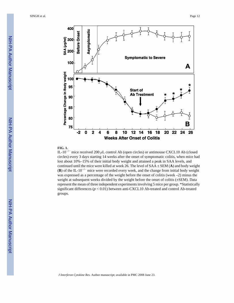
FIG. 1.IL-10−/− mice received 200 μL control Ab (open circles) or antimouse CXCL10 Ab (closedcircles) every 3 days starting 14 weeks after the onset of symptomatic colitis, when mice hadlost about 10%–15% of their initial body weight and attained a peak in SAA levels, andcontinued until the mice were killed at week 26. The level of SAA ± SEM (A) and body weight(B) of the IL-10−/− mice were recorded every week, and the change from initial body weightwas expressed as a percentage of the weight before the onset of colitis (week –2) minus theweight at subsequent weeks divided by the weight before the onset of colitis (±SEM). Datarepresent the mean of three independent experiments involving 5 mice per group. *Statisticallysignificant differences (p < 0.01) between anti-CXCL10 Ab-treated and control Ab-treatedgroups.
SINGH et al. Page 12
J Interferon Cytokine Res. Author manuscript; available in PMC 2008 June 23.
NIH
-PA Author Manuscript
NIH
-PA Author Manuscript
NIH
-PA Author Manuscript
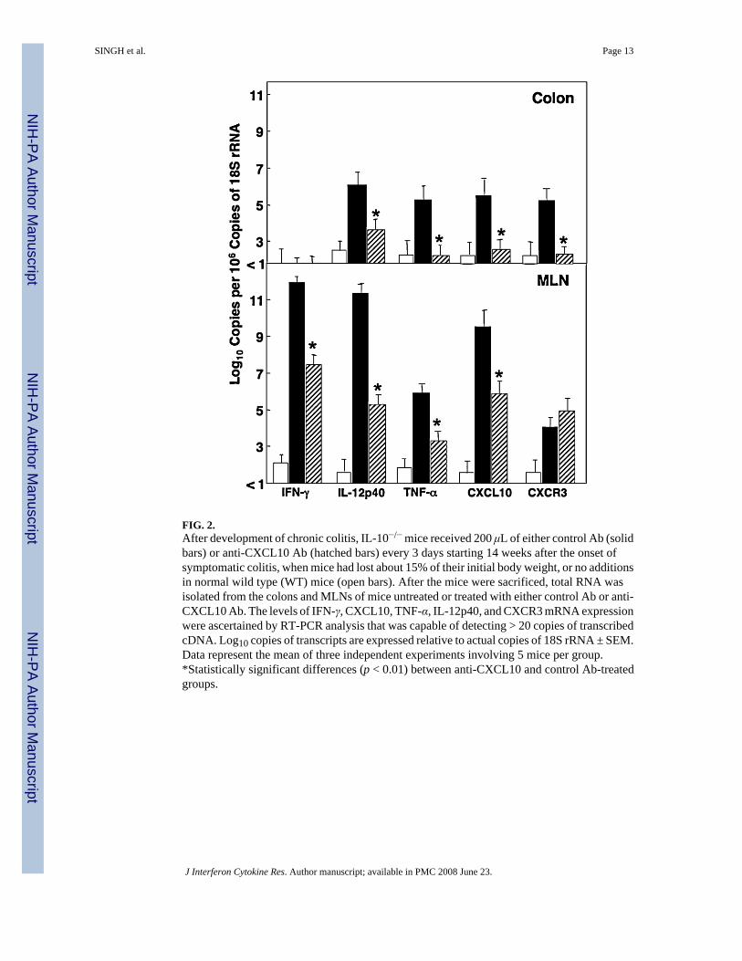
FIG. 2.After development of chronic colitis, IL-10−/− mice received 200 μL of either control Ab (solidbars) or anti-CXCL10 Ab (hatched bars) every 3 days starting 14 weeks after the onset ofsymptomatic colitis, when mice had lost about 15% of their initial body weight, or no additionsin normal wild type (WT) mice (open bars). After the mice were sacrificed, total RNA wasisolated from the colons and MLNs of mice untreated or treated with either control Ab or anti-CXCL10 Ab. The levels of IFN-γ, CXCL10, TNF-α, IL-12p40, and CXCR3 mRNA expressionwere ascertained by RT-PCR analysis that was capable of detecting > 20 copies of transcribedcDNA. Log10 copies of transcripts are expressed relative to actual copies of 18S rRNA ± SEM.Data represent the mean of three independent experiments involving 5 mice per group.*Statistically significant differences (p < 0.01) between anti-CXCL10 and control Ab-treatedgroups.
SINGH et al. Page 13
J Interferon Cytokine Res. Author manuscript; available in PMC 2008 June 23.
NIH
-PA Author Manuscript
NIH
-PA Author Manuscript
NIH
-PA Author Manuscript
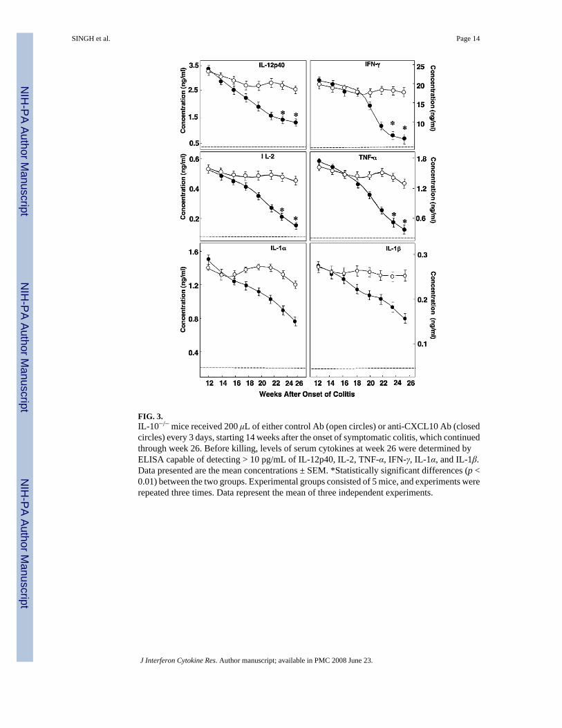
FIG. 3.IL-10−/− mice received 200 μL of either control Ab (open circles) or anti-CXCL10 Ab (closedcircles) every 3 days, starting 14 weeks after the onset of symptomatic colitis, which continuedthrough week 26. Before killing, levels of serum cytokines at week 26 were determined byELISA capable of detecting > 10 pg/mL of IL-12p40, IL-2, TNF-α, IFN-γ, IL-1α, and IL-1β.Data presented are the mean concentrations ± SEM. *Statistically significant differences (p <0.01) between the two groups. Experimental groups consisted of 5 mice, and experiments wererepeated three times. Data represent the mean of three independent experiments.
SINGH et al. Page 14
J Interferon Cytokine Res. Author manuscript; available in PMC 2008 June 23.
NIH
-PA Author Manuscript
NIH
-PA Author Manuscript
NIH
-PA Author Manuscript
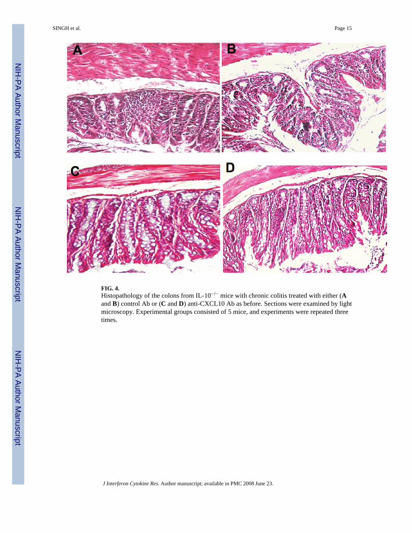
FIG. 4.Histopathology of the colons from IL-10−/− mice with chronic colitis treated with either (Aand B) control Ab or (C and D) anti-CXCL10 Ab as before. Sections were examined by lightmicroscopy. Experimental groups consisted of 5 mice, and experiments were repeated threetimes.
SINGH et al. Page 15
J Interferon Cytokine Res. Author manuscript; available in PMC 2008 June 23.
NIH
-PA Author Manuscript
NIH
-PA Author Manuscript
NIH
-PA Author Manuscript
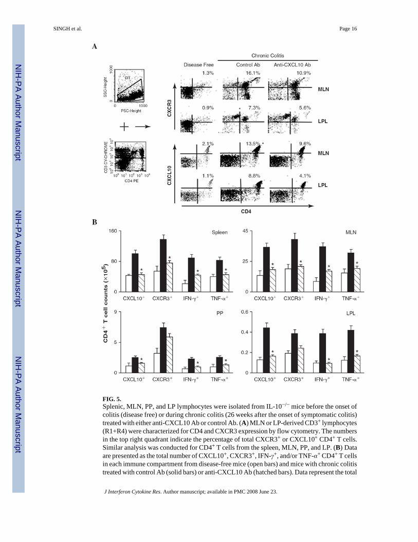
FIG. 5.Splenic, MLN, PP, and LP lymphocytes were isolated from IL-10−/− mice before the onset ofcolitis (disease free) or during chronic colitis (26 weeks after the onset of symptomatic colitis)treated with either anti-CXCL10 Ab or control Ab. (A) MLN or LP-derived CD3+ lymphocytes(R1+R4) were characterized for CD4 and CXCR3 expression by flow cytometry. The numbersin the top right quadrant indicate the percentage of total CXCR3+ or CXCL10+ CD4+ T cells.Similar analysis was conducted for CD4+ T cells from the spleen, MLN, PP, and LP. (B) Dataare presented as the total number of CXCL10+, CXCR3+, IFN-γ+, and/or TNF-α+ CD4+ T cellsin each immune compartment from disease-free mice (open bars) and mice with chronic colitistreated with control Ab (solid bars) or anti-CXCL10 Ab (hatched bars). Data represent the total
SINGH et al. Page 16
J Interferon Cytokine Res. Author manuscript; available in PMC 2008 June 23.
NIH
-PA Author Manuscript
NIH
-PA Author Manuscript
NIH
-PA Author Manuscript
number of cells ± SEM from three independent experiments involving 5 mice per group.Statistically significant differences (p < 0.01) between anti-CXCL10-treated and control Ab-treated groups.
SINGH et al. Page 17
J Interferon Cytokine Res. Author manuscript; available in PMC 2008 June 23.
NIH
-PA Author Manuscript
NIH
-PA Author Manuscript
NIH
-PA Author Manuscript
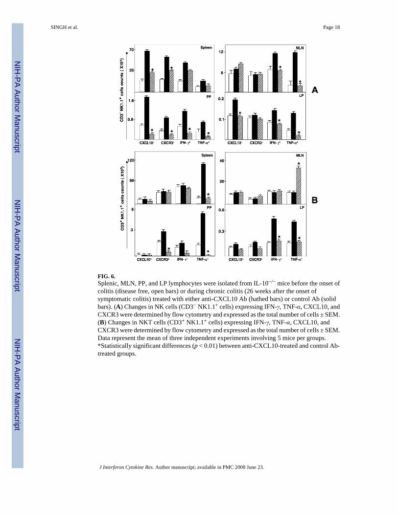
FIG. 6.Splenic, MLN, PP, and LP lymphocytes were isolated from IL-10−/− mice before the onset ofcolitis (disease free, open bars) or during chronic colitis (26 weeks after the onset ofsymptomatic colitis) treated with either anti-CXCL10 Ab (hathed bars) or control Ab (solidbars). (A) Changes in NK cells (CD3− NK1.1+ cells) expressing IFN-γ, TNF-α, CXCL10, andCXCR3 were determined by flow cytometry and expressed as the total number of cells ± SEM.(B) Changes in NKT cells (CD3+ NK1.1+ cells) expressing IFN-γ, TNF-α, CXCL10, andCXCR3 were determined by flow cytometry and expressed as the total number of cells ± SEM.Data represent the mean of three independent experiments involving 5 mice per groups.*Statistically significant differences (p < 0.01) between anti-CXCL10-treated and control Ab-treated groups.
SINGH et al. Page 18
J Interferon Cytokine Res. Author manuscript; available in PMC 2008 June 23.
NIH
-PA Author Manuscript
NIH
-PA Author Manuscript
NIH
-PA Author Manuscript
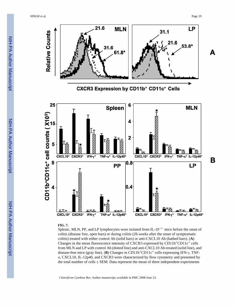
FIG. 7.Splenic, MLN, PP, and LP lymphocytes were isolated from IL-10−/− mice before the onset ofcolitis (disease free, open bars) or during colitis (26 weeks after the onset of symptomaticcolitis) treated with either control Ab (solid bars) or anti-CXCL10 Ab (hathed bars). (A)Changes in the mean fluorescence intensity of CXCR3 expressed by CD11b+CD11c+ cellsfrom MLN and LP with control Ab (dotted line) and anti-CXCL10 Ab-treated (solid line), anddisease-free mice (gray line). (B) Changes in CD11b+CD11c+ cells expressing IFN-γ, TNF-α, CXCL10, IL-12p40, and CXCR3 were characterized by flow cytometry and presented bythe total number of cells ± SEM. Data represent the mean of three independent experiments
SINGH et al. Page 19
J Interferon Cytokine Res. Author manuscript; available in PMC 2008 June 23.
NIH
-PA Author Manuscript
NIH
-PA Author Manuscript
NIH
-PA Author Manuscript
involving 10 mice per group. *Statistically significant differences (p < 0.01) between anti-CXCL10 Ab-treated and control Ab-treated groups.
SINGH et al. Page 20
J Interferon Cytokine Res. Author manuscript; available in PMC 2008 June 23.
NIH
-PA Author Manuscript
NIH
-PA Author Manuscript
NIH
-PA Author Manuscript
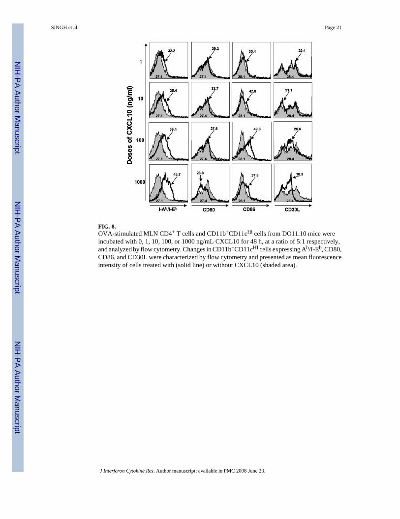
FIG. 8.OVA-stimulated MLN CD4+ T cells and CD11b+CD11cHi cells from DO11.10 mice wereincubated with 0, 1, 10, 100, or 1000 ng/mL CXCL10 for 48 h, at a ratio of 5:1 respectively,and analyzed by flow cytometry. Changes in CD11b+CD11cHI cells expressing Ab/I-Eb, CD80,CD86, and CD30L were characterized by flow cytometry and presented as mean fluorescenceintensity of cells treated with (solid line) or without CXCL10 (shaded area).
SINGH et al. Page 21
J Interferon Cytokine Res. Author manuscript; available in PMC 2008 June 23.
NIH
-PA Author Manuscript
NIH
-PA Author Manuscript
NIH
-PA Author Manuscript
NIH
-PA Author Manuscript
NIH
-PA Author Manuscript
NIH
-PA Author Manuscript
SINGH et al. Page 22
Table 1Histologic Evaluation of Control and Anti-CXCL10 Ab-Treated IL-10−/− Mice with Chronic Colitisa
Treatment/group Number of mice Colitis disease score (0–12) SAA (μg/mL)
Anti-CXCL10 Ab 15 4.17 ± 0.40* 113.6 ± 16*Control Ab 15 8.01 ± 0.91 387.3 ± 17Untreated WT C57BL/6 2 0 13.4 ± 0.6
aThe persistence or improvement of chronic/severe colitis was monitored by evaluating SAA levels and histopathologic changes in the coons of
IL-10−/− mice that received 200 μL of either control or anti-CXCL10 Ab every 3 days, starting at 14 weeks after the establishment of asymptomaticcolitis (or 10 weeks after symptomatic disease). After the mice were killed, intestines were fixed, sectioned at 6 μm, and stained with H&E. The sectionswere examined microscopically at magnifications of 40× and 100× and scored for the severity of colitis.
*Statistically significant differences (p < 0.01) between control Ab-treated and anti-CXCL10 Ab-treated groups.
J Interferon Cytokine Res. Author manuscript; available in PMC 2008 June 23.
NIH
-PA Author Manuscript
NIH
-PA Author Manuscript
NIH
-PA Author Manuscript
SINGH et al. Page 23
Table 2Percent Change in CD11b+ CD11cHi Cell B7, Ab/I-Eb and CD30 Ligand Expressiona
CD11b+CD11cHi cell costimulatory molecules Dose of CXCL10 (ng/mL)
1 10 100 1000
CD80 (14.2%) ± 2.1 (10.2%) ± 1.2 (11.82) ± 1.6 (9.04%) ± 0.9CD86 29.2% ± 4.2 42.2% ± 5.4* 63.2% ± 5.2* 59.6% ± 6.1*CD30L (16.5%) ± 1.9 (15.3%) ± 2.3 (12.5%) ± 1.1 (1.9%) ± 0.3*Ab/I-Eb 27.9% ± 3.3 35.6% ± 4.1 53.9% ± 7.2* 61.3% ± 6.9*
aOVA-stimulated MLN CD4+ T cells and CD11b CD11cHi from DO11.10 mice were incubated with 0, 1, 10, 100, and 1000 ng/mL of CXCL10 for 48
h, at a ratio of 5:1 respectively, and prepared for FACS analysis. The percent increase (or decrease) in costimulatory molecule surface expression was
calculated as the percentage of CD11b+ CD11cHi cells that were CD80+, CD86+, Ab/I-Eb+, or CD30L+ in cultures containing CXCL10 minus thepercent gated double-positive cells in cultures without CXCL10 addition divided by the latter. Studies were repeated three times and presented as themean percent increase or (decrease) in expression.
*Statistically significant differences (p < 0.01) relative to culture without CXCL10.
J Interferon Cytokine Res. Author manuscript; available in PMC 2008 June 23.


