Embed Size (px)
Citation preview
Original Contribution
EVIDENCE FOR OXIDATIVE STRESS IN NSAID-INDUCED COLITIS INIL10�/� MICE
SEIKO NARUSHIMA,* DOUGLAS R. SPITZ,† LARRY W. OBERLEY,† SHINYA TOYOKUNI,§ TOSHIO MIYATA ,¶
CAROL A. GUNNETT,* GARRY R. BUETTNER,† JUAN ZHANG,* HANAN ISMAIL,* RICHARD G. LYNCH,‡ and
DANIEL J. BERG**Department of Internal Medicine,†Free Radical and Radiation Biology Program,‡Department of Pathology, University of Iowa
College of Medicine, Iowa City, IA, USA;§Department of Pathology and Biology of Diseases, Graduate School of Medicine,Kyoto University, Kyoto, Japan;¶Molecular and Cellular Nephrology, Institute of Medical Sciences and Department of Internal
Medicine, Tokai University School of Medicine, Kanagawa, Japan
(Received 10 September 2002;Revised 21 January 2003;Accepted 24 January 2003)
Abstract—The goal of this study was to evaluate for evidence of oxidative stress in colonic inflammation in a novelmodel of inflammatory bowel disease, nonsteroidal anti-inflammatory drug- (NSAID-) treated interleukin-10-deficient(IL10�/�) mice. IL10�/� and wild-type (wt) mice were treated with a nonselective NSAID (piroxicam, 200 ppm in thediet) for 2 weeks to induce colitis, and parameters for oxidative stress in the colonic tissues were evaluated. Meanchemiluminescence enhanced with lucigenin in the colons from IL10�/� mice treated with piroxicam was more than5-fold higher than that of the control wt group. Chemiluminescence was inhibited with diphenylethylene iodinium, butnot allopurinol, indomethacin, or N-�-nitro-L-arginine, indicating that flavin-containing enzymes were the source of thereactive oxygen species. Colonic aconitase activity in NSAID-treated IL10�/� mice decreased to 50% of the activityof control mice. There was no difference in the total glutathione levels in the colonic mucosa among the groups;however, glutathione disulfide levels were approximately 2-fold greater in the colon of NSAID-treated IL10�/� miceas compared with control groups. Immunohistochemistry studies of colons from NSAID-treated IL10�/� micedemonstrated intense staining with two antibodies that recognize advanced glycation endproducts formed throughglycation and oxidation: anticarboxymethylysine and antipentosidine. The epithelial cells and lamina propria cells in thecolons of NSAID-treated IL10�/� mice showed immunostaining with antinitrotyrosine, indicating the presence ofreactive nitrogen species. Colonic epithelium of IL10�/� mice with colitis showed moderate immunostaining for8-hydroxy-2'-deoxyguanosine in the nuclei. NSAID-treated IL10�/� mice treated with diphenylene idodonium chloride(DPI), an irreversible inhibitor of flavoprotein enzymes, experienced significantly reduced inflammation. Takentogether, these results strongly indicate the presence of oxidative stress in the inflammatory bowel disease inNSAID-treated IL10�/� mice and suggests a role for oxidative stress in the pathophysiology of this model ofinflammatory bowel disease. © 2003 Elsevier Inc.
Keywords—Inflammatory bowel disease, Interleukin-10, Nonsteroidal anti-inflammatory drug, Free radicals, Reactiveoxygen species, Reactive nitrogen species
INTRODUCTION
Oxidative stress may play an important role in the patho-physiology of inflammatory bowel disease (IBD). Thecolons of individuals with IBD are infiltrated with neu-trophils and activated macrophages that are capable of
producing high levels of reactive oxygen and nitrogenspecies. In addition, inflammatory cytokines such asTNF-� and IFN-�, which are overproduced in IBD, arepotent inducers of reactive oxygen species (ROS) andNO. Increased luminol or lucigenin-amplified chemilu-minescence indicating the presence of reactive oxygenspecies have been reported in colon biopsies from indi-viduals with IBD [1,2]. Additionally, increased nitroty-rosine levels, which indicate increased levels of reactivenitrogen species [3], have been documented in colonic
Address correspondence to: Dr. Daniel J. Berg, University of IowaHospitals, Department of Internal Medicine, C32-GH, 200 HawkinsDrive, Iowa City, IA 52242, USA; Tel: (319) 353-7800; Fax: (319)353-8383; E-Mail: [email protected].
Free Radical Biology & Medicine, Vol. 34, No. 9, pp. 1153–1166, 2003Copyright © 2003 Elsevier Inc.
Printed in the USA. All rights reserved0891-5849/03/$–see front matter
doi:10.1016/S0891-5849(03)00065-0
1153
biopsies from individuals with IBD, further suggesting arole for oxidative stress in this disease. Reactive oxygenand nitrogen species are highly reactive in vivo andexcessive production can lead to tissue damage of thehost via oxidation of lipids, proteins, and DNA. More-over, chronic inflammatory bowel diseases, both ulcer-ative colitis and Crohn’s disease, are significant riskfactors for the development of colon cancer [4,5]. Al-though the mechanisms by which colitis promotes thedevelopment of colon cancer are not fully understood,inflammation-induced oxidants may be carcinogenicsince they can induce mutations in proto-oncogenes andtumor suppressor genes and, thus, promote the develop-ment of colon cancer.
Interleukin-10 (IL10) is a cytokine with potent anti-inflammatory and immune regulatory activity. IL10 in-hibits the production of inflammatory cytokines, such asIL1 and TNF-�, which stimulate production of reactiveoxygen species [6]. IL10 also inhibits production ofreactive oxygen species in neutrophils and human mono-cytes [7,8]. We have reported previously that IL10-defi-cient mice (IL10�/�) develop a spontaneous inflamma-tory bowel disease 3–6 months after birth [9]. Thisenterocolitic inflammation is associated with uncon-trolled cytokine production by activated macrophages aswell as IFN-� production from CD4� Th1-like T cells.Anti-IFN-� antibody (Ab) or anti-IL12 treatment [10]significantly attenuated intestinal inflammation in youngIL10�/� mice, further suggesting that a pathogenic Th1response plays a major role for the inflammation [9,10].Moreover, T cell transfer studies demonstrate that IBD inIL10�/� mice is T cell dependent [11]. The IL10�/�
model is considered to be a well-characterized modelthat has many features similar to human IBD. Indeed,based on this model there have been several clinical trialsusing IL10 treatment for IBD in human patients [12].
Recently, we reported that treatment with piroxicam,a nonselective NSAID, rapidly induces IBD in IL10�/�
mice [13] and that NSAID treatment increases parame-ters indicative of oxidative stress in HepG2 cells [14].This mimics what can be seen in humans with IBD, as itis well known that NSAID treatment can exacerbate IBDand reactivate quiescent disease [15,16]. NSAID-in-duced colitis in IL10�/� mice is characterized by infil-tration of the colon with CD4� T cells and macrophages,increased inflammatory cytokine production and IFN-�production, and colonic epithelial proliferation, all ofwhich are similar to those of spontaneous colitis inIL10�/� mice. NSAID-induced colitis has a great advan-tage over the spontaneous IBD model in that the timerequired for development of colitis is substantially short-ened and the variability between animals is significantlyreduced, making this a much more useful model for the
study of the mechanisms underlying the pathophysiologyof the inflammatory bowel disease.
There are numerous reports suggesting a role foroxidative stress in animal models of IBD [17–19]. How-ever, most of the animal studies of oxidative stress in thedevelopment of colitis have used acute disease modelsthat may not necessarily reflect T cell-mediated IBD. Thegoal of this study was to determine the extent to whichIBD in NSAID-induced colitis in IL10�/� mice is ac-companied by oxidative stress.
MATERIALS AND METHODS
Animals
Healthy 4 to 5 week old IL10�/�mice generated on a129/SvEv background [9] and maintained in our animalcare facility were used for the study. Wild-type (wt)129/SvEv mice were obtained from Taconic Farms (Ger-mantown, NY, USA). Mice were maintained in mi-croisolator cages under specific pathogen-free conditionsat the animal care facility at the University of Iowa.Research was conducted according to the principles ofthe Institutional Animal Care and Use Committee of theUniversity of Iowa.
Induction of colitis
The rapid induction of colitis in IL10�/� mice bynonsteroidal anti-inflammatory drug was described pre-viously [13]. Both wt and IL10�/� mice were fed eithercontrol diet (NIH-31M) or diet supplemented with pi-roxicam (4-hydroxy-2-methyl-3-[pyrid-2-yl-carbamoyl]-2H-1,2-benzothiazine 1,1-dioxide; Sigma Chemical Co.,St. Louis, MO, USA) at a dose of 200 ppm for 2 weeks.Mice that received piroxicam for the 2 week feedingperiod are referred to as having acute colitis. In someexperiments, after the 2 week piroxicam treatment, themice were subsequently placed on standard rodent chowfor 4 to 5 weeks. Prior studies [13] demonstrate thatcolitis persists at the same intensity after discontinuationof NSAID. Animals treated in this manner are referred toas having chronic colitis.
Histopathology
Two weeks after being fed piroxicam, mice wereeuthanized and colon specimens were fixed in 10% neu-tral-buffered formalin, embedded in paraffin, and 6 �msections were stained with hematoxylin and eosin.
Chemiluminescence response of colonic tissue
The proximal 2 cm of mouse colon, not including thececum, was removed and rinsed with ice-cold PBS,opened, and longitudinally cut into two strips. One of thestrips was used for chemiluminescence assay and the
1154 S. NARUSHIMA et al.
other half was immediately frozen in dry ice and storedat �80°C for the measurement of aconitase activity. Thecolonic tissue strips were transferred to 5 �M lucigeninimmediately prior to assessment of the chemilumines-cence response [20]. The chemiluminescence was mea-sured in Femtomaster FB12 (Zylux Corp., Maryville,TN, USA). Sample photon emission was measured for 5min, after 1.5 min dark adaptation. Subsequently, 10 �M(final concentration) of NADPH dissolved in PBS wasadded to the vial and chemiluminescence was measured.
To assess the source of the reactive oxygen species,colonic tissue was incubated in the presence of inhibitorsincluding: diphenileneiodonium chloride (DPI, 100 �M),allopurinol (100 �M), indomethacin (100 �M), or N-�-nitro-L-arginine (L-NA, 100 �M). Indomethacin andL-NA were dissolved in PBS. Allopurinol was dissolvedin 1 N NaOH in high concentration and diluted in PBS.The final pH of the solution was confirmed to be 7.0. DPIwas dissolved in DMSO and further diluted in PBS.Preincubation with DMSO at the same concentration asin the DPI solution did not change colonic tissue chemi-luminescence (data not shown). The proximal colon fromfive NSAID-treated IL10�/� mice was divided into fivestrips and each strip was preincubated with the reagentsor vehicle, and the lucigenin-amplified signal was mea-sured. The doses of the inhibitors were based on previousstudies [1,2,20–22]. The colonic tissue strips werefreeze-dried and lucigenin-amplified chemiluminescenceis expressed as the number of photons/s/mg dry weightof tissue after subtraction of the background count. Theblank value (chemiluminescence without tissue) wassubtracted from each value. In some studies, lucigenin-enhanced chemiluminescence with colonic strips wasalso measured in the presence of either Cu,Zn superoxidedismutase (SOD) from bovine erythrocytes (Oxis Inter-national, Portland, OR, USA) at a final concentration of1000 U/ml or catalase (Sigma Chemical Co.) at 1000U/ml.
Aconitase activity
Frozen colonic tissue was thawed in 1000 �l of buffer(50 mM Tris-HCl, pH 7.4, 0.5 mM MnCl2, 0.2 mMsodium citrate, 1 �g/ml leupeptin, 1 �g/ml aprotinin, and100 �g/ml PMSF) and homogenized on ice. After cen-trifugation at 1000 � g for 10 min at 4°C, supernatantswere collected for measurement of aconitase activity.Tissue aconitase activity was measured spectrophoto-metrically either by direct assay of measuring cis-aconi-tate absorbance decrease at 240 nm [23,24] or by coupledassay with isocitrate dehydrogenase, in which NADPreduction is monitored [25]. The former assay was per-formed in 50 mM Tris HCl (pH 7.4) containing 20 mMDL-trisodium isocitrate. An extinction coefficient for
cis-aconitate of 3.6 mM�1 at 240 nm was used. The latterassay was performed with a commercial kit (BioxytechAconitase-340, Oxis International) according to the man-ufacturer’s instructions. Protein concentration was mea-sured using a commercial kit (Bio-Rad Laboratories,Hercules, CA, USA), based on the method of Bradford,and activity was expressed as mU (nmol/min)/mg pro-tein.
Measurement of glutathione content
The proximal 2 cm portion of the colon (excluding thececum) was flushed with ice-cold PBS to remove fecalmaterial, cut longitudinally, and opened flat. The mucosawas scraped with glass slides and immediately put into5% (w/v) 5-sulfosalicylic acid solution. Colonic mucosawas then homogenized with pestle and centrifuged10,000 � g for 5 min at 4°C. The supernatant was usedfor glutathione assay. Total glutathione content was de-termined by the method of Anderson [26]. Glutathione(GSH) and glutathione disulfide (GSSG) were distin-guished by the addition of 4 �l of a 1:1 mixture of2-vinylpyridine and ethanol per 20 �l of sample, fol-lowed by incubation for 2.5 h and assay as describedpreviously by Griffith [27]. All glutathione determina-tions were normalized to the precipitated tissue proteinsof the acid-treated mucosa samples. Protein pellets wereresuspended in 5% (w/v) SDS in 0.1 M NaOH, and theconcentrations were measured using the method of bicin-choninic acid protein assay with the Micro BCA proteinassay reagent kit (Pierce Biotechnology, Inc., Rockford,IL, USA), with bovine serum albumin as standard.
Immunohistochemical detection of biomarkers relatedto oxidative stress
N�-carboxymethyllysine (CML), pentosidine, and nitro-tyrosine. Colonic tissue from mice after 2 weeks ofNSAID treatment were analyzed for the immunohisto-chemical detection of markers of oxidative stress. Todetermine whether biomarkers of oxidative stress wouldbe altered in chronic colitis, after the 2 week piroxicamtreatment the mice were subsequently placed on standardrodent chow for 4 to 5 weeks. Colonic tissue withchronic colitis was subsequently assessed for CML andpentosidine staining.
Colonic specimens were fixed in 10% neutral-buff-ered formalin, routinely processed, and sectioned at 6�m. After deparaffinization and rehydration, antigen re-trieval was performed for detection of 3-nitrotyrosine byheating the glass slides in 10 mM citrate buffer, pH 6.0,with a microwave oven at 500 W for 10 min. Aftercooling to room temperature, the sections were blockedwith endogenous peroxidases by incubating sectionswith 0.3% hydrogen peroxide for 30 min. Specimenswere subsequently incubated with 1% BSA for 60 min
1155Oxidative stress in IL10�/� mice
followed by 5% normal goat serum and 1% BSA for 60min. Slides were incubated with primary antibody over-night at 4°C. Primary antibodies include: polyclonal an-tinitrotyrosine antibody raised in rabbit (1 �l/ml) sup-plied by Upstate Biotechnology (Lake Placid, NY,USA), purified antipentosidine raised in rabbit (5 �l/ml)[28], and purified anti-CML (AGE) polyclonal antibody(5 �l/ml) [29] or isotype control antibody (Vector Lab-oratories Inc., Burlingame, CA, USA). The majorepitope structure of anti-AGE antibody is identified asN�-carboxymethyllysine (CML) [29]. Biotin-labeledgoat antirabbit IgG serum (diluted 1:200, Vector Labo-ratories Inc.) was used as the secondary antibody. All theglass slides were then incubated with avidin-biotininy-lated horseradish peroxidase macromolecular complex(ABC Kit, Vectastain, Vector Laboratories Inc.), fol-lowed by the peroxidase substrate 3,3'-diaminobenzidine(DAB, Vector Laboratories Inc.). Absorption tests withantibody preincubated with nitrotyrosine-labeled proteinprevented the immunostaining, demonstrating the speci-ficity of the antibody.
Western blotting for iNOS protein
After feeding piroxicam for 2 weeks, mice were eutha-nized and the colon was removed, rinsed in ice-cold PBS,and homogenized in lysis buffer (100 �g/ml PMSF, 1�g/ml aprotinin, 1 �g/ml leupeptin, in PBS pH 7.2). Aftercentrifuging at 15,000 rpm for 20 min at 4°C, the superna-tant was used for the assay. Protein concentration wasmeasured using a commercial reagent based on BCA stain-ing from Pierce Biotechnology, Inc. Equal amounts (25 �g)of protein were loaded onto a 7.5% polyacrylamide gel andseparated by electrophoresis (100 V, 90 min). Proteins werethen transferred to nitrocellulose (0.14 A, 12 h) and themembrane was blocked with 5% nonfat dry milk. Themembrane was incubated with a rabbit polyclonal primaryanti-iNOS antibody (1:5000, BD Transduction, San Diego,CA, USA) overnight at 4°C. Antibody labeling was de-tected using enhanced chemiluminescence (ECL, Amer-sham Pharmacia Biotech, Newark, NJ, USA), according tothe manufacturer’s instructions. Specificity of the antibodywas confirmed with the use of mouse macrophage RAW264.7 cell lysate stimulated with IFN-� and LPS (BDTransduction). Equivalent loading of protein was verifiedby Coomassie blue staining of the membrane. Films ofWestern blots were scanned in at 600 dpi using an EpsonExpression 1600 scanner (Epson America, Long Beach,CA, USA).
Immunohistochemical detection of8-hydroxy-2'-deoxyguanosine
Purified mouse monoclonal antibody against 8-hy-droxy-2�-deoxyguanosine N45.1 (10 �g/ml) was used
for the immunostaining of 8-hydroxy-2'-deoxyguanosine(8-OHdG) [30]. Colon specimens were fixed in neutral-buffered formalin solution and embedded in paraffin.The antigen retrieval was performed by autoclaving theslides at 121°C for 5 min in 10 mM citrate buffer. Theavidin-biotin complex method with alkaline phosphatase(Vector Laboratories Inc.) was used for the immunohis-tochemical study.
Treatment of mice with diphenylene iodiniumchloride(DPI)
IL10-deficient mice were placed on piroxicam-con-taining diet as described above. With initiation ofNSAID treatment, mice were randomized into twogroups. DPI was dissolved in a minimal volume ofdimethylsulfoxide (DMSO) and diluted with sterilephosphate-buffered saline (PBS). Mice received 20�mol/kg of DPI by intraperitoneal injection [31]. Controlmice received injections of solvent (DMSO in PBS).Injections were repeated daily. Severity of colitis wasdetermined by analysis of histopathology at day 7 or day14 after the initiation of treatment.
Analysis of histopatholgy
Samples from the entire gastrointestinal tract wereexamined by the same pathologist (R.G.L.) withoutknowledge of which treatment group the samples werefrom. Because intestinal lesions were multifocal and ofvariable severity, the grades given to any section ofintestine took into account the number of lesions as wellas their severity. A grade from 0 to 4 was based on thefollowing criteria:
1) Grade 0 indicates no change from normal tissue.2) Grade 1 indicates one or a few multifocal mononu-
clear cell infiltrates in the lamina propria, accompa-nied by minimal epithelial hyperplasia and slight tono depletion of mucus from goblet cells.
3) Grade 2 indicates lesions were more frequent andtypical changes included several multifocal, mild in-flammatory cell infiltrates in the lamina propria, com-posed primarily of mononuclear cells with a fewneutrophils. Mild epithelial hyperplasia and mucindepletion were also seen. Small epithelial erosionswere occasionally present and inflammation rarelyinvolved the submucosa.
4) Grade 3 indicates lesions involved a large area of themucosa or were more frequent than grade 2 lesions.Inflammation was moderate and often involved thesubmucosa but was rarely transmural. Inflammatorycells were a mixture of mononuclear cells as well asneutrophils, and crypt abscesses were sometimes ob-served. Moderate epithelial hyperplasia and mucin
1156 S. NARUSHIMA et al.
depletion were seen. Ulcers were occasionally ob-served.
5) Grade 4 indicates lesions usually involved most of theintestinal section and were more severe than grade 3lesions. Inflammation was intense, including mono-nuclear cells and neutrophils, and was sometimestransmural. Epithelial hyperplasia was marked withcrowding of epithelial cells in elongated glands. Fewmucin-containing cells were seen. Crypt abscessesand ulcers were present and foci of fibrinoid necrosiswere present in the submucosa contiguous to ulcer-ations and crypt abscesses.
Statistical analysis
The data are expressed as means � SD. Differencesamong the groups were tested using one-way analysis ofvariance (ANOVA) followed by Scheffe’s test. The levelof significance was p � .05.
All data handling and statistics generating were per-formed using the statistical software package StatView5.0 for Windows (SAS Institute Inc., Cary, NC, USA).
RESULTS
Lucigenin-amplified chemiluminescence of colon tissuefrom wt and IL10�/� mice
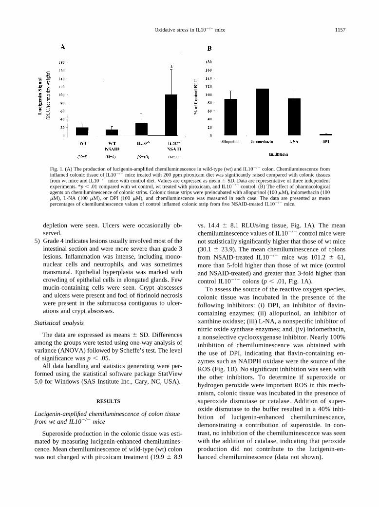
Superoxide production in the colonic tissue was esti-mated by measuring lucigenin-enhanced chemilumines-cence. Mean chemiluminescence of wild-type (wt) colonwas not changed with piroxicam treatment (19.9 � 8.9
vs. 14.4 � 8.1 RLU/s/mg tissue, Fig. 1A). The meanchemiluminescence values of IL10�/� control mice werenot statistically significantly higher that those of wt mice(30.1 � 23.9). The mean chemiluminescence of colonsfrom NSAID-treated IL10�/� mice was 101.2 � 61,more than 5-fold higher than those of wt mice (controland NSAID-treated) and greater than 3-fold higher thancontrol IL10�/� colons (p � .01, Fig. 1A).
To assess the source of the reactive oxygen species,colonic tissue was incubated in the presence of thefollowing inhibitors: (i) DPI, an inhibitor of flavin-containing enzymes; (ii) allopurinol, an inhibitor ofxanthine oxidase; (iii) L-NA, a nonspecific inhibitor ofnitric oxide synthase enzymes; and, (iv) indomethacin,a nonselective cyclooxygenase inhibitor. Nearly 100%inhibition of chemiluminescence was obtained withthe use of DPI, indicating that flavin-containing en-zymes such as NADPH oxidase were the source of theROS (Fig. 1B). No significant inhibition was seen withthe other inhibitors. To determine if superoxide orhydrogen peroxide were important ROS in this mech-anism, colonic tissue was incubated in the presence ofsuperoxide dismutase or catalase. Addition of super-oxide dismutase to the buffer resulted in a 40% inhi-bition of lucigenin-enhanced chemiluminescence,demonstrating a contribution of superoxide. In con-trast, no inhibition of the chemiluminescence was seenwith the addition of catalase, indicating that peroxideproduction did not contribute to the lucigenin-en-hanced chemiluminescence (data not shown).
Fig. 1. (A) The production of lucigenin-amplified chemiluminescence in wild-type (wt) and IL10�/� colon. Chemiluminescence frominflamed colonic tissue of IL10�/� mice treated with 200 ppm piroxicam diet was significantly raised compared with colonic tissuesfrom wt mice and IL10�/� mice with control diet. Values are expressed as mean � SD. Data are representative of three independentexperiments. *p � .01 compared with wt control, wt treated with piroxicam, and IL10�/� control. (B) The effect of pharmacologicalagents on chemiluminescence of colonic strips. Colonic tissue strips were preincubated with allopurinol (100 �M), indomethacin (100�M), L-NA (100 �M), or DPI (100 �M), and chemiluminscence was measured in each case. The data are presented as meanpercentages of chemiluminescence values of control inflamed colonic strip from five NSAID-treated IL10�/� mice.
1157Oxidative stress in IL10�/� mice
Aconitase activity in the colon of wt and IL10�/� mice
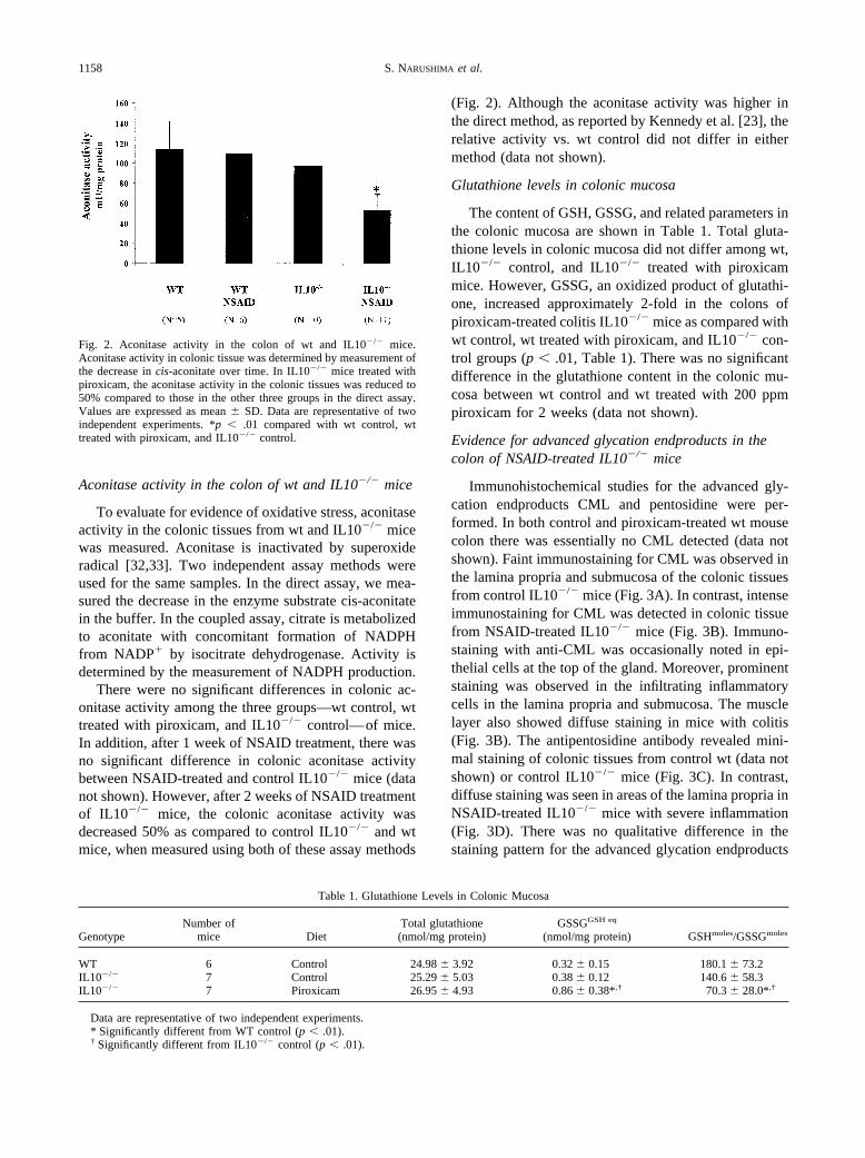
To evaluate for evidence of oxidative stress, aconitaseactivity in the colonic tissues from wt and IL10�/� micewas measured. Aconitase is inactivated by superoxideradical [32,33]. Two independent assay methods wereused for the same samples. In the direct assay, we mea-sured the decrease in the enzyme substrate cis-aconitatein the buffer. In the coupled assay, citrate is metabolizedto aconitate with concomitant formation of NADPHfrom NADP� by isocitrate dehydrogenase. Activity isdetermined by the measurement of NADPH production.
There were no significant differences in colonic ac-onitase activity among the three groups—wt control, wttreated with piroxicam, and IL10�/� control—of mice.In addition, after 1 week of NSAID treatment, there wasno significant difference in colonic aconitase activitybetween NSAID-treated and control IL10�/� mice (datanot shown). However, after 2 weeks of NSAID treatmentof IL10�/� mice, the colonic aconitase activity wasdecreased 50% as compared to control IL10�/� and wtmice, when measured using both of these assay methods
(Fig. 2). Although the aconitase activity was higher inthe direct method, as reported by Kennedy et al. [23], therelative activity vs. wt control did not differ in eithermethod (data not shown).
Glutathione levels in colonic mucosa
The content of GSH, GSSG, and related parameters inthe colonic mucosa are shown in Table 1. Total gluta-thione levels in colonic mucosa did not differ among wt,IL10�/� control, and IL10�/� treated with piroxicammice. However, GSSG, an oxidized product of glutathi-one, increased approximately 2-fold in the colons ofpiroxicam-treated colitis IL10�/� mice as compared withwt control, wt treated with piroxicam, and IL10�/� con-trol groups (p � .01, Table 1). There was no significantdifference in the glutathione content in the colonic mu-cosa between wt control and wt treated with 200 ppmpiroxicam for 2 weeks (data not shown).
Evidence for advanced glycation endproducts in thecolon of NSAID-treated IL10�/� mice
Immunohistochemical studies for the advanced gly-cation endproducts CML and pentosidine were per-formed. In both control and piroxicam-treated wt mousecolon there was essentially no CML detected (data notshown). Faint immunostaining for CML was observed inthe lamina propria and submucosa of the colonic tissuesfrom control IL10�/� mice (Fig. 3A). In contrast, intenseimmunostaining for CML was detected in colonic tissuefrom NSAID-treated IL10�/� mice (Fig. 3B). Immuno-staining with anti-CML was occasionally noted in epi-thelial cells at the top of the gland. Moreover, prominentstaining was observed in the infiltrating inflammatorycells in the lamina propria and submucosa. The musclelayer also showed diffuse staining in mice with colitis(Fig. 3B). The antipentosidine antibody revealed mini-mal staining of colonic tissues from control wt (data notshown) or control IL10�/� mice (Fig. 3C). In contrast,diffuse staining was seen in areas of the lamina propria inNSAID-treated IL10�/� mice with severe inflammation(Fig. 3D). There was no qualitative difference in thestaining pattern for the advanced glycation endproducts
Fig. 2. Aconitase activity in the colon of wt and IL10�/� mice.Aconitase activity in colonic tissue was determined by measurement ofthe decrease in cis-aconitate over time. In IL10�/� mice treated withpiroxicam, the aconitase activity in the colonic tissues was reduced to50% compared to those in the other three groups in the direct assay.Values are expressed as mean � SD. Data are representative of twoindependent experiments. *p � .01 compared with wt control, wttreated with piroxicam, and IL10�/� control.
Table 1. Glutathione Levels in Colonic Mucosa
GenotypeNumber of
mice DietTotal glutathione(nmol/mg protein)
GSSGGSH eq
(nmol/mg protein) GSHmoles/GSSGmoles
WT 6 Control 24.98 � 3.92 0.32 � 0.15 180.1 � 73.2IL10�/� 7 Control 25.29 � 5.03 0.38 � 0.12 140.6 � 58.3IL10�/� 7 Piroxicam 26.95 � 4.93 0.86 � 0.38*,† 70.3 � 28.0*,†
Data are representative of two independent experiments.* Significantly different from WT control (p � .01).† Significantly different from IL10�/� control (p � .01).
1158 S. NARUSHIMA et al.
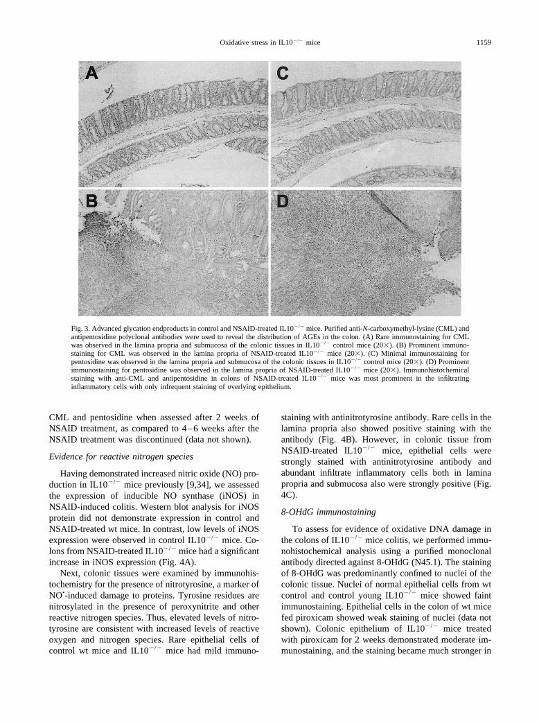
CML and pentosidine when assessed after 2 weeks ofNSAID treatment, as compared to 4–6 weeks after theNSAID treatment was discontinued (data not shown).
Evidence for reactive nitrogen species
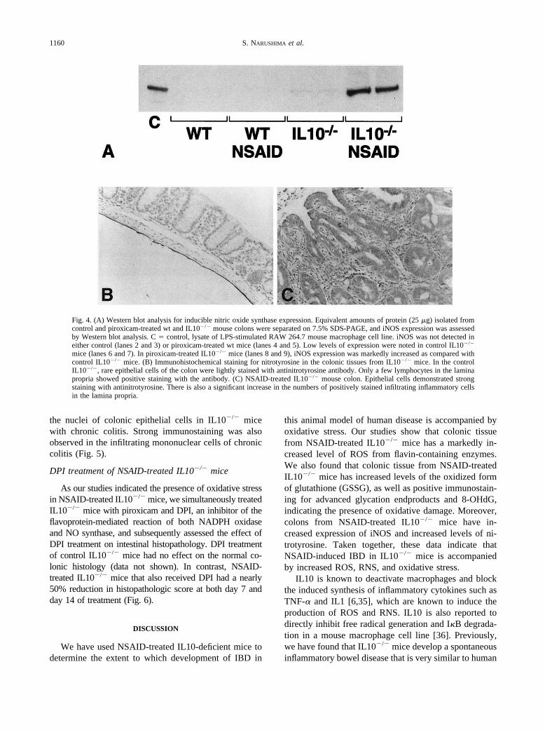
Having demonstrated increased nitric oxide (NO) pro-duction in IL10�/� mice previously [9,34], we assessedthe expression of inducible NO synthase (iNOS) inNSAID-induced colitis. Western blot analysis for iNOSprotein did not demonstrate expression in control andNSAID-treated wt mice. In contrast, low levels of iNOSexpression were observed in control IL10�/� mice. Co-lons from NSAID-treated IL10�/� mice had a significantincrease in iNOS expression (Fig. 4A).
Next, colonic tissues were examined by immunohis-tochemistry for the presence of nitrotyrosine, a marker ofNO•-induced damage to proteins. Tyrosine residues arenitrosylated in the presence of peroxynitrite and otherreactive nitrogen species. Thus, elevated levels of nitro-tyrosine are consistent with increased levels of reactiveoxygen and nitrogen species. Rare epithelial cells ofcontrol wt mice and IL10�/� mice had mild immuno-
staining with antinitrotyrosine antibody. Rare cells in thelamina propria also showed positive staining with theantibody (Fig. 4B). However, in colonic tissue fromNSAID-treated IL10�/� mice, epithelial cells werestrongly stained with antinitrotyrosine antibody andabundant infiltrate inflammatory cells both in laminapropria and submucosa also were strongly positive (Fig.4C).
8-OHdG immunostaining
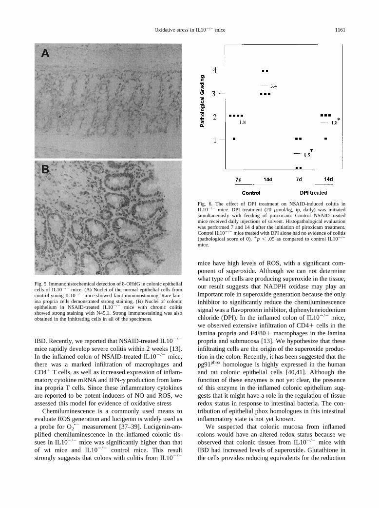
To assess for evidence of oxidative DNA damage inthe colons of IL10�/� mice colitis, we performed immu-nohistochemical analysis using a purified monoclonalantibody directed against 8-OHdG (N45.1). The stainingof 8-OHdG was predominantly confined to nuclei of thecolonic tissue. Nuclei of normal epithelial cells from wtcontrol and control young IL10�/� mice showed faintimmunostaining. Epithelial cells in the colon of wt micefed piroxicam showed weak staining of nuclei (data notshown). Colonic epithelium of IL10�/� mice treatedwith piroxicam for 2 weeks demonstrated moderate im-munostaining, and the staining became much stronger in
Fig. 3. Advanced glycation endproducts in control and NSAID-treated IL10�/� mice. Purified anti-N-carboxymethyl-lysine (CML) andantipentosidine polyclonal antibodies were used to reveal the distribution of AGEs in the colon. (A) Rare immunostaining for CMLwas observed in the lamina propria and submucosa of the colonic tissues in IL10�/� control mice (20�). (B) Prominent immuno-staining for CML was observed in the lamina propria of NSAID-treated IL10�/� mice (20�). (C) Minimal immunostaining forpentosidine was observed in the lamina propria and submucosa of the colonic tissues in IL10�/� control mice (20�). (D) Prominentimmunostaining for pentosidine was observed in the lamina propria of NSAID-treated IL10�/� mice (20�). Immunohistochemicalstaining with anti-CML and antipentosidine in colons of NSAID-treated IL10�/� mice was most prominent in the infiltratinginflammatory cells with only infrequent staining of overlying epithelium.
1159Oxidative stress in IL10�/� mice
the nuclei of colonic epithelial cells in IL10�/� micewith chronic colitis. Strong immunostaining was alsoobserved in the infiltrating mononuclear cells of chroniccolitis (Fig. 5).
DPI treatment of NSAID-treated IL10�/� mice
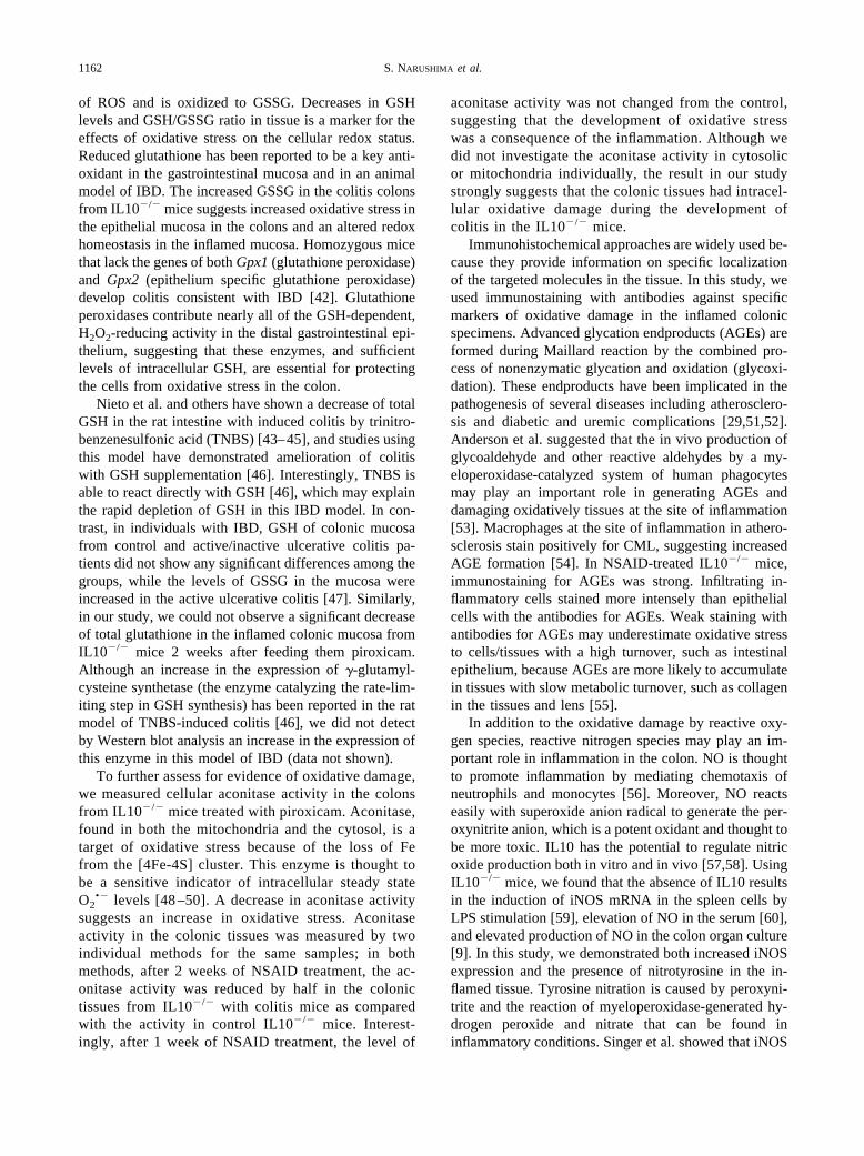
As our studies indicated the presence of oxidative stressin NSAID-treated IL10�/� mice, we simultaneously treatedIL10�/� mice with piroxicam and DPI, an inhibitor of theflavoprotein-mediated reaction of both NADPH oxidaseand NO synthase, and subsequently assessed the effect ofDPI treatment on intestinal histopathology. DPI treatmentof control IL10�/� mice had no effect on the normal co-lonic histology (data not shown). In contrast, NSAID-treated IL10�/� mice that also received DPI had a nearly50% reduction in histopathologic score at both day 7 andday 14 of treatment (Fig. 6).
DISCUSSION
We have used NSAID-treated IL10-deficient mice todetermine the extent to which development of IBD in
this animal model of human disease is accompanied byoxidative stress. Our studies show that colonic tissuefrom NSAID-treated IL10�/� mice has a markedly in-creased level of ROS from flavin-containing enzymes.We also found that colonic tissue from NSAID-treatedIL10�/� mice has increased levels of the oxidized formof glutathione (GSSG), as well as positive immunostain-ing for advanced glycation endproducts and 8-OHdG,indicating the presence of oxidative damage. Moreover,colons from NSAID-treated IL10�/� mice have in-creased expression of iNOS and increased levels of ni-trotyrosine. Taken together, these data indicate thatNSAID-induced IBD in IL10�/� mice is accompaniedby increased ROS, RNS, and oxidative stress.
IL10 is known to deactivate macrophages and blockthe induced synthesis of inflammatory cytokines such asTNF-� and IL1 [6,35], which are known to induce theproduction of ROS and RNS. IL10 is also reported todirectly inhibit free radical generation and I�B degrada-tion in a mouse macrophage cell line [36]. Previously,we have found that IL10�/� mice develop a spontaneousinflammatory bowel disease that is very similar to human
Fig. 4. (A) Western blot analysis for inducible nitric oxide synthase expression. Equivalent amounts of protein (25 �g) isolated fromcontrol and piroxicam-treated wt and IL10�/� mouse colons were separated on 7.5% SDS-PAGE, and iNOS expression was assessedby Western blot analysis. C � control, lysate of LPS-stimulated RAW 264.7 mouse macrophage cell line. iNOS was not detected ineither control (lanes 2 and 3) or piroxicam-treated wt mice (lanes 4 and 5). Low levels of expression were noted in control IL10�/�
mice (lanes 6 and 7). In piroxicam-treated IL10�/� mice (lanes 8 and 9), iNOS expression was markedly increased as compared withcontrol IL10�/� mice. (B) Immunohistochemical staining for nitrotyrosine in the colonic tissues from IL10�/� mice. In the controlIL10�/�, rare epithelial cells of the colon were lightly stained with antinitrotyrosine antibody. Only a few lymphocytes in the laminapropria showed positive staining with the antibody. (C) NSAID-treated IL10�/� mouse colon. Epithelial cells demonstrated strongstaining with antinitrotyrosine. There is also a significant increase in the numbers of positively stained infiltrating inflammatory cellsin the lamina propria.
1160 S. NARUSHIMA et al.
IBD. Recently, we reported that NSAID-treated IL10�/�
mice rapidly develop severe colitis within 2 weeks [13].In the inflamed colon of NSAID-treated IL10�/� mice,there was a marked infiltration of macrophages andCD4� T cells, as well as increased expression of inflam-matory cytokine mRNA and IFN-� production from lam-ina propria T cells. Since these inflammatory cytokinesare reported to be potent inducers of NO and ROS, weassessed this model for evidence of oxidative stress
Chemiluminescence is a commonly used means toevaluate ROS generation and lucigenin is widely used asa probe for O2
•� measurement [37–39]. Lucigenin-am-plified chemiluminescence in the inflamed colonic tis-sues in IL10�/� mice was significantly higher than thatof wt mice and IL10�/� control mice. This resultstrongly suggests that colons with colitis from IL10�/�
mice have high levels of ROS, with a significant com-ponent of superoxide. Although we can not determinewhat type of cells are producing superoxide in the tissue,our result suggests that NADPH oxidase may play animportant role in superoxide generation because the onlyinhibitor to significantly reduce the chemiluminescencesignal was a flavoprotein inhibitor, diphenyleneiodoniumchloride (DPI). In the inflamed colon of IL10�/� mice,we observed extensive infiltration of CD4� cells in thelamina propria and F4/80� macrophages in the laminapropria and submucosa [13]. We hypothesize that theseinfiltrating cells are the origin of the superoxide produc-tion in the colon. Recently, it has been suggested that thepg91phox homologue is highly expressed in the humanand rat colonic epithelial cells [40,41]. Although thefunction of these enzymes is not yet clear, the presenceof this enzyme in the inflamed colonic epithelium sug-gests that it might have a role in the regulation of tissueredox status in response to intestinal bacteria. The con-tribution of epithelial phox homologues in this intestinalinflammatory state is not yet known.
We suspected that colonic mucosa from inflamedcolons would have an altered redox status because weobserved that colonic tissues from IL10�/� mice withIBD had increased levels of superoxide. Glutathione inthe cells provides reducing equivalents for the reduction
Fig. 5. Immunohistochemical detection of 8-OHdG in colonic epithelialcells of IL10�/� mice. (A) Nuclei of the normal epithelial cells fromcontrol young IL10�/� mice showed faint immunostaining. Rare lam-ina propria cells demonstrated strong staining. (B) Nuclei of colonicepithelium in NSAID-treated IL10�/� mice with chronic colitisshowed strong staining with N45.1. Strong immunostaining was alsoobtained in the infiltrating cells in all of the specimens.
Fig. 6. The effect of DPI treatment on NSAID-induced colitis inIL10�/� mice. DPI treatment (20 �mol/kg, ip, daily) was initiatedsimultaneously with feeding of piroxicam. Control NSAID-treatedmice received daily injections of solvent. Histopathological evaluationwas performed 7 and 14 d after the initiation of piroxicam treatment.Control IL10�/� mice treated with DPI alone had no evidence of colitis(pathological score of 0). �p � .05 as compared to control IL10�/�
mice.
1161Oxidative stress in IL10�/� mice
of ROS and is oxidized to GSSG. Decreases in GSHlevels and GSH/GSSG ratio in tissue is a marker for theeffects of oxidative stress on the cellular redox status.Reduced glutathione has been reported to be a key anti-oxidant in the gastrointestinal mucosa and in an animalmodel of IBD. The increased GSSG in the colitis colonsfrom IL10�/� mice suggests increased oxidative stress inthe epithelial mucosa in the colons and an altered redoxhomeostasis in the inflamed mucosa. Homozygous micethat lack the genes of both Gpx1 (glutathione peroxidase)and Gpx2 (epithelium specific glutathione peroxidase)develop colitis consistent with IBD [42]. Glutathioneperoxidases contribute nearly all of the GSH-dependent,H2O2-reducing activity in the distal gastrointestinal epi-thelium, suggesting that these enzymes, and sufficientlevels of intracellular GSH, are essential for protectingthe cells from oxidative stress in the colon.
Nieto et al. and others have shown a decrease of totalGSH in the rat intestine with induced colitis by trinitro-benzenesulfonic acid (TNBS) [43–45], and studies usingthis model have demonstrated amelioration of colitiswith GSH supplementation [46]. Interestingly, TNBS isable to react directly with GSH [46], which may explainthe rapid depletion of GSH in this IBD model. In con-trast, in individuals with IBD, GSH of colonic mucosafrom control and active/inactive ulcerative colitis pa-tients did not show any significant differences among thegroups, while the levels of GSSG in the mucosa wereincreased in the active ulcerative colitis [47]. Similarly,in our study, we could not observe a significant decreaseof total glutathione in the inflamed colonic mucosa fromIL10�/� mice 2 weeks after feeding them piroxicam.Although an increase in the expression of �-glutamyl-cysteine synthetase (the enzyme catalyzing the rate-lim-iting step in GSH synthesis) has been reported in the ratmodel of TNBS-induced colitis [46], we did not detectby Western blot analysis an increase in the expression ofthis enzyme in this model of IBD (data not shown).
To further assess for evidence of oxidative damage,we measured cellular aconitase activity in the colonsfrom IL10�/� mice treated with piroxicam. Aconitase,found in both the mitochondria and the cytosol, is atarget of oxidative stress because of the loss of Fefrom the [4Fe-4S] cluster. This enzyme is thought tobe a sensitive indicator of intracellular steady stateO2
•� levels [48 –50]. A decrease in aconitase activitysuggests an increase in oxidative stress. Aconitaseactivity in the colonic tissues was measured by twoindividual methods for the same samples; in bothmethods, after 2 weeks of NSAID treatment, the ac-onitase activity was reduced by half in the colonictissues from IL10�/� with colitis mice as comparedwith the activity in control IL10�/� mice. Interest-ingly, after 1 week of NSAID treatment, the level of
aconitase activity was not changed from the control,suggesting that the development of oxidative stresswas a consequence of the inflammation. Although wedid not investigate the aconitase activity in cytosolicor mitochondria individually, the result in our studystrongly suggests that the colonic tissues had intracel-lular oxidative damage during the development ofcolitis in the IL10�/� mice.
Immunohistochemical approaches are widely used be-cause they provide information on specific localizationof the targeted molecules in the tissue. In this study, weused immunostaining with antibodies against specificmarkers of oxidative damage in the inflamed colonicspecimens. Advanced glycation endproducts (AGEs) areformed during Maillard reaction by the combined pro-cess of nonenzymatic glycation and oxidation (glycoxi-dation). These endproducts have been implicated in thepathogenesis of several diseases including atherosclero-sis and diabetic and uremic complications [29,51,52].Anderson et al. suggested that the in vivo production ofglycoaldehyde and other reactive aldehydes by a my-eloperoxidase-catalyzed system of human phagocytesmay play an important role in generating AGEs anddamaging oxidatively tissues at the site of inflammation[53]. Macrophages at the site of inflammation in athero-sclerosis stain positively for CML, suggesting increasedAGE formation [54]. In NSAID-treated IL10�/� mice,immunostaining for AGEs was strong. Infiltrating in-flammatory cells stained more intensely than epithelialcells with the antibodies for AGEs. Weak staining withantibodies for AGEs may underestimate oxidative stressto cells/tissues with a high turnover, such as intestinalepithelium, because AGEs are more likely to accumulatein tissues with slow metabolic turnover, such as collagenin the tissues and lens [55].
In addition to the oxidative damage by reactive oxy-gen species, reactive nitrogen species may play an im-portant role in inflammation in the colon. NO is thoughtto promote inflammation by mediating chemotaxis ofneutrophils and monocytes [56]. Moreover, NO reactseasily with superoxide anion radical to generate the per-oxynitrite anion, which is a potent oxidant and thought tobe more toxic. IL10 has the potential to regulate nitricoxide production both in vitro and in vivo [57,58]. UsingIL10�/� mice, we found that the absence of IL10 resultsin the induction of iNOS mRNA in the spleen cells byLPS stimulation [59], elevation of NO in the serum [60],and elevated production of NO in the colon organ culture[9]. In this study, we demonstrated both increased iNOSexpression and the presence of nitrotyrosine in the in-flamed tissue. Tyrosine nitration is caused by peroxyni-trite and the reaction of myeloperoxidase-generated hy-drogen peroxide and nitrate that can be found ininflammatory conditions. Singer et al. showed that iNOS
1162 S. NARUSHIMA et al.
protein is expressed in polymorphonuclear cells in thelumen and in limited numbers of mononuclear cells inthe lamina propria in other than epithelial cells of thecolon specimens from IBD patients [3].
However, the role of nitric oxide in the develop-ment of colitis is uncertain. Tissue damage was sig-nificantly diminished in iNOS knockout mice whencolitis was induced by dextran sodium sulfate treat-ment [61]. On the other hand, inhibition of nitric oxidesynthesis by aminoguanidine increased intestinal dam-age in the acute phase of rat colitis induced by TNBS[62]. Moreover, McCafferty et al. reported that theabsence of iNOS in IL10/iNOS double knockout micedid not have any effect on the spontaneous develop-ment of colonic inflammation [63]. Further study willbe required to determine the functional significance ofRNS in the development of NSAID-induced colitis inIL10�/� mice. In this study, however, increased RNSare consistent with our other data and lend additionalsupport to the hypothesis that oxidative stress is ele-vated in this model of IBD.
The DNA base-modified product 8-hydroxy-2'-deoxy-guanidine is one of the most commonly used markers forthe evaluation of oxidative damage of DNA. 8-OHdG isinduced by hydroxy radical, singlet oxygen, or photody-namic action [30]. We clearly showed that oxidativestress in the nucleus is induced in the epithelial cells inthe colitis colon. We also observed that the anti-8-OHdGstaining was variable in the colons from IL10�/� micetreated with piroxicam for 2 weeks, and stronger immu-nostaining was observed in the colons from IL10�/�
with chronic colitis. Previously, we reported that a highincidence of colorectal adenocarcinomas (67%) was ob-served in spontaneous colitis in IL10�/� mice [9]. In ourNSAID-induced colitis model, when mice were placedon standard diet for several weeks after 2 weeks ofpiroxicam feeding, chronic inflammation with subse-quent cellular atypia, nuclear and cytologic pleomor-phrism, and invasive epithelium were seen in the colon[13], suggesting the potential to progress to colon cancer.In our present study, we observed strong immunostainingwith anti-8-OHdG in the nuclei of the colonic epitheliumof chronic colitis in IL10�/� mice. Accumulation ofDNA damage induced by oxidative stress in the epithe-lial cells in the colon could lead to the transformation ofcells, increasing the risk of development of colon cancer.Moreover, increased oxidative stress may promote epi-thelial proliferation, as has been reported by Kondo et al.[64], for colorectal carcinoma, which may further pre-dispose the epithelium to cellular transformation. Inter-estingly, we found intense 8-OHdG immunostaining inthe infiltrating mononuclear cells in lamina propria andsubmucosa. This result was consistent with that ofKondo et al. [64], who observed strong immunostaining
in the infiltrating mononuclear cells throughout the hu-man colonic specimens, suggesting that the cells produc-ing reactive oxygen species also may have evidence ofoxidative damage.
Treatment with the flavoprotein inhibitor DPI re-sulted in a significant reduction in pathology inNSAID-treated IL10�/� mice. DPI can irreversiblyinactivate NADPH oxidase and NO synthase [65,66].Suppression of ROS/RNS from inflammatory cellsmay have led to decreased pathological damage aswell as altered ROS/NO-dependent signal transductionpathways [67,68], potentially altering the immune re-sponse as well. However, DPI is not specific forNADPH oxidase or iNOS. DPI can inhibit other fla-voproteins such as dehydrogenases, monooxygenases,and H2O2-producing oxidases [65]. Further studieswill be required to determine the key source(s) of theROS and the mechanism(s) by which their inhibitionin turn inhibits the development of colitis in thismodel. Nevertheless, our findings do provide a linkbetween oxidative stress and the development of IBDin this model.
IL10�/� mice serve as a well-established model ofhuman inflammatory bowel disease and colitis-associ-ated cancer. We have used a variation of this model,NSAID-treated IL10�/� mice, to address potential un-derlying mechanisms of the pathology of IBD. The goalof this study was to determine the extent to which IBD inNSAID-treated IL10�/� mice is accompanied by oxida-tive stress. We evaluated various markers for oxidativestress in the colons from wt and IL10�/� mice treatedwith piroxicam. The increased levels of ROS in theinflamed colon from IL10�/� mice directly suggest theincreased risk of oxidative damage in these tissues. Thechange in the redox status of mucosal glutathione (in-creased GSSG) clearly indicates a cellular response tooxidative stress. Decreased aconitase activity and thedetection of AGEs and nitrotyrosine in the colon fromIL10�/� mice treated with piroxicam suggest the oxida-tive damage to protein by ROS and NO. Furthermore,increased immunostaining with anti-8-OHdG, a signa-ture of oxidative damage to nucleic acids, is furtherevidence of oxidative stress in this model. Our findingthat treatment with DPI significantly reduces pathologyin NSAID-treated IL10�/� mice links oxidative stresswith the development of pathology in this model. Takentogether, the results from this study demonstrate signif-icant oxidative stress in NSAID-induced IBD in IL10�/�
mice. Increased oxidative stress may play an importantrole in the immunopathologic damage in IBD and mayserve as a promoter of the development of inflammation-associated colon cancer.
1163Oxidative stress in IL10�/� mice
Acknowledgements — We are grateful to Dr. Freya Q. Schafer forhelpful discussion. We thank Susan Walsh for expert technical assis-tance. The study was supported by a Translational Research Grant(D. J. B.) from the University of Iowa Holden Comprehensive CancerCenter.
REFERENCES
[1] Sedghi, S.; Fields, J. Z.; Klamut, M.; Urban, G.; Durkin, M.;Winship, D.; Fretland, D.; Olyaee, M.; Keshavarzian, A. In-creased production of luminol enhanced chemiluminescence bythe inflamed colonic mucosa in patients with ulcerative colitis.Gut 34:1191–1197; 1993.
[2] Simmonds, N. J.; Allen, R. E.; Stevens, T. R.; Van Someren,R. N.; Blake, D. R.; Rampton, D. S. Chemiluminescence assay ofmucosal reactive oxygen metabolites in inflammatory bowel dis-ease. Gastroenterology 103:186–196; 1992.
[3] Singer, I. I.; Kawka, D. W.; Scott, S.; Weidner, J. R.; Mumford,R. A.; Riehl, T. E.; Stenson, W. F. Expression of inducible nitricoxide synthase and nitrotyrosine in colonic epithelium in inflam-matory bowel disease. Gastroenterology 111:871–885; 1996.
[4] Sachar, D. B. Cancer in Crohn’s disease: dispelling the myths.Gut 35:1507–1508; 1994.
[5] Ransohoff, D. F. Colon cancer in ulcerative colitis. Gastroenter-ology 94:1089–1091; 1988.
[6] Fiorentino, D. F.; Zlotnik, A.; Mosmann, T. R.; Howard, M.;O’Garra, A. IL10 inhibits cytokine production by activated mac-rophages. J. Immunol. 147:3815–3822; 1991.
[7] Laichalk, L. L.; Danforth, J. M.; Standiford, T. J. Interleukin-10inhibits neutrophil phagocytic and bactericidal activity. FEMSImmunol. Med. Microbiol. 15:181–187; 1996.
[8] Kuga, S.; Otsuka, T.; Niiro, H.; Nunoi, H.; Nemoto, Y.; Nakano,T.; Ogo, T.; Umei, T.; Niho, Y. Suppression of superoxide anionproduction by interleukin-10 is accompanied by a downregulationof the genes for subunit proteins of NADPH oxidase. Exp. He-matol. 24:151–157; 1996.
[9] Berg, D. J.; Davidson, N.; Kuhn, R.; Muller, W.; Menon, S.;Holland, G.; Thompson-Snipes, L.; Leach, M. W.; Rennick, D.Enterocolitis and colon cancer in interleukin-10-deficient mice areassociated with aberrant cytokine production and CD4(�) TH1-like responses. J. Clin. Invest. 98:1010–1020; 1996.
[10] Davidson, N. J.; Hudak, S. A.; Lesley, R. E.; Menon, S.; Leach,M. W.; Rennick, D. M. IL12, but not IFN-�, plays a major role insustaining the chronic phase of colitis in IL10-deficient mice.J. Immunol. 161:3143–3149; 1998.
[11] Davidson, N. J.; Leach, M. W.; Fort, M. M.; Thompson-Snipes,L.; Kuhn, R.; Muller, W.; Berg, D. J.; Rennick, D. M. T helpercell 1-type CD4� T cells, but not B cells, mediate colitis ininterleukin-10-deficient mice. J. Exp. Med. 184:241–251; 1996.
[12] Schreiber, S.; Fedorak, R. N.; Nielsen, O. H.; Wild, G.; Williams,C. N.; Nikolaus, S.; Jacyna, M.; Lashner, B. A.; Gangl, A.;Rutgeerts, P.; Isaacs, K.; van Deventer, S. J.; Koningsberger,J. C.; Cohard, M.; LeBeaut, A.; Hanauer, S. B. Safety and efficacyof recombinant human interleukin-10 in chronic active Crohn’sdisease. Crohn’s disease IL10 cooperative study group. Gastro-enterology 119:1461–1472; 2000.
[13] Berg, D. J.; Zhang, J.; Weinstock, J. V.; Ismail, H. F.; Earle,K. A.; Alila, H.; Pamukcu, R.; Moore, S.; Lynch, R. G. Rapiddevelopment of colitis in NSAID-treated IL10-deficient mice.Gastroenterology 123:1527–1542; 2002.
[14] Sekhar, K. R.; Spitz, D. R.; Harris, S.; Nguyen, T. T.; Meredith,M. J.; Holt, J. T.; Guis, D.; Marnett, L. J.; Summar, M. L.;Freeman, M. L. Redox-sensitive interaction between KIAA0132and Nrf2 mediates indomethacin-induced expression of �-glu-tamylcysteine synthetase. Free Radic. Biol. Med. 32:650–662;2002.
[15] Bjarnason, I.; Hayllar, J.; MacPherson, A. J.; Russell, A. S. Sideeffects of nonsteroidal anti-inflammatory drugs on the small andlarge intestine in humans. Gastroenterology 104:1832–1847;1993.
[16] Kaufmann, H. J.; Taubin, H. L. Nonsteroidal anti-inflammatorydrugs activate quiescent inflammatory bowel disease. Ann. Intern.Med. 107:513–516; 1987.
[17] Millar, A. D.; Rampton, D. S.; Chander, C. L.; Claxson, A. W.;Blades, S.; Coumbe, A.; Panetta, J.; Morris, C. J.; Blake, D. R.Evaluating the antioxidant potential of new treatments for inflam-matory bowel disease using a rat model of colitis. Gut 39:407–415; 1996.
[18] Keshavarzian, A.; Sedghi, S.; Kanofsky, J.; List, T.; Robinson, C.;Ibrahim, C.; Winship, D. Excessive production of reactive oxygenmetabolites by inflamed colon: analysis by chemiluminescenceprobe. Gastroenterology 103:177–185; 1992.
[19] Yue, G.; Sun, F. F.; Dunn, C.; Yin, K.; Wong, P. Y. The21-aminosteroid tirilazad mesylate can ameliorate inflammatorybowel disease in rats. J. Pharmacol. Exp. Ther. 276:265–270;1996.
[20] Didion, S. P.; Faraci, F. M. Effects of NADH and NADPH onsuperoxide levels and cerebral vascular tone. Am. J. Physiol.Heart Circ. Physiol. 282:H688–H695; 2002.
[21] Gunnett, C. A.; Heistad, D. D.; Berg, D. J.; Faraci, F. M. IL10deficiency increases superoxide and endothelial dysfunction dur-ing inflammation. Am. J. Physiol. Heart Circ. Physiol. 279:H1555–H1562; 2000.
[22] Gantt, K. R.; Goldman, T. L.; McCormick, M. L.; Miller, M. A.;Jeronimo, S. M.; Nascimento, E. T.; Britigan, B. E.; Wilson,M. E. Oxidative responses of human and murine macrophagesduring phagocytosis of Leishmania chagasi. J. Immunol. 167:893–901; 2001.
[23] Kennedy, M. C.; Emptage, M. H.; Dreyer, J. L.; Beinert, H. Therole of iron in the activation-inactivation of aconitase. J. Biol.Chem. 258:11098–11105; 1983.
[24] Drapier, J. C.; Hibbs, J. B. Jr. Aconitases: a class of metallopro-teins highly sensitive to nitric oxide synthesis. Methods Enzymol.269:26–36; 1996.
[25] Rose, I. A.; O’Connell, E. L. Mechanism of aconitase action. I.The hydrogen transfer reaction. J. Biol. Chem. 242:1870–1879;1967.
[26] Anderson, M. E. Determination of glutathione and glutathionedisulfide in biological samples. Methods Enzymol. 113:548–555;1985.
[27] Griffith, O. W. Determination of glutathione and glutathionedisulfide using glutathione reductase and 2-vinylpyridine. Anal.Biochem. 106:207–212; 1980.
[28] Miyata, T.; Taneda, S.; Kawai, R.; Ueda, Y.; Horiuchi, S.; Hara,M.; Maeda, K.; Monnier, V. M. Identification of pentosidine as anative structure for advanced glycation endproducts in �-2-mi-croglobulin-containing amyloid fibrils in patients with dialysis-related amyloidosis. Proc. Natl. Acad. Sci. USA 93:2353–2358;1996.
[29] Horie, K.; Miyata, T.; Maeda, K.; Miyata, S.; Sugiyama, S.;Sakai, H.; Strihou, C. Y.; Monnier, V. M.; Witztum, J. L.;Kurokawa, K. Immunohistochemical colocalization of glycoxida-tion products and lipid peroxidation products in diabetic renalglomerular lesions. Implication for glycoxidative stress in thepathogenesis of diabetic nephropathy. J. Clin. Invest. 100:2995–3004; 1997.
[30] Toyokuni, S.; Tanaka, T.; Hattori, Y.; Nishiyama, Y.; Yoshida,A.; Uchida, K.; Hiai, H.; Ochi, H.; Osawa, T. Quantitative im-munohistochemical determination of 8-hydroxy-2'-deox-yguanosine by a monoclonal antibody N45.1: its application toferric nitrilotriacetate-induced renal carcinogenesis model. Lab.Invest. 76:365–374; 1997.
[31] Miesel, R.; Kurpisz, M.; Kroger, H. Suppression of inflammatoryarthritis by simultaneous inhibition of nitric oxide synthase andNADPH oxidase. Free Radic. Biol. Med. 20:75–81; 1996.
[32] Gardner, P. R.; Nguyen, D. D.; White, C. W. Aconitase is asensitive and critical target of oxygen poisoning in cultured mam-malian cells and in rat lungs. Proc. Natl. Acad. Sci. USA 91:12248–12252; 1994.
[33] Hausladen, A.; Fridovich, I. Superoxide and peroxynitrite inacti-
1164 S. NARUSHIMA et al.
vate aconitases, but nitric oxide does not. J. Biol. Chem. 269:29405–29408; 1994.
[34] Berg, D. J.; Zhang, J.; Lauricella, D. M.; Moore, S. A. IL10 is acentral regulator of cyclooxygenase-2 expression and prostaglan-din production. J. Immunol. 166:2674–2680; 2001.
[35] de Waal Malefyt, R.; Abrams, J.; Bennett, B.; Figdor, C. G.; deVries, J. E. Interleukin-10 (IL10) inhibits cytokine synthesis byhuman monocytes: an autoregulatory role of IL10 produced bymonocytes. J. Exp. Med. 174:1209–1220; 1991.
[36] Dokka, S.; Shi, X.; Leonard, S.; Wang, L.; Castranova, V.;Rojanasakul, Y. Interleukin-10-mediated inhibition of free radicalgeneration in macrophages. Am. J. Physiol. Lung Cell. Mol.Physiol. 280:L1196–L1202; 2001.
[37] Davies, G. R.; Simmonds, N. J.; Stevens, T. R.; Sheaff, M. T.;Banatvala, N.; Laurenson, I. F.; Blake, D. R.; Rampton, D. S.Helicobacter pylori stimulates antral mucosal reactive oxygenmetabolite production in vivo. Gut 35:179–185; 1994.
[38] Williams, A. J.; Cole, P. J. The onset of polymorphonuclearleucocyte membrane-stimulated metabolic activity. Immunology43:733–739; 1981.
[39] Li, Y.; Zhu, H.; Kuppusamy, P.; Roubaud, V.; Zweier, J. L.;Trush, M. A. Validation of lucigenin (bis-N-methylacridinium) asa chemilumigenic probe for detecting superoxide anion radicalproduction by enzymatic and cellular systems. J. Biol. Chem.273:2015–2023; 1998.
[40] Suh, Y. A.; Arnold, R. S.; Lassegue, B.; Shi, J.; Xu, X.; Sorescu,D.; Chung, A. B.; Griendling, K. K.; Lambeth, J. D. Cell trans-formation by the superoxide-generating oxidase Mox1. Nature401:79–82; 1999.
[41] Kikuchi, H.; Hikage, M.; Miyashita, H.; Fukumoto, M. NADPHoxidase subunit, gp91(phox) homologue, preferentially expressedin human colon epithelial cells. Gene 254:237–243; 2000.
[42] Esworthy, R. S.; Aranda, R.; Martin, M. G.; Doroshow, J. H.;Binder, S. W.; Chu, F. F. Mice with combined disruption of Gpx1and Gpx2 genes have colitis. Am. J. Physiol. Gastrointest. LiverPhysiol. 281:G848–G855; 2001.
[43] Nieto, N.; Torres, M. I.; Fernandez, M. I.; Giron, M. D.; Rios, A.;Suarez, M. D.; Gil, A. Experimental ulcerative colitis impairsantioxidant defense system in rat intestine. Dig. Dis. Sci. 45:1820–1827; 2000.
[44] Sanchez de Medina, F.; Galvez, J.; Romero, J. A.; Zarzuelo, A.Effect of quercitrin on acute and chronic experimental colitis inthe rat. J. Pharmacol. Exp. Ther. 278:771–779; 1996.
[45] Zea-Iriarte, W. L.; Makiyama, K.; Goto, S.; Murase, K.; Urata,Y.; Sekine, I.; Hara, K.; Kondo, T. Impairment of antioxidants incolonic epithelial cells isolated from trinitrobenzene sulphonicacid-induced colitis rats. Protective effect of rebamipide. Scand.J. Gastroenterol. 31:985–992; 1996.
[46] Ardite, E.; Sans, M.; Panes, J.; Romero, F. J.; Pique, J. M.;Fernandez-Checa, J. C. Replenishment of glutathione levels im-proves mucosal function in experimental acute colitis. Lab. Invest.80:735–744; 2000.
[47] Holmes, E. W.; Yong, S. L.; Eiznhamer, D.; Keshavarzian, A.Glutathione content of colonic mucosa: evidence for oxidativedamage in active ulcerative colitis. Dig. Dis. Sci. 43:1088–1095;1998.
[48] Yan, L. J.; Levine, R. L.; Sohal, R. S. Oxidative damage duringaging targets mitochondrial aconitase. Proc. Natl. Acad. Sci. USA94:11168–11172; 1997.
[49] Gardner, P. R.; Raineri, I.; Epstein, L. B.; White, C. W. Super-oxide radical and iron modulate aconitase activity in mammaliancells. J. Biol. Chem. 270:13399–13405; 1995.
[50] Fridovich, I. Superoxide anion radical (O2•�), superoxide dis-
mutases, and related matters. J. Biol. Chem. 272:18515–18517;1997.
[51] Miyata, T.; Kurokawa, K.; Van Ypersele De Strihou, C. Ad-vanced glycation and lipoxidation endproducts: role of reactivecarbonyl compounds generated during carbohydrate and lipidmetabolism. J. Am. Soc. Nephrol. 11:1744–1752; 2000.
[52] Miyata, T.; Van Ypersele De Strihou, C.; Kurokawa, K.; Baynes,J. W. Alterations in nonenzymatic biochemistry in uremia: origin
and significance of “carbonyl stress” in long-term uremic com-plications. Kidney Int. 55:389–399; 1999.
[53] Anderson, M. M.; Requena, J. R.; Crowley, J. R.; Thorpe, S. R.;Heinecke, J. W. The myeloperoxidase system of human phago-cytes generates N�- (carboxymethyl)lysine on proteins: a mecha-nism for producing advanced glycation endproducts at sites ofinflammation. J. Clin. Invest. 104:103–113; 1999.
[54] Schleicher, E. D.; Wagner, E.; Nerlich, A. G. Increased accumu-lation of the glycoxidation product N�-(carboxymethyl)lysine inhuman tissues in diabetes and aging. J. Clin. Invest. 99:457–468;1997.
[55] Dunn, J. A.; Patrick, J. S.; Thorpe, S. R.; Baynes, J. W. Oxidationof glycated proteins: age-dependent accumulation of N�-(car-boxymethyl)lysine in lens proteins. Biochemistry 28:9464–9468;1989.
[56] Kimura, H.; Hokari, R.; Miura, S.; Shigematsu, T.; Hirokawa, M.;Akiba, Y.; Kurose, I.; Higuchi, H.; Fujimori, H.; Tsuzuki, Y.;Serizawa, H.; Ishii, H. Increased expression of an inducible iso-form of nitric oxide synthase and the formation of peroxynitrite incolonic mucosa of patients with active ulcerative colitis. Gut42:180–187; 1998.
[57] Cunha, F. Q.; Moncada, S.; Liew, F. Y. Interleukin-10 (IL10)inhibits the induction of nitric oxide synthase by interferon-� inmurine macrophages. Biochem. Biophys. Res. Commun. 182:1155–1159; 1992.
[58] Roilides, E.; Dimitriadou, A.; Kadiltsoglou, I.; Sein, T.; Karpou-zas, J.; Pizzo, P. A.; Walsh, T. J. IL10 exerts suppressive andenhancing effects on antifungal activity of mononuclear phago-cytes against Aspergillus fumigatus. J. Immunol. 158:322–329;1997.
[59] Berg, D. J.; Zhang, J.; Lauricella, D.; Moore, S. A. Interleukin-10is a central regulator of cyclooxygenase-2 expression and prosta-glandin production. J. Immunol. 166:2674–2680; 2001.
[60] Berg, D. J.; Kuhn, R.; Rajewsky, K.; Muller, W.; Menon, S.;Davidson, N.; Grunig, G.; Rennick, D. Interleukin-10 is a centralregulator of the response to LPS in murine models of endotoxicshock and the Shwartzman reaction but not endotoxin tolerance.J. Clin. Invest. 96:2339–2347; 1995.
[61] Hokari, R.; Kato, S.; Matsuzaki, K.; Kuroki, M.; Iwai, A.;Kawaguchi, A.; Nagao, S.; Miyahara, T.; Itoh, K.; Sekizuka, E.;Nagata, H.; Ishii, H.; Miura, S. Reduced sensitivity of induciblenitric oxide synthase-deficient mice to chronic colitis. Free Radic.Biol. Med. 31:153–163; 2001.
[62] Dikopoulos, N.; Nussler, A. K.; Liptay, S.; Bachem, M.; Rein-shagen, M.; Stiegler, M.; Schmid, R. M.; Adler, G.; Weidenbach,H. Inhibition of nitric oxide synthesis by aminoguanidine in-creases intestinal damage in the acute phase of rat TNB-colitis.Eur. J. Clin. Invest. 31:234–239; 2001.
[63] McCafferty, D. M.; Sihota, E.; Muscara, M.; Wallace, J. L.;Sharkey, K. A.; Kubes, P. Spontaneously developing chroniccolitis in IL10/iNOS double-deficient mice. Am. J. Physiol. Gas-trointest. Liver Physiol. 279:G90–G99; 2000.
[64] Kondo, S.; Toyokuni, S.; Iwasa, Y.; Tanaka, T.; Onodera, H.;Hiai, H.; Imamura, M. Persistent oxidative stress in human colo-rectal carcinoma, but not in adenoma. Free Radic. Biol. Med.27:401–410; 1999.
[65] Yea, C. M.; Cross, A. R.; Jones, O. T. Purification and someproperties of the 45 kDa diphenylene iodonium-binding flavopro-tein of neutrophil NADPH oxidase. Biochem. J. 265:95–100;1990.
[66] Stuehr, D. J.; Fasehun, O. A.; Kwon, N. S.; Gross, S. S.; Gonza-lez, J. A.; Levi, R.; Nathan, C. F. Inhibition of macrophage andendothelial cell nitric oxide synthase by diphenyleneiodoniumand its analogs. FASEB J. 5:98–103; 1991.
[67] Toledano, M. B.; Leonard, W. J. Modulation of transcriptionfactor NF-�B binding activity by oxidation-reduction in vitro.Proc. Natl. Acad. Sci. USA 88:4328–4332; 1991.
[68] Turpaev, K. T. Reactive oxygen species and regulation of geneexpression. Biochemistry (Moscow) 67:281–292; 2002.
1165Oxidative stress in IL10�/� mice
ABBREVIATIONS
AGE—advanced glycation endproductCML—carboxymethyllysineCOX—cyclooxygenaseDAB—3,3'-diaminobenzidineDPI—diphenyleneiodinium chloride8-OHdG—8-hydroxy-2'-deoxyguanosineGSH—glutathioneGSSG—glutathione disulfide
IBD—inflammatory bowel diseaseIFN-�—interferon-�L-NA—N-�-nitro-L-arginineNADPH—nicotineamide adenine dinucleotide phos-
phate (reduced)NSAID—nonsteroidal anti-inflammatory drugPG—prostaglandinTNBS—trinitrobenzenesulfonic acidTNF-�—tumor necrosis factor �
1166 S. NARUSHIMA et al.


![[Mast cells in ulcerative colitis]](https://img.pdfslide.net/doc/110x75/634e26a23bdc8e881007bdae/mast-cells-in-ulcerative-colitis.jpg)
![[Acute colitis in Wegener's disease: a case report]](https://img.pdfslide.net/doc/110x75/6351f9645c21d80fde0ac222/acute-colitis-in-wegeners-disease-a-case-report.jpg)

