Embed Size (px)
Citation preview

[Frontiers in Bioscience 17, 1389-1401, January 1, 2012]
1389
Deer antler innervation and regeneration
Manuel Nieto-Diaz1, Daniel Wolfgang Pita-Thomas2, Teresa Munoz-Galdeano1, Cayetana Martinez-Maza3, Rosa Navarro-Ruiz1, David Reigada1, Monica Yunta1, Marcos Javier Caballero-Lopez1, Manuel Nieto-Sampedro4, Rodrigo Martinez-Maza1
1Molecular Neuroprotection Group, Hospital Nacional de Paraplejicos (SESCAM), Finca la Peraleda s/n, 45071 Toledo (Spain), 2Bascom Palmer Eye Institute, University of Miami Miller School of Medicine, Miami, FL 33136 (USA), 3Museo Nacional de Ciencias Naturales (CSIC), C/ Jose Gutierrez Abascal 2, 28006 Madrid, Spain, 4Neural Plasticity Group, Instituto Cajal de Neurobiologia (CSIC), Avda. Doctor Arce 37, 28002 Madrid, Spain
TABLE OF CONTENTS
1. Abstract 2. Introduction 2.1. Nervous system injuries 2.2. Therapeutic potential of the antler’s nerve regeneration 3. Antler innervation 3.1. Antler nerve supply 3.2. Nerve fiber distribution in the antler 3.3. Types of antler nerve fibers 4. Antler regulators of axon regeneration 4.1. Paracrine regulation 4.2 Endocrine regulation and other promoting mechanisms 5. Summary and perspectives 6. Acknowledgments 7. References
1. ABSTRACT Nervous system injuries are a major cause of
impairment in the human society. Up to now, clinical approaches have failed to adequately restore function following nervous system damage. The regenerative cycle of deer antlers may provide basic information on mechanisms underlying nervous system regeneration. The present contribution reviews the actual knowledge on the antler innervation and the factors responsible for its regeneration and fast growth. Growing antlers are profusely innervated by sensory fibers from the trigeminal nerve, which regenerate every year reaching elongation rates up to 2 cm a day. Antler nerves grow through the velvet in close association to blood vessels. This environment is rich in growth promoting molecules capable of inducing and guiding neurite outgrowth of rat sensory neurons in vitro. Conversely, endocrine regulation failed to show effects on neurite outgrowth in vitro, in spite of including hormones of known promoting effects on axon growth. Additional studies are needed to analyze unexplored factors promoting on growth in antlers such as electric potentials or mechanical stretch, as well as on the survival of antler innervating neurons.
2. INTRODUCTION 2.1. Nervous system injuries
Accidents causing spinal cord injury, traumatic brain injury, stroke, or peripheral nerve injuries prevent the correct functioning of the nervous system, causing total or partial loss of sensory, motor, autonomic or cognitive functions. Nervous system deficits are a major cause of disability in developed countries. It has been estimated that every year in the United States, 1.4 million people sustain a traumatic brain injury (1), and 12,000 spinal cord injury (2) while the incidence of nerve injury affects each year 13.9 per 100,000 persons in Sweden (3). In most cases, resulting deficits are permanent. Less than 40 percent of the patients suffering spinal cord injury (2) or 60 percent of those with different peripheral nerve lesions (4) return to work after injury. The social consequences are enormous since, contrary to other pathologies like cancer or cardiovascular diseases, neurological injuries frequently affect people under 45 years of age, and the medical and social care is required for decades.
Functional deficits following nervous system
damage are the direct consequence of the interruption of

Deer antler innervation
1390
Figure 1. Illustration of the processes, regenerative capacities and functional outcomes that experience innervation following peripheral nervous system (PNS) and central nervous system (CNS) injuries compared to those taking place during deer antler regeneration. For each system, direct effects of damage are listed on top, below are the subsequent processes followed by the regenerative capabilities of the system and the resulting functional outcome.
neural connections, largely due to the interruption of axons (axotomy) and the death of neural cells (5) but also due to inflammation, ischemia, and other processes that result in extended cell death and disconnection (6-7, Figure 1). Moreover, the reaction to damage of the nervous system may also alter the neural circuitry and cause undesirable side effects like neuropathic pain (5). Prognostic of nervous system injuries depends on many parameters, including type, location or extension of the lesion, age, etc. Functional deficits resulting from severe injuries become permanent due to the limited regeneration capacity of the nervous system. The high differentiation degree of some neural cells, particularly neurons or oligodendrocytes, prevents their proliferation and replacement (8). The neurons surviving axotomy assume a regenerative phenotype (9-11) but effective axon growth depends on local environmental factors (see reviews in 12-13). Mature central nervous system (CNS) environment is inhibitory for axon growth (11,13), with few exceptions like the olfactory or hippocampal tracts (see, for example, 14). Adult CNS neurons also seem to present intrinsic properties that reduce their regeneration capabilities (15). On the other hand, peripheral nervous system (PNS) will regenerate within the permissive growth environment of the Schwann cells (12), although it progressively fails to sustain regenerative response with time after injury (12). Thus, prolonged
denervations and axotomies result in poor functional recoveries in most cases (16-17). Functional recovery following injury is also made difficult by the high specificity that neurons exhibit, which, in practice, means that any new connections may not result in the recovery of original circuits but the formation of new aberrant ones (18).
2.2. Therapeutic potential of the antler’s nerve regeneration
Despite more than a century of neurological research and surgical innovation, clinical approaches have failed to adequately restore function following central or even peripheral nervous system damage (7). The scientific and clinical communities have realized that the development of efficient therapeutic tools depends on our understanding of the damaged nervous system and our capacity to manipulate regeneration (19). Much work has dealt with developmental mechanisms leading to the formation of the nervous system assuming that their reactivation may overcome the regrowth limitations after injury. Less attention has been paid to systems that can regenerate spontaneously even though these systems may provide basic information on the mechanisms that rule nervous system regeneration (20).

Deer antler innervation
1391
The capability to regenerate large sections of the body plan is typical of some invertebrates and urodele amphibians (21-23). Among adult mammals, full organ regeneration is exceptional, being restricted to deer antlers (24). Every year, male deers shed (cast) their antlers and fulfill a complete regeneration process that leads to the formation of a new set of antlers. Antler regeneration cycle has been recognized as a valuable model to study the mechanisms underlying organ regeneration and rapid tissue growth in mammals (25). The whole growth period takes place in about 3 months, reaching growth rates above 2 cm a day to build up structures of more than 3 Kg (25), up to a 20 percent of the whole skeleton weight (26). The growing antler is an extension of the antler pedicle periostium (27) that proliferates and differentiates into cartilage and bone tissue to form the bone core of the new antlers. Growing antlers are enveloped in a hair-covered skin known as velvet that presents several peculiarities, including lack of sweat glands and arrector pili muscles and the presence of abundant multilobullated sebaceous glands (28). At the end of the summer, antlers become calcified and velvet sheds, leaving the dead bony core used in agonistic encounters during the rut season (29). Every year, antler innervation regenerates to provide the antler with nerve supply (30-33). This growth supposes an extraordinary enlargement of the peripheral field that likely arises as a local extension of the nerve fibers that supply the forehead and the pedicle. Once antlers stop growing and become mineralized, nerve fibers at the velvet die back to the pedicle where they apparently remain encapsulated (34). By the end of the winter, when dry antlers are cast and new ones begin to grow, nerves reenter into a regenerative state to supply the velvet antler.
Deer antlers are a very interesting source of
information on the mechanisms underlying the nerve regeneration and functional recovery following injury (20,23,35). This spontaneous regeneration model is particularly interesting for therapeutic studies because it occurs in an adult mammal and involves cells, mechanisms and/or biochemical pathways which are more likely to be similar to those in humans than other regenerating models as the non-mammalian vertebrates (newts or fishes) or invertebrates. Moreover, antler regeneration takes place in adult individuals and affects adult neural cells like those typically involved in nerve injuries and different from those of embryos with different growth capabilities (see for example, 15, 36). The extraordinary extension and growth rate that the nerve fibers achieve during antler regeneration is particularly outstanding from both the clinical and biological points of view (Figure 1). Gray and colleagues (32) demonstrated that nerve supply grows together with all antler tissues to cover all antler (see also 33,37,38), reaching elongation rates over 2 to 3 cm a day in the largest species like the moose (Alces alces; 24) or the wapiti (Cervus canadenisis; 39). From the biological point of view, it is particularly remarkable the transport rate and the amounts of cell material needed to enlarge axons more than one meter at rates above 2 cm per day, especially if we consider that maximum axon growth by growth-cone extension only reaches 1 mm per day (40-41). In fact, such an extremely fast growth is not consistent with current understanding of the transport of essential structural elements such as neurofilament proteins, for which the
average transport speed is limited to 1 mm per day (42-45). The rapid and sustained nerve growth observed in the antlers indicates that the physiological capacity of axons to expand rapidly and continuously is not limited by protein synthesis, transport rates, or the availability of structural constituents (43-44,46-48). The clinical consequences of understanding this phenomenon would be enormous, considering that in many injuries, growing axons have to reach far away targets, up to one meter in the case of some human nerve fibers. Reconnecting these targets would take years for axons growing at elongation rates around 1 mm per day, resulting in functional recovery failure (17). However, at the rate observed in deer antlers, it would take a few months of growth. 3. ANTLER INNERVATION 3.1. Antler nerve supply
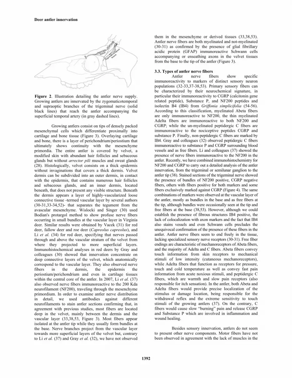
The first description on the anatomy of the deer antler innervation dates back to the 19th century (49). Later studies on Virginia deer (Odocoileus virginianus; 30), red deer (Cervus elaphus, 50), wapiti (cervus canadensis, 51), and fallow deer (Dama dama, 51) showed that the nerves supplying the antlers come from the supraoptic (infratrochlear) branch of the ophtalmic division and the zygomaticotemporal branch of the maxillary division, both from the trigeminal (5th cranial) nerve (Figure 2). The supraoptic branch is a single bundle that emerges from the skull beneath the upper edge of the orbit (1.5-2 cm from the medial canthus of the eye in red deer according to Adams, 50) and courses medially over the dorsal rim, near the medial angle of the eye (51). It then courses caudally through the orbicularis oculi muscle and sends several nerves toward the base of the antlers where 6 or more small branches in Virginia deer (30) or a full web in wapiti (51) are given off to the anterior and medial surfaces of the pedicle and the antler. The zygomaticotemporal branch of the maxillary nerve emerges onto the scalp as a large nerve near the zygomatic arch, at the caudal margin of the zygomatic process of the frontal bone, and divides immediately, producing a number of branches coursing toward the antler base and the ear. The branches that supply the antler pass caudodorsally through the retrorbital (periorbital) fat and beneath the frontalis muscle. Midway from the orbit to the pedicle they branch into several nerves (6 or more in Virginia deer according to 30), which disperse on the lateral and posterior surfaces of the antler pedicle and then onto the antler. Both trigeminal nerve supplies follow very closely the respective distribution of the lateral and medial coronary branches of the superficial temporal artery (the last branch of the external carotid) that provides the antler blood supply (30,51). Besides the trigeminal innervation, fibers from the zygomatic branch of the auriculopalpebral nerve (a branch of the facial or 7th cranial nerve) have been observed to reach the medial surface of the pedicle (50-51) although there is no evidence of its extension into the antler. According to Woodbory and Haigh (51), no other nerves could be traced to the pedicle. 3.2. Nerve fiber distribution in the antler
Several authors have studied the location of the nerve supply in the growing antler (30-32,34,37-38).
Deer antler innervation
1392
Figure 2. Illustration detailing the antler nerve supply. Growing antlers are innervated by the zygomaticotemporal and supraoptic branches of the trigeminal nerve (solid black lines) that reach the antler accompanying the superficial temporal artery (in gray dashed lines).
Growing antlers consist on tips of densely packed
mesenchymal cells which differentiate proximally into cartilage and bone tissue (Figure 3). Overlaying cartilage and bone, there is a layer of perichondrium/periostium that ultimately shows continuity with the mesenchyme primordia. The entire antler is covered by velvet, a modified skin with abundant hair follicles and sebaceous glands but without arrector pili muscles and sweat glands (28). Histologically, velvet consists on a thick epidermis without invaginations that covers a thick dermis. Velvet dermis can be subdivided into an outer dermis, in contact with the epidermis, that contains numerous hair follicles and sebaceous glands, and an inner dermis, located beneath, that does not present any visible structure. Beneath the dermis appears a layer of highly-vascularized loose connective tissue -termed vascular layer by several authors (30-31,33-34,52)- that separates the tegument from the avascular mesenchyme. Wislocki and Singer (30) used Bodian's protargol method to show profuse nerve fibers occurring in small bundles at the vascular layer in Virginia deer. Similar results were obtained by Vacek (31) for red deer, fallow deer and roe deer (Capreolus capreolus), and Li et al. (34) for red deer, specifying that nerves passed through and above the vascular stratum of the velvet from where they projected to more superficial layers. Immunohistochemical analyses in red deers by Gray and colleagues (30) showed that innervation concentrate on deep connective layers of the velvet, which anatomically correspond to the vascular layer. They also observed nerve fibers in the dermis, the epidermis the periostium/perichondrium and even in cartilage tissues within the central core of the antler. In 2007, Li et al. (37) also observed nerve fibers immunoreactive to the 200 Kda neurofilament (NF200), traveling through the mesenchyme primordium. In order to examine antler nerve distribution in detail, we used antibodies against different neurofilaments to stain antler sections confirming that, in agreement with previous studies, most fibers are located deep in the velvet, mainly between the dermis and the vascular layer (33,38,53, Figure 3). Most fibers appear isolated at the antler tip while they usually form bundles at the base. Nerve branches project from the vascular layer towards more superficial layers of the velvet but, contrary to Li et al. (37) and Gray et al. (32), we have not observed
them in the mesenchyme or derived tissues (33,38,53). Antler nerve fibers are both myelinated and not-myelinated (30-31) as confirmed by the presence of glial fibrillary acidic protein (GFAP) immunoreactive Schwann cells accompanying or enseathing axons in the velvet tissues from the base to the tip of the antler (Figure 3). 3.3. Types of antler nerve fibers
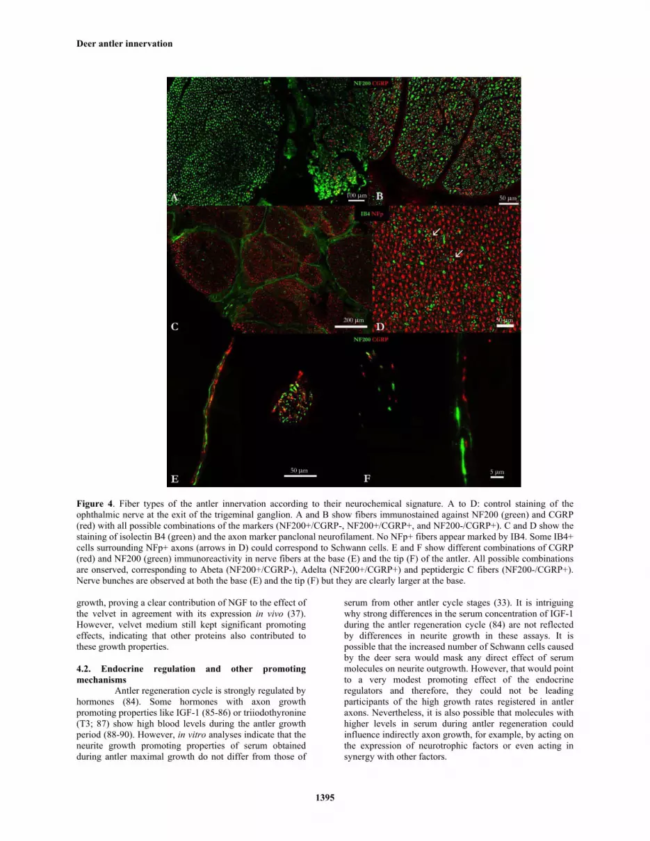
Antler nerve fibers show specific immunoreactivity to markers of distinct sensory neuron populations (32-33,37-38,53). Primary sensory fibers can be characterized by their neurochemical signature, in particular their immunoreactivity to CGRP (calcitonin gene related peptide), Substance P, and NF200 peptides and isolectin B4 (IB4) from Griffonia simplicifolia (54-56). According to this classification, myelinated Abeta fibers are only immunoreactive to NF200, the thin myelinated Adelta fibers are immunoreactive to both NF200 and CGRP; while the un-myelinated peptidergic C fibers are immunoreactive to the nociceptive peptides CGRP and substance P. Finally, non-peptidergic C fibers are marked by IB4. Gray and colleagues (32) observed peptidergic C fibers immunoreactive to substance P and CGRP surrounding blood vessels and as free fibers. Li and colleagues (37) showed the presence of nerve fibers immunoreactive to the NF200 in the antler. Recently, we have combined immunohistochemistry for NF200 and CGRP to carry out a detailed analysis of the antler innervation, from the trigeminal or semilunar ganglion to the antler tip (38). Stained sections of the trigeminal nerve showed the presence of bundles of NF200 positive, CGRP negative fibers, others with fibers positive for both markers and some fibers exclusively marked against CGRP (Figure 4). The same combinations of markers were observed at the vascular layer of the antler, mostly as bundles in the base and as free fibers at the tip, although bundles were occasionally seen at the tip and free fibers at the base (38,53). However, although we could establish the presence of fibrous structures IB4 positive, the lack of colocalization with axon markers and the fact that IB4 also stains vessels and even Schwann cells precludes the unequivocal confirmation of the presence of these fibers in the antler. Antler nerve fibers seem to end freely in the tissue, lacking specialized sensory nerve receptors (30-31). Free fiber endings are characteristic of mechanoreceptors of Abeta fibers, and the majority of Adelta and C fibers. Abeta fibers convey touch information from skin receptors to mechanical stimuli of low intensity (cutaneous mechanoreceptors), while Adelta fibers that function as receptors for pressure, touch and cold temperature as well as convey fast pain information from acute noxious stimuli, and peptidergic C fibers, which are warmth and slow pain receptors (also responsible for itch sensation). In the antler, both Abeta and Adelta fibers would provide precise localization of the stimulus or damage location, being responsible for the withdrawal reflex and the extreme sensitivity to touch stimuli of the growing antlers (37). On the contrary, C fibers would cause slow “burning” pain and release CGRP and Substance P which are involved in inflammation and wound healing.
Besides sensory innervation, antlers do not seem to present other nerve components. Motor fibers have not been observed in agreement with the lack of muscles in the

Deer antler innervation
1393
Figure 3. Anatomy of the antler innervation. A) Histology of the antler tip (hematoxilin eosin) specifying its constituting tissues. B) Detail of the antler tip (hematoxilin eosin). E=epidermis, OD=outer dermis, ID=inner dermis, VL=vascular layer, M=mesenchyme, PC=precartilague. C) Corresponding serial section showing nerve fibers (green) immunostained with antibody against Neurofilament and cell nuclei stained with Hoechst (blue). Axons are located over the vascular layer (arrow), but also at the dermis (star) or within the vascular layer (asterisk). D) Schwann cells (GFAP, red) enseathing an axon (Neurofilament panclonal, green) at the antler tip.
antler. Sympathetic innervation of the blood vessels was suggested by Vacek (31), who described nerve fibers in the adventitia and the media of the antler arteries. However, neither previous anatomical studies by Wislocki and Singer (30) nor later studies by Rayner and Ewen (57) and Gray et al. (32) have confirmed the presence adrenergic nerve fibers in the blood vessels of any part of the antler, although they were evident at the pedicle (57). In fact, description, antler arteries seem to be constructed to close themselves by constriction in the
event of being severed, being independent from innervation (30). 4. ANTLER REGULATORS OF AXON REGENERATION 4.1. Paracrine regulation
The factors underlying nerve fiber growth in the antler remain largely unknown. However, a paracrine regulation can be expected, due to the presence in the antler

Deer antler innervation
1394
Table 1. Axon growth promoters identified in the growing antler
Trophic factors Neurotrophins
• Nerve Growth Factor • Neuritrophin-3 • Brain Derived Growth Factor
Epitelial growth factors • Epidermal Growth Factor
Fibroblast growth factors • basic Fibroblast Growth Factor
Insulin-like growth factors • Insulin-like Growth Factor-1 • Insulin-like Growth Factor-2
Transforming growth factor family • Bone Morphogenetic Protein 2 • Bone Morphogenetic Protein 3B • Bone Morphogenetic Protein 4 • Transforming Growth Factor beta
Vascular endotelial growth factor Neurite growth promoting factors
• Midkine • Pleiotrophin
Serpins • Pigment Epitelium Derived Factor
Other factors • Glucose-6-Phosphate Isomerase • Meteorin • Retinoic acid
Extracellular matrix molecules • Laminin • Collagen I
Glycosaminoglycans • Heparan sulfate
Cell adhesion molecules • Neuronal Cell Adhesion Molecule
For references see 57
of Schwann cells and endothelial cells which constitute a normal source of neural growth promoters. In agreement, antlers express nerve growth factor (NGF, 37) and neurotrophin 3 (NT3, 56), two well-known axon growth factors of the neurotrophin family. Recently, we combined different molecular techniques to determine the gene expression of axon growth promoters in the antler velvet (59). Microarray analysis allowed us to hypothesize the expression or change in expression of 90 promoters or regulators of the axonal growth. 15 of them were sequenced and analyzed by semiquantitative RT-PCR, establishing the expression in the antler velvet of brain derived neurotrophic factor (BDNF), glucose phosphate isomerase (GPI), meteorin (MTRN), midkine (MDK), and neuronal cell adhesion molecule (NRCAM), not previously observed in deer. Combining these data with previous analyses, a list of more than 20 axon growth promoters can be obtained drawing a picture of the growth promoting environment of the antler innervation during its annual regeneration. The list (Table 1) comprises several neurotrophins and growth factors, such as basic fibroblast growth factor (FGFb), epidermal growth factor (EGF), pleiotrophin, or pigment epithelium derived factor (PEDF), morphogens of the transforming growth factor (TGFbeta) family, members of the insulin-like growth factor (IGF) family, together with retinoic acid, and several substrate molecules like collagen, laminin and heparan sulphate. All these molecules have demonstrated direct or indirect axon growth promoting
properties for different neuronal types, including sensory neurons like those innervating the antler, either in culture or in vivo. Most of them also promote axon regeneration following nervous system damage (60-67) or have been identified in models of epimorphic regeneration, like in the newt limbs, or in the fish fins. Classic among them are retinoic acid (68-69), bone morphogenetic proteins (BMPs, 70-71) or FGFb (69-70,72-73), but also collagen (70,74), heparan sulphates (75), laminin (76), IGF (77) or TGFbeta (78). In most cases, these molecules are necessary for the organ regeneration to be completed or even initiated, but little is known on their roles in the axon growth during the epimorphic regeneration process. Table 1 also shows that most factors are present or expressed in the velvet skin, where nerves are located, except for the BMPs. In fact, immunohistochemical and in situ hybridization studies indicate the presence or expression of most promoters in the inner vascular layer of the velvet dermis where most nerve fibers are observed. This is the case of NGF (37), pleiotrophin (79), EGF (52), FGFb (80), and vascular endothelial growth factor (VEGF, 80). Such spatial co-localization indicates a direct exposure of the axons to these molecules and would confirm a relevant role for them in antler nerve growth, either promoting axon regrowth or meeting the trophic requirements of the axons. However, comparisons with published information on individual molecules or from human gene expression profiles stored in the GeneNote database (81) reveals that all identified promoters are also expressed in normal or wounded skin. Moreover, comparison of the gene expression between the antler velvet and the skin overlaying the antler pedicle or the frontal bone confirms this similarity (59). The only exception is Midkine, which is significantly overexpressed in the velvet respect to frontal skin samples, though not respect to pedicle skin. Somehow, Midkine expression profile fits with what can be expected from a molecule involved in the process of rapid axonal growth in the antler.
The capabilities of the antler molecules to
promote neurite growth were first evaluated by Huo and colleagues (82), who observed neurite outgrowth from PC12 cells cultured with growing antler tissue extract. We further analyzed the axon growth promoting properties of the different tissues of the growing antler (33). The analysis of the effects of antler extracellular matrix and cell adhesion molecules on neurite outgrowth from dorsal root ganglia (DRG) demonstrated that the antler vascular layer has the capability to guide but not to promote axon growth. These results suggest the action of axonal guidance systems, in agreement with a strong orientation of the collagen fibers in deep velvet layers (83) or the predominant expression of NGF in the vascular layer of the dermis (37). On the other hand, soluble molecules secreted by the antler tip velvet promote a significant increase in neuritogenesis and neurite outgrowth in DRG neurons from rat embryos (33), trigeminal neurons from adult rats (53) and PC12 cells (in preparation), as illustrated by Figure 5. Biochemical treatments using enzymatic digestion, heat denaturation, and size filtering treatments proved that the neurite outgrowth promoters are most likely proteins. Experiments using blocking antibodies (33,53) showed that NGF blockage caused a significant reduction of neurite
Deer antler innervation
1395
Figure 4. Fiber types of the antler innervation according to their neurochemical signature. A to D: control staining of the ophthalmic nerve at the exit of the trigeminal ganglion. A and B show fibers immunostained against NF200 (green) and CGRP (red) with all possible combinations of the markers (NF200+/CGRP-, NF200+/CGRP+, and NF200-/CGRP+). C and D show the staining of isolectin B4 (green) and the axon marker panclonal neurofilament. No NFp+ fibers appear marked by IB4. Some IB4+ cells surrounding NFp+ axons (arrows in D) could correspond to Schwann cells. E and F show different combinations of CGRP (red) and NF200 (green) immunoreactivity in nerve fibers at the base (E) and the tip (F) of the antler. All possible combinations are onserved, corresponding to Abeta (NF200+/CGRP-), Adelta (NF200+/CGRP+) and peptidergic C fibers (NF200-/CGRP+). Nerve bunches are observed at both the base (E) and the tip (F) but they are clearly larger at the base.
growth, proving a clear contribution of NGF to the effect of the velvet in agreement with its expression in vivo (37). However, velvet medium still kept significant promoting effects, indicating that other proteins also contributed to these growth properties. 4.2. Endocrine regulation and other promoting mechanisms
Antler regeneration cycle is strongly regulated by hormones (84). Some hormones with axon growth promoting properties like IGF-1 (85-86) or triiodothyronine (T3; 87) show high blood levels during the antler growth period (88-90). However, in vitro analyses indicate that the neurite growth promoting properties of serum obtained during antler maximal growth do not differ from those of
serum from other antler cycle stages (33). It is intriguing why strong differences in the serum concentration of IGF-1 during the antler regeneration cycle (84) are not reflected by differences in neurite growth in these assays. It is possible that the increased number of Schwann cells caused by the deer sera would mask any direct effect of serum molecules on neurite outgrowth. However, that would point to a very modest promoting effect of the endocrine regulators and therefore, they could not be leading participants of the high growth rates registered in antler axons. Nevertheless, it is also possible that molecules with higher levels in serum during antler regeneration could influence indirectly axon growth, for example, by acting on the expression of neurotrophic factors or even acting in synergy with other factors.

Deer antler innervation
1396
Figure 5. Neurite outgrowth induced by soluble molecules secreted by the antler velvet. Illustrative images showing the differences in neurite outgrowth of adult rat trigeminal neurons (upper row), rat embryo DRG neurons (middle row), and PC12 line cells (lower row) cultured with basal medium (negative control, left column), medium supplemented with NGF (center column), and conditioned (supplemented) by antler velvet tissue (right column).
In addition to paracrine and endocrine regulation,
other less-classical factors may contribute to cause the very fast axonal growth observed in the deer antlers, including mechanical stretch or electric fields. Growing antlers present negative electrical potentials between their tip and base, which vary in marked correlation with the growth rate of the antler (91). Similar electric potentials have been observed to promote and orientate axonal growth during development and regeneration (see 92 for review). The influence of electric fields on antler nerve growth was already proposed by Bubenik (23,93), who also proposed that electric stimulation of the growth of the nerve branches innervating the antler would also induce a general growth of the antler, both in size and complexity. Parallel evidences also support the contribution of mechanical stretch to the antler nerve growth. Antler velvet experience strong mechanical stretch due to the fast growth of the underlying mesenchyme (83). According to Li and Suttie (28), this stretch may be responsible for the growth and properties of the antler velvet. As shown in different in vitro studies, such tensions can induce higher axon growth rates than any other promoter (94-95). In fact, mechanical stretch seems to be responsible for the axon elongation during postnatal development, when axons have already connected to their targets but still have to grow (95).
5. SUMMARY AND PERSPECTIVES
We have just begun to analyze the axon regeneration in the deer antler (33,37,53,58,59,95). The analyses have shown that the antler velvet provides the growing axons with a environment rich in promoting factors, with NGF having a basic role (33,37,82,96), although other paracrine regulators should also contribute (33). In this respect, our group is using proteomic techniques to isolate and identify axon growth promoters secreted by the velvet. Preliminary results have identified more than 80 secreted or membrane proteins with documented relationships to axon growth, including cell adhesion, extracellular matrix and soluble molecules. However, it is also necessary to evaluate the effect of unexplored factors like mechanical stretch, or electric fields, which may also contribute or even determine the growing characteristics of the antler innervation.
Despite the potential of these approaches,
understanding the nerve regeneration process and its regulators would greatly benefit from the direct study of the antler neurons. The study of the antler’s transcriptome, particularly the analysis of the trigeminal neurons

Deer antler innervation
1397
innervating the antler, would be also very helpful to identify and characterize the processes taking place during the antler regeneration. This approach, followed by other cell and molecular analyses, would allow the characterization of these neurons and the pathways activated during the antler regeneration. The recent sequencing and annotation of the red deer genome by researchers from New Zealand and the development of the High Throughput Sequencing methodologies capable of sequencing hundreds of millions RNA molecules have opened the opportunity to carry out these analyses. The resulting dataset would help many research groups working on this model as well as researchers from related fields to access detailed information on all tissues involved in the process at different times.
Besides nerve regeneration, functional repair of
injured nervous system also depends on protecting nerve cells from cell death. Apoptotic cell death following CNS injury is responsible for a significant loss of functional capabilities (97-98). Even in the peripheral nervous system, 20 to 50 percent of sensory neurons die through apoptosis or similar processes during the weeks following injury (6,7,99-100), and some specific populations almost completely disappear (100). The total number of dying cells varies depending on factors like the age, the location of the injury or its extent (7). Apoptosis of axotomized neurons may be triggered by antidromic electrical activity or neurotoxic inflammatory agents, or most likely by the subsequent loss of target-derived neurotrophic support (101). Conversely, the sensory neurons supplying the growing antler do not seem to experience relevant cell death processes even 8 months after axotomy. Although direct evidence of neuron survival or death after antler mineralization is still lacking, the presence of encapsulated axon ends at the pedicle (34) suggests that neurons innervating the antler survive axotomy and regenerate as collaterals from these encapsulated fibers to supply the new antler one year later. 6. AKNOWLEDGEMENTS
The present contribution was supported by funding from the Health Department of the Castilla-La Mancha government (projects ICS-06025 and PI2008-38). We are deeply in debt to Andres García, Tomas Landete, Laureano Gallego from IREC and UCLM, and Javier Martin from VenissonDeer for providing the deer samples used in our studies. We also thank an anonimous reviewer for his/her helful comments on the manuscript. 7. REFERENCES 1. Centers for Disease Control (CDC). Traumatic Brain Injury in the United States: A Report to Congress. http://www.cdc.gov/ncipc/pub-res/tbicongress.htm. (2001) 2. National Spinal Cord Injury Statistical Center (NSCISC). Facts and figures at a glance-February 2010. https://www.nscisc.uab.edu/ (2010). 3. Maria Asplunda, Mats Nilssona, Anders Jacobsson, Hans von Holst: Incidence of Traumatic Peripheral Nerve
Injuries and Amputations in Sweden between 1998 and 2006. Neuroepidemiol 32, 217-228 (2009) 4. Coen NP Bruyns, Jean-Bart Jaquet, Ton AR Schreuders, Sandra Kalmijn, Paul DL Kuypers, Steven ER Hovius: Predictors for return to work in patients with median and ulnar nerve injuries. J Hand Surg Am 28 (1), 28-34 (2003) 5. Xavier Navarro: Neural plasticity after nerve injury and regeneration. Int J Neurobiol 87, 483-505 (2009) 6. Mike J Groves, T Christopherson, Bruno Giometto, Francesco Scaravilli: Axotomy-induced apoptosis in adult rat primary sensory neurons. J Neurocytol 26, 615-624 (1997) 7. Andrew M Hart, Giorgio Terenghi, Mikael Wiberg: Neuronal death after peripheral nerve injury and experimental strategies for neuroprotection. Neurol Res 30 (10), 999-1011 (2008) 8. Ferdinando Rossi, Elena Cattaneo: Neural stem cell therapy for neurological diseases: dreams and reality. Nat Rev Neurosci 3, 401-409 (2002) 9. Frank Bosse, Kerstin Hasenpusch-Theil, Patrick Kury, Hans Werner Müller: Gene expression profiling reveals that peripheral nerve regeneration is a consequence of both novel injury-dependent and reactivated developmental processes. J Neurochem 96 (5), 1441-1457 (2006) 10. Yi Yang, Yuanyuen Xie, Hong Chai, Ming Fan, Shuhong Liu, Hong Liu, Iain Bruce and Wutian Wu: Microarray analysis of gene expression patterns in adult spinal motoneurons after different types of axonal injuries. Bran Res 1075, 1-12 (2006) 11. Santiago Ramon y Cajal: Degeneration and regeneration of the nervous system. Eds: DeFelipe J, Jones, EG, Hafner Press, NY (1928) 12. Keith Fenrich, Tessa Gordon: Axonal regeneration in the peripheral and central nervous systems – current issues and advances. Can J Neurol Sci 31, 142-156 (2004) 13. Michael E Selzer: Promotion of axonal regeneration in the injured CNS. Lancet Neurol 2, 157-166 (2003) 14. Pasquale PC Graziadei, Ariella G Monti Graziadei: Regeneration in the olfactory system of vertebrates. Am J Otolaryngol. 4 (4), 228-33 (1983) 15. Jeffrey L Goldberg, Matthew P Klassen, Ying Hua, Ben A Barres: Amacrine-signaled loss of intrinsic axon growth ability by retinal ganglion cells. Science 296, 1860-1864 (2002) 16. Susan Y Fu, Tessa Gordon: Contributing factors to poor functional recovery after delayed nerve repair: prolonged axotomy. J Neurosci 15, 3876-3885 (1995)

Deer antler innervation
1398
17. Susan Y Fu, Tessa Gordon: Contributing factors to poor functional recovery after delayed nerve repair: prolonged denervation. J Neurosci 15, 3886-3895 (1995) 18. Antoni Valero-Cabre, Konstantin Tsironis, Emmanouil Skouras, Xavier Navarro, Wolfram F. Neiss: Peripheral and spinal motor reorganization after nerve injury and repair. J Neurotrauma 21, 95-108 (2004) 19. Simon PJ Kay, Mikael Wiberg, Daniel JA Thornton: Nerves are living structures whose injury requires urgent repair. J Plast Reconstr Aesthet Surg 63, 139-1940 (2010) 20. Patrizia Ferretti, Fang Zhang, Paul O'Neill: Changes in spinal cord regenerative ability through phylogenies and development: lessons to be learnt. Dev Dyn 226, 245-256 (2003) 21. Richard J Goss: Principles of regeneration. Academic Press, NY (1969) 22. Miranda Mladinic, Kenneth J Muller, John G Nicholls: Central nervous system regeneration: from leech to opossum. J Physiol 587, 2775-2782 (2009) 23. Ioannis V Yannas: Tissue and Organ Regeneration in Adults. New York, Springer (2001) 24. Richard J Goss: Problems of antlerogenesis. Clin Orthop Relat Res 69, 227-238 (1970) 25. Joanna S Price, Steve Allen, Corrine Faucheux, Thnaian Althnaian, James G Mount: Deer antlers: a zoological curiosity or the key to understand organ regeneration in mammals? J Anat 207, 603-618 (2005) 26. Tomas Landete-Castillejos, Jose A Estevez, Fernando Ceacero, Andres J Garcia, Laureano Gallego: A review of factors affecting antler composition and mechanics. Front Biosci (this volume) 27. Chunyi Li, James M Suttie: Deer antlerogenic periostium: a piece of postnatally retained embryonic tissue? Anat Embryol 204, 375-388 (2001) 28. Chunyi Li, James M Suttie: Histological studies of pedicle skin formation and its transformation to antler velvet in red deer (Cervus elaphus). Anat Rec 260, 62-71 (2000) 29. John D Currey, Tomas Landete-Castillejos, Jose A Estevez, Fernando Ceacero, Augusto Olguin, Andres J Garcia, Laureano Gallego. The mechanical properties of red deer antler bone when used in fighting. J Exp Biol 212, 3895-3993 (2009) 30. George B. Wislocki, Marcus Singer: The occurrence and function of nerves in the growing antlers of deer. J Comp Neurol 85, 1-19 (1946) 31. Zdenek Vacek: Innervace lyci rostoucia parohu u cervidu. Cslkgl Morf 3, 249–264 (1955)
32. Collin Gray, Mika Hukkanen, Yrjo T Konttinen, Giorgio Terenghi, Timothy R Arnett, Sheila J Jones, Geoffrey Burnstock, Julia M Polak: Rapid neural growth: calcitonin gene-related peptide and substance P-containing nerves attain exceptional growth rates in regenerating deer antler. Neurosci 50, 953-963 (1992) 33. Daniel Wolfgang Pita-Thomas, Manuel Nieto-Sampedro, Rodrigo M Maza, Manuel Nieto-Diaz: Factors promoting neurite outgrowth during deer antler regeneration. J Neurosci Res 88, 3034-3047 (2010) 34. Chunyi Li, James M Suttie, Dawn E Clark: Histological examination of antler regeneration in red deer (Cervus elaphus). Anat Rec A Discov Mol Cell Evol Biol 282, 163-174 (2005) 35. Brian W Payton. History of medicinal leeching and early medical references. In: Neurobiology of the Leech. Eds: Muller K, Nicholls J, Stent G, Cold Spring Harbor, NY: Cold Spring Harbor Laboratory, pp. 27–34. (1981) 36. Phillip L Lamoureux, Matthew R O'Toole, Steven R Heidemann, Kyle E Mille: Slowing of axonal regeneration is correlated with increased axonal viscosity during aging. BMC Neurosci 11, 140 (2010) 37. Chunyi Li, Jo-Ann L Stanton, Tracy M Robertson, James M Suttie, Philip W Sheard, A John Harris, Dawn E Clark: Nerve Growth Factor mRNA Expression in the Regenerating Antler Tip of Red Deer (Cervus elaphus). PLoS ONE 2 (1), e148 (2007) 38. Daniel Wolfgang Pita-Thomas, Rodrigo Martinez-Maza, Monica Yunta, David Reigada, Rosa Navarro-Ruiz, Marcos Javier Lopez Rodriguez, Manuel Nieto Sampedro, Manuel Nieto-Diaz: Caracterización inmunohistoquímica de la inervación de las astas de ciervo en crecimiento. Resumenes del Primer Encuentro Internacional Virtual de Educación e Investigación en Ciencias Morfológicas, Argentina (2009) 39. Enrique Gaspar-Lopez, Tomas Landete-Castillejos, Laureano Gallego, Andres Garcia: Antler growth rate in yearling iberian red deer (Cervus elaphus hispanicus). Eur J Wildl Res 54, 753-755 (2008) 40. Phillip Lamoureux, Jing Zheng, Robert E. Buxbaum, Steven R. Heidemann: A cytomechanical investigation of neurite growth on different culture surfaces. J Cell Biol 118, 655-661 (1992) 41. Hellen M Buettner, Randall N Pittman, Jonathan K Ivins: A model of neurite extension across regions of nonpermissive substrate: simulations based on experimental measurement of growth cone motility and filopodial dynamics. Dev Biol 163, 407-422 (1994) 42. Ralph A Nixon: The slow axonal transport of cytoskeletal proteins. Curr Opin Cell Biol 10, 87-92 (1998)

Deer antler innervation
1399
43. Anthony Brown: Slow axonal transport: stop and go traffic in the axon. Nat Rev Mol Cell Biol 1, 153-156 (2000) 44. Subhojit Roy, Pilar Coffee, George Smith, Ronald KH Liem, Scott T Brady, Mark M Black: Neurofilaments are transported rapidly but intermittently in axons: implications for slow axonal transport. J Neurosci 20, 6849-6861 (2000) 45. Jagesh V Shah, Don W Cleveland: Slow axonal transport: fast motors in the slow lane. Curr Opin Cell Biol 14, 58-62 (2002) 46. Phillip Lamoureux, Robert E Buxbaum, Steven R Heidemann: Axonal outgrowth of cultured neurons is not limited by growth cone competition. J Cell Sci 111, 3245-3252 (1998) 47. Jaime Alvarez, Antonio Giuditta, Edward Koenig: Protein synthesis in axons and terminals: significance for maintenance, plasticity and regulation of phenotype with a critique of slow transport theory. Prog Neurobiol 62, 1-62 (2000) 48. Perry A Brittis, Qiang Lu, John G Flanagan: Axonal protein synthesis provides a mechanism for localized regulation at an intermediate target. Cell 110, 223-235 (2002) 49. Arnold Adolph Berthold: Uber das Wachstum, den abfall und die wiedererzogung der Hirchgeweihe. Beitrag z Anat Zool u Physiol 5, 39-96 (1831) 50. John Lewis Adams: Innervation and blood supply of the antler pedicle of the red deer. New Zealand Vet J 27, 200-201 (1979) 51. Murray R Woodbury, Jerry C Haigh: Innervation and anesthesia of the antler pedicle in wapiti and fallow deer. Can Vet J 37, 486-489 (1996) 52. Peter M Barling, Angela KW Lai, Louise FB Nicholson: Distribution of EGF and its receptor in growing red deer antler. Cell Biol Int 29, 229-36 (2005) 53. Daniel Wolfgang Pita-Thomas: Estudio de los factores responsables de la regeneracion axonal en astas de ciervo adulto. PhD Thesis, University Complutense of Madrid (2009) 54. Sally N Lawson, Mark J Perry, Elizabeth Prabhakar, Peter W McCarthy: Primary sensory neurones: neurofilament, neuropeptides, and conduction velocity. Brain Res Bull 30, 239-243 (1993) 55. Theodore J Price, Christopher M Flores: Critical evaluation of the colocalization between calcitonin gene-related peptide, substance P, transient receptor potential vanilloid subfamily type 1 immunoreactivities and isolectin B4 binding in primary afferent neurons of the rat and mouse. J Pain 8, 263-272 (2007) 56. Jason J Ivanusic: Size, neurochemistry, and segmental distribution of sensory neurons innervating rat tibia. J Comp Neurol 517, 276-283 (2009)
57. Vernon Rayner, Stanley W Ewen: Do the blood vessels of the antler velvet of the red deer have an adrenergic innervation? Q J Exp Physiol 66, 81-6 (1981) 58. R L Garcia, Mehri Sadighi, Susan M Francis, James M Suttie, Jean S Fleming: Expression of neurotrophin-3 in the growing velvet antler of the red deer Cervus elaphus. J Mol Endocrinol 19 (2), 173-182 (1997) 59. Daniel Wolfgang Pita-Thomas, Carmen Fernandez-Martos, Monica Yunta, Rodrigo M. Maza, Rosa Navarro-Ruiz, Marcos Javier Lopez-Rodriguez, David Reigada, Manuel Nieto-Sampedro, Manuel Nieto-Diaz: Gene expression of axon growth promoting factors in the deer antler. PLoS One 5 (12), e15706 (2010) 60. Hoke A, Redett R, Hameed H, Jari R, Zhou C, Li ZB, Griffin JW, Brushart TM: Schwann cells express motor and sensory phenotypes that regulate axon regeneration. J Neurosci 26 (38), 9646-9655 (2006) 61. Zarife Sahenk, Janet Oblinger, Chris Edwards: Neurotrophin-3 deficient Schwann cells impair nerve regeneration Exp Neurol, 212 (2), 552-556. Epub (2008) 62. Claudia Grothe, Guido Nikkhah: The role of basic fibroblast growth factor in peripheral nerve regeneration. Anat Embryol (Berl) 204, 171-177 (2001) 63. Jorg Mey: New therapeutic target for CNS injury? The role of retinoic acid signaling after nerve lesions. J Neurobiol 66, 757-779 (2006) 64. Chongyang Fu, Guangxiang Hong, Fabin Wang: Favorable effect of local VEGF gene injection on axonal regeneration in the rat sciatic nerve. J Huazhong Univ Sci Technolog Med Sci 27, 186-189 (2007) 66. Ruifa Mi, Weiran Chen, Ahmet Hoke: Pleiotrophin is a neurotrophic factor for spinal motor neurons. Proc Natl Acad Sci USA 104, 4664-4669 (2007) 66. Charles Q. Yu, Min Zhang, Krisztina I. Matis, Charles Kim, Mark I. Rosenblatt:Vascular endothelial growth factor mediates corneal nerve repair. Invest Ophthalmol Vis Sci 49, 3870-3878 (2008) 67. Sawako Unezaki, Satoru Yoshii, Tamaki Mabuchi, Akira Saito, Seiji Ito: Effects of neurotrophic factors on nerve regeneration monitored by in vivo imaging in thy1-YFP transgenic mice. J Neurosci Methods 178, 308-315 (2009) 68. Lijoy K Mathew, Sumitra Sengupta, Jill A Franzosa, Jessica Perry, Jane La Du, Eric A Andreasen, Robert L Tanguay: Comparative expression profiling reveals an essential role for raldh2 in epimorphic regeneration. J Biol Chem 284, 33642-33653 (2009) 69. Tamara L Tal, Jill A Franzosa, Robert L Tanguay: Molecular signaling networks that choreograph epimorphic

Deer antler innervation
1400
fin regeneration in zebrafish - a mini-review. Gerontology 56, 231-240 (2010) 70. Rei Katogi, Yuki Nakatani, Tadasu Shin-i, Yuji Kohara, Keiji Inohaya, Akira Kudo: Large-scale analysis of the genes involved in fin regeneration and blastema formation in the medaka, Oryzias latipes. Mech Dev 121, 861-872 (2004) 71. Esther J Pearl, Donna Barker, Robert C Day, Caroline W Beck: Identification of genes associated with regenerative success of Xenopus laevis hindlimbs. BMC Dev Biol 8, 66 (2008) 72. Kenneth D Poss, Jiaxiang Shen, Alex Nechiporuk, Gerald McMahon, Bernard Thisse, Christine Thisse, Mark T Keating: Roles for Fgf signaling during zebrafish fin regeneration. Dev Biol 222, 347-358 (2000) 73. Mohamed Bouzaffour, Pascale Dufourcq, Virginie Lecaudey, Petra Haas, Sophie Vriz: Fgf and Sdf-1 pathways interact during zebrafish fin regeneration. PLoS One 4, e5824 (2009) 74. Jesus A Santamaria, Jose Becerra: Tail fin regeneration in teleosts: cell-extracellular matrix interaction in blastemal differentiation. J Anat 176, 9-21 (1991) 75. Henri E Young, Bernell K Dalley, Roger R Markwald: Glycoconjugates in normal wound tissue matrices during the initiation phase of limb regeneration in adult Ambystoma. Anat Rec 223, 231-241 (1989) 76. Liria M Masuda-Nakagawa, Kenneth J Muller, John G Nicholls: Accumulation of laminin and microglial cells at sites of injury and regeneration in the central nervous system of the leech. Proc Biol Sci 241, 201-206 (1990) 77. Fabian Chablais, Anna Jazwinska: IGF signaling between blastema and wound epidermis is required for fin regeneration. Development 137, 871-879 (2010) 78. Marco Patruno, Michael C Thorndyke, Maria Daniela Candia Carnevali, Francesco Bonasoro, Philip W Beesley: Growth factors, heat-shock proteins and regeneration in echinoderms. J Exp Biol 204, 843-848 (2001) 79. Dawn E Clark, Eric A Lord, James M Suttie: Expression of VEGF and pleiotrophin in deer antler. Anat Rec A Discov Mol Cell Evol Biol 288, 1281-1293 (2006) 80. Angela K W Lai, Wei Lin Hou, Daniel John Verdon, Louise F B Nicholson, Peter M. Barling: The distribution of the growth factors FGF-2 and VEGF, and their receptors, in growing red deer antler. Tissue Cell 39, 35-46 (2007) 81. GeneNote database Weizmann Institute of Science http://bioinfo2.weizmann.ac.il/cgi-bin/genenote/home_page.pl) 82. Yu Huo, Virgil R Schirf, Wendell D Winter: The differential expression of NGFS-like substance from fresh
pilose antler of Cervus nippon Temminck. Biomed Sci Instrum 33, 541-543 (1997) 83. Donald Speer: The collagenous architecture of antler velvet. In: Antler development in cervidae. Eds: Brown R, Caesar Klever Wildlife Research Institute, TX (1982) 84. George A Bubenik: Neuroendocrine regulation of the antler cycle. In: Horns, pronghorns and antlers. Eds: Bubenik GA, Bubenik AB, Springer-Verlag, NY (1990) 85. Erik D Rabinovsky: The multifunctional role of IGF-1 in peripheral nerve regeneration. Neurol Res 26, 204-210 (2004) 86. Staci D Sanford, Jesse C Gatlin, Tomas Hokfelt, Karl H Pfenninger: Growth cone responses to growth and chemotropic factors. Eur J Neurosci 28 (2), 268-278 (2008) 87. Michel Schenker, Beat Michel Riederer, Thierry Kuntzer, Ibtissam Barakat-Walter: Thyroid hormones stimulate expression and modification of cytoskeletal protein during rat sciatic nerve regeneration. Brain Res 957 (2), 259-270 (2002) 88. James M Suttie, Peter D Gluckman, John H Butler, Peter F Fennessy, Ian D Corson, Frans J Laas: Insulin-like growth factor 1 (IGF-1) antler-stimulating hormone? Endocrinology 116 (2), 846-848 (1985) 89. James M Suttie, Peter F Fennessy, Ian D Corson, Frans J Laas, Stuart F Crosbie, John H Butler, Peter D Gluckman: Pulsatile growth hormone, insulin-like growth factors and antler development in red deer (Cervus elaphus scoticus) stags. J Endocrinol 121 (2), 351-360 (1989) 90. George A Bubenik, Antoine J Sempere, Joe Hamr: Developing antler, a model for endocrine regulation of bone growth. Concentration gradient of T3, T4, and alkaline phosphatase in the antler, jugular, and the saphenous veins. Calcif Tissue Int 41 (1), 38-43 (1987) 91. Francis T Lake, Gordon C Solomon, Robert W Davis, Noel Pace, John R Morgan: Bioelectric potentials associated with the growing deer antler. Clin Orthop Relat Res 142, 237-43 (1979) 92. Colin D McCaig, Ann M Rajnicek, Bing Song, Min Zhao: Has electrical growth cone guidance found its potential? Trends Neurosci 25 (7), 354-359 (2002) 93. George A Bubenik, Anthony B Bubenik, E Don Stevens, Allan G Binnington: The effect of neurogenic stimulation on the development and growth of bony tissues. J Exp Zool 219 (2), 205-216 (1982) 94. Bryan J Pfister, Akira Iwata, David F Meaney, Douglas H Smith: Extreme stretch growth of integrated axons. J Neurosci 24 (36), 7978-7983 (2004)
Deer antler innervation
1401
95. Douglas H Smith: Stretch growth of integrated axon tracts: extremes and exploitations. Prog Neurobiol 89 (3), 231-239 Epub (2009) 96. Manuel Nieto-Diaz, Daniel Wolfgang Pita-Thomas, Rodrigo M Maza, Monica Yunta, Marcos Javier Lopez-Rodriguez, Rosa Navarro-Ruiz, David Reigada, Carmen Fernandez-Martos, Manuel Nieto-Sampedro: Factors promoting axon growth in the deer antler. Animal Prod Science 51, 1-4 (2011) 97. Xiao Z Liu, Xiao M Xu, Rong Hu, Cheng Du, Shu X Zhang, John W McDonald, Hong X Dong, Ying J Wu, Guang S Fan, Mark F Jacquin, Chung Y Hsu, Dennis W Choi: Neuronal and glial apoptosis after traumatic spinal cord injury. J Neurosci 17, 5395-5406 (1997) 98. Michael S Beattie, Akhlaq A Farooqui, Jaqueline C Bresnahan: Review of current evidence for apoptosis after spinal cord injury. J Neurotrauma 17, 915-925 (2000) 99. Per A R Ekstrom: Neurones and glial cells of the mouse sciatic nerve undergo apoptosis after injury in vivo and in vitro. Neuroreport 6 (7), 1029-1032 (1995) 100. Ivan S Raginov, Yu A Chelyshev: Post-traumatic survival of sensory neurons of different subpopulations. Neurosci Behav Physiol 35 (1), 17-20 (2005) 101. Susan Y Fu, Tessa Gordon: The cellular and molecular basis of peripheral nerve regeneration. Mol Neurobiol 14 (1-2), 67-116 (1997) Abbreviations: CNS central nervous system, PNS peripheral nervous system, NF200 200Kda neurofilament, CGRP Calcitonin-gene related peptide, IB4 isolecting B4, GFAP glial fibrillary acidic protein, NGF nerve growth factor, NT-3 neurotrophin 3, BDNF brain derived neurotrophic factor, GPI glucose phosphate isomerase, MTRN meteorin, MDK midkine, NRCAM neuronal cell adhesion molecule, FGFb basic fibroblast growth factor, EGF epidermal growth factor, PEDF pigment epithelium derived factor, TGFbeta transforming growth factor beta, BMP bone morphogenetic protein, IGF insulin-like growth factor, VEGF vascular endothelial growth factor, DRG dorsal root ganglia. Key Words Deer antler, Velvet, Endocrine, Paracrine regulation, Fast axon growth, Trigeminal nerve, Nervous system injury, Spontaneous regeneration, Neuron, Gene expression, in vitro analysis Send correspondence to: Manuel Nieto-Diaz, Molecular Neuroprotection Group, Hospital Nacional de Paraplejicos (SESCAM), Finca La Peraleda s/n. 45071 Toledo, Spain, Tel: 34 925396834, Fax: 34 925247745, E-mail: [email protected] http://www.bioscience.org/current/vol17.htm