Embed Size (px)
Citation preview
Dépistage du cancer du col de l�’utérus et recherche du Papillomavirus humain
(HPV)
KCE reports vol. 38B
Federaal Kenniscentrum voor de gezondheidszorg Centre fédéral d�’expertise des soins de santé
2006

Le Centre fédéral d�’expertise des soins de santé
Présentation : Le Centre Fédéral d�’Expertise des Soins de Santé est un parastatal, créé le 24 décembre 2002 par la loi-programme (articles 262 à 266), sous tutelle du Ministre de la Santé publique et des Affaires sociales, qui est chargé de réaliser des études éclairant la décision politique dans le domaine des soins de santé et de l�’assurance maladie.
Conseil d�’administration
Membres effectifs : Gillet Pierre (Président), Cuypers Dirk (Vice-Président), Avontroodt Yolande, De Cock Jo (Vice-Président), De Meyere Frank, De Ridder Henri, Gillet Jean-Bernard, Godin Jean-Noël, Goyens Floris, Kesteloot Katrien, Maes Jef, Mertens Pascal, Mertens Raf, Moens Marc, Perl François Smiets Pierre, Van Massenhove Frank, Vandermeeren Philippe, Verertbruggen Patrick, Vermeyen Karel
Membres suppléants : Annemans Lieven, Boonen Carine, Collin Benoît, Cuypers Rita, Dercq Jean-Paul, Désir Daniel, Lemye Roland, Palsterman Paul, Ponce Annick, Pirlot Viviane, Praet Jean-Claude, Remacle Anne, Schoonjans Chris, Schrooten Renaat, Vanderstappen Anne,
Commissaire du gouvernement : Roger Yves
Direction
Directeur général : Dirk Ramaekers
Directeur général adjoint : Jean-Pierre Closon
Contact
Centre fédéral d�’expertise des soins de santé (KCE). 62 Rue de la Loi B-1040 Bruxelles Belgium
Tel: +32 [0]2 287 33 88 Fax: +32 [0]2 287 33 85
Email : [email protected] Web : http://www.centredexpertise.fgov.be
Dépistage du cancer du col de l�’utérus et recherche du
Papillomavirus humain (HPV)
KCE reports vol. 38B
HULSTAERT F, ARBYN M, HUYBRECHTS M, VINCK I, PUDDU M, RAMAEKERS D
Federaal Kenniscentrum voor de Gezondheidszorg Centre Fédéral d�’Expertise des Soins de Santé
2006

KCE reports vol.38B
Titre : Dépistage du cancer du col de l�’utérus et recherche du Papillomavirus humain (HPV)
Auteurs : Frank Hulstaert (KCE), Marc Arbyn (IPH), Michel Huybrechts (KCE), Imgard Vinck (KCE), Marina Puddu (IPH), Dirk Ramaekers (KCE)
Experts externes : John-Paul Bogers (Pathology, Antwerp), Frank Buntinx (General Practice, Leuven), Philippe Delvenne (Pathology, Liège), Ria Drijkoningen (Pathology, Leuven), Patricia Eeckeleers (General Practice, SSMG), Wilfried Gyselaers (Gynaecology, Genk and Maastricht), Geert Page (Gynaecology, VVOG), Willy Poppe (Gynaecology, Leuven), Frank Smeets (General Practice, Domus Medica), Erik Van Limbergen (Center for Prevention of Cancer, Leuven), Marc Van Ranst (Virology, Leuven), Steven Weyers (Gynaecology, Gent)
Validateurs : Foidart Jean-Michel (Gynaecology, Liège), Vandenbroucke Ann (Cliniques Universitaires Saint-Luc), Van Ballegooijen Marjolein (Department of Public Health, Erasmus Medical Center, Rotterdam, The Netherlands)
Conflit d�’intérêts : John-Paul Bogers déclare avoir une activité d�’anatomo-pathologiste à temps partiel dans un laboratoire d�’analyses médicales qui effectue des tests HPV. Marc Arbyn déclare avoir bénéficié de bourses de voyage de la part de MSD Sanofi-Pasteur et GSK, et est membre d�’un conseil consultatif auprès de GSK.
Disclaimer: Les experts externes et validateurs ont collaboré à la rédaction du rapport scientifique mais ne sont pas responsables des recommandations aux Autorités. Les recommandations aux Autorités ont été rédigées par le Centre d�’Expertise (KCE).
Mise en Page : Dimitri Bogaerts
Bruxelles, décembre 2006 (2nd print; 1st print, September 2006)
Etude nr 2005-13 Domaine : Health Technology Assessment (HTA)
MeSH : Cervix Neoplasms ; Papillomavirus, Human ; Mass Screening ; Vaginal Smears NLM classification : WP 480
Langage : français, anglais Format : Adobe® PDF�™ (A4)
Dépôt légal : D/2006/10.273/36
La reproduction partielle de ce document est autorisée à condition que la source soit mentionnée. Ce document est disponible en téléchargement sur le site Web du Centre Fédéral d�’Expertise des Soins de Santé.
Comment citer ce rapport ? Hulstaert F, Arbyn M, Huybrechts M, Vinck I, Puddu M, Ramaekers D. Dépistage du cancer du col de l�’utérus et recherche du Papillomavirus humain (HPV). Health Technology Assessment (HTA). Bruxelles: Centre fédéral d'expertise des soins de santé (KCE); 2006. KCE reports 38B (D/2006/10.273/36)
KCE reports vol.38B Dépistage du cancer du col de l�’utérus et HPV i
Préface La particularité du cancer du col est qu�’il survient uniquement chez les femmes contaminées par certains types de papillomavirus humains (HPV). L�’infection disparaît spontanément chez la plupart d�’entre elles. Dans de rares cas, une transformation maligne survient. Le frottis du col permet la détection à un stade précoce de cette transformation et le traitement en temps utile.
La prévention du cancer du col de l�’utérus par le frottis annuel est fortement enracinée dans l�’esprit de beaucoup de femmes. Que nous apprennent les études scientifiques et quelles sont les recommandations à l�’intention des médecins traitants et des gynécologues à propos de l�’utilité de cet examen annuel ?
Depuis quelques années un nouvel acteur est apparu sur le marché: la recherche d�’HPV au moyen d�’un test diagnostique moléculaire. A l�’occasion des nombreux contacts que nous avons eus avec les experts qui nous ont accompagnés au cours de cette étude, il ressort que certains laboratoires assortissent systématiquement frottis du col et recherche d�’HPV par test diagnostique moléculaire, et envoient tout simplement la facture �– d�’un montant variable mais non remboursable par l�’assurance-maladie �– à la patiente. Vous apprendrez dans ce rapport ce que nous savons actuellement de l�’utilité de ce test et s�’il peut remplacer le frottis classique. Les conséquences psychologiques possibles de la recherche systématique d�’HPV pour la personne testée et son partenaire sont aussi abordées car elles ne sont pas sans importance. Quelle attitude adopter en présence d�’un test HPV positif mais en l�’absence d�’anomalies sérieuses du frottis du col? Et faut-il informer au préalable une femme de la signification d�’un test HPV positif ?
Une autre observation belge est l�’étonnante variabilité du recours à un deuxième examen chez les femmes : la colposcopie ou examen visuel du col de l�’utérus. Existe-t-il des preuves scientifiques qui fondent l�’utilisation de cet examen à des fins de dépistage ?
Enfin, plus de 40% des femmes de 25 à 64 ans n�’ont que rarement ou jamais bénéficié d�’un frottis du col. Le recrutement dans ce groupe constitue un défi pour la médecine préventive. Ce défi peut être gagné au prix d�’une organisation solide. En témoigne le succès déjà obtenu à l�’étranger. Ce n�’est aujourd�’hui pas le cas dans notre pays.
En matière de politique de santé, on peut donc mieux faire. Ce rapport d�’évaluation des technologies de santé (HTA) a l�’ambition d�’aider nos décideurs à mettre sur pied un dépistage du cancer du col de grande qualité et largement accessible et propose une série de pistes pour une utilisation plus efficiente des moyens disponibles.
Un remerciement tout particulier va aux nombreux gynécologues, médecins généralistes, anatomo-pathologistes et experts des communautés pour leur contribution scientifique de haut niveau. Les enquêtes menées tant par la Société Belge de Cytologie Clinique que par l�’Association Flamande d�’Obstétrique et de Gynécologie méritent également notre sincère admiration. Cette connaissance du terrain est apparue très utile lors des discussions fructueuses que nous avons eues pour préparer ce rapport d�’évaluation des technologies de santé consacré au dépistage du cancer du col de l�’utérus.
Jean-Pierre CLOSON Dirk RAMAEKERS
Directeur Général Adjoint Directeur Général
ii Dépistage du cancer du col de l�’utérus et HPV KCE reports vol.38B
Résumé du rapport
INTRODUCTION Ce rapport HTA du KCE a été réalisé en collaboration avec l�’Institut de Santé Publique, Bruxelles. Le but de ce rapport est d�’étayer l�’efficacité du dépistage du cancer du col et de documenter en particulier l�’apport de la recherche du papillomavirus humain (HPV).
Les tests de dépistage étudiés comprennent le frottis conventionnel et la cytologie basée sur une collecte des cellules en milieu liquide qui permet aussi la recherche d�’HPV. Nous documentons l�’état du dépistage en Belgique et dans d�’autres pays mais sans comparer explicitement les résultats de ces dépistages. Nous décrivons également les attentes et les attitudes des femmes lors des tests d�’HPV. Nous estimons le budget nécessaire pour l�’exécution des tests HPV lorsque ce test est cliniquement indiqué et formulons des recommandations à l�’intention des décideurs. Nous ne présentons pas d�’étude coût-efficacité ni de revue de la littérature.
L�’infection du col par un ou plusieurs types à haut risque d�’HPV constitue la condition nécessaire mais pas suffisante du développement ultérieur d�’un cancer. La plupart des femmes (comme des hommes) développent au cours de leur vie sexuelle des infections asymptomatiques par l�’un ou l�’autre type à haut risque d�’HPV, infections qui disparaissent spontanément. Parfois certaines infections conduisent à des néoplasies intra-épithéliales (CIN) du col, qui laissées à elles-mêmes, peuvent évoluer en cancer invasif. Le but du dépistage est de détecter ces lésions à potentiel malin puis en les éliminant, d�’éviter la transformation en cancer invasif.
Le succès du dépistage dépend avant tout du taux de participation de la population cible, de la qualité du test de dépistage et de l�’efficacité du traitement des lésions observées au dépistage.
Le dépistage du cancer du col en Belgique ne couvre actuellement que 59% des femmes de 25 à 64 ans et ceci en l�’absence de tout contrôle de qualité externe officiel. Nous renvoyons le lecteur au rapport du KCE consacré aux tests diagnostiques moléculaires pour les recommandations à propos de la qualité de ces tests y compris les tests HPV. Il est cependant clair qu�’une meilleure couverture des populations cibles et l�’amélioration de la qualité des différentes étapes du dépistage procurent un gain de santé bien plus important que la mise en place appropriée de la recherche d�’ HPV. La vaccination vis-à-vis d�’HPV n�’est pas analysée puisqu�’elle fera l�’objet d�’un rapport futur par le KCE. Les résultats d�’études avec groupes contrôles sur le dépistage basé d�’abord sur la recherche d�’HPV, l�’instauration d�’une vaccination vis-à-vis de celui-ci, les développements méthodologiques dans la détection d�’HPV et les conséquences au niveau cellulaire de l�’incorporation du génome viral peuvent influencer le dépistage du cancer du col et justifier une mise à jour de ce document.
EFFICACITE CLINIQUE
Frottis classique selon Papanicolaou réalisé à la consultation
Dès le moment où la population cible est identifiée, le dépistage basé sur la cytologie comprend 3 étapes: la recherche d�’anomalies cellulaires sur frottis après coloration de Papanicolaou, la confirmation sur biopsie tissulaire obtenue sous contrôle colposcopique et le traitement de la lésion, qui laissée à elle-même, pourrait évoluer vers un cancer invasif. L�’expression « CIN » recouvre des lésions in situ observées lors de l�’examen histologique. CIN1, CIN2 et CIN3 décrivent des niveaux de sévérité croissante de la dysplasie. Les observations cytologiques sont classées selon le système de Bethesda. Le terme LSIL+ recouvre des lésions intra-épithéliales squameuses peu ou plus inquiétantes tandis que le terme HSIL+ dénote au moins des lésions intra-épithéliales squameuses inquiétantes voire carrément malignes. La valeur du LSIL+ observée sur un frottis classique comme prédictive d�’un CIN2+ en histologie n�’est pas
KCE reports vol.38B Dépistage du cancer du col de l�’utérus et HPV iii
connue avec certitude. La sensibilité varie de 52% à 77% et la spécificité de 96% à 92% selon que l�’on considère uniquement les études contrôlées ou toutes les études. Un dépistage tous les 3 à 5 ans chez les femmes de 30 à 60 ans au moyen d�’un frottis de col classique réduit d�’au moins 80% l�’incidence du cancer du col. Une réduction de la mortalité due au cancer du col après dépistage par cytologie classique n�’a jamais été prouvée par des essais cliniques avec tirage aléatoire mais une évidence d�’efficacité est cependant largement acceptée sur base d�’études observationnelles (cohortes et cas-contrôles). Seul un dépistage bien organisé assorti d�’une assurance de la qualité à tous les niveaux peut conduire à une réduction de l�’incidence du cancer du col.
Cytologie basée sur prélèvement liquide
La cytologie basée sur un prélèvement liquide comme la cytologie classique prédisent aussi bien l�’existence d�’un CIN2+. On pourrait objecter que la LBC fut désavantagée dans les études avec fractionnement de l�’échantillon ; toutefois, une précision supérieure de la LBC reste à prouver lors d�’un essai avec tirage aléatoire. La LBC est plus facile à réaliser, donne moins de frottis non satisfaisants, permet une lecture plus rapide et autorise un test HPV sur le même échantillon. Le résultat est seulement bien documenté pour les systèmes SurePath et ThinPrep, tous deux approuvés par la FDA.
Systèmes automatisés
La lecture de frottis classiques, qu�’elle soit manuelle ou assistée par ordinateur, est également précise mais la lecture assistée permet un débit plus rapide. Les systèmes avec assistance à la lecture sont encore en cours d�’évaluation.
Colposcopie
La colposcopie n�’est pas recommandée comme outil de dépistage compte tenu de sa pauvre spécificité. C�’est par contre un outil diagnostique en cas de cytologie anormale.
Détection d�’HPV
L�’existence d�’un lien de cause à effet entre une infection persistante du tractus génital par l�’HPV à haut risque et le développement du cancer du col a stimulé la mise au point de différents systèmes de détection de l�’HPV par amplification de l�’ADN ou de l�’ARN. Un test diagnostique validé d�’HPV donne une réponse objective (oui ou non) qui n�’est pas affectée par la variabilité de l�’interprétation cytologique entre différents laboratoires ou pathologistes.
A ce jour, un nombre considérable de données probantes extraites d�’études de cohortes indiquent que la détection d�’HPV prédit à long terme la survenue de CIN à haut risque. Aucune de ces études longitudinales n�’a comparé les différents tests HPV pour identifier lequel de ces tests aurait les caractéristiques idéales comme test pronostique d�’un effet protecteur à long terme contre le cancer du col. On peut en conclure que les tests avec amorces PCR comme le système Hybrid Capture II approuvé par la FDA ont au moins une certaine valeur pronostique d�’un effet protecteur à long terme.
La recherche d�’HPV peut être envisagée dans différents contextes et indications :
Triage des cellules atypiques de signification non précisée (ASC-US)
La recherche des types d�’HPV à risque élevé est indiquée. Il va sans dire que seul un test diagnostique validé pour HPV devrait être utilisé en clinique. Le test de capture (HC2) a montré une meilleure sensibilité que la répétition du frottis bien que la spécificité soit équivalente. Un second frottis est un choix acceptable si l�’observance du suivi est certaine ou si le test HPV n�’est pas disponible. La colposcopie est un troisième choix.
iv Dépistage du cancer du col de l�’utérus et HPV KCE reports vol.38B
Triage des lésions squameuses intra-épithéliales de faible risque (LSIL)
La recherche immédiate d�’HPV au moyen d�’un test non spécifique en présence de LSIL est en général une solution inutile : la plupart des échantillons seront positifs. Néammoins, cette recherche immédiate peut être coût-efficace chez des patientes plus âgées avec LSIL parce que la prévalence d�’infections par HPV est beaucoup plus faible. Il n�’existe cependant pas suffisamment de données d�’études stratifiées par âge. Des études sont nécessaires avant de proposer un test de triage satisfaisant pour les patientes avec LSIL.
Suivi du traitement pour néoplasie intra-épithéliale du col à haut potentiel malin
Les tests HPV sont plus sensibles que la cytologie pour repérer un CIN résiduel ou une rechute. Le suivi d�’exérèse pour néoplasie peut être espacé si cytologie et HPV sont négatifs 6 mois après le traitement. Il existe peu d�’évidences cliniques en faveur d�’un schéma de suivi précis après traitement.
Dépistage primaire
La sensibilité et la spécificité du test HC2 pour la présence d�’anomalies histologiques de niveau CIN2+ dans six études menées en Europe et en Amérique du Nord étaient de 97,9% (IC 95% : 95,9-99,9%) et de 91,3% (IC 95%: 89,5-93,1%). La sensibilité et la spécificité de l�’association des tests HC2 et ASCUS pour prédire un CIN2+ dans une analyse groupée de 6 études nord-américaines et européennes s�’élevaient à respectivement 99,2% (CI 95%: 97,4-100%) et 87,3% (CI 95%: 84,2-90,4%). Globalement, 14,5% (IC 95% : 11,0-18,1%) des femmes dépistées avaient au moins un test anormal. La spécificité du dépistage d�’HPV est meilleure si elle se limite aux femmes de plus de 30 à 35 ans. En ce qui concerne la PCR, l�’utilisation de différentes amorces et de systèmes différents de détection des séquences génétiques amplifiées ne permet pas de généraliser les conclusions obtenues à partir d�’essais isolés.
Des études supplémentaires sont nécessaires avant de pouvoir proposer des indicateurs de performance à long terme. Des essais avec tirage aléatoire, qui comparent la présence d�’HPV vis à vis de la combinaison HPV et frottis et vis à vis du seul frottis, sont en cours. Les résultats de ces études longitudinales seront publiés en 2006-2008. Les résultats de ces grandes études peuvent conduire à une révision des recommandations pour le dépistage du cancer du col.
SITUATION INTERNATIONALE Dans la plupart des pays européens, le dépistage du cancer du col fut à l�’origine pratiqué de manière opportuniste à l�’initiative des femmes ou des médecins. L�’approche opportuniste prévaut encore en Europe. Cette activité de dépistage était souvent proposée dans le contexte du planning familial, de telle sorte que la cible originelle était les femmes jeunes et que le dépistage ne concernait pas les femmes plus âgées.
Les programmes de dépistage bien organisés ont un meilleur impact que le dépistage opportuniste parce qu�’ils peuvent inclure davantage de femmes et particulièrement celles qui éprouvent des difficultés matérielles à adhérer au dépistage et présentent en même temps les risques les plus graves de cancer du col. Un dépistage organisé se prête aussi beaucoup mieux à la mise en place et à la surveillance des mesures d�’assurance de qualité.
Nous avons suivi l�’évolution de l�’incidence et de la mortalité du cancer du col au Danemark, en Finlande, Islande, Norvège et Suède depuis les années 50 par rapport à la diffusion et à l�’intensité des programmes de dépistage dans ces pays. Nous retrouvons une corrélation frappante entre la couverture atteinte par les programmes de dépistage organisé et la diminution de l�’incidence du cancer du col et de la mortalité dues aux formes invasives de ce cancer. En Norvège, l�’augmentation substantielle de la couverture depuis le début du dépistage organisé en 1995, spécialement dans le groupe 50 �– 69 ans, s�’est traduite par une chute de 22% des cancers invasifs.
KCE reports vol.38B Dépistage du cancer du col de l�’utérus et HPV v
La Grande-Bretagne a mis sur pied en 1988 un système national d�’appel et de rappel. L�’analyse temporelle de l�’évolution de l�’incidence et de la mortalité attribués au cancer du col en fonction du taux de dépistage et d�’autres indicateurs a permis de mesurer l�’effet de ce programme. La couverture moyenne des femmes ciblées est passée de 42% en 1988 à 85% en 1994, taux qui s�’est ensuite maintenu. L�’augmentation de la couverture, qui touchait tous les groupe d�’âges mais surtout les femmes de 55 à 64 ans, s�’est traduite par une chute de 35% de l�’incidence de cancers invasifs.
Pour conclure, il ressort qu�’un dépistage bien organisé est plus efficace qu�’une recherche opportuniste et utilise de manière plus efficiente les ressources mises à disposition. Pour maximiser les effets positifs et minimiser les effets non désirés éventuels, le Conseil de l�’Union Européenne recommande un dépistage structuré (Commission des Communautés Européennes, 2003/0093 ; Conseil de l�’Union Européenne, 2003/87/EC).
La création d�’un registre de dépistage est capitale pour la réussite des objectifs du programme. Doivent y figurer la participation au dépistage, les résultats, les actions entreprises auprès des femmes positives (observance et résultats). Ce registre devrait être couplé aux registres de population et du cancer.
SITUATION EN BELGIQUE On estime que 700 cancers invasifs du col utérin sont diagnostiqués chaque année en Belgique. Plus d�’un tiers des patientes décéderont de ce cancer. Les activités de prévention sont de la compétence des régions tandis que les activités médicales sont prises en charge par l�’Assurance Maladie nationale. Le dépistage du cancer du col reste en Belgique essentiellement opportuniste. Des initiatives de dépistage organisé ont débuté dans 4 des 5 provinces flamandes, avec chaque fois un registre distinct. Les efforts pour organiser un registre central de dépistage du col ont jusqu�’ici échoué. Il n�’existe pas de programme externe d�’assurance de qualité des frottis du col. La couverture du dépistage tous les 3 ans chez les femmes de 25 à 64 ans n�’atteint en moyenne que 59%, avec un taux de dépistage excessif (d�’un frottis par an) chez beaucoup d�’entre elles.
Le prélèvement s�’effectue pour 90% chez les gynécologues, pour 10% chez les médecins traitants. Dans le cadre de ce projet, l�’Association flamande des obstétriciens-gynécologues (VVOG) et la Société belge de cytologie clinique (BSCC) ont toutes deux mené une enquête auprès de leurs membres sur les pratiques en 2006. La plupart des laboratoires de pathologie utilisent en routine le prélèvement en milieu liquide (principalement les systèmes SurePath and ThinPrep) et demandent habituellement la recherche d�’HPV. Ces tests HPV sont souvent sous-traités par d�’autres laboratoires de cyto-pathologie ou de microbiologie/biologie clinique. La recherche d�’HPV s�’appuie principalement sur les méthodes HC2 et PCR. Les indications retenues pour la recherche d�’HPV comprennent le triage de frottis ASC-US mais cette pratique diffère considérablement d�’un laboratoire à l�’autre et touche de moins d�’1% à 7,5% des frottis analysés. Plus du quart des gynécologues signale en outre que certains laboratoires associent systématiquement un dépistage primaire d�’HPV à la cytologie. La communication du résultat des tests HPV par les laboratoires prend jusqu�’à 21 jours (= valeur médiane ; intervalle de 8 à 90 jours). Dans certains laboratoires, le résultat du test HPV peut affecter le protocole cyto-pathologique initial ou final. Presque tous les laboratoires utilisent la classification de Bethesda.
Environ deux-tiers des gynécologues informent leurs patientes de la possibilité d�’un test HPV. Plus de 90% d�’entre eux ne communiquent pas explicitement les résultats négatifs de la cytologie ou du test HPV (�‘�‘pas de nouvelle, bonne nouvelle�’�’). La plupart d�’entre eux transmettent un résultat d�’HPV positif, ce qui conduit le plus souvent à une augmentation du nombre de consultations. En Belgique, les coûts de la recherche d�’HPV ne sont plus pris en charge par l�’INAMI/RIZIV puisqu�’une décision de justice au début de 2005 a considéré comme illégal le financement des centres de diagnostic moléculaire. Environ la moitié des gynécologues font état d�’une facturation directe aux patientes par
vi Dépistage du cancer du col de l�’utérus et HPV KCE reports vol.38B
le laboratoire pour un montant qui varie de 10 à 50 euro par test HPV. Certains laboratoires de pathologie réalisent gratuitement ces tests HPV.
Impact budgétaire
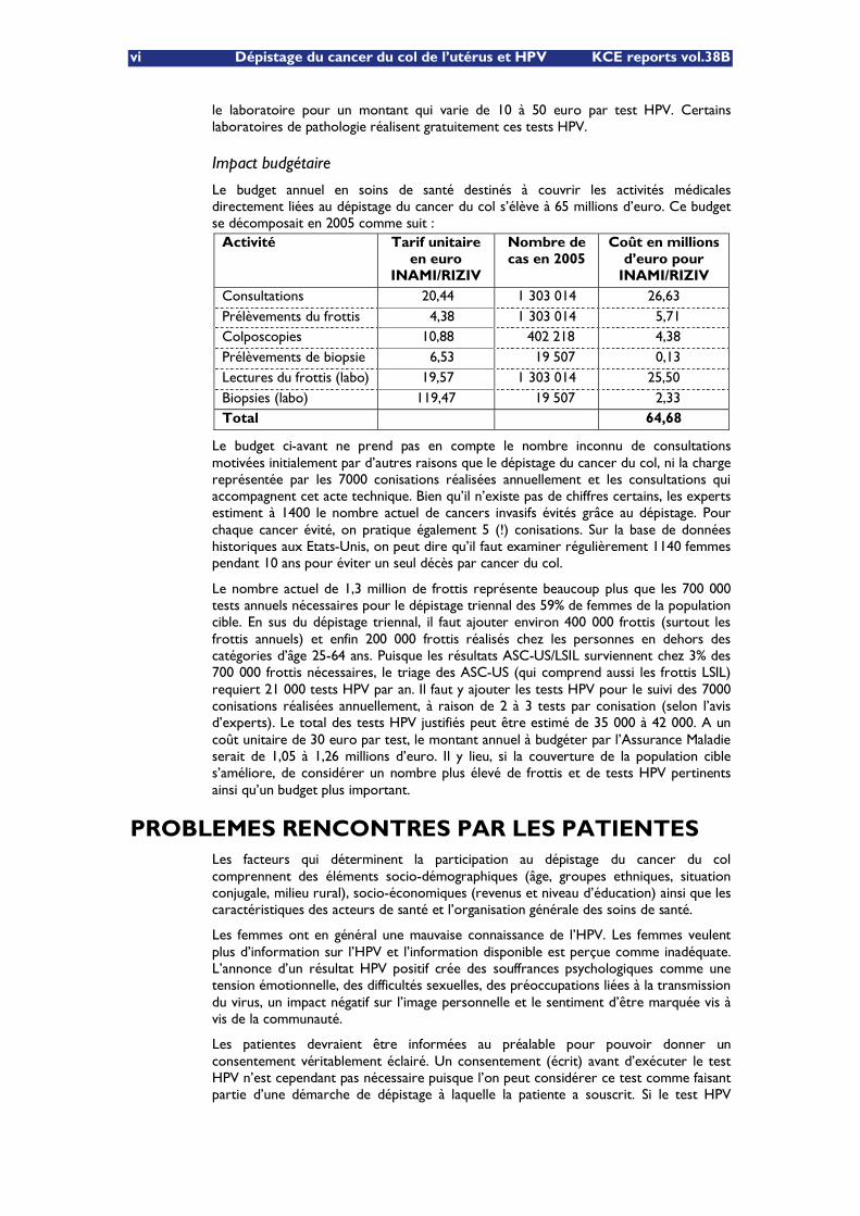
Le budget annuel en soins de santé destinés à couvrir les activités médicales directement liées au dépistage du cancer du col s�’élève à 65 millions d�’euro. Ce budget se décomposait en 2005 comme suit :
Activité Tarif unitaire en euro
INAMI/RIZIV
Nombre de cas en 2005
Coût en millions d�’euro pour
INAMI/RIZIV Consultations 20,44 1 303 014 26,63 Prélèvements du frottis 4,38 1 303 014 5,71 Colposcopies 10,88 402 218 4,38 Prélèvements de biopsie 6,53 19 507 0,13 Lectures du frottis (labo) 19,57 1 303 014 25,50 Biopsies (labo) 119,47 19 507 2,33 Total 64,68
Le budget ci-avant ne prend pas en compte le nombre inconnu de consultations motivées initialement par d�’autres raisons que le dépistage du cancer du col, ni la charge représentée par les 7000 conisations réalisées annuellement et les consultations qui accompagnent cet acte technique. Bien qu�’il n�’existe pas de chiffres certains, les experts estiment à 1400 le nombre actuel de cancers invasifs évités grâce au dépistage. Pour chaque cancer évité, on pratique également 5 (!) conisations. Sur la base de données historiques aux Etats-Unis, on peut dire qu�’il faut examiner régulièrement 1140 femmes pendant 10 ans pour éviter un seul décès par cancer du col.
Le nombre actuel de 1,3 million de frottis représente beaucoup plus que les 700 000 tests annuels nécessaires pour le dépistage triennal des 59% de femmes de la population cible. En sus du dépistage triennal, il faut ajouter environ 400 000 frottis (surtout les frottis annuels) et enfin 200 000 frottis réalisés chez les personnes en dehors des catégories d�’âge 25-64 ans. Puisque les résultats ASC-US/LSIL surviennent chez 3% des 700 000 frottis nécessaires, le triage des ASC-US (qui comprend aussi les frottis LSIL) requiert 21 000 tests HPV par an. Il faut y ajouter les tests HPV pour le suivi des 7000 conisations réalisées annuellement, à raison de 2 à 3 tests par conisation (selon l�’avis d�’experts). Le total des tests HPV justifiés peut être estimé de 35 000 à 42 000. A un coût unitaire de 30 euro par test, le montant annuel à budgéter par l�’Assurance Maladie serait de 1,05 à 1,26 millions d�’euro. Il y lieu, si la couverture de la population cible s�’améliore, de considérer un nombre plus élevé de frottis et de tests HPV pertinents ainsi qu�’un budget plus important.
PROBLEMES RENCONTRES PAR LES PATIENTES Les facteurs qui déterminent la participation au dépistage du cancer du col comprennent des éléments socio-démographiques (âge, groupes ethniques, situation conjugale, milieu rural), socio-économiques (revenus et niveau d�’éducation) ainsi que les caractéristiques des acteurs de santé et l�’organisation générale des soins de santé.
Les femmes ont en général une mauvaise connaissance de l�’HPV. Les femmes veulent plus d�’information sur l�’HPV et l�’information disponible est perçue comme inadéquate. L�’annonce d�’un résultat HPV positif crée des souffrances psychologiques comme une tension émotionnelle, des difficultés sexuelles, des préoccupations liées à la transmission du virus, un impact négatif sur l�’image personnelle et le sentiment d�’être marquée vis à vis de la communauté.
Les patientes devraient être informées au préalable pour pouvoir donner un consentement véritablement éclairé. Un consentement (écrit) avant d�’exécuter le test HPV n�’est cependant pas nécessaire puisque l�’on peut considérer ce test comme faisant partie d�’une démarche de dépistage à laquelle la patiente a souscrit. Si le test HPV
KCE reports vol.38B Dépistage du cancer du col de l�’utérus et HPV vii
s�’avère positif, il faut informer la patiente sur la signification de ce résultat et obtenir son consentement à poursuivre le traitement. Il faut encourager la mise à disposition avant le prélèvement de moyens concrets d�’information comme par exemple des dépliants sur le dépistage du cancer du col et sur la recherche d�’HPV (avec des brochures distinctes pour les interventions de suivi) ou l�’accès à un site internet central. L�’influence d�’interventions qui aident à la prise d�’une décision éclairée lorsqu�’il s�’agit de participer à un dépistage est toutefois modeste.
CONCLUSIONS ET RECOMMANDATIONS POUR LES PRENEURS DE DECISIONS
La cytologie classique ou validée en milieu liquide demeure le pilier du dépistage du cancer du col. La recherche de l�’HPV par un test validé s�’indique uniquement pour l�’orientation des ASC-US chez les femmes de 25 à 64 et pour le suivi après traitement de lésions néoplasiques. Les résultats de la cytologie (selon la classification de Bethesda) et de la recherche de l�’HPV devraient être mentionnés séparément mais faire l�’objet d�’un rapport unique.
Le niveau de connaissance des femmes à propos de l�’HPV est généralement faible. Un test positif peut inquiéter inutilement et semer un doute entre la femme et son partenaire. Il faut dès lors s�’interroger sur l�’opportunité d�’un dépistage non sélectif de l�’HPV tant que les résultats des études en cours ne seront pas connus. Il faut encourager la mise à disposition avant le prélèvement de moyens concrets d�’information comme par exemple des dépliants sur le dépistage du cancer du col et sur la recherche d�’HPV.
En Belgique, le dépistage du cancer du col est d�’abord opportuniste et non structuré. Le dépistage tous les 3 ans couvre seulement 59% des femmes de 25 à 64 ans alors que nombre d�’entre elles ont une fréquence exagérée d�’un frottis annuel. En Grande-Bretagne et en Scandinavie, un dépistage organisé permet d�’atteindre au moins 80% de la population cible. Un meilleur taux de dépistage dans la population concernée et l�’amélioration de la qualité des différentes étapes du dépistage procureront un bénéfice de santé de loin supérieur à celui espéré en cas d�’utilisation pertinente des tests HPV.
Il existe au niveau européen un large consensus pour que les activités de dépistage soient menées de préférence de façon organisée. La structure devrait adhérer aux recommandations européennes pour l�’assurance de qualité en matière de dépistage du cancer du col. Un programme de dépistage devrait être conçu de telle sorte qu�’il se prête à une évaluation régulière par l�’autorité compétente. La première étape doit être la création d�’un registre détaillé du dépistage. Ce registre devrait contenir les résultats de la cytologie et des tests HPV réalisés au cours du dépistage organisé ou en dehors de celui-ci, ainsi que (ou être couplé à) certains résultats anormaux et les mesures prises. Un tel registre devrait se positionner entre le registre de population et le registre du cancer et utiliser un code d�’identification gérable par la Sécurité Sociale. Il existe déjà un projet d�’Arrêté Royal portant sur l�’agrément des laboratoires de cytologie et de pathologie qui oblige ces laboratoires à participer à aux programmes externes d�’assurance qualité. La participation obligatoire de ces laboratoires à l�’enregistrement des données de dépistage pourrait s�’y ajouter s�’ils désirent bénéficier du remboursement de leur activité médicale par l�’INAMI/RIZIV.
Si plusieurs options s�’offrent pour l�’organisation du dépistage du cancer du col, toutes s�’articulent autour de la mise sur pied d�’un registre exhaustif. Ce n�’est qu�’ainsi que les femmes non examinées actuellement pourront être contactées. A l�’inverse, les personnes déjà examinées ne devront pas être contactées. Plusieurs options sont possibles et pour le contact et pour le prélèvement, pour autant qu�’un enregistrement correct et complet soit réalisé et reste accessible dans le respect du secret médical. Dans tous les cas, le médecin traitant et le gynécologue doivent être informés du résultat du dépistage du cancer du col. L�’organisation d�’un dépistage systématique du cancer du col devrait tenir compte des points suivants :
viii Dépistage du cancer du col de l�’utérus et HPV KCE reports vol.38B
La situation actuelle où le frottis est prélevé par le gynécologue ou le médecin traitant est probablement la solution la plus réaliste pour les femmes qui bénéficient déjà d�’un dépistage. Il n�’est pas certain que cette approche soit la plus coût-efficace. Cependant, la relation interpersonnelle entre la femme et son médecin permet d�’aborder en confiance d�’autres problèmes et contribue à la santé féminine en général.
Les femmes non encore prises en charge nécessiteront d�’autres approches. Certaines femmes, en particulier socio-économiquement défavorisées, ne se présenteront pas spontanément à un médecin pour un frottis. L�’organisation et l�’invitation des femmes au dépistage pourraient se faire au niveau régional, provincial ou local. Plusieurs options sont possibles pour l�’organisation comme par exemple le prélèvement du frottis par une infirmière spécialisée travaillant dans un local fixe ou une antenne mobile. L�’approche la plus coût-efficace n�’est pas connue, et une évaluation périodique s�’indique.
Comme le volume des examens réalisés chaque année est considérable, des contrats prix-volumes pourraient être envisagés entre l�’Assurance Maladie et les laboratoires de cyto-pathologie qui présentent les garanties nécessaires de qualité et de service.
Le budget annuel des soins de santé consacré aux activités médicales directement liées au dépistage du cancer du col doit être employé de manière plus efficiente et les activités doivent être coordonnées entre les communautés en charge de la prévention. Les activités médicales de sur-dépistage ne doivent pas être financées par l�’INAMI/RIZIV.
Le budget annuel pour la recherche d�’HPV dans les indications reconnues s�’élève à environ 1,2 millions d�’euro. Les moyens financiers devraient être dégagés pour créer et maintenir un système informatisé d�’enregistrement du dépistage, identifier et informer correctement la population cible, rendre le dépistage plus accessible, y compris sur le plan financier, pour atteindre les 41% de la population cible non encore dépistée.
Points clés
Aujourd�’hui, le dépistage opportuniste couvre moins de 59% des femmes de 25 à 64 ans alors que la couverture atteint au moins 80% dans les pays qui disposent d�’un dépistage bien organisé. Si les décideurs veulent réduire la mortalité liée au cancer du col, un dépistage bien organisé assorti d�’une assurance qualité �– au lieu du dépistage opportuniste actuel - s�’impose.
La mise sur pied d�’un registre obligatoire et exhaustif des résultats du dépistage et des cancers du col est essentielle.
La recherche d�’HPV n�’a pas d�’utilité avérée dans le dépistage primaire. Il y a lieu d�’attendre le résultat des études en cours. Les femmes ont droit à une information claire sur le test HPV pour éviter de les blesser psychologiquement. Un dépistage d�’HPV en cas de frottis ASC-US est indiqué, seulement pour environ 3% des patientes lors du dépistage, et pour le suivi après traitement de lésions cancéreuses.
Le budget annuel en soins de santé consacrés à la couverture des activités médicales directement liées au dépistage du cancer du col peut être employé de manière plus efficiente. Il est inconvenant que les prestations en rapport avec l�’excès de dépistage (le frottis annuel) soient encore financées par l�’Assurance Maladie.
Les diverses activités de dépistage qui existent ou démarreront doivent être intégrées de façon organisée et coordonnées au sein des différents programmes de dépistage en accord avec toutes les autorités concernées.
KCE reports vol.38 Cervical cancer screening and HPV 1
Table of contents
1 INTRODUCTION.............................................................................................................. 4
2 EFFECTIVENESS ............................................................................................................... 5 2.1 INTRODUCTION ........................................................................................................................................... 5 2.2 ASSESSMENT OF THE PERFORMANCE OF SCREENING TESTS.................................................... 6 2.3 CONVENTIONAL CYTOLOGY .............................................................................................................. 10
2.3.1 Description.......................................................................................................................................... 10 2.3.2 Performance........................................................................................................................................ 11 2.3.3 Conclusions......................................................................................................................................... 12
2.4 LIQUID-BASED CYTOLOGY .................................................................................................................... 13 2.4.1 Description.......................................................................................................................................... 13 2.4.2 Performance........................................................................................................................................ 14 2.4.3 Conclusions......................................................................................................................................... 19
2.5 AUTOMATED CYTOLOGY ...................................................................................................................... 19 2.5.1 Description of automated screening devices ............................................................................. 20 2.5.2 Performance........................................................................................................................................ 20 2.5.3 Conclusions......................................................................................................................................... 20
2.6 COLPOSCOPY............................................................................................................................................... 21 2.6.1 Description.......................................................................................................................................... 21 2.6.2 Performance of colposcopy ............................................................................................................ 21 2.6.3 Conclusions......................................................................................................................................... 21
2.7 HUMAN PAPILLOMAVIRUS (HPV) TESTING...................................................................................... 22 2.7.1 Description.......................................................................................................................................... 22 2.7.2 Performance in triage of minor cytological abnormalities ...................................................... 26 2.7.3 Performance in follow-up after treatment.................................................................................. 32 2.7.4 Performance in primary screening ................................................................................................ 33 2.7.5 Conclusions......................................................................................................................................... 39
2.8 GENERAL CONCLUSIONS ....................................................................................................................... 40
3 CERVICAL CANCER SCREENING IN OTHER EUROPEAN COUNTRIES ......... 41 3.1 INTRODUCTION ......................................................................................................................................... 41 3.2 ASPECTS OF ORGANISED SCREENING............................................................................................... 41
3.2.1 Rational of organised screening ..................................................................................................... 41 3.2.2 European Council Recommendation............................................................................................ 42 3.2.3 Target age groups.............................................................................................................................. 43
3.3 AN OVERVIEW OF CERVICAL CANCER SCREENING SYSTEMS IN EUROPE........................ 44 3.3.2 Evidence for finding that well-organised screening is more effective................................... 47 3.3.3 Two highlighted screening programmes...................................................................................... 51
3.4 RECOMMENDATIONS ............................................................................................................................... 53
4 CERVICAL CANCER SCREENING IN BELGIUM...................................................... 54 4.1 INTRODUCTION ......................................................................................................................................... 54 4.2 CERVICAL CANCER IN BELGIUM.......................................................................................................... 54 4.3 CERVICAL CANCER SCREENING ACTIVITY..................................................................................... 55
4.3.1 Regional initiatives for organized screening................................................................................ 55 4.3.2 Cytology-based testing..................................................................................................................... 57 4.3.3 HPV testing.......................................................................................................................................... 58
4.4 BUDGET, TESTING VOLUME AND COVERAGE............................................................................... 59

2 Cervical cancer screening and HPV KCE reports vol.38
5 PATIENT ISSUES ............................................................................................................ 64 5.1 SEARCH STRATEGY..................................................................................................................................... 64 5.2 DETERMINANTS OF PARTICIPATION................................................................................................. 64 5.3 INTERVENTIONS TO ENCOURAGE PARTICIPATION.................................................................. 65 5.4 CURRENT KNOWLEDGE OF HPV AND INFORMATION NEEDS............................................. 66 5.5 PSYCHOLOGICAL IMPACT AND ATTITUDES TOWARDS HPV TESTING............................ 66 5.6 INFORMED DECISION MAKING............................................................................................................. 67
6 APPENDICES ................................................................................................................... 71 6.1 APPENDIX 1. SEARCH STRATEGY LITERATURE ON PATIENT ISSUES.................................... 71

KCE reports vol.38 Cervical cancer screening and HPV 3
LIST OF ABBREVIATIONS AGC Atypical Glandular Cells ASC-US Atypical Squamous Cells of Undetermined Significance CI Confidence Interval CIN Cervical Intra-epithelial Neoplasia CP Conventional Pap smear HC2 Hybrid Capture II HPV Human Papilloma Virus HSIL High grade Squamous Intra-epithelial Lesions IARC International Agency for Research in Cancer, Lyon IPH Institute of Public Health, Brussels LBC Liquid Based Cytology LSIL Low grade Squamous Intra-epithelial Lesions NNS Number Needed to Screen NOS No Other Specification OR Odds Ratio Pap Papanicolaou PCR Polymerase Chain Reaction TBS The Bethesda System
4 Cervical cancer screening and HPV KCE reports vol.38
1 INTRODUCTION This KCE HTA project was conducted in collaboration with the Institute of Public Health, Brussels. The aim of this project was to document the effectiveness of cervical cancer screening and in particular the role of Human Papilloma Virus (HPV) testing. Screening tests considered include the conventional and liquid based cytology and in particular the HPV test. The cervical cancer screening situation in other countries as well as in Belgium is documented. However, performance of existing screening programmes was not explicitly compared. Patient issues such as women�’s expectations and attitudes concerning the HPV testing are reviewed. For those indications where HPV testing is found clinically effective we estimate the budget required, and formulate recommendations for the decision makers. No formal cost-effectiveness study or literature review was performed.
Infection of the cervix with one or more high-risk types of HPV is a necessary but insufficient condition for the later development of cervical cancer. Most of the women (and men) get asymptomatic infection with high-risk HPV types at some point during their sexually active life and most HPV infections will become undetectable without intervention. However, some infections will lead to cervical intra-epithelial neoplasia (CIN), which if left untreated may progress to invasive cancer. The aim of cervical cancer screening is to detect progressive CIN and, by its treatment, prevent progression to invasive cancer. The success of screening depends essentially on the participation of the target population, the quality of the screening test, and the efficacy of treatment of screen-detected lesions.
The current screening coverage in Belgium is only 59% in women 25 to 64 years old. External quality assurance of the current testing has yet to be implemented. We refer to the KCE report on molecular diagnostics for recommendations to assure the quality of molecular tests, including HPV tests. It is clear that beyond the appropriate introduction of HPV testing, a much larger population health improvement can be expected from an increased screening coverage of the target population and a quality improvement of different steps in the screening process. Preventive HPV vaccination is not discussed as it will be the subject of a subsequent KCE project and no immediate consequences on screening policy are expected. The outcome of the randomized trials on HPV primary screening, the full introduction of HPV vaccination as well as evolutions in methods to detect HPV and its cellular consequences may impact on cervical cancer screening and may require this document to be updated.
KCE reports vol.38 Cervical cancer screening and HPV 5
2 EFFECTIVENESS
2.1 INTRODUCTION
Screening for cervical cancer requires the use of a test, which is easy to perform by medical or paramedical personnel, available at an acceptable cost, causing minimal discomfort to the woman and has a high sensitivity and specificity for progressive intra-epithelial lesions (CIN), which are the precursor stages that precede the occurrence of invasive cancer. Evidence of effectiveness of a given cancer screening procedure should be based on its potential to reduce the morbidity and especially the mortality from the particular cancer. High sensitivity for the detection of CIN is an insufficient criterion for effectiveness, since CIN often regresses spontaneously. High specificity is required to avoid anxiety, subsequent unnecessary investigations and unnecessary treatment and side effects.
Cervical cancer screening using the conventional Pap smear partially fulfils these criteria. Cytological screening every three to five years can reduce morbidity and mortality from cervical cancer by 80% or more, if offered in a well-organised setting. Cytology-based screening traditionally involves 3 steps: finding cytological abnormalities in a Pap smear; histological confirmation of a biopsy taken under colposcopic control and treatment of the lesion that otherwise could develop into cancer.
Nevertheless, the test-validity, in particular the test sensitivity of the conventional Pap smear for CIN, is moderate: between 50 to 70% for CIN; but between 70 and 80% for high-grade CIN. Evidence of effectiveness of cytological screening using the Pap smear has essentially been derived from organised screening programmes. However, cytological screening in opportunistic settings is in general less effective (see chapter on international situation) and less cost-effective.
Occurrence of false-negative and unsatisfactory Pap smears was considered as a justification to develop new technologies such as liquid based cytology and automated screening devices. The quality of the evaluations of their performance was often poor, essentially limited to cross-sectional cytological outcomes and rarely verified by a valid gold standard. This chapter aims to assess differences in test performance and quality characteristics between the current standard screening test, which is the conventional Pap smear, and the newer alternatives of cervical cytology.
Colposcopy is only shortly described since it is not an appropriate screening method but rather a tool which is essential in the diagnostic work-up of screen-positive women.
Finally, this chapter presents the current state of the art concerning Human Papilloma Virus (HPV) testing evaluated in three possible settings: 1) primary screening; 2) triage of minor cervical lesions and 3) in follow-up after treatment of high-grade CIN. As the HPV test can be considered an additional test to or a replacement of the existing Pap smear, large studies are needed. Where cytological screening is already well-organised and quality assured, this also means that any possible gains after adding new tests are limited. Any loss in specificity, leading to increased costs should therefore be treated cautiously.
Before addressing the performance and the quality of all these test procedures we develop a methodology on how to evaluate their performance.
The Scientific Institute of Public Health, Brussels, (IPH) was in charge of the preparation of the new European Guidelines for Quality Assurance in Cervical Cancer Screening and collaborated with the International Agency for Research in Cancer (Lyon) and with the Gynaecological Cancer Cochrane Review Collaboration. In those frameworks several systematic reviews were conducted which concerned test performance of liquid-based cytology and the different applications of HPV testing 1-3. The current report contains updated summaries of this work. Interested readers, who want more information on the retrieval methods of references and on the applied statistical meta-analytical procedures, should contact IPH and request the specific reports (http://www.iph.fgov.be/epidemio/).
6 Cervical cancer screening and HPV KCE reports vol.38
2.2 ASSESSMENT OF THE PERFORMANCE OF SCREENING TESTS
The aim of cervical cancer screening is to detect progressive cervical intra-epithelial neoplasia (CIN1) and, by their treatment, prevent progression to invasive cancer 4.
The effectiveness of a screening programme is determined by the programme sensitivity. This programme sensitivity depends on the sensitivity of the chosen screening test for CIN of a given degree, the natural history of this degree of CIN, and the screening policy (the target age group, screening interval, and procedures for follow-up of positive screenees). The essential elements in the natural evolution of the disease are the rates of onset, progression and regression of precursor lesions and the distribution of their sojourn times. The mean sojourn time of CIN is at least 10 years and the probability of detection increases as the preclinical phase progresses 5, 6. Therefore, repetition of a moderately sensitive screen test, such as the Pap smear can reduce incidence of and mortality from cervical cancer to a low residual level 7. The reduction in the cumulative incidence of cancer is estimated to be respectively 91 and 84% due to well organised cytological screening every 3 or 5 years 8, 6.
The success of screening depends essentially on the participation of the target population and the quality of the screening test and further on the compliance and efficacy of treatment of screen-detected lesions.
In this chapter we focus on the performance of screening methods. We will describe and assess the performance of 5 main types of tests that are currently used in cervical cancer screening in Europe or that are proposed as an alternative or supplement for current methods:
The conventional Pap smear
Liquid based cytology
Automated cytology
Colposcopy
Detection of nucleic acid sequences of oncogenic Human papilloma viruses
For an overview of principles of good diagnostic research to evaluate test accuracy, we refer to The Cochrane Methods Group on Systematic Review of Screening and Diagnostic Tests: Recommended Methods 9 and Bossuyt 10.
Classifications
The 1988 version of The Bethesda Reporting System (TBS) was used for the cytological classification of the test result 11. We considered three threshold levels for positive cytology: atypical squamous cells of undetermined significance or worse (ASCUS+), low-grade squamous intra-epithelial lesions or worse (LSIL+) and high-grade intra-epithelial lesions or worse (HSIL+). Atypical glandular lesions were assimilated together within the ASCUS category. Categories of cytological abnormality, defined according to other reporting formats, were converted into TBS using translation tables as established before 1. At the 1991 Bethesda Workshop, it was proposed to sub-classify ASCUS into three sub-classes: "atypical squamous cells favouring a benign reactive process" (ASC-R), "atypical squamous cells of undetermined significance" (ASC-US) and ASC-H, "atypical squamous cells, HSIL cannot be ruled out" 12. At the 2001 Workshop, it was decided to integrate henceforth ASC-R into the group of "negative for intraepithelial lesion or malignancy" and to distinguish only "ASC-US" (with hyphen) and "ASC-H" (Solomon 2002). In our main meta-analyses, we accepted studies using TBS2001 and providing data for equivocal cytology, if they included ASC-US (alone) or ASC-US and ASC-H (combined). Studies considering ASC-H alone or atypical glandular cells alone were excluded.
1 In this chapter �“CIN�” (cervical intra-epithelial neoplasia) is used for histologically confirmed lesions, while the �“SIL�” (Bethesda) terminology is used to describe cytological findings.

KCE reports vol.38 Cervical cancer screening and HPV 7
We used the CIN nomenclature to describe histological outcomes 13.
A list of outcomes for programme effectiveness of cervical cancer screening methods, assessed by different study methods, is enumerated in Table 1 and ranked from high to low according to the level of evidence that such studies provide.
In Table 2, we show a short list of six design topics (a to f) that are particularly important in the evaluation of the accuracy of cervical cancer screening tests; within each topic study types are ranked by quality of design.
Five categories of parameters are compared between LBC and conventional cytology.
The observed test positivity rates defined at different cytological cut-offs
The positive predictive value at each level of cytological abnormality to find histologically confirmed cervical intraepithelial neoplasia of grade 2 of worse (CIN2+)
Diagnostic accuracy (sensitivity, specificity) for CIN2+
The proportion of unsatisfactory preparations, the proportion of smears lacking endocervical cells and the reasons for judging as unsatisfactory and, fifth
The time needed for cytological reading.
Table 1 Ranking of studies by level of decreasing evidence for effectiveness of cervical cancer screening methods according to the studied outcome and the used study design.
Study outcome:
1 Reduction of mortality from cervical cancer, life-years gained
2 Reduction of morbidity due to cervical cancer: incidence of cancer (Ib+), Quality adjusted life-years gained
3 Reduction of incidence of cancer (including micro-invasive cancer).
4 Reduction of incidence of CIN3 or worse disease (CIN3+).
5 Increased detection rate of CIN3+ or CIN2+.
6 Increased test positivity with increased, similar or hardly reduced positive predictive value
Study design2:
1 Randomised clinical trials, randomised population based trials
2 Cohort studies
3 Case-control studies
4 Trend studies, ecological studies on routinely collected data
5 Cross-sectional studies evaluating diagnostic test accuracy
2 Only controlled studies are considered, this means studies where two or more screening methods are compared.
8 Cervical cancer screening and HPV KCE reports vol.38
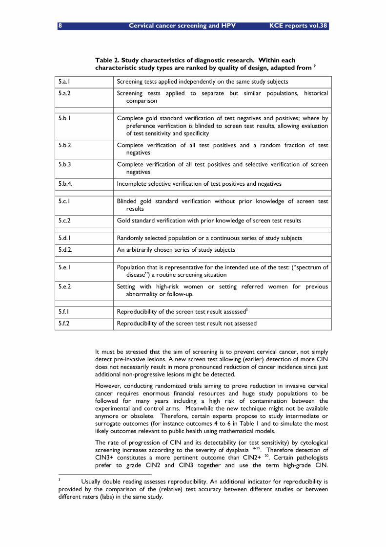
Table 2. Study characteristics of diagnostic research. Within each characteristic study types are ranked by quality of design, adapted from 9
5.a.1 Screening tests applied independently on the same study subjects
5.a.2 Screening tests applied to separate but similar populations, historical comparison
5.b.1 Complete gold standard verification of test negatives and positives; where by preference verification is blinded to screen test results, allowing evaluation of test sensitivity and specificity
5.b.2 Complete verification of all test positives and a random fraction of test negatives
5.b.3 Complete verification of all test positives and selective verification of screen negatives
5.b.4. Incomplete selective verification of test positives and negatives
5.c.1 Blinded gold standard verification without prior knowledge of screen test results
5.c.2 Gold standard verification with prior knowledge of screen test results
5.d.1 Randomly selected population or a continuous series of study subjects
5.d.2. An arbitrarily chosen series of study subjects
5.e.1 Population that is representative for the intended use of the test: (�“spectrum of disease�”) a routine screening situation
5.e.2 Setting with high-risk women or setting referred women for previous abnormality or follow-up.
5.f.1 Reproducibility of the screen test result assessed3
5.f.2 Reproducibility of the screen test result not assessed
It must be stressed that the aim of screening is to prevent cervical cancer, not simply detect pre-invasive lesions. A new screen test allowing (earlier) detection of more CIN does not necessarily result in more pronounced reduction of cancer incidence since just additional non-progressive lesions might be detected.
However, conducting randomized trials aiming to prove reduction in invasive cervical cancer requires enormous financial resources and huge study populations to be followed for many years including a high risk of contamination between the experimental and control arms. Meanwhile the new technique might not be available anymore or obsolete. Therefore, certain experts propose to study intermediate or surrogate outcomes (for instance outcomes 4 to 6 in Table 1 and to simulate the most likely outcomes relevant to public health using mathematical models.
The rate of progression of CIN and its detectability (or test sensitivity) by cytological screening increases according to the severity of dysplasia 14-19. Therefore detection of CIN3+ constitutes a more pertinent outcome than CIN2+ 20. Certain pathologists prefer to grade CIN2 and CIN3 together and use the term high-grade CIN.
3 Usually double reading assesses reproducibility. An additional indicator for reproducibility is provided by the comparison of the (relative) test accuracy between different studies or between different raters (labs) in the same study.
KCE reports vol.38 Cervical cancer screening and HPV 9
Nevertheless, in screening research, CIN3 should be the aimed outcome since the diagnosis of CIN2 is contaminated by under-reported CIN1. Moreover the diagnosis of CIN3 has a higher reproducibility than CIN2. CIN1 is a much less relevant outcome since most mild dysplasia does not progress 16, 21.
The assessment of the diagnostic validity, expressed in terms of sensitivity, requires the explicit definition of test-thresholds for test positivity and disease. It can be evaluated by application of screen tests to a relevant screening population followed by verification of all subjects with an accurate gold standard. It can be assumed that histological examination of material obtained by colposcopy/biopsy, loop excision or endocervical curettage, provides complete ascertainment of the true disease status. This might in fact not be true, but independent verification with an imperfect gold standard will attract sensitivity and specificity ratios (sensitivytest1/sentistivitytest2; specificitytest1/specificitytest2) towards unity. Therefore observed accuracy ratios are to be considered as minimum estimates. When tests and gold standard are positively correlated, then, sensitivity and specificity will be systematically overestimated.
In routine practice and even in many studies, colposcopy and histology are not applied to screen negatives, which includes a serious risk of verification bias. Nevertheless, when 2 screen tests are applied to the same study subjects and all subjects, positive for one or both tests, are verified with an acceptable gold standard, unbiased estimation of the test positive predictive value, the relative sensitivity and detection rate of true positives is possible 22, 23 4 . The same is true for randomized clinical trials, where different tests are applied to different subjects. When a random sample of screen-negatives are verified, an inferred sensitivity and specificity can be computed 24-26.
When the prevalence of disease is low, an approximated test specificity can be computed, even without systematic verification of a random sample of test-negatives, from the ratio of the number of test-negatives over the total number of study subjects minus the true positives 4. (Specificityapprox= # test negatives / (N �– # true positives); where N = the number of all tested individuals).
The reliability or reproducibility of a test expresses the capacity to obtain the same test result �– correct or not �– when the screening test is repeated on the same individual. The reliability depends on the definition of distinct test criteria that can be applied by skilled personnel. Poor reproducibility automatically yields low average sensitivity and specificity.
Once again, it must be repeated that the observation of increased sensitivity of a new test for histologically confirmed CIN does not necessarily imply that its inclusion in a screening programme will yield a reduction in incidence of lethal cervical cancer with respect to conventional cytological screening. Nevertheless, when biological and epidemiological arguments justify the assumption that the lesions detected in excess by the new method have a substantial chance of progression (acceptable longitudinal positive predictive value) and that screen negatives have a substantially lower chance to develop cancer in the future (higher longitudinal negative predictive value), planning of the new test in a randomized population- based trial in an organized setting can be considered.
Until now we studied essentially programme effectiveness stressing test sensitivity. Cervical cancer screening addresses large populations and are therefore extremely costly. Costs are largely determined by the test specificity.
An overview of the cost components attributed to screening is presented in Table 3.
4 The same is true when different tests are studied in different populations as long as the prevalence of disease can be assumed to be the same (e.g. in randomised trials) .

10 Cervical cancer screening and HPV KCE reports vol.38
Table 3. Overview of cost components of a screening programme
1 Cost price of the screen-test (investment and recurrent costs); fees of health professionals (time for preparation, interpretation of the screen test, documentation, training); information of the client (obtaining informed consent if required); logistical costs (transport, processing, storage); administrative costs (invitation, registration and analysis of data).
2 Specificity of the screen test: cost of follow-up and treatment of women with false-positive results or having non-progressive screen-detected lesions (over-diagnosis).
3 Sensitivity of the screen test (longitudinal): cost for follow-up and treatment of true positives; this cost may be off-set by cost savings in avoided treatment of advanced disease.
4 Human costs: time spent by women to be screened, anxiety and discomfort for follow-up and/or treatment of women with true and false-positive results and consequences of delay in detection of cancer in false-negative women.
5 Specificity of quality control, triage and diagnostic follow-up procedures, contributing to increased positive predictive value and savings by avoiding treatment of false-positive women.
6 Quality of screen test procedures; satisfactory rate influencing the need for repeat tests.
A small decrease in specificity can have dramatic consequences on costs. The number of additional false positives is computed from nearly the complete target population, since the prevalence of progressive cervical cancer precursors is low. Nevertheless, the loss in specificity can be limited by raising the screening interval, by increasing the age at onset of screening and by increasing the cut-off for test positivity.
2.3 CONVENTIONAL CYTOLOGY
2.3.1 Description
Cells are collected with a sampling device from the surface of the transformation zone of the uterine cervix. It is important to ensure the entire squamocolumnar junction is sampled, since this is the site where most CIN lesions develop. Cells are either directly smeared on a glass slide, dried and ethanol-fixed, or transferred to a liquid medium. For microscopic evaluation by a cytologist the cells must be stained. The cells are then analysed using a microscope.
The basic assumption of cytological diagnosis is that it is related to the histology of the relevant tissue. This means that there is an equivalent appearance of cells even after the cells are detached from tissue and all three-dimensional information is lost. Cytological findings should be categorised according to an established reporting system. The European guidelines strongly recommend that all terminology systems should be translatable into the categories of the Bethesda system (TBS) 27.
Conventional cytology is still the standard method for primary cervical cancer screening. Repetition of the Pap smear is used as triage method in case of minor cytological lesions and as follow-up method after treatment of lesions.
The judgement of the quality of a smear is an essential component of the cytological interpretation of a Pap smear. At a minimum, TBS criteria for conventional smear and LBC should be used and reasons for inadequacy should be provided on the cytology report 27.
If HPV testing is done in addition to cytology, the virological result and the cytological findings should be integrated in one report under the responsibility of a cytopathologist.
KCE reports vol.38 Cervical cancer screening and HPV 11
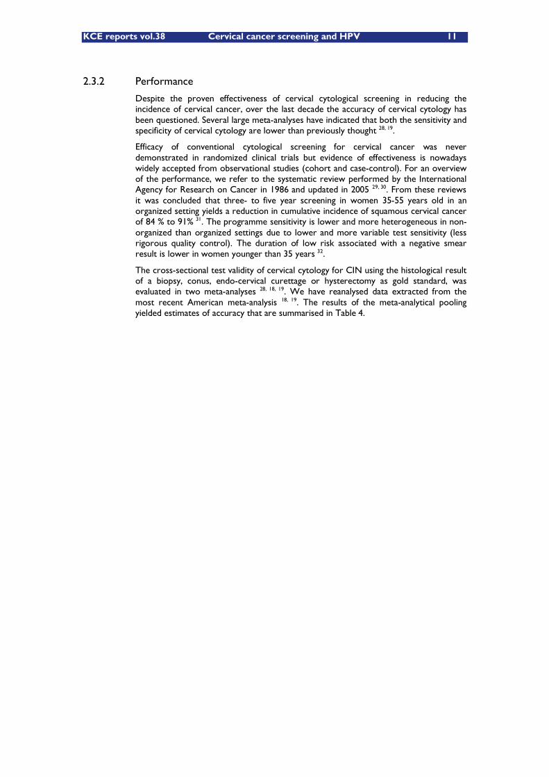
2.3.2 Performance
Despite the proven effectiveness of cervical cytological screening in reducing the incidence of cervical cancer, over the last decade the accuracy of cervical cytology has been questioned. Several large meta-analyses have indicated that both the sensitivity and specificity of cervical cytology are lower than previously thought 28, 19.
Efficacy of conventional cytological screening for cervical cancer was never demonstrated in randomized clinical trials but evidence of effectiveness is nowadays widely accepted from observational studies (cohort and case-control). For an overview of the performance, we refer to the systematic review performed by the International Agency for Research on Cancer in 1986 and updated in 2005 29, 30. From these reviews it was concluded that three- to five year screening in women 35-55 years old in an organized setting yields a reduction in cumulative incidence of squamous cervical cancer of 84 % to 91% 31. The programme sensitivity is lower and more heterogeneous in non-organized than organized settings due to lower and more variable test sensitivity (less rigorous quality control). The duration of low risk associated with a negative smear result is lower in women younger than 35 years 32.
The cross-sectional test validity of cervical cytology for CIN using the histological result of a biopsy, conus, endo-cervical curettage or hysterectomy as gold standard, was evaluated in two meta-analyses 28, 18, 19. We have reanalysed data extracted from the most recent American meta-analysis 18, 19. The results of the meta-analytical pooling yielded estimates of accuracy that are summarised in Table 4.
12 Cervical cancer screening and HPV KCE reports vol.38
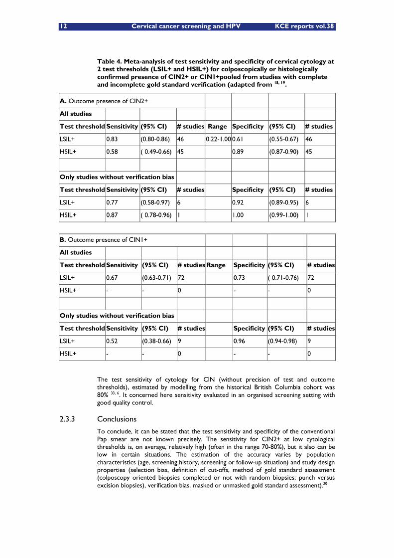
Table 4. Meta-analysis of test sensitivity and specificity of cervical cytology at 2 test thresholds (LSIL+ and HSIL+) for colposcopically or histologically confirmed presence of CIN2+ or CIN1+pooled from studies with complete and incomplete gold standard verification (adapted from 18, 19.
A. Outcome presence of CIN2+
All studies
Test threshold Sensitivity (95% CI) # studies Range Specificity (95% CI) # studies
LSIL+ 0.83 (0.80-0.86) 46 0.22-1.00 0.61 (0.55-0.67) 46
HSIL+ 0.58 ( 0.49-0.66) 45 0.89 (0.87-0.90) 45
Only studies without verification bias
Test threshold Sensitivity (95% CI) # studies Specificity (95% CI) # studies
LSIL+ 0.77 (0.58-0.97) 6 0.92 (0.89-0.95) 6
HSIL+ 0.87 ( 0.78-0.96) 1 1.00 (0.99-1.00) 1
B. Outcome presence of CIN1+
All studies
Test threshold Sensitivity (95% CI) # studies Range Specificity (95% CI) # studies
LSIL+ 0.67 (0.63-0.71) 72 0.73 ( 0.71-0.76) 72
HSIL+ - - 0 - - 0
Only studies without verification bias
Test threshold Sensitivity (95% CI) # studies Specificity (95% CI) # studies
LSIL+ 0.52 (0.38-0.66) 9 0.96 (0.94-0.98) 9
HSIL+ - - 0 - - 0
The test sensitivity of cytology for CIN (without precision of test and outcome thresholds), estimated by modelling from the historical British Columbia cohort was 80% 33, 6. It concerned here sensitivity evaluated in an organised screening setting with good quality control.
2.3.3 Conclusions
To conclude, it can be stated that the test sensitivity and specificity of the conventional Pap smear are not known precisely. The sensitivity for CIN2+ at low cytological thresholds is, on average, relatively high (often in the range 70-80%), but it also can be low in certain situations. The estimation of the accuracy varies by population characteristics (age, screening history, screening or follow-up situation) and study design properties (selection bias, definition of cut-offs, method of gold standard assessment (colposcopy oriented biopsies completed or not with random biopsies; punch versus excision biopsies), verification bias, masked or unmasked gold standard assessment).30
KCE reports vol.38 Cervical cancer screening and HPV 13
Nevertheless, convincing evidence is available with respect to the effectiveness of cytological screening, if offered in a well organised setting with quality control at all levels.
2.4 LIQUID-BASED CYTOLOGY
2.4.1 Description
Liquid-Based cytology (LBC) was introduced in the mid-1990s as a way to improve the performance of the conventional test. The cells are transferred into a vial with a liquid preservative solution that is transported to the laboratory where the slide is prepared. The cells are not spread directly onto a slide to obtain a conventional Pap (CP) smear but transferred into a vial with a fixative liquid. This vial is then sent to a specially equipped laboratory. Several systematic reviews regarding the performance of LBC to detect cervical cancer precursors were performed over the last 8 years 34, 35, 18, 36-47}. Conclusions formulated by the reviewing authors were disparate and depended largely on selection criteria to include individual studies and the considered performance parameters. Studies comparing detection rates for low grade cytological abnormalities often yielded more favourable results for LBC 34, 36, 39, 48, whereas in studies focusing on accuracy for biopsy-confirmed high-grade CIN (cervical intraepithelial neoplasia), no significant differences between the CP (conventional Pap smear) and LBC were found 40,
43, 47.
2.4.1.1 Liquid-based cytology techniques
A number of different LBC techniques are in use worldwide. These include ThinPrep®, Surepath® (formerly, CytoRich and AutoCyte PREP), Cytoscreen®, Cyteasy®, Labonord Easy Prep, Cytoslide, SpinThin and PapSpin.
So far, ThinPrep® and Surepath® are approved for use in the USA by the Food and Drug Administration (FDA) allowing the claim of increased detection of squamous intraepithelial lesions and a reduction of the number of unsatisfactory smears compared to the CP 49, 50. In the ThinPrep the liquid preservative solution is filtered through a membrane filter with a pore size specifically designed to trap epithelial cells. The epithelial cells collected on the membrane filter are then transferred on a glass slide and stained. In the Surepath system, concentration of cells is based on sedimentation through a density gradient.
2.4.1.2 Study design
Numerous studies have evaluated the comparative performance of the two most commonly used LBC methods, ThinPrep and Surepath, and conventional cytology with respect to test positivity, their sensitivity and specificity for identification of CIN, the specimen adequacy and the time required for evaluation of the specimens. Although there is a reasonable agreement that LBC improves specimen adequacy and reduces screening time compared to conventional cytology, there is considerable controversy surrounding the relative sensitivity and specificity of the two approaches, largely due to a lack of well designed studies 30.
Most of the comparative studies have utilized one of two types of study design:
The concomitant testing design (mainly �“split-sample�”)
The two-cohort design (�“direct-to-vial�”)
In the concomitant testing design, 2 samples are prepared from the same subject. Most often one single sample is taken from the uterine cervix and a CP (conventional smear) is prepared first, followed by transfer of the residual cellular material remnant on the sampling device into a vial with fixative liquid ("split-sample"). Occasionally, two separate samples are collected: one for the CP and another one for LBC. In the two-cohort design, CP samples and LBC samples are taken from separate populations.
14 Cervical cancer screening and HPV KCE reports vol.38
Both study design have significant limitations. With split-sample studies, it is difficult to ensure that the two cytology specimens are comparable and this design would seem to lead to bias against LBC since only the material remnant on the sampling device after preparation of a conventional smear can be used for LBC. It is possible that some diagnostic elements included in the CP-split sample are not available anymore for the LBC. In the two-cohort design it has been argued that the historical controls introduce other biases as the comparability of the populations being compared and expectation bias.
The other major limitations found in most of the studies evaluating LBC are the lack of comparison of test performance with a gold standard (�“blinded colposcopy/biopsy) and study population of women followed-up for a previous abnormal test result rather than women undergoing routine screening. Large, randomized controlled clinical trials need to be conducted. One large randomized trial is currently ongoing in The Netherlands but the results are not yet available.
2.4.2 Performance
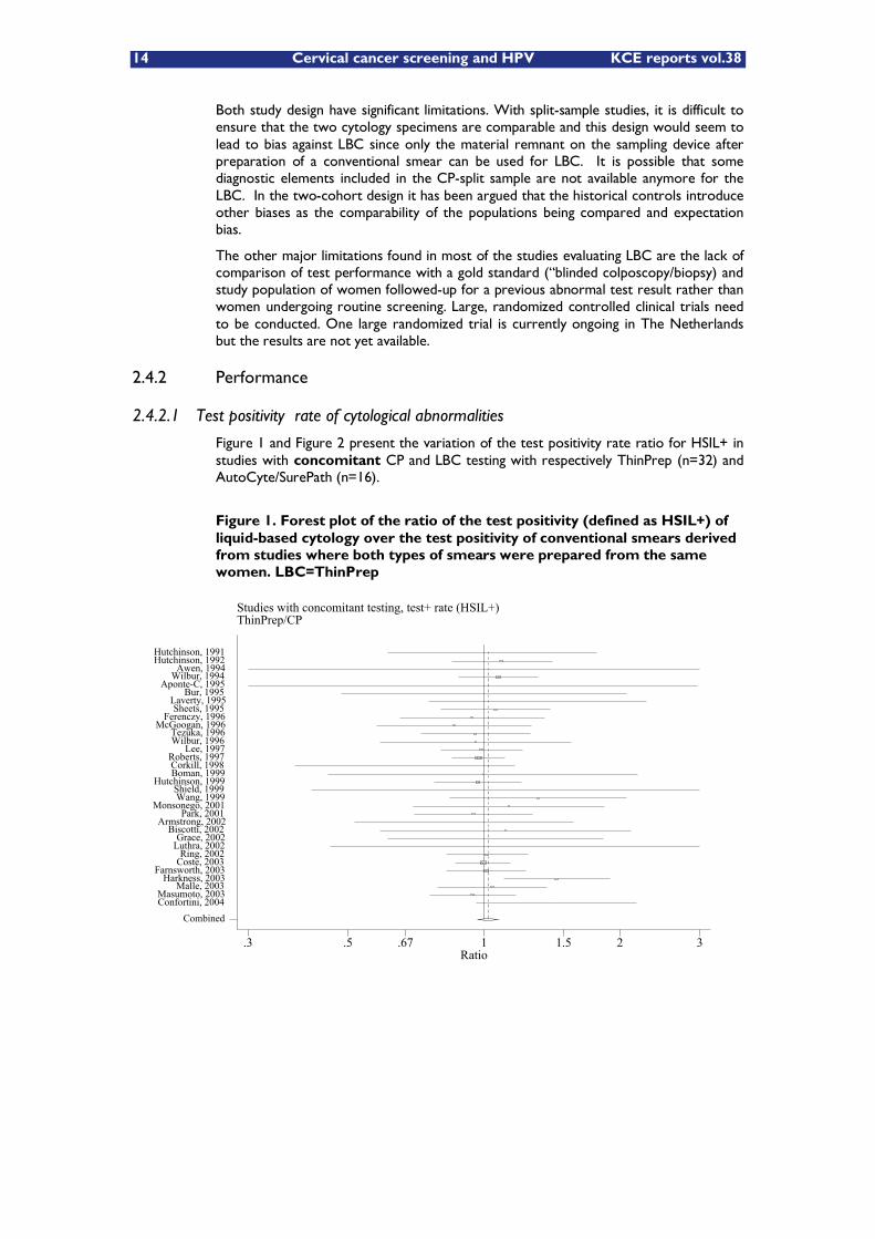
2.4.2.1 Test positivity rate of cytological abnormalities
Figure 1 and Figure 2 present the variation of the test positivity rate ratio for HSIL+ in studies with concomitant CP and LBC testing with respectively ThinPrep (n=32) and AutoCyte/SurePath (n=16).
Figure 1. Forest plot of the ratio of the test positivity (defined as HSIL+) of liquid-based cytology over the test positivity of conventional smears derived from studies where both types of smears were prepared from the same women. LBC=ThinPrep
Studies with concomitant testing, test+ rate (HSIL+)ThinPrep/CP
Ratio.3 .5 .67 1 1.5 2 3
Combined Confortini, 2004 Masumoto, 2003
Malle, 2003 Harkness, 2003
Farnsworth, 2003 Coste, 2003 Ring, 2002
Luthra, 2002 Grace, 2002
Biscotti, 2002 Armstrong, 2002
Park, 2001 Monsonego, 2001
Wang, 1999 Shield, 1999
Hutchinson, 1999 Boman, 1999 Corkill, 1998 Roberts, 1997
Lee, 1997 Wilbur, 1996 Tezuka, 1996
McGoogan, 1996 Ferenczy, 1996
Sheets, 1995 Laverty, 1995
Bur, 1995 Aponte-C, 1995
Wilbur, 1994 Awen, 1994
Hutchinson, 1992 Hutchinson, 1991
KCE reports vol.38 Cervical cancer screening and HPV 15
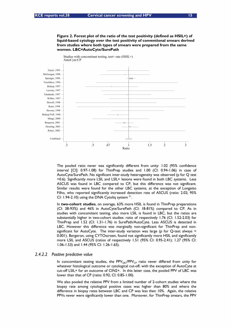
Figure 2. Forest plot of the ratio of the test positivity (defined as HSIL+) of liquid-based cytology over the test positivity of conventional smears derived from studies where both types of smears were prepared from the same women. LBC=AutoCyte/SurePath
The pooled ratio never was significantly different from unity: 1.02 (95% confidence interval [CI]: 0.97-1.08) for ThinPrep studies and 1.00 (CI: 0.94-1.06) in case of AutoCyte/SurePath. No significant inter-study heterogeneity was observed (p for Q test >0.6). Significantly more LSIL and LSIL+ lesions were found in both LBC systems. Less ASCUS was found in LBC compared to CP, but this difference was not significant. Similar results were found for the other LBC systems, at the exception of Longatto Filho, who reported significantly increased detection rate of ASCUS (ratio: 2.02; 95% CI: 1.94-2.10) using the DNA Cytoliq system 51.
In two-cohort studies, on average, 63% more HSIL is found in ThinPrep preparations (CI: 38-93%) and 46% in AutoCyte/SurePath (CI: 18-81%) compared to CP. As in studies with concomitant testing, also more LSIL is found in LBC, but the ratios are substantially higher in two-cohort studies: ratio of respectively 1.76 (CI: 1.52-2.03) for ThinPrep and 1.52 (CI: 1.31-1.76) in SurePath/AutoCyte. Less ASCUS is detected in LBC. However this difference was marginally non-significant for ThinPrep and non-significant for AutoCyte. The inter-study variation was large (p for Q-test always < 0.001). Bergeron, using CYTOscreen, found not significantly more HSIL and significantly more LSIL and ASCUS (ratios of respectively 1.51 (95% CI: 0.95-2.41); 1.27 (95% CI: 1.06-1.53) and 1.44 (95% CI: 1.26-1.65).
2.4.2.2 Positive predictive value
In concomitant testing studies, the PPVLBC/PPVCP ratio never differed from unity for whatever histological outcome or cytological cut-off, with the exception of AutoCyte at cut-off LSIL+ for an outcome of CIN2+. In this latter case, the pooled PPV of LBC was lower than that of CP (ratio: 0.92; CI: 0.85-1.00).
We also pooled the relative PPV from a limited number of 2-cohort studies where the biopsy rate among cytological positive cases was higher than 80% and where the difference in biopsy rates between LBC and CP was less then 10%. Again, the relative PPVs never were significantly lower than one. Moreover, for ThinPrep smears, the PPV
Studies with concomitant testing, test+ rate (HSIL+)AutoCyte/CP
Ratio.3 .5 .67 1 1.5 2 3
Combined
Robyr, 2002 Hessling, 2001
Bergeron, 2001 Minge, 2000
Bishop/TriP, 1998
Stevens, 1998 Kunz, 1998
Howell, 1998 Wilbur, 1997
Takahashi, 1997 Laverty, 1997 Bishop, 1997
Vassilakos, 1996 Sprenger, 1996
McGoogan, 1996 Geyer, 1993
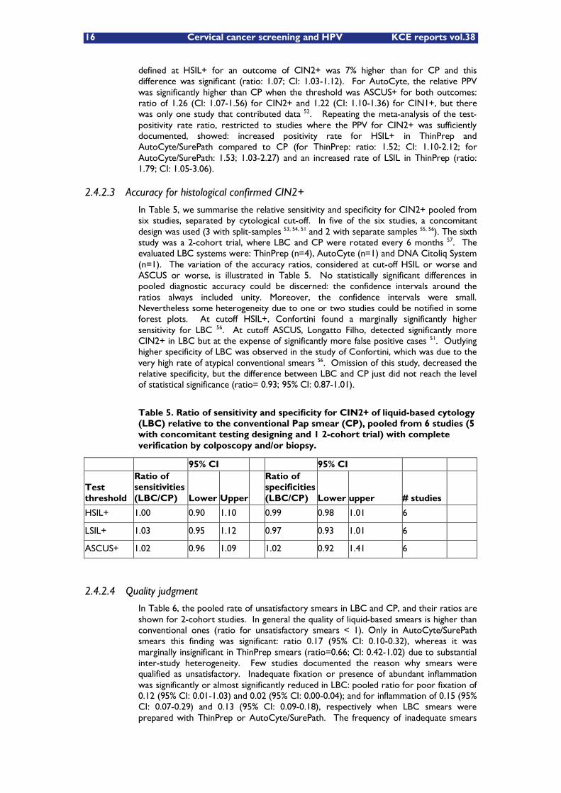
16 Cervical cancer screening and HPV KCE reports vol.38
defined at HSIL+ for an outcome of CIN2+ was 7% higher than for CP and this difference was significant (ratio: 1.07; CI: 1.03-1.12). For AutoCyte, the relative PPV was significantly higher than CP when the threshold was ASCUS+ for both outcomes: ratio of 1.26 (CI: 1.07-1.56) for CIN2+ and 1.22 (CI: 1.10-1.36) for CIN1+, but there was only one study that contributed data 52. Repeating the meta-analysis of the test-positivity rate ratio, restricted to studies where the PPV for CIN2+ was sufficiently documented, showed: increased positivity rate for HSIL+ in ThinPrep and AutoCyte/SurePath compared to CP (for ThinPrep: ratio: 1.52; CI: 1.10-2.12; for AutoCyte/SurePath: 1.53; 1.03-2.27) and an increased rate of LSIL in ThinPrep (ratio: 1.79; CI: 1.05-3.06).
2.4.2.3 Accuracy for histological confirmed CIN2+
In Table 5, we summarise the relative sensitivity and specificity for CIN2+ pooled from six studies, separated by cytological cut-off. In five of the six studies, a concomitant design was used (3 with split-samples 53, 54, 51 and 2 with separate samples 55, 56). The sixth study was a 2-cohort trial, where LBC and CP were rotated every 6 months 57. The evaluated LBC systems were: ThinPrep (n=4), AutoCyte (n=1) and DNA Citoliq System (n=1). The variation of the accuracy ratios, considered at cut-off HSIL or worse and ASCUS or worse, is illustrated in Table 5. No statistically significant differences in pooled diagnostic accuracy could be discerned: the confidence intervals around the ratios always included unity. Moreover, the confidence intervals were small. Nevertheless some heterogeneity due to one or two studies could be notified in some forest plots. At cutoff HSIL+, Confortini found a marginally significantly higher sensitivity for LBC 56. At cutoff ASCUS, Longatto Filho, detected significantly more CIN2+ in LBC but at the expense of significantly more false positive cases 51. Outlying higher specificity of LBC was observed in the study of Confortini, which was due to the very high rate of atypical conventional smears 56. Omission of this study, decreased the relative specificity, but the difference between LBC and CP just did not reach the level of statistical significance (ratio= 0.93; 95% CI: 0.87-1.01).
Table 5. Ratio of sensitivity and specificity for CIN2+ of liquid-based cytology (LBC) relative to the conventional Pap smear (CP), pooled from 6 studies (5 with concomitant testing designing and 1 2-cohort trial) with complete verification by colposcopy and/or biopsy.
95% CI 95% CI
Test threshold
Ratio of sensitivities (LBC/CP) Lower Upper
Ratio of specificities (LBC/CP) Lower upper # studies
HSIL+ 1.00 0.90 1.10 0.99 0.98 1.01 6
LSIL+ 1.03 0.95 1.12 0.97 0.93 1.01 6
ASCUS+ 1.02 0.96 1.09 1.02 0.92 1.41 6
2.4.2.4 Quality judgment
In Table 6, the pooled rate of unsatisfactory smears in LBC and CP, and their ratios are shown for 2-cohort studies. In general the quality of liquid-based smears is higher than conventional ones (ratio for unsatisfactory smears < 1). Only in AutoCyte/SurePath smears this finding was significant: ratio 0.17 (95% CI: 0.10-0.32), whereas it was marginally insignificant in ThinPrep smears (ratio=0.66; CI: 0.42-1.02) due to substantial inter-study heterogeneity. Few studies documented the reason why smears were qualified as unsatisfactory. Inadequate fixation or presence of abundant inflammation was significantly or almost significantly reduced in LBC: pooled ratio for poor fixation of 0.12 (95% CI: 0.01-1.03) and 0.02 (95% CI: 0.00-0.04); and for inflammation of 0.15 (95% CI: 0.07-0.29) and 0.13 (95% CI: 0.09-0.18), respectively when LBC smears were prepared with ThinPrep or AutoCyte/SurePath. The frequency of inadequate smears
KCE reports vol.38 Cervical cancer screening and HPV 17
due to obscuration by blood was not significantly different in LBC or CP. Significantly less smears judged unsatisfactory because of scanty cells was found in AutoCyte smears (ratio=0.13; 95% CI: 0.02-0.94). Absence of endocervical columnar cells (EC-) was noted more frequently in ThinPrep (ratio=1.15; 95% CI: 0.78-1.70) and less frequently in AutoCyte (ratio=0.87; 95% CI: 0.42-1.83), but these differences were statistically insignificant.
2.4.2.5 Duration of cytological reading
In 10 studies, the average duration to read the smear was measured (data not shown but available at the aforementioned website address). The simple mean was 237 and 338 seconds respectively for LBC and CP (a reduction of 30%). No confidence intervals could be computed since most studies did not report standard errors.
2.4.2.6 Impact of co-variate factors
The impact of the following covariates on study outcomes was assessed: composition of the study population, clinical setting (screening, follow-up or mixed), the version of the LBC-system (betaTP, TP2000, TP3000; CytoRich, AutoCyte PREP, SurePath, other LBC systems), collection devices, training of smear takers and readers, blinding of screeners, reviewers, colposcopists, and histologists, quality control of cytotechnologists' 1st diagnosis, definition and completeness of golden standard verification, thresholds for cytology and histology, length of follow-up period, and last but not least the disclosed interests of the researcher and involvement of the manufacturers of devices
Meta-regression did not reveal any significant influence of study characteristics on the ratio of detection of HSIL or LSIL+ in split-sample studies. In two-cohort studies however, training of cytologists and year of publication contributed significantly in explaining a part of the inter-study heterogeneity. The HSIL detection ratio was on average 39.9% (95% CI: 33.8-46.2%) higher when cytologists were trained just before the study compared to studies where cytologists had a thorough experience in reading LBC. On average, the HSIL and LSIL detection rate ratios were respectively 26.5% (24.7-28.3%) and 20.5% (95% CI 18.5-22.6%) lower if the study was published after 2000, compared to studies published earlier.
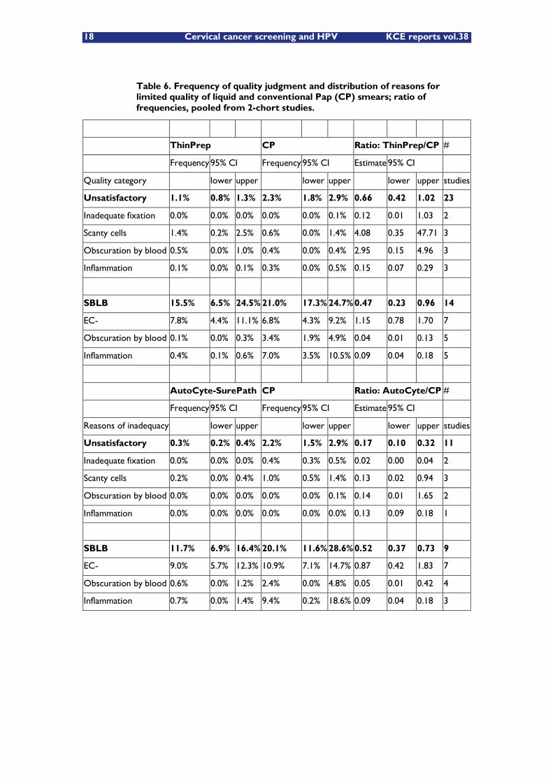
18 Cervical cancer screening and HPV KCE reports vol.38
Table 6. Frequency of quality judgment and distribution of reasons for limited quality of liquid and conventional Pap (CP) smears; ratio of frequencies, pooled from 2-chort studies.
ThinPrep CP Ratio: ThinPrep/CP #
Frequency95% CI Frequency95% CI Estimate 95% CI
Quality category lower upper lower upper lower upper studies
Unsatisfactory 1.1% 0.8% 1.3% 2.3% 1.8% 2.9% 0.66 0.42 1.02 23
Inadequate fixation 0.0% 0.0% 0.0% 0.0% 0.0% 0.1% 0.12 0.01 1.03 2
Scanty cells 1.4% 0.2% 2.5% 0.6% 0.0% 1.4% 4.08 0.35 47.71 3
Obscuration by blood 0.5% 0.0% 1.0% 0.4% 0.0% 0.4% 2.95 0.15 4.96 3
Inflammation 0.1% 0.0% 0.1% 0.3% 0.0% 0.5% 0.15 0.07 0.29 3
SBLB 15.5% 6.5% 24.5% 21.0% 17.3% 24.7%0.47 0.23 0.96 14
EC- 7.8% 4.4% 11.1% 6.8% 4.3% 9.2% 1.15 0.78 1.70 7
Obscuration by blood 0.1% 0.0% 0.3% 3.4% 1.9% 4.9% 0.04 0.01 0.13 5
Inflammation 0.4% 0.1% 0.6% 7.0% 3.5% 10.5% 0.09 0.04 0.18 5
AutoCyte-SurePath CP Ratio: AutoCyte/CP #
Frequency95% CI Frequency95% CI Estimate 95% CI
Reasons of inadequacy lower upper lower upper lower upper studies
Unsatisfactory 0.3% 0.2% 0.4% 2.2% 1.5% 2.9% 0.17 0.10 0.32 11
Inadequate fixation 0.0% 0.0% 0.0% 0.4% 0.3% 0.5% 0.02 0.00 0.04 2
Scanty cells 0.2% 0.0% 0.4% 1.0% 0.5% 1.4% 0.13 0.02 0.94 3
Obscuration by blood 0.0% 0.0% 0.0% 0.0% 0.0% 0.1% 0.14 0.01 1.65 2
Inflammation 0.0% 0.0% 0.0% 0.0% 0.0% 0.0% 0.13 0.09 0.18 1
SBLB 11.7% 6.9% 16.4% 20.1% 11.6% 28.6%0.52 0.37 0.73 9
EC- 9.0% 5.7% 12.3% 10.9% 7.1% 14.7% 0.87 0.42 1.83 7
Obscuration by blood 0.6% 0.0% 1.2% 2.4% 0.0% 4.8% 0.05 0.01 0.42 4
Inflammation 0.7% 0.0% 1.4% 9.4% 0.2% 18.6% 0.09 0.04 0.18 3
KCE reports vol.38 Cervical cancer screening and HPV 19
2.4.3 Conclusions
From our meta-analyses we can make the following conclusions:
No difference in detection of HSIL+ in studies with concomitant testing
Increased detection of HSIL by LBC in direct-to-vial studies
PPV for CIN2+ not significantly lower in LBC, considering LSIL+, HSIL+
From b and c: an increase in sensitivity without significant loss in specificity can be assumed. The level of evidence for this deduction is low (needs assumption of complete comparability of study groups and absence of expectation bias)
Sensitivity and specificity for CIN2+ are equal in studies with complete verification (5 split sample studies/1 direct to vial study)
Superior accuracy of LBC should be confirmed using RCTs before definitive conclusions can be made
The percentage of unsatisfactory smears is significantly lower in LBC
LBC preparations can be interpreted in a shorter time
Advantage to perform ancillary HPV testing (for instance ASC-US triage)
The microscopic interpretation of LBC is more comfortable than a CP.
No firm conclusions can currently be drawn on performance of LBC systems other than SurePath/AutoCyte and ThinPrep by lack of study data.
No evidence is available indicating higher accuracy of LBC for high-grade cervical intra-epithelial neoplasia. Nevertheless, from six studies with complete colposcopy and or biopsy verification, evidence can be derived indicating equal cross-sectional sensitivity and specificity for both preparation systems. Therefore, implementation of LBC in screening needs to be based on cost and local feasibility. Results from well-conducted trials comparing LBC and CP are waited for.
Based on these conclusions, the European Cervical Cancer Screening Network decided that both CP and LBC are accepted screening methods for use in the EU.
2.5 AUTOMATED CYTOLOGY
Automation assisted screening is aimed to enhance sensitivity and specificity by finding e.g., small atypical cells, as squamous and glandular cells, known to be very difficult to find in manual microscopic screening. The performance could be increased by excluding part of the normal slides from manual screening or by relocating the most suspicious cells down the microscope or by enriching the most atypical cells to images to be studied by the microscope. By enhancing the effectiveness of the screening work, automation is thought to allow more slides to be screened without changing the number of staff. This would be an advantage, especially in countries with severe shortage of cyto-technicians. These automated devices can process either conventional or liquid based smears, and they can be used in different kinds of screening programmes.
The aims for automated screening are: (1) increasing sensitivity and specificity of cytological screening; (2) decreasing the screening false negative rate due to human error, decreasing the workload of technicians; (3) decreasing the cost of the screening programmes; (4) and finally, decreasing the incidence and mortality of cervical cancer.

20 Cervical cancer screening and HPV KCE reports vol.38
2.5.1 Description of automated screening devices
Two commercial systems were extensively studied in the 1990s: PAPNET (Neuromedical Systems Inc. (NSI), Suffern, New York, USA) and the AUTOPAP System (NeoPath Inc., Redmond, Washington, USA).
PAPNET includes neural network software and traditional imaging technology. It selects 128 of the most suspicious fields in conventional Pap smears and presents this on a PC monitor. A cytologist interprets the images on the screen and decides to carry out manual screening when abnormalities are recognised or suspected. PAPNET is FDA-approved for quality control of slides interpreted as negative after conventional screening.
AUTOPAP is a computerised scanning device designed for algorithmic classification of conventional Pap smears. It designates a score based on the likelihood that the slide contains an abnormality. AUTOPAP selects a predetermined proportion of slides that need further manual screening. AUTOPAP is FDA-approved for quality control and for primary screening 58.
In the meantime, newer devices targeting liquid based cytology smears, are available on the market: for instance: FOCAL POINT (TriPath Imaging Inc.) and IMMAGER (Cytyc, Boxborough, MA, US). However, by lack of insufficient high-quality data, no systematic review could be performed.
2.5.2 Performance
There are several studies on performance of automated screening devices 28, 59-68. They show generally a better sensitivity with at least the same specificity as conventional screening. Most of these studies were retrospective (quality control) and/or involved relatively small numbers of smears. The Prismatic study 65, showed also equal sensitivity but better specificity for automated screening as well as better productivity (faster screening) in a prospective study with 21 700 smears. Also Ronco et al 69 found substantially reduced interpretation time and good agreement in classification with map-guided vs. conventional interpretation. Only two randomised prospective trials in a primary screening setting using the obsolete PAPNET have been published so far 59 70. They show that automation-assisted screening is feasible in routine primary screening and that it performs in organized screening programmes at least as well as conventional manual microscopy. Equal sensitivity, specificity and positive predictive value were reported when compared to manual conventional screening.
2.5.3 Conclusions
The few studies applying a robust design have shown that automation assisted screening performs equally well compared to conventional screening in an organised quality controlled setting. There is no current evidence of increased sensitivity and specificity for relevant pre-invasive lesions with computer assisted cytology. The advantage relies on increased productivity and must be compared with the costs of the device.
Currently a new generation of screening devices are in phase of evaluation targeting essentially liquid based cytology smears. These new models should also be tested in prospective randomised trials before adopting them in to the routine screening. Insufficient high-quality data are available to formulate evidence-based recommendations for clinical practice regarding regarding automated interpretation of liquid-based cytology.
KCE reports vol.38 Cervical cancer screening and HPV 21
2.6 COLPOSCOPY
2.6.1 Description
The colposcope is an optical instrument, which allows observation of the cervix and vagina, under optimal illumination at magnification between x6 and x40. The aim of colposcopy is to allow the trained colposcopist to identify a premalignant disease of the cervix.
After macroscopic examination of the vulva, an appropriate vaginal speculum is inserted taking care not to injure the cervix. A 3 or 5% acetic acid solution is then applied to the cervix. An aceto-white reaction occurs when the squamous epithelium is abnormal. Unfortunately, not all the areas of aceto-white epithelium indicate the presence of premalignant disease, for example areas of immature metaplasia are aceto-white.
Complete colposcopic examination requires observation of the original squamous epithelium, the entire transformation zone, the squamocolumnar junction and as much of the columnar epithelium of the cervix as possible. Locating the squamocolumnar junction is a key procedure in colposcopic assessment. If the squamocolumnar junction is not visible, or only partially visible, then the colposcopy should be considered unsatisfactory.
Colposcopy requires long-term experience to acquire an expertise in colposcopic pattern recognition. The expert colposcopist may be able to predict the histological diagnosis quite accurately, but in general, the colpo-histological correlation is only moderate. Even after several years of colposcopic practice, inter-observer and intra-observer variations of colposcopic interpretations may not reach Kappa values greater than 0.50. The specificity of colposcopy is low, due to over-interpretation of aceto-white areas in the transformation zone. However, the specificity is higher with high-grade lesions. The sensitivity is low with regard to endocervical lesions. Micro-invasive carcinoma can also be misdiagnosed.
2.6.2 Performance of colposcopy
The assessment of the diagnostic performance of colposcopy is particularly difficult, since it is - in general - the colposcopic impression that determinates the decision of taking a biopsy. Because of this intrinsic correlation, estimates of the accuracy to identify high-grade CIN by colpscopy are inflated. Moreover, glandular lesions or squamous lesions with endocervical location cannot be visualised colposcopically. In a meta-analysis, conducted by Mitchell, based on 9 studies, the sensitivity and specificity of colposcopy in detecting CIN2+, was estimated to be 96% and 48%. However, most studies included in the meta-analysis suffered from the correlation bias outlined above. In one particular study, conducted in China, a more unbiased assessment of colposcopic accuracy was revealed 71. Biopsies were taken not only from colposcopically suspect areas but also from the four quadrants of the transformation zone in colposcopically negative cases. Moreover, endo-cervical curettage was performed in every woman. In this study the sensitivity of colposcopy directed biopsy for CIN2+ in women with satisfactory colposcopy was only 57% (95% CI: 52-62%). In the ALTS, immediate colposcopy at enrolment, detected 64% (95% CI: 57-71%) of the 2-year clinical cumulative diagnoses of CIN2+ 72.
2.6.3 Conclusions
Because of its low specificity, colposcopy is not recommended as a screening tool. However it continues to be used routinely as a part of a standard of gynaecological examination by many clinicians in some European and Latin American countries.
Colposcopy is a diagnostic tool for the management of women with abnormal cytology. It is also indicated in case of presence of external genital warts 73 and in women at increased risk of cervical neoplasia.
22 Cervical cancer screening and HPV KCE reports vol.38
2.7 HUMAN PAPILLOMAVIRUS (HPV) TESTING
2.7.1 Description
The recognition of the strong causal relationship between persistent infection of the genital tract with high-risk human papillomavirus (HPV) types and occurrence of cervical cancer 74, 30 has resulted in the development of a series of HPV DNA or RNA detection systems.
2.7.1.1 HPV tests: principles and laboratory practises
Reproducibility
The early literature on epidemiology of HPV based on HPV DNA testing was inconsistent. Careful validation of technologies subsequently showed that some of the early assays commonly gave misclassified results. Even a moderate amount of misclassification in HPV testing can lead to severe underestimations of relative risks. This has been most clearly pointed out by Schiffman and Schatzkin 75 who found that 2 essentially similar studies performed in the same laboratory, one study with moderately reproducible technology, the other with carefully validated PCR technology resulted in completely different conclusions: Estimations of the relative risk for CIN in case of HPV positivity of 2.3 or >10, respectively.
Blinded reanalysis of a panel containing the same set of samples, on 2 different occasions, is a simple method to assess the current standards of testing.
HPV testing with a validated test is objective and lacks the interlaboratory/interobserver variability of cervical cytology. Castle found a good agreement by retesting frozen samples from a Costa Rican population with the HC2 assay (un-weighted kappa of 0.72) 76. High agreement in HC2 results was also found in a quality assurance experience in seven Italian laboratories (overall kappa=0.95 with ThinPrep samples and 0.96 with STM samples) 77.
Sensitivity and specificity
Assessing the sensitivity and specificity of HPV tests is not straightforward, as these measures are dependent on knowledge of results of a �“Gold standard test�” that should reflect the truth. For clinical practice evaluation purposes, it is also more useful to consider test performances in relation to the desired properties of the test, rather than in relation to some form of laboratory standard. In the context of screening the desired purpose of testing is to reduce the risk of cervical cancer. Two major conditions should be fulfilled:
1. Negative women should have a low risk of developing cervical cancer. The duration of this low risk determines testing frequency and general cost-efficiency of a screening program.
2. Positive women should have a high risk of developing cervical cancer. For positive women there should be a treatment and surveillance option that reduces their risk for cervical cancer.
These basic criteria are more complicated than they may seem at a first glance. Whereas it is abundantly clear from a large amount of studies that HPV-negative women are at a very low risk to have high-grade CIN or cancer at the time they are tested, much less data exists regarding the duration of this low risk. A 1997 modelling study that assumed the duration of low risk lasts only 1 year, found that HPV screening was not advantageous over presently used programs, but would be both more effective and cost-effective if the low risk lasted for 10 years. Today, there exists a substantial amount of longitudinal evidence that has found that HPV-testing has indeed a long-term predictive value for future occurrence of high-grade CIN or cervical cancer 78, 79. None of these longitudinal studies has compared different HPV tests to determine which one has the best desirable test characteristics in providing a long-term protective effect
KCE reports vol.38 Cervical cancer screening and HPV 23
against cervical cancer, but it can be concluded that both one of the general primer PCR test systems and the Hybrid Capture II do confer at least some long-term protection.
2.7.1.2 HPV nucleic acid detection systems
Hybrid Capture�™ assay
The majority of clinical research of HPV testing has used the first or second generation Hybrid Capture�™ assays (Digene Corp., Gaithersburg, Maryland, USA). The Hybrid Capture-II (HC2) is the only HPV test currently approved by the FDA for triage of women with equivocal cytology or for cervical cancer screening in combination with cytology after the age of 30. The HC system is a nucleic acid hybridization assay with signal amplification for the qualitative detection of DNA of high risk, cancer associated HPV types in cervical specimens. It cannot determine the specific HPV type present, since detection is performed with a combined probe mix 80. Detection of HPV DNA yields a light signal whose intensity is related to the viral load. The first HC assay (HC I) was a tube-based system for only nine high-risk HPV types: 16,18,31,33,35,45,51,52 and 56. The second-generation assay (HC II), based on a microplate assay, targets 13 high-risk types (16,18,31,33,35,39,45,51,52,56,58,59 and 68), indicated as probe A. There is another probe, probe B, targeting 5 low-risk HPV types (6,11,42,43and 44).
HC2 is a kit containing the brush for sampling, a vial with specimen transport medium and the solution hybridization assay. This assay uses long synthetic RNA probes that are complementary to DNA sequence of the high-risk HPV types.
This test has the obvious advantage of availability in a standardized kit format that can be used by most laboratories. The test has been used in several cross-sectional and longitudinal studies and has been shown to have a high sensitivity for detection of high-grade CIN and cancer 80, 78, 30.
A disadvantage is that the test does not provide the possibility to determine the HPV type in the sample. There is only an answer for presence or absence of oncogenic HPV, as the test hybridizes with a mixture of probes.
The test has also been found to detect additional HPV types that cross-hybridise with the probe mix 81-83. Cross contamination with other high-risk types can be considered as beneficial but take-up of low-risk types clearly involves decrease in specificity 84. A higher specificity with a negligible decrease in sensitivity was observed in European trials when increasing the cut-off from 1 to 2 pg/ml 85, 86. However, in a high-risk population in Costa Rica the optimal cut-off was at 1pg/ml 87.
Polymerase chain reaction (PCR)
PCR is based on the repetitive replication of a target sequence of DNA flanked at each end by a pair of specific oligonucleotide primers, which initiate de polymerase-catalysed reaction. PCR has very high levels of molecular sensitivity and permits the detections of less than 10 copies of HPV DNA in a mixture. Therefore, PCR has a lower threshold of molecular detection for HPV DNA than the HC assay. The very high sensitivity of PCR is its very limiting factor in terms of clinical application. Molecular threshold does not correlate directly with clinical sensitivity and specificity 88. Because millions of copies of the DNA target can be produced from a single molecule, there is a high probability of contamination of other specimens and control samples with HPV sequences in airborne droplets and aerosolized reaction mixture. In fact, cross-contamination was a major problem in some early applications of PCR in HPV testing.
General primer PCR based on the primer pair GP5+/GP6+
The GP5+/6+ polymerase chain reaction system is an extended version of the GP5/6 PCR, which uses a simple pair of consensus primers. The GP5+/6+ test amplifies a 140 bp region in the L1 gene and has shown a high sensitivity and specificity for prediction of high-grade CIN (Jacobs, 1997). The test has been developed to a simple, rapid enzyme immunoassay-PCR (EIA-PCR) format that is suitable for processing very large amounts of samples. An international validation study that was performed before the start of a primary HPV screening trial in Sweden found limited interlaboratory variation (Kappa statistics of at worst 0.88, at best 1.0) 89.
24 Cervical cancer screening and HPV KCE reports vol.38
Comparison of reproducibility between different HPV tests in the same study found comparatively low agreement, implying that intermethod variability is considerably greater than intramethod interlaboratory variation.
HPV typing of positive samples can be accomplished by several methods. The most commonly used method is reverse hybridization, originally reported by Forslund et al, 1994, which hybridizes the labelled PCR products with HPV genomes or probes immobilized on membranes. Comparison of reproducibility of different HPV tests for determining the exact HPV type in the sample found unacceptably low agreements. At present, it cannot be investigated with certainty, which HPV types that it is cost-effective to screen for, because meta-analyses of literature using different HPV typing methods cannot be performed.
General primer MY09/11 system
This PCR test amplifies a 450 bp region in the L1 gene. The test is presently used with an improved primer design (2 sets of non-degenerated PGMY09/11 primers), that has been found to have better consistency and better sensitivity for a broad range of HPV types than the original MY09/11 primers 90.
There are several methodological studies that have compared this test to either the Hybrid Capture or the GP5+/GP6+ PCR system. The sensitivity for detection of cervical neoplasia appears to be about the same, but there is a disturbing amount of discrepant results. Qu et al found an overall agreement of 0.79 (kappa statistic) 91 and Elfgren et al reported a kappa statistic of 0.68 when comparing MY09/11 and GP5+/GP6+ 89. Peyton et al found a kappa of 0.58 when comparing MY09/11 and Hybrid Capture 81.
Part of the discrepancies, but only part, can be explained by differential sensitivities for certain HPV types 92, 93. For instance, the MY09/11 primers are less sensitive for amplification of HPV 35 and GP5+/GP6+ are less sensitive for amplifying HPV 53 and 61 94.
There is also a striking difference in the amount of samples that are simultaneously positive for several HPV types by the different systems, with MY09/11 assays reporting much more multiple HPV positivities 95. The difference is based on the fact that GP uses one consensus primer pair that will selectively bind with highest affinity in the first amplification round to one HPV type in a mixture, whereas a mix of primers allows binding of different types with comparable affinity at the same type.
SPF10 PCR
The SPF10 PCR amplifies a DNA sequence of only 65 bp from a highly conserved region of the viral L1 gene 96, 97. Given the shortage of the amplicon, the analytical sentivity is very high, but for the same reason type discrimination is complex 98. SPF10 amplification was shown to be useful for HPV DNA testing in archived smears, where parts of the viral genome can be damaged . It is also used included in the LiPA HPV typing system (see below).
Amplicor Human Papillomavirus Test
The Amplicor Human Papillomavirus Test (Roche Molecular Diagnostics) is the first commercially available PCR kit. It uses a nondegenerate set of primers that targets a short 170 bp fragment of the L1 gene of the same 13 high-risk HPV types as included in Hybrid Capture II assay. The kit employs the TaqGold DNA polymerase, which minimizes non-specific amplification and increases sensitivity 98. Since it targets only a short DNA sequence, analytical sensitivity is higher than systems targeting longer fragments. The Amplicor kit has obtained the CE label in 2004, FDA submission is planned 2006.
KCE reports vol.38 Cervical cancer screening and HPV 25
Real time PCR
In real-time PCR (RT-PCR), fluorescein bound to the primer, is released by the 5'-exonuclease activity of the Taq DNA polymerase. The intensity of fluorescence is directly proportional to the amount of amplified DNA and is measured in real-time by an automated fluorometer. It therefore allows a precise estimate of the quantity of target DNA that is present in a sample 99, 100. RT PCR can also be applied in multiplex format, where presence of and viral load of multiple HPV types can be assessed simultaneously and with control of amount of input DNA 101, 102
HPV DNA typing methods
After PCR amplification, distinction of HPV types by can be achieved by hybridisation with type-specific probes using a variety of formats such as line strip assays and micro-titre plates 98. Van den Brulle developed a reverse line blot analysis enabling rapid and high-throughput identification of 37 human papillomavirus genotypes after GP5+/GP6+ amplification 103. The LiPA HPV genotyping kit (Innogenetics, Gent, Belgium), is a commercially available line probe assay allowing detection of 25 HPV genotypes after SPF-10 PCR amplification 104. Identification of types can be done with PCR using type-specific primers which often target DNA sequences of the viral E genes.
DNA micro-array chips
In the DNA microarray detection system developed by Biomedlab Company (Seoul, South-Korea) type specific oligonucleotide probes and a control probe for beta-globine DNA are fixed to a slide. The sample is first submitted to PCR amplification in the presence of fluoresceinated nucleotides. The amplicons are subsequently hybridised on the slide and laser-scanned 105.
Detection of viral oncogene transcripts
Viral mRNA can be detected using (nested) real-time-PCR (nRT-PCR) or nucleic acid sequence based amplification assay (NASBA) 106, 107. Presence of viral mRNA transcripts coding for the oncoproteins E6 and E7 from high-risk papilloma viruses might be a more specific predictor of progressive infection than simple presence of HPV DNA 108, 109. A commercial kit exists (PreTect HPV-Proofer, NorChip AS, Kokkastua, Norway) which detects E6 mRNA from HPV 16 and E7 mRNA from the HPV types 18, 31, 33 and 45.
Presence of E6 and E7 mRNA and absence of viral E2 DNA (negative test result on consensus-PCR) indicates integration of viral DNA in the human genome yielding enhanced transcription of the E6-E7 sequence. Molden found rates of HPV-Proofer positivity and presence of HPV DNA (measured with GP5/6+ consensus PCR and type specific PCR) that increased with severity of cytological or histological cervical abnormality 110. Nevertheless, lower proportions of mRNA-positive results were observed in normal cases, ASCUS, and LSIL, which could be interpreted as a possible increase in specificity compared to HPV DNA testing.
2.7.1.3 Applications of HPV testing
Detection of high-risk HPV DNA is considered to be potentially useful in three clinical applications: first as a primary screening test, solely or in combination with a Pap smear to detect cervical cancer precursors; further as a triage test to select women showing minor cytological lesions in their Pap smears needing referral for diagnosis and treatment and, finally, as a follow-up test for women treated for high-grade intra-epithelial lesion with local ablative or excisional therapy to predict cure or failure of treatment 111.
In this chapter we will summarize and update recently conducted meta-analyses and systematic reviews which synthesize current knowledge on the performance of HPV DNA testing in each of these 3 clinical applications 3.
26 Cervical cancer screening and HPV KCE reports vol.38
2.7.2 Performance in triage of minor cytological abnormalities
A first meta-analysis 112, 113 concerned the cross-sectional accuracy of HPV DNA testing to triage women with an index smear showing ASCUS (atypical squamous cells of undetermined significance) or AGUS (atypical squamous cells of undetermined significance) for detecting histologically confirmed cervical intraepithelial neoplasia of grade II or worse disease (CIN2+).
Studies were included if the high-risk probe cocktail of the Hybrid Capture II assay was applied to women with a prior ASCUS result and if presence or absence of cervical intra-epithelial was verified by colposcopy and subsequent biopsy and/or endocervical curettage when colposcopy indicated. Data from two of the three trial arms of the ASCUS-LSIL triage study (ALTS) were included as well: those referred to colposcopy and those triaged by Hybrid Capture II 114. We computed sensitivity and specificity for two outcome thresholds, CIN 2 or worse and CIN3 or worse (CIN3+), based on the histological result of the biopsy and assuming that a negative colposcopic impression corresponds with absence of high-grade CIN. For studies, where also the result of a repeat Pap smear was documented, we assessed the ratio of the sensitivity and specificity of HPV testing relative to repeat cytology, using 3 different cytological cut-offs: ASCUS+, LSIL+ (low-grade squamous intra-epithelial lesion or worse) and HSIL+ (high-grade squamous intra-epithelial lesion or worse). Random effect models were used for meta-analytical pooling 115.
A similar second meta-analysis included studies fulfilling the same criteria but where women with cytological findings of LSIL were enrolled 116.
2.7.2.1 Triage of atypical cells of undetermined significance
Absolute accuracy
We retrieved 20 studies, where the accuracy of HC2 for triage of women with findings of ASCUS could be assessed 117-123, 114, 124-131, 77, 132-134. On average, in 9.7% (95% CI: 7.7-11.71%) and 4.3% (95% CI: 2.7-5.9%) of cases, underlying CIN2+ or CIN3+ was found Table 7. The variation of the accuracy of HC2 triage in detecting these high-grade CIN is displayed in the forest plots in Figure 3. Overall, HC2 had a sensitivity of 92.5% (95% CI: 90.1-94.9%) and 95.6% (95% CI: 92.8-98.4%) for detecting respectively CIN2+ or CIN3+. The pooled specificity was 62.5% (95% CI: 57.8-67.3%) when the outcome was CIN2+ and 59.3% (51.2-67.4%) for CIN3+. Inter-study heterogeneity was not statistically significant for sensitivity but very significant for specificity. Between 23 and 57% of women tested positive (pooled rate of 42.2; 95% CI: 38.1-46.3%).
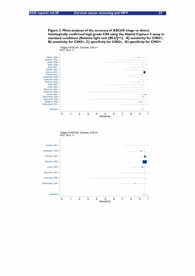
KCE reports vol.38 Cervical cancer screening and HPV 27
Figure 3. Meta-analyses of the accuracy of ASCUS triage to detect histologically confirmed high-grade CIN using the Hybrid Capture 2 assay in standard conditions (Relative light unit [RLU]>1). A) sensitivity for CIN2+, B) sensitivity for CIN3+, C) specificity for CIN2+, D) specificity for CIN3+
Triage of ASCUS. Outome: CIN 2+HC2, RLU >1
Sensitivity0 .1 .2 .3 .4 .5 .6 .7 .8 .9 1
Combined
Kiatpongsan, 2006 Bergeron, 2006
Giovannelli, 2005 Dalla Palma, 2005
Carrozi, 2005 Andersson, 2005 Wensveen, 2003
Lonky, 2003 Guyot, 2003
Pretorius, 2002 Pambucian, 2002 Kulasingam, 2002
Zielinski, 2001 Solomon, 2001 Rebello, 2001
Morin, 2001 Shlay, 2000
Lytwyn, 2000 Bergeron, 2000
Manos, 1999
Triage of ASCUS. Outome: CIN 3+HC2, RLU >1
Sensitivity0 .1 .2 .3 .4 .5 .6 .7 .8 .9 1
Combined
Kiatpongsan, 2006
Andersson, 2005
Wensveen, 2003
Lonky, 2003
Sherman, 2002
Pretorius, 2002
Kulasingam, 2002
Zielinski, 2001
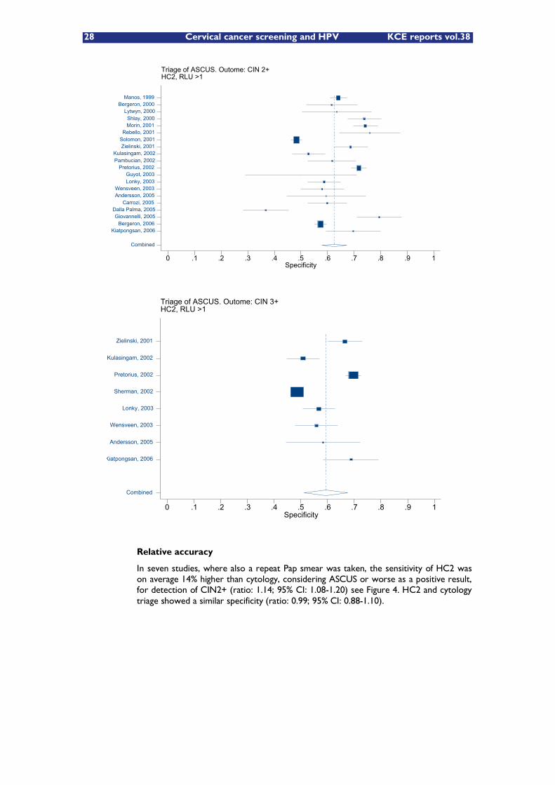
28 Cervical cancer screening and HPV KCE reports vol.38
Triage of ASCUS. Outome: CIN 2+HC2, RLU >1
Specificity0 .1 .2 .3 .4 .5 .6 .7 .8 .9 1
Combined
Kiatpongsan, 2006 Bergeron, 2006
Giovannelli, 2005 Dalla Palma, 2005
Carrozi, 2005 Andersson, 2005 Wensveen, 2003
Lonky, 2003 Guyot, 2003
Pretorius, 2002 Pambucian, 2002 Kulasingam, 2002
Zielinski, 2001 Solomon, 2001 Rebello, 2001
Morin, 2001 Shlay, 2000
Lytwyn, 2000 Bergeron, 2000
Manos, 1999
Relative accuracy
In seven studies, where also a repeat Pap smear was taken, the sensitivity of HC2 was on average 14% higher than cytology, considering ASCUS or worse as a positive result, for detection of CIN2+ (ratio: 1.14; 95% CI: 1.08-1.20) see Figure 4. HC2 and cytology triage showed a similar specificity (ratio: 0.99; 95% CI: 0.88-1.10).
Triage of ASCUS. Outome: CIN 3+HC2, RLU >1
Specificity0 .1 .2 .3 .4 .5 .6 .7 .8 .9 1
Combined
Kiatpongsan, 2006
Andersson, 2005
Wensveen, 2003
Lonky, 2003
Sherman, 2002
Pretorius, 2002
Kulasingam, 2002
Zielinski, 2001
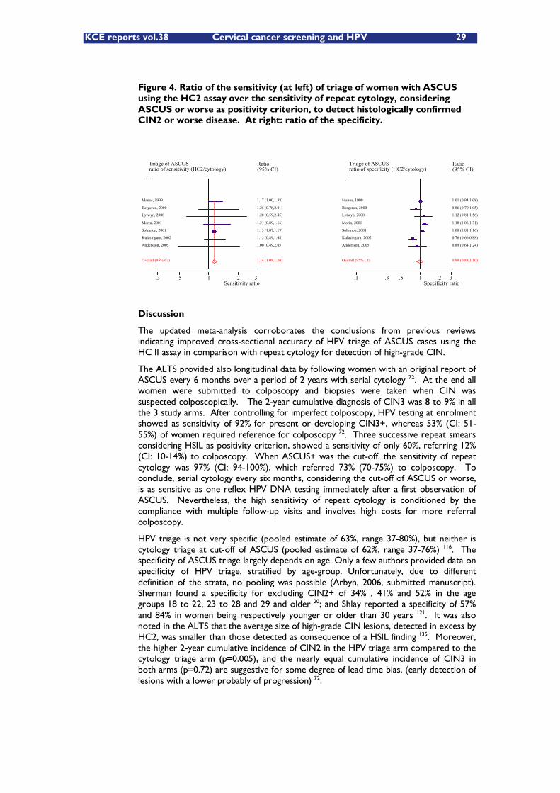
KCE reports vol.38 Cervical cancer screening and HPV 29
Triage of ASCUSratio of sensitivity (HC2/cytology)
Sensitivity ratio.3 .5 1 2 3
Ratio (95% CI)
1.17 (1.00,1.38) Manos, 1999
1.25 (0.78,2.01) Bergeron, 2000
1.20 (0.59,2.45) Lytwyn, 2000
1.21 (0.89,1.66) Morin, 2001
1.13 (1.07,1.19) Solomon, 2001
1.15 (0.89,1.48) Kulasingam, 2002
1.00 (0.49,2.05) Andersson, 2005
1.14 (1.08,1.20) Overall (95% CI)
Triage of ASCUSratio of specificity (HC2/cytology)
Specificity ratio.1 .3 .5 1 2 3
Ratio (95% CI)
1.01 (0.94,1.08) Manos, 1999
0.86 (0.70,1.05) Bergeron, 2000
1.12 (0.81,1.56) Lytwyn, 2000
1.18 (1.06,1.31) Morin, 2001
1.08 (1.01,1.16) Solomon, 2001
0.76 (0.66,0.88) Kulasingam, 2002
0.89 (0.64,1.24) Andersson, 2005
0.99 (0.88,1.10) Overall (95% CI)
Figure 4. Ratio of the sensitivity (at left) of triage of women with ASCUS using the HC2 assay over the sensitivity of repeat cytology, considering ASCUS or worse as positivity criterion, to detect histologically confirmed CIN2 or worse disease. At right: ratio of the specificity.
Discussion
The updated meta-analysis corroborates the conclusions from previous reviews indicating improved cross-sectional accuracy of HPV triage of ASCUS cases using the HC II assay in comparison with repeat cytology for detection of high-grade CIN.
The ALTS provided also longitudinal data by following women with an original report of ASCUS every 6 months over a period of 2 years with serial cytology 72. At the end all women were submitted to colposcopy and biopsies were taken when CIN was suspected colposcopically. The 2-year cumulative diagnosis of CIN3 was 8 to 9% in all the 3 study arms. After controlling for imperfect colposcopy, HPV testing at enrolment showed as sensitivity of 92% for present or developing CIN3+, whereas 53% (CI: 51-55%) of women required reference for colposcopy 72. Three successive repeat smears considering HSIL as positivity criterion, showed a sensitivity of only 60%, referring 12% (CI: 10-14%) to colposcopy. When ASCUS+ was the cut-off, the sensitivity of repeat cytology was 97% (CI: 94-100%), which referred 73% (70-75%) to colposcopy. To conclude, serial cytology every six months, considering the cut-off of ASCUS or worse, is as sensitive as one reflex HPV DNA testing immediately after a first observation of ASCUS. Nevertheless, the high sensitivity of repeat cytology is conditioned by the compliance with multiple follow-up visits and involves high costs for more referral colposcopy.
HPV triage is not very specific (pooled estimate of 63%, range 37-80%), but neither is cytology triage at cut-off of ASCUS (pooled estimate of 62%, range 37-76%) 116. The specificity of ASCUS triage largely depends on age. Only a few authors provided data on specificity of HPV triage, stratified by age-group. Unfortunately, due to different definition of the strata, no pooling was possible (Arbyn, 2006, submitted manuscript). Sherman found a specificity for excluding CIN2+ of 34% , 41% and 52% in the age groups 18 to 22, 23 to 28 and 29 and older 20; and Shlay reported a specificity of 57% and 84% in women being respectively younger or older than 30 years 121. It was also noted in the ALTS that the average size of high-grade CIN lesions, detected in excess by HC2, was smaller than those detected as consequence of a HSIL finding 135. Moreover, the higher 2-year cumulative incidence of CIN2 in the HPV triage arm compared to the cytology triage arm (p=0.005), and the nearly equal cumulative incidence of CIN3 in both arms (p=0.72) are suggestive for some degree of lead time bias, (early detection of lesions with a lower probably of progression) 72.
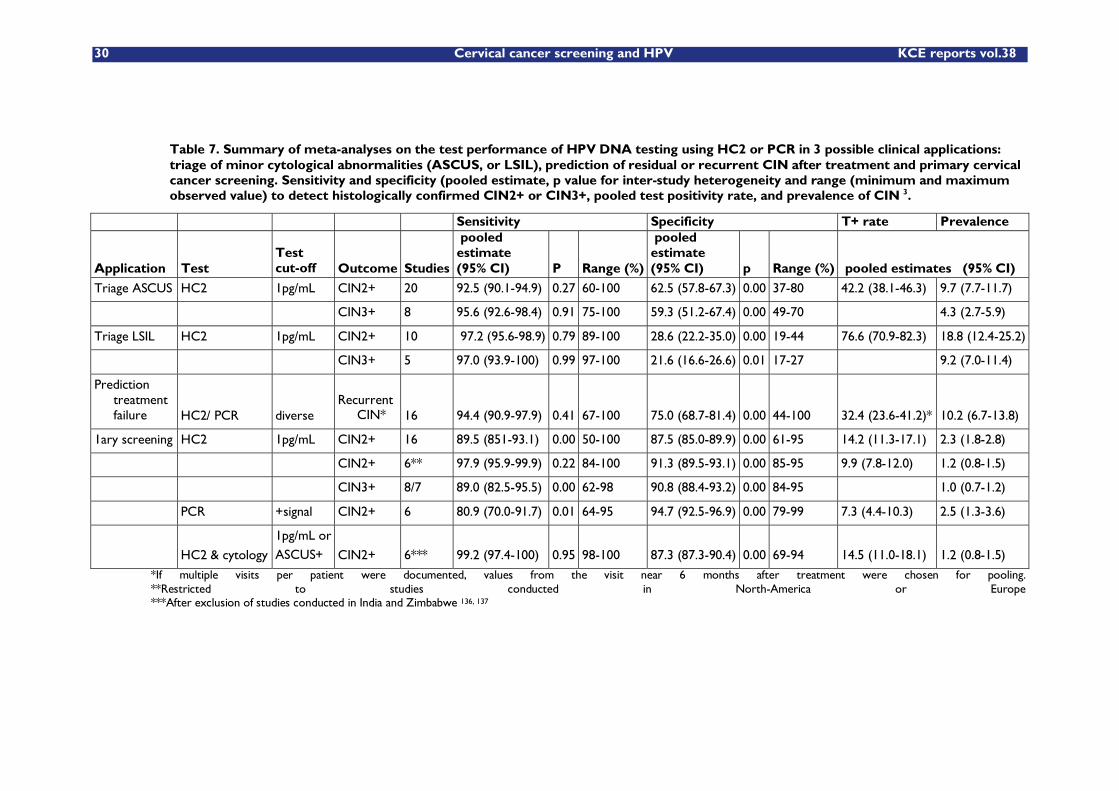
30 Cervical cancer screening and HPV KCE reports vol.38
Table 7. Summary of meta-analyses on the test performance of HPV DNA testing using HC2 or PCR in 3 possible clinical applications: triage of minor cytological abnormalities (ASCUS, or LSIL), prediction of residual or recurrent CIN after treatment and primary cervical cancer screening. Sensitivity and specificity (pooled estimate, p value for inter-study heterogeneity and range (minimum and maximum observed value) to detect histologically confirmed CIN2+ or CIN3+, pooled test positivity rate, and prevalence of CIN 3.
Sensitivity Specificity T+ rate Prevalence
Application Test Test cut-off Outcome Studies
pooled estimate (95% CI) P Range (%)
pooled estimate (95% CI) p Range (%) pooled estimates (95% CI)
Triage ASCUS HC2 1pg/mL CIN2+ 20 92.5 (90.1-94.9) 0.27 60-100 62.5 (57.8-67.3) 0.00 37-80 42.2 (38.1-46.3) 9.7 (7.7-11.7)
CIN3+ 8 95.6 (92.6-98.4) 0.91 75-100 59.3 (51.2-67.4) 0.00 49-70 4.3 (2.7-5.9)
Triage LSIL HC2 1pg/mL CIN2+ 10 97.2 (95.6-98.9) 0.79 89-100 28.6 (22.2-35.0) 0.00 19-44 76.6 (70.9-82.3) 18.8 (12.4-25.2)
CIN3+ 5 97.0 (93.9-100) 0.99 97-100 21.6 (16.6-26.6) 0.01 17-27 9.2 (7.0-11.4)
Prediction treatment failure HC2/ PCR diverse
Recurrent CIN* 16 94.4 (90.9-97.9) 0.41 67-100 75.0 (68.7-81.4) 0.00 44-100 32.4 (23.6-41.2)* 10.2 (6.7-13.8)
1ary screening HC2 1pg/mL CIN2+ 16 89.5 (851-93.1) 0.00 50-100 87.5 (85.0-89.9) 0.00 61-95 14.2 (11.3-17.1) 2.3 (1.8-2.8)
CIN2+ 6** 97.9 (95.9-99.9) 0.22 84-100 91.3 (89.5-93.1) 0.00 85-95 9.9 (7.8-12.0) 1.2 (0.8-1.5)
CIN3+ 8/7 89.0 (82.5-95.5) 0.00 62-98 90.8 (88.4-93.2) 0.00 84-95 1.0 (0.7-1.2)
PCR +signal CIN2+ 6 80.9 (70.0-91.7) 0.01 64-95 94.7 (92.5-96.9) 0.00 79-99 7.3 (4.4-10.3) 2.5 (1.3-3.6)
HC2 & cytology
1pg/mL or ASCUS+ CIN2+ 6*** 99.2 (97.4-100) 0.95 98-100 87.3 (87.3-90.4) 0.00 69-94 14.5 (11.0-18.1) 1.2 (0.8-1.5)
*If multiple visits per patient were documented, values from the visit near 6 months after treatment were chosen for pooling. **Restricted to studies conducted in North-America or Europe ***After exclusion of studies conducted in India and Zimbabwe 136, 137
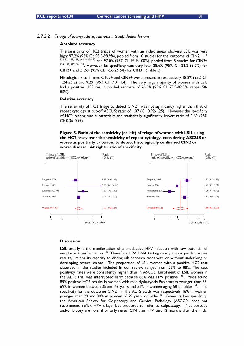
KCE reports vol.38 Cervical cancer screening and HPV 31
Triage of LSILratio of sensitivity (HC2/cytology)
Sensitivity ratio.3 .5 1 2 3
Ratio (95% CI)
0.93 (0.80,1.07) Bergeron, 2000
3.00 (0.61,14.86) Lytwyn, 2000
1.30 (1.05,1.60) Kulasingam, 2002
1.05 (1.01,1.10) Sherman, 2002
1.07 (0.92,1.25) Overall (95% CI)
Triage of LSILratio of specificity (HC2/cytology)
Specificity ratio.1 .3 .5 1 2 3
Ratio (95% CI)
0.97 (0.79,1.17) Bergeron, 2000
0.49 (0.22,1.07) Lytwyn, 2000
0.29 (0.19,0.42) Kulasingam, 2002
0.82 (0.66,1.01) Sherman, 2002
0.60 (0.36,0.99) Overall (95% CI)
2.7.2.2 Triage of low-grade squamous intraepithelial lesions
Absolute accuracy
The sensitivity of HC2 triage of women with an index smear showing LSIL was very high: 97.2% (95% CI: 95.6-98.9%), pooled from 10 studies for the outcome of CIN2+ 118,
120, 123-125, 127, 20, 128, 138, 77 and 97.0% (95% CI: 93.9-100%), pooled from 5 studies for CIN3+ 124, 125, 127, 20, 138. However its specificity was very low: 28.6% (95% CI: 22.2-35.0%) for CIN2+ and 21.6% (95% CI: 16.6-26.6%) for CIN3+ (Table 5).
Histologically confirmed CIN2+ and CIN3+ were present in respectively 18.8% (95% CI: 1.24-25.2) and 9.2% (95% CI: 7.0-11.4). The very large majority of women with LSIL had a positive HC2 result: pooled estimate of 76.6% (95% CI: 70.9-82.3%; range: 58-85%).
Relative accuracy
The sensitivity of HC2 triage to detect CIN2+ was not significantly higher than that of repeat cytology at cut-off ASCUS: ratio of 1.07 (CI: 0.92-1.25). However the specificity of HC2 testing was substantially and statistically significantly lower: ratio of 0.60 (95% CI: 0.36-0.99).
Figure 5. Ratio of the sensitivity (at left) of triage of women with LSIL using the HC2 assay over the sensitivity of repeat cytology, considering ASCUS or worse as positivity criterion, to detect histologically confirmed CIN2 or worse disease. At right: ratio of specificity.
Discussion
LSIL usually is the manifestation of a productive HPV infection with low potential of neoplastic transformation 139. Therefore HPV DNA testing nearly always yields positive results, limiting its capacity to distinguish between cases with or without underlying or developing severe lesions. The proportion of LSIL women with a positive HC2 test observed in the studies included in our review ranged from 59% to 88%. The test positivity rates were consistently higher than in ASCUS. Enrolment of LSIL women in the ALTS trial was interrupted early because 83% was HPV positive 140. Moss found 89% positive HC2 results in women with mild dyskaryosis Pap smears younger than 35, 69% in women between 35 and 49 years and 51% in women aging 50 or older 141. The specificity for the outcome CIN2+ in the ALTS study was respectively 16% in women younger than 29 and 30% in women of 29 years or older 20. Given its low specificity, the American Society for Colposcopy and Cervical Pathology (ASCCP) does not recommend reflex HPV triage, but proposes to refer to colposcopy. If colposcopy and/or biopsy are normal or only reveal CIN1, an HPV test 12 months after the initial
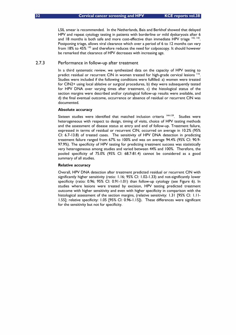
32 Cervical cancer screening and HPV KCE reports vol.38
LSIL smear is recommended. In the Netherlands, Bais and Berkhof showed that delayed HPV and repeat cytology testing in patients with borderline or mild dyskaryosis after 6 and 18 months is both safe and more cost-effective than immediate HPV triage 142, 143. Postponing triage, allows viral clearance which over a period of 6 to 12 months can vary from 18% to 45% 142 and therefore reduces the need for colposcopy. It should however be remarked that clearance of HPV decreases with increasing age.
2.7.3 Performance in follow-up after treatment
In a third systematic review, we synthesized data on the capacity of HPV testing to predict residual or recurrent CIN in women treated for high-grade cervical lesions 116. Studies were included if the following conditions were fulfilled: a) women were treated for CIN2+ using local ablative or surgical procedures, b) they were subsequently tested for HPV DNA over varying times after treatment, c) the histological status of the section margins were described and/or cytological follow-up results were available, and d) the final eventual outcome, occurrence or absence of residual or recurrent CIN was documented.
Absolute accuracy
Sixteen studies were identified that matched inclusion criteria 144-159. Studies were heterogeneous with respect to design, timing of visits, choice of HPV testing methods and the assessment of disease status at entry and end of follow-up. Treatment failure, expressed in terms of residual or recurrent CIN, occurred on average in 10.2% (95% CI: 6.7-13.8) of treated cases. The sensitivity of HPV DNA detection in predicting treatment failure ranged from 67% to 100% and was on average 94.4% (95% CI: 90.9-97.9%). The specificity of HPV testing for predicting treatment success was statistically very heterogeneous among studies and varied between 44% and 100%. Therefore, the pooled specificity of 75.0% (95% CI: 68.7-81.4) cannot be considered as a good summary of all studies.
Relative accuracy
Overall, HPV DNA detection after treatment predicted residual or recurrent CIN with significantly higher sensitivity (ratio: 1.16; 95% CI: 1.02-1.33) and not-significantly lower specificity (ratio: 0.96; 95% CI: 0.91-1.01) than follow-up cytology (see Figure 6). In studies where lesions were treated by excision, HPV testing predicted treatment outcome with higher sensitivity and even with higher specificity in comparison with the histological assessment of the section margins, (relative sensitivity: 1.31 [95% CI: 1.11-1.55]; relative specificity: 1.05 [95% CI: 0.96-1.15]). These differences were significant for the sensitivity but not for specificity.
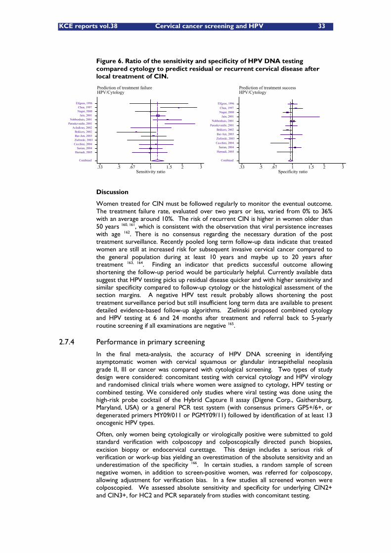
KCE reports vol.38 Cervical cancer screening and HPV 33
Prediction of treatment failureHPV/Cytology
Sensitivity ratio.33 .5 .67 1 1.5 2 3
Combined
Hernadi, 2005 Sarian, 2004
Cecchini, 2004 Zielinski, 2003 Bar-Am, 2003 Bekkers, 2002
Acladious, 2002 Paraskevaidis, 2001
Nobbenhuis, 2001 Jain, 2001
Nagai, 2000 Chua, 1997
Elfgren, 1996
Prediction of treatment successHPV/Cytology
Specificity ratio.33 .5 .67 1 1.5 2 3
Combined
Hernadi, 2005 Sarian, 2004
Cecchini, 2004 Zielinski, 2003 Bar-Am, 2003 Bekkers, 2002
Paraskevaidis, 2001 Nobbenhuis, 2001
Jain, 2001 Nagai, 2000 Chua, 1997
Elfgren, 1996
Figure 6. Ratio of the sensitivity and specificity of HPV DNA testing compared cytology to predict residual or recurrent cervical disease after local treatment of CIN.
Discussion
Women treated for CIN must be followed regularly to monitor the eventual outcome. The treatment failure rate, evaluated over two years or less, varied from 0% to 36% with an average around 10%. The risk of recurrent CIN is higher in women older than 50 years 160, 161, which is consistent with the observation that viral persistence increases with age 162. There is no consensus regarding the necessary duration of the post treatment surveillance. Recently pooled long term follow-up data indicate that treated women are still at increased risk for subsequent invasive cervical cancer compared to the general population during at least 10 years and maybe up to 20 years after treatment 163, 164. Finding an indicator that predicts successful outcome allowing shortening the follow-up period would be particularly helpful. Currently available data suggest that HPV testing picks up residual disease quicker and with higher sensitivity and similar specificity compared to follow-up cytology or the histological assessment of the section margins. A negative HPV test result probably allows shortening the post treatment surveillance period but still insufficient long term data are available to present detailed evidence-based follow-up algorithms. Zielinski proposed combined cytology and HPV testing at 6 and 24 months after treatment and referral back to 5-yearly routine screening if all examinations are negative 165.
2.7.4 Performance in primary screening
In the final meta-analysis, the accuracy of HPV DNA screening in identifying asymptomatic women with cervical squamous or glandular intraepithelial neoplasia grade II, III or cancer was compared with cytological screening. Two types of study design were considered: concomitant testing with cervical cytology and HPV virology and randomised clinical trials where women were assigned to cytology, HPV testing or combined testing. We considered only studies where viral testing was done using the high-risk probe cocktail of the Hybrid Capture II assay (Digene Corp., Gaithersburg, Maryland, USA) or a general PCR test system (with consensus primers GP5+/6+, or degenerated primers MY09/011 or PGMY09/11) followed by identification of at least 13 oncogenic HPV types.
Often, only women being cytologically or virologically positive were submitted to gold standard verification with colposcopy and colposcopically directed punch biopsies, excision biopsy or endocervical curettage. This design includes a serious risk of verification or work-up bias yielding an overestimation of the absolute sensitivity and an underestimation of the specificity 166. In certain studies, a random sample of screen negative women, in addition to screen-positive women, was referred for colposcopy, allowing adjustment for verification bias. In a few studies all screened women were colposcopied. We assessed absolute sensitivity and specificity for underlying CIN2+ and CIN3+, for HC2 and PCR separately from studies with concomitant testing.
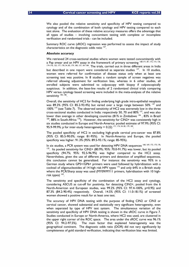
34 Cervical cancer screening and HPV KCE reports vol.38
We also pooled the relative sensitivity and specificity of HPV testing compared to cytology and of the combination of both cytology and HPV testing compared to each test alone. The evaluation of these relative accuracy measures offers the advantage that all types of studies �– involving concomitant testing with complete or incomplete verification and randomised trials - can be included.
Summary ROC curve (sROC) regression was performed to assess the impact of study characteristics on the diagnostic odds ratio 167.
Absolute accuracy
We retrieved 24 cross-sectional studies where women were tested concomitantly with a Pap smear and an HPV assay in the framework of primary screening 168-171, 87, 172, 173, 136,
174-176, 125, 177, 178, 54, 85, 179, 180, 137, 181-184. The trials, carried out in three different areas in India but described in one report were considered as separate studies 137. In 10 studies, women were referred for confirmation of disease status only when at least one screening test was positive. In 8 studies a random sample of screen negatives was referred allowing adjustment for verification bias, whereas in 6 other studies, all enrolled subjects were submitted to colposcopy with biopsy if colposcopically suspicious. In addition, the base-line results of 2 randomized clinical trials comparing HPV versus cytology based screening were included in the meta-analyses of the relative sensitivity 185, 186.
Overall, the sensitivity of HC2 for finding underlying high grade intra-epithelial neoplasia was 89.3% (95% CI: 85.2-93.4%) but varied over a large range between 50% 137 and 100% 174 (see Table 7). The observed sensitivity of HC2 was extremely low in the three cross-sectional studies conducted in India: respectively 50, 70 and 80% 137, and was also lower than average in other developing countries (81% in Zimbabwe 136 , 83% in Brazil 184, 88% in South-Africa 170). However, the sensitivity for CIN2+ was consistently high in six studies conducted in Europe and North-America: pooled estimate of 97.9% (95% CI: 95.9-99.9%; p for inter-study heterogeneity = 0.22) 171, 174, 54, 85, 179, 183.
The pooled specificity of HC2 in excluding high-grade cervical pre-cancer was 87.8% (95% CI: 85.5-90.0%; range: 81-95%). In North-America and Europe, the pooled specificity was higher: 91.3% (95%: 89.5-93.1%; range: 85-95%).
In six studies, a PCR system was used for detecting HPV DNA sequences 168, 169, 172, 175, 176,
125. Its pooled sensitivity for CIN2+ (80.9%; 95%: 70.0-91.7%) was lower, but its pooled specificity (94.7%; 95%: 92.5-96.9%) was higher compared to the HC2 assay. Nevertheless, given the use of different primers and detection of amplified sequences, this conclusion cannot be generalized. For instance: the sensitivity was 95% in a German study where GP5+/GP6+ primers were used followed by hybridization with a cocktail of oligonucleotides of 14 high risk HPV types 172 and only 64% in a British study where the PCR/Sharp assay was used (MY09/MY11 primers, hybridisation with 10 high-risk types) 169.
The sensitivity and specificity of the combination of the HC2 assay and cytology, considering ASCUS as cut-off for positivity, for detecting CIN2+, pooled from the 6 North-American and European studies, was 99.2% (95% CI: 97.4-100%, p=0.95) and 87.3% (84.2-90.4%) respectively. Overall, 14.5% (95% CI: 11.0-18.1%) of screened women showed a positive result for at least one test.
The accuracy of HPV DNA testing with the purpose of finding CIN2 or CIN3 or cervical cancer, showed substantial and statistically very significant heterogeneity, even when separated by type of HPV test system. The simultaneous variation of the sensitivity and specificity of HPV DNA testing is shown in the sROC curve in Figure 7. Studies conducted in Europe or North-America, where HC2 was used, are clustered in the upper right corner of the ROC space. The area under the sROC curve was 96.1% (95% CI: 94.2-97.5%). The main factor that explained heterogeneity was the geographical continent. The diagnostic odds ratio (DOR) did not vary significantly by completeness of gold standard verification, indicating that verification bias was limited.
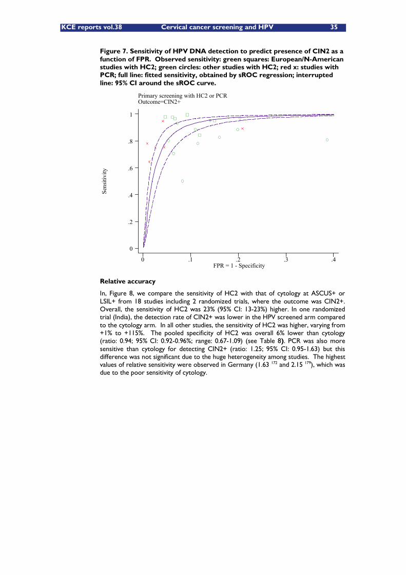
KCE reports vol.38 Cervical cancer screening and HPV 35
Figure 7. Sensitivity of HPV DNA detection to predict presence of CIN2 as a function of FPR. Observed sensitivity: green squares: European/N-American studies with HC2; green circles: other studies with HC2; red x: studies with PCR; full line: fitted sensitivity, obtained by sROC regression; interrupted line: 95% CI around the sROC curve.
Primary screening with HC2 or PCROutcome=CIN2+
Sens
itivi
ty
FPR = 1 - Specificity0 .1 .2 .3 .4
0
.2
.4
.6
.8
1
Relative accuracy
In, Figure 8, we compare the sensitivity of HC2 with that of cytology at ASCUS+ or LSIL+ from 18 studies including 2 randomized trials, where the outcome was CIN2+. Overall, the sensitivity of HC2 was 23% (95% CI: 13-23%) higher. In one randomized trial (India), the detection rate of CIN2+ was lower in the HPV screened arm compared to the cytology arm. In all other studies, the sensitivity of HC2 was higher, varying from +1% to +115%. The pooled specificity of HC2 was overall 6% lower than cytology (ratio: 0.94; 95% CI: 0.92-0.96%; range: 0.67-1.09) (see Table 8). PCR was also more sensitive than cytology for detecting CIN2+ (ratio: 1.25; 95% CI: 0.95-1.63) but this difference was not significant due to the huge heterogeneity among studies. The highest values of relative sensitivity were observed in Germany (1.63 172 and 2.15 179), which was due to the poor sensitivity of cytology.
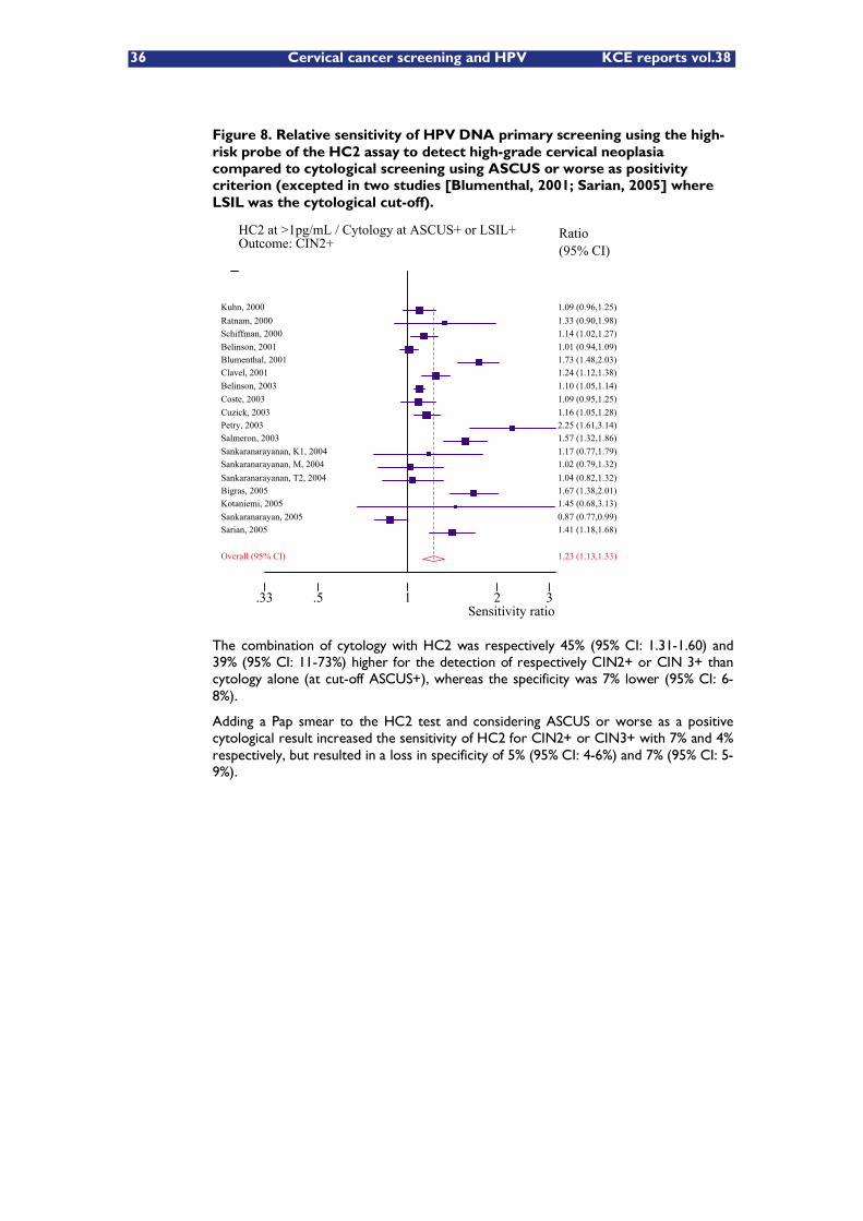
36 Cervical cancer screening and HPV KCE reports vol.38
Figure 8. Relative sensitivity of HPV DNA primary screening using the high-risk probe of the HC2 assay to detect high-grade cervical neoplasia compared to cytological screening using ASCUS or worse as positivity criterion (excepted in two studies [Blumenthal, 2001; Sarian, 2005] where LSIL was the cytological cut-off).
HC2 at >1pg/mL / Cytology at ASCUS+ or LSIL+Outcome: CIN2+
Sensitivity ratio.33 .5 1 2 3
Ratio (95% CI)
1.09 (0.96,1.25) Kuhn, 2000 1.33 (0.90,1.98) Ratnam, 2000 1.14 (1.02,1.27) Schiffman, 2000 1.01 (0.94,1.09) Belinson, 2001 1.73 (1.48,2.03) Blumenthal, 2001 1.24 (1.12,1.38) Clavel, 2001 1.10 (1.05,1.14) Belinson, 2003 1.09 (0.95,1.25) Coste, 2003 1.16 (1.05,1.28) Cuzick, 2003 2.25 (1.61,3.14) Petry, 2003 1.57 (1.32,1.86) Salmeron, 2003 1.17 (0.77,1.79) Sankaranarayanan, K1, 2004 1.02 (0.79,1.32) Sankaranarayanan, M, 2004 1.04 (0.82,1.32) Sankaranarayanan, T2, 2004 1.67 (1.38,2.01) Bigras, 2005 1.45 (0.68,3.13) Kotaniemi, 2005 0.87 (0.77,0.99) Sankaranarayan, 2005 1.41 (1.18,1.68) Sarian, 2005
1.23 (1.13,1.33) Overall (95% CI)
The combination of cytology with HC2 was respectively 45% (95% CI: 1.31-1.60) and 39% (95% CI: 11-73%) higher for the detection of respectively CIN2+ or CIN 3+ than cytology alone (at cut-off ASCUS+), whereas the specificity was 7% lower (95% CI: 6-8%).
Adding a Pap smear to the HC2 test and considering ASCUS or worse as a positive cytological result increased the sensitivity of HC2 for CIN2+ or CIN3+ with 7% and 4% respectively, but resulted in a loss in specificity of 5% (95% CI: 4-6%) and 7% (95% CI: 5-9%).

KCE reports vol.38 Cervical cancer screening and HPV 37
Table 8. Relative accuracy of virological versus cytological screening or of combined screening versus testing with one test in order to find underlying CIN2 or CIN3 or worse
Comparison Outcome Relative sensitivity Range Relative specificity Range # studies
HC2/cyto (ASCUS+) CIN2+ 1.19 (1.11-1.29) 0.97-2.25 0.97 (0.96-0.98) 0.86-1.10 14
HC2/cyto (LSIL+) 1.39 (1.30-1.48) 1.09-2.35 0.89 (0.88-0.91) 0.67-0.98 11
HC2/cyto (ASCUS/LSIL+) 1.23 (1.13-1.33) 0.87-2.25 0.94 (0.92-0.96) 0.67-1.10 18/16*
PCR/cyto (ASCUS+) 1.25 (0.95-1.63) 0.75-3.57 0.99 (0.96-1.02) 0.86-1.08 6
PCR/cyto (LSIL+) 1.61 (0.84-3.09) 0.82-5.10 0.92 (0.89-0.95) 0.89-1.00 3
HC2/cyto (ASCUS+) CIN3+ 1.28 (1.12-1.47) 0.97-2.12 1.00 (0.99-1.01) 0.96-1.10 7
HC2/cyto (LSIL+) 1.37 (1.14-1.64) 0.97-2.32 0.93 (0.91-0.95) 0.85-0.98 7
Cyto (ASC+) & HC2/Cyto (ASCUS+) CIN2+ 1.45 (1.31-1.60) 1.06-2.30 0.93 (0.92-0.94) 0.89-0.96 9
Cyto (ASC+) & HC2/Cyto (ASCUS+) CIN3+ 1.39 (1.11-1.73) 1.02-2.18 0.93 (0.92-0.94) 0.89-0.95 6
Cyto (ASCUS+) & HC2/HC2+ CIN2+ 1.07 (1.06-1.08) 1.02-1.37 0.95 (0.94-0.96) 0.81-0.99 9
Cyto (ASCUS+) & HC2/HC2+ CIN3+ 1.04 (1.03-1.04) 1.02-1.17 0.93 (0.91-0.95) 0.81-0.99 6
* The meta-analysis of relative sensitivity includes 2 RCTs, the meta-analysis of relative specificity does not include RCTs
38 Cervical cancer screening and HPV KCE reports vol. 38
Discussion
A consistently high sensitivity for high-grade CIN was demonstrated for HC2 in the six North American and European studies (pooled average: 98%), whereas the sensitivity in India, Zimbabwe and South-Africa, was substantially lower (range: 50-88%). In these last countries, visual inspection of the cervix after application of diluted acetic acid (VIA) was also included in the screening trials. A certain amount of misclassification of the final cervical status due to the use of an imperfect colposcopy-based gold standard, correlated to VIA, cannot be excluded 187. Presence of oncogenic HPV types, not included in the HC2 probe cocktail is another possibility to explain the low sensitivity of HPV testing. Nevertheless, this last possibility looks less probable given our current knowledge of HPV type distribution in high-grade cervical intraepithelial neoplasia and cancer in Africa and Asia 188-190.
Based on the accuracy data from 9 of the 18 cross-sectional studies included our meta-analyses, and considering also longitudinal results form the Portland study, the Food and Drugs administration approved the use of high-risk probe cocktail of HC2 as an adjunct to cervical cytology screening in women age 30 years or more. In the Portland study, the longitudinal sensitivity to predict subsequent CIN3+ within 5 year or 10 years was respectively 49% and 35% for cytology screening, 75% and 64% for HC2-based screening and 86% and 72% for combined cytological and virological testing. The 5-year cumulative risk of CIN3, was 4.4% for women being HC2-positive at base-line whereas only 0.24% among women with a negative HC2-test and 0.16% when both the HC2-test and Pap smear were negative 191. The longitudinal negative predictive value of a combined negative test, computed over a 5-year period, was very high: 99.91% (95% CI: 99.85-99.95%). This means that 9 over 10,000 (95% CI: 5-15/10,000), will develop CIN3+ over a 5 year period in spite of a double negative test. In women having only a negative Pap smear, this risk is 30/10 000 screened women (95% CI: 23-38/10 000).
The ASCCP recommends adding HPV testing to cytology screening after the age of 30 at an interval of 3 year if both tests are negative 192. When HC2 is positive and cytology is normal, a repetition of both tests after 6 to 12 months is proposed. The woman should be referred to colposcopy if results of either test are positive.
In Europe, however, use of HPV tests is currently not included in the basic screening policy. The results of ongoing randomized screening trials are waited for, where cytology screening is compared with HPV screening or combined cytology and HPV screening. The main postulated outcome of these trials is a reduction in cumulative incidence of CIN3 3-to 5 years after screening among HPV-negative compared to cytology-negative women 193. The results on these endpoints will be published in the period 2006-2008. Meanwhile the Pap smear continues to be the standard screen test in the European Union 194.
Age plays a tremendously important role in the determination of the target population. The change of HPV acquisition rises quickly after onset of sexual activity with peak prevalence of HPV positivity occurring near the late teens or early twenties.195 At that age, HPV infections almost always clear spontaneously. HPV prevalence declines but viral persistence tends to increase with age 162. On the other hand, the incidence of several cervical dysplasia starts rising in the late twenties-early thirties and cervical cancer in the late thirties. HPV screening at young age is therefore inefficient.
One of the draw-backs of primary HPV screening is its lower specificity in excluding absence of high-grade CIN compared to cytology screening. Cuzick showed that specificity of HPV screening is on average 7% higher in women of 35 years or older compared to younger women.195 High viral load or viral persistence are often proposed to increase the positive predictive value in identifying progressive lesions, but published results are conflicting. Schiffman showed that HPV 16, in particular, is likely to persist and predicts presence or development of CIN3 or cancer in the subsequent five years in one over five cases 196.
The 10-year cumulative risk of CIN3+ associated with HPV16, HPV18 or other risk HPV infection, among women included in the Portland study was 17%, 14% and 3% respectively 79. Cytology triage is another method, which can improve the specificity of
KCE reports vol.38 Cervical cancer screening and HPV 39
HPV primary screening. Testing for mRNA, coding for E6 or E7 oncoproteins from a limited set of oncogenic HPV types, or immunostaining of certain cell-cycle regulating proteins are candidate markers which could triage HPV positive women, but all of these are still insufficiently documented and require more research.
Adenocarcinoma
Most cervical cancers are squamous cell carcinomas. The incidence of squamous cervical cancer has declined in areas with well-organised cytological screening. However, the incidence of adenocarnima has increased or was not affected 197. Recently, a strong and systematic association was demonstrated between presence of oncogenic HPV types and development of adenocarnoma and adenocarinoma in situ of the uterine cervix 198. DNA of high-risk HPV types is detectable in most mucinous adenocarcinomas and adenosquamous carcinomas of the cervix 199, 200. Only rare histological variants of cervical adenocarcinoma seem unrelated to HPV infection 199. Therefore it is expected that HPV-based screening is likely to decrease also the incidence of cervical adenocarcinomas.
2.7.5 Conclusions
Triage of atypical cells of undetermined significance
HPV triage, with a general validated HPV test, is a recommended management option in case of a cytological result of ASC-US. Repeat cytology is still an acceptable option if compliance with follow-up recommendations can be assured or when HPV tests are not available. Colposcopy is a third option.
Triage of low grade squamous intraepithelial lesions
Reflex HPV triage, using a non-specific HPV-test is, in general, not a useful management option in case of LSIL. Nevertheless, it is possible that reflex HPV testing can be cost-effective for older women with LSIL, where the prevalence of infection is considerably lower. Repetition of cytology at 6 to 12 months or HPV testing at 12 months, with or without colposcopy are possible management options. The American Society for Colposcopy and Cervical Pathology (ASCCP) proposes to refer to colposcopy. Research is needed to identify a good reflex triage test for women with LSIL. Future reports of studies should contain sufficient age-stratified details
Follow-up after treatment of high-grade cervical intra-epithelial neoplasia
With regard to the fact that, women who have been treated for high-grade CIN still have an increased risk for invasive cervical cancer, there is a definite need for improved follow-up regimens.
Evidence supports the use of a double cytology and HPV test at 6 months post treatment for improved safety of post-treatment follow-up. While there is evidence to suggest that subsequent follow-up of women negative for both HPV and in cytology needs to be less intense, evidence cannot distinguish which specific follow-up regimen that should be used.
Further research on long-term protection of HPV-negativity as well as of joint cytology and HPV-negativity is warranted.
Primary screening
Further research is necessary to better define the longitudinal performance indicators (sensitivity, specificity and positive and negative predictive values) of HPV DNA testing as well as of combined HPV DNA testing and cytology.
Adequate triage methods are needed to identify those HPV-positive women that are at risk of developing cancer and to minimise surveillance with intensified screening among those who are not at risk.
A judicious use of a combination of randomised trials, modelling studies and randomised health care policies is suggested. Modelling should be used to investigate optimal settings. Effects on intermediate endpoints can then be used in further modelling studies
40 Cervical cancer screening and HPV KCE reports vol. 38
to estimate effects on late endpoints such as mortality and/or to design randomized health care policies.
Cost-effectiveness modelling studies need to be repeated in different populations that may differ in associated costs, rates of HPV infection of different types as well as in background rates of other risk factors for cervical cancer.
2.8 GENERAL CONCLUSIONS
From the reviews outlined above in this chapter, the following concluding statements can be formulated.
In well-organised settings, with a high level of quality assurance, conventional cytological screening reduces the incidence of squamous cervical cancer by 80% or more (Outcome 1-3; study types 2-4, see Table 1). Nevertheless, drawbacks of cytological screening are its low to moderate reproducibility. The cross-sectional sensitivity for high-grade lesions is highly variable.
Liquid-based cytology showed similar sensitivity and specificity as conventional cytology with respect to detection of high-grade CIN. The percentage of unsatisfactory smear usually is lower and the interpretation requires less time compared to conventional smears. The quality of evaluation described in the literature is quite poor. (Outcome 6, study type 2).
One large population-based randomised trial comparing automated versus manual conventional cytology showed equal detection rates of high-grade CIN and cancer and similar specificity compared to high-quality conventional cytology (Outcome 5, study types 1-2).
HPV DNA testing with validated methods is highly reproducible. The high-risk cocktail of Hybrid-Capture II is more sensitive and equally specific compared to repeat cytology to triage women with equivocal cytology to select women who need further management. Most women with LSIL are HPV positive, limiting the efficiency of reflex HPV triage. After conservative treatment of cervical lesions, HPV testing picks up more quickly, with higher sensitivity and not lower specificity residual or recurrent CIN than follow-up cytology.
Primary screening with HC2 or validated PCR systems is substantially more sensitive for identifying CIN2+ compared to cytology at cut-off ASCUS or LSIL, but it is less specific. By combined HPV and cytology screening (a positive test is defined as positive either for HPV, for cytology, or for both) still a small gain in sensitivity for high-grade CIN lesions can be obtained but at the expense of a considerable loss in specificity, in comparison with isolated HC2 screening; Further research is necessary to better define the longitudinal performance indicators (sensitivity, specificity and positive and negative predictive values) of HPV DNA testing as well as of combined HPV DNA testing and cytology. Randomised controlled trials aiming to demonstrate a lower cumulative incidence of CIN3 in HPV negative compared to cytology negative women are waited for before primary HPV screening can be recommended.
The specificity of HPV screening can be enhanced by restriction to women older than 30-35 years. Potential methods to triage HPV positive women are: cytology, repetition of the HPV test 6-12 months later, typing for a limited set of HPV types (including HPV 16), assessment of the (type-specific) viral load, viral integration, mRNA or cell-cycle regulating proteins. The selection of the best triage option is still an object of research.
If HPV testing is done in addition to cytology, the virological result and the cytological findings should be integrated in one report under the responsibility of a cytopathologist.

KCE reports vol.38 Cervical cancer screening and HPV 41
3 CERVICAL CANCER SCREENING IN OTHER EUROPEAN COUNTRIES
3.1 INTRODUCTION
In most European countries, cervical cancer screening started as an opportunistic activity, performed on the initiative of women or doctors. This opportunistic screening activity is still predominant in Europe. Cervical cancer screening often was offered in the context of anti-conception, so that the target group was younger women. Because such services are frequently not integrated with the second level of care, it was not always possible to ensure adequate diagnosis and treatment of women with a positive test result 30.
Well-organized screening programmes have a greater impact than opportunistic screening because they have the potential to achieve greater participation and this can improve equity of access and the likelihood of reaching women at higher risk. Moreover organised screening implicates the implementation of quality assurance measures, which are actively monitored. Implementation of a national programme requires that there be a national policy that defines the screening age and interval and what method of screening will be used, as well sufficient political and financial investment.
The objective of this chapter is to describe and compare the screening activities and policies in Europe. The screening systems are compared in terms of the definition of the target population, (start and end age), screening interval, the type of organisation and quality assurance methodology and the monitoring of quality and impact indicators.
Until recently, cervical cancer screening was based on the microscopic examination of cells collected with a spatula or brush from the cervix (Papanicolau test). HPV detection is used as a triage test in some countries but not as screening test. Therefore this chapter will only describe systems, where primary screening is based on cytology.
Further more, details of two selected countries running well-organized screening programmes (Norway and Sweden) will be presented in more detail because they are particularly useful for the Belgian situation. Norway recently switched from opportunistic to organised screening, whereas in Sweden, several county-based policies co-exist. However in both countries a nation-wide screening registry was set up. Such a screening registry is the corner stone of an organised programme.
3.2 ASPECTS OF ORGANISED SCREENING
3.2.1 Rational of organised screening
To maximise the positive impact and minimise the adverse effects, screening should be offered only in organised settings 194. Designing a programme includes defining the screening policy: this means determining the target age group, the screening interval, choice of the screening test, and the establishment of follow-up and treatment strategies for screen-positive women. The screening policy should be defined taking into account the natural history of the disease and the variation in background risk. Moreover, a screening programme must reach high population acceptance and coverage, and assure and demonstrate good quality at all levels.
Population-based information systems need to be set up allowing continuous monitoring of screening process indicators. A legal framework should be established permitting registration of individual data and linkage between population databases, screening files, cancer and mortality registers 201, 202, 194, 203, 204. The information system is an essential tool to run the programme, to compute the indicators of attendance, compliance, quality and impact, and to provide feed back to involved health professionals, stake holders and health authorities.

42 Cervical cancer screening and HPV KCE reports vol. 38
A concern is the completeness of the recorded information of the programme. Reliable cancer registration is important. Individual-level links between population, screening, cancer registry, and treatment data are needed.
As with any public health policy, a screening programme should be designed in such a way that it can be evaluated. Key components in the monitoring and evaluation of screening are: regularly published results on the screening performance so that it is clear for the decision-makers, key personnel groups, and for the general public how well the programme is running; scientific evaluation of the effectiveness and outcomes of the screening programme based on established epidemiological methods; and ascertaining and feed-back of information about invasive cancers detected in connection or after screening.
Effectiveness of an organised programme is a function of the quality of its individual components. Epidemiology provides instruments that permit planning, guidance and evaluation of the entire process of a screening programme, from the organisational and administrative aspects up to assessment of the impact.
Organised cervical cancer screening is a multi-step process including:
Identification of the target population
Reaching women belonging to the target population
Collection of an adequate Pap smear
Examination of the Pap smear and reporting
Communication of smear test results to women with a normal result.
Recall of women with unsatisfactory/inadequate smears
Follow-up of women with abnormal smears i.e. diagnostic procedures and treatment if needed, including a fail-safe system to make sure this actually happens
In some countries, among which Belgium, re-allocation of resources already used for screening activities will theoretically be sufficient to cover the entire target population within a defined screening interval 205, 206. Different solutions can be proposed to implement organised screening (depending e.g. if opportunistic activity currently exists, or does not exist). In general, systems that have demonstrated effectiveness can be recommended and also relevant cost-effectiveness aspects and aspects to minimise potential adverse effects need to be considered.
3.2.2 European Council Recommendation
The European Union has recommended organising cervical cancer screening since the start of the Europe against Cancer programme in 1987. The first edition of European Guidelines for Quality Assurance in Cervical Cancer Screening was issued in 1993 207.
On 2 December 2003, the ministers responsible for public health of the Member States endorsed, upon proposition of the European Commission and after consultation of the European Parliament, the �“Council Recommendation on Cancer Screening 194. The recommendation was based on the Vienna consensus of 1999 208 which stressed the need for the adoption of organised screening programmes with personal invitations and quality assurance 209. Given the scientific evidence of effectiveness, the Council endorsed the recommendation for organised screening for 3 cancers, among which cervical cancer (see Table 9).
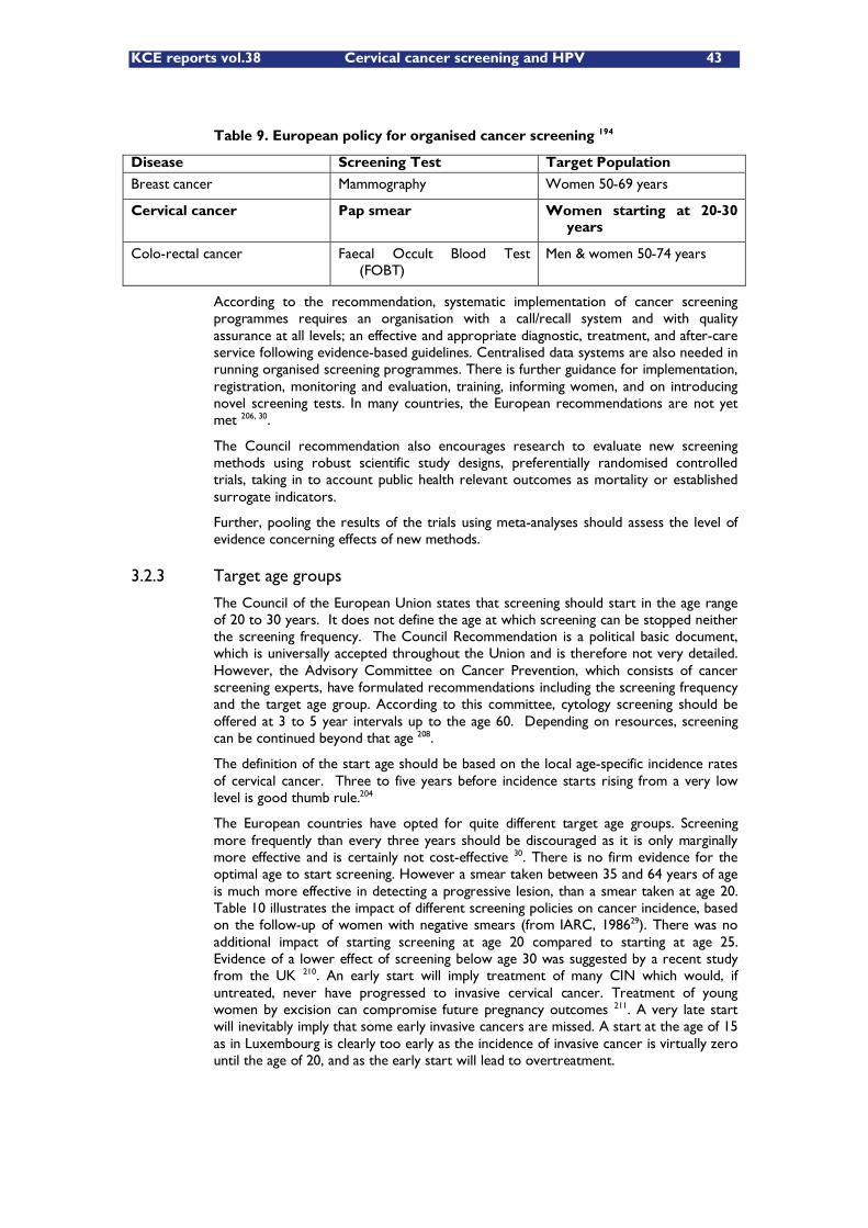
KCE reports vol.38 Cervical cancer screening and HPV 43
Table 9. European policy for organised cancer screening 194
Disease Screening Test Target Population
Breast cancer Mammography Women 50-69 years
Cervical cancer Pap smear Women starting at 20-30 years
Colo-rectal cancer Faecal Occult Blood Test (FOBT)
Men & women 50-74 years
According to the recommendation, systematic implementation of cancer screening programmes requires an organisation with a call/recall system and with quality assurance at all levels; an effective and appropriate diagnostic, treatment, and after-care service following evidence-based guidelines. Centralised data systems are also needed in running organised screening programmes. There is further guidance for implementation, registration, monitoring and evaluation, training, informing women, and on introducing novel screening tests. In many countries, the European recommendations are not yet met 206, 30.
The Council recommendation also encourages research to evaluate new screening methods using robust scientific study designs, preferentially randomised controlled trials, taking in to account public health relevant outcomes as mortality or established surrogate indicators.
Further, pooling the results of the trials using meta-analyses should assess the level of evidence concerning effects of new methods.
3.2.3 Target age groups
The Council of the European Union states that screening should start in the age range of 20 to 30 years. It does not define the age at which screening can be stopped neither the screening frequency. The Council Recommendation is a political basic document, which is universally accepted throughout the Union and is therefore not very detailed. However, the Advisory Committee on Cancer Prevention, which consists of cancer screening experts, have formulated recommendations including the screening frequency and the target age group. According to this committee, cytology screening should be offered at 3 to 5 year intervals up to the age 60. Depending on resources, screening can be continued beyond that age 208.
The definition of the start age should be based on the local age-specific incidence rates of cervical cancer. Three to five years before incidence starts rising from a very low level is good thumb rule.204
The European countries have opted for quite different target age groups. Screening more frequently than every three years should be discouraged as it is only marginally more effective and is certainly not cost-effective 30. There is no firm evidence for the optimal age to start screening. However a smear taken between 35 and 64 years of age is much more effective in detecting a progressive lesion, than a smear taken at age 20. Table 10 illustrates the impact of different screening policies on cancer incidence, based on the follow-up of women with negative smears (from IARC, 198629). There was no additional impact of starting screening at age 20 compared to starting at age 25. Evidence of a lower effect of screening below age 30 was suggested by a recent study from the UK 210. An early start will imply treatment of many CIN which would, if untreated, never have progressed to invasive cervical cancer. Treatment of young women by excision can compromise future pregnancy outcomes 211. A very late start will inevitably imply that some early invasive cancers are missed. A start at the age of 15 as in Luxembourg is clearly too early as the incidence of invasive cancer is virtually zero until the age of 20, and as the early start will lead to overtreatment.
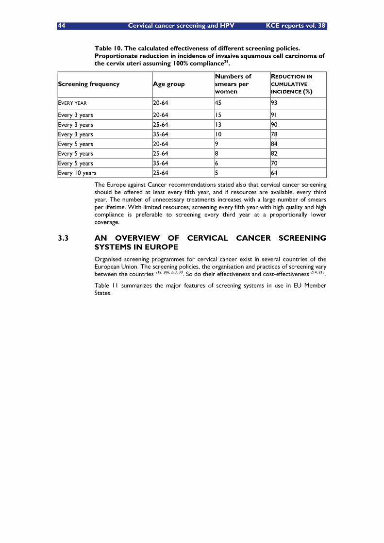
44 Cervical cancer screening and HPV KCE reports vol. 38
Table 10. The calculated effectiveness of different screening policies. Proportionate reduction in incidence of invasive squamous cell carcinoma of the cervix uteri assuming 100% compliance29.
Screening frequency Age group Numbers of smears per women
REDUCTION IN
CUMULATIVE
INCIDENCE (%)
EVERY YEAR 20-64 45 93
Every 3 years 20-64 15 91
Every 3 years 25-64 13 90
Every 3 years 35-64 10 78
Every 5 years 20-64 9 84
Every 5 years 25-64 8 82
Every 5 years 35-64 6 70
Every 10 years 25-64 5 64
The Europe against Cancer recommendations stated also that cervical cancer screening should be offered at least every fifth year, and if resources are available, every third year. The number of unnecessary treatments increases with a large number of smears per lifetime. With limited resources, screening every fifth year with high quality and high compliance is preferable to screening every third year at a proportionally lower coverage.
3.3 AN OVERVIEW OF CERVICAL CANCER SCREENING SYSTEMS IN EUROPE
Organised screening programmes for cervical cancer exist in several countries of the European Union. The screening policies, the organisation and practices of screening vary between the countries 212, 206, 213, 30. So do their effectiveness and cost-effectiveness 214, 215.
Table 11 summarizes the major features of screening systems in use in EU Member States.
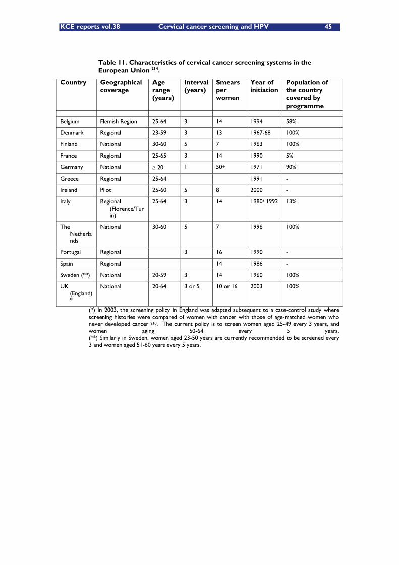
KCE reports vol.38 Cervical cancer screening and HPV 45
Table 11. Characteristics of cervical cancer screening systems in the European Union 214.
Country Geographical coverage
Age range (years)
Interval (years)
Smears per women
Year of initiation
Population of the country covered by programme
Belgium Flemish Region 25-64 3 14 1994 58%
Denmark Regional 23-59 3 13 1967-68 100%
Finland National 30-60 5 7 1963 100%
France Regional 25-65 3 14 1990 5%
Germany National 20 1 50+ 1971 90%
Greece Regional 25-64 1991 -
Ireland Pilot 25-60 5 8 2000 -
Italy Regional (Florence/Turin)
25-64 3 14 1980/ 1992 13%
The Netherlands
National 30-60 5 7 1996 100%
Portugal Regional 3 16 1990 -
Spain Regional 14 1986 -
Sweden (**) National 20-59 3 14 1960 100%
UK (England)*
National 20-64 3 or 5 10 or 16 2003 100%
(*) In 2003, the screening policy in England was adapted subsequent to a case-control study where screening histories were compared of women with cancer with those of age-matched women who never developed cancer 210. The current policy is to screen women aged 25-49 every 3 years, and women aging 50-64 every 5 years. (**) Similarly in Sweden, women aged 23-50 years are currently recommended to be screened every 3 and women aged 51-60 years every 5 years.
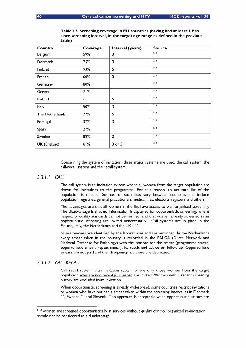
46 Cervical cancer screening and HPV KCE reports vol. 38
Table 12. Screening coverage in EU countries (having had at least 1 Pap since screening interval, in the target age range as defined in the previous table)
Country Coverage Interval (years) Source
Belgium 59% 3 216
Denmark 75% 3 212
Finland 93% 5 212
France 60% 3 217
Germany 80% 1 212
Greece 71% 212
Ireland - 5 212
Italy 50% 3 212
The Netherlands 77% 5 212
Portugal 37% 3 212
Spain 27% 212
Sweden 82% 3 212
UK (England) 61% 3 or 5 212
Concerning the system of invitation, three major systems are used: the call system, the call-recall system and the recall system.
3.3.1.1 CALL
The call system is an invitation system where all women from the target population are drawn for invitations to the programme. For this reason, an accurate list of the population is needed. Sources of such lists vary between countries and include population registries, general practitioners medical files, electoral registers and others.
The advantages are that all women in the list have access to well-organised screening. The disadvantage is that no information is captured for opportunistic screening, where respect of quality standards cannot be verified, and that women already screened in an opportunistic screening are invited unnecessarily5 . Call systems are in place in the Finland, Italy, the Netherlands and the UK 218-221.
Non-attendees are identified by the laboratories and are reminded. In the Netherlands every smear taken in the country is recorded in the PALGA (Dutch Network and National Database for Pathology) with the reasons for the smear (programme smear, opportunistic smear, repeat smear), its result and advice on follow-up. Opportunistic smears are not paid and their frequency has therefore decreased.
3.3.1.2 CALL-RECALL
Call recall system is an invitation system where only those women from the target population who are not recently screened are invited. Women with a recent screening history are excluded from invitation
When opportunistic screening is already widespread, some countries restrict invitations to women who have not had a smear taken within the screening interval as in Denmark 207, Sweden 222 and Slovenia. This approach is acceptable when opportunistic smears are
5 If women are screened opportunistically in services without quality control, organized re-invitation should not be considered as a disadvantage;
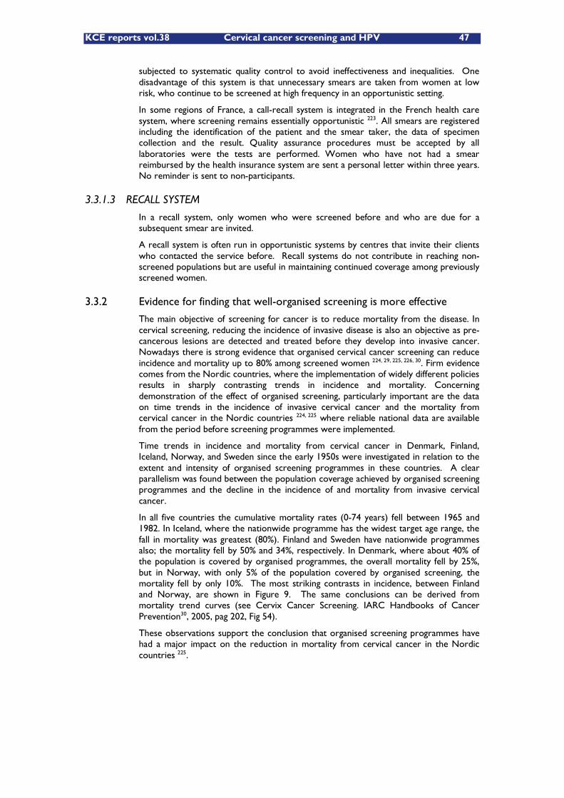
KCE reports vol.38 Cervical cancer screening and HPV 47
subjected to systematic quality control to avoid ineffectiveness and inequalities. One disadvantage of this system is that unnecessary smears are taken from women at low risk, who continue to be screened at high frequency in an opportunistic setting.
In some regions of France, a call-recall system is integrated in the French health care system, where screening remains essentially opportunistic 223. All smears are registered including the identification of the patient and the smear taker, the data of specimen collection and the result. Quality assurance procedures must be accepted by all laboratories were the tests are performed. Women who have not had a smear reimbursed by the health insurance system are sent a personal letter within three years. No reminder is sent to non-participants.
3.3.1.3 RECALL SYSTEM
In a recall system, only women who were screened before and who are due for a subsequent smear are invited.
A recall system is often run in opportunistic systems by centres that invite their clients who contacted the service before. Recall systems do not contribute in reaching non-screened populations but are useful in maintaining continued coverage among previously screened women.
3.3.2 Evidence for finding that well-organised screening is more effective
The main objective of screening for cancer is to reduce mortality from the disease. In cervical screening, reducing the incidence of invasive disease is also an objective as pre-cancerous lesions are detected and treated before they develop into invasive cancer. Nowadays there is strong evidence that organised cervical cancer screening can reduce incidence and mortality up to 80% among screened women 224, 29, 225, 226, 30. Firm evidence comes from the Nordic countries, where the implementation of widely different policies results in sharply contrasting trends in incidence and mortality. Concerning demonstration of the effect of organised screening, particularly important are the data on time trends in the incidence of invasive cervical cancer and the mortality from cervical cancer in the Nordic countries 224, 225 where reliable national data are available from the period before screening programmes were implemented.
Time trends in incidence and mortality from cervical cancer in Denmark, Finland, Iceland, Norway, and Sweden since the early 1950s were investigated in relation to the extent and intensity of organised screening programmes in these countries. A clear parallelism was found between the population coverage achieved by organised screening programmes and the decline in the incidence of and mortality from invasive cervical cancer.
In all five countries the cumulative mortality rates (0-74 years) fell between 1965 and 1982. In Iceland, where the nationwide programme has the widest target age range, the fall in mortality was greatest (80%). Finland and Sweden have nationwide programmes also; the mortality fell by 50% and 34%, respectively. In Denmark, where about 40% of the population is covered by organised programmes, the overall mortality fell by 25%, but in Norway, with only 5% of the population covered by organised screening, the mortality fell by only 10%. The most striking contrasts in incidence, between Finland and Norway, are shown in Figure 9. The same conclusions can be derived from mortality trend curves (see Cervix Cancer Screening. IARC Handbooks of Cancer Prevention30, 2005, pag 202, Fig 54).
These observations support the conclusion that organised screening programmes have had a major impact on the reduction in mortality from cervical cancer in the Nordic countries 225.
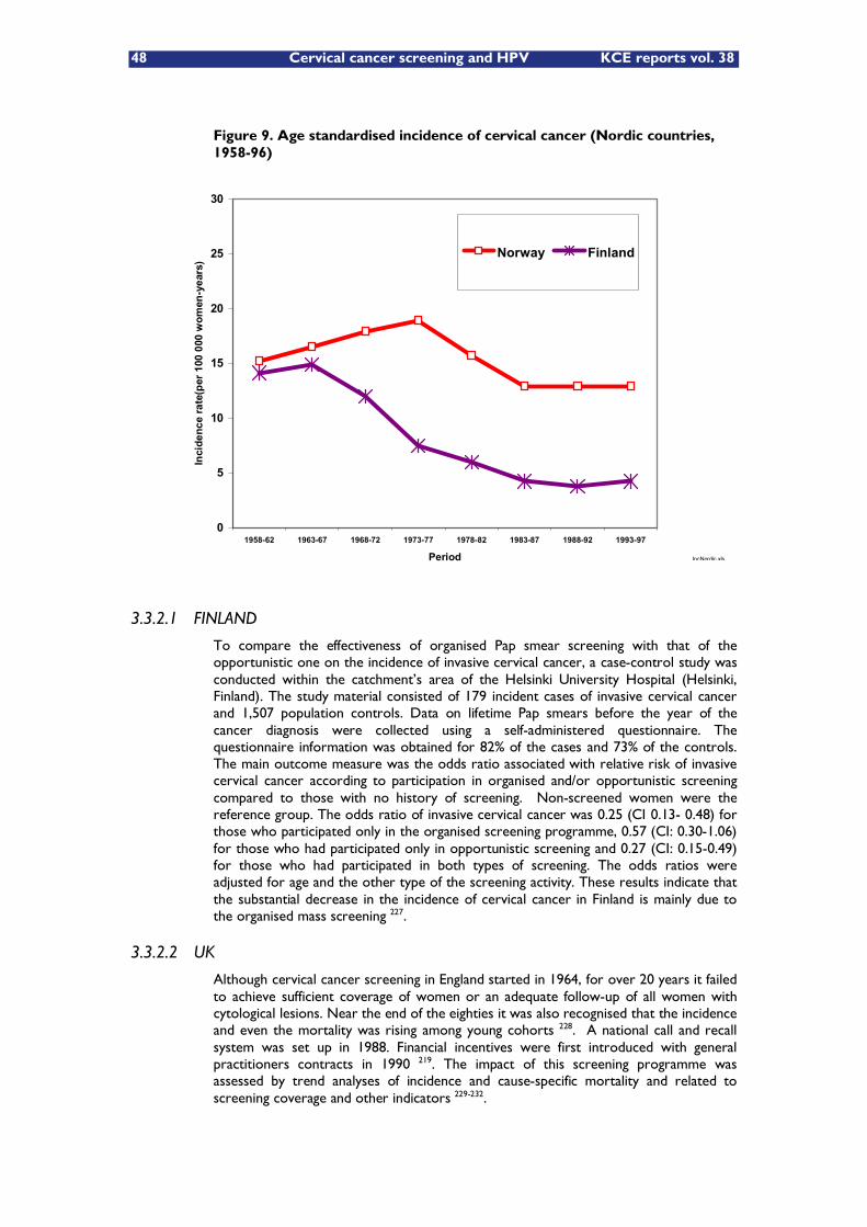
48 Cervical cancer screening and HPV KCE reports vol. 38
Figure 9. Age standardised incidence of cervical cancer (Nordic countries, 1958-96)
3.3.2.1 FINLAND
To compare the effectiveness of organised Pap smear screening with that of the opportunistic one on the incidence of invasive cervical cancer, a case-control study was conducted within the catchment�’s area of the Helsinki University Hospital (Helsinki, Finland). The study material consisted of 179 incident cases of invasive cervical cancer and 1,507 population controls. Data on lifetime Pap smears before the year of the cancer diagnosis were collected using a self-administered questionnaire. The questionnaire information was obtained for 82% of the cases and 73% of the controls. The main outcome measure was the odds ratio associated with relative risk of invasive cervical cancer according to participation in organised and/or opportunistic screening compared to those with no history of screening. Non-screened women were the reference group. The odds ratio of invasive cervical cancer was 0.25 (CI 0.13- 0.48) for those who participated only in the organised screening programme, 0.57 (CI: 0.30-1.06) for those who had participated only in opportunistic screening and 0.27 (CI: 0.15-0.49) for those who had participated in both types of screening. The odds ratios were adjusted for age and the other type of the screening activity. These results indicate that the substantial decrease in the incidence of cervical cancer in Finland is mainly due to the organised mass screening 227.
3.3.2.2 UK
Although cervical cancer screening in England started in 1964, for over 20 years it failed to achieve sufficient coverage of women or an adequate follow-up of all women with cytological lesions. Near the end of the eighties it was also recognised that the incidence and even the mortality was rising among young cohorts 228. A national call and recall system was set up in 1988. Financial incentives were first introduced with general practitioners contracts in 1990 219. The impact of this screening programme was assessed by trend analyses of incidence and cause-specific mortality and related to screening coverage and other indicators 229-232.
0
5
10
15
20
25
30
1958-62 1963-67 1968-72 1973-77 1978-82 1983-87 1988-92 1993-97
Period
Inci
denc
e ra
te(p
er 1
00 0
00 w
omen
-yea
rs)
Norway Finland
IncNordic.xls
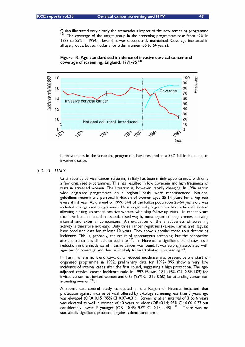
KCE reports vol.38 Cervical cancer screening and HPV 49
Quinn illustrated very clearly the tremendous impact of the new screening programme 230. The coverage of the target group in the screening programme rose from 42% in 1988 to 85% in 1994, a level that was subsequently maintained. Coverage increased in all age groups, but particularly for older women (55 to 64 years).
Figure 10. Age standardised incidence of invasive cervical cancer and coverage of screening, England, 1971-95 230
Improvements in the screening programme have resulted in a 35% fall in incidence of invasive disease.
3.3.2.3 ITALY
Until recently cervical cancer screening in Italy has been mainly opportunistic, with only a few organised programmes. This has resulted in low coverage and high frequency of tests in screened women. The situation is, however, rapidly changing. In 1996 nation wide organised programmes on a regional basis, were recommended. National guidelines recommend personal invitation of women aged 25-64 years for a Pap test every third year. At the end of 1999, 34% of the Italian population 25-64 years old was included in organised programmes. Most organised programmes have a fail-safe system allowing picking up screen-positive women who skip follow-up visits. In recent years data have been collected in a standardised way by most organised programmes, allowing internal and external comparisons. An evaluation of the effectiveness of screening activity is therefore not easy. Only three cancer registries (Varese, Parma and Ragusa) have produced data for at least 10 years. They show a secular trend to a decreasing incidence. This is, probably, the result of spontaneous screening, but the proportion attributable to it is difficult to estimate 220. In Florence, a significant trend towards a reduction in the incidence of invasive cancer was found. It was strongly associated with age-specific coverage, and thus most likely to be attributed to screening 233.
In Turin, where no trend towards a reduced incidence was present before start of organised programme in 1992, preliminary data for 1992-1995 show a very low incidence of interval cases after the first round, suggesting a high protection. The age-adjusted cervical cancer incidence ratio in 1992-98 was 0.81 (95% C.I. 0.59-1.09) for invited versus not invited women and 0.25 (95% CI 0.13-0.50) for attending versus non attending women 234.
A recent case-control study conducted in the Region of Firenze, indicated that protection against invasive cervical offered by cytology screening less than 3 years ago was elevated (OR= 0.15 (95% CI 0.07�–0.31). Screening at an interval of 3 to 6 years was elevated as well in women of 40 years or older (OR=0.14; 95% CI: 0.06�–0.33 but considerably lower if younger (OR= 0.45; 95% CI 0.14�–1.48) 235. There was no statistically significant protection against adeno-carcinoma.
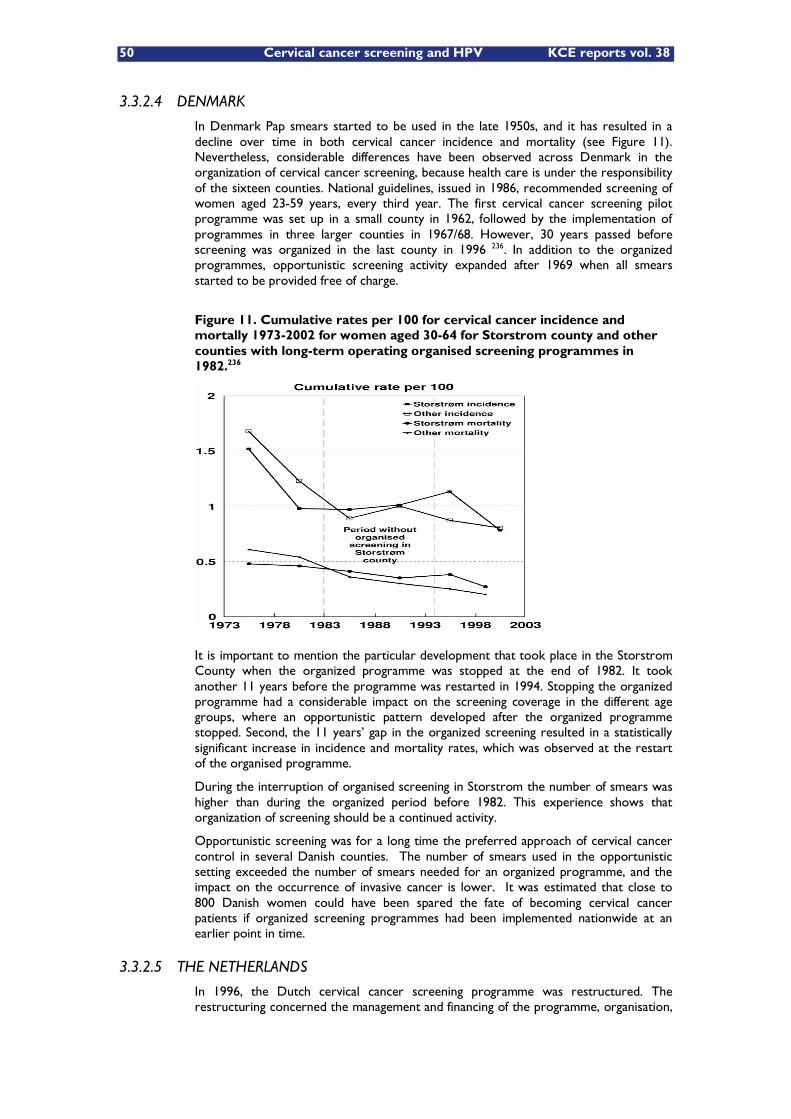
50 Cervical cancer screening and HPV KCE reports vol. 38
3.3.2.4 DENMARK
In Denmark Pap smears started to be used in the late 1950s, and it has resulted in a decline over time in both cervical cancer incidence and mortality (see Figure 11). Nevertheless, considerable differences have been observed across Denmark in the organization of cervical cancer screening, because health care is under the responsibility of the sixteen counties. National guidelines, issued in 1986, recommended screening of women aged 23-59 years, every third year. The first cervical cancer screening pilot programme was set up in a small county in 1962, followed by the implementation of programmes in three larger counties in 1967/68. However, 30 years passed before screening was organized in the last county in 1996 236. In addition to the organized programmes, opportunistic screening activity expanded after 1969 when all smears started to be provided free of charge.
Figure 11. Cumulative rates per 100 for cervical cancer incidence and mortally 1973-2002 for women aged 30-64 for Storstrom county and other counties with long-term operating organised screening programmes in 1982.236
It is important to mention the particular development that took place in the Storstrom County when the organized programme was stopped at the end of 1982. It took another 11 years before the programme was restarted in 1994. Stopping the organized programme had a considerable impact on the screening coverage in the different age groups, where an opportunistic pattern developed after the organized programme stopped. Second, the 11 years�’ gap in the organized screening resulted in a statistically significant increase in incidence and mortality rates, which was observed at the restart of the organised programme.
During the interruption of organised screening in Storstrom the number of smears was higher than during the organized period before 1982. This experience shows that organization of screening should be a continued activity.
Opportunistic screening was for a long time the preferred approach of cervical cancer control in several Danish counties. The number of smears used in the opportunistic setting exceeded the number of smears needed for an organized programme, and the impact on the occurrence of invasive cancer is lower. It was estimated that close to 800 Danish women could have been spared the fate of becoming cervical cancer patients if organized screening programmes had been implemented nationwide at an earlier point in time.
3.3.2.5 THE NETHERLANDS
In 1996, the Dutch cervical cancer screening programme was restructured. The restructuring concerned the management and financing of the programme, organisation,
KCE reports vol.38 Cervical cancer screening and HPV 51
target age ranges and interval, follow-up of abnormal results, and evaluation 221. When comparing before (1996) and after (2003) the restructuration the most important achievements are the following 237:
Substantial increase of the five-year coverage in the added target age groups (30-34, and 54-60) while in the old target age group (35-53 years) it remained around 80%.
Decrease of the proportion of screened women sent to follow-up from almost 19% to 3% per screening round.
Improvement of the follow-up compliance among screened women.
Shortening of the average time until a woman is either referred or rejoins the regular screening schedule
Reduction of the test positivity rate from over 10% up to approximately 2%
Reduction in the number of smears taken outside the target age group by 20% while maintaining high coverage rate
No increased in interval cancer rate, in spite of less screening and lower percentage of women under follow-up
Compared to other countries with organised national programmes, the Netherlands has been successful in limiting the number of excess smears while maintaining a high coverage rate. The procedures in the Netherlands allow sorting out women with recent smears. Further, smears taken outside of the regular screening schedule are only reimbursed when the women have medical complaints. Last, Pap smear screening in the Netherlands is principally performed in the GP practices.
3.3.2.6 NORWAY
In Norway, a 20% of reduction in incidence of cervical cancer has been observed since the initiation of organised screening in 1995. This was achieved trough more efficient use of Pap smears (by fixing the screening interval at 3 years) yielding lower consumption of screening examinations but also an increased population coverage. More details about the situation in Norway are given in the next paragraph.
3.3.3 Two highlighted screening programmes
3.3.3.1 The Norwegian cervical cancer screening programme
In Norway, a centralized system has been set up comprising obligatory registration of screen tests carried out in the organised programme or in an opportunistic setting. The Norwegian screening programme was introduced in 1995. It is population-based, nationwide, and recommends women of 25 to 69 years of age to have a Pap smear taken every 3 years. However in Norway, spontaneous screening activities were present since the early 1960s. Those activities brought a 50% reduction of the invasive cervical cancer incidence in women of 40-59 years old 238. From 1990, the incidence has remained stable. Several attempts during the 1960s, 1970s and 1980s to introduce an organised screening programme in Norway failed. In that period screening was characterised by frequent testing of young women at low risk and low-coverage in among women older than 50 and among women at high risk.
The introduction of a screening programme into a population where screening is already widespread poses problems different from those when implementing a novel programme. The Norwegian challenge was therefore to try to implement an organised programme in coordination with the spontaneous screening activity. The choice was made to integrate spontaneous screening into the organised programme in order to minimise changes in the healthcare system. By establishing a Cytology Register that registered every Pap smear taken in Norway, and by linking information at the individual level, the Norwegian coordinated screening programme started posting recommendation letters in 1995 to women who had not had a Pap smear in the
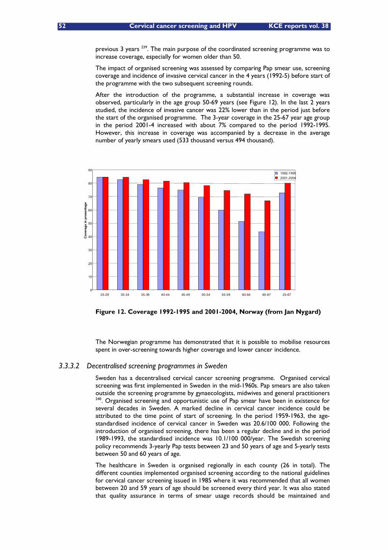
52 Cervical cancer screening and HPV KCE reports vol. 38
previous 3 years 239. The main purpose of the coordinated screening programme was to increase coverage, especially for women older than 50.
The impact of organised screening was assessed by comparing Pap smear use, screening coverage and incidence of invasive cervical cancer in the 4 years (1992-5) before start of the programme with the two subsequent screening rounds.
After the introduction of the programme, a substantial increase in coverage was observed, particularly in the age group 50-69 years (see Figure 12). In the last 2 years studied, the incidence of invasive cancer was 22% lower than in the period just before the start of the organised programme. The 3-year coverage in the 25-67 year age group in the period 2001-4 increased with about 7% compared to the period 1992-1995. However, this increase in coverage was accompanied by a decrease in the average number of yearly smears used (533 thousand versus 494 thousand).
Figure 12. Coverage 1992-1995 and 2001-2004, Norway (from Jan Nygard)
The Norwegian programme has demonstrated that it is possible to mobilise resources spent in over-screening towards higher coverage and lower cancer incidence.
3.3.3.2 Decentralised screening programmes in Sweden
Sweden has a decentralised cervical cancer screening programme. Organised cervical screening was first implemented in Sweden in the mid-1960s. Pap smears are also taken outside the screening programme by gynaecologists, midwives and general practitioners 240. Organised screening and opportunistic use of Pap smear have been in existence for several decades in Sweden. A marked decline in cervical cancer incidence could be attributed to the time point of start of screening. In the period 1959-1963, the age-standardised incidence of cervical cancer in Sweden was 20.6/100 000. Following the introduction of organised screening, there has been a regular decline and in the period 1989-1993, the standardised incidence was 10.1/100 000/year. The Swedish screening policy recommends 3-yearly Pap tests between 23 and 50 years of age and 5-yearly tests between 50 and 60 years of age.
The healthcare in Sweden is organised regionally in each county (26 in total). The different counties implemented organised screening according to the national guidelines for cervical cancer screening issued in 1985 where it was recommended that all women between 20 and 59 years of age should be screened every third year. It was also stated that quality assurance in terms of smear usage records should be maintained and
0
10
20
30
40
50
60
70
80
90
25-29 30-34 35-39 40-44 45-49 50-54 55-59 60-64 65-67 25-67
Cov
erag
e in
pro
sent
age
1992-19952001-2004
KCE reports vol.38 Cervical cancer screening and HPV 53
registry linkages with cancer registries be set up. The population registry is used. Every person has a personal identification number (PIN) and screening registries, cancer registries, pathology and cytology registries are all based on the PIN 222.
Sweden applied a call-recall invitation system. By a linkage with the population register and cytology registries all women who had a spontaneous smear taken in the past 18 months are sorted out and not invited for screening. The situation is heterogeneous in with respect to coverage and consumption of Pap tests. In certain counties over-screening is a substantial problem. The very high (86%) coverage of Pap smears in Stockholm has also led to a remarkable decrease in both incidence and mortality of cervical cancer. Rodvall demonstrated, in the Stockholm area, that organised screening reached high coverage among certain groups such as older and immigrant women, which are usually less covered in opportunistic settings.
The screening programme in Sweden is heterogeneous in quality. The new national guidelines seek to remedy some of the major limitations in particular by means of a national working group responsible for reviewing the programme.
It is of particular importance that registration of screening tests, in Norway and in Sweden, is compulsory and based on the national ID numbers and allowing linkages with other databases. Besides the advantages, outlined above for public health, this system offers enormous possibilities for bio-bank research. It is not by coincidence that HPV vaccine manufacturing companies have chosen these countries for post-licence surveillance.
3.4 RECOMMENDATIONS
In conclusion, well-organised screening appears more effective and a fortiori more cost-effective than the opportunistic activity. To maximise the positive impacts and minimise potential adverse effects, it is recommended that screening should be offered in organised settings (the Commission of the European Communities, 2003/0093; the Council of the European Union, 2003/87/EC).
The existence of a screening register is of great importance to achieve the objectives of the programme. It should contain information on participation in screening, the screen test results, the subsequent management of screen-positive women (compliance and results) and it should be linked to the cancer register and the population register.
54 Cervical cancer screening and HPV KCE reports vol. 38
4 CERVICAL CANCER SCREENING IN BELGIUM
4.1 INTRODUCTION
In this chapter we provide data on cervical cancer incidence and the related screening activities. Cervical cancer screening in Belgium is essentially opportunistic and integrated in the routine curative care system. Health insurance is obligatory in Belgium and is organized at a national level. Whereas financing of sampling and examination of Pap smears remains with the federal social security system, since 1980, the organisation of preventive healthcare in Belgium is confined to the Flemish, the French and German community. Organized cervical cancer screening initiatives started in 4 out of 5 Flemish provinces, using separate registers. Efforts to start a central cervical cancer screening programme have failed so far. There is no national external quality assurance programme for Pap smear analysis. The 3-year Pap screening coverage in women 25-64 years old is only 59%, while many of the women screened are over-screened.
In addition to the literature references a number of organisations were contacted as data source. The incidence of invasive cervical cancer is based on local cancer registry data provided by the Belgian Cancer Registry Foundation. The yearly volume of specific medical and laboratory activities associated with cervical cancer prevention was obtained from the National Health Insurance Institute (RIZIV/INAMI). In the context of this project, the Belgian Society of Clinical Cytology (data provided by Ph Delvenne) and the Flemish Society of Gynaecologists (VVOG, data provided by G Page) each conducted mid 2006 a survey documenting the current practice with regard to cervical cancer screening and HPV. Responses were obtained from 24 cyto-pathology labs and 79 gynaecologists.
4.2 CERVICAL CANCER IN BELGIUM
Based on the National Cancer Registry, 749 cases of cervical cancer were reported in 1993241, of which 482 (64%) cases occurred in the Flemish Region, 206 (28%) in the Walloon Region and 61 (8%) in the Capital Region of Brussels. The numbers should be interpreted with caution because of a possible under-declaration of new invasive cancers and an inconsistent inclusion of carcinoma in situ. For 2000 and 2001, reliable Flemish data are available for invasive cervical cancer (given for the year 2000 in Figure 13). According to the cancer register of the Flemish Region for the year 2000, 562 women were diagnosed with carcinoma in situ and 411 women with invasive cancer of the cervix (Figure 13).242 For 2000 and 2001 together the number of new cases of invasive cancer of the cervix was 803.243. The carcinoma in situ incidence cases however (pink curve in Figure 13) constitute only a fraction of all CIN3 diagnoses, the other part being cases of serious dysplasia which are not included here. Mortality due to cervical cancer is not known exactly because a substantial proportion of cervical cancers may get coded as unspecified uterine cancer. In 1993, 477 women died of cervical cancer or unspecified uterine cancer. It has been estimated that in the nineties between 300 and 350 died from cervical cancer in Belgium each year.244 The observed 5 year survival of invasive cancer of the cervix is 65.2% (1997-2001).243 No hard data are available for the number of deaths prevented by cervical cancer screening in Belgium, nor for the number needed to screen (NNS) to prevent one death. Based on historical data from British Columbia the NNS was 1140, meaning that 1140 women would have to be regularly screened over 10 years to prevent one death from cervical cancer.245
In July 2006, a new federal structure, the Belgian Cancer Registry Foundation (Stichting Kankerregister), took over the cancer registry role of the Flemish League against Cancer.243 This initiative should improve the quality of the cancer registration for the whole country. A 98.2% completeness of the cancer registry for breast cancer in 2000 was found based on a cross-check with the 57 screen detected breast cancers at The Leuven University Centre for Cancer Prevention. Validation of cancer registry data remains an issue. An on-site validity check of the registry data is not possible for
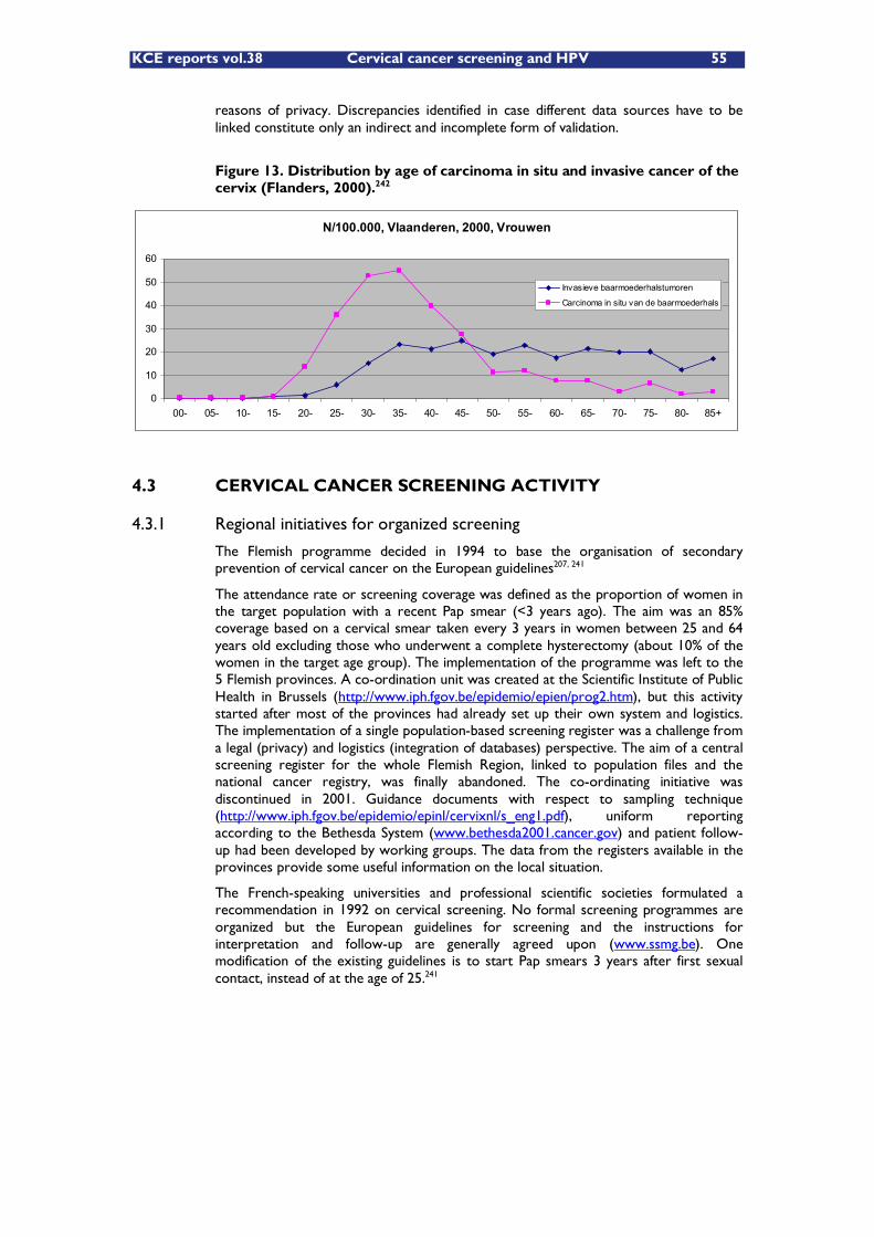
KCE reports vol.38 Cervical cancer screening and HPV 55
reasons of privacy. Discrepancies identified in case different data sources have to be linked constitute only an indirect and incomplete form of validation.
Figure 13. Distribution by age of carcinoma in situ and invasive cancer of the cervix (Flanders, 2000).242
4.3 CERVICAL CANCER SCREENING ACTIVITY
4.3.1 Regional initiatives for organized screening
The Flemish programme decided in 1994 to base the organisation of secondary prevention of cervical cancer on the European guidelines207, 241
The attendance rate or screening coverage was defined as the proportion of women in the target population with a recent Pap smear (<3 years ago). The aim was an 85% coverage based on a cervical smear taken every 3 years in women between 25 and 64 years old excluding those who underwent a complete hysterectomy (about 10% of the women in the target age group). The implementation of the programme was left to the 5 Flemish provinces. A co-ordination unit was created at the Scientific Institute of Public Health in Brussels (http://www.iph.fgov.be/epidemio/epien/prog2.htm), but this activity started after most of the provinces had already set up their own system and logistics. The implementation of a single population-based screening register was a challenge from a legal (privacy) and logistics (integration of databases) perspective. The aim of a central screening register for the whole Flemish Region, linked to population files and the national cancer registry, was finally abandoned. The co-ordinating initiative was discontinued in 2001. Guidance documents with respect to sampling technique (http://www.iph.fgov.be/epidemio/epinl/cervixnl/s_eng1.pdf), uniform reporting according to the Bethesda System (www.bethesda2001.cancer.gov) and patient follow-up had been developed by working groups. The data from the registers available in the provinces provide some useful information on the local situation.
The French-speaking universities and professional scientific societies formulated a recommendation in 1992 on cervical screening. No formal screening programmes are organized but the European guidelines for screening and the instructions for interpretation and follow-up are generally agreed upon (www.ssmg.be). One modification of the existing guidelines is to start Pap smears 3 years after first sexual contact, instead of at the age of 25.241
N/100.000, Vlaanderen, 2000, Vrouwen
0
10
20
30
40
50
60
00- 05- 10- 15- 20- 25- 30- 35- 40- 45- 50- 55- 60- 65- 70- 75- 80- 85+
Invasieve baarmoederhalstumoren
Carcinoma in situ van de baarmoederhals
56 Cervical cancer screening and HPV KCE reports vol. 38
In 2001 the Belgian Parliament also discussed the matter and proposed a resolution246
to organize a national cervical cancer screening programme, in collaboration with the regions, including the use of HPV testing in case of abnormal cytological result
to direct the concerned medical specialists and generalists to work out guidelines for such screening, criteria for the tests to be used in a quality assurance program, as well as a system for follow-up of the whole population
to stimulate EU coordination, consensus and research on HPV screening.247
The initiatives to improve cervical cancer screening are still funded by the Flemish Region and the provinces in 4 out of the 5 provinces of the Flemish Region. The initiatives have been described in a report published by the IPH, Brussels.248 In the provinces of Antwerp, Flemish-Brabant and Limburg separate registers for Pap smear results have been set up. Laboratories transfer dates and/or results to the screening register on a voluntary basis, respecting the privacy law.
In the province of West-Flanders no register with Pap smear results has been set up. The campaign activities are confined to specific regions. In 2002, the campaign resulted in a few more consultations for most of the physicians who completed a questionnaire (http://www.vvog.be/docs/2003/05/24034402.doc). Half of the physicians noticed an increased attendance of populations at risk (older women, never screened or lower socio-economic status).
For Antwerp, a report based on the registered results of over 76 000 Pap smears (1997-1999) showed abnormal results in 5.5% of the smears.249. In the 2002 Antwerp report the proportion of ASCUS was 1.9%, LSIL 1.1% and HSIL 0.4%. In the 2005 report, results of a call-recall system are described by LOGO (a number of communities grouped). The overall screening coverage was 51% and varied by LOGO from 39% to 59%. The average coverage decreased from 54% for 25-44 y old women to 40% for 55-64 y old women. The proportion of women over 55 who underwent a hysterectomy is 12.6%. Efforts are ongoing to further increase the participation of all cyto-pathology labs. The quality of the smear was reported as optimal in 74%. Epithelial lesions are reported in nearly 4% of 295 636 cases, and include ASCUS in 1.9%, LSIL in 1.3%, and HSIL in 0.4%. A total of 24 cases of invasive cervical cancer were reported as well as 193 cases of epithelial carcinoma in situ. AGUS was found in 0.4% of 202 735 cases and 46 cases of adenocarcinoma were reported (report by E Van de Mieroop, and provided by F Smeets).
For Limburg, results of cervical smears are stored in the LIKAR register (Limburger Cancer Register) since 1996. The LIKAR Cervix cytology Register (http://likas.edm.uhasselt.be/likar/) covers Pap-smears of women in the province of Limburg (7.6% of the Belgian population). All cytology laboratories in Limburg, as well as some neighbouring laboratories examining Pap-smears of women, contribute to this Register. The goals of the register are
to monitor the epidemiology of cervical cancer, mortality, screening coverage
to monitor and optimize screening participation (reduce overscreening, increase coverage in underscreened populations)
improve quality of samples taken by providing feedback to sampling physicians
evaluate and support follow-up of screen positives and evaluate follow-up of screen negatives
link screening register with LIKAR cancer register to detect cases of interval cancer
Due to incompleteness of voluntary cytology registration and due to inaccuracies in unique identification of women, no reliable estimates of screening coverage can be derived from the current registries. Initial problems for data analysis concerning
KCE reports vol.38 Cervical cancer screening and HPV 57
address changes, double records and records with male sex were resolved. In the report covering the period 1996-2000 (http://likas.edm.uhasselt.be/likar/scripts/php/showdoc.php?id=83) the screening register was estimated to be 75% complete. Gynaecologist-connexists (reading Pap smears of own patients) were not included and account for an estimated 20% of the smears. A hysterectomy is performed in 8-12% of women aged 24-65 years. Data were exchanged with the provinces of Antwerp and Flemish-Brabant on a yearly basis. Standardisation of the coding system for Pap smear results is a requirement for a well functioning register. Unfortunately, many Pap results had to be recoded to the Bethesda terminology.
Overscreening (more than once per 3 year period) was observed in about a third of the women, and was observed only slightly more frequently in gynaecology practices compared with GPs. Smears assessed for quality were in 77% of optimal quality, 23% suboptimal and 1% of bad quality. The proportion of abnormal smear results was 3.31% on a total of 299 642 smears collected in the period 1996-2000 (ASC-US: 1.72%, AGUS: 0.07%, LSIL: 0.45%, HSIL: 1.04%, carcinoma: 0.03%).
The registry of Flemish-Brabant contains data provided by 13 laboratories and is unique because all results are directly coded using the Bethesda 2001 terminology. This is not yet the case for the two other registers (Antwerp and Limburg). The Flemish-Brabant data for 2004 and the previous years show ASC-US/LSIL results in 3% of the 300 000 Pap smears (Bourgain C., personal communication, see also248)
Smears include both screening and follow-up samples. The proportion of ASC-US reported needs however to be interpreted with caution as many laboratories already perform HPV testing in such cases and no guidance is provided on how to integrate the HPV findings. The HPV result might thus theoretically result in a change in the pathological diagnosis finally reported and thus in fewer ASC-US reports.
No screening is organized in East-Flanders, the Walloon Region or Brussels. The cost of opportunistic screening has been reported to be 42% higher compared with organized screening.250, 251.
4.3.2 Cytology-based testing
Cervical cancer screening in Belgium remains essentially opportunistic and integrated in the routine curative care system. Processing and reading of Pap smears is mainly performed by laboratories for pathology, and in 2004 in 3.8% by gynaecologists (for their own patients only). Screening of cervical smears is mostly (in 60% of all laboratories) carried out by cytotechnicians, under the supervision of a senior cytotechnician (60%) or a pathologist (35%).252 In 38% of the labs smears are read by a pathologist (38%) or resident (2%).
Quality assurance is considered important by 93% of pathologists, and is common practice in about three-quarters of cytology laboratories.252 Targeted review by a cyto-technologist of Pap smears read as negative for patients at increased risk of positivity (eg based on history or clinical information) is routine practice in about half of the laboratories.252 There is no formal long term training of cytotechnicians, most of them being graduate laboratory technicians having been trained at the bench. Continuing education of cytotechnicians is the rule in 88% of cytology labs.252 Quality of the smears is good in about three quarters of the cases, and this proportion could further be increased eg by avoiding smear taking during pregnancy or during the 6 months postpartum, as recommended.249
There is no national external quality assurance program for Pap smear analysis. Pathology historically moved away from the clinical biology field. In order to enforce quality assurance, a legal basis for the recognition of cytology and pathology labs is needed. A draft Royal Decree has been prepared for this purpose. This requires that pathology/cytology labs participate to external QA programmes, organised by the Institute for Public Health (IPH).(J-C Libeer, IPH, personal communication).
Methods (use of LBC), reporting standard (use of Bethesda standard), and measures taken to guarantee the quality of Pap smears are included in an non-exhaustive and not completely up to date overview table by Domus Medica / WVVH (Organisation of GP�’s
58 Cervical cancer screening and HPV KCE reports vol. 38
in Flanders) of laboratories offering Pap smear interpretation in the Flemish region (http://www.wvvh.be/files/Labos%20cervix%202004.pdf).
LBC was introduced in Belgium from 1998 onwards. In a survey published in 2005 about half of the cytology labs performed cervical LBC. Prepstain (Tripath Imaging, Burlington, NC, US) was the preferred instrument, while in the Walloon region ThinPrep 2000 (Cytyc Corporation, Boxborough, MA, USA) was predominantly used. Small pathology services often applied manual methods (Tripath 2, ThermoShandon, Turbitec Labonord�…) or cooperated with larger services for cytopreparation.252
In the recent 2006 survey among the cyto-pathology labs responses were obtained for 24 labs, representing the different regions of the country. 19 out of 24 labs report to use LBC routinely. The cytology medium used is reported for 21 labs: 10 use SurePath from Tripath (CytoRich Blue, CytoRich Red, AutoCyte) 5 use ThinPrep from CYTYC, 4 use Easyfix from Labonord, 2 use a Thermo-Electron medium. 23 out of the 24 labs use Bethesda reporting for cervical cytology.
4.3.3 HPV testing
In agreement with international data, HPV genotype 16 was identified as the most common genotype in cervical smears with abnormal cytology result. This was the case for studies based on routinely obtained Pap smears in Belgium. One group used MY09/MY11 consensus primers for HPV DNA detection and specific primers for identifying 14 high-risk types and identified in high-grade lesions essentially HPV 16, 18, 31, 33, 35 and 51.253 A second group used PCR detection based on the SPF primer set, followed by reverse hybridization allowing the identification of 25 HPV genotypes. The most frequently detected genotype in high-grade lesions was HPV 16, followed by HPV 52, 51 and 31.254 In a population of sex workers in East-Flanders (mean age 28 years, 39% of non-Belgian nationality) HPV genotype 16, 31 and 52 were identified most frequently using the same method. This population also showed significantly more high-grade Pap smear results, as well as more HPV positive results versus an age-matched control group.255
HPV tests are most conveniently performed on cell brush material collected for liquid based cytology. Collection of a separate liquid sample for HPV testing is needed when a conventional smear is used. The guidelines of the Belgian Society for Clinical Cytology, developed with the participation of the societies of gynaecologists256 list the following indications for HPV testing:
ASCUS, mainly ASC-US, but also ASC-H
AGC-ecc, both NOS and possibly neoplastic
Follow-up after treated HSIL or AGS-ecc, in case the Pap result is negative.
These guidelines do not include LSIL as an indication, in accordance with TBS2001, and overruled the indications for testing used before by the Centres for Molecular Diagnosis (CMD�’s): cervical ASCUS/AGUS in women over 30 years old, LSIL, or follow-up for residual HPV (maximum 2 times in 6 months after the intervention).247
Molecular diagnostics were introduced in 1998 into the Belgian health care system based on the funding of CMD�’s, as documented in a KCE report257. We refer to this report for recommendations to assure the quality of molecular tests, including HPV tests. The yearly overall CMD health insurance budget remained fixed at 6.5 million Euros. After an increase in the number of centres from 10 to 18 based on legal judgment, the legal basis of the CMD�’s was rejected early 2005 by the Council of State. The Human Papilloma Virus (HPV) test was one of the 94 molecular tests performed at the CMD�’s. In addition to the HPV tests performed at the CMD�’s, HPV testing is also routinely performed by some private laboratories.
In 2001, HPV testing was provided by 53% of cytology laboratories.252, and according to the 2006 survey HPV testing is used by the vast majority of reporting gynaecologists (69 out of 77). No less than 19 out of 69 gynaecologists report in 2006 the routine practice of primary HPV screening, mainly simultaneously with cytology. In case gynaecologists
KCE reports vol.38 Cervical cancer screening and HPV 59
use HPV testing for purposes other than primary screening, the HPV test request is made by the cytopathologist 35 times out of 50.
In the 2006 survey nearly all cyto-pathology labs (23 out of 24) report the use of HPV testing: 22 labs for ASC-US (or repeat ASC-US), 14 for ASC-H, 15 for LSIL, 3 for HSIL, 11 for Atypical Glandular Cells (AGC), 8 for other (mainly treatment follow-up). The number of HPV yearly tests per reporting lab varies from 8 to 1560, median 253 HPV tests per year. This represents about 2% of all PAP tests performed at the lab (from < 1% to 7.5% for the 6 labs which reported their overall volume).
In 2003-2004, the HPV tests at the CMD�’s were performed mainly using the kit Hybrid Capture II (HC2) from Digene. In the 2006 surveys 10 labs report using the HC2 HPV test method, 4 labs report using a PCR method. Of the 48 gynaecologists reporting the HPV testing method, 32 reported PCR and 16 the Hybrid Capture method.
The average turnaround time for an HPV test was 9.5 days in the CMD�’s. The 2006 cyto-pathology lab survey shows a large variation in maximum turnaround time, from 8 to 90 days, median 21 days. All but one lab integrate the HPV result into the first or a second pathology report, 8 labs integrate HPV results already into the first report. No guidance exists on how to integrate the HPV findings. In some labs the HPV test may result in a change of the pathology result finally reported, and eg in fewer ASC-US reports.
Nine of the 24 reporting labs do the HPV testing at their own pathology lab. Direct requests for HPV testing by gynaecologists or GPs constitute only a minority of all HPV test requests (from few tests to up to 20%).
The performance and results of Pap tests and possible HPV tests are not always routinely communicated between health care professionals, mainly gynaecologists and GPs. GPs have information on cervical cancer screening for no more than a third of the target population in their practice (personal communication F Smeets, Domus Medica). No local data are available verifying whether this situation is preferred by the patients.
Gynaecologists responding to the 2006 survey (n=61) report women are informed orally (n=45) prior to (possible) HPV testing. The vast majority of responding gynaecologists (72 out of 78) do not explicitly communicate normal Pap/HPV test results (no news is good news). Most of the responding gynaecologists will communicate a positive HPV result (59 out of 74). A total of 46 out of 76 responding gynaecologists report that a positive HPV test will increase the frequency of consultations. Note that this is the intended purpose for HPV tests performed in the appropriate indications and at the appropriate intervals. As a side remark some gynaecologists note that HPV testing is performed without request on every sample shipped. Such testing may lead to problems for the gynaecologist, in case of a positive HPV result in a woman with negative cytological results. Also the question is raised whether such generalised testing (for research purposes?) has been approved by any Ethics Committee.
4.4 BUDGET, TESTING VOLUME AND COVERAGE
The yearly Belgian health insurance budget used for covering medical activities directly linked to cervical cancer screening amounts up to 65 million euro. The 2005 budget was composed as detailed in Table 13.
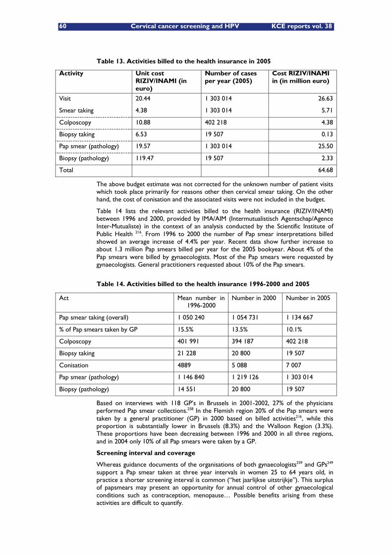
60 Cervical cancer screening and HPV KCE reports vol. 38
Table 13. Activities billed to the health insurance in 2005
Activity Unit cost RIZIV/INAMI (in euro)
Number of cases per year (2005)
Cost RIZIV/INAMI in (in million euro)
Visit 20.44 1 303 014 26.63
Smear taking 4.38 1 303 014 5.71
Colposcopy 10.88 402 218 4.38
Biopsy taking 6.53 19 507 0.13
Pap smear (pathology) 19.57 1 303 014 25.50
Biopsy (pathology) 119.47 19 507 2.33
Total 64.68
The above budget estimate was not corrected for the unknown number of patient visits which took place primarily for reasons other then cervical smear taking. On the other hand, the cost of conisation and the associated visits were not included in the budget.
Table 14 lists the relevant activities billed to the health insurance (RIZIV/INAMI) between 1996 and 2000, provided by IMA/AIM (Intermutualistisch Agentschap/Agence Inter-Mutualiste) in the context of an analysis conducted by the Scientific Institute of Public Health 216. From 1996 to 2000 the number of Pap smear interpretations billed showed an average increase of 4.4% per year. Recent data show further increase to about 1.3 million Pap smears billed per year for the 2005 bookyear. About 4% of the Pap smears were billed by gynaecologists. Most of the Pap smears were requested by gynaecologists. General practitioners requested about 10% of the Pap smears.
Table 14. Activities billed to the health insurance 1996-2000 and 2005
Act Mean number in 1996-2000
Number in 2000 Number in 2005
Pap smear taking (overall) 1 050 240 1 054 731 1 134 667
% of Pap smears taken by GP 15.5% 13.5% 10.1%
Colposcopy 401 991 394 187 402 218
Biopsy taking 21 228 20 800 19 507
Conisation 4889 5 088 7 007
Pap smear (pathology) 1 146 840 1 219 126 1 303 014
Biopsy (pathology) 14 551 20 800 19 507
Based on interviews with 118 GP�’s in Brussels in 2001-2002, 27% of the physicians performed Pap smear collections.258 In the Flemish region 20% of the Pap smears were taken by a general practitioner (GP) in 2000 based on billed activities216, while this proportion is substantially lower in Brussels (8.3%) and the Walloon Region (3.3%). These proportions have been decreasing between 1996 and 2000 in all three regions, and in 2004 only 10% of all Pap smears were taken by a GP.
Screening interval and coverage
Whereas guidance documents of the organisations of both gynaecologists259 and GPs249 support a Pap smear taken at three year intervals in women 25 to 64 years old, in practice a shorter screening interval is common (�“het jaarlijkse uitstrijkje�”). This surplus of papsmears may present an opportunity for annual control of other gynaecological conditions such as contraception, menopause�… Possible benefits arising from these activities are difficult to quantify.
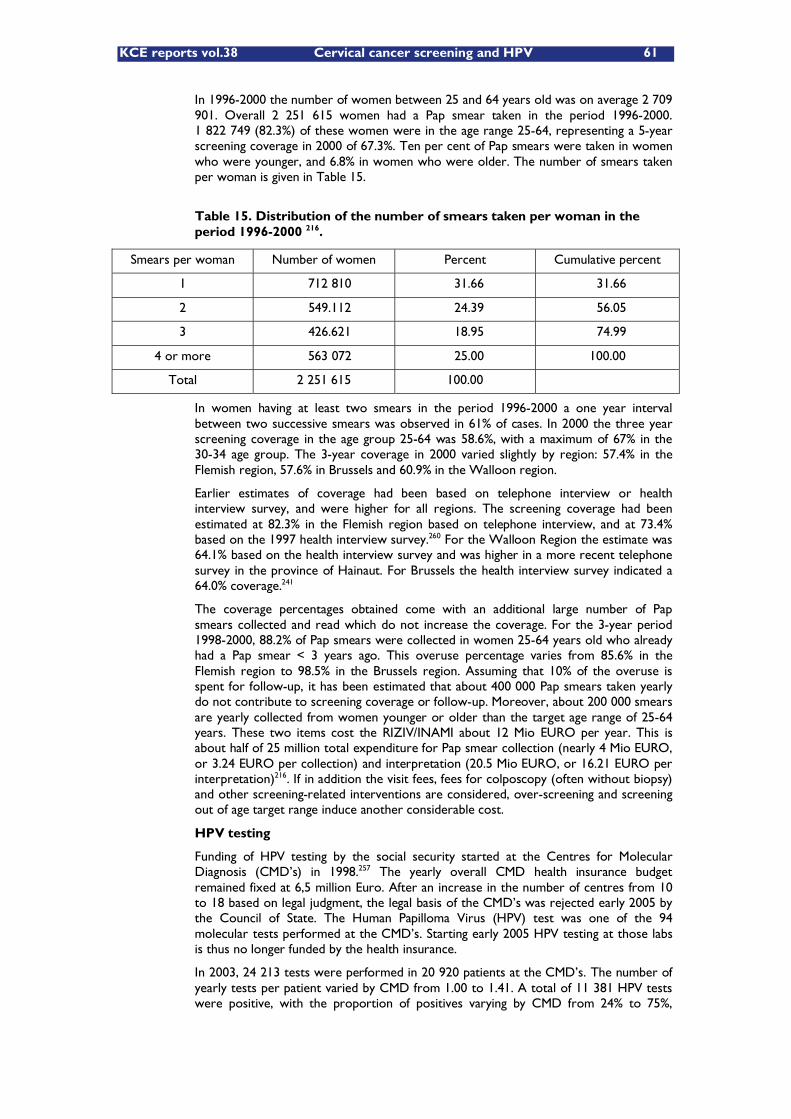
KCE reports vol.38 Cervical cancer screening and HPV 61
In 1996-2000 the number of women between 25 and 64 years old was on average 2 709 901. Overall 2 251 615 women had a Pap smear taken in the period 1996-2000. 1 822 749 (82.3%) of these women were in the age range 25-64, representing a 5-year screening coverage in 2000 of 67.3%. Ten per cent of Pap smears were taken in women who were younger, and 6.8% in women who were older. The number of smears taken per woman is given in Table 15.
Table 15. Distribution of the number of smears taken per woman in the period 1996-2000 216.
Smears per woman Number of women Percent Cumulative percent
1 712 810 31.66 31.66
2 549.112 24.39 56.05
3 426.621 18.95 74.99
4 or more 563 072 25.00 100.00
Total 2 251 615 100.00
In women having at least two smears in the period 1996-2000 a one year interval between two successive smears was observed in 61% of cases. In 2000 the three year screening coverage in the age group 25-64 was 58.6%, with a maximum of 67% in the 30-34 age group. The 3-year coverage in 2000 varied slightly by region: 57.4% in the Flemish region, 57.6% in Brussels and 60.9% in the Walloon region.
Earlier estimates of coverage had been based on telephone interview or health interview survey, and were higher for all regions. The screening coverage had been estimated at 82.3% in the Flemish region based on telephone interview, and at 73.4% based on the 1997 health interview survey.260 For the Walloon Region the estimate was 64.1% based on the health interview survey and was higher in a more recent telephone survey in the province of Hainaut. For Brussels the health interview survey indicated a 64.0% coverage.241
The coverage percentages obtained come with an additional large number of Pap smears collected and read which do not increase the coverage. For the 3-year period 1998-2000, 88.2% of Pap smears were collected in women 25-64 years old who already had a Pap smear < 3 years ago. This overuse percentage varies from 85.6% in the Flemish region to 98.5% in the Brussels region. Assuming that 10% of the overuse is spent for follow-up, it has been estimated that about 400 000 Pap smears taken yearly do not contribute to screening coverage or follow-up. Moreover, about 200 000 smears are yearly collected from women younger or older than the target age range of 25-64 years. These two items cost the RIZIV/INAMI about 12 Mio EURO per year. This is about half of 25 million total expenditure for Pap smear collection (nearly 4 Mio EURO, or 3.24 EURO per collection) and interpretation (20.5 Mio EURO, or 16.21 EURO per interpretation)216. If in addition the visit fees, fees for colposcopy (often without biopsy) and other screening-related interventions are considered, over-screening and screening out of age target range induce another considerable cost.
HPV testing
Funding of HPV testing by the social security started at the Centres for Molecular Diagnosis (CMD�’s) in 1998.257 The yearly overall CMD health insurance budget remained fixed at 6,5 million Euro. After an increase in the number of centres from 10 to 18 based on legal judgment, the legal basis of the CMD�’s was rejected early 2005 by the Council of State. The Human Papilloma Virus (HPV) test was one of the 94 molecular tests performed at the CMD�’s. Starting early 2005 HPV testing at those labs is thus no longer funded by the health insurance.
In 2003, 24 213 tests were performed in 20 920 patients at the CMD�’s. The number of yearly tests per patient varied by CMD from 1.00 to 1.41. A total of 11 381 HPV tests were positive, with the proportion of positives varying by CMD from 24% to 75%,
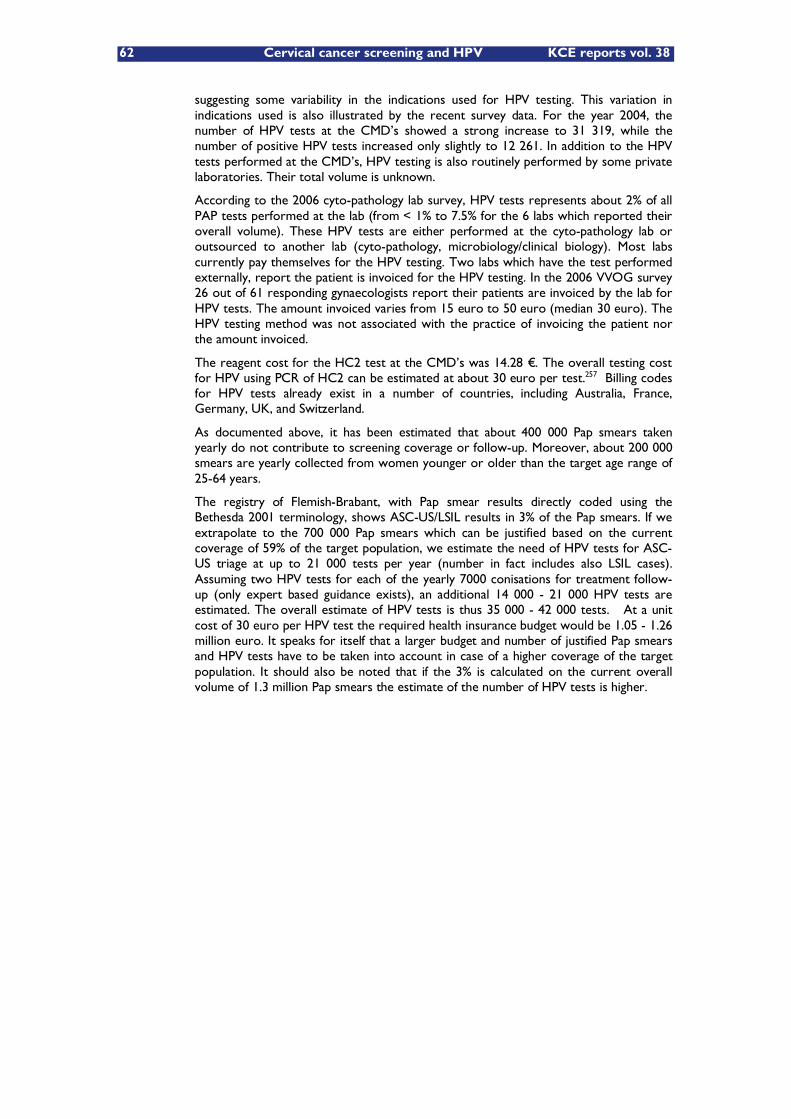
62 Cervical cancer screening and HPV KCE reports vol. 38
suggesting some variability in the indications used for HPV testing. This variation in indications used is also illustrated by the recent survey data. For the year 2004, the number of HPV tests at the CMD�’s showed a strong increase to 31 319, while the number of positive HPV tests increased only slightly to 12 261. In addition to the HPV tests performed at the CMD�’s, HPV testing is also routinely performed by some private laboratories. Their total volume is unknown.
According to the 2006 cyto-pathology lab survey, HPV tests represents about 2% of all PAP tests performed at the lab (from < 1% to 7.5% for the 6 labs which reported their overall volume). These HPV tests are either performed at the cyto-pathology lab or outsourced to another lab (cyto-pathology, microbiology/clinical biology). Most labs currently pay themselves for the HPV testing. Two labs which have the test performed externally, report the patient is invoiced for the HPV testing. In the 2006 VVOG survey 26 out of 61 responding gynaecologists report their patients are invoiced by the lab for HPV tests. The amount invoiced varies from 15 euro to 50 euro (median 30 euro). The HPV testing method was not associated with the practice of invoicing the patient nor the amount invoiced.
The reagent cost for the HC2 test at the CMD�’s was 14.28 �€. The overall testing cost for HPV using PCR of HC2 can be estimated at about 30 euro per test.257 Billing codes for HPV tests already exist in a number of countries, including Australia, France, Germany, UK, and Switzerland.
As documented above, it has been estimated that about 400 000 Pap smears taken yearly do not contribute to screening coverage or follow-up. Moreover, about 200 000 smears are yearly collected from women younger or older than the target age range of 25-64 years.
The registry of Flemish-Brabant, with Pap smear results directly coded using the Bethesda 2001 terminology, shows ASC-US/LSIL results in 3% of the Pap smears. If we extrapolate to the 700 000 Pap smears which can be justified based on the current coverage of 59% of the target population, we estimate the need of HPV tests for ASC-US triage at up to 21 000 tests per year (number in fact includes also LSIL cases). Assuming two HPV tests for each of the yearly 7000 conisations for treatment follow-up (only expert based guidance exists), an additional 14 000 - 21 000 HPV tests are estimated. The overall estimate of HPV tests is thus 35 000 - 42 000 tests. At a unit cost of 30 euro per HPV test the required health insurance budget would be 1.05 - 1.26 million euro. It speaks for itself that a larger budget and number of justified Pap smears and HPV tests have to be taken into account in case of a higher coverage of the target population. It should also be noted that if the 3% is calculated on the current overall volume of 1.3 million Pap smears the estimate of the number of HPV tests is higher.

KCE reports vol.38 Cervical cancer screening and HPV 63
Key points
In Belgium, preventive healthcare resides within the responsibilities of the three communities, while the medical activities concerned are being paid by the federal social security.
Despite a 65 million euro Belgian health insurance budget used for covering medical activities directly linked to cervical cancer screening, an estimated 700 women are diagnosed with invasive cervical cancer each year. About a third of these women will die from cervical cancer.
Cervical cancer screening is essentially opportunistic. Screening initiatives were set up in the Flemish provinces. Efforts to start a central cervical cancer screening programme have failed so far.
There is no national external quality assurance programme for Pap smear analysis.
The 3-year Pap screening coverage in women 25-64 years old is only 59% in Belgium.
Many of the women screened are over-screened.
Introducing HPV testing for ASC-US triage and treatment follow-up requires a health-insurance budget of about 1.2 million euro.
The available resources, if used more efficiently, are theoretically sufficient to cover the whole target population.
64 Cervical cancer screening and HPV KCE reports vol. 38
5 PATIENT ISSUES The issue and practice of screening for cervical cancer raises several ethical considerations about the detection in pre-symptomatic stages. It has been recognized that screening may not only have benefits but also associated harms for participants. Women may experience psychological harm such as anxiety, false alarms, false reassurance and side effects such as unnecessary colposcopies and biopsies, over-diagnosis, and over-treatment 261. Hence the issue of participation following informed decision making is of particular interest. A good understanding of the behavioural factors linked to cervical cancer screening also urges for a deeper consideration of factors that determine the participation in cervical screening programs and interventions to encourage participation.
The role of HPV testing in cervical cancer screening offers an interesting approach. Whereas routine HPV screening is not recommended (yet), the use of HPV testing to triage women whose pap smear results show ASCUS is a recommended management option. Hence, HPV testing in the management of ASCUS or in the context of cervical screening raises important questions about informed participation in cervical screening. Especially the question how to provide information to patients enabling informed decisions has to be discussed.
5.1 SEARCH STRATEGY
First systematic reviews were searched in the Cochrane library. The key words �‘cervical cancer screening�’ and �‘Human papillomavirus�’ were used. One relevant systematic review was found 262. Secondly, we looked for original research. We searched for systematic reviews and original research in the databases Pubmed, Embase and Cinahl. A description of the detailed search strategy can be found in annex. Additional references were located through searching by keywords, the bibliographies of identified studies, related papers and by contacts with specialists in the subject area. The search for literature has been performed in May 2006.
5.2 DETERMINANTS OF PARTICIPATION
The uptake of pap smears varies between and within countries. A report of the International Agency for research on cancer (IARC) reviewed studies of the last 10 years on predictors of participation in cervical cancer screening programs 30 In many studies attendance was associated with a higher income and educational level 263. Employed women were found to be more likely to attend screening than unemployed women 240. A more recent US study however found that screening participation was higher in non employed women. A possible explanation is that the segment of individuals working for minimum wages and for employers that don�’t provide an insurance to their employees increases in the US and is represented in the sample 264. Another reported predictor that is often closely linked to the socio-economic status 265 was the ethnicity 266. Ethnicity as a predictor of attendance of cervical cancer screening also reflects the influence of cultural barriers 267. Less conclusive was the impact of the health status. The prevalence of risk factors such as sexual intercourse at young age, multiple sex partners, contraceptive pill use, smoking have increased over time but women at higher risk are generally better screened in the Flemish region of Belgium 260. Age was found to be an important predictor for attendance 265. Most studies in the IARC report found that younger women were more likely to participate in cervical screening programs than older women. A US study using a national interview survey among 18 388 women older than 18 years who had a pap test in the last 12 months however found that women of childbearing age (18-44) had a lower percentage of cervical cancers screening in the last 12 months than the women older than 44 264. The authors suggest that issues related to insurance provider and benefits type could be an explanatory factor.
KCE reports vol.38 Cervical cancer screening and HPV 65
Overscreening (interval between pap smear less than 3 years) was found to be an important phenomenon among screened women especially within the younger age groups.
Participation also has been reported to vary by marital status. Singles were less likely to participate in screening than married, divorced and widowed women 260, 240, 268. Moreover evidence was found that women who live in rural areas are less likely to attend for screening than those from urban areas 269. Among the rural population farm lifestyle was revealed to be a predictor for failure of participation 270.
Better knowledge about the screening procedure (the screening interval, the perceived necessity...) increases the attendance. Anticipated embarrassment and attitudes to screening (for instance women don�’t go for screening if they don�’t have any symptoms) are also strongly associated with participation 267, 268. Anxiety and fear of cancer were identified by several studies reviewed in the IARC report30 as factors of non attendance of screening.
One of the main predictors of attendance reported were the interactions with the health provider characteristics and health care organisation. Attendance increases when the physician is female, when a recommendation is made by a doctor to attend screening and to health insurance issues 271. A UK population based study reported the impact factors of primary care service delivery. Independent predictors were general practice structure, workload and GP characteristics²272.
5.3 INTERVENTIONS TO ENCOURAGE PARTICIPATION
All of the trials included in the Cochrane review 273 on interventions targeted at women to encourage the participation in cervical screening were based on the assumption that screening was beneficial and high level of participation should be achieved at all costs. The review showed some evidence to support the use of invitation letters. This finding was confirmed by the IARC report 30. Invitation letters with fixed appointments were more effective than invitations with open appointments. The accuracy of population registers was identified as a key issue. Limited evidence is available to support educational interventions but it was unclear what format was most effective (i.e. printed, video/slide or face to face presentations). Other interventions such as revealing the gender of the smear taker in the invitation letter and the use of a health promotion nurse appeared promising approaches but their effectiveness was only assessed in a limited number of studies. In the UK it has been recommended that a leaflet emphasising the risks and benefits should be included with every invitation for screening 274.
An RCT including all women invited to organised screening in Sweden (n=12.240) evaluated the effectiveness of 3 different interventions: a) an invitation letter accompanied by an information brochure versus a control group with a standard invitation letter. b) a reminder letter to non-attendees after the first intervention compared with women who did not receive a reminder letter. c) a phone reminder to non�–attenders compared with non phone reminder 275. Enclosing an information leaflet did not increase attendance. This finding was consistent with earlier studies 30. A reminder letter and a phone reminder however significantly increased women�’s attendance 275.
A Dutch study revealed that invitation for a national screening programme for cervical cancer by a GP resulted in a higher participation rate than a letter by the local health authority. The differences were the greatest among non western women, women who lived in highly urban areas and in the youngest age group 269. Evidence was found that personal approach such as face-to-face contact, home visits that included delivering educational material and providing tailored counselling increase participation rates.30 Multi-component interventions are most effective. Of the studies included in the IARC report 30 that evaluated the effect of physician�’s reminders only 2 found a significant increase in screening participation compared with no intervention. Studies regarding community orientated strategies reported that mass media campaigns were effective if they were linked to other strategies. Mass media alone was reported to be effective to
66 Cervical cancer screening and HPV KCE reports vol. 38
increase participation in only one study. A Polish study found that the use of a website on cervical cancer increased the participation of screening 276.
Most of the assessed studies apply to local situations. Drawing general conclusions from local experiences however is difficult. Moreover the quality of the literature on this issue is often poor.
5.4 CURRENT KNOWLEDGE OF HPV AND INFORMATION NEEDS
Extensive -mostly UK and US based- qualitative research regarding the knowledge level on HPV has been carried out. Knowledge and awareness of HPV was reported to be low by several studies performed in the general population (http://www.kff.org/womenshealth/upload/The-HPV-Test-Coming-Soon-to-a-Doctor-s-Office-Near-You-Is-It-Better-than-the-Pap-Smear-for-Detecting-Cervical-Cancer-Chart-Pack.pdf, 277 as well as in targeted groups such as patients in health clinics 278-283, university students and staff 284-291 and adolescents 292, 293. Similar low percentages were found for knowledge of cervical cancer and risk factors in general (IARC report).
Knowledge of HPV was also tested among Mexican physicians 294. Most of the physicians identified the HPV virus as the main cause of cervical cancer. The questionnaire however included an informational paragraph on HPV. A study among Belgian GP�’s and trainees found that they have appropriate knowledge of the relationship between HPV and cervical cancer. They underestimated however the role of smoking and the correct chance of survival for women in whom cervical cancer is detected within the frame of the cervical smear program 295.
Several studies expressed women�’s need for more information on HPV and found that existing information was perceived to be inadequate 296, 281, 297. Women wanted more information on different HPV viral types, transmission, implications for sexual partners, prevalence, latency and regression of HPV, their management options and the implications of infection for cancer risk and fertility 297.
The information needs on HPV were also explored in a 2002 study using focus groups in ethnically diverse, low-income women in Massachusetts, US 280. Most of the women had not heard of HPV before. Areas of confusion identified were the distinction between low-risk and high-risk strains of HPV, the meaning of HPV tests versus Pap smear results, and the level of concern warranted by HPV infection. The core areas of desired information were similar among women of different age, ethnic and income group. Younger women however focused on the sexual transmission of HPV, rather than on its potential to cause cancer. Following these findings the study suggests that education on HPV must
Include accurate information regarding transmission, prevention, treatment and cervical carcinoma risk
Tailor messages to describe HPV susceptibility according to age, risk profile and literacy
Present clarification regarding HPV strains and their consequence
Offer explanation of different types of tests and their results
Provide information in a manner that balances accurate discussion of cancer risks with the reassurance that following recommended screening practices will reduce risk to negligible levels.
5.5 PSYCHOLOGICAL IMPACT AND ATTITUDES TOWARDS HPV TESTING
The psychological impact on tested women for HPV in case when smear test results are borderline or mildly dyskaryotic, was assessed using a questionnaire 298 The survey demonstrated that HPV testing was associated with increased anxiety, even in those with a negative result. Women with abnormal smear test result experience a higher
KCE reports vol.38 Cervical cancer screening and HPV 67
level of anxiety and are less likely to attend a follow �–up smear test within the recommended time frame 299. At 6 months follow-up the concern was greatest in women who did not undergo HPV testing 300. Women reported that the psychological strain of HPV testing is affected by the effectiveness of health information about the virus, its transmission, latency, prevention and association with CIN 297.
A UK focus study 296 McCaffery et al. studied the attitudes towards HPV testing within primary cervical cancer screening among a sample of 71 women of varying ethnicity. HPV was stigmatised as an STI and associated with genital warts. There was concern about cervical cancer risk and transmission and confusion between high risk HPV, genital warts and other low risk wart types. The author argues that this might have some implications for the provision of information to women as far as it suggests that the use of the term �‘wart virus�’ to describe high �– risk HPV, may encourage confusion and exacerbate stigma 285. Feelings of anger, distress, anxiety and relationship difficulties were expressed by participants in all groups, but particularly in the Indian and Pakistani group. More positive views however were demonstrated by the African �– Caribbean and white British group. They expressed feelings of relief that �‘something�’ could be found at an early stage. Feelings of anger, fear, anxiety, regret and confusion were also predicted for female college students who were asked to imagine testing positive for HPV 284.
A review article on existing research on the topic found that testing positive for HPV indeed causes psychological harm such as emotional distress, sexual problems, concerns about transmission, negative impact on self image, feelings of stigma 301. Mc Caffery et al. demonstrated in a survey of adult women completed 1 week after having received the results of HPV and PAP test that those women who were HPV positive had higher levels of anxiety and interpersonal concern than those who were HPV negative 302. The response to a the test result and psychological burden of HPV infection relates to women�’s understanding of the key features of HPV but also to their relationship status and history, their social and cultural norms and practices around sex and relationships 303, 304. The style in which results are reported by physicians and the mode of delivering the result was also revealed as a factor that influence women�’s psychological response to the diagnosis of HPV 297.
Although HPV testing as primary screening tool is not recommended in adolescents and young women, the short�–term psychological, behavioural and interpersonal impact of HPV and PAP results has been studied for this group of women. Similar to the findings that assessed the impact of HPV testing for adult women, relief was reported if results of HPV testing were negative and anxiety or distress if the result was negative 79,305. The participants however also reported a number of positive responses to test results, including empowerment, intention to practice safer sexual behaviours; intention to return for STI or pap screening and belief that disclosure of test results to partners is a valuable communication tool 79.
A UK study on how women make sense of information about HPV identified aspects of HPV knowledge as key components to minimise the potential negative impact of a positive HPV -test result 306. The participants mentioned that they wanted to be aware of the high prevalence, the fact that testing positive for HPV would not cause the development of warts, the spontaneous clearance, the dormancy of the virus, that future transmission of the virus to male partners is not a reason for concern.
5.6 INFORMED DECISION MAKING
Informed decision making can be defined as �“an individuals overall process of gathering relevant information from both his or her clinician and from other clinical and non clinical sources, with or without independent clarification of values (United States Preventive Services Task Force http://www.ahrq.gov/clinic/uspstfix.htm). Increased patient involvement may lead to better decision �– making, as the likelihood increases that decisions reflect the patients�’ needs, preferences and values (PSA report �– KCE http://kce.fgov.be/index_nl.aspx?ID=0&SGREF=5272&CREF=6705). In the particular case of HPV testing in cervical cancer screening, tensions arise between promoting informed decision making and the risk to decrease attendance in participation in cervical
68 Cervical cancer screening and HPV KCE reports vol. 38
cancer screening. A possible scenario is that if women are fully informed on HPV testing the participation rate will decline as a result of social stigma or as women who do not regard themselves being at risk of an STI may stop attending screening 301. Irwig et al. argue that concordance between consumer preferences and screening behaviour should replace participation as one of the measurements of success for screening programmes 307.
To enable patients to make an informed decision they need the appropriate information 308. An informed decision making intervention is any intervention in a community or health care system that promotes informed decision making. Informed decision making interventions can be targeted at individuals or at the population concerned. The supply of information by health care providers is often not optimal. A common criticism by health care providers is that informing the patient extensively is a time consuming activity. Moreover not all providers have the skills and training to provide information in an appropriate way. In a study among Mexican physicians roughly 20% thought that providers would not know how to counsel women on this topic, although nearly all physicians acknowledge the necessity of providing information to women about the relation between HPV and cervical cancer. Following this finding the author suggests that appropriate education is needed 294. Alternatively other clinical providers can play an important role in providing HPV information to patients 280. An intervention by a practice nurse delivering an educational package on mildly dyskaryotic smears was reported to be effective on women�’s knowledge and appreciation 309.
Although it is not recommended to use HPV as a primary screen, an example of the pathway of provision of information was given in literature 310. It was suggested that generalised information could be given to every woman with the letter of invitation for screening, followed by specific information dependent on the woman�’s result. The information pathway if HPV is used for triage of atypical cells of undetermined significance was not described. Mc Caffery et al. suggest that women participating in cervical screening should obtain information on HPV and it�’s role in cervical cancer screening prior to screening rather than afterwards 297.
Shared decision making goes beyond informed decision making by emphasizing that the decision process is joint and shared between the patient and the health care provider (The concepts of informed decision making and shared decision making have extensively been discussed in a KCE report on PSA screening, 311. The model of shared decision making is reported to be a good model for HPV testing in the scope of cervical cancer screening 312. The variety of options available for management of ASC-US results may create some confusion for patients but more than one option also provides a unique opportunity for women to participate in decision making 262. Women with mildly abnormal pap smears were found to be not indifferent to alternative screening protocols. Melnikow et al. evaluated 313, 314 preferences among ethnically diverse women for the management of a low �– grade abnormal Pap smear result: early colposcopy or observation with repeat pap smears. HPV-testing however was not included as an option. Wide variation was found in women�’s preferences. The impact of the different interventions was found not to be confined to health outcomes 315. The authors suggest that a flexible and approach needs to be adopted by physicians. In that way clinicians should provide technical information and the potential effects on patient�’s psychological and social well being. On the other hand the woman can express her anxieties, beliefs and preferences towards HPV and cervical cancer screening.
Besides information provided at the individual level, information strategies at population level can support informed decision making. Information can be delivered through different channels, such as mass media, small media, group education. The quality of information offered however is an issue. Anhang and al. reviewed the information on HPV in mass media and found that new stories in qualitative newspapers often don�’t make the link to cervical cancer or does not mention that condoms are imperfect at preventing HPV infection. Similarly, the authors found that only a minority of sources mention that HPV can be asymptomatic and often shows regression without treatment. Only a quarter of the sources mention most women with HPV do not develop cancer. Also the distinction in HPV genotypes associated with genital wards from those associated with cervical cancer is rarely made 262.

KCE reports vol.38 Cervical cancer screening and HPV 69
Interventions to support patients in making informed decisions about cancer screening increase knowledge, accuracy of beliefs and perceptions of screening or both 316. Little evidence is available about whether the interventions promoted decisions are consistent with individual preferences and values or whether the interventions resulted in individuals participating in decision making.
Jepson defined a set of factors to indicate informed choice in cancer screening programs and to evaluate the effectiveness of interventions to increase informed choice 308:
How informed is the person when making his/her choice
Preferred and/or intended choice
Barriers towards carrying out the choice
Values and beliefs
Degree of preferred involvement
Degree of coercion or control
Perceived availability of choice
Behaviour carried out
A review on literature of informed decision making interventions and decisions aids, lead Rimer and colleagues to identify seven lessons regarding informed decision making in cancer screening:
Informed decision making interventions increase short �–term improvements in knowledge, beliefs and accuracy of cancer risk perceptions.
There is insufficient evidence to conclude whether informed decision making interventions result in decisions that are consistent with patients�’ preferences.
The impact of informed decision making interventions on screening is modest. Informed decision making interventions generally have resulted in small increases of attending for cervical cancer screening programs
Informed decision making interventions are needed, especially for those cancer screening tests for which the evidence is uncertain or is very sensitive to patients�’ preferences
In the short run, participation in informed decision making should be facilitated for those patients who want it. Greater number of individuals should be encouraged to participate more fully in their health care.
Decision making information can be provided to individuals outside clinical encounters. This not only may attenuate health disparities but may enhance the efficiency of patient �– physician interactions.
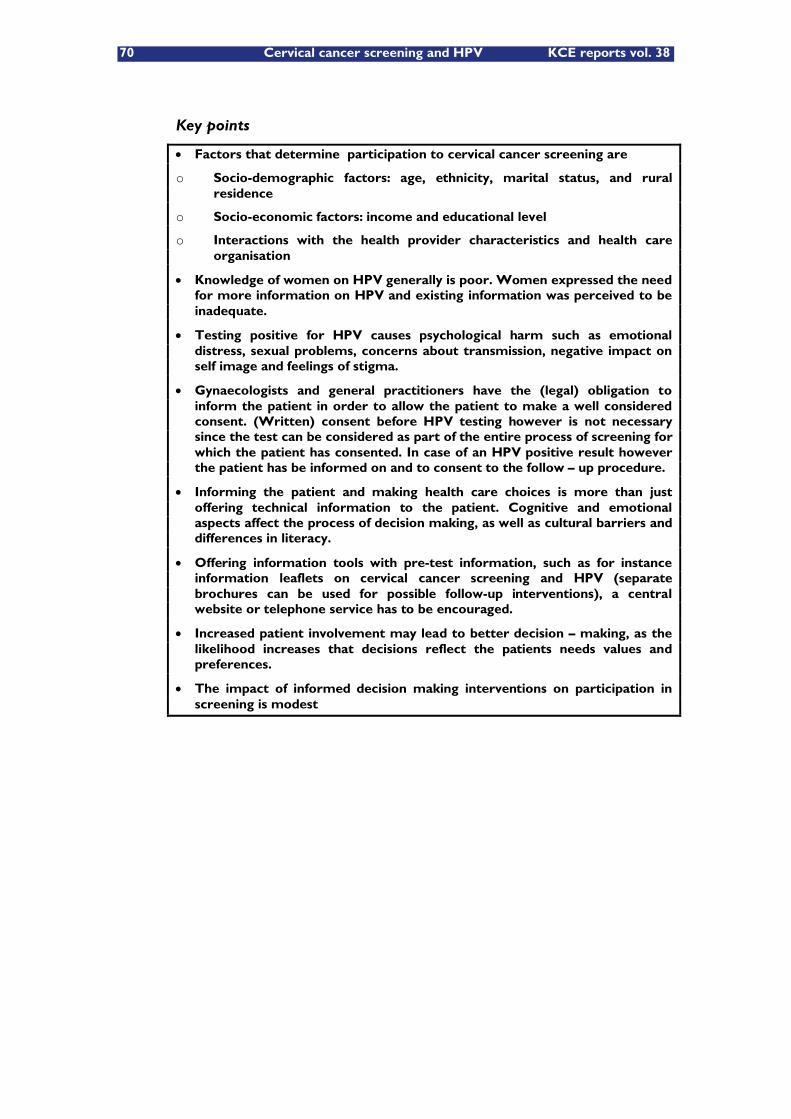
70 Cervical cancer screening and HPV KCE reports vol. 38
Key points
Factors that determine participation to cervical cancer screening are
o Socio-demographic factors: age, ethnicity, marital status, and rural residence
o Socio-economic factors: income and educational level
o Interactions with the health provider characteristics and health care organisation
Knowledge of women on HPV generally is poor. Women expressed the need for more information on HPV and existing information was perceived to be inadequate.
Testing positive for HPV causes psychological harm such as emotional distress, sexual problems, concerns about transmission, negative impact on self image and feelings of stigma.
Gynaecologists and general practitioners have the (legal) obligation to inform the patient in order to allow the patient to make a well considered consent. (Written) consent before HPV testing however is not necessary since the test can be considered as part of the entire process of screening for which the patient has consented. In case of an HPV positive result however the patient has be informed on and to consent to the follow �– up procedure.
Informing the patient and making health care choices is more than just offering technical information to the patient. Cognitive and emotional aspects affect the process of decision making, as well as cultural barriers and differences in literacy.
Offering information tools with pre-test information, such as for instance information leaflets on cervical cancer screening and HPV (separate brochures can be used for possible follow-up interventions), a central website or telephone service has to be encouraged.
Increased patient involvement may lead to better decision �– making, as the likelihood increases that decisions reflect the patients needs values and preferences.
The impact of informed decision making interventions on participation in screening is modest
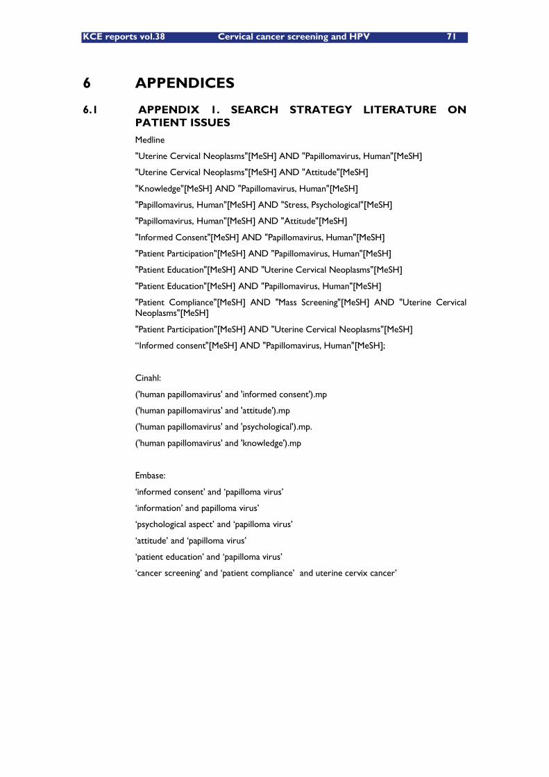
KCE reports vol.38 Cervical cancer screening and HPV 71
6 APPENDICES
6.1 APPENDIX 1. SEARCH STRATEGY LITERATURE ON PATIENT ISSUES
Medline
"Uterine Cervical Neoplasms"[MeSH] AND "Papillomavirus, Human"[MeSH]
"Uterine Cervical Neoplasms"[MeSH] AND "Attitude"[MeSH]
"Knowledge"[MeSH] AND "Papillomavirus, Human"[MeSH]
"Papillomavirus, Human"[MeSH] AND "Stress, Psychological"[MeSH]
"Papillomavirus, Human"[MeSH] AND "Attitude"[MeSH]
"Informed Consent"[MeSH] AND "Papillomavirus, Human"[MeSH]
"Patient Participation"[MeSH] AND "Papillomavirus, Human"[MeSH]
"Patient Education"[MeSH] AND "Uterine Cervical Neoplasms"[MeSH]
"Patient Education"[MeSH] AND "Papillomavirus, Human"[MeSH]
"Patient Compliance"[MeSH] AND "Mass Screening"[MeSH] AND "Uterine Cervical Neoplasms"[MeSH]
"Patient Participation"[MeSH] AND "Uterine Cervical Neoplasms"[MeSH]
�“Informed consent"[MeSH] AND "Papillomavirus, Human"[MeSH];
Cinahl:
('human papillomavirus' and 'informed consent').mp
('human papillomavirus' and 'attitude').mp
('human papillomavirus' and 'psychological').mp.
('human papillomavirus' and 'knowledge').mp
Embase:
�‘informed consent�’ and �‘papilloma virus�’
�‘information�’ and papilloma virus�’
�‘psychological aspect�’ and �‘papilloma virus�’
�‘attitude�’ and �‘papilloma virus�’
�‘patient education�’ and �‘papilloma virus�’
�‘cancer screening�’ and �‘patient compliance�’ and uterine cervix cancer�’

72 Cervical cancer screening and HPV KCE reports vol. 38
REFERENCES
1. Arbyn M, Buntinx F, Van Ranst M, Cortinas Abrahantes J. Triage of women with atypical or low-grade cytological abnormalities of the cervix by HPV testing: systematic review and meta-analysis. Brussels: Scientific Institute of Public Health; 2002.
2. Arbyn M, Abarca M. Is Liquid Based Cytology an Effective Alternative for the Conventional Pap Smear to Detect Cervical Cancer Precursors? A Systematic Review and Meta-analysis. Brussels: Scientific Institute of Public Health; 2003.
3. Arbyn M, Sasieni P, Meijer CJLM, Clavel C, Koliopoulos G, Dillner J. Clinical applications of HPV testing: a summary of meta-analyses. Vaccine. 2006;24(S3):78-89.
4. Morrison AS. Screening in Chronic Disease. 2nd. Kelsey JL, Marmot MG, Stolley PD, Vessey MP, editor.: Oxford University Press Inc.; 1992.
5. Hakama M, Chamberlain J, Day NE, Miller AB, Prorok PC. Evaluation of screening programmes for gynaecological cancer. Brit J Cancer. 1985;52:669-73.
6. van Oortmarssen GJ, Habbema JD. invasive cervical cancer. Brit J Cancer. 1991;64:559-65.
7. van Oortmarssen GJ, Habbema JDF, van Ballegooijen M. Predicting mortality from cervical cancer after negative smear test results. Bmj. 1992;305:449-51.
8. Day NE. Screening for cancer of the cervix. J.Epidemiol.Community Health. 1989;43(2):103-6.
9. Cochrane Methods Group on Systematic Review of Screening, Diagnostic Tests. The Cochrane methods working group on systematic review of screening and diagnosic tests : recommended methods. 1996:1-15.
10. Bossuyt PM, Reitsma JB, Bruns DE, Gatsonis CA, Glasziou PP, Irwig LM, et al. Towards complete and accurate reporting of studies of diagnostic accuracy: the STARD initiative. Bmj. 2003;326(7379):41-4.
11. Lundberg GD, National Cancer Institute. The 1988 Bethesda System for Reporting Cervical/Vaginal Cytologic Diagnoses. Jama. 1989;262(7):931-4.
12. Luff RD, National Cancer Institute. The Bethesda System for Reporting Cervical/Vaginal Cytologic diagnoses: a Report of the 1991 Bethesda Workshop. Hum Pathol. 1992;23(7):719-21.
13. Richart RM. Cervical intraepithelial neoplasia. Pathol Annu. 1973;8:301-23.
14. Canadian Task Force. Cervical cancer screening programs: I. Epidemiology and natural history of carcinoma of the cervix. Cmaj. 1976;114:1003-33.
15. Narod SA, Thompson DW, Jain M, Wall C, Green LM, Miller AB. Dysplasia and the natural history of cervical cancer: early results of the Toronto Cohort Study. Eur J Cancer. 1991;27:1411-6.
16. Ostor AG. Natural history of cervical intraepithelial neoplasia: a critical review. Int.J.Gynecol.Pathol. 1993;12(2):186-92.
17. Melnikow J, Nuovo J, Willan AR, Chan BK, Howell LP. Natural History of cervical squamous intraepithelial lesions : a meta-analysis. Obstet.Gynecol. 1998;92:727-35.
18. McCrory DC, Matchar DB, Bastian L, Datta S, Hasselblad V, Hickey J, et al. Evaluation of cervical cytology. Rockville (MD): AHCPR; 1999.
19. Nanda K, McCrory DC, Myers ER, Bastian LA, Hasselblad V, Hickey JD, et al. Accuracy of the Papanicolaou Test in Screening for and Follow-up of Cervical Cytologic Abnormalities: A Systematic Review. Ann Intern Med. 2000;132:810-9.

KCE reports vol.38 Cervical cancer screening and HPV 73
20. Sherman ME, Schiffman MA, Cox JT. Effects of age and human papilloma viral load on colposcopy triage: data from the randomised atypical squamous cells of undetermined significance/low-grade intraepithelial lesion triage study (ALTS). J.Natl.Cancer Inst. 2002;94(2):102-7.
21. Holowaty P, Miller AB, Rohan T, To T. Natural History of Dysplasia of the Uterine Cervix. J.Natl.Cancer Inst. 1999;91(3):252-8.
22. Schatzkin A, Connor RJ, Taylor PR, Bunnag B. Comparing new and old screening tests when a reference procedure cannot be performed on all screenees. Example of automated cytometry for early detection of cervical cancer. Am.J.Epidemiol. 1987;125:672-8.
23. Chock C, Irwig I, Berry G, Glasziou P. Comparing dichotomous screening tests when individuals negative on both tests are not verified. J.Clin.Epidemiol. 1997;50:1211-7.
24. Begg CB, Greenes RA. Assessment of diagnostic tests when disease verification is subject to selection bias. Biometrics. 1983;39:207-15.
25. Choi BC. Sensitivity and specificity of a single diagnostic test in the presence of work-up bias. J.Clin.Epidemiol. 1992;45(6):581-6.
26. Irwig L, Glasziou PP, Berry G, Chock C, Mock P, Simpson JM. Efficient Study Designs to Assess the Accuracy of Screening Tests. Am.J.Epidemiol. 1994;140(8):759-69.
27. Solomon D, Davey D, Kurman R, Moriarty A, O'Connor D, Prey M, et al. The 2001 Bethesda System: terminology for reporting results of cervical cytology. Jama. 2002;287:2114-9.
28. Fahey MT, Irwig L, Macaskill P. Meta-analysis of pap test accuracy. Am.J.Epidemiol. 1995;141(7):680-9.
29. Hakama M, Miller AB, Day NE. Screening for cancer of the uterine cervix. From the IARC Working Group on Cervical Cancer Screening and the UICC Project Group on the Evaluation of Screening Programmes for Cancer. Lyon; 1986.
30. IARC. Cervix Cancer Screening. IARC Handbooks of Cancer Prevention. Vol. 10. Lyon; 2005.
31. Day N, Moss S, Berrino F, Choi NW, Clarke EA, D�”br�”ssy L, et al. Screening for squamous cervical cancer: duration of low risk after negative results of cervical cytology and its implication for screening policies. Bmj. 1986;293:659-64.
32. Sasieni PD, Cuzick J, Lynch-Farmery EL, National Co-ordinating Network for Cervical Screening Working Group. Estimating the efficacy of screening by auditing smear histories of women with and without cervical cancer. Brit J Cancer. 1996;73:1001-5.
33. Boyes DA, Morrison B, Knox EG, Draper GJ, Miller AB. A cohort study of cervical cancer screening in British Columbia. Clin Invest Med. 1982;5(1):1-29.
34. Austin RM, Ramzy I. Increased detection of epithelial cell abnormalities by liquid-based gynecologic cytology preparations. Acta Cytol. 1998;42:178-84.
35. Australian Health Technology Advisory Committee. Review of automated and semi-automated cervical screening devices. Canberra : Australian Health Technology Advisory Committee 1998. Health Technol Assess. 1998;4:xx.
36. Bishop JW, Marshall CJ, Bentz JS. New technologies in gynecologic cytology. J.Reprod.Med. 2000;45:701-19.
37. Broadstock M. Effectiveness and cost effectiveness of automates and semi-automated cervical cancer screening devices. Christchurch, New Zealand: NZHTA; 2000.
38. Payne N, Chilcott J, McGoogan E. Liquid-based cytology in cervical screening : a rapid and systematic review. Health Technol Assess. 2000;4:1-73.
39. Bernstein SJ, Sanchez-Ramos L, Ndubisi B. Liquid-based cervical cytologic smear study and conventional papanicolaou smears. A metaanalysis of prospective studies comparing cytologic diagnosis and sample adequacy. Am.J.Obstet.Gynecol. 2001;185:308-17.

74 Cervical cancer screening and HPV KCE reports vol. 38
40. Sulik SM, Kroeger K, Schultz JK, Brown JL, Becker LA, Grant WD. Are Fluid-based Cytologies Superior to the Conventional Papanicolaou Test? A Systematic Review. J.Fam.Pract. 2001;50(12):1040-6.
41. ANAES. Guidelines for follow-up of patients having an abnormal Pap smear. Update 2002. (in French). Paris: ANAES; 2002.
42. Moseley RP, Paget S. Liquid-based cytology: is this the way forward for cervical screening? Cytopathology. 2002;13(2):71-82.
43. MSAC. Liquid based cytology for cervical screening. 2002.
44. Karnon J, Peters J, Chilcott J, McGoogan E. Assessment report: liquid-based cytology in cervical screening: an updated rapid and systematic review. Sheffield, UK: The National Institute for Clinical Excellence (NICE); 2003.
45. Noorani HZ, Brown A, Skidmore B, Stuart GCE. Liquid-based cytology and human papillomavirus testing in cervical cancer screening. Ottawa, Ontario, Canada: CCOHTA (Canadian Coordinating Office for Health Technology Assessment); 2003.
46. Randall P, Farquhar C, Marjoribanks J. Liquid-based cytology in cervical screening: a systematic review. A report for the National Screening Unit of the Ministry of Health, April 2004. Auckland: Department of Obstetrics and Gynaecology University of Auckland New Zealand.; 2004.
47. Davey E, Barratt A, Irwig L, Chan SF, Macaskill P, Mannes P, et al. Effect of study design and quality on unsatisfactory rates, cytology classifications, and accuracy in liquid-based versus conventional cervical cytology: a systematic review. Lancet. 2006;367(9505):122-32.
48. Abulafia O, Pezzullo JC, Sherer DM. Performance of ThinPrep liquid-based cervical cytology in comparison with conventionally prepared Papanicolaou smears: a quantitative survey. Gynecol.Oncol. 2003;90(1):137-44.
49. Gutman S. Labeling liquid-based systems: FDA clarification. J.Reprod.Med. 2000;45(11):969-70.
50. Fremont-Smith M, Marino James, Griffin Bryan, Spencer Lynn, Bolick David. Comparison of the SurePath liquid-based Papanicolaou smear with the conventional Papanicolaou smear in a multi-site direct-to-vial study. Cancer Cytopathology. 2004:269-79.
51. Longatto-Filho A, Pereira SM, Di Loreto C, Utagawa ML, Makabe S, Sakamoto Maeda MY, et al. DCS liquid-based system is more effective than conventional smears to diagnosis of cervical lesions: study in high-risk population with biopsy-based confirmation. Gynecol.Oncol. 2005;97(2):497-500.
52. Vassilakos P, Schwartz D, De Marval F, Yousfi L, Broquet G, Mathez-Loic F, et al. Biopsy-based comparison of liquid-based, thin-layer preparations to conventional Pap smears. J.Reprod.Med. 2000;45(1):11-6.
53. Ferenczy A, Robitaille J, Franco EL, Arseneau J, Richart RM, Wright TC. Conventional cervical cytologic smears vs. ThinPrep smears. A paired comparison study on cervical cytology. Acta Cytol. 1996;40(6):1136-42.
54. Coste J, Cochand-Priollet B, de Cremoux P, Le Gales C, Cartier I, Molinie V, et al. Cross sectional study of conventional cervical smear, monolayer cytology, and human papillomavirus DNA testing for cervical cancer screening. Bmj. 2003;326(7392):733-6.
55. Bergeron C, Bishop J, Lemarie A, Cas F, Ayivi J, Huynh B, et al. Accuracy of Thin-Layer Cytology in Patients Undergoing Cervical Cone Biopsy. Acta Cytol. 2001;45(4):519-24.
56. Confortini M, Bulgaresi P, Cariaggi MP, Carozzi FM, Cecchini S, Cipparrone I, et al. Comparing conventional and liquid-based smears from a consecutive series of 297 subjects referred to colposcopy assessment. Cytopathology. 2004;15(3):168-9.
57. Taylor S, Kuhn L, Dupree W, Denny L, De Souza M, Wright TC, Jr. Direct comparison of liquid-based and conventional cytology in a South African screening trial. Int.J.Cancer. 2006:957-62.

KCE reports vol.38 Cervical cancer screening and HPV 75
58. Dunton CJ. New technology in Papanicolaou smear processing. Clin Obstet Gynecol. 2000;43:410-7.
59. Kok MR, Boon ME. Consequences of neural network technology for cervical screening: increase of diagnostic consistency and increase of positive scores. Cancer. 1996;78(1):112-7.
60. Wilbur DC, Bonfiglio TA, Rutkowski MA, Atkison KM, Richart RM, Lee JS, et al. Sensitivity of the AutoPap 300 QC System for cervical cytologic abnormalities. Biopsy data confirmation. Acta Cytol. 1996;40(1):127-32.
61. Koss LG, Sherman ME, Cohen MB, Anes AR, Darragh TM, Lemos LB, et al. Significant reduction in the rate of false-negative cervical smears with neural network-based technology (papnet testing system). Hum Pathol. 1997;28:1196-203.
62. Michelow PM, Hlongwane NF, Leiman G. Simulation of primary cervical cancer screening by the PAPNET system in an unscreened, high-risk community. Acta Cytol. 1997;41(1):88-92.
63. Doornewaard H, van der Schouw YT, van der Graaf Y, Bos AB, Habbema JDF, van den Tweel JG. The Diagnostic Value of Computer-Assisted Primary Cervical Smear Screening: A Longitudinal Cohort Study. Mod.Pathol. 1999;12:995-1000.
64. Halford JA, Wright RG, Ditchmen EJ. Prospective study of PAPNET : review of 25656. Pap smears negative on manual screening and rapid rescreening. Cytopathology. 1999;10(5):317-24.
65. Prismatic Project Management Team. Assessment of automated primary screening on PAPNET of cervical smears in the PRISMATIC trial. Lancet. 1999;353(9162):1381-5.
66. Bergeron C, Masseroli M, Ghezi A, Lemarie A, Mango L, Koss LG. Quality control of cervical cytology in high-risk women. PAPNET system compared with manual rescreening. Acta Cytol. 2000;44(2):151-7.
67. Duggan MA. Papnet-assisted, primary screening of cervico-vaginal smears. Eur J Gynaecol Oncol. 2000;21(1):35-42.
68. Kok MR, Boon ME, Schreiner-Kok PG, Koss LG. Cytological recognition of invasive squamous cancer of the uterine cervix: comparison of conventional light-microscopical screening and neural network-based screening. Hum Pathol. 2000;31(1):23-8.
69. Ronco G, Vineis C, Montanari G, Orlassino R, Parisio F, Arnaud S, et al. Impact of the AutoPap (currently Focalpoint) primary screening system location guide use on interpretation time and diagnosis. Cancer. 2003;99(2):83-8.
70. Nieminen P, Hakama M, Viikki M, Tarkkanen J, Anttila A. Prospective and randomised public-health trial on neural network-assisted screening for cervical cancer in Finland: results of the first year. Int.J.Cancer. 2003;103:422-6.
71. Pretorius RG, Zhang WH, Belinson JL, Huang MN, Wu LY, Zhang X, et al. Colposcopically directed biopsy, random cervical biopsy, and endocervical curettage in the diagnosis of cervical intraepithelial neoplasia II or worse. Am.J.Obstet.Gynecol. 2004;191(2):430-4.
72. ASCUS-LSIL Triage Study Group. Results of a randomized trial on the management of cytology interpretations of atypical squamous cells of undetermined significance. Am.J.Obstet.Gynecol. 2003;188(6):1383-92.
73. Howard M, Sellors J, Lytwyn A. Cervical intraepithelial neoplasia in women presenting with external genital warts. Cmaj. 2002;166(5):598-9.
74. Bosch FX, Lorincz AT, Munoz N, Meijer CJ, Shah KV. The causal relation between human papillomavirus and cervical cancer. J Clin Pathol. 2002;55(4):244-65.
75. Schiffman MH, Schatzkin A. Test reliability is critically important to molecular epidemiology: an example from studies of human papillomavirus infection and cervical neoplasia. Cancer Res. 1994;54:1944s-7s.
76. Castle PE, Lorincz AT, Mielzynska-Lohnas I, Scott DR, Glass AG, Sherman ME, et al. Results of human papillomavirus DNA testing with the hybrid capture 2 assay are reproducible. J.Clin.Microbiol. 2002;40(3):1088-90.
76 Cervical cancer screening and HPV KCE reports vol. 38
77. Carozzi FM, Confortini M, Cecchini S, Bisanzi S, Cariaggi MP, Pontenani G, et al. Triage with human papillomavirus testing of women with cytologic abnormalities prompting referral for colposcopy assessment. Cancer. 2005;105(1):2-7.
78. Sherman ME, Lorincz AT, Scott DR, Wacholder S, Castle PE, Glass AG, et al. Baseline Cytology, Human Papillomavirus Testing, and Risk for Cervical Neoplasia: A 10-Year Cohort Analysis. J.Natl.Cancer Inst. 2003;95(1):46-52.
79. Khan MJ, Castle PE, Lorincz AT, Wacholder S, Sherman M, Scott DR, et al. The elevated 10-year risk of cervical precancer and cancer in women with human papillomavirus (HPV) type 16 or 18 and the possible utility of type-specific HPV testing in clinical practice. J.Natl.Cancer Inst. 2005;97(14):1072-9.
80. Lorincz AT. Methods of DNA hybridisation and their clinical applicability to human papillomavirus detection. In: Franco EL, Monsonego J, editors. New developments in cervican cancer screening and prevention. Oxford; 1997. p. 325-37.
81. Peyton CL, Schiffman MA, L�”rincz AT, Hunt WC, Mielzynska I, Bratti C, et al. and hybrid capture-based human papillomavirus detection systems using multiple cervical specimen collection strategies. J.Clin.Microbiol. 1998;36(11):3248-54.
82. Vernon SD, Unger ER, Williams D. Comparison of human papillomavirus detection and typing by cycle sequencing, line blotting, and hybrid capture. J.Clin.Microbiol. 2000;38(2):651-5.
83. Peyton CL, Gravitt PE, Hunt WC, Hundley RS, Zhao M, Apple RJ, et al. Determinants of genital human papillomavirus detection in a US population. J.Infect.Dis. 2001;183(11):1554-64.
84. Castle PE, Schiffman MA, Burk RD, Wacholder S, Hildesheim A, Herrero R, et al. Restricted cross-reactivity of hybrid capture 2 with nononcogenic human papillomavirus types. Cancer Epidemiol.Biomarkers Prev. 2002;11(11):1394-9.
85. Cuzick J, Szarewski A, Cubie H, Hulman G, Kitchener H, Luesley D, et al. Management of women who test positive for high-risk types of human papillomavirus: the HART study. Lancet. 2003;362:1871-6.
86. Ronco G, Segnan N, Giorgi-Rossi P, Zappa M, Casadei GP, Carozzi F, et al. Human Papillomavirus testing and liquid-based cytology in primary cervical screening: results at recruitment from the New Technologies for Cervical Cancer randomized controlled trial . . J.Natl.Cancer Inst. 2006;98:765-74.
87. Schiffman MA, Herrero R, Hildesheim A, Sherman ME, Bratti M, Wacholder S, et al. HPV DNA testing in cervical cancer screening. Results from women in a high-risk province of Costa Rica. Jama. 2000;283:87-93.
88. Snijders PJ, van den Brule AJ, Meijer CJLM. The clinical relevance of human papillomavirus testing : relationship between analytical and clinical sensitivity. J Pathol. 2003;201:1-6.
89. Elfgren K. Longitudinal studies of human papillomavirus infection. Doctoral thesis. Stockholm, Sweden: Karolinska University; 2003.
90. Gravitt PE, Peyton CL, Apple RJ, Wheeler CM. Genotyping of 27 human papillomavirus types by using L1 consensus PCR products by a single-hybridization, reverse line blot detection method. J.Clin.Microbiol. 1998;36:3020-7.
91. Qu W, Jiang G, Cruz Y, Chang CJ, Ho GY, Klein RS, et al. PCR detection of human papillomavirus: comparison between MY09/MY11 and GP5+/GP6+ primer systems. J.Clin.Microbiol. 1997;35(6):1304-10.
92. Jacobs MV, Snijders PJ, Voorhorst FJ, Dillner J, Forslund O, Johansson B, et al. Reliable high risk HPV DNA testing by polymerase chain reaction: and intermethod and intramethod comparison. J Clin Pathol. 1999;52:498-503.
93. Konya J, Veress G, Juhasz A, Szarka K, Sapy T, Hernadi Z, et al. Additional human papillomavirus types detected by the hybrid capture tube test among samples from women with cytological and colposcopical atypia. J.Clin.Microbiol. 2000;38(1):408-11.

KCE reports vol.38 Cervical cancer screening and HPV 77
94. van der Graaf Y, Molijn A, Doornewaard H, Quint W, van Doorn LJ, van den Tweel J. Human papillomavirus and the long-term risk of cervical neoplasia. Am.J.Epidemiol. 2002;156:158-64.
95. Kornegay JR, Shepard AP, Hankins C, Franco EL, Lapointe N, Richardson H, et al. Nonisotopic detection of human papillomavirus DNA in clinical specimens using a consensus PCR and a generic probe mix in an enzyme-linked immunosorbent assay format. J.Clin.Microbiol. 2001;39(10):3530-6.
96. Kleter B, van Doorn LJ, Schrauwen L, van Krimpen K, Burger M, ter Harmsel B, et al. Novel short-fragment PCR assey for highly sensitive broad-spectrum detection of anogenital human papillomaviruses. Am.J.Pathol. 1998;153(6):1731-9.
97. Kleter B, van Doorn LJ, Schrauwen L, Molijn A, Sastrowijoto S, ter Schegget J, et al. Development and clinical evaluation of a highly sensitive PCR-reverse hybridization line probe assay for detection and identification of anogenital human papillomavirus. J.Clin.Microbiol. 1999;37:2508-17.
98. Iftner T, Villa LL. Chapter 12: Human papillomavirus technologies. J.Natl.Cancer Inst.Monogr. 2003(31):80-8.
99. Swan DC, Tucker RA, Holloway BP, Icenogle JP. A sensitive, type-specific, fluorogenic probe assay for detection of human papillomavirus DNA. J.Clin.Microbiol. 1997;35(4):886-91.
100. Josefsson AM, Livak K, Gillensten U. Viral load of human papilloma virus 16 as a determinant for development of cervical carcinoma in situ; a nested case-control study. J.Clin.Microbiol. 1999;37:490-6.
101. Tucker RA, Unger ER, Holloway BP, Swan DC. Real-time PCR-based fluorescent assay for quantitation of human papillomavirus types 6, 11, 16, and 18. Mol Diagn. 2001;6(1):39-47.
102. Moberg M, Gustavsson I, Gyllensten U. Real-time PCR-based system for simultaneous quantification of human papillomavirus types associated with high risk of cervical cancer. J.Clin.Microbiol. 2003;41(7):3221-8.
103. van den Brule AJ, Pol R, Fransen-Daalmeijer N, Schouls LM, Meijer CJ, Snijders PJ. GP5+/6+ PCR followed by reverse line blot analysis enables rapid and high-throughput identification of human papillomavirus genotypes. J.Clin.Microbiol. 2002;40(3):779-87.
104. Quint WG, Scholte G, van Doorn LJ, Kleter B, Smits PH, Lindeman J. Comparative analysis of human papillomavirus infections in cervical scrapes and biopsy specimens by general SPF(10) PCR and HPV genotyping. J.Pathol. 2001;194(1):51-8.
105. Kim CJ, Jeong JK, Park M, Park TS, Park TC, Namkoong SE, et al. HPV oligonucleotide microarray-based detection of HPV genotypes in cervical neoplastic lesions. Gynecol.Oncol. 2003;89(2):210-7.
106. Smits HL, van Gemen B, Schukkink R, Van der Velden J, Tjong AH, Jebbink MF, et al. Application of the NASBA nucleic acid amplification method for the detection of human papillomavirus type 16 E6-E7 transcripts. J.Virol.Methods. 1995;54(1):75-81.
107. Sotlar K, Selinka HC, Menton M, Kandolf R, Bultmann B. Detection of human papillomavirus type 16 E6/E7 oncogene transcripts in dysplastic and nondysplastic cervical scrapes by nested RT-PCR. Gynecol.Oncol. 1998;69(2):114-21.
108. Nakagawa S, Yoshikawa H, Yasugi T, Kimura M, Kawana K, Matsumoto K, et al. Ubiquitous presence of E6 and E7 transcripts in human papillomavirus-positive cervical carcinomas regardless of its type. J.Med.Virol. 2000;62(2):251-8.
109. Cuschieri KS, Whitley MJ, Cubie HA. Human papillomavirus type specific DNA and RNA persistence--implications for cervical disease progression and monitoring. J.Med.Virol. 2004;73(1):65-70.
110. Molden T, Nygard JF, Kraus I, Karlsen F, Nygard M, Skare GB, et al. Predicting CIN2+ when detecting HPV mRNA and DNA by PreTect HPV-proofer and consensus PCR: A 2-year follow-up of women with ASCUS or LSIL pap smear. Int.J.Cancer. 2005;114(6):973-6.

78 Cervical cancer screening and HPV KCE reports vol. 38
111. Cuzick J, Sasieni P, Davies P, Adams J, Normand C, Frater A, et al. A systematic review of the role of human papillomavirus testing within a cervical screening programme. Health Technol Assess. 1999;3:1-204.
112. Arbyn M, Buntinx F, Van Ranst M, Paraskevaidis E, Martin-Hirsch P, Dillner J. Virologic versus cytologic triage of women with equivocal Pap smears: a meta-analysis of the accuracy to detect high-grade intraepithelial neoplasia. J.Natl.Cancer Inst. 2004;96:280-93.
113. Arbyn M, Dillner J, Van Ranst M, Buntinx F, Martin-Hirsch P, Paraskevaidis E. Re: Have we resolved how to triage equivocal cervical cytology? J.Natl.Cancer Inst. 2004;96(18):1401-2.
114. Solomon D, Schiffman MA, Tarone B. Comparison of three management strategies for patients with atypical squamous cells of undetermined significance (ASCUS): baseline results from a randomized trial. J.Natl.Cancer Inst. 2001;93(4):293-9.
115. Dersimonian R, Laird NM. Meta-analysis in clinical trials. Controlled Clin Trials. 1986;7:177-88.
116. Arbyn M, Paraskevaidis E, Martin-Hirsch P, Prendiville W, Dillner J. Clinical utility of HPV DNA detection: triage of minor cervical lesions, follow-up of women treated for high-grade CIN. An update of pooled evidence. Gynecol.Oncol. 2005;99 (Suppl 3):7-11.
117. Manos MM, Kinney WK, Hurley LB, Sherman ME, Shieh-Ngai J, Kurman RJ, et al. Identifying Women With Cervical Neoplasia: Using Human Papillomavirus DNA Testing for Equivocal Papanicolaou Results. Jama. 1999;281(17):1605-10.
118. Bergeron C, Jeannel D, Poveda J, Cassonnet P, Orth G. Human papillomavirus testing in women with mild cytologic atypia. Obstet.Gynecol. 2000;95((6 Pt 1)):821-7.
119. Lin CT, Tseng CJ, Lai CH, Hsueh S, Huang HJ, Law KS. High-risk HPV DNA detection by Hybrid Capture II. An adjunctive test for mildly abnormal cytologic smears in women > or = 50 years of age. J.Reprod.Med. 2000;45:345-50.
120. Lytwyn A, Sellors JW, Mahony JB. Comparison of human papillomavirus DNA testing and repeat Papanicolaou test in women with low-grade cervical cytologic abnormalities: a randomized trial. Cmaj. 2000;163:701-7.
121. Shlay JC, Dunn T, Byers T, Baron AE, Douglas JM, Jr. Prediction of cervical intraepithelial neoplasia grade 2-3 using risk assessement and human papillomavirus testing in women with atypia on papanicolaou smears. Obstet.Gynecol. 2000;96(3):410-6.
122. Morin C, Bariati C, Bouchard C, Fortier M, Roy M, Moore L, et al. Managing Atypical Squamous Cells of Undetermined Significance in Papanicolaou Smears. J.Reprod.Med. 2001;46:799-805.
123. Rebello G, Hallam N, Smart G, Farquharson D, McCafferty J. Human papillomavirus testing and the management of women with mildly abnormal cervical smears: an observational study. Bmj. 2001;322:894-5.
124. Zielinski GD, Snijders PJ, Rozendaal L, Voorhorst FJ, Runsink AP, De Schipper FA, et al. High-risk HPV testing in women with borderline and mild dyskaryosis: long-term follow-up data and clinical relevance. J Pathol. 2001;195:300-6.
125. Kulasingam SL, Hughes JP, Kiviat NB, Mao C, Weiss NS, Kuypers JM, et al. Evaluation of human papillomavirus testing in primary screening cervical abnormalities. Comparison of sesitivity, specificity, and frequency of referral. Jama. 2002;288(14):1749-57.
126. Pambuccian SE, Lundeen SJ, Woronzoff K, Rohlader J, Kjeldahl K, Curran C, et al. Reflex HPV testing of ASCUS patients: initial experience with PCR-based HPV identification and typing. In: Proceedings of the Am.Soc.Cytopathol.; 2002; Salt Lake City; 2002.
127. Pretorius RG, Peterson P, Novak S, Azizi F, Sadeghi M, Lorincz AT. Comparison of two signal-amplification DNA tests for high-risk HPV as an aid to colposcopy. J.Reprod.Med. 2002;47:290-6.
128. Guyot A, Karim S, Kyi MS, Fox J. Evaluation of adjunctive HPV testing by hybrid capture II(R) in women with minor cytological abnormalities for the diagnosis of CIN2/3 and cost comparison with colposcopy. BMC Infectious Diseases. 2003;3(23):1-7.

KCE reports vol.38 Cervical cancer screening and HPV 79
129. Lonky NM, Felix JC, Naidu YM, Wolde Tsadik G. Triage of atypical squamous cells of undetermined significance with Hybrid Capture II: colposcopy and histologic human papillomavirus correlation. Obstet.Gynecol. 2003;101(3):481-9.
130. Wensveen C, Kagie M, Veldhuizen R, De Groot C, Denny L, Zwinderman K, et al. Detection of cervical intraepithelial neoplasia in women with atypical squamous or glandular cells of undetermined significance cytology: a prospective study. Acta Obstet Gynecol Scand. 2003;82(9):883-9.
131. Andersson S, Dillner L, Elfgren K, Mints M, Persson M, Rylander E. A comparison of the human papillomavirus test and Papanicolaou smear as a second screening method for women with minor cytological abnormalities. Acta Obstet.Gynecol.Scand. 2005;84(10):996-1000.
132. Dalla Palma P, Pojer A, Girlando S. HPV triage of women with atypical squamous cells of undetermined significance: a 3 year experience in an Italian organized programme. Cytopathology. 2005;16:22-6.
133. Giovannelli L, Capra G, Lama A, Bustinto T, Genco A, Valenti FM, et al. Atypical squamous cells of undetermined significance-favour reactive compared to atypical squamous cells of undetermined significance-favour dysplasia: association with cervical intraepithelial lesions and human papillomavirus infection. J.Clin.Virol. 2005;33(4):281-6.
134. Bergeron C, Cas F, Fagnani F, Contrepas A, Wadler R, Poveda JD. Assessment of human papillomavirus testing on liquid-based CYTO-screen system for women with atypical squamous cells of undetermined significance. Effect of age. (in press). Gynecol Obstet Fertil. 2006;34:312-6.
135. Sherman ME, Wang SS, Tarone R, Rich L, Schiffman MA. Histopathologic extent of cervical intraepithelial neoplasia 3 lesions in the atypical squamous cells of undetermined significance low-grade squamous intraepithelial lesion trage study: implications for subject safety and lead-time bias. Cancer Epidemiol Biomarkers Prev. 2003;12:372-9.
136. Blumenthal PD, Gaffikin L, Chirenje ZM, McGrath J, Womack S, Shah K. Adjunctive testing for cervical cancer in low resource settings with visual inspection, HPV, and the Pap smear. Int.J.Gynecol.Obstet. 2001;72(1):47-53.
137. Sankaranarayanan R, Chatterji R, Shastri SS, Basu P, Mah�‚ C, Muwonge R, et al. Accuracy of human papillomavirus testing in primary screening of cervical neoplasia: results from a multicentre study in India. Int.J.Cancer. 2004;112(2):341-7.
138. Andersson S, Mints M, Sallstrom J, Wilander E. The relative distribution of oncogenic types of human papillomavirus in benign, pre-malignant and malignant cervical biopsies. A study with human papillomavirus deoxyribonucleic acid sequence analysis. Cancer Detect.Prev. 2005;29(1):37-41.
139. Zuna RE, Wang SS, Rosenthal DL, Jeronimo J, Schiffman M, Solomon D. Determinants of human papillomavirus-negative, low-grade squamous intraepithelial lesions in the atypical squamous cells of undetermined significance/low-grade squamous intraepithelial lesions triage study (ALTS). Cancer. 2005;105(5):253-62.
140. Alts group, Anonymous. Human papillomavirus testing for triage of women with cytologic evidence of low-grade squamous intraepithelial lesions: baseline data from a randomized trial. J.Natl.Cancer Inst. 2000;92:397-402.
141. Moss S, Gray A, Legood R, Vessey M, Patnick J, Kitchener H. Effect of testing for human papillomavirus as a triage during screening for cervical cancer: observational before and after study. Bmj. 2006:83-5.
142. Bais AG, Rebolj M, Snijders PJ, De Schipper FA, van der Meulen DA, Verheijen RH, et al. Triage using HPV-testing in persistent borderline and mildly dyskaryotic smears: Proposal for new guidelines. Int.J.Cancer. 2005;116(1):122-9.
143. Berkhof J, de Bruijne MC, Zielinski GD, Bulkmans NW, Rozendaal L, Snijders PJ, et al. Evaluation of cervical screening strategies with adjunct high-risk human papillomavirus testing for women with borderline or mild dyskaryosis. Int.J.Cancer. 2006;118:1759-68.

80 Cervical cancer screening and HPV KCE reports vol. 38
144. Elfgren K, Bistoletti P, Dillner L, Walboomers JM, Meijer CJ, Dillner J. Conization for cervical intraepithelial neoplasia is followed by disappearance of human papillomavirus deoxyribonucleic acid and a decline in serum and cervical mucus antibodies against human papillomavirus antigens. Am.J.Obstet.Gynecol. 1996;174(3):937-42.
145. Chua KL, Hjerpe A. Human papillomavirus analysis as a prognostic marker following conization of the cervix uteri. Gynecol.Oncol. 1997;66(1):108-13.
146. Distefano AL, Picconi MA, Alonio LV, Dalbert D, Mural J, Bartt O, et al. Persistence of human papillomavirus DNA in cervical lesions after treatment with diathermic large loop excision. Infect Dis Obstet Gynecol. 1998;6(5):214-9.
147. Bollen LJ, Tjong-A-Hung SP, van dV, Mol BW, ten Kate FW, ter Schegget J, et al. Prediction of recurrent and residual cervical dysplasia by human papillomavirus detection among patients with abnormal cytology. Gynecol.Oncol. 1999;72(2):199-201.
148. Nagai Y, Maehama T, Asato T, Kanazawa K. Persistence of human papillomavirus infection after therapeutic conization for CIN 3: is it an alarm for disease recurrence? Gynecol.Oncol. 2000;79(2):294-9.
149. Jain S, Tseng CJ, Horng SG, Soong YK, Pao CC. Negative predictive value of human papillomavirus test following conization of the cervix uteri. Gynecol.Oncol. 2001;82(1):177-80.
150. Lin CT, Tseng CJ, Lai CH, Hsueh S, Huang KG, Huang HJ, et al. Value of human papillomavirus deoxyribonucleic acid testing after conization in. Am.J.Obstet.Gynecol. 2001;184(4):940-5.
151. Nobbenhuis MA, Meijer CJ, van den Brule AJ, Rozendaal L, Voorhorst FJ, Risse EK, et al. Addition of high-risk HPV testing improves the current guidelines on follow-up after treatment for cervical intraepithelial neoplasia. Brit J Cancer. 2001;84(6):796-801.
152. Paraskevaidis E, Koliopoulos G, Alamanos Y, Malamou-Mitsi V, Lolis ED, Kitchener HC. Human papillomavirus testing and the outcome of treatment for cervical intraepithelial neoplasia. Obstet.Gynecol. 2001;98(5):833-6.
153. Bekkers RL, Melchers WJ, Bakkers JM, Hanselaar AG, Quint WG, Boonstra H, et al. The role of genotype-specific human papillomavirus detection in diagnosing residual cervical intraepithelial neoplasia. Int.J.Cancer. 2002;102(2):148-51.
154. Bar-Am A, Gamzu R, Levin I, Fainaru O, Niv J, Almog B. Follow-up by combined cytology and human papillomavirus testing for patients post-cone biopsy: results of a long-term follow-up. Gynecol.Oncol. 2003;91(1):149-53.
155. Houfflin Debarge V, Collinet P, Vinatier D, Ego A, Dewilde A, Boman F, et al. Value of human papillomavirus testing after conization by loop electrosurgical excision for high-grade squamous intraepithelial lesions. Gynecol.Oncol. 2003;90(3):587-92.
156. Zielinski GD, Rozendaal L, Voorhorst FJ, Berkhof J, Snijders PJ, Risse EJ, et al. HPV testing can reduce the number of follow-up visits in women treated for cervical intraepithelial neoplasia grade 3. Gynecol.Oncol. 2003;91(1):67-73.
157. Cecchini S, Carozzi F, Confortini M, Zappa M, Ciatto S. Persistent human papilloma virus infection as an indicator of risk of recurrence of high-grade cervical intraepithelial neoplasia treated by the loop electrosurgical excision procedure. Tumori. 2004;90(2):225-8.
158. Sarian LO, Derchain SF, Andrade LA, Tambascia J, Morais SS, Syrjanen KJ. HPV DNA test and Pap smear in detection of residual and recurrent disease following loop electrosurgical excision procedure of high-grade cervical intraepithelial neoplasia. Gynecol.Oncol. 2004;94(1):181-6.
159. Hernadi Z, Szoke K, Sapy T, Krasznai ZT, Soos G, Veress G, et al. Role of human papillomavirus (HPV) testing in the follow-up of patients after treatment for cervical precancerous lesions. Eur.J.Obstet.Gynecol.Reprod.Biol. 2005;118(2):229-34.
160. Flannelly G, Bolger B, Fawzi H, De Lopes AB, Monaghan JM. Follow up after LLETZ: could schedules be modified according to risk of recurrence? Bjog. 2001;108(10):1025-30.
KCE reports vol.38 Cervical cancer screening and HPV 81
161. Paraskevaidis E, Kalantaridou SN, Paschopoulos M, Zikopoulos K, Diakomanolis E, Dalkalitsis N, et al. Factors affecting outcome after incomplete excision of cervical intraepithelial neoplasia. Eur J Gynaecol Oncol. 2003;24(6):541-3.
162. Castle PE, Schiffman MA, Herrero R, Hildesheim A, Rodriguez AC, Bratti MC, et al. A prospective study of age trends in cervical human papillomavirus acquisition and persistence in Guanacaste, Costa Rica. J.Infect.Dis. 2005;191(11):1808-16.
163. Kalliala I, Anttila A, Pukkala E, Nieminen P. Risk of cervical and other cancers after treatment of cervical intraepithelial neoplasia: retrospective cohort study. Bmj. 2005;331(7526):1183-5.
164. Soutter WP, Sasieni P, Panoskaltsis T. Long-term risk of invasive cervical cancer after treatment of squamous cervical intraepithelial neoplasia. Int.J.Cancer. 2005;118(8):2048-55.
165. Zielinski GD, Bais AG, Helmerhorst TJ, Verheijen RH, De Schipper FA, Snijders PJ, et al. HPV testing and monitoring of women after treatment of CIN 3: review of the literature and meta-analysis. Obstet.Gynecol.Surv. 2004;59(7):543-53.
166. Walter SD. Estimation of test sensitivity and specificity when disease confirmation is limited to positive results. Epidemiology. 1999;10(1):67-72.
167. Moses LE, Shapiro D, Littenberg B. Combining independent studies of a diagnostic test into a summary ROC curve: data-analytic approaches and some additional considerations. Stat.Med. 1993;12:1293-316.
168. Cuzick J, Szarewski A, Terry G, Ho L, Hanby A, Maddox P, et al. Human papillomavirus testing in primary cervical screening. Lancet. 1995;345:1533-6.
169. Cuzick J, Beverley E, Ho L, Terry G, Sapper H, Mielzynska I, et al. HPV testing in primary screening of older women. Brit J Cancer. 1999;81:554-8.
170. Kuhn L, Denny L, Pollack A, Lorincz AT, Richart RM, Wright TC. Human papillomavirus DNA testing for cervical cancer screening in low-resource settings. J.Natl.Cancer Inst. 2000;92(10):818-25.
171. Ratnam S, Franco EL, Ferenczy A. Human papillomavirus testing for primary screening of cervical cancer precursors. Cancer Epidemiol Biomarkers Prev. 2000;9:945-51.
172. Schneider A, Hoyer H, Lotz B, Leistritza S, Kuhne-Heid R, Nindl I, et al. Screening for high-grade cervical intra-epithelial neoplasia and cancer by testing for high-risk HPV, routine cytology or colposcopy. Int.J.Cancer. 2000;89(6):529-34.
173. Belinson JL, Qiao YL, Pretorius R, Zhang WH, Elson P, Li L, et al. Shanxi province cervical cancer screening study: a cross-sectional comparative trial multiple techniques to detect cervical neoplasia. Gynecol.Oncol. 2001;83:439-44.
174. Clavel C, Masure M, Bory JP, Putaud I, Mangeonjean C, Lorenzato M, et al. Human papillomavirus testing in primary screening for the detection of high-grade cervical lesions: a study of 7932 women. Brit J Cancer. 2001;89(12):1616-23.
175. Oh YL, Shin KJ, Han J, Kim DS. Significance of high-risk human papillomavirus detection by polymerase chain reaction in primary cervical cancer screening. Cytopathology. 2001;12(2):75-83.
176. Paraskevaidis E, Malamou-Mitsi V, Koliopoulos G, Pappa L, Lolis E, Georgiou I, et al. Expanded cytological referral criteria for colposcopy in cervical screening : comparison with human papillomavirus testing. Gynecol.Oncol. 2001;82((2)):355-9.
177. Syrjänen SM. New technologies in cervical cytology screening: a word of caution. J.Low Genit.Tract.Dis. 2002;6(2):97-110.
178. Belinson JL, Qiao YL, Pretorius RG, Zhang WH, Rong SD, Huang MN, et al. Shanxi Province cervical cancer screening study II: self-sampling for high-risk human papillomavirus compared to direct sampling for human papillomavirus and liquid based cervical cytology. Int.J.Gynecol.Cancer. 2003;13(6):819-26.
179. Petry KU, Menton S, Menton M, Loenen-Frosch F, de Carvalho GH, Holz B, et al. Inclusion of HPV testing in routine cervical cancer screening for women above 29 years in Germany: results for 8466 patients. Brit J Cancer. 2003;88(10):1570-7.
82 Cervical cancer screening and HPV KCE reports vol. 38
180. Salmeron J, Lazcano-Ponce E, Lorincz AT, Hern ndez M, Hern ndez P, Leyva A, et al. Comparison of HPV-based assays with Papaniclaou smears for cervical cancer screening in Morelos State, Mexico. Cancer Causes Control. 2003;14:505-12.
181. Sankaranarayanan R, Thara S, Sharma A, Roy C, Shastri S, Mahe C, et al. Accuracy of conventional cytology: results from a multicentre screening study in India. J.Med.Screen. 2004;11(2):77-84.
182. Agorastos T, Dinas K, Lloveras B, de Sanjose S, Kornegay JR, Bonti H, et al. Human papillomavirus testing for primary screening in women at low risk of developing cervical cancer. The Greek experience. Gynecol.Oncol. 2005;96(3):714-20.
183. Bigras G, De Marval F. The probability for a Pap test to be abnormal is directly proportional to HPV viral load: results from a Swiss study comparing HPV testing and liquid-based cytology to detect cervical cancer precursors in 13 842 women. Brit J Cancer. 2005;93(5):575-81.
184. Sarian LO, Derchain SF, Naud P, Roteli-Martins C, Longatto-Filho A, Tatti S, et al. Evaluation of visual inspection with acetic acid (VIA), Lugol's iodine (VILI), cervical cytology and HPV testing as cervical screening tools in Latin America. J.Med.Screen. 2005;12(3):142-9.
185. Kotaniemi-Talonen L, Nieminen P, Anttila A, Hakama M. Routine cervical screening with primary HPV testing and cytology triage protocol in a randomised setting. Brit J Cancer. 2005;93(8):862-7.
186. Sankaranarayanan R, Nene BM, Dinshaw KA, Mahe C, Jayant K, Shastri SS, et al. A cluster randomized controlled trial of visual, cytology and human papillomavirus screening for cancer of the cervix in rural India. Int.J.Cancer. 2005;116:617-23.
187. Arbyn M, Gaffikin L, Sankaranarayanan R, Muwonge R, Keita N, Wesley R, et al. Assessment of Innovative Approaches to Cervical Cancer Screening, Follow-up and Treatment of Screen Detected Cervical Lesions in Developing Countries. A Pooled Analysis of ACCP Trial Results. PATH-Seattle & IPH Publication; 2005.
188. Clifford GM, Smith JS, Aguado T, Franceschi S. Comparison of HPV type distribution in high-grade cervical lesions and cervical cancer: a meta-analysis. Brit J Cancer. 2003;89(1):101-5.
189. Franceschi S, Rajkumar T, Vaccarella S, Gajalakshmi V, Sharmila A, Snijders PJ, et al. Human papillomavirus and risk factors for cervical cancer in Chennai, India: a case-control study. Int.J.Cancer. 2003;107(1):127-33.
190. Munoz N, Bosch FX, de Sanjose S, Herrero R, Castellsague X, Shah KV, et al. Epidemiologic classification of human papillomavirus types associated with cervical cancer. N Engl J Med. 2003;348(6):518-27.
191. Wright TC, Schiffman MA, Solomon D, Cox JT, Garcia F, Goldie S, et al. Interim guidance for the use of human papillomavirus DNA testing as an adjunct to cervical cytology for screening. Obstet.Gynecol. 2004;103(2):304-9.
192. Lorincz AT, Richart RM. Human papillomavirus DNA testing as an adjunct to cytology in cervical screening programs. Arch.Pathol.Lab Med. 2003;127(8):959-68.
193. Davies P, Arbyn M, Dillner J, Kitchener H, Ronco G, Hakama M. A report on the current status of European research on the use of human papillomavirus testing for primary cervical cancer screening. Int.J.Cancer. 2006;118:791-6.
194. The Council of the European Union. Council Recommendation of 2 December on Cancer Screening. Off J Eur Union. 2003;878 (L327):34-8.
195. Cuzick J, Clavel C, Petry KU, Meijer CJ, Hoyer H, Ratnam S, et al. Overview of the European and North American studies on HPV testing in primary cervical cancer screening. Int J Cancer. 2006;119(5):1095-101.
196. Schiffman MA, Herrero R, Desalle R, Hildesheim A, Wacholder S, Rodriguez AC, et al. The carcinogenicity of human papillomavirus types reflects viral evolution. Virology. 2005;337(1):76-84.

KCE reports vol.38 Cervical cancer screening and HPV 83
197. Bray F, Carstensen B, Moller H, Zappa M, Zakelj MP, Lawrence G, et al. Incidence trends of adenocarcinoma of the cervix in 13 European countries. Cancer Epidemiol.Biomarkers Prev. 2005;14(9):2191-9.
198. Castellsague X, Diaz M, de Sanjose S, Munoz N, Herrero R, Franceschi S, et al. Worldwide human papillomavirus etiology of cervical adenocarcinoma and its cofactors: implications for screening and prevention. J.Natl.Cancer Inst. 2006;98(5):303-15.
199. Pirog EC, Kleter B, Olgac S, Bobkiewicz P, Lindeman J, Quint WG, et al. Prevalence of human papillomavirus DNA in different histological subtypes of cervical adenocarcinoma. Am.J.Pathol. 2000;157(4):1055-62.
200. Zielinski GD, Snijders PJ, Rozendaal L, Daalmeijer NF, Risse EK, Voorhorst FJ, et al. The presence of high-risk HPV combined with specific p53 and p16INK4a expression patterns points to high-risk HPV as the main causative agent for adenocarcinoma in situ and adenocarcinoma of the cervix. J Pathol. 2003;201(4):535-43.
201. Arbyn M, Wallyn S, Van Oyen H, Nys H, Dhont J, Seutin B. The new privacy law in Belgium: a legal basis for organised cancer screening. . Eur J Health Law. 1999;6(4):401-7.
202. Commission of the European Communities. Proposal for a Council Recommendation on Cancer Screening. 2003;2003/0093 (CNS):1-22.
203. Storm H, Buiatti E, Hakulinen T, Ziegler H. Guidelines on confidentiality in population-based cancer registration in the European Union. IARC Technical Report. Lyon: 2004.
204. Anttila A, Ronco G, Lynge E, Fender M, Arbyn M, Baldauf JJ, et al. Chapter 2: Epidemiological guidelines for quality assurance in cervical cancer screening. In: Commission of the European C, editor. European Guidelines for Quality Assurance in Cervical Cancer Screening. Luxembourg; 2006. p. 1-43.
205. Arbyn M, Van Oyen H. Cost-analysis of cervical cancer screening in the Flemish Region: the spontaneous versus the organised approach. Eur J Cancer. 1997;33(Supplement 9):15-6.
206. Anttila A, Ronco G, Clifford G, Bray F, Hakama M, Arbyn M, et al. Cervical cancer screening programmes and policies in 18 European countries. Brit J Cancer. 2004;91(5):935-41.
207. Coleman D, Day N, Douglas G, Farmery E, Lynge E, Philip J, et al. European Guidelines for Quality Assurance in Cervical Cancer Screening. Europe against cancer programme. Eur J Cancer. 1993;29A Suppl 4:S1-S38.
208. Advisory Committee on Cancer Prevention, Lynge E. Recommendations on cancer screening in the European union. Advisory Committee on Cancer Prevention. Eur J Cancer. 2000;36:1473-8.
209. Arbyn M, Van Oyen H, Lynge E, Micksche M, Faivre J, Jordan J. European Commission's proposal for a Council recommendation on cancer screening. Bmj. 2003;327:289-90.
210. Sasieni P, Adams J, Cuzick J. Benefit of cervical screening at different ages: evidence from the UK audit of screening histories. Brit J Cancer. 2003;89(1):88-93.
211. Kyrgiou M, Koliopoulos G, Martin-Hirsch P, Arbyn M, Prendiville W, Paraskevaidis E. Obstetric outcomes after conservative treatment for intra-epithelial or early invasive cervical lesions: a systematic review and meta-analysis of the literature. Lancet. 2006;367(9509):489-98.
212. Linos A, Riza E. Comparisons of cervical cancer screening programmes in the European Union. Eur.J.Cancer. 2000;36(17):2260-5.
213. Arbyn M. An overview of cervical cancer screening systems in Europe. Keynote lecture presented at the third European Congress for Colposcopy and Cervical Pathology, organised by the European Federation for Colposcopy. Paris. 2004.
214. van Ballegooijen M, van den Akker van Marle ME, Patnick J, Lynge E, Arbyn M, Anttila A, et al. Overview of important cervical cancer screening process values in EU-countries, and tentative predictions of the corresponding effectiveness and cost-effectiveness. Eur J Cancer. 2000;36(17):2177-88.
215. Miller AB. The (in)efficiency of cervical screening in Europe. Eur J Cancer. 2002;38(3):321-6.

84 Cervical cancer screening and HPV KCE reports vol. 38
216. Arbyn M, Van Oyen H. Analysis of individual health insurance data pertaining to Pap smears, colposcopies, biopsies and surgery on the uterine cervix (Belgium, 1996-2000). Brussels: Scientific Institute of Public Health; 2004.
217. Rousseau A, Bohet P, MerliSre J, Treppoz H, Heules-Bernin B, Ancelle-Park R. Evaluation du d�‚pistage organis�‚, et du d�‚pistage individuel du cancer du col de l'ut�‚rus: utilit�‚, des donn�‚es de l'Assurance maladie. Bull.Epidemiol.Hebdom. 2002;19:81-4.
218. Anttila A, Nieminen P. Cervical cancer screening programme in Finland. Eur J Cancer. 2000;36:2209-14.
219. Patnick J. Cervical cancer screening in England. Eur J Cancer. 2000;36:2205-8.
220. Segnan N, Ronco G, Ciatto S. Cervical cancer screening in Italy. Eur.J.Cancer. 2000;36(17):2235-9.
221. van Ballegooijen M, Hermens R. Cervical cancer screening in the Netherlands. Eur J Cancer. 2000;36:2244-6.
222. Dillner J. Cervical cancer screening in Sweden. Eur J Cancer. 2000;36(17):2255-9.
223. Schaffer P, Sancho-Garnier H, Fender M, Dellenbach P, Carbillet JP, Monnet E, et al. Cervical cancer screening in France. Eur J Cancer. 2000:2215-20.
224. Hakama M. Trends in the incidence of cervical cancer in the Nordic countries. In: Magnus K, editor. Trends in Cancer Incidence. Washington: Hemisphere Publishing Corporation; 1982. p. 279-92.
225. Laara E, Day NE, Hakama M. Trends in mortality from cervical cancer in the Nordic countries: association with organised screening programmes. Lancet. 1987;1:1247-9.
226. Sankila R, Demaret E, Hakama M, Lynge E, Schouten LJ, Parkin DM. Evaluation and monitoring of screening programmes. Office for official publications of the European communities edn.; 2000.
227. Nieminen P, Kallio M, Anttila A, Hakama M. Organised vs. spontaneous Pap-smear screening for cervical cancer: A case-control study. Int.J.Cancer. 1999;83(1):55-8.
228. Cook GA, Draper GJ. Trends in cervical cancer and carcinoma in situ in Great Britain. Brit J Cancer. 1984;50:367-75.
229. Sasieni P, Cuzick J, Farmery E. Accelerated decline in cervical cancer mortality in England and Wales. Lancet. 1995;346(8989):1566-7.
230. Quinn M, Babb P, Jones J. Effect of Screening on Incidence an Mortality from Cancer of the Cercix in England: Evaluation Based on Routinely Collected Statistics. Bmj. 1999;318:904-8.
231. Sasieni PD, Adams J. Analysis of cervical cancer mortality and incidence data from England and Wales: evidence of a beneficial effect of screening. J Royal Stat Soc. 2000;163(2):191-209.
232. Peto J, Gilham C, Fletcher O, Matthews FE. The cervical cancer epidemic that screening has prevented in the UK. Lancet. 2004;364(9430):249-56.
233. Ciatto S, Cecchini S, Iossa A, Grazzini G, Bonardi R, Zappa M, et al. Trends in cervical cancer incidence in the district of Florence. Eur.J.Cancer. 1995;31A(3):354-5.
234. Ronco G, Pilutti S, Patriarca S, Montanari G, Ghiringhello B, Volante R, et al. Impact of the introduction of organised screening for cervical cancer in Turin, Italy: cancer incidence by screening history 1992-98. Brit J Cancer. 2005;93(3):376-8.
235. Zappa M, Visioli CB, Ciatto S, Iossa A, Paci E, Sasieni P. Lower protection of cytological screening for adenocarcinomas and shorter protection for younger women: the results of a case-control study in Florence. Brit J Cancer. 2004;90(9):1784-6.
236. Lynge E, Clausen LB, Guignard R, Poll P. What happens when organization of cervical cancer screening is delayed or stopped? J.Med.Screen. 2006;13(1):41-6.
237. Rebolj M, van Ballegooijen M, Berkers LM, Habbema JDF. Monitoring a national cancer prevention programme: Successful changes in cervical cancer screening in the Netherlands. 2006:1-29.

KCE reports vol.38 Cervical cancer screening and HPV 85
238. Nygard JF, Skare GB, Thoresen SO. The cervical cancer screening programme in Norway, 1992-2000: changes in Pap smear coverage and incidence of cervical cancer. J.Med.Screen. 2002;9(2):86-91.
239. Bjorge T, Gunbjorud AB, Haugen OA, Skare GB, Trope C, Thoresen SO. Mass screening for cervical cancer in Norway: evaluation of the pilot project. Cancer Causes Control. 1995;6(6):477-84.
240. Rodvall Y, Kemetli L, Tishelman C, Tornberg S. Factors related to participation in a cervical cancer screening programme in urban Sweden. Eur.J.Cancer Prev. 2005;14(5):459-66.
241. Arbyn M, Van Oyen H. Cervical cancer screening in Belgium. Eur J Cancer. 2000;36(17):2191-7.
242. VLK. Kankerregister Vlaanderen 2000. Vlaamse Liga tegen Kanker; 2000. Available from: http://www.tegenkanker.be/uploadedFiles/Kankerregistratie/Kankerincidentie/2000/00-v-abs-Vl.pdf
243. Van Eycken L, De Wever N. Cancer Incidence and Survival in Flandres, 2000-2001. Brussels: Flemish Cancer Registry Network, VLK; 2006.
244. Arbyn M, Geys H. Trend of cervical cancer mortality in Belgium (1954-1994): tentative solution for the certification problem of unspecified uterine cancer. Int J Cancer. 2002;102(6):649-54.
245. Gates TJ. Screening for cancer: evaluating the evidence. Am Fam Physician. 2001;63(3):513-22.
246. Arbyn M, Temmerman M. Belgian Parliament calls for organised cervical cancer screening and HPV research throughout Europe. Lancet Oncol. 2002;3(2):74.
247. Delvenne P, Jacobs N, Lambert C, Doyen J, Kridelka F. Screening for uterine cervical cancer in Belgium in 2003. Rev Med Liege. 2003;58(5):316-8.
248. Arbyn M, Bourgain C, Cuvelier C, Drijkoningen M, Van Marck E, Willocx F, et al. The Flemish Cervical Cancer Screening Register: creation and first results. Acta Cytol. 1999;43:708-9.
249. Smeets F, De Deken L, Baeten R, F G. Cervixkankerscreening. Aanbeveling voor goede medische praktijkvoering. Gevalideerd onder No 2002 / 03. Huisarts Nu. 2002;31(6):275-95.
250. Arbyn M, Van Oyen H. Cost-analysis of cervical cancer screening in the Flemish Region: the spontaneous versus the organized approach. Eur J Cancer. 1997;33(suppl 9):15-6.
251. Castronovo V, Foidart JM, Boniver J. Cancer of the cervix uteri: analysis of the cost-benefit and efficacy of opportunistic screening versus organized screening. Rev Med Liege. 1998;53(5):305-7.
252. Drijkoningen M, Bogers JP, Bourgain C, Cuvelier C, Delvenne P, Gompel C, et al. Cytopathology in Belgium. Cytopathology. 2005;16(2):100-4.
253. Depuydt CE, Vereecken AJ, Salembier GM, Vanbrabant AS, Boels LA, van Herck E, et al. Thin-layer liquid-based cervical cytology and PCR for detecting and typing human papillomavirus DNA in Flemish women. Br J Cancer. 2003;88(4):560-6.
254. Beerens E, Van Renterghem L, Praet M, Sturtewagen Y, Weyers S, Temmerman M, et al. Human papillomavirus DNA detection in women with primary abnormal cytology of the cervix: prevalence and distribution of HPV genotypes. Cytopathology. 2005;16(4):199-205.
255. Mak R, Van Renterghem L, Cuvelier C. Cervical smears and human papillomavirus typing in sex workers. Sex Transm Infect. 2004;80(2):118-20.
256. BVKC/SBCC. Belgian follow-up expert guidelines for cervical cytology abnormal cervical smears. Presented at the 2004 Spring Meeting of the BVKC/SBCC, Aalst. 2004.
257. Hulstaert F, Huybrechts M, Van Den Bruel A, Cleemput I, Bonneux L, Vernelen K, et al. HTA Moleculaire Diagnostiek in België. KCE reports 20 A. (D2005/10.273/23). Brussels: Federaal Kenniscentrum voor de Gezondheidszorg (KCE); 2005.

86 Cervical cancer screening and HPV KCE reports vol. 38
258. Gulbis B, Barlow P. The general practitioner and screening reproductive age women. Rev Med Brux. 2001;22(4):A345-8.
259. VVOG. Opsporing van baarmoederhalskanker. 1999. Available from: http://www.vvog.be/docs/2005/08/24061202.doc
260. Arbyn M, Quataert P, Van Hal G, Van Oyen H. Cervical cancer screening in the Flemish region (Belgium): measurement of the attendance rate by telephone interview. Eur J Cancer Prev. 1997;6(4):389-98.
261. Austoker J. Gaining informed consent for screening. Is difficult--but many misconceptions need to be undone. BMJ. 1999;319(7212):722-3.
262. Anhang R, Goodman A, Goldie SJ. HPV communication: review of existing research and recommendations for patient education. CA Cancer J Clin. 2004;54(5):248-59.
263. Sabates RF, L. The role of education in the uptake of preventative health care: the case of cervical cancer screening in Britain. Social science & Medicine. 2006;62:2998-3010.
264. Lockwood-Rayermann S. Characteristics of participation in cervical cancer screening. Cancer Nurs. 2004;27(5):353-63.
265. Datta GD, Subramanian SV, Colditz GA, Kawachi I, Palmer JR, Rosenberg L. Individual, neighborhood, and state-level predictors of smoking among US Black women: A multilevel analysis. Soc Sci Med. 2006.
266. van Leeuwen AWFM, de Nooijer P, Hop WCJ. Screening for cervical carcinoma. Participation and results for ethnic groups and socioeconomic status. Cancer Cytopathology. 2005;105(5):270-6.
267. Behbakht K, Lynch A, Teal S, Degeest K, Massad S. Social and cultural barriers to Papanicolaou test screening in an urban population. Obstet Gynecol. 2004;104(6):1355-61.
268. Sutton S, Rutherford C. Sociodemographic and attitudinal correlates of cervical screening uptake in a national sample of women in Britain. Social Science & Medicine. 2005;61(11):2460-5.
269. de Nooijer DP, de Waart FG, van Leeuwen AW, Spijker WW. [Participation in the Dutch national screening programme for uterine cervic cancer higher after invitation by a general practitioner, especially in groups with a traditional low level of attendance]. Ned Tijdschr Geneeskd. 2005;149(42):2339-43.
270. Carruth AK, Browning S, Reed DB, Skarke L, Sealey L. The impact of farm lifestyle and health characteristics: cervical cancer screening among southern farmwomen. Nurs Res. 2006;55(2):121-7.
271. Ogedegbe G, Cassells AN, Robinson CM, DuHamel K, Tobin JN, Sox CH, et al. Perceptions of barriers and facilitators of cancer early detection among low-income minority women in community health centers. J Natl Med Assoc. 2005;97(2):162-70.
272. Webb R, Richardson J, Pickles A. A population-based study of primary care predictors of non-attendance for cervical screening. J Med Screen. 2004;11(3):135-40.
273. Forbes C, Jepson R, Martin-Hirsch P. Interventions targeted at women to encourage the uptake of cervical screening. Cochrane Database Syst Rev. 2002(3):CD002834.
274. Dept of Health. Cervical Screening Action Team - the report. Wetherby: Department of Health, UK; 1998.
275. Eaker S, Adami HO, Granath F, Wilander E, Sparen P. A large population-based randomized controlled trial to increase attendance at screening for cervical cancer. Cancer Epidemiol Biomarkers Prev. 2004;13(3):346-54.
276. Knapp PZ, T.; Knapp, P. The use of interactive medical data transfer to increase effectiveness of a mass screening program. Ginekol. Pol. 2004;75(11):896-903.
277. Waller J, McCaffery K, Wardle J. Beliefs about the risk factors for cervical cancer in a British population sample. Preventive Medicine. 2004;38(6):745-53.
KCE reports vol.38 Cervical cancer screening and HPV 87
278. Mays RM, Zimet GD, Winston Y, Kee R, Dickes J, Su L. Human papillomavirus, genital warts, Pap smears, and cervical cancer: knowledge and beliefs of adolescent and adult women. Health Care Women Int. 2000;21(5):361-74.
279. Waller J, McCaffery K, Forrest S, Szarewski A, Cadman L, Wardle J. Awareness of human papillomavirus among women attending a well woman clinic. Sex Transm Infect. 2003;79(4):320-2.
280. Anhang R, Wright TC, Jr., Smock L, Goldie SJ. Women's desired information about human papillomavirus. Cancer. 2004;100(2):315-20.
281. Baay MF, Verhoeven V, Avonts D, Vermorken JB. Risk factors for cervical cancer development: what do women think? Sex Health. 2004;1(3):145-9.
282. Holcomb B, Bailey JM, Crawford K, Ruffin MTt. Adults' knowledge and behaviors related to human papillomavirus infection. J Am Board Fam Pract. 2004;17(1):26-31.
283. Pruitt SL, Parker PA, Peterson SK, Le T, Follen M, Basen-Engquist K. Knowledge of cervical dysplasia and human papillomavirus among women seen in a colposcopy clinic. Gynecologic Oncology. 2005;99(3 Suppl 1):S236-44.
284. Ramirez JE, Ramos DM, Clayton L, Kanowitz S, Moscicki AB. Genital human papillomavirus infections: knowledge, perception of risk, and actual risk in a nonclinic population of young women. J Womens Health. 1997;6(1):113-21.
285. Yacobi E, Tennant C, Ferrante J, Pal N, Roetzheim R. University students' knowledge and awareness of HPV. Preventive Medicine. 1999;28(6):535-41.
286. Baer H, Allen S, Braun L. Knowledge of human papillomavirus infection among young adult men and women: implications for health education and research. Journal of Community Health. 2000;25(1):67-78.
287. Lambert EC. College students' knowledge of human papillomavirus and effectiveness of a brief educational intervention. J Am Board Fam Pract. 2001;14(3):178-83.
288. Pitts M, Clarke T. Human papillomavirus infections and risks of cervical cancer: what do women know? Health Education Research. 2002;17(6):706-14.
289. Philips Z, Johnson S, Avis M, Whynes DK. Human papillomavirus and the value of screening: young women's knowledge of cervical cancer. Health Education Research. 2003;18(3):318-28.
290. Klug SJ, Hetzer M, Blettner M. Screening for breast and cervical cancer in a large German city: participation, motivation and knowledge of risk factors. Eur J Public Health. 2005;15(1):70-7.
291. Denny-Smith T, Bairan A, Page MC. A survey of female nursing students' knowledge, health beliefs, perceptions of risk, and risk behaviors regarding human papillomavirus and cervical cancer. J Am Acad Nurse Pract. 2006;18(2):62-9.
292. Dell DL, Chen H, Ahmad F, Stewart DE. Knowledge about human papillomavirus among adolescents. Obstetrics & Gynecology. 2000;96(5 Pt 1):653-6.
293. Moreira ED, Jr., Oliveira BG, Ferraz FM, Costa S, Costa Filho JO, Karic G. Knowledge and attitudes about human papillomavirus, Pap smears, and cervical cancer among young women in Brazil: implications for health education and prevention. Int J Gynecol Cancer. 2006;16(2):599-603.
294. Aldrich T, Becker D, Garcia SG, Lara D. Mexican physicians' knowledge and attitudes about the human papillomavirus and cervical cancer: a national survey. Sex Transm Infect. 2005;81(2):135-41.
295. Baay MF, Verhoeven V, Peremans L, Avonts D, Vermorken JB. General practitioners' perception of risk factors for cervical cancer development: Consequences for patient education. Patient Educ Couns. 2006.
296. McCaffery K, Forrest S, Waller J, Desai M, Szarewski A, Wardle J. Attitudes towards HPV testing: a qualitative study of beliefs among Indian, Pakistani, African-Caribbean and white British women in the UK. Br J Cancer. 2003;88(1):42-6.

88 Cervical cancer screening and HPV KCE reports vol. 38
297. McCaffery K, Irwig L. Australian women's needs and preferences for information about human papillomavirus in cervical screening. Journal of Medical Screening. 2005;12(3):134-41.
298. Maissi E, Marteau TM, Hankins M, Moss S, Legood R, Gray A. Psychological impact of human papillomavirus testing in women with borderline or mildly dyskaryotic cervical smear test results: cross sectional questionnaire study. BMJ. 2004;328(7451):1293.
299. French DP, Maissi E, Marteau TM. Psychological costs of inadequate cervical smear test results. Br J Cancer. 2004;91(11):1887-92.
300. Maissi E, Marteau TM, Hankins M, Moss S, Legood R, Gray A. The psychological impact of human papillomavirus testing in women with borderline or mildly dyskaryotic cervical smear test results: 6-month follow-up. Br J Cancer. 2005;92(6):990-4.
301. Waller J, McCaffery KJ, Forrest S, Wardle J. Human papillomavirus and cervical cancer: issues for biobehavioral and psychosocial research. Annals of Behavioral Medicine. 2004;27(1):68-79.
302. McCaffery K, Waller J, Forrest S, Cadman L, Szarewski A, Wardle J. Testing positive for human papillomavirus in routine cervical screening: examination of psychosocial impact. Bjog. 2004;111(12):1437-43.
303. Kahn JA, Slap GB, Bernstein DI, Kollar LM, Tissot AM, Hillard PA, et al. Psychological, behavioral, and interpersonal impact of human papillomavirus and Pap test results. J Womens Health (Larchmt). 2005;14(7):650-9.
304. McCaffery K, Waller J, Nazroo J, Wardle J. Social and psychological impact of HPV testing in cervical screening: a qualitative study. Sex Transm Infect. 2006;82(2):169-74.
305. McCree DH, Dempsey AF. Psychological impact of human papillomavirus and Pap testing in adolescents and young women.[comment]. Journal of Women's Health. 2005;14(8):742-4.
306. Waller J, McCaffery K, Nazroo J, Wardle J. Making sense of information about HPV in cervical screening: a qualitative study. British Journal of Cancer. 2005;92(2):265-70.
307. Irwig L, McCaffery K, Salkeld G, Bossuyt P. Informed choice for screening: implications for evaluation. Bmj. 2006;332(7550):1148-50.
308. Jepson RG, Hewison J, Thompson AG, Weller D. How should we measure informed choice? The case of cancer screening. Journal of Medical Ethics. 2005;31(4):192-6.
309. Tristram A. HPV information needs. Best Pract Res Clin Obstet Gynaecol. 2006;20(2):267-77.
310. Harper DM. Why am I scared of HPV? CA Cancer J Clin. 2004;54(5):245-7.
311. Kaplan RM. Shared medical decision making. A new tool for preventive medicine.[comment]. American Journal of Preventive Medicine. 2004;26(1):81-3.
312. Whitney SN, McGuire AL, McCullough LB. A typology of shared decision making, informed consent, and simple consent. Ann Intern Med. 2004;140(1):54-9.
313. Melnikow J, Kuppermann M, Birch S, Chan BKS, Nuovo J. Management of the low-grade abnormal Pap smear: What are women's preferences? J Fam Pract. 2002;51(10):849-55.
314. Melnikow J, Birch S. Human papillomavirus triage of atypical squamous cells of undetermined significance: cost-effective, but at what cost? J Natl Cancer Inst. 2006;98(2):82-3.
315. Birch S, Melnikow J, Kuppermann M. Conservative versus aggressive follow up of mildly abnormal Pap smears: testing for process utility. Health Econ. 2003;12(10):879-84.
316. Briss P, Rimer B, Reilley B, Coates RC, Lee NC, Mullen P, et al. Promoting informed decisions about cancer screening in communities and healthcare systems.[see comment]. American Journal of Preventive Medicine. 2004;26(1):67-80.

This page is left intentionally blank.

Dépôt légal : D/2006/10.273/36

KCE reports
1. Efficacité et rentabilité des thérapies de sevrage tabagique. D/2004/10.273/2. 2. Etude relative aux coûts potentiels liés à une éventuelle modification des règles du droit de la responsabilité
médicale (Phase 1). D/2004/10.273/4. 3. Utilisation des antibiotiques en milieu hospitalier dans le cas de la pyélonéphrite aiguë. D/2004/10.273/6. 4. Leucoréduction. Une mesure envisageable dans le cadre de la politique nationale de sécurité des
transfusions sanguines. D/2004/10.273/8. 5. Evaluation des risques préopératoires. D/2004/10.273/10. 6. Validation du rapport de la Commission d�’examen du sous financement des hôpitaux. D/2004/10.273/12. 7. Recommandation nationale relative aux soins prénatals: Une base pour un itinéraire clinique de suivi de
grossesses. D/2004/10.273/14. 8. Systèmes de financement des médicaments hospitaliers: étude descriptive de certains pays européens et du
Canada. D/2004/10.273/16. 9. Feedback: évaluation de l'impact et des barrières à l'implémentation �– Rapport de recherche: partie 1.
D/2005/10.273/02. 10. Le coût des prothèses dentaires. D/2005/10.273/04. 11. Dépistage du cancer du sein. D/2005/10.273/06. 12. Etude d�’une méthode de financement alternative pour le sang et les dérivés sanguins labiles dans les
hôpitaux. D/2005/10.273/08. 13. Traitement endovasculaire de la sténose carotidienne. D/2005/10.273/10. 14. Variations des pratiques médicales hospitalières en cas d�’infarctus aigu du myocarde en Belgique.
D/2005/10.273/12 15. Evolution des dépenses de santé. D/2005/10.273/14. 16. Etude relative aux coûts potentiels liés à une éventuelle modification des règles du droit de la responsabilité
médicale. Phase II : développement d'un modèle actuariel et premières estimations. D/2005/10.273/16. 17. Evaluation des montants de référence. D/2005/10.273/18. 18. Utilisation des itinéraires cliniques et guides de bonne pratique afin de déterminer de manière prospective
les honoraires des médecins hospitaliers: plus facile à dire qu'à faire.. D/2005/10.273/20 19. Evaluation de l'impact d'une contribution personnelle forfaitaire sur le recours au service d'urgences.
D/2005/10.273/22. 20. HTA Diagnostic Moléculaire en Belgique. D/2005/10.273/24, D/2005/10.273/26. 21. HTA Matériel de Stomie en Belgique. D/2005/10.273.28. 22. HTA Tomographie par Emission de Positrons en Belgique. D/2005/10.273/30. 23. HTA Le traitement électif endovasculaire de l�’anévrysme de l�’aorte abdominale (AAA). D/2005/10.273.33. 24. L'emploi des peptides natriurétiques dans l'approche diagnostique des patients présentant une suspicion de
décompensation cardiaque. D/2005/10.273.35 25. Endoscopie par capsule. D2006/10.273.02. 26. Aspects médico-légaux des recommandations de bonne pratique médicale. D2006/10.273/06. 27. Qualité et organisation des soins du diabète de type 2. D2006/10.273/08. 28. Recommandations provisoires pour les évaluations pharmacoéconomiques en Belgique. D2006/10.273/11. 29. Recommandations nationales Collège d�’oncologie : A. cadre général pour un manuel d�’oncologie B. base
scientifique pour itinéraires cliniques de diagnostic et traitement, cancer colorectal et cancer du testicule. D2006/10.273/13.
30. Inventaire des bases de données de soins de santé. D2006/10.273/15. 31. Health Technology Assessment : l�’antigène prostatique spécifique (PSA) dans le dépistage du cancer de la
prostate. D2006/10.273/18. 32. Feedback: évaluation de l'impact et des barrières à l'implémentation - Rapport de recherche: partie II.
D2006/10.273/20. 33. Effets et coûts de la vaccination des enfants Belges au moyen du vaccin conjugué antipneumococcique.
D2006/10.273/22. 34. Trastuzumab pour les stades précoces du cancer du sein. D2006/10.273/24. 35. Etude relative aux coûts potentiels liés à une éventuelle modification des règles du droit de la responsabilité
médicale �– Phase III : affinement des estimations. D2006/10.273/27. 36. Traitement pharmacologique et chirurgical de l'obésité. Prise en charge résidentielle des enfants sévèrement
obèses en Belgique. D/2006/10.273/29. 37. Health Technology Assessment Imagerie par Résonance Magnétique. D/2006/10.273/33. 38. Dépistage du cancer du col de l�’utérus et recherche du Papillomavirus humain (HPV). D/2006/10.273/36