Embed Size (px)
Citation preview

Materials Science and Engineering C 64 (2016) 286–292
Contents lists available at ScienceDirect
Materials Science and Engineering C
j ourna l homepage: www.e lsev ie r .com/ locate /msec
Development of silane grafted ZnO core shell nanoparticles loadeddiglycidyl epoxy nanocomposites film for antimicrobial applications
S. Suresh a,b,⁎, P. Saravanan c, K. Jayamoorthy c, S. Ananda Kumar d, S. Karthikeyan b,e
a Department of Physics, St. Joseph's College of Engineering, Chennai 600119, Tamil Nadu, Indiab Department of Research and Development Centre, Bharathiar University, Coimbatore 641046, Tamil Nadu, Indiac Department of Chemistry, St. Joseph's College of Engineering, Chennai 600119, Tamil Nadu, Indiad Department of Chemistry, Anna University, Chennai 600 025, Tamil Nadu, Indiae Department of Physics, Dr. Ambedkar Government Arts College, Chennai 600 039, Tamil Nadu, India
⁎ Corresponding author at: Department of Physics, St.Chennai 600119, Tamil Nadu, India.
E-mail address: [email protected] (S. Suresh).
http://dx.doi.org/10.1016/j.msec.2016.03.0960928-4931/© 2016 Elsevier B.V. All rights reserved.
a b s t r a c t
a r t i c l e i n f oArticle history:Received 30 July 2015Received in revised form 5 February 2016Accepted 26 March 2016Available online 30 March 2016
In this article a series of epoxy nanocomposites film were developed using amine functionalized (ZnO-APTES)core shell nanoparticles as the dispersed phase and a commercially available epoxy resin as the matrix phase.The functional group of the samples was characterized using FT-IR spectra. The most prominent peaks ofepoxy resin were found in bare epoxy and in all the functionalized ZnO dispersed epoxy nanocomposites(ZnO-APTES-DGEBA). The XRD analysis of all the samples exhibits considerable shift in 2θ, intensity and d-spacing values but the best and optimum concentration is found to be 3% ZnO-APTES core shell nanoparticlesloaded epoxy nanocomposites supported by FT-IR results. From TGAmeasurements, 100wt% residue is obtainedin bare ZnO nanoparticles whereas in ZnO core shell nanoparticles graftedDGEBA residue percentages are 37, 41,45, 46 and 52% for 0, 1, 3, 5 and 7% ZnO-APTES-DGEBA respectively, which is confirmed with ICP-OES analysis.From antimicrobial activity test, it was notable that antimicrobial activity of 7% ZnO-APTES core shell nanoparti-cles loaded epoxy nanocomposite film has best inhibition zone effect against all pathogens under study.
© 2016 Elsevier B.V. All rights reserved.
Keywords:APTESZnO nanocomposites, XRDFT-IRAntimicrobial activity
1. Introduction
Zinc oxide nanoparticles are being widely used in health care com-mercial products due to their unique properties such asUV light absorp-tion and being catalytic, semi-conducting, magnetic and antimicrobialproperties [1–3]. During last decade, nanomaterials are of considerableinterest due to the functionalities unavailable to bulk materials. It isfound that once the materials are prepared in the nanostructuredforms, significant changes could occur to their physical, chemical andelectrical properties [4]. Metal oxides such as TiO2, ZnO, MgO and CaOare generally regarded as safe materials to human beings and animals,which not only exhibit strong antibacterial activity in small amountseven in absence of light but also stable under harsh process conditions[5]. ZnO is an important basic material due to its low cost, large bandgap (3.31 eV), large exciton binding energy (60 MeV), luminescentproperties and as biocompatible antimicrobial material. ZnO nanoparti-cles are useful as antibacterial and antifungal agents when incorporatedinto materials, such as surface coatings (paints), textiles and plastics [6,7]. The enhanced surface area of ZnOnanoparticles allows amuch stron-ger interaction with bacteria [8–11]. This permits using a smalleramount of zinc oxide for the same or improved biostatic behaviour.
Joseph's College of Engineering,
Furthermore, nanoparticles have a large surface area to volume ratiothat results in a significant increasing of the effectiveness in blockingthe UV radiation when compared to bulk materials [12]. In this work,surface modification of nZnO was achieved with a 3-aminopropyltriethoxysilane coupling agent and the surface modifiednZnO in different concentrationswas reinforcedwith epoxy resin to for-mulate nZnO reinforced epoxy nanocomposites film. The reinforcing ef-fect of nZnO particles with epoxy resin towards microbial resistancewas investigated by several techniques including Fourier transforminfra-red (FTIR) spectra, XRD, SEMand antimicrobial studies. The resultsof these studies are discussed along with supporting evidence of thenanoparticle behaviour. Epoxy resin is a well known source materialfor manufacturing antifouling paints used in marine industries. So themain focus of this work to improve the antibacterial and antifungalproperties of epoxy resin by silane grafted ZnO nanoparticles of variousconcentrations, which could be used in antifouling paints.
2. Experimental
2.1. Materials
Zinc acetate, 3-aminopropyltriethoxysilane and all other reagentshave been purchased from Sigma-Aldrich chemicals and used withoutfurther purification.

287S. Suresh et al. / Materials Science and Engineering C 64 (2016) 286–292
2.2. Measurements
FT-IR enables samples to be examined directly in nanocompositefilm without any further sample preparation. The infrared spectrawere recorded using KBr pellet technique in BRUKER IFS 66 V modelFT-IR spectrometer. The ZnO-APTES-DGEBA nanocomposites film havebeen characterized by X-ray diffraction (XRD) has been equipped witha Copper target (λ = 1.5405 Å) radiation using Guinier type cameraused as focusing geometry and a solid state detector. Curved nickel crys-tal has been used as themonochromator to produce Cu Kα1 radiation inthe range of 5°–90°. A JEOL JEM-3010 analytical transmission electronmicroscope, operating at 300 kVwith ameasured point-to-point resolu-tion of 0.23 nm, has been used to characterize the sphericalmorphologyof ZnO and ZnO-APTES core shell nanoparticles. The epoxy nanocom-posites film samples have been then coated with a thin layer of goldby vaporization and morphology has been observed by scanning elec-tron microscope (LEO 1455VP). The thermal properties of the designedsamples were studied by thermogravimetric analysis (TGA) (TAinstruments-2000 Perkin Elmer) at a heating rate of 10 °C/min in aninert N2 atmosphere. The acid digested samples were measured usingInductively Coupled Plasma Optical Emission Spectrometer (ISA JOBINYVON 24MODEL) to estimate the ZnO concentration in all the samples.
2.3. 2.3. Test-pathogenic microorganisms
Test pathogenic bacteria such as, Streptomyces, Staphylococcus aure-us and Pseudomonas aeruginosa and fungi Aspergillus nigerwere used forin vitro antimicrobial activity. These selected pathogenic strains wereobtained from Microbiological Division (Jayagen Biologics AnalyticalLaboratory, Jayagen Biologics and Chennai).
2.4. Antibacterial activity evaluation
The epoxy nanocomposite films, containing between 1, 3, 5 and7 wt% ZnO-APTES core shell nanoparticles were placed in the middleof sterile plates containing agar medium. The antibacterial activities ofnanocomposite film sampleswere tested by an inhibition zonemethod.In this method the good mobility microbes were taken as the test path-ogens. 100 ml Muller Hinton broth, 200 ml Muller Hinton agar, Petridish and the samples were autoclaved at 121 °C, 15 psi for 15 min. Aloop of the microbe's culture was inoculated from fresh colonies onagar plates into 100 ml Muller Hinton culture medium. The culturewas allowed to grow until the optical density reached 0.2 at 600 nm(OD of 0.2 corresponding to a concentration of 108 CFU ml−1of medi-um). Then it was swabbed uniformly onto individual Mueller Hintonagar plates using sterile cotton swabs. The differentwt% core shell nano-particles loaded nanocomposite films were placed in the center of theculture swabbed petriplate in such a manner that the films are in con-tact with the culture. The petri dish plates were examined for possibleclear zone formation after overnight incubation at 37 °C. The presenceof clear zone around the nanocomposite films on the petri dish plateswas recorded as an inhibition against the test microbial species. The en-tire experiments were conducted in a laminar hood to prevent anycontamination.
2.5. Anti-fungal evaluation
The epoxy nanocomposite testing filmswere gently placed onto nu-trient agar (potato dextrose agar, PDA) bywhich a fungal disk of A. nigerat the center of Petri dish (90 mm diameter) was located between thetest specimens. The distance from fungal disk to the edge of the speci-mens was fixed at 15 mm. The fungi were then incubated at 30 °C for7 days. The fungal growth area was observed and reported in terms offungal growth area.
2.6. Preparation of ZnO nanoparticles
ZnOnanoparticles weremade according to themethod of Singh et al(2013) [13]. For the synthesis of ZnO, NaOH (0.4 M) and zinc acetate(0.2 M), solutions were mixed slowly with molar ratio of 2:1, respec-tively. The above solution was stirred for 10 min. After that, 1.2 ml oftriethanolamine (TEA) was added and stirring continued for another10 min. This solution was put for microwave irradiation at 700 W intwo steps, that is, 40 °C for 20 min and 60 °C for 30 min. Resulting pre-cipitate was washed with DI water 2–3 times before drying at 70 °C for4 h, then crushed using mortar pestle and calcinated in air at 500 °C for1 h.
2.7. Surface treatment of ZnO nanoparticles
The introduction of reactive NH2 group onto the surface of ZnOnanoparticles was achieved through the reaction between 3-aminopropyltriethoxysilane and the hydroxyl groups on the ZnO nano-particle surface. Typically, 2.0 g ZnO nanoparticles and 2 ml 3-aminopropyltriethoxysilane in 40 ml O-xylene were kept at 150 °C for3 h under ultrasonic bath stirring and Argon protection. The reactionmixture was refluxed for 24 h. Rotary evaporator was used to removethe solvent form the APTES modified ZnO. After that, the ZnO nanopar-ticles were collected by filtration and rinsed three times with acetone.Afterwards, the APTES functionalized ZnO nanoparticles were driedunder vacuum for 12 h [14].
2.8. Preparation of ZnO-APTES-DGEBA nanocomposite films
The epoxy nanocomposite films were prepared using a high speeddisperser. The fabrication processes of ZnO-APTES-DGEBA nanocom-posites were as follows. Differentweight percentages of silanemodifiedZnO nanoparticles (0, 1, 3, 5 and 7 wt%) was directly added to vesselcharged with epoxy resin (DGEBA) and solvent mixture (butanol/xy-lene) followed by addition of additives. The pigment was dispersed bystirring at 400 rotations per minute (RPM) for 30min and then increas-ing the stirrer speed to 2000 RPM. The vessel was externally cooledusing cold water to avoid rise in temperature during processing. Thedispersion was continued for 45–60 min to give a uniform red nano-composites. For curing, epoxy formulation and curing agent (HY951)were mixed in a weight ratio 100:58 of epoxy to amine. The mixturewas degassed in the vacuumoven for another 20min at 40 °C to removeany gas bubbles generated during themixing process. Solvents mixtureof xylene and butanol was used for dilution as per the convenience. Bythis method, different formulations were employed for preparation ofnanocomposite films. The films were left for about 2 weeks at roomtemperature for complete curing. The reaction route of ZnO-APTES-DGEBA nano-composite is depicted in Scheme 1.
3. Results and discussion
ZnO-APTES core shell nanoparticles were analyzed by XRD, SEM,AFMmeasurements. Also, the functional group of these core shell nano-particles was confirmed by FTIR spectroscopy. These results were re-ported earlier [15].
3.1. TEM analysis unmodified ZnO and ZnO-APTES core shell nanoparticles
The unmodified ZnO nanoparticles agglomerated severely as shownin Fig. 1a and the nanoparticles cannot be distinguished separately. Fig.1b shows the TEM image of the modified ZnO-APTES nanoparticles. Itcan be clearly seen from the TEM image thatmost of the silanemodifiedZnO particles exhibit spherical morphology and no large agglomera-tions. TEM analysis is used to obtain the exact size distribution of allthe metal oxides nanoparticles taken for studies. Most of the particlesdistributed are homogeneous and holds the size less than 16 nm. The

Scheme 1. Reaction route of ZnO-APTES-DGEBA nanocomposite.
288 S. Suresh et al. / Materials Science and Engineering C 64 (2016) 286–292
morphology with smooth and fused surfaces and weak accumulation ofparticles were clearly resolved from TEM images.
3.2. XRD analysis of ZnO-APTES-DGEBA nanocomposite film
Fig. 2 shows the XRD patterns of 1%, 3%, 5% and 7% ZnO-APTES-DGEBA nanocomposites, all the patterns are similar in structure andthe peaks exhibit uniformly dispersed structure with no crystallinepeaks corresponding to silane grafted ZnO core shell nanoparticles[16]. There is no peak obtained for 2θ between 10° to 20° for samples1%, 3% and 5% ZnO-APTES-DGEBA nanocomposite films that are attrib-uted to the decrease in intensities due to the incorporation of silanegrafted ZnO core shell nanoparticles in epoxy resin. However higherconcentration of 7% ZnO-APTES-DGEBA nanocomposite films showssome increase in intensities compared to other samples 1%, 3% and 5%ZnO-APTES-DGEBA. There is shift in the 2θ theta values of many intensepeaks in all the samples 1%, 3%, 5% and 7% ZnO-APTES- DGEBA
nanocomposites compared to the DGEBA sample. The silane graftedZnO core shell nanoparticles loaded DGEBA nanocomposites are effec-tive for concentrations 1, 3 and 5%, whereas for 7% concentration it isnot suitable to disperse ZnO-APTES in epoxy resin. This might be dueto the agglomeration of nanoparticles within the epoxy resin at higherconcentration. Even though all the samples show some response, thebest and optimum concentration is 3% ZnO-APTES-DGEBA, supportedby FT-IR results.
3.3. FT-IR analysis of ZnO-APTES-DGEBA nanocomposite film
The structures of DGEBA resin and HY951 curing agent were con-firmed by IR spectral analyses. The four different coating formulationsexhibit different IR spectra. The IR spectrum of DGEBA resin revealsthe presence of characteristic absorption bands for Ar\\C_C\\Hstretching and bending\\CH2 and\\CH3 asymmetrical and symmetri-cal, \\C\\Ar\\O\\C stretching, and epoxy CH2–(O\\CH\\) ring

Fig. 1. TEM photographs of (a) unmodified ZnO nanoparticles and (b) ZnO-APTES core shell nanoparticles.
289S. Suresh et al. / Materials Science and Engineering C 64 (2016) 286–292
stretching vibration. The presence of epoxy groups in IR spectra wasproved from the presence of strong bands at 3056 cm−1 (γ C\\Hepoxy) and 915 cm−1 (γ C\\O epoxy). The 1, 4-substitution of aromaticring was seen at 830 cm−1 for DGEBA resin. There was a broad bandwith very low intensity at 3429 cm−1 corresponding to the vibrationmode of water OH group indicating the presence of small amount ofwater adsorbed on the nZnO crystal surface. The band at 1601 cm−1
was due to the OH bending of water. A strong band at 530 cm−1 is at-tributed to the nZnO stretching band which is consistent with that re-ported before. The IR analysis carried out for HY951 curing agentreveals the presence of characteristic absorption bands for N\\Hstretching and bending vibration. The broad doublet peak observed be-tween 3340 and 3200 cm−1 may be due to the\\NH2 vibration absorp-tion of amine compound. The aliphatic\\CH2 and\\CH3 vibrationswere seen between 3000 and 2850 cm−1 for the curing agent. Themost obvious distinguishing features were that the curing agent spectrahad an intense broad N\\H stretching absorption around 3300 cm−1.Fig. 3 shows the infrared spectra of all the coatings analyzed on KBrdisk between the zone 400–4000 cm−1.
Fig. 2. XRD plot of ZnO-APTES-DGEBA nanocomposite films.
3.4. SEM analysis of ZnO-APTES-DGEBA nanocomposite film
From the Fig. 4a and b, it was found that the ZnO-APTES core shellnanoparticles are spherical in shape. It shows that the ZnO-APTES coreshell nanoparticles were homogeneously dispersed in epoxy matrix.The unmodified ZnO nanoparticles aggregated and a crack around theaggregation emerged in the coating after, because there was no graftedepoxy resin on the ZnO surface, the compatibility between ZnO andepoxy resin was poor and interface bonding between nanoparticlesand epoxy resin was weak. With the increasing of the graft density,the compatibility and interface bonding between nanoparticles andepoxy resin were improved; the aggregation of ZnO-APTES and thecracks around the aggregation were decreased. When the ZnO-APTESwith the maximum graft density on the surface were added into thecoating, few ZnO-APTES agglomerations and no cracks between the in-terface of nanoparticles and epoxymatrixwere observed. Therefore, thecompatibility and interface bonding between nanoparticles and epoxyresin were improved with the increase of graft density on the surface
Fig. 3. FT-IR plot of ZnO-APTES-DGEBA nanocomposite films. (a) ‘1 wt%’, (b) ‘3 wt%’,(c) ‘5 wt%’, (d) ‘7 wt%’, (e) 0 wt% and (f) nano-ZnO.

Fig. 4. (a) SEM image of ZnO-DGEBA nanocomposite film; (b) ZnO-APTES-DGEBA nanocomposite film.
290 S. Suresh et al. / Materials Science and Engineering C 64 (2016) 286–292
of nanoparticles. As it can be observed, for sample containing unmodi-fied ZnO nanoparticles, relatively large particle aggregates with a non-uniform distribution appeared on the surface of the samples. However,with ZnO-APTES core shell nanoparticles, the size of particle aggregateson the surface of the coating film less and more uniform distribution ofnanoparticles was also achieved, as compared to its unmodified coun-terparts. However, in order to evaluate the homogeneity and distribu-tion of the nanoparticles to the entire volume of the film, furtherstudies are needed.
3.5. TGA and ICP-OES analysis of ZnO-APTES-DGEBA nanocomposite film
Fig. 5 shows the TGA curve which exhibit two decomposition stagesin all samples. In bare epoxy DGEBA sample the first decompositionstage starts at 325 °C and the second stage at
513 °C. The temperature of first decomposition stages for 1%-ZnO-APTES-DGEBA and 3%-ZnO-APTES-DGEBA increases compared to theDGEBA sample whereas it found decreased at higher concentrations of5%-ZnO-APTES-DGEBA and 7%-ZnO-APTES-DGEBA. The temperatureof second stage decomposition increases in all the samples comparedto the DGEBA sample. The TGA measurements shows that bare ZnOnanoparticles doesn't decompose upto 600 °C, hence nearly 100 wt%residue is obtained in bare ZnO nanoparticles sample whereas in silanegrafted ZnO loaded DGEBA samples residue percentages are 37, 41, 45,46 and 52% for 0, 1, 3, 5 and 7% ZnO-APTES-DGEBA respectively. Theseresults exhibit the evidence for presence of ZnO-APTES core shell nano-particles in epoxy coatings. Further the increase in residue weight per-centage is due to the increase in ZnO- APTES concentrations. The
Fig. 5. TGA curve of bare ZnO nanoparticles and ZnO-APTES-DGEBA nanocomposite film.
results has been confirmed with the ICP-OES results which shows 0.0,0.264, 0.322, 0.431 and 0.583 mg/l of zinc ion concentration for 0, 1, 3,5 and 7% ZnO-APTES-DGEBA respectively.
3.6. Antibacterial behaviour of ZnO-APTES-DGEBA nanocomposites
Fig. 6a–c, shows the antibacterial effect of ZnO-APTES-DGEBA nano-composites film against Streptomyces, S. aureus and P. aeruginosa. Fromthe results obtained due to the antimicrobial activity of ZnO-APTES-DGEBA nanocomposites film on Streptomyces was interesting to notethat as the concentration of nanoparticles increases, the zone ofinhibition also increases i.e. a minimum for control (almost none) to amaximum in 7wt% (Table 1). The inhibitory effect of various concentra-tions of ZnO-APTES core shell nanoparticles (1, 3, 5 and 7 wt%) loadedepoxy nanocomposites film was examined through agar diffusionmethod. The growth inhibition of S. aureus and P. aeruginosa was in-creased at increased concentration of ZnO-APTES nanoparticles andthe maximum inhibition of growth was obtained at 7 wt%. The mini-mum inhibitory effect of lower concentration of ZnO-APTES core shellnanoparticles loaded DGEBA was attributed to the inadequate concen-tration of nanoparticles in epoxy resin. Similarly, Li et al. [17] reportedthe antibacterial effect of ZnO powder coated PVC film against Gram-positive and Gram-negative bacteria. The larger surface area and higherconcentration are responsible for the antibacterial activity of ZnO nano-particles [18,19]. The previously reports demonstrated that the genera-tion of H2O2 from ZnO leads to the penetration of particles into the cellmembrane of bacteria leads to the formation of injuries and finally thedeath of bacterium was occurred [20,21]. Conversely, the electrostaticinteraction between bacterial cell surface and nanoparticles may beone of the reasons for the inhibition of growth [22,23]. Based upon theabove possible phenomena, our present study reports the growth inhi-bition of Streptomyces, S. aureus and P. aeruginosa may be produced bythe damage of the cell membrane. Enlarged antimicrobial image ofZnO-APTES-DGEBA nanocomposite film against Streptomyces shown inFig. S1.
3.7. Antifungal behaviour of ZnO-APTES-DGEBA nanocomposites
As per the literature survey there is limited studies carried out forantifungal activity of ZnO-APTES core shell nanoparticles. Fig. 6d,shows the inhibitory effect of ZnO-APTES core shell nanoparticlesagainst the fungus A. niger. Herein, the maximum inhibition of fungalgrowth was achieved at 7 wt% and the Fig. 6d exhibits the increasedconcentration of ZnO-APTES core shell nanoparticles resulting in the de-creased growth rate of A. niger. After 7 inoculation days, the fungal col-onieswere grownuponly to extremity of the polymer fragment. Similarbehaviour was observed at higher incubation times (14, 21, 28 and60 days). In comparison, the samples containing 5%, 7 wt% ZnO-APTEScore shell nanoparticles showed obvious inhibitory effects on fungal
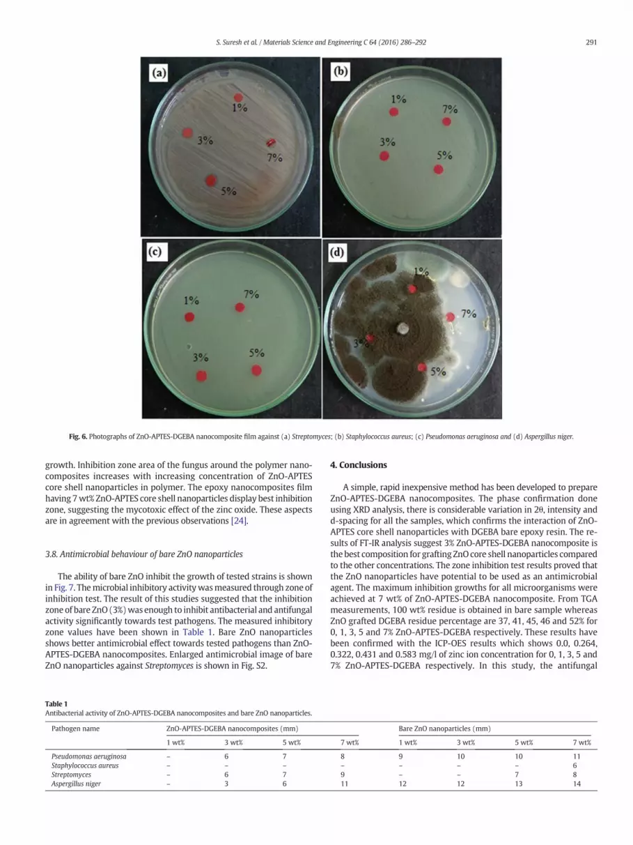
Fig. 6. Photographs of ZnO-APTES-DGEBA nanocomposite film against (a) Streptomyces; (b) Staphylococcus aureus; (c) Pseudomonas aeruginosa and (d) Aspergillus niger.
291S. Suresh et al. / Materials Science and Engineering C 64 (2016) 286–292
growth. Inhibition zone area of the fungus around the polymer nano-composites increases with increasing concentration of ZnO-APTEScore shell nanoparticles in polymer. The epoxy nanocomposites filmhaving 7wt% ZnO-APTES core shell nanoparticles display best inhibitionzone, suggesting the mycotoxic effect of the zinc oxide. These aspectsare in agreement with the previous observations [24].
3.8. Antimicrobial behaviour of bare ZnO nanoparticles
The ability of bare ZnO inhibit the growth of tested strains is shownin Fig. 7. Themicrobial inhibitory activitywasmeasured through zone ofinhibition test. The result of this studies suggested that the inhibitionzoneof bare ZnO(3%)was enough to inhibit antibacterial and antifungalactivity significantly towards test pathogens. The measured inhibitoryzone values have been shown in Table 1. Bare ZnO nanoparticlesshows better antimicrobial effect towards tested pathogens than ZnO-APTES-DGEBA nanocomposites. Enlarged antimicrobial image of bareZnO nanoparticles against Streptomyces is shown in Fig. S2.
Table 1Antibacterial activity of ZnO-APTES-DGEBA nanocomposites and bare ZnO nanoparticles.
Pathogen name ZnO-APTES-DGEBA nanocomposites (mm)
1 wt% 3 wt% 5 wt%
Pseudomonas aeruginosa – 6 7Staphylococcus aureus – – –Streptomyces – 6 7Aspergillus niger – 3 6
4. Conclusions
A simple, rapid inexpensive method has been developed to prepareZnO-APTES-DGEBA nanocomposites. The phase confirmation doneusing XRD analysis, there is considerable variation in 2θ, intensity andd-spacing for all the samples, which confirms the interaction of ZnO-APTES core shell nanoparticles with DGEBA bare epoxy resin. The re-sults of FT-IR analysis suggest 3% ZnO-APTES-DGEBA nanocomposite isthe best composition for grafting ZnOcore shell nanoparticles comparedto the other concentrations. The zone inhibition test results proved thatthe ZnO nanoparticles have potential to be used as an antimicrobialagent. The maximum inhibition growths for all microorganisms wereachieved at 7 wt% of ZnO-APTES-DGEBA nanocomposite. From TGAmeasurements, 100 wt% residue is obtained in bare sample whereasZnO grafted DGEBA residue percentage are 37, 41, 45, 46 and 52% for0, 1, 3, 5 and 7% ZnO-APTES-DGEBA respectively. These results havebeen confirmed with the ICP-OES results which shows 0.0, 0.264,0.322, 0.431 and 0.583 mg/l of zinc ion concentration for 0, 1, 3, 5 and7% ZnO-APTES-DGEBA respectively. In this study, the antifungal
Bare ZnO nanoparticles (mm)
7 wt% 1 wt% 3 wt% 5 wt% 7 wt%
8 9 10 10 11– – – – 69 – – 7 811 12 12 13 14

Fig. 7. Photographs of bare ZnO nanoparticles against (a) Streptomyces; (b) Staphylococcus aureus; (c) Pseudomonas aeruginosa and (d) Aspergillus niger.
292 S. Suresh et al. / Materials Science and Engineering C 64 (2016) 286–292
behaviour against A. niger for a set of ZnO-APTES core shell nanoparti-cles based nanocomposites was envisaged. The presence of fungigrowthwas observed for the 1 and 3wt% ZnO-APTES-DGEBA nanocom-posites but inhibition was observed at 5 and 7% ZnO-APTES-DGEBAnanocomposites.
Supplementary data to this article can be found online at http://dx.doi.org/10.1016/j.msec.2016.03.096.
References
[1] F. Porter, Zinc Handbook: Properties, Processing and Use in Design, CRC Press, BocaRaton, FL, 1991.
[2] M. Li, S. Pokhrel, X. Jin, L. Madler, R. Damoiseaux, E.M. Hoek, Environ. Sci. Technol. 45(2011) 755.
[3] A.J. Huh, Y.J. Kwon, J. Control. Release 156 (2011) 128.[4] D.W. Bahnemann, C. Kormann, M.R. Hoffmann, J. Phys. Chem. 91 (1987) 3789.[5] P.K. Stoimenov, R.L. Klinger, G.L. Marchin, Langmuir 18 (2002) 6679.[6] D. Zvekic, V.V. Srdic, M.A. Karaman,M.N. Matavulj, Processing and Application of Ce-
ramics, 52011 41.[7] C. Guo, Z. Zheng, Q. Zhu, X. Wang, Polym. Plast. Technol. 46 (2007) 1161.[8] R.H. Wang, J.H. Xin, X.M. Tao, W.A. Daoud, Chem. Phys. Lett. 398 (2004) 250.[9] R.H. Wang, J.H. Xin, X.M. Tao, Inorg. Chem. 44 (2005) 3926.
[10] R.K. Dutta, P.K. Sharma, A.C. Pandey, Dig. J. Nanomater. Bios. 4 (2009) 83.
[11] N. Vigneshwaran, S. Kumar, A.A. Kathe, P.V. Varadarajan, V. Prasad, Nanotechnology17 (2006) 5087.
[12] A. Yadav, V. Prasad, A.A. Kathe, S. Raj, D. Yadav, C. Sundaramoorthy, N.Vigneshwaran, Bull. Mater. Sci. 29 (2006) 641.
[13] Kiran Singh, Yogender Kumar, Parvesh Puri, Chetan Sharma, Kamal Rai Aneja, Arabi-an Journal of Chemistry, 2013, http://dx.doi.org/10.1016/j.arabjc.2012.12.038.
[14] D. Duraibabu, T. Ganeshbabu, R. Manjumeena, S. Ananda Kumar, Priya Dasan, Prog-ress in Organic Coatings, 772014 657.
[15] P. Saravanan, K. Jayamoorthy, S. Ananda Kumar, Sens. Actuators, B 221 (2015) 784.[16] R.A. Prates, A.M. Yamada Jr., L.C. Suzuki, M.C.K. Hashimoto, S. Cai, S. Gouw-Soares, L.
Gomes, M.S. Ribeiro, J. Photochem. Photobiol., B 86 (2007) 70.[17] Du Wen-Li, Shan-Shan Niu, Ying-Lei Xu, Zi-Rong Xu, Cheng-Li Fan, Carbohydr.
Polym. 75 (2009) 385.[18] Alexandra Muñoz-Bonilla, Marta Fernández-García, Eur. Polym. J. 65 (2015) 45.[19] (a) Alexandra Muñoz-Bonilla, Marta Fernández-García, Prog. Polym. Sci. 37 (2012)
281;(b) Felix Siedenbiedel, Joerg C. Tiller, Polymers 4 (1) (2012) 46.
[20] J. Sawai, E. Kawada, F. Kanou, H. Igarashi, A. Hashimoto, T Kokugan, J. Chem. Eng. Jpn29 (1996) 627.
[21] J. Jayabharathi, V. Thanikachalam, K. Jayamoorthy, J. Photochem, Photobiol., B 115(2012) 85.
[22] K. Ghule, A.V. Ghule, B. Chen, Y. Ling, Green Chem. 8 (2006) 1034–1041.[23] Y. Liu, L. He, A. Mustapha, H. Li, Z.Q. Hu, M. Lin, J. Appl. Microbiol. 107 (2009) 1193.[24] Sawai, Yoshikawa, J. Appl. Microbiol. 96 (2004) 803.


