Embed Size (px)
Citation preview

Citation: Jurado, C.A.; Parachuru, V.;
Villalobos Tinoco, J.; Guzman-Perez,
G.; Tsujimoto, A.; Javvadi, R.;
Afrashtehfar, K.I. Diagnostic
Mock-Up as a Surgical Reduction
Guide for Crown Lengthening:
Technique Description and Case
Report. Medicina 2022, 58, 1360.
https://doi.org/10.3390/
medicina58101360
Academic Editors: Giuseppe
Minervini and Stefania Moccia
Received: 30 August 2022
Accepted: 25 September 2022
Published: 28 September 2022
Publisher’s Note: MDPI stays neutral
with regard to jurisdictional claims in
published maps and institutional affil-
iations.
Copyright: © 2022 by the authors.
Licensee MDPI, Basel, Switzerland.
This article is an open access article
distributed under the terms and
conditions of the Creative Commons
Attribution (CC BY) license (https://
creativecommons.org/licenses/by/
4.0/).
medicina
Case Report
Diagnostic Mock-Up as a Surgical Reduction Guide for CrownLengthening: Technique Description and Case ReportCarlos A. Jurado 1 , Venkata Parachuru 1,*, Jose Villalobos Tinoco 2, Gerardo Guzman-Perez 3,Akimasa Tsujimoto 4,5, Ramya Javvadi 6 and Kelvin I. Afrashtehfar 7,8
1 Woody L Hunt School of Dental Medicine, Texas Tech University Health Sciences Center El Paso,El Paso, TX 79905, USA
2 Graduate Program in Periodontics, School of Dentistry, National University of Rosario,Rosario S2000CGK, Argentina
3 Private Practice, Uriangato, Guanajuato 36260, Mexico4 Department of Operative Dentistry, College of Dentistry and Dental Clinics, The University of Iowa,
Iowa City, IA 52242, USA5 Department of General Dentistry, School of Dentistry, Creighton University, Omaha, NE 68102, USA6 Oasis Dental, El Paso, TX 79905, USA7 Evidence-Based Practice Unit, Clinical Sciences Department, College of Dentistry, Ajman University,
Ajman City P.O. Box 346, United Arab Emirates8 Department of Reconstructive Dentistry and Gerodontology, School of Dental Medicine, University of Bern,
CH-3010 Berne, Switzerland* Correspondence: [email protected]
Abstract: Background and Objectives: The report describes a technique using a diagnostic mock-up as acrown-lengthening surgical guide to improve the gingival architecture. Materials and Methods: Thepatient’s primary concern was improving her smile due to her “gummy smile” and short clinicalcrowns. After clinical evaluation, surgical crown lengthening accompanied by maxillary centralfull-coverage single-unit prostheses and lateral incisor veneers was recommended. The diagnosticmock-up was placed in the patient’s maxillary anterior region and used as a soft tissue reductionguide for the gingivectomy. Once the planned gingival architecture was achieved, a flap was reflectedto proceed with ostectomy in order to obtain an appropriate alveolar bone crest level using the overlay.After six months, all-ceramic crowns and porcelain veneers were provided as permanent restorations.Results: A diagnostic mock-up fabricated with a putty guide directly from the diagnostic wax-up canbe an adequate surgical guide for crown-lengthening procedures. The diagnostic wax-up was used tofabricate the diagnostic mock-up. These results suggested that it can be used as a crown-lengtheningsurgical guide to modify the gingival architecture. Several advantages of the overlay used in theaesthetic complex case include: (1) providing a preview of potential restorative outcomes, (2) allowingfor the appropriate positioning of gingival margins and the desired alveolar bone crest level for thecrown-lengthening procedure, and (3) serving as a provisional restoration after surgery. Conclusions:The use of a diagnostic mock-up, which was based on a diagnostic wax-up, as the surgical guideresulted in successful crown lengthening and provisional restorations. Thus, a diagnostic overlay canbe a viable option as a surgical guide for crown lengthening.
Keywords: crowns; aesthetic dentistry; mock-up; wax-up; periodontal plastic surgery
1. Introduction
The gingival architecture surrounding natural teeth or dental implants is an importantcomponent of aesthetics in the anterior region [1–3]. When the natural dentition lacks sym-metry or has poor gingival architecture, these conditions can markedly alter the harmony ofthe dentition [4]. In recent years, it has become common for patients to have high aestheticdemands, going beyond a simple desire for a smile makeover [5–8]. Thus, clinicians mustaim for an optimal gingival architecture during treatment [9]. Crown lengthening can be
Medicina 2022, 58, 1360. https://doi.org/10.3390/medicina58101360 https://www.mdpi.com/journal/medicina
Medicina 2022, 58, 1360 2 of 10
used in several clinical situations, such as excessive gingival display or a “gummy smile”,teeth with an inadequate amount of tooth structure, or short clinical restorations [10]. Inthese cases, crown lengthening can re-establish the gingival architecture and enhance therestorative outcome.
Before initiating restorative treatment with crown lengthening, the patient’s aestheticconcerns and expectations should be evaluated in detail. A diagnostic wax-up represent-ing the desired outcome can be completed. Then, an intraoral diagnostic overlay can befabricated to provide the patient and clinician with a tactile evaluation of the proposedtreatment [11]. In addition, excellent communication between the surgeon and the restora-tive dentist is necessary to achieve the desired harmonious gingival architecture, especiallyin patients with high aesthetic demands [6,12–14]. Based on the diagnostic evaluationsmade by the restorative dentist, the surgeon can re-establish the soft and hard tissues torelocate the margins and alveolar crest and achieve periodontal health and an aestheticallypleasing gingival architecture.
Generally, a vacuum-formed surgical guide for crown lengthening is made from aduplicated cast from the diagnostic wax-up to establish the desired gingival architectureand alveolar bone crest level [15]. However, very few reports using a diagnostic overlayfabricated using a temporary bis-acrylic resin with a putty guide directly from the wax-upas a surgical guide for crown-lengthening procedures are available in the literature [16].This case report aims to describe a technique wherein a diagnostic overlay can be used as acrown-lengthening surgical guide to help a surgeon achieve optimal gingival architecture.
2. Materials and Methods
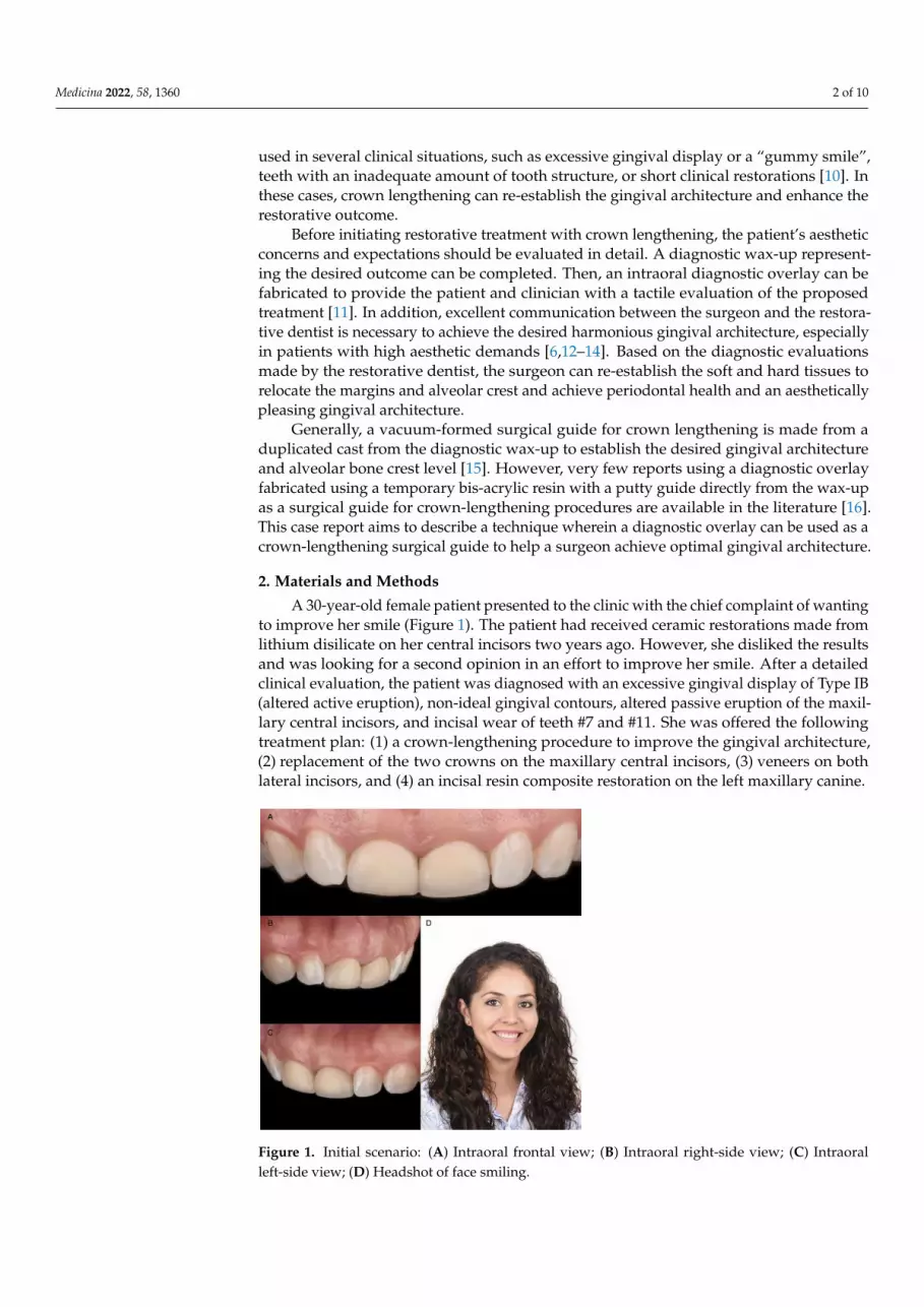
A 30-year-old female patient presented to the clinic with the chief complaint of wantingto improve her smile (Figure 1). The patient had received ceramic restorations made fromlithium disilicate on her central incisors two years ago. However, she disliked the resultsand was looking for a second opinion in an effort to improve her smile. After a detailedclinical evaluation, the patient was diagnosed with an excessive gingival display of Type IB(altered active eruption), non-ideal gingival contours, altered passive eruption of the maxil-lary central incisors, and incisal wear of teeth #7 and #11. She was offered the followingtreatment plan: (1) a crown-lengthening procedure to improve the gingival architecture,(2) replacement of the two crowns on the maxillary central incisors, (3) veneers on bothlateral incisors, and (4) an incisal resin composite restoration on the left maxillary canine.
Medicina 2022, 58, x FOR PEER REVIEW 3 of 11
Figure 1. Initial scenario: (A) Intraoral frontal view; (B) Intraoral right-side view; (C) Intraoral left-
side view; (D) Headshot of face smiling.
Figure 2. Crown prostheses removal: (A) Rubber dam isolation; (B) Initial channel in buccal surface;
(C) Use of hand instrument for wedging; (D) Abutment assessment.
Figure 1. Initial scenario: (A) Intraoral frontal view; (B) Intraoral right-side view; (C) Intraoralleft-side view; (D) Headshot of face smiling.

Medicina 2022, 58, 1360 3 of 10
Diagnostic casts were made, and a wax-up (Wax GEO Classic, Renfert, Hilzingen,Germany) was fabricated to generate a harmonious smile according to the patient’s wishes.After showing the patient the diagnostic wax-up, a diagnostic overlay was made withtemporary bis-acrylic resin (Structur Premium, VOCO, Cuxhaven, Germany). The patientconsented to the treatment after approving the diagnostic wax-up and overlay. Rubberdam isolation (Nic Tone Dental Dam, MDC Dental, Guadalajara, Mexico) was placed, andthe existing ceramic restorations on the central incisors were sectioned with a diamond bur(Conical End 850, Jota AG, Rüthi, Switzerland) and removed (Figure 2).
Medicina 2022, 58, x FOR PEER REVIEW 3 of 11
Figure 1. Initial scenario: (A) Intraoral frontal view; (B) Intraoral right-side view; (C) Intraoral left-
side view; (D) Headshot of face smiling.
Figure 2. Crown prostheses removal: (A) Rubber dam isolation; (B) Initial channel in buccal surface;
(C) Use of hand instrument for wedging; (D) Abutment assessment. Figure 2. Crown prostheses removal: (A) Rubber dam isolation; (B) Initial channel in buccal surface;(C) Use of hand instrument for wedging; (D) Abutment assessment.
The diagnostic overlay was placed over the teeth to guide the desired contour throughgingivoplasty with an electrosurgical unit (Sensimatic 700SE Electrosurge, Parkell, Edge-wood, NY, USA) (Figures 3 and 4).
Medicina 2022, 58, x FOR PEER REVIEW 4 of 11
Figure 3. Diagnostic overlay and tissue recontour: (A) Placement of the diagnostic overlay intra-
orally; (B) Gingivoplasty with an electrosurgical unit; (C) Finishing the contouring with the laser.
Figure 4. Diagnostic overlay removal and evaluation: (A) Removal of the diagnostic overlay; (B)
Gingival tissue architecture evaluation.
After the new gingival architecture was achieved, buccal flap reflection provided a
clear view for the surgeon performing the ostectomy. Flap reflection revealed the proper
position of the osseous crest relative to the cemento-enamel junction (CEJ), which, in this
case, was at the CEJ (Figure 5). An ostectomy procedure was performed using the diag-
nostic overlay as a guide to remove the alveolar bone. The crown-lengthening procedure
was conducted within the recommended range of the Root/Crown (R/C) ratio: (1) R/C
ratio of at least 1/1.5 for an abutment, and (2) R/C ratio of at least 1/1 for a crown.
Figure 3. Diagnostic overlay and tissue recontour: (A) Placement of the diagnostic overlay intra-orally; (B) Gingivoplasty with an electrosurgical unit; (C) Finishing the contouring with the laser.

Medicina 2022, 58, 1360 4 of 10
Medicina 2022, 58, x FOR PEER REVIEW 4 of 11
Figure 3. Diagnostic overlay and tissue recontour: (A) Placement of the diagnostic overlay intra-
orally; (B) Gingivoplasty with an electrosurgical unit; (C) Finishing the contouring with the laser.
Figure 4. Diagnostic overlay removal and evaluation: (A) Removal of the diagnostic overlay; (B)
Gingival tissue architecture evaluation.
After the new gingival architecture was achieved, buccal flap reflection provided a
clear view for the surgeon performing the ostectomy. Flap reflection revealed the proper
position of the osseous crest relative to the cemento-enamel junction (CEJ), which, in this
case, was at the CEJ (Figure 5). An ostectomy procedure was performed using the diag-
nostic overlay as a guide to remove the alveolar bone. The crown-lengthening procedure
was conducted within the recommended range of the Root/Crown (R/C) ratio: (1) R/C
ratio of at least 1/1.5 for an abutment, and (2) R/C ratio of at least 1/1 for a crown.
Figure 4. Diagnostic overlay removal and evaluation: (A) Removal of the diagnostic overlay;(B) Gingival tissue architecture evaluation.
After the new gingival architecture was achieved, buccal flap reflection provided aclear view for the surgeon performing the ostectomy. Flap reflection revealed the properposition of the osseous crest relative to the cemento-enamel junction (CEJ), which, in thiscase, was at the CEJ (Figure 5). An ostectomy procedure was performed using the diagnosticoverlay as a guide to remove the alveolar bone. The crown-lengthening procedure wasconducted within the recommended range of the Root/Crown (R/C) ratio: (1) R/C ratio ofat least 1/1.5 for an abutment, and (2) R/C ratio of at least 1/1 for a crown.
Medicina 2022, 58, x FOR PEER REVIEW 5 of 11
Figure 5. Crown-lengthening procedure: (A) Gingivectomy completed; (B) Initiation of flap; (C)
Flap reflation.
The flap was repositioned (Figure 6), crown margins were refined, and provisional
restorations (adjusted diagnostic overlay) were placed on the central incisors with tempo-
rary resin luting cement.
Figure 6. Flap release and reposition: (A) Flap release; (B) Flap reposition after suturing.
After six months, veneer preparations were performed on lateral incisors to allow for
the proper healing of the periodontal complex (Figure 7), and a final impression was made
with polyvinyl siloxane impression material (Virtual 380, Ivoclar Vivadent, Schaan, Liech-
tenstein).
Figure 5. Crown-lengthening procedure: (A) Gingivectomy completed; (B) Initiation of flap;(C) Flap reflation.

Medicina 2022, 58, 1360 5 of 10
The flap was repositioned (Figure 6), crown margins were refined, and provisionalrestorations (adjusted diagnostic overlay) were placed on the central incisors with tempo-rary resin luting cement.
Medicina 2022, 58, x FOR PEER REVIEW 5 of 11
Figure 5. Crown-lengthening procedure: (A) Gingivectomy completed; (B) Initiation of flap; (C)
Flap reflation.
The flap was repositioned (Figure 6), crown margins were refined, and provisional
restorations (adjusted diagnostic overlay) were placed on the central incisors with tempo-
rary resin luting cement.
Figure 6. Flap release and reposition: (A) Flap release; (B) Flap reposition after suturing.
After six months, veneer preparations were performed on lateral incisors to allow for
the proper healing of the periodontal complex (Figure 7), and a final impression was made
with polyvinyl siloxane impression material (Virtual 380, Ivoclar Vivadent, Schaan, Liech-
tenstein).
Figure 6. Flap release and reposition: (A) Flap release; (B) Flap reposition after suturing.
After six months, veneer preparations were performed on lateral incisors to allowfor the proper healing of the periodontal complex (Figure 7), and a final impressionwas made with polyvinyl siloxane impression material (Virtual 380, Ivoclar Vivadent,Schaan, Liechtenstein).
Medicina 2022, 58, x FOR PEER REVIEW 6 of 11
Figure 7. Lateral incisors veneer preparations: (A) Right side of the lateral veneer preparation with
the reduction guide; (B) Left side of the lateral veneer preparation with the reduction guide.
The final master cast was fabricated with type IV stone (Fujirock, GC, Tokyo, Japan).
Restorations, fabricated following the contours of the diagnostic wax-up, were made of
refractory feldspathic porcelain (Noritake Super Porcelain EX-3, Kuraray Noritake Dental,
Tokyo, Japan) for the veneers and full-coverage crowns (Figure 8).
Figure 8. Feldspathic veneers on the master cast: (A) Porcelain build-up before baking; (B) Definitive
restorations.
A try-in of the final ceramic restorations was performed to evaluate the fit and con-
tours, and the patient approved the final appearance. For bonding the ceramic restora-
tions, isolation was provided via rubber dam placement. The teeth were air-abraded with
20-micron aluminum oxide particles (AquaCare Aluminium Oxide Air Abrasion Powder,
Figure 7. Lateral incisors veneer preparations: (A) Right side of the lateral veneer preparation withthe reduction guide; (B) Left side of the lateral veneer preparation with the reduction guide.

Medicina 2022, 58, 1360 6 of 10
The final master cast was fabricated with type IV stone (Fujirock, GC, Tokyo, Japan).Restorations, fabricated following the contours of the diagnostic wax-up, were made ofrefractory feldspathic porcelain (Noritake Super Porcelain EX-3, Kuraray Noritake Dental,Tokyo, Japan) for the veneers and full-coverage crowns (Figure 8).
Medicina 2022, 58, x FOR PEER REVIEW 6 of 11
Figure 7. Lateral incisors veneer preparations: (A) Right side of the lateral veneer preparation with
the reduction guide; (B) Left side of the lateral veneer preparation with the reduction guide.
The final master cast was fabricated with type IV stone (Fujirock, GC, Tokyo, Japan).
Restorations, fabricated following the contours of the diagnostic wax-up, were made of
refractory feldspathic porcelain (Noritake Super Porcelain EX-3, Kuraray Noritake Dental,
Tokyo, Japan) for the veneers and full-coverage crowns (Figure 8).
Figure 8. Feldspathic veneers on the master cast: (A) Porcelain build-up before baking; (B) Definitive
restorations.
A try-in of the final ceramic restorations was performed to evaluate the fit and con-
tours, and the patient approved the final appearance. For bonding the ceramic restora-
tions, isolation was provided via rubber dam placement. The teeth were air-abraded with
20-micron aluminum oxide particles (AquaCare Aluminium Oxide Air Abrasion Powder,
Figure 8. Feldspathic veneers on the master cast: (A) Porcelain build-up before baking; (B) Definitiverestorations.
A try-in of the final ceramic restorations was performed to evaluate the fit and contours,and the patient approved the final appearance. For bonding the ceramic restorations, isola-tion was provided via rubber dam placement. The teeth were air-abraded with 20-micronaluminum oxide particles (AquaCare Aluminium Oxide Air Abrasion Powder, Velopex,London, UK). The teeth receiving veneers were surface treated with 37% phosphoric acid(Total Etch, Ivoclar Vivadent, Schaan, Liechtenstein) for 15 s and then rinsed with water.A primer (Syntac Primer, Ivoclar Vivadent, Schaan, Liechtenstein ) was applied, and anyexcess was gently removed with air. Adhesive (Syntac Adhesive, Ivoclar Vivadent, Schaan,Liechtenstein) was applied, and any excess was removed with air according to the manu-facturer’s recommendations. The intaglio surfaces of the ceramic restorations were etchedwith 5% hydrofluoric acid (IPS Ceramic Etching Gel, Ivoclar Vivadent, Schaan, Liechten-stein) for 60 s, and Monobond Plus (Ivoclar Vivadent, Schaan, Liechtenstein) was applied tothe etched surfaces. Light-cure resin luting cement (Variolink Esthetic LC, Ivoclar Vivadent,Schaan, Liechtenstein) was applied to the veneers, and they were seated. Excess cementwas removed, and the restorations were cured using an LED light-curing unit (VALO Cord-less, Ultradent, South Jordan, UT, USA) on each surface (facial, palatal, mesial, and distal)for 20 s. The crowns were cemented with a dual-cure resin luting cement (Panavia V5,Kuraray Noritake Dental, Tokyo, Japan) and light-cured, followed by applying appropriatepre-treatments to the teeth and ceramic restorations.
After adjusting the occlusion as needed, the restorations were finalized with polishingpoints (Dialite Feather Lite, Brasseler USA Dental, Savannah, GA, USA) and polishingpaste (Dialite Intra-Oral Polishing Paste, Brasseler USA Dental, Savannah, GA, USA). Theincisal wear was addressed as follows. The maxillary left canine received 37% phosphoricacid etching gel for 15 s, and an adhesive (Tetric N-Bond Universal, Ivoclar Vivadent,Schaan, Liechtenstein) was applied for 20 s, gently air-thinned, and light-cured for 20 s. Anano-hybrid flowable composite resin (Tetric N-Flow, Shade A1, Ivoclar Vivadent, Schaan,Liechtenstein) was placed on the incisal edge and light-cured for 30 s. The resin composite

Medicina 2022, 58, 1360 7 of 10
restoration was re-shaped on the incisal edge with a fine diamond bur (Diamond bur FG859012, Jota AG, Rüthi, Switzerland). The restoration was final-polished with green andgrey composite polishers (Composite Diamond Polisher, Jota AG, Ruthi, Switzerland) usinga polishing paste (Diamond Polish Mint, Ultradent, South Jordan, UT, USA) and a polishingbrush (Jiffy Composite Polishing Brush, Ultradent, South Jordan, UT, USA). The patientapproved of the shape and size of the final restorations, and the treatment fulfilled heraesthetic desire (Figure 9).
Medicina 2022, 58, x FOR PEER REVIEW 7 of 11
Velopex, London, UK). The teeth receiving veneers were surface treated with 37% phos-
phoric acid (Total Etch, Ivoclar Vivadent, Schaan, Liechtenstein) for 15 s and then rinsed
with water. A primer (Syntac Primer, Ivoclar Vivadent, Schaan, Liechtenstein ) was ap-
plied, and any excess was gently removed with air. Adhesive (Syntac Adhesive, Ivoclar
Vivadent, Schaan, Liechtenstein) was applied, and any excess was removed with air ac-
cording to the manufacturer’s recommendations. The intaglio surfaces of the ceramic res-
torations were etched with 5% hydrofluoric acid (IPS Ceramic Etching Gel, Ivoclar Viva-
dent, Schaan, Liechtenstein) for 60 s, and Monobond Plus (Ivoclar Vivadent, Schaan,
Liechtenstein) was applied to the etched surfaces. Light-cure resin luting cement (Vario-
link Esthetic LC, Ivoclar Vivadent, Schaan, Liechtenstein) was applied to the veneers, and
they were seated. Excess cement was removed, and the restorations were cured using an
LED light-curing unit (VALO Cordless, Ultradent, South Jordan, UT, USA) on each sur-
face (facial, palatal, mesial, and distal) for 20 s. The crowns were cemented with a dual-
cure resin luting cement (Panavia V5, Kuraray Noritake Dental, Tokyo, Japan) and light-
cured, followed by applying appropriate pre-treatments to the teeth and ceramic restora-
tions.
After adjusting the occlusion as needed, the restorations were finalized with polish-
ing points (Dialite Feather Lite, Brasseler USA Dental, Savannah, GA, USA) and polishing
paste (Dialite Intra-Oral Polishing Paste, Brasseler USA Dental, Savannah, GA, USA). The
incisal wear was addressed as follows. The maxillary left canine received 37% phosphoric
acid etching gel for 15 s, and an adhesive (Tetric N-Bond Universal, Ivoclar Vivadent,
Schaan, Liechtenstein) was applied for 20 s, gently air-thinned, and light-cured for 20 s. A
nano-hybrid flowable composite resin (Tetric N-Flow, Shade A1, Ivoclar Vivadent,
Schaan, Liechtenstein) was placed on the incisal edge and light-cured for 30 s. The resin
composite restoration was re-shaped on the incisal edge with a fine diamond bur (Dia-
mond bur FG 859012, Jota AG, Rüthi, Switzerland). The restoration was final-polished
with green and grey composite polishers (Composite Diamond Polisher, Jota AG, Ruthi,
Switzerland) using a polishing paste (Diamond Polish Mint, Ultradent, South Jordan, UT,
USA) and a polishing brush (Jiffy Composite Polishing Brush, Ultradent, South Jordan,
UT, USA). The patient approved of the shape and size of the final restorations, and the
treatment fulfilled her aesthetic desire (Figure 9).
Figure 9. Final restorations: (A) Frontal view; (B) Lateral view; (C) Frontal in occlusion; (D) Final
smile. Figure 9. Final restorations: (A) Frontal view; (B) Lateral view; (C) Frontal in occlusion; (D) Final smile.
An occlusal night guard was also provided to prevent damage to the final restorations.At the patient’s five-year follow-up, she was fascinated with the clinical outcome (Figure 10).
Medicina 2022, 58, x FOR PEER REVIEW 8 of 11
An occlusal night guard was also provided to prevent damage to the final restora-
tions. At the patient’s five-year follow-up, she was fascinated with the clinical outcome
(Figure 10).
Figure 10. Five-year follow-up: (A) Frontal view; (B) Lateral view.
3. Discussion
Despite the lack of clinical case reports, evidence from this case suggests that a diag-
nostic overlay for crown lengthening allows outcomes to be predictable for follow-up pe-
riods of at least five years. These findings are essential for the anterior region, where soft
tissue changes may compromise treatment outcomes without a vacuum-formed surgical
guide. Typically, the restorative process of crown lengthening using a vacuum-formed
surgical guide needs both provisional restorations and a surgical guide. However, this
clinical case shows that a diagnostic overlay can be used to confirm the proposed treat-
ment plan with the patient and provide a two-for-one surgical guide and provisional res-
torations, thus reducing costs. Recently, a fully digital workflow for crown lengthening,
using a single surgical guide, was reported [17–20]. However, this technique requires an
intraoral scanner, 3D printer, and cone beam computed tomography (CBCT) scan, relying
on many kinds of expertise in the digital workflow. Most clinicians are still not familiar
with digital workflow [21], and the additional time and costs required before surgery are
disincentives for its introduction.
In contrast, it is simpler to use conventional methods to prepare the soft and hard
tissues based on a diagnostic overlay when it is placed in the mouth if the clinician knows
the appropriate distances to the alveolar bone crest level from the gingival margins. Most
clinicians know that the distance from the alveolar crest to the gingival margin on the
facial and palatal aspects is in the range of 3 mm, while the distance from the alveolar crest
to the gingival margin on the interproximal aspect is about 5 mm due to the height of the
interproximal papilla [22]. Thus, it appears easier to determine the desired alveolar bone
crest level using only a diagnostic overlay. This would be a simplified approach to per-
forming crown lengthening without a traditional surgical guide.
Figure 10. Five-year follow-up: (A) Frontal view; (B) Lateral view.
Medicina 2022, 58, 1360 8 of 10
3. Discussion
Despite the lack of clinical case reports, evidence from this case suggests that a di-agnostic overlay for crown lengthening allows outcomes to be predictable for follow-upperiods of at least five years. These findings are essential for the anterior region, where softtissue changes may compromise treatment outcomes without a vacuum-formed surgicalguide. Typically, the restorative process of crown lengthening using a vacuum-formedsurgical guide needs both provisional restorations and a surgical guide. However, thisclinical case shows that a diagnostic overlay can be used to confirm the proposed treatmentplan with the patient and provide a two-for-one surgical guide and provisional restorations,thus reducing costs. Recently, a fully digital workflow for crown lengthening, using asingle surgical guide, was reported [17–20]. However, this technique requires an intraoralscanner, 3D printer, and cone beam computed tomography (CBCT) scan, relying on manykinds of expertise in the digital workflow. Most clinicians are still not familiar with digitalworkflow [21], and the additional time and costs required before surgery are disincentivesfor its introduction.
In contrast, it is simpler to use conventional methods to prepare the soft and hardtissues based on a diagnostic overlay when it is placed in the mouth if the clinician knowsthe appropriate distances to the alveolar bone crest level from the gingival margins. Mostclinicians know that the distance from the alveolar crest to the gingival margin on thefacial and palatal aspects is in the range of 3 mm, while the distance from the alveolarcrest to the gingival margin on the interproximal aspect is about 5 mm due to the heightof the interproximal papilla [22]. Thus, it appears easier to determine the desired alveolarbone crest level using only a diagnostic overlay. This would be a simplified approach toperforming crown lengthening without a traditional surgical guide.
In the present case, the use of a diagnostic overlay, based on the diagnostic wax-up,was an easy and powerful tool for the diagnostic planning of a treatment with high aestheticdemands. Given this outcome, the placement of a diagnostic overlay could be adoptedas a routine protocol by clinicians, as it provides a high predictability of outcomes inaesthetically complex cases. Furthermore, the overlay can also be considered as a usefulpromotional tool for acquiring the patient approval of the treatment plan presented bythe dental professional. In this case report, the diagnostic overlay was made based on thepatient’s requests, and after it was placed in her mouth, the patient immediately indicatedthat she liked the result and requested the treatment.
Soft tissue crown lengthening is performed with gingivoplasty using a scalpel, anelectrosurgical unit, a radiosurgical unit, or a laser [23]. If the new gingival margin positionis near the underlying bone, a flap should be reflected for an ostectomy to re-establish anadequate biologic width. In the current case, the diagnostic overlay was placed and guidedthe use of the electrosurgical unit for the external gingivoplasty. A flap was reflected torecontour hard tissues and re-establish the biologic width of 3 mm. Compared with atraditional scalpel, an electrosurgical unit allows the clinician to cut, ablate, and re-shapesoft tissues with no resulting bleeding and no need for suturing. The diagnostic overlaywas an excellent guide.
Another consideration when performing crown lengthening is the healing period.The periodontal phenotype is a crucial factor, especially in aesthetic outcomes, because itimpacts both the healing and final position of the gingival margin [24]. Research suggeststhat a thin biotype has a thickness of 1.5 mm or less, and a thick biotype has a thickness of2.0 mm or more. Patients with a thin biotype may experience more gingival recession thanthose with a thick one [25]. The patient in this case had a thick biotype; thus, the likelihoodof gingival recession was minimal. When considering aesthetic outcomes during this kindof treatment, the ideal healing time ranges from six weeks to six months, and a longer timemay be required for patients with a thin biotype [26,27]. In the present case, the cliniciansdecided to wait six months before finalizing the ceramic restorations. This period providedadequate time for tissue healing and resulted in a stable gingival margin position for a

Medicina 2022, 58, 1360 9 of 10
pleasing aesthetic result. The diagnostic overlay was used as the provisional restorationand performed well during this time.
A limitation of this traditional workflow can happen if the diagnostic wax-up isexcessive; then, it will create bulky restorations. In order to prevent this, the lip support isevaluated by the clinician and patient during the mock-up. Thus, the diagnostic overlay isnecessary to demonstrate the likely outcome to the patient, and, as a provisional restorationduring the healing of the hard and soft tissue, it can also be used as the surgical guidewhile attaining good aesthetic results. This suggests that a separate surgical guide may beunnecessary in many cases, and the assumed additional precision resulting from the use ofa dedicated surgical guide may similarly be unnecessary. A simplified procedure using thediagnostic overlay may achieve all treatment goals at a lower cost.
4. Conclusions
The case presented in this clinical report shows that using a diagnostic overlay, whichwas based on a diagnostic wax-up, as the surgical guide resulted in successful crownlengthening. These results suggest that a diagnostic overlay may be viable for surgicallyguiding crown lengthening in aesthetically complex cases.
Author Contributions: Conceptualization, C.A.J. and J.V.T.; methodology, V.P.; investigation, G.G.-P.;resources, A.T. and R.J.; data curation, K.I.A.; writing—original draft preparation, C.A.J.; writing—reviewand editing, A.T. and K.I.A. All authors have read and agreed to the published version of the manuscript.
Funding: This research received no external funding.
Institutional Review Board Statement: The study was conducted in accordance with the Declarationof Helsinki, and approved by the Institutional Review Board of Cenro de Estudios Odontologicos deQueretaro (protocol code XXXDENT/0430121-16 and 11/01/2016).” for studies involving humans.
Informed Consent Statement: Written informed consent has been obtained from the patient topublish this paper.
Data Availability Statement: Not applicable.
Acknowledgments: K. I. Afrashtehfar thanks the Universität Bern for partially supporting the open-access publication of this work.
Conflicts of Interest: The authors declare no conflict of interest.
References1. Jurado, C.A.; Tinoco, J.V.; Tsujimoto, A.; Barkmeier, W.; Fischer, N.; Markham, M. Clear matrix use for composite resin core
fabrication. Int. J. Esthet. Dent. 2020, 15, 108–117. [PubMed]2. Afrashtehfar, K.I.; Assery, M.K.A.; Bryant, S.R. Aesthetic Parameters and patient-perspective assessment tools for maxillary
anterior single implants. Int. J. Dent. 2021, 2021, 6684028. [CrossRef] [PubMed]3. Del Monte, S.; Afrashtehfar, K.I.; Emami, E.; Abi Nader, S.; Tamimi, F. Lay preferences for dentogingival esthetic parameters: A
systematic review. J. Prosthet. Dent. 2017, 118, 717–724. [CrossRef] [PubMed]4. Miranda, M.E.; Olivieri, K.A.; Rigolin, F.J.; de Vasconcellos, A.A. Esthetic challenges in rehabilitating the anterior maxilla: A case
report. Oper. Dent. 2016, 41, 2–7. [CrossRef] [PubMed]5. Afrashtehfar, K.I.; Assery, M.K. Five considerations in cosmetic and esthetic dentistry. J. New Jersey Dent. Assoc. 2014, 85, 14–15.6. Afrashtehfar, K.I.; Assery, M.K.A.; Bryant, S.R. Patient Satisfaction in Medicine and Dentistry. Int. J. Dent. 2020, 2020, 6621848.
[CrossRef]7. Alikhasi, M.; Yousefi, P.; Afrashtehfar, K.I. Smile Design: Mechanical Considerations. Dent. Clin. North Am. 2022, 66, 477–487.
[CrossRef]8. Afrashtehfar, K.I.; Bryant, S.R. Understanding the lived experience of north american dental patients with a single-tooth implant
in the upper front region of the mouth: Protocol for a qualitative Study. JMIR Res. Protoc. 2021, 10, e25767. [CrossRef]9. Jurado, C.A.; Tsujimoto, A.; Guzman, L.G.; Fischer, N.G.; Markham, M.D.; Barkmeier, W.W.; Latta, M.A. Implant therapy with
monolithic translucent zirconia restorations in the esthetic zone. Gen. Dent. 2020, 68, 46–49.10. Marzadori, M.; Stefanini, M.; Sangiorgi, M.; Mounssif, I.; Monaco, C.; Zucchelli, G. Crown lengthening and restorative procedures
in the esthetic zone. Periodontol. 2000 2018, 77, 84–92. [CrossRef]11. Simon, H.; Magne, P. Clinically based diagnostic wax-up for optimal esthetics: The diagnostic mock-up. J. Calif. Dent. Assoc. 2008,
36, 355–362. [PubMed]

Medicina 2022, 58, 1360 10 of 10
12. Jurado, C.; Watanabe, H.; Tinoco, J.V.; Valenzuela, H.U.; Perez, G.G.; Tsujimoto, A. A conservative approach to ceramic veneers: Acase report. Oper. Dent. 2020, 45, 229–234. [CrossRef] [PubMed]
13. Afrashtehfar, K.I.; Igarashi, K.; Bryant, S.R. Canadian Dental Patients with a Single-Unit Implant-Supported Restoration in theAesthetic Region of the Mouth: Qualitative and Quantitative Patient-Reported Outcome Measures (PROMs). Data 2021, 6, 90.[CrossRef]
14. Afrashtehfar, K.I. Conventional free-hand, dynamic navigation and static guided implant surgery produce similar short-termpatient-reported outcome measures and experiences. Evid.-Based Dent. 2021, 22, 143–145. [CrossRef]
15. Longo, E.; Frosecchi, M.; Marradi, L.; Signore, A.; de Angelis, N. Guided periodontal surgery: A novel approach for the treatmentof gummy smile. A case report. Int. J. Esthet. Dent. 2019, 14, 384–392.
16. Gurrea, J.; Bruguera, A. Wax-up and mock-up. A guide for anterior periodontal and restorative treatments. Int. J. Esthet. Dent.2014, 9, 146–162. [PubMed]
17. Liu, X.; Yu, J.; Zhou, J.; Tan, J. A digitally guided dual technique for both gingival and bone resection during crown lengtheningsurgery. J. Prosthet. Dent. 2018, 119, 345–349. [CrossRef]
18. Kongkiatkamon, S.; Rokaya, D. Full digital workflow in the esthetic dental restoration. Case Rep Dent. 2022, 2022, 8836068.[CrossRef]
19. Mendoza-Azpur, G.; Cornejo, H.; Villanueva, M.; Alva, R.; Barbisan de Souza, A. Periodontal plastic surgery for esthetic crownlengthening by using data merging and a CAD-CAM surgical guide. J. Prosthet. Dent. 2022, 127, 556–559. [CrossRef]
20. Jurado, C.A.; Tsujimoto, A.; Watanabe, H.; Villalobos-Tinoco, J.; Garaicoa, J.L.; Markham, M.D.; Barkmeier, W.W.; Latta, M.A.Chair-side CAD/CAM fabrication of a single-retainer resin bonded fixed dental prosthesis: A case report. Restor. Dent. Endod.2020, 45, e15. [CrossRef]
21. Alazmi, S.O. Three dimensional digitally designed surgical guides in esthetic crown lengthening: A clinical case report with12 months follow up. Clin. Cosmet Investig. Dent. 2022, 14, 55–59. [CrossRef] [PubMed]
22. Takei, H.H.; Bevilacqua, F.; Cooney, J. Surgical crown lengthening of the maxillary anterior dentition: Aesthetic considerations.Pract. Periodontics Aesthetic Dent. 1999, 11, 639–644.
23. Hempton, T.J.; Dominici, J.T. Contemporary crown-lengthening therapy: A review. J. Am. Dent. Assoc. 2010, 141, 647–655.[CrossRef] [PubMed]
24. Pontoriero, R.; Carnevale, G. Surgical crown lengthening: A 12-month clinical wound healing study. J. Periodontol. 2001, 72,841–848. [CrossRef]
25. Kois, J.C. Altering gingival levels: The restorative connection part I: Biologic variables. J. Esthet. Restor. Dent. 1994, 6, 3–7.[CrossRef]
26. Deas, D.E.; Moritz, A.J.; McDonnell, H.T.; Powell, C.A.; Mealey, B. Osseous surgery for crown lengthening: A 6-month clinicalstudy. J. Periodontol. 2004, 75, 1288–1294. [CrossRef] [PubMed]
27. Afrashtehfar, K.I.; Moshaverinia, A. Five things to know about regenerative periodontal therapies in dental medicine. J. NewJersey Dent. Assoc. 2015, 86, 12–13.


