Embed Size (px)
Citation preview

Differential gene expression in cumulus cells as a prognosticindicator of embryo viability: a microarray analysis
Aafke P.A. van Montfoort1,4, Joep P.M. Geraedts2, John C.M. Dumoulin1, AlphonsP.M. Stassen3, Johannes L.H. Evers1 and Torik A.Y. Ayoubi3
1Department of Obstetrics and Gynaecology, Research Institute Growth and Development (GROW), Academic Hospital Maastricht,
Maastricht, The Netherlands; 2Department of Clinical Genetics, Research Institute Growth and Development (GROW), Academic
Hospital Maastricht, Maastricht, The Netherlands; 3Department of Population Genetics, Genomics and Bioinformatics, Maastricht
University, and Research Institute Growth and Development (GROW), Maastricht, The Netherlands
4Correspondence address. E-mail: [email protected]
Besides the established selection criteria based on embryo morphology and blastomere number, new parameters for embryo
viability are needed to improve the clinical outcome of IVF and more particular of elective single-embryo transfer. Genome-
wide gene expression in cumulus cells was studied, since these cells surround the oocyte inside the follicle and therefore possibly
reflect oocyte developmental potential. Early cleavage (EC) was chosen as a parameter for embryo viability. Gene expression in
cumulus cells from eight oocytes resulting in an EC embryo (EC-CC; n 5 8) and from eight oocytes resulting in a non-EC (NEC)
embryo (NEC-CC; n 5 8) was analysed using microarrays (n 5 16). A total of 611 genes were differentially expressed (P < 0.01),
mainly involved in cell cycle, angiogenesis, apoptosis, epidermal growth factor, fibroblast growth factor and platelet-derived
growth factor signalling, general vesicle transport and chemokine and cytokine signalling. Of the 25 selected differentially
expressed genes analysed by quantitative real-time PCR 15 (60%) genes could be validated in the original samples. Of these 8
(53%) could also be validated in 24 (12-EC-CC and 12 NEC-CC) extra independent samples. The most differentially expressed
genes among these were CCND2, CXCR4, GPX3, CTNND1 DHCR7, DVL3, HSPB1 and TRIM28, which probably point to
hypoxic conditions or a delayed oocyte maturation in NEC-CC samples. This opens up perspectives for new molecular embryo
or oocyte selection parameters which might also be useful in countries where the selection has to be made at the oocyte stage
before fertilization instead of at the embryonic stage.
Keywords: assisted reproductive technology; early cleavage; gene expression; cumulus cells; microarray
Introduction
The only way to prevent a dizygotic twin pregnancy in IVF, which is
regarded as one of the most serious complications, is single-embryo
transfer (SET). As most patients have more than one embryo available
for transfer, selecting the most viable one is of pivotal importance.
Most clinics rely for embryo selection on the non-invasive examin-
ation of developmental and morphological aspects of the embryos.
In every stage of oocyte and embryonic development, characteristics
have been defined which appear to be prognostic indicators of success-
ful pregnancy. Among these are zona pellucida thickness and cyto-
plasmic granularity of the oocyte, size of pronuclei and alignment of
nuclear polar bodies in the zygote, early cleavage (EC) in the cleavage
stage embryo and number and size of blastomeres, fragmentation and
multinucleation in the 4–8-cell stage embryo [see Borini et al. (2005)
for a review and Gerris (2005) for a more extensive list of references].
Especially, EC appears to be a good parameter for embryo viability as
it is highly correlated with the blastocyst formation rate (Fenwick
et al., 2002) and the implantation and pregnancy rate (Shoukir
et al., 1997; Sakkas et al., 1998; Lundin et al., 2001), not only in
double but also in SETs (Salumets et al., 2003; Van Montfoort
et al., 2004). For embryos in the intermediate syngamy state, the
pregnancy rate was in between that of EC and non-early cleavage
(NEC) embryos (Wharf et al., 2004).
As the developmental potential of an embryo cannot be fully deter-
mined by characteristics visible by microscopy alone, several other
markers are studied (Pearson, 2006). For instance, several investi-
gators focused on the influence of the follicular micro-environment
on subsequent embryonic development. The follicular fluid LH and
growth hormone levels at the time of oocyte retrieval were higher in
embryos with good morphology (Mendoza et al., 2002). Furthermore,
the concentrations of hormones (17b-estradiol, LH, growth hormone,
prolactin, leptin), growth factors (insulin-like growth factor-I), cyto-
kines (interleukin-1) and proteinases (matrix-metalloproteinase-9) in
follicular fluid differ according to the probability of pregnancy
(Mendoza et al., 2002; Anifandis et al., 2005; Hammadeh et al.,
2005; Lee et al., 2005). Also vascularization of the follicles has
been examined as a potential marker for the developmental potential
of an embryo. The peri-follicular blood flow characteristics are
related to oocyte oxygenation (Van Blerkom et al., 1997) and can
differ between the follicles in one ovary. Nargund et al. (1996)
found, by Doppler imaging of the follicular blood flow, that oocytes
from poorly vascularized follicles developed in morphologically
inferior embryos as compared to those from well-vascularized
# The Author 2008. Published by Oxford University Press on behalf of the European Society of Human Reproduction and Embryology. All rights reserved.
For Permissions, please email: [email protected] 157
MHR-Basic Science of Reproductive Medicine Vol.14, No.3 pp. 157–168, 2008
Advance Access publication on January 18, 2008 doi:10.1093/molehr/gam088
by guest on March 14, 2016
http://molehr.oxfordjournals.org/
Dow
nloaded from

follicles. Several other studies confirmed a positive relationship
between perifollicular vascularization and pregnancy (Chui et al.,
1997; Van Blerkom et al., 1997; Borini et al., 2004). Pregnancies
were only achieved with embryos from oocytes which had vascularity
detected in .50% of their follicular circumference and live births
only from oocytes with .75% follicular vascularity (Chui et al.,
1997). Gaulden et al. (1992) suggested that hypoxic intracellular con-
ditions might result in a diminished level of oxidative metabolism in
the oocyte and a lower intracellular pH. The latter in turn could lead
to meiotic spindle instabilities and chromosomal abnormalities.
Indeed,Chui et al. (1997) and Van Blerkom et al. (1997) reported a
significantly higher incidence of aneuploidy and spindle defects in
oocytes derived from follicles with poor vascularization as compared
to those from well-vascularized follicles. In addition, ATP content of
the oocyte and dissolved oxygen content of the follicle fluid are related
to oocyte/embryo development (Van Blerkom et al., 1995,1997).
As the oocyte is in dialogue with the surrounding cumulus cells via
paracrine and gap-junctional signalling (Sutton et al., 2003), we
hypothesized that differences in intra-follicular processes which are
responsible for oocyte and embryonic development and subsequently
implantation are reflected in the gene expression pattern of cumulus
cells. The bi-directional communication between the oocyte and the
cumulus cells is necessary for oocyte development as oocytes fail to
grow in the absence of (a connection with) cumulus cells (Ackert
et al., 2001; Matzuk et al., 2002). Zhang et al. (2005) reported that
the expression of several genes in cumulus cells, particularly pentraxin
3 (PTX3), was indicative of oocyte and embryo quality. In addition,
the expression of cyclooxygenase 2 (COX2), gremlin (GREM) and
hyaluronic acid synthase 2 (HAS2) is also positively correlated with
embryo quality (McKenzie et al., 2004). In turn, oocyte factors like
growth and differentiation factor-9 (GDF-9) are necessary for
cumulus expansion (Sutton et al., 2003).
The aim of this study was to analyse the genome-wide expression of
genes in cumulus cells as indicators of embryo viability. By analysing
gene expression in cumulus cells, the understanding of the regulation
of oogenesis and embryonic development might be improved. This
information might lead to new molecular non-invasive embryo selec-
tion parameters reflected in cumulus cells that can be used in addition
to the existing morphological parameters or might result in an oocyte
selection tool for those who are obliged to select a limited number of
oocytes for fertilization (Ludwig et al., 2000).
Materials and Methods
Patients and human cumulus cell collection
Patients visiting the IVF clinic of the academic hospital Maastricht underwent
an IVF or ICSI treatment as described previously (Van Montfoort et al., 2006).
For the study, which was approved by the local Ethics Committee, in consent-
ing patients, immediately following ultrasound-guided cumulus–oocyte
complex (COC) retrieval, a proportion of the cumulus cells surrounding a
single oocyte were removed using a sharp needle, lysed in 100 ml Trizol
reagent (Invitrogen, Carlsbad, USA) supplemented with 1% (v/v)
2-mercapto-ethanol (Merck, Darmstadt, Germany), snap-frozen in liquid nitro-
gen and stored at –808C (cumulus cells from one oocyte per vial). The oocytes
were cultured and fertilized individually in 5 ml droplets covered by mineral
oil. Between 23–26 h post-injection or 25–28 h post-insemination EC status
of embryos was assessed. A 2 h time difference is necessary to compensate
for the time difference in early development between IVF- and ICSI-derived
embryos (Van Montfoort et al., 2004). Subsequently, on Day 2 of development,
the embryos were examined for morphology, number of blastomeres and the
presence or absence of multinucleated blastomeres (MNBs) (Van Montfoort
et al., 2005).
Experimental design
EC was chosen as a marker for embryo viability. Gene expression in cumulus
cells from eight oocytes resulting in an EC embryo (EC-CC; n ¼ 8) and from
eight oocytes resulting in a non-EC embryo (NEC-CC; n ¼ 8) derived from six
patients were analysed using microarrays (n ¼ 16). To exclude a differential
gene expression due to differences in patient characteristics, samples were
paired. From four patients both an EC-CC and a NEC-CC sample were used.
From two additional patients two EC-CC as well as two NEC-CC samples
were used. The microarray results were validated by quantitative RT–PCR
(qRT–PCR) on the original samples analysed by microarray as well as on 24
new samples.
The cumulus cell samples (for microarray and RT–PCR) from EC and NEC
embryos were derived from normally fertilized (2PN) oocytes, which devel-
oped into embryos with comparable characteristics on Day 2, i.e. 4-cell with
good morphology and no MNBs present.
RNA isolation
Total RNA was extracted using Trizol reagent (Invitrogen) according to the
manufacturer’s instructions with some adaptations for the small quantity of
RNA. RNA was precipitated with isopropyl alcohol for 2 h and the RNA
pellet was washed three times with 75% ethanol. To be able to track the
small RNA pellet, 5 mg glycogen (Ambion, Woodward, USA) was added to
the sample before RNA precipitation. Total RNA was resuspended in 20 ml
RNase-free water and stored at 2808C. For all RNA samples quantity and
purity were determined using the Nanodrop ND-1000 spectrophotometer
(Nanodrop Technologies, Wilmington, USA) and RNA integrity was deter-
mined using the Bioanalyzer 2100 (Agilent Technologies, Palo Alto, USA).
Two cycle amplification and microarray hybridization
Fifty nanogram total RNA was amplified using the two-cycle cDNA synthesis
kit (Affymetrix, Santa Clara, USA) in combination with the MEGAscript T7 in
vitro transcription system (Ambion). Biotin labelled target complementary
RNA was fractionated and hybridized to Human Genome U133A Plus 2.0
Arrays (Affymetrix). Each array contained .54 000 oligonucleotide probe-sets
corresponding to 38 500 characterized human genes.
Microarray analysis
To identify probe sets which were differentially expressed between eight
EC-CC and eight NEC-CC samples, a three step process was applied. First,
Affymetrix GeneChip Operating Software (GCOS, version 1.4) was used to
analyse image data. For each transcript represented on the array by a probe
set, the expression algorithm computed the detection call (present, absent or
marginal), the detection P-value, and the signal which is an average intensity
value for each probe set. This resulted in a table with 54 675 probe sets.
Second, for each probe set the 16 detection calls were used to determine
whether the probe set was reliably detected or not and should or should not
be selected for further analysis (McClintick and Edenberg, 2006). To this
end, for every group of eight arrays (the early and the non-early) the number
of present calls was counted (a number ranging from 0 to 8). If six or more
calls were present, the probe set was denoted present. If the probe set was in
at least one of the two groups denoted present, it was selected for further analy-
sis. Finally, the over- or underexpression of the remaining probe sets in one of
the two groups was analysed using the class comparison method in BRB Array-
Tools software package applying a univariate test composed of a paired t-test
with random variance model. This was developed by the Biometric Research
Branch of the US National Cancer Institute (http://linus.nci.nih.gov/
BRB-ArrayTools.html). Hierarchical clustering of samples was also performed
using BRB ArrayTools. Samples were clustered by comparing their expression
profiles.
The genes showing significant differential expression between both groups
(P , 0.01) were classified into functional groups using the Panther classifi-
cation system (http://www.pantherdb.org) (Thomas et al., 2003). The gene
expression data analysis tool (Thomas et al., 2006) was used to determine
which biological process or pathway was significantly overexpressed in one
of the two groups. This program uses binomial statistics with Bonferroni cor-
rection to analyse whether the proportion of genes from a certain biological
process or pathway present in a gene list (i.e. the list of differentially expressed
van Montfoort et al.
158
by guest on March 14, 2016
http://molehr.oxfordjournals.org/
Dow
nloaded from
genes from an array study) is significantly different from the proportion of
genes in that process or pathway in the whole human genome (P , 0.05).
Quantitative real-time PCR
For the qRT–PCR, TaqMan low density arrays (TLDAs) (Applied Biosystems,
Foster City, USA) were used. Each 2 ml well of the TLDA contains user-
defined primers and probes selected from an online catalogue (http://
myscience.appliedbiosystems.com) for a single gene. One well contains
primers and probes for 18S rRNA, a mandatory endogenous control from the
manufacturer.
cDNA was prepared from 100 hg total RNA per sample using the High
Capacity cDNA archive kit (Applied Biosystems) according to the manufac-
turer’s instructions. To each cDNA sample (20 ml), 80 ml nuclease-free water
and 100 ml 2� TaqMan Universal PCR Master Mix (Applied Biosystems)
was added. This mixture was then equally divided over two sample-loading
ports of the TLDA, each connected to one set of all the genes of interest.
The arrays were centrifuged twice (10, 331 g) to equally distribute the sample
over the wells. Subsequently, the card was sealed to prevent an exchange
between wells. qRT–PCR amplification was performed using an Applied Bio-
systems Prism 7900HT sequence detection system with the following thermal
cycler conditions: 2 min at 508C and 10 min at 94.58C, followed by 40 cycles of
30 s at 978C and 1 min at 59.78C.
qRT–PCR analysis
The RQ manager 1.2 software was used to generate Ct values corrected for var-
iances in fluorescent signal strength by using a passive reference dye. The
geNorm program (Vandesompele et al., 2002) was used to determine the
most stably expressed housekeeping genes. Briefly, the average pair-wise vari-
ation of a housekeeping gene with all other housekeeping genes was calculated.
Stepwise exclusion of the gene with the highest variation resulted in a combi-
nation of two housekeeping genes that have the most stable expression. The
geometrical mean of the Ct values of these two genes was used as a normaliza-
tion factor which was substracted from the Ct values of the genes of interest to
obtain normalized Ct values (DCt). Subsequently, the mean DCt of the
NEC-CC samples was substracted from the mean DCt of the EC-CC samples
generating a DDCt. This DDCt was recalculated into a relative expression quan-
tity (22DDCt) of the gene of interest in EC-CC as compared with NEC-CC
samples (Livak and Schmittgen, 2001).
Results
Microarray analysis
For the gene expression analysis, 8 EC-CC and 8 NEC-CC samples
(from 6 patients) have been analysed using 16 microarrays. The raw
microarray data have been deposited in NCBIs Gene Expression
Omnibus (GEO, http://www.ncbi.nlm.nih.gov/geo/) and are accessible
through GEO Series accession number GSE9526. From the 54 675
probe sets on the array, 18 480 had a present call. Most of these
probe sets showed similar expression between the EC-CC and
NEC-CC group, except for 737 probe sets that were differentially
expressed (P , 0.01). For 59 of these probe sets, the corresponding
gene is not yet known. Of the 678 remaining probe sets, which corre-
spond to 611 different genes, 162 (24%) were up-regulated and 516
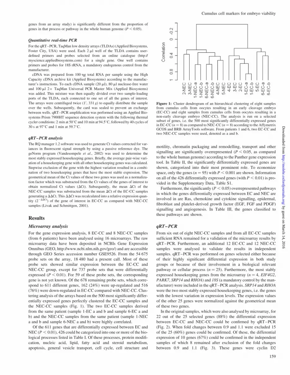
(76%) were down-regulated in EC-CC compared with NEC-CC. Clus-
tering analysis of the arrays based on the 500 most significantly differ-
entially expressed genes perfectly clustered the EC-CC samples and
the NEC-CC samples (Fig. 1). The two EC-CC samples derived
from the same patient (sample 1-EC a and b and sample 6-EC a and
b) and the NEC-CC samples from the same patient (sample 1-NEC
a and b and sample 6-NEC a and b) were highly correlated.
Of the 611 genes that are differentially expressed between EC and
NEC (P , 0.01), 426 could be categorized into one or more of the bio-
logical processes listed in Table I. Of these processes, protein modifi-
cation, nucleic acid, lipid, fatty acid and steroid metabolism,
apoptosis, general vesicle transport, cell cycle, cell structure and
motility, chromatin packaging and remodelling, transport and other
signalling are significantly overrepresented (P , 0.05, as compared
to the whole human genome) according to the Panther gene expression
tool. In Table II, the significantly differentially expressed genes are
shown, categorized into their most prominent role. To economize
space, only the genes (n ¼ 95) with P , 0.001 are shown. Information
on all of the 426 differentially expressed genes (with P , 0.01) is pro-
vided in the Supplementary Data, Table S1.
Furthermore, the significantly (P , 0.05) overrepresented pathways
in which the genes differentially expressed between EC and NEC are
involved in are Ras, chemokine and cytokine signalling, epidermal,
fibroblast and platelet-derived growth factor (EGF, FGF and PDGF)
signalling and angiogenesis. In Table III, the genes classified to
these pathways are shown.
qRT–PCR
From six out of eight NEC-CC samples and from all EC-CC samples
sufficient RNA remained for a validation of the microarray results by
qRT–PCR. Furthermore, an additional 12 EC-CC and 12 NEC-CC
samples were analysed to validate the results in independent
samples. qRT–PCR was performed on genes selected either because
of their highly significant differential expression in both study
groups or because of their involvement in a biological relevant
pathway or cellular process (n ¼ 25). Furthermore, the most stably
expressed housekeeping genes from the microarray (n ¼ 4, EIF4G2,
PARK7, SRP14 and RHOA) and 18S (a mandatory control by the man-
ufacturer) were included in the qRT–PCR analysis. SRP14 and RHOA
were the two most stably expressed housekeeping genes, i.e. the genes
with the lowest variation in expression levels. The expression values
of the other 25 genes were normalized against the geometrical mean
of these two genes.
In the original samples, which were also analysed by microarray, for
22 out of the 25 selected genes (88%) the differential expression
between EC-CC and NEC-CC could be confirmed by qRT–PCR
(Fig. 2). When fold changes between 0.9 and 1.1 were excluded 15
of the 25 (60%) genes could be confirmed. Of these, the differential
expression of 10 genes (67%) could be confirmed in the independent
samples of which 8 remained after exclusion of the fold changes
between 0.9 and 1.1 (Fig. 3). These genes were cyclin D2
Figure 1: Cluster dendrogram of an hierarchical clustering of eight samplesfrom cumulus cells from oocytes resulting in an early cleavage embryo(EC-CC) and eight samples from cumulus cells from oocytes resulting in anon-early cleavage embryo (NEC-CC). The analysis is run on a selectedsubset of genes, i.e. the 500 most significantly differentially expressed genesin EC-CC (n ¼ 8) as compared to NEC-CC (n ¼ 8) according to the AffymetrixGCOS and BRB ArrayTools software. From patients 1 and 6, two EC-CC andtwo NEC-CC samples were used, denoted as a and b.
Cumulus cell markers for embryo viability
159
by guest on March 14, 2016
http://molehr.oxfordjournals.org/
Dow
nloaded from
(CCND2), catenin delta-1 (CTNND1), CXC chemokine receptor 4
(CXCR4), 7-dehydrocholesterol reductase (DHCR7), dishevelled dsh
homolog 3 (DVL3), glutathione peroxidase 3 (GPX3), heatshock
27 kDa protein 1 (HSPB1) and tripartite motif-containing 28
(TRIM28).
Discussion
To improve the clinical outcome of elective single-embryo transfer
(eSET), the embryo selection needs to be optimized. Besides the
established selection criteria based on embryo morphology and blasto-
mere number, new selection parameters should be developed. Infor-
mation about the oocyte and its development might be a valuable
contribution to the existing selection criteria. As cumulus cells sur-
round the oocyte inside the follicle, a microarray analysis was per-
formed on these cells. Both cumulus cells from oocytes developing
into an EC-CC as well as from oocytes developing into a NEC-CC
embryo were compared. Our analysis revealed that 18 480 genes
were expressed in cumulus cells, 611 of which showed significant
differential expression between EC-CC and NEC-CC. A cluster analy-
sis could perfectly separate the EC-CC and NEC-CC samples, indicat-
ing that differences in embryonic implantation potential can already
be detected as early as folliculogenesis. These differences were not
manifested in blastomere number and morphology of the embryo as
these were similar in both groups.
The differences in gene expression could not be due to differences
in age, ovarian stimulation or other patient characteristics as from each
patient one EC-CC and one NEC-CC (n ¼ 4 patients) sample or two
EC-CC and two NEC-CC samples (n ¼ 2 patients) were used. By
pairing the samples from each patient, the differential gene expression
due to different patient characteristics could be ruled out. Furthermore,
while other studies analyzing gene expression in human cumulus
cells pooled the cumulus cells from several oocytes (Zhang et al.,
2005; Assou et al., 2006), in this study each sample consisted
of the cumulus cells from one oocyte. This prevented loss of
information.
Of the 611 differentialy expressed genes 24% was overexpressed in
EC-CC, whereas 76% was overexpressed in NEC-CC. The most abun-
dant functions or pathways these genes were involved in were EGF,
FGF and PDGF signalling as well as chemokine and cytokine signal-
ling, lipid, fatty acid and steroid metabolism, cell cycle, apoptosis and
angiogenesis. Twenty-five genes were selected for validation by quan-
titative real-time PCR. The gene expression profile found by microar-
ray analysis could be validated for 15 of the 25 (60%) selected genes.
In literature, a 84–88% concordance between microarray and quanti-
tative real-time PCR has been described (Rajeevan et al., 2001a;
Dallas et al., 2005). Microarray results can be influenced by labelling
Table I. Number of significantly differentially expressed transcripts (P , 0.01) up or down-regulated in EC-CC as compared toNEC-CC, categorized per biological process (% per category).
Up-regulated (%) Down-regulated (%)
Antioxidation and free radical removal 0 (0) 3 (100)Apoptosis* 6 (19) 26 (81)Extracellular matrix, cell communicationand cell adhesion
16 (29) 39 (71)
Cell cycle* 16 (33) 33 (67)DNA metabolism, repair and replication 4 36) 7 (64)Cell motility and structure* 8 (16) 41 (84)Chromatin packaging and remodelling* 2 (15) 11 (85)Defense 12 (26) 34 (74)Growth factor 5 (83) 1 (17)Amino acid metabolism and transport 1 (7) 14 (93)Carbohydrate metabolism 5 (19) 21 (81)Lipid, fatty acid and steroid transport andmetabolism*
7 (21) 26 (79)
Phospholipid metabolism 1 (14) 6 (86)Oxidative phosphorylation 0 (0) 3 (100)Protein biosynthese 5 (25) 15 (75)Protein modification* 17 (26) 49 (74)Proteolysis 5 (15) 28 (85)Calcium mediated signalling 3 (30) 7 (70)Cytokine and chemokine mediatedsignalling
5 (50) 5 (50)
G-protein mediated signalling 3 (16) 16 (84)Other signalling* 22 (27) 60 (73)Stress response 03 (23) 10 (77)Transcription factor 12 (21) 44 (79)mRNA transcription and posttranslationalmodification
14 (19) 58 (81)
Nucleoside, nucleotide and nucleic acidmetabolism*
23 (21) 89 (79)
Purine metabolism 3 (30) 7 (70)RNA processing 0 (0) 3 (100)Cation transport 6 (33) 12 (67)Mitochondrial transport 1 (17) 5 (83)General vesicle transport* 5 (23) 17 (77)Transporter* 15 (21) 56 (79)Other or unknown function 53 (27) 146 (73)
*Biological processes significantly overrepresented as compared to the whole human genome.
van Montfoort et al.
160
by guest on March 14, 2016
http://molehr.oxfordjournals.org/
Dow
nloaded from

Table II. Genes differentially expressed (P , 0.001) in EC-CC versus NEC-CC categorized into biological process.
Gene ID Gene description Probe set Folda
Antioxidation and freeradical removalGPX3 Glutathione peroxidase 3 (plasma) 201348_at 0.5PRDX2 Peroxiredoxin 2 39 729_at 0.7ApoptosisCLU Clusterin (complement lysis inhibitor, SP-40,40, sulphated
glycoprotein 2, testosterone-repressed prostate message 2,apolipoprotein J)
208791_at 0.6208792_s_at 0.6
Extracellular matrix, cellcommunication and celladhesionCSPG2 Chondroitin sulfate proteoglycan 2 (versican) 211571_s_at 0.6CTNND1 Catenin (cadherin-associated protein), delta 1 211240_x_at 0.7GPC4 Glypican 4 204983_s_at 0.6ITGB1 Integrin, beta 1 (fibronectin receptor, beta polypeptide, antigen CD29
includes MDF2, MSK12)1553678_a_at 0.7
Cell cycle76P Gamma tubulin ring complex protein (76p gene) 213266_at 0.8APRIN Androgen-induced proliferation inhibitor 229704_at 1.3CCND2 Cyclin D2 200953_s_at 0.6PRC1 Protein regulator of cytokinesis 1 218009_s_at 0.6DNA metabolism, repairand replicationDKFZP564I0422 THAP domain containing, apoptosis associated protein 2 212202_s_at 0.7Cell motility and structureCFL1 Cofilin 1 (non-muscle) 1555730_a_at 0.6MSN Moesin 200600_at 0.7PFN1 Profilin 1 200634_at 0.6WASL Wiskott–Aldrich syndrome-like 205809_s_at 1.6Chromatin packaging andremodellingARID1A AT rich interactive domain 1A (SWI- like) 210649_s_at 0.7DefenseIFITM1 Interferon induced transmembrane protein 1 (9–27) 214022_s_at 0.7ILF2 Interleukin enhancer binding factor 2, 45 kDa 200052_s_at 0.7Amino acid metabolismand transportAKAP13 A kinase (PRKA) anchor protein 13 237018_at 0.8GATM Glycine amidinotransferase (L-arginine:glycine amidinotransferase) 216733_s_at 0.6Carbohydrate metabolismC17orf25 Chromosome 17 open reading frame 25 209092_s_at 0.8PGD Phosphogluconate dehydrogenase 201118_at 0.7UGP2 UDP-glucose pyrophosphorylase 2 231698_at 1.6Lipid, fatty acid and steroidtransport and metabolismACAD8 Acyl-coenzyme A dehydrogenase family, member 8 221669_s_at 0.7DHCR7 7-dehydrocholesterol reductase 201791_s_at 0.8ELOVL5 ELOVL family member 5, elongation of long chain fatty acids
(FEN1/Elo2, SUR4/Elo3-like, yeast)1567219_at 1.5
PLD3 Phospholipase D family, member 3 201050_at 0.7Protein biosyntheseMETAP2 Methionyl aminopeptidase 2 209861_s_at 0.7RPL14 Ribosomal protein L14 219138_at 1.5RPS3 Ribosomal protein S3 208692_at 0.8Protein modificationDNAJB6 DnaJ (Hsp40) homolog, subfamily B, member 6 208810_at 0.7OGT O-linked N-acetylglucosamine (GlcNAc) transferase
(UDP-N-acetylglucosamine:polypeptide-N-acetylglucosaminyltransferase)
229787_s_at 1.4
RYK RYK receptor-like tyrosine kinase 216976_s_at 0.7SUMO2 SMT3 suppressor of mif two 3 homolog 2 (yeast) 208739_x_at 0.7ProteolysisCST3 Cystatin C (amyloid angiopathy and cerebral haemorrhage) 201360_at 0.6HTRA1 HtrA serine peptidase 1 201185_at 0.7USP11 Ubiquitin specific peptidase 11 208723_at 0.7XPNPEP1 X-prolyl aminopeptidase (aminopeptidase P) 1, soluble 208453_s_at 0.7Cytokine and chemokinemediated signallingCXCR4 Chemokine (C-X-C motif) receptor 4 209201_x_at 0.5
211919_s_at 0.5
Continued
Cumulus cell markers for embryo viability
161
by guest on March 14, 2016
http://molehr.oxfordjournals.org/
Dow
nloaded from

Table II. Continued
Gene ID Gene description Probe set Folda
G-protein mediatedsignallingAPLP2 Amyloid beta (A4) precursor-like protein 2 208703_s_at 0.6
208248_x_at 0.7HRB HIV-1 Rev-binding protein 213926_s_at 1.7Other signallingCBL Cas-Br-M (murine) ecotropic retroviral transforming sequence 229010_at 1.6LMBR1 Limb region 1 homolog (mouse) 224410_s_at 0.7MAP3K7 Mitogen-activated protein kinase kinase kinase 7 211536_x_at 0.8RAB5B RAB5B, member RAS oncogene family 201276_at 0.7Stress responseHSPB1 Heat shock 27 kDa protein 1 201841_s_at 0.6Transcription factorNFIB Nuclear factor I/B 209289_at 1.4PQBP1 Polyglutamine-binding protein 1 214527_s_at 0.7TRIM28 Tripartite motif-containing 28 200990_at 0.6TSC22D3 TSC22 domain family, member 3 208763_s_at 0.8WWTR1 WW domain containing transcription regulator 1 202132_at 1.5mRNA transcription andposttranslationalmodificationDHX9 DEAH (Asp-Glu-Ala-His) box polypeptide 9 212107_s_at 1.5SNRP70 Small nuclear ribonucleoprotein 70 kDa polypeptide (RNP antigen) 201221_s_at 0.7SNRPA Small nuclear ribonucleoprotein polypeptide A 201770_at 0.8Nucleoside, nucleotide andnucleic acid metabolismAK1 Adenylate kinase 1 202587_s_at 0.7BAT1 HLA-B associated transcript 1 200041_s_at 0.7CSNK2A1 Casein kinase 2, alpha 1 polypeptide 229212_at 1.3DDX3X DEAD (Asp-Glu-Ala-Asp) box polypeptide 3, X-linked 212515_s_at 0.7RANBP9 RAN-binding protein 9 242143_at 0.7Purine metabolismDKC1 Dyskeratosis congenita 1, dyskerin 201478_s_at 0.7
201479_at 0.7GUK1 Guanylate kinase 1 213621_s_at 1.7LARP1 La ribonucleoprotein domain family, member 1 212193_s_at 0.6PRPS2 Phosphoribosyl pyrophosphate synthetase 2 230352_at 1.5Cation transportSLC39A9 Solute carrier family 39 (zinc transporter), member 9 217859_s_at 1.3General vesicle transportARF6 ADP-ribosylation factor 6 214182_at 1.6DCTN1 Dynactin 1 (p150, glued homolog, Drosophila) 201082_s_at 0.7RAB6IP2 RAB6 interacting protein 2 1563947_a_at 1.5TRAPPC1 Trafficking protein particle complex 1 225294_s_at 0.8TransporterFOLR1 FOLAte receptor 1 (adult);FOLR1 211074_at 2.2Unknown functionAG1 AG1 protein 201104_x_at 0.7C18orf10 CHromosome 18 open reading frame 10 212055_at 0.7C20orf45 Chromosome 20 open reading frame 45 229835_s_at 1.4C21orf34 Chromosome 21 open reading frame 34 239999_at 0.5C3orf10 Chromosome 3 open reading frame 10 224023_s_at 1.6CKLFSF3 Chemokine-like factor superfamily 3 1555705_a_at 0.7CKS1B CDC28 protein kinase regulatory subunit 1B 201897_s_at 0.8CRTAP Cartilage associated protein 1554464_a_at 0.7DJ328E19.C1.1 Hypothetical protein DJ328E19.C1.1 212854_x_at 0.8EHBP1 EH-domain-binding protein 1 212650_at 1.4ILF3 Interleukin enhancer binding factor 3, 90kDa 208931_s_at 0.7KIAA0256 KIAA0256 gene product 212451_at 1.4KIAA0652 KIAA0652 203364_s_at 0.7LY6E Lymphocyte antigen 6 complex, locus E 202145_at 0.7MEIS4 Meis1, myeloid ecotropic viral integration site 1 homolog 4 (mouse) 214077_x_at 1.3MGEA5 MEningioma expressed antigen 5 (hyaluronidase) 223494_at 0.6NAG8 Nasopharyngeal carcinoma associated gene protein-8 210109_at 0.7PMF1 Polyamine-modulated factor 1 202337_at 0.7RIG Regulated in glioma 221127_s_at 1.5RTN3 Reticulon 3 219549_s_at 0.8SEL1L Sel-1 suppressor of lin-12-like (C. elegans) 230265_at 1.5SPTAN1 Spectrin, alpha, non-erythrocytic 1 (alpha-fodrin) 208611_s_at 0.7THOC3 THO complex 3 224623_at 0.7UST Uronyl-2-sulfotransferase 205139_s_at 0.7WBP11 WW-domain-binding protein 11 217821_s_at 0.8
a EC-CC : NEC-CC expression ratio.
van Montfoort et al.
162
by guest on March 14, 2016
http://molehr.oxfordjournals.org/
Dow
nloaded from

Table III. Genes differentially expressed (P , 0.01) in EC-CC versus NEC-CC per significantly overrepresented (P , 0.05) pathway.
Gene ID Gene description Probe set Folda
Ras pathwayAKT1 v-akt murine thymoma viral oncogene homolog 1 207163_s_at 0.8ARAF v-raf murine sarcoma 3611 viral oncogene homolog 201895_at 0.8JUN v-jun sarcoma virus 17 oncogene homolog (avian) 201466_s_at 1.3MAP2K2 Mitogen-activated protein kinase kinase 2 202424_at 0.8MAP3K7 Mitogen-activated protein kinase kinase kinase 7 211536_x_at 0.8MAP3K7 Mitogen-activated protein kinase kinase kinase 7 211537_x_at 0.8RAF1 v-raf-1 murine leukaemia viral oncogene homolog 1 201244_s_at 0.8RHOA ras homolog gene family. member A 1555814_a_at 0.8SEC5L1 SEC5-like 1 (S. cerevisiae) 226270_at 0.7SOS2 Son of sevenless homolog 2 (Drosophila) 233369_at 0.7STAT1 Signal transducer and activator of transcription 1 200887_s_at 0.8STK4 Serine/threonine kinase 4 211085_s_at 1.3Inflammation mediated by chemokine and cytokine signallingAKT1 v-akt murine thymoma viral oncogene homolog 1 207163_s_at 0.8ARAF v-raf murine sarcoma 3611 viral oncogene homolog 201895_at 0.8C15orf24 Chromosome 15 open reading frame 24 217898_at 0.8CAMK1 Calcium/calmodulin-dependent protein kinase I;CAMK1 204392_at 0.7CXCR4 Chemokine (C-X-C motif) receptor 4 209201_x_at 0.5CXCR4 Chemokine (C-X-C motif) receptor 4 211919_s_at 0.5CXCR4 Chemokine (C-X-C motif) receptor 4 217028_at 0.7GNAQ Guanine nucleotide-binding protein (G protein). q polypeptide 224862_at 0.7IFNAR2 Interferon (alpha, beta and omega) receptor 2 204786_s_at 1.3ITGB1 Integrin, beta 1 1553678_a_at 0.7ITPR1 Inositol 1.4.5-triphosphate receptor. type 1 231329_at 0.7ITPR2 Inositol 1.4.5-triphosphate receptor. type 2 240458_at 0.7JAK1 Janus kinase 1 1552610_a_at 0.7JAK1 Janus kinase 1 201648_at 0.8JUN v-jun sarcoma virus 17 oncogene homolog (avian) 201466_s_at 1.3MAP3K7 Mitogen-activated protein kinase kinase kinase 7 211537_x_at 0.8MPP1 Membrane protein. palmitoylated 1. 55kDa 202974_at 0.7NFATC3 Nuclear factor of activated T-cells. cytoplasmic.
calcineurin-dependent 3210555_s_at 0.7
RAF1 v-raf-1 murine leukaemia viral oncogene homolog 1 201244_s_at 0.8RHOA ras homolog gene family. member A 1555814_a_at 0.8SOS2 Son of sevenless homolog 2 (Drosophila) 233369_at 0.7STAT1 Signal transducer and activator of transcription 1 200887_s_at 0.8STK4 Serine/hreonine kinase 4 211085_s_at 1.3EGF receptor signalling pathwayAKT1 v-akt murine thymoma viral oncogene homolog 1 207163_s_at 0.8ARAF v-raf murine sarcoma 3611 viral oncogene homolog 201895_at 0.8CBL Cas-Br-M (murine) ecotropic retroviral transforming sequence 229010_at 1.6MAP2K2 Mitogen-activated protein kinase kinase 2 202424_at 0.8MRPL38 Mitochondrial ribosomal protein L38 225103_at 0.7PKD2 Polycystic kidney disease 2 203688_at 0.8PPP2CA Protein phosphatase 2 (formerly 2A). catalytic subunit. alpha isoform 208652_at 0.7PPP6C Protein phosphatase 6. catalytic subunit 206174_s_at 0.8RAF1 v-raf-1 murine leukaemia viral oncogene homolog 1 201244_s_at 0.8SOS2 Son of sevenless homolog 2 (Drosophila) 233369_at 0.7STAT1 Signal transducer and activator of transcription 1. 91kDa 200887_s_at 0.8YWHAH Tyrosine 3-monooxygenase/tryptophan 5-monooxygenase activation
protein. eta polypeptide201020_at 0.7
FGF signalling pathwayAKT1 v-akt murine thymoma viral oncogene homolog 1 207163_s_at 0.8ARAF v-raf murine sarcoma 3611 viral oncogene homolog 201895_at 0.8FGFR2 Fibroblast growth factor receptor 2 208229_at 1.3MAP2K2 Mitogen-activated protein kinase kinase 2 202424_at 0.8MRPL38 Mitochondrial ribosomal protein L38 225103_at 0.7PPP2CA Protein phosphatase 2 (formerly 2A). catalytic subunit. alpha isoform 208652_at 0.7PPP2R1A Protein phosphatase 2 (formerly 2A). regulatory subunit A (PR 65).
alpha isoform200695_at 0.7
PPP6C Protein phosphatase 6. catalytic subunit 206174_s_at 0.8RAF1 v-raf-1 murine leukaemia viral oncogene homolog 1 201244_s_at 0.8SOS2 Son of sevenless homolog 2 (Drosophila) 233369_at 0.7YWHAH Tyrosine 3-monooxygenase/tryptophan 5-monooxygenase activation
protein. eta polypeptide201020_at 0.7
PDGF signalling pathwayAKT1 v-akt murine thymoma viral oncogene homolog 1 207163_s_at 0.8ARAF v-raf murine sarcoma 3611 viral oncogene homolog 201895_at 0.8ARHGAP10 Rho GTPase activating protein 10 239567_at 0.7
Continued
Cumulus cell markers for embryo viability
163
by guest on March 14, 2016
http://molehr.oxfordjournals.org/
Dow
nloaded from

and hybridization efficiency, whereas quantitative PCR is dependent
on the efficiency of the enzymes and primers. Especially, small fold
changes between study groups are sensitive to these variations,
which explains the lower concordance in our study (Rajeevan et al.,
2001b). The differential expression of 8 of the 15 genes (53%)
could be verified in extra independent samples. This indicates that
gene expression validation in independent samples is very important
to control for genes not consistently over- or underexpressed in the
tested conditions and that with microarray analysis alone some
genes can show differential expression between the examined con-
ditions by chance. The validated genes are CCND2, CTNND1,
CXCR4, DHCR7, DVL3, GPX3, HSPB1 and TRIM28.
CCND2 is an important cell cycle regulator and plays an essential
role in cumulus cell proliferation as a null mutation in Ccnd2 in
mice impairs cumulus cell proliferation and leads to small follicles
unable to ovulate (Sicinski et al., 1996). Cumulus cell proliferation
Table III. Continued
Gene ID Gene description Probe set Folda
ITPR1 Inositol 1.4.5-triphosphate receptor. type 1 231329_at 0.7ITPR2 Inositol 1.4.5-triphosphate receptor. type 2 240458_at 0.7JAK1 Janus kinase 1 1552610_a_at 0.7JAK1 Janus kinase 1 201648_at 0.8JUN v-jun sarcoma virus 17 oncogene homolog (avian) 201466_s_at 1.3MAP2K2 Mitogen-activated protein kinase kinase 2 202424_at 0.8PDGFA Platelet-derived growth factor alpha polypeptide; 205463_s_at 1.7PDGFA Platelet-derived growth factor alpha polypeptide; 229830_at 1.4PDGFC Platelet-derived growth factor C 222719_s_at 0.7RAF1 v-raf-1 murine leukaemia viral oncogene homolog 1 201244_s_at 0.8RHOA ras homolog gene family. member A 1555814_a_at 0.8SOS2 Son of sevenless homolog 2 (i) 233369_at 0.7STAT1 Signal transducer and activator of transcription 1 200887_s_at 0.8AngiogenesisAKT1 v-akt murine thymoma viral oncogene homolog 1 207163_s_at 0.8ARAF v-raf murine sarcoma 3611 viral oncogene homolog 201895_at 0.8DVL3 Dishevelled. dsh homolog 3 (Drosophila) 201908_at 0.8FGFR2 Fibroblast growth factor receptor 2 208229_at 1.3JAK1 Janus kinase 1 1552610_a_at 0.7JAK1 Janus kinase 1 201648_at 0.8JUN v-jun sarcoma virus 17 oncogene homolog (avian) 201466_s_at 1.3PDGFA Platelet-derived growth factor alpha polypeptide; 205463_s_at 1.7PDGFA Platelet-derived growth factor alpha polypeptide; 229830_at 1.4PDGFC Platelet-derived growth factor C 222719_s_at 0.7PKD2 Polycystic kidney disease 2 203688_at 0.8RAF1 v-raf-1 murine leukaemia viral oncogene homolog 1 201244_s_at 0.8RHOA ras homolog gene family. member A 1555814_a_at 0.8SOS2 Son of sevenless homolog 2 (Drosophila) 233369_at 0.7STAT1 Signal transducer and activator of transcription 1 200887_s_at 0.8STK4 Serine/threonine kinase 4 211085_s_at 1.3WNT5A Wingless-type MMTV integration site family. member 5A 213425_at 0.6
a EC-CC : NEC-CC expression ratio.
Figure 2: Relative gene expression of 25 genes expressed in cumulus cells from oocytes resulting in an early cleavage embryo (EC-CC) as compared to cumuluscells from oocytes resulting in an non-early cleavage embryo (NEC-CC). The relative gene expression in EC-CC and NEC-CC samples derived from 6 patients wasmeasured using two different platforms. The gray bars show the relative gene expression as measured with Affymetrix microarrays and the BRB ArrayTools soft-ware (EC-CC: n ¼ 8; NEC-CC: n ¼ 8). The white bars show the relative gene expression measured with qRT–PCR using TaqMan low density arrays and analysedwith the delta Ct method (EC-CC: n ¼ 8; NEC-CC: n ¼ 6). For microarray analysis and qRT–PCR the same samples were used.
van Montfoort et al.
164
by guest on March 14, 2016
http://molehr.oxfordjournals.org/
Dow
nloaded from

is regulated by factors secreted by fully grown oocytes (Eppig, 2001).
At least in bovine oocytes, this cumulus cell promoting activity
decreases upon meiotic maturation (Lanuza et al., 1998). The
increased proliferation in the NEC-CC group as compared to the
EC-CC group therefore suggests that the enclosed oocyte in the first
group was less mature at the time of oocyte retrieval. This delayed
maturation might also explain the increased CTNND1 expression in
NEC-CC. d-Catenin forms, together with E-cadherin, a major cell–
cell adhesion protein complex (Keirsebilck et al., 1998). This might
result in an enhanced cumulus cell adhesion as compared to EC-CC,
and thus a delayed cumulus expansion, which was not visible by
microscopy at the time of cumulus cell collection. The cumulus cell
membrane receptor CD44 (Schoenfelder and Einspanier, 2003),
which attaches the extracellular matrix to the cumulus cells (Yokoo
et al., 2002) was significantly underexpressed in NEC-CC, also con-
firming a disturbed cumulus cell expansion. Cumulus expansion is
necessary for a proper oocyte and embryo development (Zhang
et al., 1995) and is induced by the oocyte factors BMP-15 and
GDF-9 (Sutton et al., 2003). In our study, no indication was found
for underexpression of BMP-15 or GDF-9, as the expression of the
target genes PTGS2, HAS2, GREM1 and PTX3 (McKenzie et al.,
2004; Zhang et al., 2005) was not different between the two groups.
There might be an oocyte factor other than BMP-15 or GDF-9 that
induces cumulus expansion.
An increase in cumulus cell apoptosis has also been associated with
immaturity of oocytes and with an impaired fertilization (Host et al.,
2002) and pregnancy rate (Lee et al., 2001). In our study, of the 10 dif-
ferentially expressed genes (P , 0.01) whose most prominent func-
tion is a role in apoptosis, 8 point to an increase in apoptosis in the
NEC-CC samples (6 pro-apoptotic genes were overexpressed and 2
anti-apoptotic genes were underexpressed as compared to EC-CC).
It is not exactly known how these apoptotic signals exert their negative
effect on oocyte and embryo development. The apoptotic signals can
easily be transferred from the cumulus cells to the oocyte through the
gap junctions. After the LH peak, at the time the oocyte reaches meta-
phase I, these junctions close (Sutton et al., 2003). The timing of gap
junction closure might be important for oocyte and embryo develop-
ment. A delayed response to LH also emerges from the increased
expression of both CCND2 (Muniz et al., 2006) and CTNND1
(Sasson et al., 2004), whose expression is inhibited by LH and the
increased expression of CD44, which is induced by LH (Schoenfelder
and Einspanier, 2003).
TRIM28 (also known as KAP-1 or TIF1beta) is a universal
co-repressor of gene transcription that acts via interaction with
KRAB domains in Kruppel-type zinc-finger proteins. It also has a
role in DNA repair. Upon DNA damage, KAP1 is phosphorylated
through which the damaged DNA can decondensate (Ziv et al.,
2006). The increased expression of TRIM28 in NEC-CC might thus
lead to a reduced response to DNA damage or to transcriptional sup-
pression of several genes.
The chemokine receptor CXCR4 was up-regulated in NEC-CC
samples. The expression of Cxcr4 in cumulus cells was confirmed
by Hernandez et al. (2006) who localized the protein at the cell
surface. The role of this receptor and its ligand CXCL12 in oocyte
development is however still undefined. CXCR4 in endothelial and
cancer cells is expressed via hypoxia inducible factor 1a (HIF1a)-
mediated transcription, which in turn is activated under hypoxic con-
ditions via the phosphatidylinositol 3-kinase (PI 3-kinase) pathway
(Schioppa et al., 2003; Staller et al., 2003; Phillips et al., 2005).
EGF can also induce PI 3-kinase signalling and up-regulate CXCR4
transcription via HIF-1a (Phillips et al., 2005). HIF-1a is a regulator
of oxygen homeostasis by functioning as a transcription factor for
genes involved in angiogenesis, erythropoiesis, glycolysis and cell
proliferation and survival (Semenza, 2002). How CXCR4 can
relieve hypoxic stress in cumulus cells needs further investigation.
Another gene which might indicate a hypoxic environment in
NEC-CC samples is GPX3. Hypoxia is a strong transcriptional regulator
of this gene through its HIF-1 binding sites. It is the only glutathione
Figure 3: Relative gene expression of 15 genes expressed in cumulus cells from oocytes resulting in an early cleavage embryo (EC-CC) as compared to cumuluscells from oocytes resulting in an non-early cleavage embryo (NEC-CC). The relative gene expression was measured using two different platforms. The gray barsshow the relative gene expression as measured with Affymetrix microarrays and the BRB ArrayTools software (EC-CC: n ¼ 8; NEC-CC: n ¼ 8). The black barsshow the relative gene expression measured with qRT–PCR using TaqMan low density arrays and analysed with the delta Ct method (EC-CC: n ¼ 12; NEC-CC:n ¼ 12). For microarray analysis and qRT–PCR different independent samples were used.
Cumulus cell markers for embryo viability
165
by guest on March 14, 2016
http://molehr.oxfordjournals.org/
Dow
nloaded from

peroxidase in which these binding sites have been detected (Bierl et al.,
2004). Hypoxia leads to the formation of reactive oxygen species (ROS)
which can cause lipid peroxidation, enzyme inactivation and cell
damage, resulting in apoptosis (Buttke and Sandstrom, 1994) not only
in cumulus cells, but also in the oocyte (Tatemoto et al., 2000). Both
hypoxia (Van Blerkom et al., 1997) and a concentration of ROS
above a certain level in follicular fluid have also been negatively associ-
ated with embryonic development, pregnancy outcome (Pasqualotto
et al., 2004; Das et al., 2006) and a significantly higher incidence of
aneuploidy and spindle defects in oocytes (Chui et al., 1997; Van
Blerkom et al., 1997). Nowadays some clinics screen the embryos for
aneuploidy by fluorescence in-situ hybridization on one or two blasto-
meres biopsied from the embryo (Munne et al., 2003). Although our
data need further examination, it would be very promising if intrafolli-
cular hypoxia and thus the enhanced chance for aneuploidy in the corre-
sponding embryo can be analysed by using the cumulus cells instead of
removing one or two blastomeres from the embryo.
A common response to hypoxia is the stimulation of angiogenesis.
Recently, it has been established that Wnt signalling, besides in
embryogenesis, also plays a role in angiogenesis (Masckauchan
et al., 2006). WNT5A, which is up-regulated in NEC-CC (only ana-
lysed with microarray, but not included in the qRT–PCR assay) acts
via non-canonical Wnt signalling to promote angiogenesis. It exerts
its signal through DVL3 phosphorylation (also up-regulated in
NEC-CC) (Schulte et al., 2005).
In agreement with the existence of a stressful environment in
NEC-CC is the increased expression of the stress-induced apoptosis
inhibitor HSPB1 (Arya et al., 2007). HSPB1 can however also be
expressed in the presence of estrogen instead of stress factors and
act as a co-repressor of estrogen signalling by binding to the estrogen
receptor beta (Al-Madhoun et al., 2007).
DHCR7 converts 7-dehydrocholesterol to cholesterol, which is a
component necessary for progesterone and estrogen synthesis. A sup-
pression of the cholesterol biosynthesis pathway in COC led to a
decrease in progesterone production and subsequently to a decreased
rate of germinal vesicle breakdown (Yamashita et al., 2003). It is
however unknown how an increased cholesterol biosynthesis is
related to a reduced embryonic development (as is the case in NEC
samples), but it is consistent with the estrogen-related HSPB1
expression and it fits with the theory of a delayed follicle maturation
as estrogen and progesterone levels start to rise during follicle
maturation.
The fact that EGF as well as PDGF and FGF signalling is overrepre-
sented is mainly because the pathways share common components like
AKT1, ARAF, MAPK2K, RAF1 and SOS2. These factors were all more
abundant in NEC-CC as compared to EC-CC. The EGF-like ligands
epiregulin, amphiregulin and betacellulin, which have recently been
discovered to be induced by LH (Conti et al., 2006) were not differen-
tially expressed in this study. As these are the ligands that are mainly
involved in oocyte maturation, this suggests that a disturbed EGF sig-
nalling might not be the cause of the decreased implantation potential
in the NEC-CC group. The FGF signalling specific FGFR2 was
up-regulated in EC-CC as compared to NEC-CC and in the PDGF sig-
nalling PDGFA was down-regulated and PDGFC up-regulated, all
known to be involved in angiogenesis. Chen et al. (2006) found in
mice ovaries that FSH, partly via estradiol, modulated PDGF
members, including e.g. Pdgfa and Pdgfc, in an opposite way,
suggesting different functions for these two proteins. Angiogenesis
is important for folliculogenesis as the concentration of VEGF in
follicular fluid has been negatively correlated to IVF outcome and
embryo development (Friedman et al., 1998; Barroso et al., 1999).
Although the ultimate goal was to find a new parameter predicting
a pregnancy, in this study, EC, which has previously been shown to be
a good marker for pregnancy, was used as an end-point. To correlate a
differential gene expression directly to pregnancy, only cycles with
SET should be included, as with double-embryo transfer it is not
known which embryo implanted. SET, however, would make it
impossible to perfectly match samples with a positive and samples
with a negative pregnancy outcome for several patient characteristics
which probably influence gene expression in cumulus cells. Further-
more, as pregnancy or implantation not only depends on embryonic
factors, but also on e.g. endometrial receptivity, a considerable
number of extra microarrays would have been needed in order to
find significant intrafollicular differences in gene expression. As EC
is a good parameter for pregnancy, independent of blastomere
number and morphology (Salumets et al., 2003; Van Montfoort
et al., 2004), this was chosen as a marker. Besides, there are indi-
cations that whether or not an embryo cleaves early is determined
during oogenesis as the human embryonic genome is only activated
between the four- and eight-cell stage (Braude et al., 1988;
Eichenlaub-Ritter and Peschke, 2002). This means that the mature
oocyte at ovulation must contain the proteins and mRNA necessary
for fertilization and the early stages of embryonic development,
including the first cleavage division. Several intrafollicular processes
might influence the accumulation of these transcripts. Most investi-
gators of EC concluded indeed after eliminating several explanations
for EC like differences in oocyte maturity, that EC might be the result
of an as yet unknown intrinsic oocyte factor (Shoukir et al., 1997;
Sakkas et al., 1998; Lundin et al., 2001; Fenwick et al., 2002).
In conclusion, this study provides evidence that embryo viability is
reflected in differential gene expression in the cumulus cells. The mol-
ecular discrimination of cumulus cells from different oocytes might
lead to an improved embryo selection with improved eSET results
or might serve as a tool for oocyte selection necessary in countries
where not all oocytes are allowed to be fertilized. Although some
genes point to hypoxic conditions as a negative regulator or to
a delayed oocyte maturation, other processes or conditions might be
disturbed as well. Probably, among different IVF patients, different
processes can be disturbed leading to an impaired embryo develop-
ment. To generate a group of genes predictive of embryo quality or
even pregnancy, genes belonging to several processes that might be
disturbed should be included.
Supplementary data
Supplementary data are available at http://molehr.oxfordjournals.org
Acknowledgements
The authors want to thank Bieke Vanherle for her technical assistance and JohnBaeten and Ronald Bergkamp from Applied Biosystems for their equipmentsupport. Microarray analyses were performed using BRB ArrayTools devel-oped by Dr Richard Simon and Amy Peng Lam.
Funding
This study was supported by a research grant (945-12-014) from the
Dutch Organisation for Health Research and Development
(ZonMW) and the Dutch Health Insurance Board (CvZ) in a joined
research program on health technology assessment of infertility.
References
Ackert CL, Gittens JE, O’Brien MJ, Eppig JJ, Kidder GM. Intercellularcommunication via connexin43 gap junctions is required for ovarianfolliculogenesis in the mouse. Dev Biol 2001;233:258–270.
Al-Madhoun AS, Chen YX, Haidari L, Rayner K, Gerthoffer W, McBride H,O’Brien ER. The interaction and cellular localization of HSP27 and
van Montfoort et al.
166
by guest on March 14, 2016
http://molehr.oxfordjournals.org/
Dow
nloaded from

ERbeta are modulated by 17beta-estradiol and HSP27 phosphorylation. MolCell Endocrinol 2007;270:33–42.
Anifandis G, Koutselini E, Louridas K, Liakopoulos V, Leivaditis K,Mantzavinos T, Sioutopoulou D, Vamvakopoulos N. Estradiol and leptinas conditional prognostic IVF markers. Reproduction 2005;129:531–534.
Arya R, Mallik M, Lakhotia SC. Heat shock genes—integrating cell survivaland death. J Biosci 2007;32:595–610.
Assou S, Anahory T, Pantesco V, Le Carrour T, Pellestor F, Klein B,Reyftmann L, Dechaud H, De Vos J, Hamamah S. The human cumulus–oocyte complex gene-expression profile. Hum Reprod 2006;21:1705–1719.
Barroso G, Barrionuevo M, Rao P, Graham L, Danforth D, Huey S, AbuhamadA, Oehninger S. Vascular endothelial growth factor, nitric oxide, and leptinfollicular fluid levels correlate negatively with embryo quality in IVFpatients. Fertil Steril 1999;72:1024–1026.
Bierl C, Voetsch B, Jin RC, Handy DE, Loscalzo J. Determinants of humanplasma glutathione peroxidase (GPx-3) expression. J Biol Chem2004;279:26839–26845.
Borini A, Tallarini A, Maccolini A, Prato LD, Flamigni C. Perifollicularvascularity monitoring and scoring: a clinical tool for selecting the bestoocyte. Eur J Obstet Gynecol Reprod Biol 2004;115(Suppl 1):S102–S105.
Borini A, Lagalla C, Cattoli M, Sereni E, Sciajno R, Flamigni C, Coticchio G.Predictive factors for embryo implantation potential. Reprod Biomed Online2005;10:653–668.
Braude P, Bolton V, Moore S. Human gene expression first occurs between thefour- and eight-cell stages of preimplantation development. Nature1988;332:459–461.
Buttke TM, Sandstrom PA. Oxidative stress as a mediator of apoptosis.Immunol Today 1994;15:7–10.
Chen X, Aravindakshan J, Yang Y, Tiwari-Pandey R, Sairam MR. Aberrantexpression of PDGF ligands and receptors in the tumor prone ovary offollitropin receptor knockout (FORKO) mouse. Carcinogenesis 2006;27:903–915.
Chui DK, Pugh ND, Walker SM, Gregory L, Shaw RW. Follicularvascularity—the predictive value of transvaginal power Dopplerultrasonography in an in-vitro fertilization programme: a preliminarystudy. Hum Reprod 1997;12:191–196.
Conti M, Hsieh M, Park JY, Su YQ. Role of the epidermal growth factornetwork in ovarian follicles. Mol Endocrinol 2006;20:715–723.
Dallas PB, Gottardo NG, Firth MJ, Beesley AH, Hoffmann K, Terry PA, FreitasJR, Boag JM, Cummings AJ, Kees UR. Gene expression levels assessed byoligonucleotide microarray analysis and quantitative real-time RT–PCR—how well do they correlate? BMC Genomics 2005;6:59.
Das S, Chattopadhyay R, Ghosh S, Ghosh S, Goswami SK, Chakravarty BN,Chaudhury K. Reactive oxygen species level in follicular fluid–embryoquality marker in IVF? Hum Reprod 2006;21:2403–2407.
Eichenlaub-Ritter U, Peschke M. Expression in in-vivo and in-vitro growingand maturing oocytes: focus on regulation of expression at thetranslational level. Hum Reprod Update 2002;8:21–41.
Eppig JJ. Oocyte control of ovarian follicular development and function inmammals. Reproduction 2001;122:829–838.
Fenwick J, Platteau P, Murdoch AP, Herbert M. Time from insemination to firstcleavage predicts developmental competence of human preimplantationembryos in vitro. Hum Reprod 2002;17:407–412.
Friedman CI, Seifer DB, Kennard EA, Arbogast L, Alak B, Danforth DR.Elevated level of follicular fluid vascular endothelial growth factor is amarker of diminished pregnancy potential. Fertil Steril 1998;70:836–839.
Gaulden ME. Maternal age effect: the enigma of Down syndrome and othertrisomic conditions. Mutat Res 1992;296:69–88.
Gerris JM. Single embryo transfer and IVF/ICSI outcome: a balanced appraisal.Hum Reprod Update 2005;11:105–121.
Hammadeh ME, Fischer-Hammadeh C, Amer AS, Rosenbaum P, Schmidt W.Relationship between cytokine concentration in serum and preovulatoryfollicular fluid and in vitro fertilization/intracytoplasmic sperm injectionoutcome. Chem Immunol Allergy 2005;88:80–97.
Hernandez-Gonzalez I, Gonzalez-Robayna I, Shimada M, Wayne CM, OchsnerSA, White L, Richards JS. Gene expression profiles of cumulus cell oocytecomplexes during ovulation reveal cumulus cells express neuronal andimmune-related genes: does this expand their role in the ovulationprocess? Mol Endocrinol 2006;20:1300–1321.
Host E, Gabrielsen A, Lindenberg S, Smidt-Jensen S. Apoptosis in humancumulus cells in relation to zona pellucida thickness variation, maturationstage, and cleavage of the corresponding oocyte after intracytoplasmicsperm injection. Fertil Steril 2002;77:511–515.
Keirsebilck A, Bonne S, Staes K, van Hengel J, Nollet F, Reynolds A,van Roy F. Molecular cloning of the human p120ctn catenin gene(CTNND1): expression of multiple alternatively spliced isoforms.Genomics 1998;50:129–146.
Lanuza GM, Fischman ML, Baranao JL. Growth promoting activity of oocyteson granulosa cells is decreased upon meiotic maturation. Dev Biol1998;197:129–139.
Lee DM, Lee TK, Song HB, Kim CH. The expression of matrixmetalloproteinase-9 in human follicular fluid is associated with in vitrofertilisation pregnancy. Bjog 2005;112:946–951.
Lee KS, Joo BS, Na YJ, Yoon MS, Choi OH, Kim WW. Cumulus cellsapoptosis as an indicator to predict the quality of oocytes and the outcomeof IVF-ET. J Assist Reprod Genet 2001;18:490–498.
Livak KJ, Schmittgen TD. Analysis of relative gene expression data usingreal-time quantitative PCR and the 2(-Delta Delta C(T)) Method. Methods2001;25:402–408.
Ludwig M, Schopper B, Katalinic A, Sturm R, Al-Hasani S, Diedrich K.Experience with the elective transfer of two embryos under the conditionsof the german embryo protection law: results of a retrospective dataanalysis of 2573 transfer cycles. Hum Reprod 2000;15:319–324.
Lundin K, Bergh C, Hardarson T. Early embryo cleavage is a strongindicator of embryo quality in human IVF. Hum Reprod 2001;16:2652–2657.
Masckauchan TN, Agalliu D, Vorontchikhina M, Ahn A, Parmalee NL, Li CM,Khoo A, Tycko B, Brown AM, Kitajewski J. Wnt5a signaling inducesproliferation and survival of endothelial cells in vitro and expression ofMMP-1 and Tie-2. Mol Biol Cell 2006;17:5163–5172.
Matzuk MM, Burns KH, Viveiros MM, Eppig JJ. Intercellular communicationin the mammalian ovary: oocytes carry the conversation. Science2002;296:2178–2180.
McClintick JN, Edenberg HJ. Effects of filtering by Present call on analysis ofmicroarray experiments. BMC Bioinformatics 2006;7:49.
McKenzie LJ, Pangas SA, Carson SA, Kovanci E, Cisneros P, Buster JE,Amato P, Matzuk MM. Human cumulus granulosa cell gene expression: apredictor of fertilization and embryo selection in women undergoing IVF.Hum Reprod 2004;19:2869–2874.
Mendoza C, Ruiz-Requena E, Ortega E, Cremades N, Martinez F, Bernabeu R,Greco E, Tesarik J. Follicular fluid markers of oocyte developmentalpotential. Hum Reprod 2002;17:1017–1022.
Muniz LC, Yehia G, Memin E, Ratnakar PV, Molina CA. Transcriptionalregulation of cyclin D2 by the PKA pathway and inducible cAMP earlyrepressor in granulosa cells. Biol Reprod 2006;75:279–288.
Munne S, Sandalinas M, Escudero T, Velilla E, Walmsley R, Sadowy S, CohenJ, Sable D. Improved implantation after preimplantation genetic diagnosis ofaneuploidy. Reprod Biomed Online 2003;7:91–97.
Nargund G, Doyle PE, Bourne TH, Parsons JH, Cheng WC, Campbell S,Collins WP. Ultrasound derived indices of follicular blood flow beforeHCG administration and the prediction of oocyte recovery andpreimplantation embryo quality. Hum Reprod 1996;11:2512–2517.
Pasqualotto EB, Agarwal A, Sharma RK, Izzo VM, Pinotti JA, Joshi NJ, RoseBI. Effect of oxidative stress in follicular fluid on the outcome of assistedreproductive procedures. Fertil Steril 2004;81:973–976.
Pearson H. Safer embryo tests could boost IVF pregnancy rates. Nature2006;444:12–13.
Phillips RJ, Mestas J, Gharaee-Kermani M, Burdick MD, Sica A, Belperio JA,Keane MP, Strieter RM. Epidermal growth factor and hypoxia-inducedexpression of CXC chemokine receptor 4 on non-small cell lung cancercells is regulated by the phosphatidylinositol 3-kinase/PTEN/AKT/mammalian target of rapamycin signaling pathway and activation ofhypoxia inducible factor-1alpha. J Biol Chem 2005;280:22473–22481.
Rajeevan MS, Ranamukhaarachchi DG, Vernon SD, Unger ER. Use ofreal-time quantitative PCR to validate the results of cDNA array anddifferential display PCR technologies. Methods 2001a;25:443–451.
Rajeevan MS, Vernon SD, Taysavang N, Unger ER. Validation of array-basedgene expression profiles by real-time (kinetic) RT–PCR. J Mol Diagn2001b;3:26–31.
Sakkas D, Shoukir Y, Chardonnens D, Grace Bianchi P, Campana A. Earlycleavage of human embryos to the two-cell stage after intracytoplasmicsperm injection as an indicator of embryo viability. Hum Reprod1998;13:182–187.
Salumets A, Hyden-Granskog C, Makinen S, Suikkari A, Tiitinen A, Tuuri T.Early cleavage predicts the viability of human embryos in elective singleembryos transfer procedures. Hum Reprod 2003;18:821–825.
Cumulus cell markers for embryo viability
167
by guest on March 14, 2016
http://molehr.oxfordjournals.org/
Dow
nloaded from

Sasson R, Rimon E, Dantes A, Cohen T, Shinder V, Land-Bracha A,Amsterdam A. Gonadotrophin-induced gene regulation in humangranulosa cells obtained from IVF patients. Modulation of steroidogenicgenes, cytoskeletal genes and genes coding for apoptotic signalling andprotein kinases. Mol Hum Reprod 2004;10:299–311.
Schioppa T, Uranchimeg B, Saccani A, Biswas SK, Doni A, Rapisarda A,Bernasconi S, Saccani S, Nebuloni M, Vago L et al. Regulation ofthe chemokine receptor CXCR4 by hypoxia. J Exp Med 2003;198:1391–1402.
Schoenfelder M, Einspanier R. Expression of hyaluronan synthases andcorresponding hyaluronan receptors is differentially regulated duringoocyte maturation in cattle. Biol Reprod 2003;69:269–277.
Schulte G, Bryja V, Rawal N, Castelo-Branco G, Sousa KM, Arenas E. PurifiedWnt-5a increases differentiation of midbrain dopaminergic cells anddishevelled phosphorylation. J Neurochem 2005;92:1550–1553.
Semenza G. Signal transduction to hypoxia-inducible factor 1. BiochemPharmacol 2002;64:993–998.
Shoukir Y, Campana A, Farley T, Sakkas D. Early cleavage of in-vitrofertilized human embryos to the 2-cell stage: a novel indicator of embryoquality and viability. Hum Reprod 1997;12:1531–1536.
Sicinski P, Donaher JL, Geng Y, Parker SB, Gardner H, Park MY, Robker RL,Richards JS, McGinnis LK, Biggers JD et al. Cyclin D2 is anFSH-responsive gene involved in gonadal cell proliferation andoncogenesis. Nature 1996;384:470–474.
Staller P, Sulitkova J, Lisztwan J, Moch H, Oakeley EJ, Krek W. Chemokinereceptor CXCR4 downregulated by von Hippel-Lindau tumour suppressorpVHL. Nature 2003;425:307–311.
Sutton ML, Gilchrist RB, Thompson JG. Effects of in-vivo and in-vitroenvironments on the metabolism of the cumulus-oocyte complex and itsinfluence on oocyte developmental capacity. Hum Reprod Update2003;9:35–48.
Tatemoto H, Sakurai N, Muto N. Protection of porcine oocytes againstapoptotic cell death caused by oxidative stress during In vitro maturation:role of cumulus cells. Biol Reprod 2000;63:805–810.
Thomas PD, Campbell MJ, Kejariwal A, Mi H, Karlak B, Daverman R, DiemerK, Muruganujan A, Narechania A. PANTHER: a library of protein familiesand subfamilies indexed by function. Genome Res 2003;13:2129–2141.
Thomas PD, Kejariwal A, Guo N, Mi H, Campbell MJ, Muruganujan A,Lazareva-Ulitsky B. Applications for protein sequence-function evolutiondata: mRNA/protein expression analysis and coding SNP scoring tools.Nucleic Acids Res 2006;34:W645–W650.
Van Blerkom J, Davis PW, Lee J. ATP content of human oocytes anddevelopmental potential and outcome after in-vitro fertilization andembryo transfer. Hum Reprod 1995;10:415–424.
Van Blerkom J, Antczak M, Schrader R. The developmental potential of thehuman oocyte is related to the dissolved oxygen content of follicular fluid:association with vascular endothelial growth factor levels andperifollicular blood flow characteristics. Hum Reprod 1997;12:1047–1055.
Van Montfoort AP, Dumoulin JC, Kester AD, Evers JL. Early cleavage is avaluable addition to existing embryo selection parameters: a study usingsingle embryo transfers. Hum Reprod 2004;19:2103–2108.
Van Montfoort AP, Dumoulin JC, Land JA, Coonen E, Derhaag JG, Evers JL.Elective single embryo transfer (eSET) policy in the first three IVF/ICSItreatment cycles. Hum Reprod 2005;20:433–436.
Van Montfoort AP, Fiddelers AA, Janssen JM, Derhaag JG, Dirksen CD,Dunselman GA, Land JA, Geraedts JP, Evers JL, Dumoulin JC. Inunselected patients, elective single embryo transfer prevents all multiples,but results in significantly lower pregnancy rates compared with doubleembryo transfer: a randomized controlled trial. Hum Reprod 2006;21:338–343.
Vandesompele J, De Preter K, Pattyn F, Poppe B, Van Roy N, De Paepe A,Speleman F. Accurate normalization of real-time quantitative RT–PCRdata by geometric averaging of multiple internal control genes. GenomeBiol 2002;3: RESEARCH0034.
Wharf E, Dimitrakopoulos A, Khalaf Y, Pickering S. Early embryodevelopment is an indicator of implantation potential. Reprod BiomedOnline 2004;8:212–218.
Yamashita Y, Shimada M, Okazaki T, Maeda T, Terada T. Production ofprogesterone from de novo-synthesized cholesterol in cumulus cells andits physiological role during meiotic resumption of porcine oocytes. BiolReprod 2003;68:1193–1198.
Yokoo M, Miyahayashi Y, Naganuma T, Kimura N, Sasada H, Sato E.Identification of hyaluronic acid-binding proteins and their expressions inporcine cumulus-oocyte complexes during in vitro maturation. BiolReprod 2002;67:1165–1171.
Zhang L, Jiang S, Wozniak PJ, Yang X, Godke RA. Cumulus cell functionduring bovine oocyte maturation, fertilization, and embryo development invitro. Mol Reprod Dev 1995;40:338–344.
Zhang X, Jafari N, Barnes RB, Confino E, Milad M, Kazer RR. Studies of geneexpression in human cumulus cells indicate pentraxin 3 as a possible markerfor oocyte quality. Fertil Steril 2005;83(Suppl 1):1169–1179.
Ziv Y, Bielopolski D, Galanty Y, Lukas C, Taya Y, Schultz DC, Lukas J,Bekker-Jensen S, Bartek J, Shiloh Y. Chromatin relaxation in response toDNA double-strand breaks is modulated by a novel ATM- and KAP-1dependent pathway. Nat Cell Biol 2006;8:870–876.
Submitted on September 19, 2007; resubmitted on November 6, 2007; acceptedon November 27, 2007
van Montfoort et al.
168
by guest on March 14, 2016
http://molehr.oxfordjournals.org/
Dow
nloaded from


