Embed Size (px)
Citation preview

Differential Sensitivity of Src-Family Kinases toActivation by SH3 Domain DisplacementJamie A. Moroco1, Jodi K. Craigo1,2, Roxana E. Iacob3, Thomas E. Wales3, John R. Engen3,
Thomas E. Smithgall1*
1 Department of Microbiology and Molecular Genetics, University of Pittsburgh School of Medicine, Pittsburgh, Pennsylvania, United States of America, 2 Center for
Vaccine Research, University of Pittsburgh School of Medicine, Pittsburgh, Pennsylvania, United States of America, 3 Department of Chemistry and Chemical Biology,
Northeastern University, Boston, Massachusetts, United States of America
Abstract
Src-family kinases (SFKs) are non-receptor protein-tyrosine kinases involved in a variety of signaling pathways in virtuallyevery cell type. The SFKs share a common negative regulatory mechanism that involves intramolecular interactions of theSH3 domain with the PPII helix formed by the SH2-kinase linker as well as the SH2 domain with a conservedphosphotyrosine residue in the C-terminal tail. Growing evidence suggests that individual SFKs may exhibit distinctactivation mechanisms dictated by the relative strengths of these intramolecular interactions. To elucidate the role of theSH3:linker interaction in the regulation of individual SFKs, we used a synthetic SH3 domain-binding peptide (VSL12) toprobe the sensitivity of downregulated c-Src, Hck, Lyn and Fyn to SH3-based activation in a kinetic kinase assay. All fourSFKs responded to VSL12 binding with enhanced kinase activity, demonstrating a conserved role for SH3:linker interactionin the control of catalytic function. However, the sensitivity and extent of SH3-based activation varied over a wide range. Inaddition, autophosphorylation of the activation loops of c-Src and Hck did not override regulatory control by SH3:linkerdisplacement, demonstrating that these modes of activation are independent. Our results show that despite the similarity oftheir downregulated conformations, individual Src-family members show diverse responses to activation by domaindisplacement which may reflect their adaptation to specific signaling environments in vivo.
Citation: Moroco JA, Craigo JK, Iacob RE, Wales TE, Engen JR, et al. (2014) Differential Sensitivity of Src-Family Kinases to Activation by SH3 DomainDisplacement. PLoS ONE 9(8): e105629. doi:10.1371/journal.pone.0105629
Editor: Heinrich Sticht, Universitat Erlangen-Nurnberg, Germany
Received May 9, 2014; Accepted July 22, 2014; Published August 21, 2014
Copyright: � 2014 Moroco et al. This is an open-access article distributed under the terms of the Creative Commons Attribution License, which permitsunrestricted use, distribution, and reproduction in any medium, provided the original author and source are credited.
Data Availability: The authors confirm that all data underlying the findings are fully available without restriction. All relevant data are within the paper and itsSupporting Information files.
Funding: This work was supported by National Institutes of Health grants AI057083 and AI102724 (to TES) and the Pittsburgh AIDS Research Training Program(T32 AI065380 to JAM). The funders had no role in study design, data collection and analysis, decision to publish, or preparation of the manuscript.
Competing Interests: The authors have declared that no competing interests exist.
* Email: [email protected]
Introduction
The Src family of non-receptor protein-tyrosine kinases is the
largest tyrosine kinase family in the human kinome, with multiple
members expressed in virtually every cell type. Three Src-family
kinases (SFKs), c-Src, Fyn, and c-Yes, are found in most cell types
while the remaining members have more restricted expression
patterns, primarily in hematopoietic cell lineages [1]. SFKs
regulate multiple cellular processes, including proliferation,
survival, differentiation, adhesion and migration [2]. Strict control
of SFK activity is essential to normal cellular and tissue
homeostasis, while deregulation of SFK activity contributes to
malignant transformation. Increased c-Src expression and activity
are both observed in many forms of cancer and contribute to
tumor cell proliferation, migration, and invasiveness [3].
All SFKs share a high degree of amino acid sequence homology
and identical domain organization. The N-terminus of each family
member encodes a signal sequence for myristoylation, a lipid
modification critical to membrane localization. The myristoylation
site is followed by a relatively short unique domain, which is the
only non-homologous region across the kinase family. Most SFKs
are also palmitoylated within this region, which contributes to
membrane targeting [4]. Following the unique region is an SH3
domain, which binds proline-rich sequences that adopt a polypro-
line type-II (PPII) helical structure, an SH2 domain, which binds
phosphotyrosine-containing sequences, an SH2-kinase linker, a
bilobed kinase domain, and a C-terminal negative regulatory tail.
All SFKs contain two conserved tyrosine phosphorylation sites
important for regulation of kinase activity, which serve opposing
roles [5]. Phosphorylation of Tyr416 (all residue numbering as per
the structure of c-Src; PDB code 2SRC) in the activation loop
stabilizes the active conformation of the kinase domain. Con-
versely, phosphorylation of Tyr527 in the C-terminal tail is
required for downregulation of kinase activity. This site is
phosphorylated by the separate regulatory kinases Csk and Chk
[6].
Biochemical and structural studies have provided detailed
insight regarding the mechanisms that keep SFKs in the inactive
state. Both c-Src and the hematopoietic family member Hck have
been crystallized in their inactive conformations [7–10], revealing
two intramolecular interactions essential for downregulation of
kinase activity (Figure 1). One interaction involves pTyr527 in the
C-terminal tail and the SH2 domain, while the other includes the
SH3 domain and the PPII helix formed by the SH2-kinase linker.
PLOS ONE | www.plosone.org 1 August 2014 | Volume 9 | Issue 8 | e105629

When both interactions are present, the SH3 and SH2 domains
pack against the back of the kinase domain to stabilize the inactive
conformation. In the inactive state, helix aC in the N-lobe is
rotated away from the active site, preventing a conserved
glutamate residue (Glu310 in c-Src) from forming a salt bridge
with Lys295, a conserved interaction critical to the activity of c-Src
and virtually all other protein kinases [8,10]. As a consequence,
the activation loop adopts a partially helical structure with the
autophosphorylation site (Tyr416) pointed inward.
Sequence homology and shared domain organization suggest
that the structural basis of kinase downregulation is conserved
across all Src-family members. However, distinct mechanisms of
kinase activation have been reported for individual SFKs,
including displacement of the SH3 domain from the SH2-kinase
linker [11], displacement of the C-terminal tail from the SH2
Figure 1. Structural features of Src-family kinases. A) Crystal structure of inactive c-Src (PDB: 2SRC) [8] showing the intramolecular interactionsnecessary for downregulation of kinase activity. Shown are the SH3 domain (red), 3–2 connector (gray), SH2 domain (blue), SH2-kinase linker (orange),kinase domain (N-lobe, pink; C-lobe, light blue), and the C-terminal pTyr tail (cyan, pTyr527 side chain shown in green). The N-lobe aC-helix is shownin green. The SH3 domain interacts with the PPII helix formed by the linker, while the SH2 domain interacts with the pTyr tail. In the inactive state, theactivation loop (purple) adopts a partially helical conformation and the autophosphorylation site (Tyr416) points inward towards the catalytic cleft. B)Sequence alignment of Src-family kinase SH3 domains and SH2-kinase linkers. The Src family can be divided into two subfamilies based on sequencehomology as shown (A and B subgroups). Key hydrophobic residues that contribute to the binding surface are highlighted in bold and marked withan asterisk (their positions in the structure of the Src SH3 domain are modeled in Figure 2B). The conserved aspartate residues (Asp99 in c-Src) thatcontribute to VSL12 peptide binding are also bolded and marked with a {. SFK linker sequences are more diverse and display suboptimal residues atkey positions that face the SH3 domain in the inactive state. The positions of linker residues that contact the SH3 domain in the inactive structure ofc-Src are modeled in Figure 2B.doi:10.1371/journal.pone.0105629.g001
SFK Activation by SH3 Domain Displacement
PLOS ONE | www.plosone.org 2 August 2014 | Volume 9 | Issue 8 | e105629
domain [12], displacement of both of these intramolecular
interactions [13], and trans-phosphorylation of the activation loop
tyrosine by another kinase [5]. Further evidence suggests that these
activation mechanisms may be unique to individual family
members. For example, Hck is activated by the HIV-1 virulence
factor Nef, which binds to the Hck SH3 domain. SH3 domain
engagement by Nef displaces the Hck linker, leading to kinase
activation without displacement of the tail from the SH2 domain
[14,15]. In contrast, both the SH3:linker and SH2:tail interactions
are disrupted when c-Src is activated by focal adhesion kinase
(FAK), which contains tandem docking sites for the c-Src SH3 and
SH2 domains [13].
Other evidence suggests that individual SFKs serve unique and
non-redundant roles within the same cell type, despite their close
structural homology. Recent work in murine embryonic stem (ES)
cells provides a dramatic example of this phenomenon. Although
c-Src and its closest phylogenetic relative, c-Yes, are co-expressed
in this cell type, c-Src activity drives ES cell differentiation while c-
Yes suppresses differentiation and drives self-renewal [16–18].
Along similar lines, RNAi-mediated knock-down of c-Yes expres-
sion in colon cancer cell lines increased apoptosis and reduced cell
migration and tumor growth, while c-Src knock-down yielded a
much less dramatic effect on the same phenotypes, suggestive of a
dominant role for c-Yes vs. c-Src in colon cancer [19]. These
differences in individual functions raise fundamental questions
about what mechanisms selectively regulate specific family
members, especially in cellular contexts where multiple SFKs are
co-expressed.
Biological evidence for diversity in SFK signaling prompted us
to investigate whether individual family members are controlled by
regulatory domain displacement and autophosphorylation in an
equivalent manner. For this study, we focused on four Src family
members, c-Src, Lyn, Fyn and Hck, because they are broadly
representative of the overall family from a phylogenetic point of
view. Using a synthetic SH3-binding peptide, we demonstrate that
all four SFKs are susceptible to activation by SH3 domain
displacement, although the sensitivity to activation by this
mechanism varied widely. In addition, we observed a major
difference in sensitivity to activation of c-Src vs. Hck by SH3
domain displacement following autophosphorylation of the
activation loop. Overall, these observations suggest that the mode
of activation (activation loop phosphorylation vs. regulatory
domain displacement) may reflect the evolution of individual
SFKs to accommodate specific signaling environments.
Results
Structural basis of high affinity VSL12 peptide binding toSFK SH3 domains
To test c-Src, Fyn, Hck, and Lyn for their susceptibility to
activation by SH3 domain displacement, we first identified a
peptide ligand with similar affinity for each of their SH3 domains.
VSL12, a 12-mer peptide with the sequence VSLARRPLPPLP,
was originally discovered in a phage-display screen for sequences
that bind to the c-Src SH3 domain in the low micromolar range
[20]. Subsequent structural studies showed that VSL12 binds to
the Src SH3 domain through a polyproline type II helix found in
the C-terminal end of the peptide, with the adjacent arginine
making contact with an aspartate in the RT-loop of the c-Src SH3
domain [21]. These observations suggest that VSL12 may bind
SFK SH3 domains with similar potency, thereby representing a
useful ligand to probe the role of the SH3 domain in kinase
regulation across the Src family.
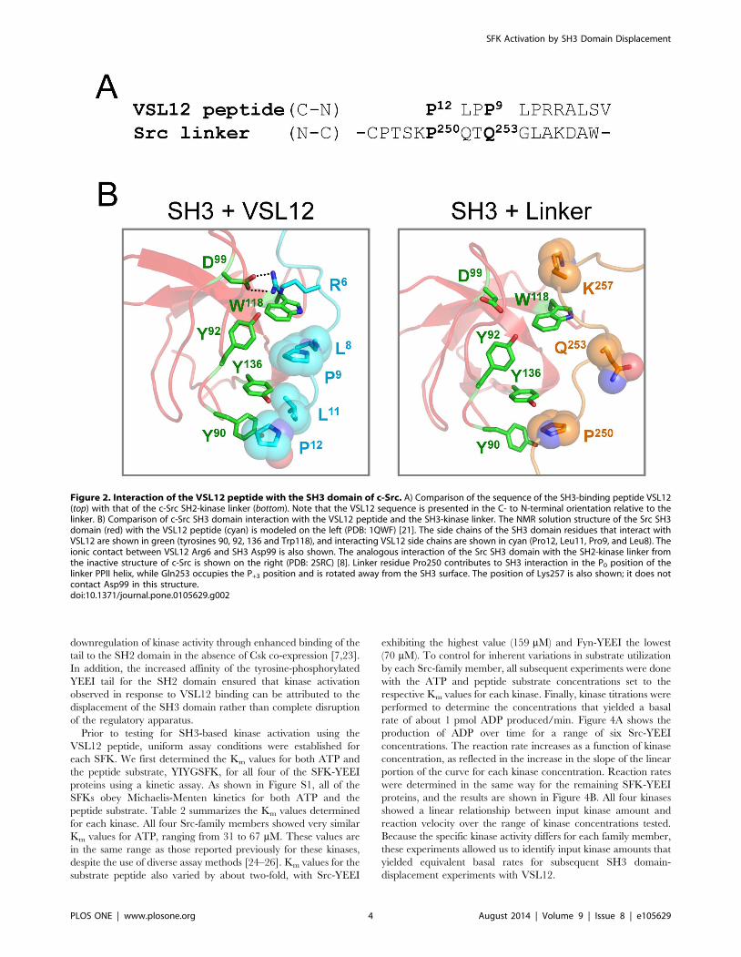
The amino acid sequence of VSL12 is aligned with the SH2-
kinase linker of c-Src in Figure 2A, and structural models of the c-
Src SH3 domain bound to VSL12 vs. its own SH2-kinase linker
are presented in Figure 2B. Comparison of these structures reveals
that VSL12 forms a much more extensive interface with the SH3
domain compared to the natural linker. Proline residues at VSL12
positions 12 and 9, together with adjacent leucine residues (Leu11
and Leu8), occupy grooves formed by conserved SH3 domain
residues Tyr90, Tyr92, Trp118, and Tyr136 (see Figure 1B for
SH3 domain alignment). In addition, VSL12 has an Arg residue in
position 6, which forms a salt bridge with Asp99 in the Src SH3
domain; aspartate is found at this position in all SFK SH3 domains
as well (Figure 1B). By comparison, the c-Src SH2-kinase linker
forms fewer contacts with the SH3 domain in the context of the
overall downregulated structure of c-Src. While the linker does
form a PPII helix, only one proline is present in the sequence
(Pro250) and packs between SH3 Tyr90 and Tyr 136 in a similar
position as Pro12 in the SH3:VSL12 peptide structure (Figure 2B).
However, Gln253 occupies the position normally occupied by
proline in high-affinity SH3 ligands (Pro9 in VSL12). The long
polar side chain of Gln253 is too bulky to fit in the pocket formed
by SH3 Tyr92, Trp118, and Tyr136, and points away from the
SH3 domain. While a basic residue (Lys257) is present near the C-
terminal end of the linker SH3-binding sequence, it does not form
a salt bridge with the conserved aspartate (Asp99) in the SH3
domain. These observations suggest that the linker sequence
represents a suboptimal SH3 ligand, and that VSL12 may be able
to compete with the linker for SH3 binding.
The VSL12 peptide binds SFK SH3 domains with similaraffinity and competes with the linker for near-full-lengthkinase binding
We first measured the affinity of the isolated SFK SH3 domains
for the VSL12 peptide using surface plasmon resonance (SPR).
For these experiments, VSL12 was immobilized on a biosensor
chip and the recombinant SH3 domains were flowed past the
peptide as described under Materials and Methods. Surface
densities were kept low to ensure that binding conformed to a 1:1
Langmuir interaction. As shown in Figure 3, all four SH3 domains
bound to VSL12 in a concentration-dependent manner with rapid
on-rates, and showed complete dissociation from the peptide
following washout. Table 1 summarizes the resulting association
(ka) and dissociation (kd) rate constants as well as the equilibrium
dissociation constants derived from them (KD). All four SH3
domains bound to VSL12 with low micromolar affinities,
consistent with previously published values for the c-Src and
Hck SH3 domains [20,22]. As negative controls, we also
performed SPR experiments with the recombinant SH2 domains
of all four kinases as well as the kinase domains of c-Src and Hck.
As expected, no binding was detected, demonstrating that the
VSL12 peptide interacts exclusively with the SH3 domain of each
kinase (Table 1).
Characterization of recombinant SFK-YEEI protein kinaseactivity in vitro
Characterization of the sensitivity of individual SFKs to SH3-
based activation required expression and purification of each Src-
family member in the downregulated conformation. Therefore, we
used purified recombinant SFK proteins in which the natural C-
terminal tails were replaced with the sequence pTyr-Glu-Glu-Ile-
Pro (referred to hereafter as SFK-YEEI proteins). This tail
modification creates an optimal SH2-binding sequence and has
been used in previous structural and enzymatic studies to ensure
SFK Activation by SH3 Domain Displacement
PLOS ONE | www.plosone.org 3 August 2014 | Volume 9 | Issue 8 | e105629
downregulation of kinase activity through enhanced binding of the
tail to the SH2 domain in the absence of Csk co-expression [7,23].
In addition, the increased affinity of the tyrosine-phosphorylated
YEEI tail for the SH2 domain ensured that kinase activation
observed in response to VSL12 binding can be attributed to the
displacement of the SH3 domain rather than complete disruption
of the regulatory apparatus.
Prior to testing for SH3-based kinase activation using the
VSL12 peptide, uniform assay conditions were established for
each SFK. We first determined the Km values for both ATP and
the peptide substrate, YIYGSFK, for all four of the SFK-YEEI
proteins using a kinetic assay. As shown in Figure S1, all of the
SFKs obey Michaelis-Menten kinetics for both ATP and the
peptide substrate. Table 2 summarizes the Km values determined
for each kinase. All four Src-family members showed very similar
Km values for ATP, ranging from 31 to 67 mM. These values are
in the same range as those reported previously for these kinases,
despite the use of diverse assay methods [24–26]. Km values for the
substrate peptide also varied by about two-fold, with Src-YEEI
exhibiting the highest value (159 mM) and Fyn-YEEI the lowest
(70 mM). To control for inherent variations in substrate utilization
by each Src-family member, all subsequent experiments were done
with the ATP and peptide substrate concentrations set to the
respective Km values for each kinase. Finally, kinase titrations were
performed to determine the concentrations that yielded a basal
rate of about 1 pmol ADP produced/min. Figure 4A shows the
production of ADP over time for a range of six Src-YEEI
concentrations. The reaction rate increases as a function of kinase
concentration, as reflected in the increase in the slope of the linear
portion of the curve for each kinase concentration. Reaction rates
were determined in the same way for the remaining SFK-YEEI
proteins, and the results are shown in Figure 4B. All four kinases
showed a linear relationship between input kinase amount and
reaction velocity over the range of kinase concentrations tested.
Because the specific kinase activity differs for each family member,
these experiments allowed us to identify input kinase amounts that
yielded equivalent basal rates for subsequent SH3 domain-
displacement experiments with VSL12.
Figure 2. Interaction of the VSL12 peptide with the SH3 domain of c-Src. A) Comparison of the sequence of the SH3-binding peptide VSL12(top) with that of the c-Src SH2-kinase linker (bottom). Note that the VSL12 sequence is presented in the C- to N-terminal orientation relative to thelinker. B) Comparison of c-Src SH3 domain interaction with the VSL12 peptide and the SH3-kinase linker. The NMR solution structure of the Src SH3domain (red) with the VSL12 peptide (cyan) is modeled on the left (PDB: 1QWF) [21]. The side chains of the SH3 domain residues that interact withVSL12 are shown in green (tyrosines 90, 92, 136 and Trp118), and interacting VSL12 side chains are shown in cyan (Pro12, Leu11, Pro9, and Leu8). Theionic contact between VSL12 Arg6 and SH3 Asp99 is also shown. The analogous interaction of the Src SH3 domain with the SH2-kinase linker fromthe inactive structure of c-Src is shown on the right (PDB: 2SRC) [8]. Linker residue Pro250 contributes to SH3 interaction in the P0 position of thelinker PPII helix, while Gln253 occupies the P+3 position and is rotated away from the SH3 surface. The position of Lys257 is also shown; it does notcontact Asp99 in this structure.doi:10.1371/journal.pone.0105629.g002
SFK Activation by SH3 Domain Displacement
PLOS ONE | www.plosone.org 4 August 2014 | Volume 9 | Issue 8 | e105629

Figure 3. Surface plasmon resonance (SPR) analysis of SH3 interactions with the VSL12 peptide. SPR was used to evaluate VSL12peptide binding kinetics and affinity for the isolated SH3 domains of c-Src, Hck, Fyn and Lyn as indicated. The biotinylated peptide was immobilizedto 80 Response Units (RU) on the surface of a streptavidin (SA) biosensor chip. The recombinant purified Src SH3 domain proteins were flowed pastthe peptide over the concentration ranges shown. Association was measured for 180 s, followed by a 300 s dissociation phase. Each panel shows arepresentative sensorgram, with the double-referenced binding data (black traces) fit to a 1:1 Langmuir binding model (red trace). Kinetic constantsderived from this experiment are presented in Table 1.doi:10.1371/journal.pone.0105629.g003
Table 1. Binding Affinities of SFK SH3 Domains for the VSL12 Peptide as Measured by SPR.
Protein ka (M21s21±SE) kd (s21±SE) Kinetic KD (M) Kinetic x2Steady-State KD
(M)Steady Statex2
Src SH3a 1.21610560.036105 4.146102160.08 61021 3.4261026 0.570 4.0061026 0.260
Fyn SH3b 1.06610660.036106 3.726102160.1061021 3.5161027 1.890 3.7061027 1.070
Hck SH3c 7.88610460.366104 2.176102160.0461021 2.7661026 1.360 2.9961026 0.390
Lyn SH3b 2.12610560.106105 1.376102160.0461021 6.4761027 1.850 5.6261027 0.225
SH2d ND ND - - - -
Kinased ND ND - - - -
Analyses were performed with biotinylated VSL12 peptide bound to a streptavidin biosensor chip as described under Materials and Methods. Each protein was flowedpast the chip surface over the concentration ranges indicated in the footnotes. Duplicate runs were performed for each concentration. A control cycle of buffer only wassubtracted from all concentrations of reference-subtracted curves. To calculate the kinetic KD, interaction data were curve-fit using a 1:1 Langmuir model, with bindingconstants and chi-squared values calculated using the BIAevaluation software. To calculate the steady state KD, the analyte response at equilibrium was plotted againstthe analyte concentration, and resulting curves were fit with the steady state model in the BIAevaluation software.a31.25, 62.5, 125, 250, 500, 1000 nM.b31.25, 62.5, 125, 250 nM.c31.25, 62.5, 125, 250, 500 nM.dSH2 domains tested: Src, Hck, Fyn and Lyn; kinase domains tested, Src and Hck. ND, binding not detected with 1 mM protein input.doi:10.1371/journal.pone.0105629.t001
SFK Activation by SH3 Domain Displacement
PLOS ONE | www.plosone.org 5 August 2014 | Volume 9 | Issue 8 | e105629

Figure 4. Linear relationship between SFK-YEEI activity and kinase protein input. A) Representative time course of ADP production for sixconcentrations of Src-YEEI. At higher kinase concentrations, the reaction rates plateau as the fluorescence reading reaches saturation. The linearportion of each curve was fit by linear regression analysis to provide the slope, which corresponds to the rate of the reaction in pmol ADP produced/min. B) Reaction rates for each SFK-YEEI protein are plotted against input kinase concentration. Curves were best-fit by linear regression analysis(dotted lines) and used to estimate the specific activity for each kinase (inset).doi:10.1371/journal.pone.0105629.g004
SFK Activation by SH3 Domain Displacement
PLOS ONE | www.plosone.org 6 August 2014 | Volume 9 | Issue 8 | e105629

Differential sensitivity of Src-family members toactivation by VSL12 peptide binding
We next tested the susceptibility of each Src-family member to
SH3-based activation using the VSL12 peptide as an SH3 domain
‘agonist’. Using the established conditions for substrate, ATP, and
kinase concentration as described above, the VSL12 peptide was
titrated into the assay over a concentration range of 0.1 to
300 mM. Figure 5A shows the reaction velocity for each kinase as
a function of VSL12 peptide concentration. These data show that
all four Src-family members are susceptible to activation by the
VSL12 peptide, providing strong evidence that SH3:linker
interaction has an important role in kinase regulation across the
entire Src family.
Data presented in Figure 5A demonstrate that the extent of
kinase activation is saturable at higher concentrations of the
VSL12 peptide. These data were therefore fit to Equation 1 (see
Materials and Methods) to determine the Vmax for each kinase
reaction as a measure of the highest level of SH3-dependent
activation attainable for each kinase. Figure 5B compares the
basal reaction rate to the Vmax for each kinase in the presence of
VSL12. This analysis revealed that Src-family kinases have
remarkably different responses to SH3-based kinase activation.
Lyn-YEEI showed the smallest response, with a Vmax of about
5 pmol ADP produced/min, while Hck-YEEI showed a much
higher response, approaching 30 pmol ADP produced/min. Fyn-
YEEI and Src-YEEI exhibited intermediate responses with values
of 11 and 16 pmol ADP produced/min, respectively. These results
support the idea that individual Src-family members have very
different inherent enzymatic capacities, at least in response to SH3
domain-based activation.
In addition to defining the extent to which each kinase can be
activated by VSL12 (Vmax), we also calculated an activation
constant (Kact) for each kinase in response to this SH3-binding
peptide. Kact defines the concentration of VSL12 required to
increase the reaction rate to one-half of the Vmax, and is calculated
by subtracting the basal kinase activity (without VSL12) from the
reaction rate observed at each VSL12 concentration and
performing nonlinear regression analysis using Equation 2 (see
Materials and Methods). As shown in Table 3, the Kact values for
VSL12-based activation of Src-YEEI, Fyn-YEEI, and Hck-YEEI
were all remarkably similar (22–25 mM). In contrast, Lyn-YEEI
displayed a lower Kact compared to the other kinases (4 mM),
which may suggest higher sensitivity to activation by SH3 domain
displacement, although the maximum response observed was the
lowest among the four kinases tested. Interestingly, Lyn and Hck
are the most closely related among the SFKs tested in terms of
phylogeny, yet their sensitivity to VSL12-induced activation is the
most disparate.
Hck and c-Src remain susceptible to activation by SH3domain displacement after autophosphorylation
In addition to regulatory domain engagement, SFK activity is
also regulated by the phosphorylation state of the kinase domain
activation loop (Figure 1A). However, whether or not activation
loop phosphorylation results in maximal kinase activation or
whether the autophosphorylated kinase is still susceptible to
further activation by SH3 domain displacement is not known. To
explore this question, we focused on c-Src and Hck, because these
are the only two family members for which crystal structures have
been solved for the downregulated states [7–10]. Before examining
the impact of VSL12 binding on SFK activity as a function of
autophosphorylation, we first confirmed that the activation loops
of recombinant Hck-YEEI and Src-YEEI were not phosphorylat-
ed prior to addition to the assay. To do this, the recombinant
purified kinases were digested with pepsin and the phosphorylation
states of the activation loop and tail tyrosines were determined by
mass spectrometry. As shown in Figure S2, the C-terminal tail
tyrosines are phosphorylated in both Src-YEEI and Hck-YEEI as
expected for the inactive conformations, while no activation loop
tyrosine phosphorylation was detected in either case. No unpho-
sphorylated tail peptide was detected for either Src-YEEI or Hck-
YEEI.
To test whether Src-YEEI and Hck-YEEI can be activated
further by SH3 domain displacement after activation loop
autophosphorylation, both kinases were preincubated in the
presence or absence of ATP prior to testing for activation by
VSL12. Time-course experiments revealed that kinase autophos-
phorylation reached a plateau after three hours of incubation with
ATP (data not shown), so preincubation was conducted for this
time period prior to assessment of the VSL12-induced response.
The preincubated samples were subsequently analyzed by pepsin
digestion and mass spectrometry to confirm phosphorylation of the
activation loop (Figure S3). Activation loop phosphorylation of
both Src-YEEI and Hck-YEEI was confirmed while the corre-
sponding unphosphorylated peptides were not observed.
We observed a dramatic increase in the basal rate of Src-YEEI
and Hck-YEEI kinase activity after pre-incubation with ATP. For
Src-YEEI, the basal rate increased from 1.74 pmol to 6.38 ADP
produced/min following ATP preincubation, while Hck-YEEI
activity increased from 1.27 to 9.04 pmol ADP produced/min.
This increase is presumably due to stabilization of the active site
conformation as a result of autophosphorylation of the activation
loop tyrosine. Note that the basal rate of each kinase following
preincubation in the absence of ATP is consistent with the basal
rates shown in Figure 5B and Table 3, indicating that the three
hour preincubation period did not compromise kinase activity.
The effects of SH3 domain displacement by VSL12 on Src-
YEEI and Hck-YEEI activity as a function of autophosphorylation
Table 2. Km Values for ATP and Peptide Substrate for Near-Full-Length SFKs.
Kinase ATP (mM) YIYGSFK (mM)
Src-YEEI 67.0610.3 159.1620.9
Fyn-YEEI 49.768.2 69.663.7
Hck-YEEI 55.8618.5 83.8612.5
Lyn-YEEI 31.169.70 83.264.9
The Km values for ATP and the substrate peptide, YIYGSFK, were determined for each SFK-YEEI protein using the ADP Quest assay as described under Materials andMethods. ATP experiments were performed three times for each kinase and substrate experiments were performed four times for each kinase, except for Src-YEEI,where ATP experiments were performed twice and substrate experiments were performed three times. Mean values are shown for each kinetic constant 6S.E.doi:10.1371/journal.pone.0105629.t002
SFK Activation by SH3 Domain Displacement
PLOS ONE | www.plosone.org 7 August 2014 | Volume 9 | Issue 8 | e105629

are shown in Figure 6A and 6B, respectively. In each case, the
basal rate observed in the absence of VSL12 was subtracted from
the rate at each peptide concentration to reveal additional
activation resulting from VSL12 binding to the SH3 domain.
Autophosphorylated Src-YEEI and Hck-YEEI were both activat-
ed by VSL12 in a concentration-dependent manner, demonstrat-
ing that phosphorylation of the activation loop does not uncouple
the kinase domain from the regulatory influence of the SH3
domain. However, we observed remarkable differences in the
responses of Src-YEEI vs. Hck-YEEI to VSL12 following
Figure 5. Differential sensitivity of individual SFK-YEEI proteins to activation by SH3 domain displacement. Each of the SFK-YEEIproteins shown was assayed in the presence of VSL12 over a range of concentrations (0.1 to 300 mM). ATP and substrate concentrations were set tothe Km for each kinase, and input kinase concentrations were set to achieve a basal reaction velocity of 1 pmol ADP produced/min. A) Each of thekinases is activated by VSL12 in a concentration-dependent manner. Plots of reaction velocity vs. VSL12 concentration were best-fit by the Michaelis-Menten equation, allowing for the determination of the Vmax. Each data point was assayed in triplicate and is shown as the mean 6S.E. B)Comparison of basal rate (left) and Vmax (right) for each kinase in the presence of VSL12. Bars heights correspond to the mean values from triplicateexperiments 6S.E.doi:10.1371/journal.pone.0105629.g005
SFK Activation by SH3 Domain Displacement
PLOS ONE | www.plosone.org 8 August 2014 | Volume 9 | Issue 8 | e105629

preincubation with ATP. For Src-YEEI, preincubation with ATP
enhanced the response to VSL12, indicating that autophosphory-
lation of Src-YEEI sensitizes the kinase domain to further
activation by SH3 domain displacement. In contrast, the curves
for Hck-YEEI activation by VSL12 were identical, whether the
kinase was preincubated with ATP or not. This observation
suggests that activation of Hck-YEEI by SH3 domain displace-
ment is independent of activation loop phosphorylation. Alterna-
tively, Hck-YEEI may autophosphorylate more rapidly than Src-
YEEI, such that Hck-YEEI reaches maximum activation loop
phosphorylation without ATP pre-incubation. To examine this
possibility, we measured the effect of VSL12 on the rates of Hck-
YEEI and Src-YEEI autophosphorylation by repeating the
VSL12-activation reactions in the absence of the peptide substrate.
Figure 6C shows that Hck-YEEI undergoes autophosphorylation
more rapidly than Src-YEEI as a function of VSL12 input. This
finding suggests that the autophosphorylation-dependent respons-
es to VSL12 observed between Src-YEEI and Hck-YEEI are the
result of inherent differences in autophosphorylation rates between
these two Src family members. These differences also suggest that
c-Src and Hck may have evolved to respond to different types of
cellular inputs for activation that have important implications for
signaling (see Discussion).
Activation by VSL12 requires an intact SH3:linkerinterface in the downregulated kinases
Data presented in the previous sections suggest that the VSL12
peptide selectively interacts with downregulated SFKs through
their SH3 domains, resulting in disruption of intramolecular
SH3:linker interaction and subsequent kinase activation. Such a
model predicts that SFK linker mutants should show higher basal
kinase activity, and also be refractory to further activation by
VSL12 binding. To test this idea, we created mutants of Hck-
YEEI and Src-YEEI in which linker residues involved in SH3
binding and kinase downregulation are replaced with alanines
(Figure 7A). Both mutants displayed higher basal kinase activity
than their wild-type counterparts in the absence of the VSL12
peptide, consistent with loss of SH3 interaction with the modified
linker (Figure 7B). Addition of VSL12 did not increase the activity
of these mutants further (Figure 7C), consistent with the notion
that VSL12 activates the wild-type kinases by disrupting
SH3:linker interaction.
Discussion
The mechanisms responsible for SFK downregulation have
been established by extensive biochemical and structural studies
and appear to be conserved among all family members (see
Introduction). The precise mechanisms driving kinase activation,
and whether or not they are also conserved across this kinase
family, are less clear. Displacement of intramolecular regulatory
interactions involving the SH2 and/or SH3 domains can cause
kinase activation, suggesting that individual family members may
be finely tuned to respond to inputs through their regulatory
domains in specific ways. In this study, we provide direct evidence
in support of this idea. Using a peptide ligand (VSL12) to induce
kinase activation by SH3 domain displacement, we showed a wide
range of sensitivities to this allosteric activation mechanism across
the Src kinase family. Furthermore, we observed that c-Src
becomes more susceptible to activation by SH3 domain displace-
ment after autophosphorylation of the activation loop tyrosine. In
contrast, activation of Hck through its SH3 domain appears to be
independent of kinase domain autophosphorylation, revealing a
very different response to this activating input despite the
remarkable similarity to c-Src in terms of the structure of the
inactive state [7–10].
We chose the VSL12 peptide as the SH3 ligand for these studies
because previous work showed that it binds to the c-Src, Fyn, and
Lyn SH3 domains with similar micromolar affinities [20,27]. SPR
results presented here show that VSL12 binds to the SH3 domains
of these three SFKs as well as that of Hck with similar dissociation
constants, providing a useful peptide ligand to compare the effect
of SH3 domain displacement on kinase activity across the kinase
family. The solution structure of the Src SH3 domain in complex
with VSL12 reveals the key SH3 residues that interact with this
peptide and suggests a mechanism for linker displacement [21].
The hydrophobic pocket formed by SH3 Tyr90 and Tyr136 is
occupied by the lone proline residue in the Src SH2-kinase linker
(Pro250; Figure 2B). This interaction is likely displaced by Leu11
and Pro12 in the VSL12 peptide, as these residues occupy the
same pocket in the NMR structure. In addition, VSL12 Leu8 and
Pro9 both make contacts with the SH3 domain, while the
analogous positions are occupied by Thr252 and Gln253 in the
linker; Gln253 does not contact the SH3 domain in the inactive c-
Src structure (Figure 2B). Furthermore, VSL12 Arg6 forms a
stabilizing ionic contact with conserved SH3 Asp99, and also
makes a hydrogen bond with the side chain of Trp118. This
interaction is missing in the SH3:linker interface in downregulated
c-Src. The SH3 domain residues involved in VSL12 binding are
conserved across the four SFKs studied here, consistent with the
similarities in binding kinetics and affinities observed by SPR
analysis (Table 1).
Work presented here supports the idea that Src-family kinases
are uniformly susceptible to activation by SH3 domain displace-
ment. In addition, VSL12-based activation of recombinant SFK
proteins with YEEI tails suggests that disruption of SH2:tail
interaction is not required for SH3-based activation, as the pYEEI
sequence has a much higher affinity for the SH2 domain than the
Table 3. Basal Rate, Maximum Velocity, and Activation Constants for VSL12 with each SFK.
Kinase Basal reaction velocity (pmol ADP produced/min) Vmax (pmol ADP produced/min) Kact (mM)
Src-YEEI 1.2760.08 15.7760.67 22.4262.23
Fyn-YEEI 1.2260.10 11.4160.55 24.9561.91
Hck-YEEI 1.9760.14 27.9461.02 22.3862.18
Lyn-YEEI 0.9360.20 5.0060.24 4.0360.61
The basal reaction velocity, maximum velocity (Vmax), and activation constant (Kact) were determined for each kinase in the ADP Quest assay as described underMaterials and Methods. Basal velocity is the rate of kinase activation in the absence of the VSL12 peptide. Kinetic constants were determined in triplicate and arepresented as the mean 6S.E.doi:10.1371/journal.pone.0105629.t003
SFK Activation by SH3 Domain Displacement
PLOS ONE | www.plosone.org 9 August 2014 | Volume 9 | Issue 8 | e105629

Figure 6. Src-YEEI and Hck-YEEI retain sensitivity to activation by SH3 domain displacement after autophosphorylation. Src-YEEI (A)and Hck-YEEI (B) were incubated in the absence or presence of ATP (at the Km) for 3 hours at 25uC to induce autophosphorylation of Tyr416 in theactivation loop. The autophosphorylated kinases were then assayed for responsiveness to the SH3-binding peptide, VSL12. C) Autophosphorylation
SFK Activation by SH3 Domain Displacement
PLOS ONE | www.plosone.org 10 August 2014 | Volume 9 | Issue 8 | e105629

natural tail sequence [7,28]. While the VSL12 peptide bound to
and activated all four of the kinases tested, differences in the extent
of activation were striking. Particularly surprising was the
difference in sensitivity to SH3-based activation between Hck
and Lyn, as they are the most similar in amino acid sequence
among the entire Src kinase family. Our observation that Hck and
Lyn demonstrate very different responses to SH3-based activation
by VSL12 is consistent with previous reports with the Nef protein
of HIV-1. Like VSL12, Nef binds to the SH3 domains of Hck and
Lyn with similar affinity, yet Nef activates Hck more strongly both
in vitro and in cell-based assays [29,30]. One possibility is that
maximal activation of Lyn may also require displacement of the C-
terminal tail from the SH2 domain, which does not appear to be
the case for Hck.
To understand how interaction with VSL12 induces kinase
activation, the structures of Src-family kinases in the active vs.
inactive conformations must be considered. In the downregulated
structure of c-Src, the SH3 domain contacts both the SH2-kinase
linker (as described above) and Arg318 in the N-lobe of the kinase
domain (through SH3 Asp117). Another key residue known to
couple the SH3-SH2 region to the kinase domain is Trp260 at the
C-terminal end of the linker [31]. This conserved residue packs
against helix C, helping to position it away from the active site and
preventing formation of a salt bridge between Glu310 and Lys295
that is required for kinase function. Because VSL12 has a relatively
high affinity for the SH3 domains of the SFKs, it is likely to bind to
the SH3 domain, displace the linker and shift Trp260 away from
the C helix. The stabilizing interaction between SH3 domain
Asp117 and N-lobe Arg318 may also be disrupted as a
consequence. Removal of these inhibitory constraints would allow
the C helix to rotate inward, with Glu310 forming the critical salt
bridge with Lys295. This rearrangement also exposes activation
loop Tyr416 for autophosphorylation. Further support for this
linker displacement mechanism in the activation of SFKs by
VSL12 comes from studies of linker mutants. Alanine substitution
of the key residues in the linker that pack against the SH3 domain
in the downregulated structure results in constitutive activation of
both Src-YEEI and Hck-YEEI. As a result, both kinases are
refractory to further activation by VSL12.
Another remarkable difference between the SFKs relates to the
coupling of activation loop autophosphorylation and sensitivity to
SH3 domain displacement. Autophosphorylation of Tyr416
strongly stimulated both Hck and c-Src kinase activity in the
absence of the VSL12 peptide. In addition, both kinases were
further activated by VSL12 in a concentration-dependent manner,
demonstrating that activation loop phosphorylation enhances
kinase activity independently of regulatory domain displacement.
This observation suggests that Tyr416 phosphorylation alone is
sufficient to reorganize the active site for phosphotransfer.
However, our data show that autophosphorylation alone does
not maximally increase kinase activity, because addition of VSL12
enhanced both c-Src and Hck kinase activity to an even greater
extent. The additional impact of this SH3 domain ligand on
autophosphorylated SFKs implies that displacement of the SH3
domain relieves additional allosteric constraints on the kinase
domain, possibly mediated through Trp260 or direct SH3
interaction with the N-lobe as described above. Furthermore, this
observation implies that autophosphorylation of the activation
loop does not cause release of the SH3 domain from the linker, at
least for c-Src and Hck.
Our finding that autophosphorylated c-Src and Hck are still
under the allosteric control of the SH3 domain is consistent with
recent studies of c-Abl kinases. Like SFKs, the c-Abl kinase ‘core’
consists of a similar arrangement of SH3 and SH2 domains, an
SH2-kinase linker, followed by the kinase domain. In this system,
intramolecular interaction of the SH3 domain with the linker is
also essential for downregulation of Abl kinase activity [32].
Interestingly, active mutants of c-Abl, as well as the constitutively
active Bcr-Abl fusion protein associated with chronic myelogenous
leukemia, are both inhibited by linker proline substitutions that
enhance interaction with the SH3 domain [33]. Together with our
data, these observations support the idea that activation loop
phosphorylation and regulatory domain displacement represent
independent modes of SFK and Abl regulation.
In summary, observations presented here suggest distinct
regulatory controls for individual Src-family members, despite
the fact that they are composed of the same component parts.
These differences may have evolved to meet specific needs for
kinase regulation under distinct physiological conditions. For
example, c-Src is locally active in focal adhesions, where it
regulates their turnover during cell adhesion and migration. One
important mechanism of c-Src activation in focal contacts involves
direct interaction with FAK. In this case, the SH3 and SH2
domains of c-Src interact with FAK, integrating the localization of
c-Src to focal adhesions with kinase activation via regulatory
domain displacement [13]. This interaction juxtaposes the c-Src
and FAK kinase domains, raising the possibility of direct
phosphorylation of the c-Src activation loop by FAK [34].
Maximal c-Src activation in focal adhesions may therefore require
displacement of both regulatory domains plus trans-phosphoryla-
tion by another kinase, consistent with our findings. In contrast,
Hck is expressed primarily in cells of innate immunity and is
activated by diverse upstream signals including hematopoietic
cytokines and Fc receptors [35–38]. In this case, receptor
engagement with Hck through its SH3 and/or SH2 domains
may be sufficient to induce a rapid and transient response, without
the need for secondary phosphorylation on the activation loop.
These intrinsic differences in the sensitivity of c-Src, Hck and other
SFKs to activating signals may provide opportunities for discovery
of selective small molecule probes of their functions.
Materials and Methods
Cloning, expression, and purification of recombinant SFKdomains
The coding sequences for the isolated SH3 and SH2 domains of
human c-Src, Fyn, Hck, and Lyn, and the isolated kinase domains
(KD) of c-Src and Hck(corresponding to residues 81–142 (SH3),
143–244 (SH2), and 251–533 (KD) of c-Src, numbering according
to PDB ID: 2SRC) were amplified by PCR and subcloned into the
bacterial expression vector pET21a (EMD Millipore) using NdeI
and XhoI restriction sites. The C-terminus of each construct was
modified by the addition of the sequence LPHHHHHH as
encoded by the XhoI site and the His6 tag in the vector. For
expression of the SH3 and SH2 domains, E. coli strain Rosetta
2(DE3)pLysS (EMD Millipore) was transformed with each plasmid
of Src-YEEI and Hck-YEEI was measured in response to the VSL12 peptide by running the kinase reactions in the absence of the substrate peptide. Forall three panels, the basal reaction velocity (no VSL12) was subtracted from the rate at each VSL12 concentration, and the resulting linear reactionvelocities were plotted as a function of VSL12 concentration as shown. Each data point was assayed in triplicate and the average value is plotted6S.E.doi:10.1371/journal.pone.0105629.g006
SFK Activation by SH3 Domain Displacement
PLOS ONE | www.plosone.org 11 August 2014 | Volume 9 | Issue 8 | e105629

Figure 7. SFK linker mutants display higher basal kinase activity than their wild-type counterparts and are refractory to activationby VSL12. A) Sequences of the wild-type (WT) linkers of Src and Hck are shown. Residues involved in intramolecular engagement of the SH3 domainare highlighted in bold, and are replaced with alanines in the respective Src-3A and Hck-2A mutants as shown. B) Reaction velocities for equivalentamounts (125 ng/well) of Src-YEEI and Hck-YEEI with wild-type vs. mutant linkers were determined using the ADP Quest assay. Results are shown asthe mean velocity for three replicate determinations 6S.E. C) Each of the SFK-YEEI proteins shown was assayed in the presence of VSL12 over a rangeof concentrations (0 to 300 mM). ATP and substrate concentrations were set to the Km for each wild-type kinase, and input kinase concentrations wereset to achieve a basal reaction velocity of 1 pmol ADP produced/min. Plots of reaction velocity vs. VSL12 concentration were best-fit by the Michaelis-Menten equation for the wild-type kinases, indicative of saturable activation kinetics by VSL12. Each data point was assayed in triplicate and is plottedas the mean velocity 6S.E.doi:10.1371/journal.pone.0105629.g007
SFK Activation by SH3 Domain Displacement
PLOS ONE | www.plosone.org 12 August 2014 | Volume 9 | Issue 8 | e105629

and cultures were grown at 37uC until the OD600 reached 0.8–1.0.
Protein expression was induced with 0.4 mM isopropyl -D-
thiogalactopyranoside (IPTG) for 4 h at 25uC. The isolated KDs
were expressed as previously described by Seeliger, et al. [39] For
purification, cell pellets were resuspended in ion-exchange start
buffer (20 mM Tris-HCl, pH 8.5, 5 mM -mercaptoethanol, 10%
glycerol), sonicated, and clarified by centrifugation. Soluble
domain proteins were purified from the clarified lysates by
anion-exchange (HiPrep Q FF; GE Healthcare), immobilized
metal ion affinity (HiTrap Chelating HP; GE Healthcare), and
size-exclusion chromatography (HiLoad 26/60 Superdex 75; GE
Healthcare). Purified proteins were buffer-exchanged with HBS-
EP (10 mM HEPES, pH 7.4, 150 mM NaCl, 3 mM EDTA,
0.05% v/v P20 surfactant) using Amicon Ultra-15 Centrifugal
Filter Units with Ultracel-10 Membranes (EMD Millipore) for
storage and surface plasmon resonance experiments. Recombinant
purified protein concentrations were quantified by Bradford assay
using the BioRad Protein Assay Dye Reagent Concentrate and
purified BSA as a reference standard (Pierce).
Expression and purification of recombinant SFK-YEEIproteins
Human Hck, Lyn, Fyn and c-Src clones were modified on their
C-terminal tails to encode the sequence Tyr-Glu-Glu-Ile-Pro
(YEEI variants) to enable autophosphorylation of the tail and
high-yield purification in the down-regulated state without co-
expression of Csk [7,28]. In addition, the N-terminal unique
domain of each kinase was replaced with a hexahistidine tag. The
linker mutant proteins Src-3A-YEEI and Hck-2A-YEEI were
produced using the QuikChange II XL Site-Directed Mutagenesis
Kit (Agilent Technologies) using the Src-YEEI and Hck-YEEI
cDNA clones, respectively, as templates. Each construct was used
to produce a recombinant baculovirus in Sf9 insect cells using
BaculoGold DNA and the manufacturer’s protocol (BD Pharmin-
gen) as previously described [23]. Src-YEEI, Src-3A-YEEI, and
Hck-2A-YEEI were co-expressed with the Yersinia pestis YopH
phosphatase to promote dephosphorylation of the activation loop
tyrosine to help downregulate kinase activity [39,40]. For these
proteins, Sf9 cells were grown in a monolayer and infected with
SFK-YEEI and YopH baculoviruses. Cells were harvested 72 h
after infection for SFK-YEEI purification. All kinases were
purified as previously described [23]. Purified proteins were stored
in 20 mM Tris-HCl, pH 8.3, containing 100 mM NaCl. SFK-
YEEI protein concentrations were determined by running
dilutions of each recombinant protein on an SDS-PAGE gel
together with dilutions of a BSA protein standard. Each gel was
stained with Coomassie blue and scanned using a LICOR
Odyssey system for densitometric determination of protein
concentration.
Surface Plasmon Resonance (SPR)SPR experiments were performed using a Biacore 3000
Instrument (GE Healthcare) on a streptavidin (SA) biosensor chip.
The high-affinity Src SH3-binding peptide VSLARRPLPPLP [20]
was synthesized and biotinylated by the University of Pittsburgh
Peptide Synthesis Core. The peptide was solubilized in HBS-EP
buffer to a concentration of 5 nM, injected onto the SA surface at
10 l/min and immobilized to a level of 80 Response Units (RU). A
biotinylated IB kinase substrate peptide, RHDSGLDSMKD
(Enzo Life Sciences), was immobilized to 80 RU on the reference
flow cells as a control for nonspecific binding of analytes to the
peptide-SA surface. SFK SH3, SH2 and kinase domain proteins
were injected in duplicate over a range of concentrations at 25uCat a flow rate of 30 l/min. Association was measured for 180 s,
followed by 300 s of dissociation in HBS-EP running buffer. The
chip surface was regenerated with HBS-EP buffer after each run.
In addition to reference flow cell subtraction, HBS-EP buffer-only
cycles were used to allow double referencing for all analyses.
Binding curves were fit using a 1:1 Langmuir binding model and
the BIAevaluation software v. 4.1.1 (GE Healthcare) which was
used to generate all kinetic data. To determine steady state KD
values, equilibrium responses were plotted against analyte
concentrations, and the resulting curves were fit with the steady
state model in the BIAevaluation software.
Kinetic kinase assaysKinetic kinase assays were performed using the ADP Quest
Assay (DiscoveRx), which fluorimetrically monitors the production
of ADP [41]. Briefly, the conversion of ATP to ADP is coupled to
the production of pyruvate from phosphoenolpyruvate (PEP) by
pyruvate kinase (PK). Pyruvate is converted to hydrogen peroxide
by pyruvate oxidase, which in turn oxidizes Amplex Red to the
fluorescent product, resorufin. Accumulation of ADP is measured
as the increase in resorufin fluorescence at excitation and emission
wavelengths of 530 nm and 590 nm, respectively. All assays were
performed in quadruplicate in black 384-well microplates (Corn-
ing Catalog # 3571). ATP stocks (10 mM; Sigma) were prepared
in 10 mM Tris-HCl, pH 7.0. The SFK substrate peptide (5 mM;
sequence YIYGSFK; Anaspec) [42] was prepared in ADP Quest
assay buffer (15 mM HEPES, pH 7.4, 20 mM NaCl, 1 mM
EGTA, 0.02% Tween-20, 10 mM MgCl2, 0.1 mg/ml bovine -
globulins). Kinase reactions were initiated by the addition of 5 l
ATP to each well at 10 times the final concentration. The
reactions were performed in a final assay volume of 50 l/well at
25uC. Assay plates were read at 5 min intervals for 3 h on a
Molecular Devices SpectraMax M5 microplate reader.
Substrate and ATP Km determination in the ADP Questassay
To measure the Km for the YIYGSFK substrate peptide, the
ATP concentration was held constant at 200 M, the maximum
tolerated concentration in the assay. ATP is regenerated in the
assay and is therefore not depleted as a function of substrate
phosphorylation. Kinase concentrations were held constant at
150 ng/well for Src-YEEI, Fyn-YEEI, and Hck-YEEI, and
250 ng/well for Lyn-YEEI. The substrate peptide was titrated
into the assay at various concentrations ranging from 1.95 to
500 M. To measure the Km for ATP, the peptide substrate
concentration was set to the Km for each SFK, and kinase
concentrations were held constant as stated above. ATP was
titrated into the assay over a concentration range of 6.25 to
200 M. The resulting rate data were fit as described below.
ADP Quest Data AnalysisEach ADP Quest assay included controls for non-enzymatic
production of ADP and kinase autophosphorylation. Raw
fluorescence data obtained from quadruplicate wells for each
condition were averaged and corrected for the non-enzymatic rate
of ADP production and SFK autophosphorylation. Corrected raw
fluorescence units (RFU) were then plotted against time, in
minutes, to determine the rate of each reaction. Linear regression
analysis (GraphPad Prism) was performed on the linear portion of
each corrected progress curve, and the slope yielded the reaction
rate. Reaction rates were converted to pmol ADP produced/min
using the correction factor 4.2 RU/pmol ADP, which was
generated from a standard curve under identical reaction
conditions. For ATP and substrate Km experiments, plots of the
SFK Activation by SH3 Domain Displacement
PLOS ONE | www.plosone.org 13 August 2014 | Volume 9 | Issue 8 | e105629

reaction rates at each substrate or ATP concentration obeyed
Michaelis-Menten kinetics and were best-fit by nonlinear regres-
sion analysis (GraphPad Prism). Km values were determined using
the equation,
v~ Vmax S½ �= Kmz S½ �ð Þ ð1Þ
where v = the measured velocity, Vmax = the maximal reaction
velocity, [S] = the substrate (peptide or ATP) concentration, and
Km = the concentration of substrate or ATP at which the reaction
velocity is half of the maximal velocity.
SFK activation by the SH3 binding peptide in the ADPQuest Assay
To test each SFK for sensitivity to activation by VSL12, ATP
and substrate concentrations were set to their respective Km values
for each SFK. Kinase concentrations were held constant for Src-
YEEI (25 ng/well), Fyn-YEEI (30 ng/well), Hck-YEEI (80 ng/
well), and Lyn-YEEI (120 ng/well), so as to yield a basal reaction
rate of about 1 pmol ADP produced/min in each case. The
VSL12 peptide was solubilized in ADP Quest assay buffer to
10 mM. The VSL12 peptide was titrated into the assay from 0.1 to
300 M. VSL12 peptide and kinase were pre-incubated for 15 min
at 25uC before the addition of substrate, assay reagents, and ATP.
To test Src-YEEI and Hck-YEEI for sensitivity to activation by
VSL12 after autophosphorylation, each kinase was pre-incubated
in 550 l of ADP Quest assay buffer with or without ATP at the Km
for each kinase for 3 h at 25uC. Following pre-incubation,
responsiveness to VSL12 activation was assayed as described
above.
Kinase reaction rates at each VSL12 peptide concentration
obeyed Michaelis-Menten kinetics and were best-fit by nonlinear
regression. The Vmax of each kinase reaction in the presence of
VSL12 was determined using Equation 1. To calculate the
activation constant (Kact), the basal rate (rate in the absence of
VSL12) was subtracted from the rate in the presence of VSL12 at
each concentration. The transformed rates were plotted against
VSL12 concentration. The resulting curves also obeyed Michaelis-
Menten kinetics and were best-fit by nonlinear regression as per
the method of Boerner, et al., 1996 [43]. Kact was calculated from
va~ Vact L½ �= Kactz L½ �ð Þ ð2Þ
where va = the measured velocity in the presence of VSL12 minus
the velocity in the absence of VSL12, Vact = the maximal reaction
velocity in the presence of VSL12 minus the velocity in the
absence of VSL12, [L] = the VSL12 concentration, and Kact =
the concentration of VSL12 at which the reaction velocity is half of
the Vact.
Pepsin digestion and mass analysisFor the elucidation of kinase autophosphorylation sites, 50 pmol
of Src-YEEI and Hck-YEEI were digested online with pepsin in
potassium phosphate buffer (150 mM KH2PO4/150 mM
K2HPO4, pH 2.5). The resulting peptides were separated using
a Waters nanoAcquity UPLC system (Waters Corp, Milford, MA),
trapped and desalted for 3 min at 100 mL/min and then separated
in 8 min by an 8%–40% acetonitrile:water gradient at 40 mL/
min. The separation column was a 1.06100.0 mm Acquity UPLC
C18 BEH (Waters) containing 1.7 mm particles. Mass spectra were
obtained with a Waters Xevo-QTOF equipped with standard ESI
source (Waters Corp., Milford, MA, USA). Mass spectra were
acquired over an m/z range of 100 to 1900. Mass accuracy was
ensured by calibration with 100 fmol/mL Glu-fibrinopeptide, and
was less than 10 ppm throughout all experiments. Identification of
the peptic fragments was accomplished through a combination of
exact mass analysis and MSE using custom IdentityE PLGS 2.5
Software from the Waters Corporation [44]. MSE was performed
by a series of low-high collision energies ramping from 5–30 V,
therefore ensuring proper fragmentation of all the peptic peptides
eluting from the LC system.
Supporting Information
Figure S1 Recombinant near-full-length Src-family ki-nases obey Michaelis-Menten kinetics. Initial reaction
velocities for each of the SFK-YEEI proteins shown were
determined over a range of ATP and peptide substrate (sequence
YIYGSFK) concentrations as described under Materials and
Methods. Plots of reaction velocity vs. the concentration of ATP
(left panels) and substrate (right panels) exhibited saturation
kinetics and were fit to the Michaelis-Menten equation by non-
linear regression analysis (GraphPad Prism Software). The
resulting Km and Vmax values are presented in Table 2 in the
main text.
(PDF)
Figure S2 Mass spectrometric analysis of Src-YEEI andHck-YEEI tail and activation loop peptides. A) ESI-MS/
MS spectra of Src-YEEI peptic peptide YFTSTESQpY527EEIP
([M+H]+ = 1683.68 Da), which is derived from the C-terminal tail,
indicates that Tyr527 is phosphorylated (numbering as per crystal
structure of c-Src; PDB ID: 2SRC). The mass difference between
fragment ions b8 and b9 (blue) indicates the presence of a
phosphate group attached to the tyrosine residue (243 Da). The y
ions are colored in red. B) ESI-MS/MS spectra of Src-YEEI
peptic peptide Y416TARQGAKF ([M+H]+ = 1041.54 Da), which
maps to the activation loop in the kinase domain, indicates that
Tyr416 is not phosphorylated (163 Da). The mass difference
between fragment ions y8 and y9 (red) corresponds to the
unphosphorylated tyrosine. C) ESI-MS/MS spectra of Hck-YEEI
peptic peptide ESQpY527EEIP ([M+H]+ = 1074.40 Da), derived
from the C-terminal tail, indicates that Tyr527 is phosphorylated.
The mass difference between fragment ions b3 and b4 (blue)
indicates the presence of a phosphate group attached to the
tyrosine residue. D) ESI-MS/MS spectra of the Hck-YEEI peptic
peptide Y416TAREGAKF ([M+H]+ = 1042.54 Da), derived from
the activation loop, indicates that Tyr416 is not phosphorylated.
The mass difference between fragment ions y8 and y9 (red)
corresponds to the unphosphorylated tyrosine.
(PDF)
Figure S3 Mass spectrometric analysis reveals quanti-tative phosphorylation of the Src-YEEI and Hck-YEEIactivation loop tyrosines following preincubation withATP. Src-YEEI and Hck-YEEI were preincubated with ATP for
3 h followed by pepsin digestion and mass spectral analysis as
described under Materials and Methods. A) ESI-MS/MS spectra
of Src-YEEI peptic peptide IEDNEpY416TARQGAKF ([M+H]+ = 1721.75 Da), derived from the activation loop, indicates
that Tyr416 is phosphorylated (numbering as per crystal structure
of c-Src; PDB ID: 2SRC). The mass difference between fragment
ions y9 and y7 (red) matches that of phosphotyrosine plus
threonine for a total mass of 344 Da. B) ESI-MS/MS spectra of
the Hck-YEEI peptic peptide ARVIEDNEpY416TARQGAKF
([M+H]+ = 2048.95 Da), derived from the activation loop, indi-
cates that Tyr416 is phosphorylated. The mass difference between
fragment ions y8 and y9 (red) corresponds to phosphotyrosine.
SFK Activation by SH3 Domain Displacement
PLOS ONE | www.plosone.org 14 August 2014 | Volume 9 | Issue 8 | e105629

Fragment b-series ions are also present (blue). We were unable to
detect the corresponding unphosphorylated activation loop
peptides in either spectrum, suggesting that preincubation with
ATP under these conditions leads to stoichiometric phosphoryla-
tion of the activation loop.
(PDF)
Author Contributions
Conceived and designed the experiments: JAM JKC JRE TES. Performed
the experiments: JAM JKC REI TEW. Analyzed the data: JAM JKC REI
TEW JRE TES. Contributed to the writing of the manuscript: JAM JKC
REI JRE TES.
References
1. Parsons SJ, Parsons JT (2004) Src family kinases, key regulators of signal
transduction. Oncogene 23: 7906–7909.2. Thomas SM, Brugge JS (1997) Cellular functions regulated by Src family
kinases. Annu Rev Cell Dev Biol 13: 513–609.
3. Yeatman TJ (2004) A renaissance for SRC. Nat Rev Cancer 4: 470–480.4. Resh MD (1999) Fatty acylation of proteins: new insights into membrane
targeting of myristoylated and palmitoylated proteins. Biochim Biophys Acta1451: 1–16.
5. Boggon TJ, Eck MJ (2004) Structure and regulation of Src family kinases.Oncogene 23: 7918–7927.
6. Chong YP, Ia KK, Mulhern TD, Cheng HC (2005) Endogenous and synthetic
inhibitors of the Src-family protein tyrosine kinases. Biochim Biophys Acta 1754:210–220.
7. Schindler T, Sicheri F, Pico A, Gazit A, Levitzki A, et al. (1999) Crystal structureof Hck in complex with a Src family-selective tyrosine kinase inhibitor. Mol Cell
3: 639–648.
8. Xu W, Doshi A, Lei M, Eck MJ, Harrison SC (1999) Crystal structures of c-Srcreveal features of its autoinhibitory mechanism. Mol Cell 3: 629–638.
9. Sicheri F, Moarefi I, Kuriyan J (1997) Crystal structure of the Src family tyrosinekinase Hck. Nature 385: 602–609.
10. Xu W, Harrison SC, Eck MJ (1997) Three-dimensional structure of the tyrosinekinase c-Src. Nature 385: 595–602.
11. Briggs SD, Smithgall TE (1999) SH2-kinase linker mutations release Hck
tyrosine kinase and transforming activities in rat-2 fibroblasts. J Biol Chem 274:26579–26583.
12. Brown MT, Cooper JA (1996) Regulation, substrates, and functions of Src.Biochim Biophys Acta 1287: 121–149.
13. Thomas JW, Ellis B, Boerner RJ, Knight WB, White GC, et al. (1998) SH2- and
SH3-mediated interactions between focal adhesion kinase and Src. J Biol Chem273: 577–583.
14. Moarefi I, LaFevre-Bernt M, Sicheri F, Huse M, Lee C-H, et al. (1997)Activation of the Src-family tyrosine kinase Hck by SH3 domain displacement.
Nature 385: 650–653.15. Briggs SD, Sharkey M, Stevenson M, Smithgall TE (1997) SH3-mediated Hck
tyrosine kinase activation and fibroblast transformation by the Nef protein of
HIV-1. J Biol Chem 272: 17899–17902.16. Meyn MA, III, Smithgall TE (2009) Chemical genetics identifies c-Src as an
activator of primitive ectoderm formation in murine embryonic stem cells. SciSignal 2: ra64.
17. Anneren C, Cowan CA, Melton DA (2004) The Src family of tyrosine kinases is
important for embryonic stem cell self-renewal. J Biol Chem 279: 31590–31598.18. Zhang X, Meyn MA, III, Smithgall TE (2013) c-Yes Tyrosine Kinase Is a Potent
Suppressor of ES Cell Differentiation and Antagonizes the Actions of Its ClosestPhylogenetic Relative, c-Src. ACS Chem Biol 9: 139–146.
19. Sancier F, Dumont A, Sirvent A, Paquay de PL, Edmonds T, et al. (2011)Specific oncogenic activity of the Src-family tyrosine kinase c-Yes in colon
carcinoma cells. PLoS One 6: e17237.
20. Rickles RJ, Botfield MC, Zhou XM, Henry PA, Brugge JS, et al. (1995) Phagedisplay selection of ligand residues important for Src homology 3 domain
binding specificity. Proc Natl Acad Sci U S A 92: 10909–10913.21. Feng S, Kasahara C, Rickles RJ, Schreiber SL (1995) Specific interactions
outside the proline-rich core of two classes of Src homology 3 ligands. Proc Natl
Acad Sci U S A 92: 12408–12415.22. Lerner EC, Trible RP, Schiavone AP, Hochrein JM, Engen JR, et al. (2005)
Activation of the Src Family Kinase Hck without SH3-Linker Release. J BiolChem 280: 40832–40837.
23. Trible RP, Emert-Sedlak L, Smithgall TE (2006) HIV-1 Nef selectively activates
SRC family kinases HCK, LYN, and c-SRC through direct SH3 domaininteraction. J Biol Chem 281: 27029–27038.
24. Boerner RJ, Barker SC, Knight WB (1995) Kinetic mechanisms of the forwardand reverse pp60c-src tyrosine kinase reactions. Biochemistry 34: 16419–16423.
25. Barker SC, Kassel DB, Weigl D, Huang X, Luther MA, et al. (1995)Characterization of pp60c-src tyrosine kinase activities using a continuous assay:
autoactivation of the enzyme is an intermolecular autophosphorylation process.
Biochemistry 34: 14843–14851.
26. Knight ZA, Shokat KM (2005) Features of selective kinase inhibitors. Chem Biol
12: 621–637.
27. Rickles RJ, Botfield MC, Weng Z, Taylor JA, Green OM, et al. (1994)
Identification of Src, Fyn, Lyn, PI3K and Abl SH3 domain ligands using phage
display libraries. EMBO J 13: 5598–5604.
28. Porter M, Schindler T, Kuriyan J, Miller WT (2000) Reciprocal regulation of
Hck activity by phosphorylation of Tyr(527) and Tyr(416). Effect of introducing
a high affinity intramolecular SH2 ligand. J Biol Chem 275: 2721–2726.
29. Saksela K, Cheng G, Baltimore D (1995) Proline-rich (PxxP) motifs in HIV-1
Nef bind to SH3 domains of a subset of Src kinases and are required for the
enhanced growth of Nef+ viruses but not for down-regulation of CD4. EMBO J
14: 484–491.
30. Briggs SD, Lerner EC, Smithgall TE (2000) Affinity of Src family kinase SH3
domains for HIV Nef in vitro does not predict kinase activation by Nef in vivo.
Biochemistry: 489–495.
31. LaFevre-Bernt M, Sicheri F, Pico A, Porter M, Kuriyan J, et al. (1998)
Intramolecular regulatory interactions in the Src family kinase Hck probed by
mutagenesis of a conserved tryptophan residue. Journal of Biological Chemistry
273: 32129–32134.
32. Hantschel O, Superti-Furga G (2004) Regulation of the c-Abl and Bcr-Abl
tyrosine kinases. Nat Rev Mol Cell Biol 5: 33–44.
33. Panjarian S, Iacob RE, Chen S, Wales TE, Engen JR, et al. (2013) Enhanced
SH3/linker interaction overcomes Abl kinase activation by gatekeeper and
myristic acid binding pocket mutations and increases sensitivity to small
molecule inhibitors. J Biol Chem 288: 6116–6129.
34. Mitra SK, Schlaepfer DD (2006) Integrin-regulated FAK-Src signaling in
normal and cancer cells. Curr Opin Cell Biol 18: 516–523.
35. Wang AV, Scholl PR, Geha RS (1994) Physical and functional association of the
high-affinity immunoglobulin G receptor (Fc gamma RI) with the kinases Hck
and Lyn. J Exp Med 180: 1165–1170.
36. Kedzierska K, Vardaxis NJ, Jaworowski A, Crowe SM (2001) FcgammaR-
mediated phagocytosis by human macrophages involves Hck, Syk, and Pyk2 and
is augmented by GM-CSF. J Leukoc Biol 70: 322–328.
37. Durden DL, Kim HM, Calore B, Liu Y (1995) The Fc gamma RI receptor
signals through the activation of Hck and MAP kinase. J Immunol 154: 4039–
4047.
38. Bhattacharjee A, Pal S, Feldman GM, Cathcart MK (2011) Hck is a key
regulator of gene expression in alternatively activated human monocytes. J Biol
Chem 286: 36709–36723.
39. Seeliger MA, Young M, Henderson MN, Pellicena P, King DS, et al. (2005)
High yield bacterial expression of active c-Abl and c-Src tyrosine kinases. Protein
Sci 14: 3135–3139.
40. Iacob RE, Pene-Dumitrescu T, Zhang J, Gray NS, Smithgall TE, et al. (2009)
Conformational disturbance in Abl kinase upon mutation and deregulation.
Proc Natl Acad Sci U S A 106: 1386–1391.
41. Charter NW, Kauffman L, Singh R, Eglen RM (2006) A generic, homogenous
method for measuring kinase and inhibitor activity via adenosine 59-diphosphate
accumulation. J Biomol Screen 11: 390–399.
42. Lam KS, Wu J, Lou Q (1995) Identification and characterization of a novel
synthetic peptide substrate specific for Src-family protein tyrosine kinases.
Int J Pept Protein Res 45: 587–592.
43. Boerner RJ, Kassel DB, Barker SC, Ellis B, DeLacy P, et al. (1996) Correlation
of the phosphorylation states of pp60c-src with tyrosine kinase activity: the
intramolecular pY530-SH2 complex retains significant activity if Y419 is
phosphorylated. Biochemistry 35: 9519–9525.
44. Plumb RS, Johnson KA, Rainville P, Smith BW, Wilson ID, et al. (2006)
UPLC/MS(E); a new approach for generating molecular fragment information
for biomarker structure elucidation. Rapid Commun Mass Spectrom 20: 1989–
1994.
SFK Activation by SH3 Domain Displacement
PLOS ONE | www.plosone.org 15 August 2014 | Volume 9 | Issue 8 | e105629


