Embed Size (px)
Citation preview
REVIEW ARTICLEpublished: 28 November 2014doi: 10.3389/fphar.2014.00255
Discovery of GPCR ligands for probing signal transductionpathwaysSimone Brogi 1,2 , Andrea Tafi 2, Laurent Désaubry 3 and Canan G. Nebigil 4*
1 European Research Centre for Drug Discovery and Development (NatSynDrugs), University of Siena, Siena, Italy2 Department of Biotechnology, Chemistry and Pharmacy, University of Siena, Siena, Italy3 Therapeutic Innovation Laboratory, UMR7200, CNRS/University of Strasbourg, Illkirch, France4 Receptor Signaling and Therapeutic Innovations, GPCR and Cardiovascular and Metabolic Regulations, Biotechnology and Cell Signaling Laboratory,
UMR 7242, CNRS/University of Strasbourg – LabEx Medalis, Illkirch, France
Edited by:
Dominique Massotte, Institut desNeurosciences Cellulaires etIntégratives, France
Reviewed by:
Andrzej Pilc, Polish Academy ofSciences, PolandSebastien Granier, Institut National dela Santé et de la Recherche Médicale,FranceHelmut Schmidhammer, University ofInnsbruck, Austria
*Correspondence:
Canan G. Nebigil, Receptor Signalingand Therapeutic Innovations, GPCRand Cardiovascular and MetabolicRegulations, Biotechnology and CellSignaling Laboratory, UMR 7242,CNRS/University of Strasbourg –LabEx Medalis, ESBS Pole API,Boulevard Sébastien Brandt, BP10413 Illkirch, Francee-mail: [email protected]
G protein-coupled receptors (GPCRs) are seven integral transmembrane proteins that arethe primary targets of almost 30% of approved drugs and continue to represent a majorfocus of pharmaceutical research. All of GPCR targeted medicines were discovered byclassical medicinal chemistry approaches. After the first GPCR crystal structures weredetermined, the docking screens using these structures lead to discovery of more noveland potent ligands. There are over 360 pharmaceutically relevant GPCRs in the humangenome and to date about only 30 of structures have been determined. For these reasons,computational techniques such as homology modeling and molecular dynamics simulationshave proven their usefulness to explore the structure and function of GPCRs. Furthermore,structure-based drug design and in silico screening (High Throughput Docking) are still themost common computational procedures in GPCRs drug discovery. Moreover, ligand-basedmethods such as three-dimensional quantitative structure–selectivity relationships, are theideal molecular modeling approaches to rationalize the activity of tested GPCR ligands andidentify novel GPCR ligands. In this review, we discuss the most recent advances for thecomputational approaches to effectively guide selectivity and affinity of ligands. We alsodescribe novel approaches in medicinal chemistry, such as the development of biasedagonists, allosteric modulators, and bivalent ligands for class A GPCRs. Furthermore, wehighlight some knockout mice models in discovering biased signaling selectivity.
Keywords: G protein-coupled receptors, GPCR, homology modeling, high throughput docking, biased agonists,
biased signaling, allosteric modulators, bivalent ligands
INTRODUCTIONG protein-coupled receptors (GPCRs) use canonical (G protein-mediated) and non-canonical (G protein-independent, β-arrestindependent) signaling pathways to assert their biological functions(Luttrell et al., 1999; Beaulieu et al., 2005; Lefkowitz and Shenoy,2005; Abbas and Roth, 2008).
The ligands can bind to receptor either competitively (orthos-terically) by interacting with the same receptor-binding site as theendogenous agonist or allosterically by exerting effects through adistinct binding site. Ligands binding at the orthosteric sites havebeen classified as agonists, antagonists, and/or inverse agonistsbased on their ability to mainly modulate G protein signaling. Theligands can directly stabilize the “active” receptor conformationsvia a non-standard binding site (known as allosteric agonism) ormodulate the binding of orthosteric ligands (known as allostericmodulation). Those ligands acting outside the orthosteric hor-mone binding sites can selectively engage subsets of signalingresponses as “functional selectivity” or “ligand-biased signaling”(Khoury et al., 2014).
Several studies have shown that multivalent ligands, but not amonovalent ligands bind to the extracellular domains of receptorsand trigger intracellular signaling by ligand-promoted receptorclustering (Sigalov, 2012). Ligands can be monovalent or bivalent,
targeting specific GPCR dimers that may provide drugs withenhanced potency, selectivity, and therapeutic index. Biased lig-ands at GPCRs preferentially stimulate one intracellular signalingpathway over another (Violin et al., 2014). This functional selec-tivity of the ligands is extremely useful for elucidating the signaltransduction pathways for both the therapeutic actions and theside effects of drugs. There is growing interest in developing biasedGPCR ligands to yield safer, better tolerated, and more effectivedrugs.
Here, we discuss the discovery of GPCR ligands includ-ing biased agonists, allosteric modulators, and bivalent ligandsand biased signaling selectivity for the class A GPCRs focusingon structure-based drug design (SBDD) and in silico screening(High Throughput Docking), medicinal chemistry, and geneticloss-of-function strategies.
STRUCTURE-BASED DRUG DESIGN AND IN SILICOSCREENING (HIGH THROUGHPUT DOCKING) IN GPCRs DRUGDISCOVERYComputational methods represent invaluable tools in medicinalchemistry, including drug discovery step. Concerning the lig-and discovery in GPCRs field, different techniques have beenapplied for selecting potential and selective chemical derivatives
www.frontiersin.org November 2014 | Volume 5 | Article 255 | 1
Brogi et al. GPCR ligands in drug discovery
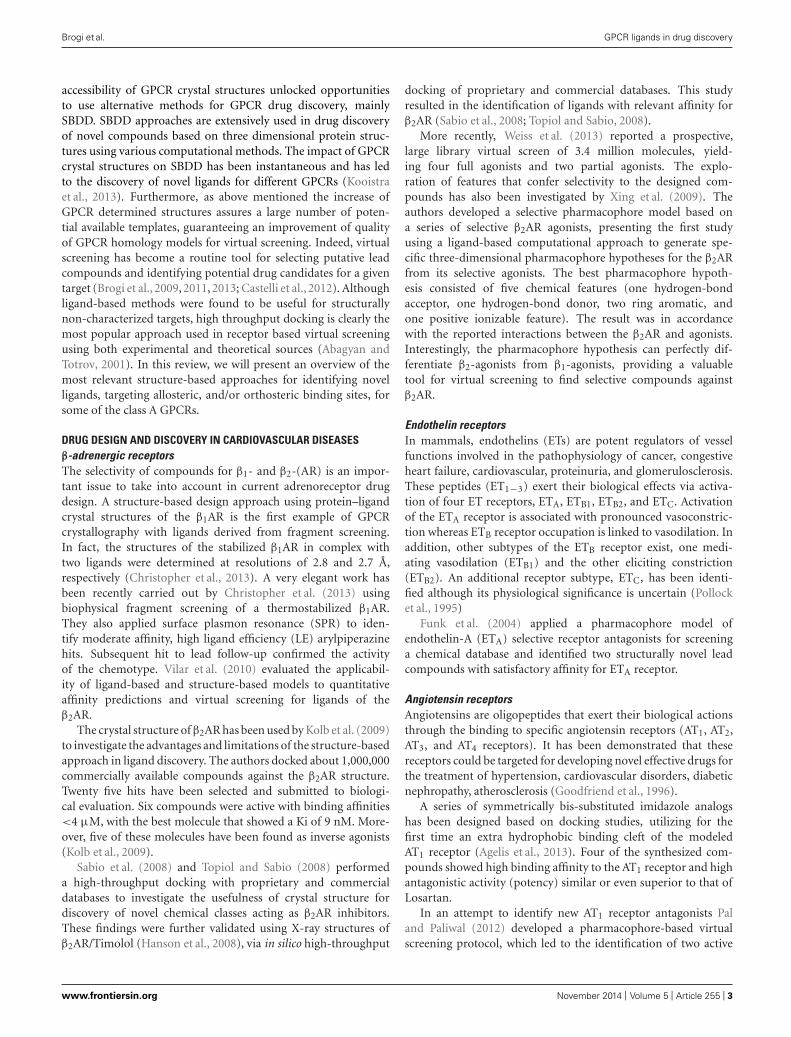
that bind to GPCRs (Andrews et al., 2014). Homology modelingand ligand screening, utilizing structure-, and/or ligand-basedapproaches represent the most common approaches to discoverin silico novel ligands. Recently, fragment-based protocols havealso been used. The impact of computational techniques inGPCR drug discovery has been relevant, due to the extremedifficulties for obtaining experimental high-resolution structuralinformation on the active and inactive state of GPCRs. After thecrystallization of the first mammalian GPCR (bovine rhodopsin;Palczewski et al., 2000; Figure 1), homology-modeling tech-nique has been extensively adopted to predict structures andfunctions of different GPCRs and also to perform in silicoscreening.
In fact, sequence analysis suggested that family A GPCRsshare the same arrangement, showing a high sequence similarityof the seven transmembrane helices, confirming the suitabil-ity of rhodopsin as a template (Li et al., 2010). During the lastdecade, we have seen a dramatic improvement in crystalliza-tion methods. Indeed, after about 7 years from the first solvedstructure of a mammalian GPCR, several three-dimensional struc-tures have been published. The second crystallized GPCR wasβ2-adrenergic receptor (β2AR; Cherezov et al., 2007; Rasmussenet al., 2007) and then the β1AR (Warne et al., 2008). Subsequently,an exponential growth of crystallized GPCR structures in theprotein data bank was observed. Actually, the three dimensionalstructures available of class A GPCRs comprise: the adenosineA2A receptor (Jaakola et al., 2008), the D3 dopamine receptor(Chien et al., 2010), the chemokine receptors CXCR1 (Park et al.,2012), CXCR4 (Wu et al., 2010), and CCR5 (Tan et al., 2013)the histamine H1 receptor (Shimamura et al., 2011), the sph-ingosine 1 phosphate receptor (Hanson et al., 2012), the M2
and M3 muscarinic receptors (Haga et al., 2012; Kruse et al.,2012), the μ, k, and δ opioid receptors (Manglik et al., 2012;Wu et al., 2012; Fenalti et al., 2014) as well as the nociceptin
receptor (NOP; Thompson et al., 2012), bovine opsin receptors(Park et al., 2008; Scheerer et al., 2008), neurotensin receptor(White et al., 2012), the serotonin 5HT1B and 5HT2B receptors(Wacker et al., 2013; Wang et al., 2013a), protease-activated recep-tor 1 (PAR1; Zhang et al., 2012), the smoothened receptor (SMO;Wang et al., 2013b), and P2Y12 receptor (Zhang et al., 2014). Veryrecently, also a crystal structure of class B and C GPCRs such asglucagon receptor (Siu et al., 2013), corticotropin-releasing fac-tor 1 (CRF1) receptor (Hollenstein et al., 2013) and metabotropicglutamate receptor 5 (Dore et al., 2014) respectively, have beenreported.
These achievements are largely attributable to the applicationof high-throughput methods for lipidic cubic phase (LCP) crystal-lography (Cherezov et al., 2004) and protein engineering with thegeneration of GPCR-T4 lysozyme (Rosenbaum et al., 2007) andGPCR–BRIL fusion proteins (Chun et al., 2012). Thermo stabiliza-tion (Serrano-Vega et al., 2008) methods represent another usefultool appropriate to GPCRs crystallization. Notably, these tech-niques can be generally applicable to structurally diverse GPCRs.Moreover, a relevant number of receptors have been solved withboth bound antagonists and agonists.
The availability of numerous different GPCR templates offersdiverse options in GPCR modeling. In particular, the applica-tion of multiple templates to the homology modeling protocolshas been demonstrated to improve the reliability of the computa-tional models including GPCRs (Fernandez-Fuentes et al., 2007;Mobarec et al., 2009; Sokkar et al., 2011; Cappelli et al., 2013;Gemma et al., 2014b).
In conclusion, the availability of a relevant number of crys-tal structures improves results of homology modeling proceduresby using novel methodology such as multiple-templates basedalignment for building the structure of GPCRs as well as thethree-dimensional structure of any type of proteins (Cappelli et al.,2013; Gemma et al., 2014a; Giovani et al., 2014). Moreover, the
FIGURE 1 | Structure of rhodopsin. (A) Crystal structure of bovine rhodopsin covalently linked with retinal adapted from PDB file 1F88. (B) Snake-like diagramfor the bovine rhodopsin highlighting extracellular (EC) and intracellular (IC) loops.
Frontiers in Pharmacology | Neuropharmacology November 2014 | Volume 5 | Article 255 | 2
Brogi et al. GPCR ligands in drug discovery
accessibility of GPCR crystal structures unlocked opportunitiesto use alternative methods for GPCR drug discovery, mainlySBDD. SBDD approaches are extensively used in drug discoveryof novel compounds based on three dimensional protein struc-tures using various computational methods. The impact of GPCRcrystal structures on SBDD has been instantaneous and has ledto the discovery of novel ligands for different GPCRs (Kooistraet al., 2013). Furthermore, as above mentioned the increase ofGPCR determined structures assures a large number of poten-tial available templates, guaranteeing an improvement of qualityof GPCR homology models for virtual screening. Indeed, virtualscreening has become a routine tool for selecting putative leadcompounds and identifying potential drug candidates for a giventarget (Brogi et al., 2009, 2011, 2013; Castelli et al., 2012). Althoughligand-based methods were found to be useful for structurallynon-characterized targets, high throughput docking is clearly themost popular approach used in receptor based virtual screeningusing both experimental and theoretical sources (Abagyan andTotrov, 2001). In this review, we will present an overview of themost relevant structure-based approaches for identifying novelligands, targeting allosteric, and/or orthosteric binding sites, forsome of the class A GPCRs.
DRUG DESIGN AND DISCOVERY IN CARDIOVASCULAR DISEASESβ-adrenergic receptorsThe selectivity of compounds for β1- and β2-(AR) is an impor-tant issue to take into account in current adrenoreceptor drugdesign. A structure-based design approach using protein–ligandcrystal structures of the β1AR is the first example of GPCRcrystallography with ligands derived from fragment screening.In fact, the structures of the stabilized β1AR in complex withtwo ligands were determined at resolutions of 2.8 and 2.7 Å,respectively (Christopher et al., 2013). A very elegant work hasbeen recently carried out by Christopher et al. (2013) usingbiophysical fragment screening of a thermostabilized β1AR.They also applied surface plasmon resonance (SPR) to iden-tify moderate affinity, high ligand efficiency (LE) arylpiperazinehits. Subsequent hit to lead follow-up confirmed the activityof the chemotype. Vilar et al. (2010) evaluated the applicabil-ity of ligand-based and structure-based models to quantitativeaffinity predictions and virtual screening for ligands of theβ2AR.
The crystal structure of β2AR has been used by Kolb et al. (2009)to investigate the advantages and limitations of the structure-basedapproach in ligand discovery. The authors docked about 1,000,000commercially available compounds against the β2AR structure.Twenty five hits have been selected and submitted to biologi-cal evaluation. Six compounds were active with binding affinities<4 μM, with the best molecule that showed a Ki of 9 nM. More-over, five of these molecules have been found as inverse agonists(Kolb et al., 2009).
Sabio et al. (2008) and Topiol and Sabio (2008) performeda high-throughput docking with proprietary and commercialdatabases to investigate the usefulness of crystal structure fordiscovery of novel chemical classes acting as β2AR inhibitors.These findings were further validated using X-ray structures ofβ2AR/Timolol (Hanson et al., 2008), via in silico high-throughput
docking of proprietary and commercial databases. This studyresulted in the identification of ligands with relevant affinity forβ2AR (Sabio et al., 2008; Topiol and Sabio, 2008).
More recently, Weiss et al. (2013) reported a prospective,large library virtual screen of 3.4 million molecules, yield-ing four full agonists and two partial agonists. The explo-ration of features that confer selectivity to the designed com-pounds has also been investigated by Xing et al. (2009). Theauthors developed a selective pharmacophore model based ona series of selective β2AR agonists, presenting the first studyusing a ligand-based computational approach to generate spe-cific three-dimensional pharmacophore hypotheses for the β2ARfrom its selective agonists. The best pharmacophore hypoth-esis consisted of five chemical features (one hydrogen-bondacceptor, one hydrogen-bond donor, two ring aromatic, andone positive ionizable feature). The result was in accordancewith the reported interactions between the β2AR and agonists.Interestingly, the pharmacophore hypothesis can perfectly dif-ferentiate β2-agonists from β1-agonists, providing a valuabletool for virtual screening to find selective compounds againstβ2AR.
Endothelin receptorsIn mammals, endothelins (ETs) are potent regulators of vesselfunctions involved in the pathophysiology of cancer, congestiveheart failure, cardiovascular, proteinuria, and glomerulosclerosis.These peptides (ET1−3) exert their biological effects via activa-tion of four ET receptors, ETA, ETB1, ETB2, and ETC. Activationof the ETA receptor is associated with pronounced vasoconstric-tion whereas ETB receptor occupation is linked to vasodilation. Inaddition, other subtypes of the ETB receptor exist, one medi-ating vasodilation (ETB1) and the other eliciting constriction(ETB2). An additional receptor subtype, ETC, has been identi-fied although its physiological significance is uncertain (Pollocket al., 1995)
Funk et al. (2004) applied a pharmacophore model ofendothelin-A (ETA) selective receptor antagonists for screeninga chemical database and identified two structurally novel leadcompounds with satisfactory affinity for ETA receptor.
Angiotensin receptorsAngiotensins are oligopeptides that exert their biological actionsthrough the binding to specific angiotensin receptors (AT1, AT2,AT3, and AT4 receptors). It has been demonstrated that thesereceptors could be targeted for developing novel effective drugs forthe treatment of hypertension, cardiovascular disorders, diabeticnephropathy, atherosclerosis (Goodfriend et al., 1996).
A series of symmetrically bis-substituted imidazole analogshas been designed based on docking studies, utilizing for thefirst time an extra hydrophobic binding cleft of the modeledAT1 receptor (Agelis et al., 2013). Four of the synthesized com-pounds showed high binding affinity to the AT1 receptor and highantagonistic activity (potency) similar or even superior to that ofLosartan.
In an attempt to identify new AT1 receptor antagonists Paland Paliwal (2012) developed a pharmacophore-based virtualscreening protocol, which led to the identification of two active
www.frontiersin.org November 2014 | Volume 5 | Article 255 | 3
Brogi et al. GPCR ligands in drug discovery
AT1 receptor antagonists with diverse structures (Pal and Paliwal,2012).
DRUG DESIGN AND DISCOVERY IN NEUROLOGICAL DISORDERS ANDPAINDopamine receptorsDopamine exerts its function via five different receptors (D1, D2,D3, D4, and D5 receptors). This system plays a pivotal role in cen-tral nervous system and has been demonstrated to be involved ina series of neurological and psychiatric diseases such as Parkin-son’s disease, schizophrenia, bipolar disorder, drug addiction,and Huntington’s disease (Pivonello et al., 2007; Beaulieu andGainetdinov, 2011). The discovery of ligands able to modulatethe dopaminergic system remains challenging and a lot of compu-tational efforts were carried out for selecting potent and selectiveligands.
In 2010, the crystal structure of D3 receptor was solved, whichdefinitely confirmed the utility of homology models in GPCRsdrug discovery (Chien et al., 2010). Indeed, Carlsson et al. (2011)docked over 3.3 million molecules against a homology model,and 26 of the highest ranking were tested for binding. Six hadaffinities ranging from 0.2 to 3.1 μM. Subsequently, the crystalstructure was used and the docking screen repeated. Of the 25compounds selected, five showed affinities ranging from 0.3 to3.0 μM. One of the new ligands from the homology model screenwas optimized reaching an affinity to 81 nM. The paper clearlydemonstrated the feasibility of high throughput docking usingmodeled GPCRs.
The solved crystal structure of D3 receptor with a D2/D3
selective antagonist provides an opportunity to identify subtlestructural differences between closely related GPCRs that canbe exploited for novel drug design. In an elegant work Laneet al. (2013) performed virtual screening for orthosteric andputative allosteric ligands of D3 receptor using two optimizedcrystal-structure-based models. The authors employed in thecomputational protocol a receptor with an empty binding pocket(D3 receptor-APO), and a receptor in complex with dopamine(D3 receptor-Dopa). Potential hits retrieved by using the twomodels were submitted to biological evaluation and functionalcharacterization. Pharmacological studies showed 14 novel ligandswith a binding affinity better than 10 μM in the D3 receptor-APO candidate list (56% hit rate), and eight novel ligands inthe D3 receptor-Dopa list (32% hit rate). Most ligands in theD3 receptor-APO model spanned both orthosteric and extendedpockets and behaved as antagonists at D3 receptor. Among theidentified ligands, one compound showed the highest potencyof dopamine inhibition (IC50 = 7 nM). In contrast, compoundsidentified by the D3 receptor-Dopa model were predicted to bindan allosteric site at the extracellular extension of the pocket.Such compounds showed a variety of functional activity pro-files. In fact, at least two compounds were non-competitiveallosteric modulator of dopamine signaling in the extracellularsignal-regulated kinase and β-arrestin recruitment assays. Thehigh affinity and LE of the chemically diverse hits identified in thismentioned study evidently demonstrated the utility of structure-based screening in targeting allosteric sites of GPCRs (Lane et al.,2013).
Very recently, Vass et al. (2014) reported a prospective structurebased virtual fragment screening on D3 and the H4 receptors.Representative receptor conformations for ensemble docking wereobtained from molecular dynamics (MD) trajectories. Biologicalevaluation confirmed hit rates ranged from 16 to 32%. Hits hadhigh LE values in the range of 0.31–0.74 and also acceptablelipophilic efficiency, demonstrating that the X-ray structure, thehomology model, and structural ensembles were all found suit-able for docking based virtual screening of fragments against theseGPCRs.
Muscarinic receptorsThe muscarinic acetylcholine receptors (M1–M5) are promisingtargets for the treatment of chronic obstructive pulmonary disease,urinary incontinence, and diabetes. Unfortunately, the lack of sub-type specificity has remained a major obstacle to develop clinicallyuseful muscarinic ligands. Very recently, Kruse et al. (2013) usedthe crystal structure of the M2 and M3 receptors as a template toidentify, by means of structure-based docking, novel muscarinicligands. Interestingly, one compound was a partial agonist at theM3 receptor without measurable M2 agonism that was able tostimulate insulin release from a mouse β-cell line (Kruse et al.,2013).
Cannabinoid (CB) receptorsThe cannabinoid 1 receptor (CB1 receptor) and the cannabi-noid 2 receptor (CB2 receptor) are members of the GPCR family(Matsuda et al., 1990). Agonists of both cannabinoid receptorsubtypes produce strong antinociceptive effects in animal mod-els of chronic, neuropathic, and inflammatory pain and areintensively investigated as potential new analgesic and antiinflam-matory agents. CB1 antagonists are clinically established to beeffective in treating obesity, obesity-related cardio-metabolic dis-orders, and substance abuse, but there are currently no marketedCB1 antagonists. The relevance of CB2-mediated therapeutics iswell established in the treatment of pain, neurodegenerative, andgastrointestinal tract disorders (Di Marzo, 2008; Brogi et al., 2011;Pasquini et al., 2012).
Pandey et al. (2014) used homology model and high through-put docking to discover new chemical classes of CB1 antagoniststhat may serve as starting point for drug development. Theauthors developed and validated a homology model of CB1 basedon a bovine rhodopsin template, which led to the discovery ofseven compounds with an inhibitory potency >50% at 10 μM(Pandey et al., 2014). Wang et al. (2008) identified a novel classof azetidinones as CB1 antagonists by also using virtual screen-ing methods. Meng et al. (2010) reported the identification of thebenzhydrylpiperazine scaffold as a potential scaffold to developnovel CB1 receptor modulators using a privileged structure-basedapproach. The authors identified a highly potent and selectiveCB1 receptor inverse agonist that was able to reduce body weightin diet-induced obese Sprague–Dawley rats.
A recent work carried out by Renault et al. (2013) highlightedthe importance related to crystallization of class-A GPCRs in arange of active states, identifying specific anchoring sites for CB2agonists retrieved in an agonist-bound homology model of CB2receptor. Docking-scoring enrichment tests of a high-throughput
Frontiers in Pharmacology | Neuropharmacology November 2014 | Volume 5 | Article 255 | 4
Brogi et al. GPCR ligands in drug discovery
virtual screening of 140 compounds led to 13 hits within the μMaffinity range. Interestingly, a relevant number of selected hitsbehaved as CB2 agonists, among them two novel unrelated fullagonists were identified. Notably, the exclusive discovery of ago-nists illustrated the reliability of this agonist-bound state modelin the discovery of GPCR ligands with desired behavior (Renaultet al., 2013).
Recently, some of us described a three-dimensional quan-titative structure–selectivity relationships (3D-QSSR) study forselectivity of a series of structurally diverse ligands character-ized by a wide range of selectivity index values for cannabinoidCB1 and CB2 receptors (Brogi et al., 2011). 3D-QSSR explo-rations were expected to provide design information for the designof selective CB2 ligands. The computational model proved tobe predictive, with r2 of 0.95 and Q2 of 0.63. In order to getprospective experimental validation, the selectivity of an externaldata set of 39 compounds reported in the literature was pre-dicted by means of 3D-QSSR model (r2 = 0.56). Subsequently,a quinolone derivative predicted to be a selective CB2 ligandwas synthesized and found to be an extremely selective CB2 lig-and displaying high CB2 affinity (Ki = 4.9 nM), while beingdevoid of CB1 affinity (Ki > 10,000 nM). This finding confirmedthat the ligand-based tool represent a valuable complementaryapproach to docking studies performed on homology models ofGPCRs.
Opioid receptorsOpioids are key medications for the treatment of pain.Theμ-opioid receptors (MORs), δ-opioid receptors (DORs), κ-opioidreceptors (KORs), and nociceptin-opioid receptor (NOP) havebeen isolated and cloned. The receptors were found through-out the peripheral and central nervous system. Their importantrole in mediating pain, drug addiction, and depression has beenestablished. Very recently crystal structures of all classes of opi-oid receptor have been solved (Granier et al., 2012; Manglik et al.,2012; Thompson et al., 2012; Wu et al., 2012; Fenalti et al., 2014).Below is reported one of the first computational efforts using thecrystal structure of the KOR.
Negri et al. (2013) applied a structure-based computationalprotocol using the crystal structure of KOR receptor, discoveringa selective novel KOR agonist, exhibiting analgesic effects with-out activating reward pathways. Remarkably, the novel derivativeshave been identified as novel pharmacological tools to study theinvolvement of KOR in the etiology of drug addiction, depression,and pain (Negri et al., 2013).
ALLOSTERIC MODULATORS AND BIVALENT LIGANDSALLOSTERIC MODULATORSThe binding site of the endogenous agonist is qualified as orthos-teric. In general, antagonists, and inverse agonists typically occupyalso this site, which is usually buried at the core of the receptoror located at its extracellular N-terminal end. In addition, existallosteric sites that bind synthetic drugs or endogenous mineralcations, such as sodium, calcium, zinc, and magnesium, which canalso modulate the activity of the receptor (Christopoulos, 2002).More specifically, allosteric ligands may promote or reduce thebinding of orthosteric ligands. Their effects on receptor activation
could be in a positive, negative, or neutral manner. Allostericmodulators offer several advantages over classical approaches.Allosteric modulator can modulate affinity via conformationalcoupling between the orthosteric and allosteric binding sites ormodulate efficacity by altering the functional response of thereceptor to orthosteric ligand binding. These mechanisms can bedominant for a particular allosteric drug candidate and have signif-icant value in the drug development process. Allosteric modulatorscan have a chemical structure unrelated to that of competitive ago-nist or antagonist drugs, offering a novel class of small moleculedrug candidates.
The orthosteric binding sites within A class GPCR family arehighly conserved due to the evolutionary pressure to retain aminoacid sequences necessary for binding of the endogenous ligand.In contrast, allosteric modulator binding sites have much greaterstructural diversity than endogenous ligand binding sites, display-ing a very high selectivity for a receptor subtype (Mohr et al.,2013).
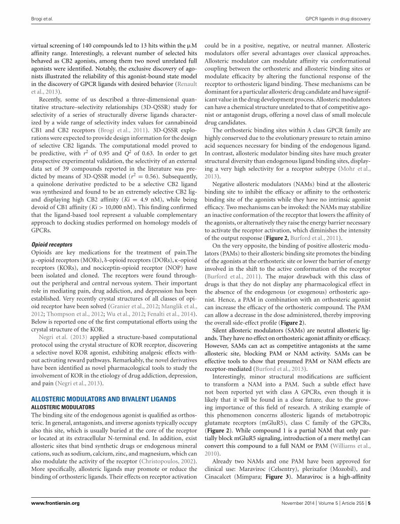
Negative allosteric modulators (NAMs) bind at the allostericbinding site to inhibit the efficacy or affinity to the orthostericbinding site of the agonists while they have no intrinsic agonistefficacy. Two mechanisms can be invoked: the NAMs may stabilizean inactive conformation of the receptor that lowers the affinity ofthe agonists, or alternatively they raise the energy barrier necessaryto activate the receptor activation, which diminishes the intensityof the output response (Figure 2, Burford et al., 2011).
On the very opposite, the binding of positive allosteric modu-lators (PAMs) to their allosteric binding site promotes the bindingof the agonists at the orthosteric site or lower the barrier of energyinvolved in the shift to the active conformation of the receptor(Burford et al., 2011). The major drawback with this class ofdrugs is that they do not display any pharmacological effect inthe absence of the endogenous (or exogenous) orthosteric ago-nist. Hence, a PAM in combination with an orthosteric agonistcan increase the efficacy of the orthosteric compound. The PAMcan allow a decrease in the dose administered, thereby improvingthe overall side-effect profile (Figure 2).
Silent allosteric modulators (SAMs) are neutral allosteric lig-ands. They have no effect on orthosteric agonist affinity or efficacy.However, SAMs can act as competitive antagonists at the sameallosteric site, blocking PAM or NAM activity. SAMs can beeffective tools to show that presumed PAM or NAM effects arereceptor-mediated (Burford et al., 2013).
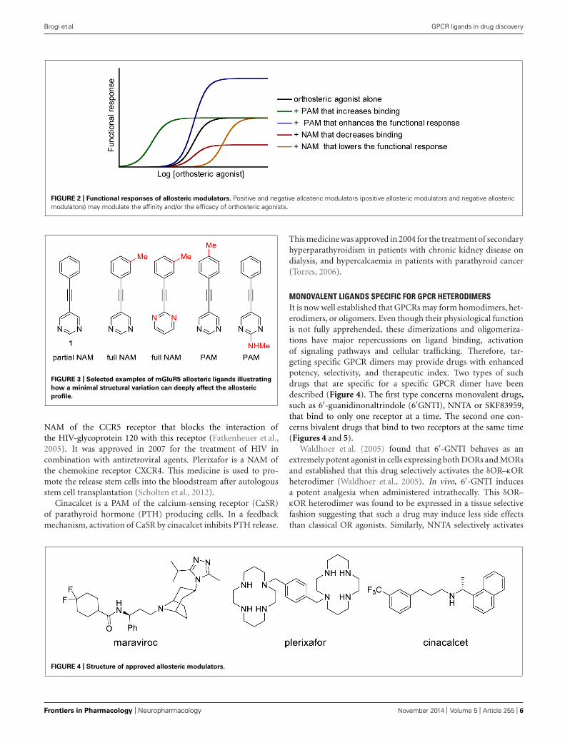
Interestingly, minor structural modifications are sufficientto transform a NAM into a PAM. Such a subtle effect havenot been reported yet with class A GPCRs, even though it islikely that it will be found in a close future, due to the grow-ing importance of this field of research. A striking example ofthis phenomenon concerns allosteric ligands of metabotropicglutamate receptors (mGluR5), class C family of the GPCRs,(Figure 2). While compound 1 is a partial NAM that only par-tially block mGluR5 signaling, introduction of a mere methyl canconvert this compound to a full NAM or PAM (Williams et al.,2010).
Already two NAMs and one PAM have been approved forclinical use: Maraviroc (Celsentry), plerixafor (Mozobil), andCinacalcet (Mimpara; Figure 3). Maraviroc is a high-affinity
www.frontiersin.org November 2014 | Volume 5 | Article 255 | 5
Brogi et al. GPCR ligands in drug discovery
FIGURE 2 | Functional responses of allosteric modulators. Positive and negative allosteric modulators (positive allosteric modulators and negative allostericmodulators) may modulate the affinity and/or the efficacy of orthosteric agonists.
FIGURE 3 | Selected examples of mGluR5 allosteric ligands illustrating
how a minimal structural variation can deeply affect the allosteric
profile.
NAM of the CCR5 receptor that blocks the interaction ofthe HIV-glycoprotein 120 with this receptor (Fatkenheuer et al.,2005). It was approved in 2007 for the treatment of HIV incombination with antiretroviral agents. Plerixafor is a NAM ofthe chemokine receptor CXCR4. This medicine is used to pro-mote the release stem cells into the bloodstream after autologousstem cell transplantation (Scholten et al., 2012).
Cinacalcet is a PAM of the calcium-sensing receptor (CaSR)of parathyroid hormone (PTH) producing cells. In a feedbackmechanism, activation of CaSR by cinacalcet inhibits PTH release.
This medicine was approved in 2004 for the treatment of secondaryhyperparathyroidism in patients with chronic kidney disease ondialysis, and hypercalcaemia in patients with parathyroid cancer(Torres, 2006).
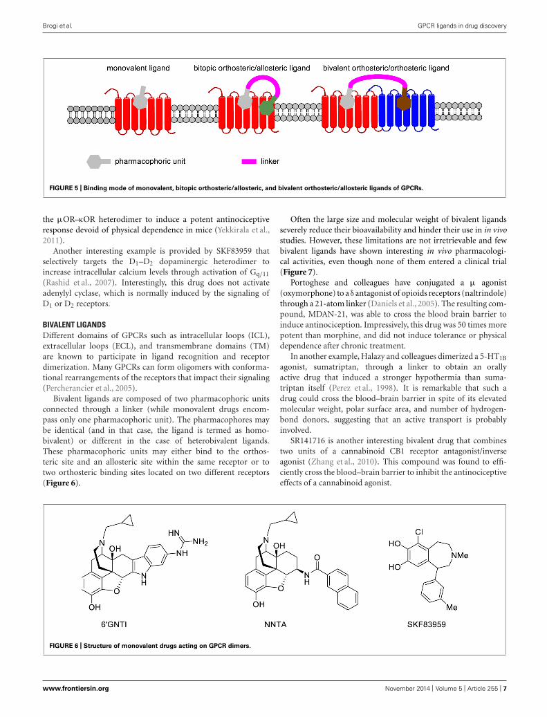
MONOVALENT LIGANDS SPECIFIC FOR GPCR HETERODIMERSIt is now well established that GPCRs may form homodimers, het-erodimers, or oligomers. Even though their physiological functionis not fully apprehended, these dimerizations and oligomeriza-tions have major repercussions on ligand binding, activationof signaling pathways and cellular trafficking. Therefore, tar-geting specific GPCR dimers may provide drugs with enhancedpotency, selectivity, and therapeutic index. Two types of suchdrugs that are specific for a specific GPCR dimer have beendescribed (Figure 4). The first type concerns monovalent drugs,such as 6′-guanidinonaltrindole (6′GNTI), NNTA or SKF83959,that bind to only one receptor at a time. The second one con-cerns bivalent drugs that bind to two receptors at the same time(Figures 4 and 5).
Waldhoer et al. (2005) found that 6′-GNTI behaves as anextremely potent agonist in cells expressing both DORs and MORsand established that this drug selectively activates the δOR–κORheterodimer (Waldhoer et al., 2005). In vivo, 6′-GNTI inducesa potent analgesia when administered intrathecally. This δOR–κOR heterodimer was found to be expressed in a tissue selectivefashion suggesting that such a drug may induce less side effectsthan classical OR agonists. Similarly, NNTA selectively activates
FIGURE 4 | Structure of approved allosteric modulators.
Frontiers in Pharmacology | Neuropharmacology November 2014 | Volume 5 | Article 255 | 6
Brogi et al. GPCR ligands in drug discovery
FIGURE 5 | Binding mode of monovalent, bitopic orthosteric/allosteric, and bivalent orthosteric/allosteric ligands of GPCRs.
the μOR–κOR heterodimer to induce a potent antinociceptiveresponse devoid of physical dependence in mice (Yekkirala et al.,2011).
Another interesting example is provided by SKF83959 thatselectively targets the D1–D2 dopaminergic heterodimer toincrease intracellular calcium levels through activation of Gq/11
(Rashid et al., 2007). Interestingly, this drug does not activateadenylyl cyclase, which is normally induced by the signaling ofD1 or D2 receptors.
BIVALENT LIGANDSDifferent domains of GPCRs such as intracellular loops (ICL),extracellular loops (ECL), and transmembrane domains (TM)are known to participate in ligand recognition and receptordimerization. Many GPCRs can form oligomers with conforma-tional rearrangements of the receptors that impact their signaling(Percherancier et al., 2005).
Bivalent ligands are composed of two pharmacophoric unitsconnected through a linker (while monovalent drugs encom-pass only one pharmacophoric unit). The pharmacophores maybe identical (and in that case, the ligand is termed as homo-bivalent) or different in the case of heterobivalent ligands.These pharmacophoric units may either bind to the orthos-teric site and an allosteric site within the same receptor or totwo orthosteric binding sites located on two different receptors(Figure 6).
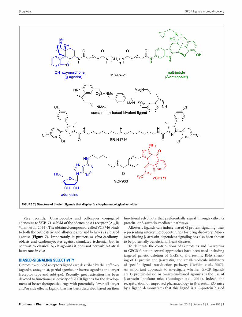
Often the large size and molecular weight of bivalent ligandsseverely reduce their bioavailability and hinder their use in in vivostudies. However, these limitations are not irretrievable and fewbivalent ligands have shown interesting in vivo pharmacologi-cal activities, even though none of them entered a clinical trial(Figure 7).
Portoghese and colleagues have conjugated a μ agonist(oxymorphone) to a δ antagonist of opioids receptors (naltrindole)through a 21-atom linker (Daniels et al., 2005). The resulting com-pound, MDAN-21, was able to cross the blood brain barrier toinduce antinociception. Impressively, this drug was 50 times morepotent than morphine, and did not induce tolerance or physicaldependence after chronic treatment.
In another example, Halazy and colleagues dimerized a 5-HT1B
agonist, sumatriptan, through a linker to obtain an orallyactive drug that induced a stronger hypothermia than suma-triptan itself (Perez et al., 1998). It is remarkable that such adrug could cross the blood–brain barrier in spite of its elevatedmolecular weight, polar surface area, and number of hydrogen-bond donors, suggesting that an active transport is probablyinvolved.
SR141716 is another interesting bivalent drug that combinestwo units of a cannabinoid CB1 receptor antagonist/inverseagonist (Zhang et al., 2010). This compound was found to effi-ciently cross the blood–brain barrier to inhibit the antinociceptiveeffects of a cannabinoid agonist.
FIGURE 6 | Structure of monovalent drugs acting on GPCR dimers.
www.frontiersin.org November 2014 | Volume 5 | Article 255 | 7
Brogi et al. GPCR ligands in drug discovery
FIGURE 7 | Structure of bivalent ligands that display in vivo pharmacological activities.
Very recently, Christopoulos and colleagues conjugatedadenosine to VCP171, a PAM of the adenosine A1 receptor (A1AR;Valant et al., 2014). The obtained compound, called VCP746 bindsto both the orthosteric and allosteric sites and behaves as a biasedagonist (Figure 7). Importantly, it protects in vitro cardiomy-oblasts and cardiomyocytes against simulated ischemia, but incontrast to classical A1AR agonists it does not perturb rat atrialheart rate in vivo.
BIASED-SIGNALING SELECTIVITYG protein-coupled receptors ligands are described by their efficacy(agonist, antagonist, partial agonist, or inverse agonist) and target(receptor type and subtype). Recently, great attention has beendevoted to functional selectivity of GPCR ligands for the develop-ment of better therapeutic drugs with potentially fewer off-targetand/or side effects. Ligand bias has been described based on their
functional selectivity that preferentially signal through either Gprotein- or β-arrestin-mediated pathways.
Allosteric ligands can induce biased G protein signaling, thusrepresenting interesting opportunities for drug discovery. More-over, biasing β-arrestin-dependent signaling has also been shownto be potentially beneficial in heart diseases.
To delineate the contributions of G proteins and β-arrestinsto GPCR function several approaches have been used includingtargeted genetic deletion of GRKs or β-arrestins, RNA silenc-ing of G protein and β-arrestin, and small-molecule inhibitorsof specific signal transduction pathways (DeWire et al., 2007).An important approach to investigate whether GPCR ligandsare G protein-biased or β-arrestin-biased agonists is the use ofβ-arrestin knockout mice (Rominger et al., 2014). Indeed, therecapitulation of improved pharmacology in β-arrestin KO miceby a ligand demonstrates that this ligand is a G-protein biased
Frontiers in Pharmacology | Neuropharmacology November 2014 | Volume 5 | Article 255 | 8
Brogi et al. GPCR ligands in drug discovery
ligand and may be particularly sensitive to the acute desensitizationeffects of β-arrestin. Inversely, minor pharmacological effects inβ-arrestin KO mice indicate that β-arrestin is required for thespecific intracellular signaling pathways of these β-arrestin-biasedligands.
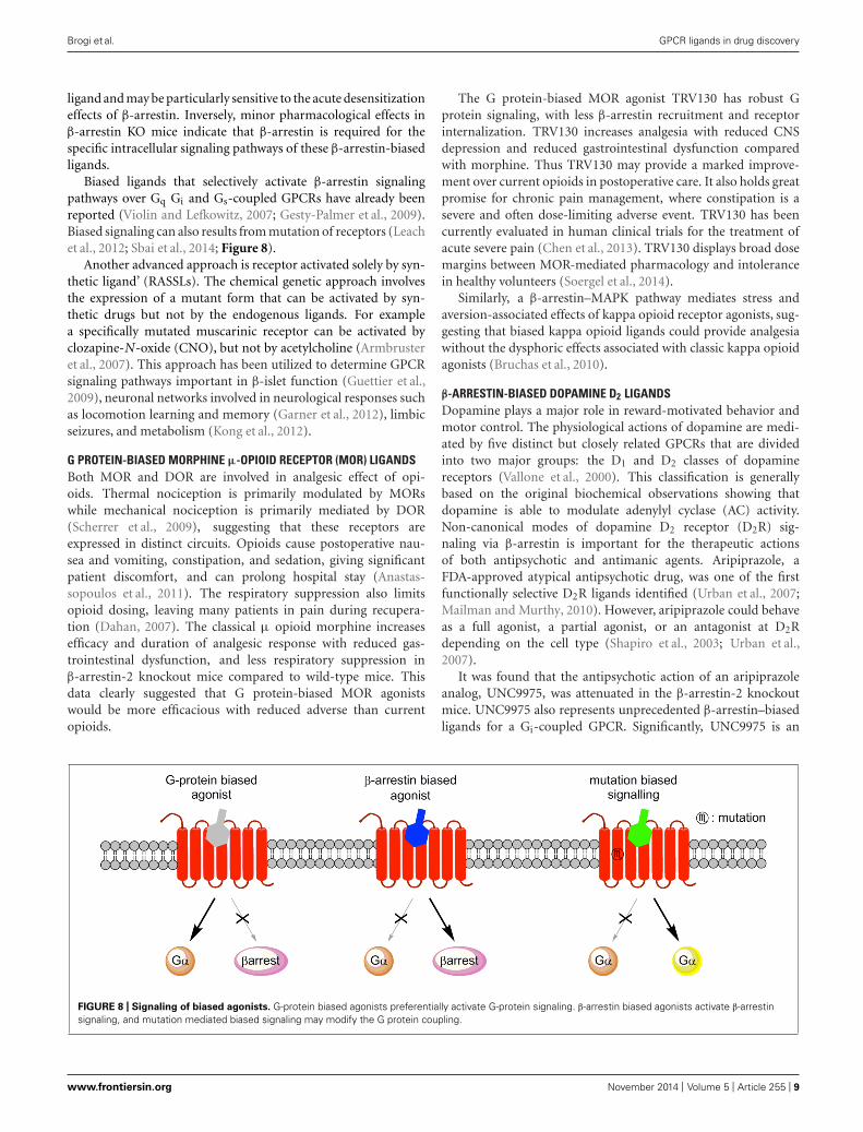
Biased ligands that selectively activate β-arrestin signalingpathways over Gq Gi and Gs-coupled GPCRs have already beenreported (Violin and Lefkowitz, 2007; Gesty-Palmer et al., 2009).Biased signaling can also results from mutation of receptors (Leachet al., 2012; Sbai et al., 2014; Figure 8).
Another advanced approach is receptor activated solely by syn-thetic ligand’ (RASSLs). The chemical genetic approach involvesthe expression of a mutant form that can be activated by syn-thetic drugs but not by the endogenous ligands. For examplea specifically mutated muscarinic receptor can be activated byclozapine-N-oxide (CNO), but not by acetylcholine (Armbrusteret al., 2007). This approach has been utilized to determine GPCRsignaling pathways important in β-islet function (Guettier et al.,2009), neuronal networks involved in neurological responses suchas locomotion learning and memory (Garner et al., 2012), limbicseizures, and metabolism (Kong et al., 2012).
G PROTEIN-BIASED MORPHINE μ-OPIOID RECEPTOR (MOR) LIGANDSBoth MOR and DOR are involved in analgesic effect of opi-oids. Thermal nociception is primarily modulated by MORswhile mechanical nociception is primarily mediated by DOR(Scherrer et al., 2009), suggesting that these receptors areexpressed in distinct circuits. Opioids cause postoperative nau-sea and vomiting, constipation, and sedation, giving significantpatient discomfort, and can prolong hospital stay (Anastas-sopoulos et al., 2011). The respiratory suppression also limitsopioid dosing, leaving many patients in pain during recupera-tion (Dahan, 2007). The classical μ opioid morphine increasesefficacy and duration of analgesic response with reduced gas-trointestinal dysfunction, and less respiratory suppression inβ-arrestin-2 knockout mice compared to wild-type mice. Thisdata clearly suggested that G protein-biased MOR agonistswould be more efficacious with reduced adverse than currentopioids.
The G protein-biased MOR agonist TRV130 has robust Gprotein signaling, with less β-arrestin recruitment and receptorinternalization. TRV130 increases analgesia with reduced CNSdepression and reduced gastrointestinal dysfunction comparedwith morphine. Thus TRV130 may provide a marked improve-ment over current opioids in postoperative care. It also holds greatpromise for chronic pain management, where constipation is asevere and often dose-limiting adverse event. TRV130 has beencurrently evaluated in human clinical trials for the treatment ofacute severe pain (Chen et al., 2013). TRV130 displays broad dosemargins between MOR-mediated pharmacology and intolerancein healthy volunteers (Soergel et al., 2014).
Similarly, a β-arrestin–MAPK pathway mediates stress andaversion-associated effects of kappa opioid receptor agonists, sug-gesting that biased kappa opioid ligands could provide analgesiawithout the dysphoric effects associated with classic kappa opioidagonists (Bruchas et al., 2010).
β-ARRESTIN-BIASED DOPAMINE D2 LIGANDSDopamine plays a major role in reward-motivated behavior andmotor control. The physiological actions of dopamine are medi-ated by five distinct but closely related GPCRs that are dividedinto two major groups: the D1 and D2 classes of dopaminereceptors (Vallone et al., 2000). This classification is generallybased on the original biochemical observations showing thatdopamine is able to modulate adenylyl cyclase (AC) activity.Non-canonical modes of dopamine D2 receptor (D2R) sig-naling via β-arrestin is important for the therapeutic actionsof both antipsychotic and antimanic agents. Aripiprazole, aFDA-approved atypical antipsychotic drug, was one of the firstfunctionally selective D2R ligands identified (Urban et al., 2007;Mailman and Murthy, 2010). However, aripiprazole could behaveas a full agonist, a partial agonist, or an antagonist at D2Rdepending on the cell type (Shapiro et al., 2003; Urban et al.,2007).
It was found that the antipsychotic action of an aripiprazoleanalog, UNC9975, was attenuated in the β-arrestin-2 knockoutmice. UNC9975 also represents unprecedented β-arrestin–biasedligands for a Gi-coupled GPCR. Significantly, UNC9975 is an
FIGURE 8 | Signaling of biased agonists. G-protein biased agonists preferentially activate G-protein signaling. β-arrestin biased agonists activate β-arrestinsignaling, and mutation mediated biased signaling may modify the G protein coupling.
www.frontiersin.org November 2014 | Volume 5 | Article 255 | 9
Brogi et al. GPCR ligands in drug discovery
antagonist of Gi-regulated cAMP production and partial ago-nist for D2R/β-arrestin-2 interactions. Importantly, UNC9975displayed potent antipsychotic-like activity without inducingmotoric side effects in vivo (Masri et al., 2008). This β-arrestin–biased ligand shows a potent ability to suppress both d-amphetamine and phencyclidine-induced hyper locomotion inmice, indicating that it possesses antipsychotic activities in vivo.β-arrestin–biased ligands induce a lack of internalization. Thus,we can assume that drugs that induce internalization would ulti-mately foster tachyphylaxis and receptor down-regulation (Allenand Roth, 2011).
MISSENSE MUTATION GPCR LEADING BIASED SIGNALING INDISEASESMany biased signaling are due to the ligand (Whalen et al., 2011),but few examples of biased signaling induced by a mutation ofreceptors have also been reported (Rajagopal et al., 2010).
A natural mutation leading to biased signaling has been iden-tified in the thyroid stimulating hormone (TSH) receptor gene.The mutant TSH receptor still couples to Gs and activates cAMPbut completely loses Gq-mediated inositol phosphate production.This mutation on TSH receptor causes euthyroid hyperthy-rotropinemia with increased radioiodine uptake (Grasberger et al.,2007).
Another example is the natural mutations in the human cal-cium sensing receptor that activate both Gq-dependent productionof inositol phosphate and the Gq- and Gi/o-dependent phospho-rylation of ERK (Leach et al., 2012; Nygaard et al., 2013). It isgenerally assumed that biased signaling is an intrinsic propertyof a given ligand-GPCR complex, whereby a GPCR exists in sev-eral conformations, each of which is preferentially stabilized andactivated by selective ligands (Nygaard et al., 2013). Likewise, themutations leading to biased signaling are supposed to affect theequilibrium between the different receptor conformations.
The mutations in the GPCRs can lead to biased downstreamsignaling and may induce pathogenic and, in some cases, protectiveroles.
Prokineticins are anorexigenic and angiogenic hormonesthat couple to two GPCRs, PKR1, and PKR2 (Nebigil, 2009;Dormishian et al., 2013; Szatkowski et al., 2013). Mutations inthe prokineticin receptor 2 (PKR2) have been found in 10%of patients with Kallmann syndrome that is characterized byhypogonadotropic hypogonadism. To date, 21 missense mutationsof PKR2 have been identified in Kallmann syndrome patients.Some of these mutations are related with the Gq-dependentsignaling pathway (Sinisi et al., 2008; Abreu et al., 2012; Sbaiet al., 2014). However, certain mutations on this receptor affectβ-arrestin recruitment (R80C) or the Gq and Gi signaling pathways(R164Q) with normal Gs signaling. The Gq-dependent signal-ing defect of the R164Q receptor makes this mutation mostlikely pathogenic. The mutation R268C affecting a residue inthe third intracellular loop of the receptor selectively impairsGi/o-dependent signaling of the receptor and is considered non-pathogenic (Sbai et al., 2014). It remains unclear whether theβ-arrestin-dependent signaling defect for the R80C mutationon PKR2 has a pathogenic effect with respect to Kallmannsyndrome.
BIASED LIGANDS IN DISEASESTwo GPCRs, the angiotensin II (AngII) type 1 receptor (AT1R) andthe β-ARs are targets of widely used cardiovascular drugs. Theyare now potential therapeutic targets for biased ligands (DeWireand Violin, 2011).
The peptide hormone angiotensin II (AngII) is a vaso-pressor that regulates salt and fluid homeostasis, modulatingvasoconstriction, and aldosterone secretion, as well as thirstand inflammation (Benigni et al., 2010). Angiotensin-convertingenzyme inhibitors that lower AngII levels and angiotensin recep-tor blockers are widely used in treating hypertension and othercardiovascular diseases. The AT1R couples primarily to Gαq
signaling, leading to phosphatidylinositol bisphosphate hydrol-ysis, generating diacylglycerol, mobilizing calcium, and acti-vating signaling enzymes such as protein kinase C. AT1R isalso involved in β-arrestin–dependent signals, activation ofepidermal growth factor receptor transactivation, Src, andJAK/STAT (Saito and Berk, 2001; Wei et al., 2003; Oliveiraet al., 2007). One body of evidence for distinct AT1R signalingcame from receptor mutagenesis. AT1R effects can be dividedinto distinct G-protein–dependent and G-protein–independentsignals in vivo. Reduction or elimination of β-arrestin-1 or β-arrestin-2 expression with siRNA in vitro or genetic deletionin vivo showed that cardioprotective effect of AT1R is medi-ated by β-arrestin-2 signaling. TRV120027, a selective andβ-arrestin–biased AT1R ligand blocks AngII-dependent hyper-tension while increasing cardiomyocyte contractility, promotingcytoprotective, or antiapoptotic signals and preserving kid-ney function to provide a great benefit in acute heart fail-ure (Monasky et al., 2013). TRV120027 is now in clinicaltrials for the treatment of acute heart failure (Soergel et al.,2014).
Endothelins play a key role in vascular homeostasis. ETA andAT1 receptor antagonists both lower blood pressure in hyperten-sive patients. Accordingly, a dual ETA and AT1 receptor antagonistmay be more efficacious antihypertensive drug than currentmedicines.
Epinephrine binds to cardiac β1AR and stimulates inotropythrough G-protein signals, resulting in increased heart rate,blood pressure, and metabolic stress, promoting cardiomyocyteapoptosis. Several studies demonstrated that β1AR G-proteinand β-arrestin pathways normally strike a balance betweenapoptosis associated with prolonged inotropy and counteract-ing cardioprotection. When this balance is disrupted in theabsence of β-arrestin signaling, apoptosis increases and car-diac function decreases. Activation of β-arrestin scaffoldedcalcium/calmodulin-dependent kinase II by the β1AR requirescAMP, thus the net effect of a β-arrestin–biased ligand is cardio-protective.
A biased ligand for β1AR, carvedilol activates the cardio-protective β-arrestin–mediated epidermal growth factor receptortransactivation-signaling pathway. Carvedilol has shown poten-tially superior clinical efficacy over other β-blockers in termsof cardiovascular events after myocardial infarction (Kopecky,2006) and perhaps mortality (Poole-Wilson et al., 2003). Thecontributions of function of GRK/β-arrestin to the clinical efficacyof carvedilol remain unclear.
Frontiers in Pharmacology | Neuropharmacology November 2014 | Volume 5 | Article 255 | 10
Brogi et al. GPCR ligands in drug discovery
Collectively, substantial data suggest that biased ligandswill have distinct and perhaps more beneficial effects thanunbiased agonists. Biased signaling is proposed to be useful inseveral diseases, including heart failure (β-ARs), hypertension(α-ARs), neuropsychiatric and/or neurodegenerative disorders(histamine H1 receptors), schizophrenia, Parkinson’s disease(dopamine receptors), psychosis and depression (serotonin recep-tors), hypothyroidism (TSH receptor), hyperlipidemia (nicotinicacid receptor), diabetes (GLP1). However, it is possible that biasedsignaling could be associated with undesirable side effects and evencontribute to disease. For example, the bacterium Neisseria menin-gitidis interacts in a biased and allosteric manner with the β2ARto initiate signaling cascades that facilitate meningeal colonization(Brissac et al., 2012).
CONCLUSIONA substantial increase in our understanding of GPCR pharmacol-ogy has provided an array of ligands that target both orthostericand allosteric sites of GPCRs as well as ligands that have prop-erties of bias stimuli. The recent identification of a PAM andNAM binding site, together with the synthesis of in vivo effec-tive ligands, represents a novel, and likely more favorable, optionfor pharmacological manipulations of the GPCRs. Biased ligandsoffer safer, better-tolerated, and more efficacious drugs. However,in some cases a path to successful drug development for targets thathave been abandoned because of on-target adverse pharmacologyin the clinical proof-of-concept studies due to additional recep-tor signaling and regulatory mechanisms rather than β-arrestinpathway.
The complexity of GPCR signaling requires a synergistic rolefor experimental and computational methods in producing noveltherapeutics with maximal clinical efficacy and lowest toxicity.Combining computational methods with sophisticated transgenicand chemical genetic animal models, the next generation of GPCRligands will unquestionably employ rational design principles todeliver GPCR ligands with minimal side-effects.
ACKNOWLEDGMENTSThe authors were supported by the Centre National de laRecherche Scientifique, Fondation pour la Recherche Médicale,Medalis/Labex, Oséo and Région Alsace.
REFERENCESAbagyan, R., and Totrov, M. (2001). High-throughput docking for lead gen-
eration. Curr. Opin. Chem. Biol. 5, 375–382. doi: 10.1016/S1367-5931(00)00217-9
Abbas, A., and Roth, B. L. (2008). Arresting serotonin. Proc. Natl. Acad. Sci. U.S.A.105, 831–832. doi: 10.1073/pnas.0711335105
Abreu, A. P., Noel, S. D., Xu, S., Carroll, R. S., Latronico, A. C., and Kaiser, U.B. (2012). Evidence of the importance of the first intracellular loop of proki-neticin receptor 2 in receptor function. Mol. Endocrinol. 26, 1417–1427. doi:10.1210/me.2012-1102
Agelis, G., Resvani, A., Koukoulitsa, C., Tumova, T., Slaninova, J., Kalavrizioti,D., et al. (2013). Rational design, efficient syntheses and biological evaluationof N,N’-symmetrically bis-substituted butylimidazole analogs as a new class ofpotent Angiotensin II receptor blockers. Eur. J. Med. Chem. 62, 352–370. doi:10.1016/j.ejmech.2012.12.044
Allen, J. A., and Roth, B. L. (2011). Strategies to discover unexpected targets fordrugs active at G protein-coupled receptors. Annu. Rev. Pharmacol. Toxicol. 51,117–144. doi: 10.1146/annurev-pharmtox-010510-100553
Anastassopoulos, K. P., Chow, W., Ackerman, S. J., Tapia, C., Benson, C., and Kim, M.S. (2011). Oxycodone-related side effects: impact on degree of bother, adherence,pain relief, satisfaction, and quality of life. J. Opioid Manag. 7, 203–215. doi:10.5055/jom.2010.0063
Andrews, S. P., Brown, G. A., and Christopher, J. A. (2014). Structure-basedand fragment-based GPCR drug discovery. ChemMedChem 9, 256–275. doi:10.1002/cmdc.201300382
Armbruster, B. N., Li, X., Pausch, M. H., Herlitze, S., and Roth, B. L. (2007). Evolvingthe lock to fit the key to create a family of G protein-coupled receptors potentlyactivated by an inert ligand. Proc. Natl. Acad. Sci. U.S.A. 104, 5163–5168. doi:10.1073/pnas.0700293104
Beaulieu, J. M., and Gainetdinov, R. R. (2011). The physiology, signaling, andpharmacology of dopamine receptors. Pharmacol. Rev. 63, 182–217. doi:10.1124/pr.110.002642
Beaulieu, J. M., Sotnikova, T. D., Marion, S., Lefkowitz, R. J., Gainetdinov, R.R., and Caron, M. G. (2005). An Akt/beta-arrestin 2/PP2A signaling complexmediates dopaminergic neurotransmission and behavior. Cell 122, 261–273. doi:10.1016/j.cell.2005.05.012
Benigni, A., Cassis, P., and Remuzzi, G. (2010). Angiotensin II revisited: new rolesin inflammation, immunology and aging. EMBO Mol. Med. 2, 247–257. doi:10.1002/emmm.201000080
Brissac, T., Mikaty, G., Dumenil, G., Coureuil, M., and Nassif, X. (2012). Themeningococcal minor pilin PilX is responsible for type IV pilus conformationalchanges associated with signaling to endothelial cells. Infect. Immun. 80, 3297–3306. doi: 10.1128/IAI.00369-12
Brogi, S., Corelli, F., Di Marzo, V., Ligresti, A., Mugnaini, C., Pasquini, S., et al.(2011). Three-dimensional quantitative structure-selectivity relationships anal-ysis guided rational design of a highly selective ligand for the cannabinoidreceptor 2. Eur. J. Med. Chem. 46, 547–555. doi: 10.1016/j.ejmech.2010.11.034
Brogi, S., Kladi, M., Vagias, C., Papazafiri, P., Roussis, V., and Tafi, A. (2009).Pharmacophore modeling for qualitative prediction of antiestrogenic activity.J. Chem. Inf. Model. 49, 2489–2497. doi: 10.1021/ci900254b
Brogi, S., Papazafiri, P., Roussis, V., and Tafi, A. (2013). 3D-QSARusing pharmacophore-based alignment and virtual screening for discovery ofnovel MCF-7 cell line inhibitors. Eur. J. Med. Chem. 67, 344–351. doi:10.1016/j.ejmech.2013.06.048
Bruchas, M. R., Land, B. B., and Chavkin, C. (2010). The dynorphin/kappa opioidsystem as a modulator of stress-induced and pro-addictive behaviors. Brain Res.1314, 44–55. doi: 10.1016/j.brainres.2009.08.062
Burford, N. T., Clark, M. J., Wehrman, T. S., Gerritz, S. W., Banks, M., O’Connell,J., et al. (2013). Discovery of positive allosteric modulators and silent allostericmodulators of Pharmacophore modeling for qualitative prediction of antiestro-genic activity-opioid receptor. Proc. Natl. Acad. Sci. U.S.A. 110, 10830–10835.doi: 10.1073/pnas.1300393110
Burford, N. T., Watson, J., Bertekap, R., and Alt, A. (2011). Strategies for theidentification of allosteric modulators of G-protein-coupled receptors. Biochem.Pharmacol. 81, 691–702. doi: 10.1016/j.bcp.2010.12.012
Cappelli, A., Manini, M., Valenti, S., Castriconi, F., Giuliani, G., Anzini, M.,et al. (2013). Synthesis and structure-activity relationship studies in serotonin5-HT(1A) receptor agonists based on fused pyrrolidone scaffolds. Eur. J. Med.Chem. 63, 85–94. doi: 10.1016/j.ejmech.2013.01.044
Carlsson, J., Coleman, R. G., Setola, V., Irwin, J. J., Fan, H., Schlessinger, A., et al.(2011). Ligand discovery from a dopamine D3 receptor homology model andcrystal structure. Nat. Chem. Biol. 7, 769–778. doi: 10.1038/nchembio.662
Castelli, M. P., Casu, A., Casti, P., Lobina, C., Carai, M. A., Colombo, G., et al.(2012). Characterization of COR627 and COR628, two novel positive allostericmodulators of the GABA(B) receptor. J. Pharmacol. Exp. Ther. 340, 529–538. doi:10.1124/jpet.111.186460
Chen, X. T., Pitis, P., Liu, G., Yuan, C., Gotchev, D., Cowan, C. L., et al. (2013).Structure-activity relationships and discovery of a G protein biased mu opioidreceptor ligand, [(3-methoxythiophen-2-yl)methyl]({2-[(9R)-9-(pyridin-2-yl)-6-oxaspiro-[4.5]decan-9-yl]ethyl})amine (TRV130), for the treatment of acutesevere pain. J. Med. Chem. 56, 8019–8031. doi: 10.1021/jm4010829
Cherezov, V., Peddi, A., Muthusubramaniam, L., Zheng, Y. F., and Caffrey, M.(2004). A robotic system for crystallizing membrane and soluble proteins inlipidic mesophases. Acta Crystallogr. D. Biol. Crystallogr. 60, 1795–1807. doi:10.1107/S0907444904019109
Cherezov, V., Rosenbaum, D. M., Hanson, M. A., Rasmussen, S. G., Thian, F. S.,Kobilka, T. S., et al. (2007). High-resolution crystal structure of an engineered
www.frontiersin.org November 2014 | Volume 5 | Article 255 | 11

Brogi et al. GPCR ligands in drug discovery
human beta2-adrenergic G protein-coupled receptor. Science 318, 1258–1265.doi: 10.1126/science.1150577
Chien, E. Y., Liu, W., Zhao, Q., Katritch, V., Han, G. W., Hanson, M. A., et al. (2010).Structure of the human dopamine D3 receptor in complex with a D2/D3 selectiveantagonist. Science 330, 1091–1095. doi: 10.1126/science.1197410
Christopher, J. A., Brown, J., Dore, A. S., Errey, J. C., Koglin, M., Marshall, F. H.,et al. (2013). Biophysical fragment screening of the beta1-adrenergic receptor:identification of high affinity arylpiperazine leads using structure-based drugdesign. J. Med. Chem. 56, 3446–3455. doi: 10.1021/jm400140q
Christopoulos, A. (2002). Allosteric binding sites on cell-surface receptors: noveltargets for drug discovery. Nat. Rev. Drug Discov. 1, 198–210. doi: 10.1038/nrd746
Chun, E., Thompson, A. A., Liu, W., Roth, C. B., Griffith, M. T., Katritch, V., et al.(2012). Fusion partner toolchest for the stabilization and crystallization of Gprotein-coupled receptors. Structure 20, 967–976. doi: 10.1016/j.str.2012.04.010
Dahan, A. (2007). Respiratory depression with opioids. J. Pain Palliat. CarePharmacother. 21, 63–66. doi: 10.1080/J354v21n01_15
Daniels, D. J., Lenard, N. R., Etienne, C. L., Law, P. Y., Roerig, S. C., and Portoghese,P. S. (2005). Opioid-induced tolerance and dependence in mice is modulated bythe distance between pharmacophores in a bivalent ligand series. Proc. Natl. Acad.Sci. U.S.A. 102, 19208–19213. doi: 10.1073/pnas.0506627102
DeWire, S. M., Ahn, S., Lefkowitz, R. J., and Shenoy, S. K. (2007).Beta-arrestins and cell signaling. Annu. Rev. Physiol. 69, 483–510. doi:10.1146/annurev.ph.69.013107.100021
DeWire, S. M., and Violin, J. D. (2011). Biased ligands for better cardiovasculardrugs: dissecting G-protein-coupled receptor pharmacology. Circ. Res. 109, 205–216. doi: 10.1161/CIRCRESAHA.110.231308
Di Marzo, V. (2008). Targeting the endocannabinoid system: to enhance or reduce?Nat. Rev. Drug Discov. 7, 438–455. doi: 10.1038/nrd2553
Dore, A. S., Okrasa, K., Patel, J. C., Serrano-Vega, M., Bennett, K., Cooke, R.M., et al. (2014). Structure of class C GPCR metabotropic glutamate receptor 5transmembrane domain. Nature 511, 557–562. doi: 10.1038/nature13396
Dormishian, M., Turkeri, G., Urayama, K., Nguyen, T. L., Boulberdaa, M.,Messaddeq, N., et al. (2013). Prokineticin receptor-1 is a new regulator ofendothelial insulin uptake and capillary formation to control insulin sensitiv-ity and cardiovascular and kidney functions. J. Am. Heart Assoc. 2, e000411.10.1161/JAHA.113.000411
Fatkenheuer, G., Pozniak, A. L., Johnson, M. A., Plettenberg, A., Staszewski, S., Hoe-pelman, A. I., et al. (2005). Efficacy of short-term monotherapy with maraviroc, anew CCR5 antagonist, in patients infected with HIV-1. Nat. Med. 11, 1170–1172.doi: 10.1038/nm1319
Fenalti, G., Giguere, P. M., Katritch, V., Huang, X. P., Thompson, A. A., Cherezov,V., et al. (2014). Molecular control of delta-opioid receptor signaling. Nature 506,191–196. doi: 10.1038/nature12944
Fernandez-Fuentes, N., Rai, B. K., Madrid-Aliste, C. J., Fajardo, J. E., and Fiser,A. (2007). Comparative protein structure modeling by combining multipletemplates and optimizing sequence-to-structure alignments. Bioinformatics 23,2558–2565. doi: 10.1093/bioinformatics/btm377
Funk, O. F., Kettmann, V., Drimal, J., and Langer, T. (2004). Chemical functionbased pharmacophore generation of endothelin-A selective receptor antagonists.J. Med. Chem. 47, 2750–2760. doi: 10.1021/jm031041j
Garner, A. R., Rowland, D. C., Hwang, S. Y., Baumgaertel, K., Roth, B. L., Kentros,C., et al. (2012). Generation of a synthetic memory trace. Science 335, 1513–1516.doi: 10.1126/science.1214985
Gemma, S., Brogi, S., Novellino, E., Campiani, G., Maga, G., Brindisi, M., et al.(2014a). HCV-targeted antivirals: current status and future challenges. Curr.Pharm. Des. 20, 3445–3464. doi: 10.2174/13816128113199990630
Gemma, S., Brogi, S., Patil, P. R., Giovani, S., Lamponi, S., Cappelli, A., et al.(2014b). From (+)-epigallocatechin gallate to a simplified synthetic analogueas a cytoadherence inhibitor for P. falciparum. RSC Adv. 4, 4769–4781. doi:10.1039/c3ra45933k
Gesty-Palmer, D., Flannery, P., Yuan, L., Corsino, L., Spurney, R., Lefkowitz, R. J.,et al. (2009). A beta-arrestin-biased agonist of the parathyroid hormone receptor(PTH1R) promotes bone formation independent of G protein activation. Sci.Transl. Med. 1, 1ra1. doi: 10.1126/scitranslmed.3000071
Giovani, S., Penzo, M., Brogi, S., Brindisi, M., Gemma, S., Novellino,E., et al. (2014). Rational design of the first difluorostatone-based PfSUB1inhibitors. Bioorg. Med. Chem. Lett. 24, 3582–3586. doi: 10.1016/j.bmcl.2014.05.044
Goodfriend, T. L., Elliott, M. E., and Catt, K. J. (1996). Angiotensinreceptors and their antagonists. N. Engl. J. Med. 334, 1649–1654. doi:10.1056/NEJM199606203342507
Granier, S., Manglik, A., Kruse, A. C., Kobilka, T. S., Thian, F. S., Weis, W. I., et al.(2012). Structure of the delta-opioid receptor bound to naltrindole. Nature 485,400–404. doi: 10.1038/nature11111
Grasberger, H., Van Sande, J., Hag-Dahood Mahameed, A., Tenenbaum-Rakover, Y.,and Refetoff, S. (2007). A familial thyrotropin (TSH) receptor mutation providesin vivo evidence that the inositol phosphates/Ca2+ cascade mediates TSH actionon thyroid hormone synthesis. J. Clin. Endocrinol. Metab. 92, 2816–2820. doi:10.1210/jc.2007-0366
Guettier, J. M., Gautam, D., Scarselli, M., Ruiz De Azua, I., Li, J. H., Rosemond,E., et al. (2009). A chemical-genetic approach to study G protein regulation ofbeta cell function in vivo. Proc. Natl. Acad. Sci. U.S.A. 106, 19197–19202. doi:10.1073/pnas.0906593106
Haga, K., Kruse, A. C., Asada, H., Yurugi-Kobayashi, T., Shiroishi, M., Zhang,C., et al. (2012). Structure of the human M2 muscarinic acetylcholine receptorbound to an antagonist. Nature 482, 547–551. doi: 10.1038/nature10753
Hanson, M. A., Cherezov, V., Griffith, M. T., Roth, C. B., Jaakola, V. P., Chien,E. Y., et al. (2008). A specific cholesterol binding site is established by the 2.8 Astructure of the human beta2-adrenergic receptor. Structure 16, 897–905. doi:10.1016/j.str.2008.05.001
Hanson, M. A., Roth, C. B., Jo, E., Griffith, M. T., Scott, F. L., Reinhart, G., et al.(2012). Crystal structure of a lipid G protein-coupled receptor. Science 335, 851–855. doi: 10.1126/science.1215904
Hollenstein, K., Kean, J., Bortolato, A., Cheng, R. K., Dore, A. S., Jazayeri, A.,et al. (2013). Structure of class B GPCR corticotropin-releasing factor receptor 1.Nature 499, 438–443. doi: 10.1038/nature12357
Jaakola, V. P., Griffith, M. T., Hanson, M. A., Cherezov, V., Chien, E. Y.,Lane, J. R., et al. (2008). The 2.6 angstrom crystal structure of a humanA2A adenosine receptor bound to an antagonist. Science 322, 1211–1217. doi:10.1126/science.1164772
Khoury, E., Clement, S., and Laporte, S. A. (2014). Allosteric and biased g protein-coupled receptor signaling regulation: potentials for new therapeutics. Front.Endocrinol. 5:68. doi: 10.3389/fendo.2014.00068
Kolb, P., Rosenbaum, D. M., Irwin, J. J., Fung, J. J., Kobilka, B. K., and Shoichet, B.K. (2009). Structure-based discovery of beta2-adrenergic receptor ligands. Proc.Natl. Acad. Sci. U.S.A. 106, 6843–6848. doi: 10.1073/pnas.0812657106
Kong, D., Tong, Q., Ye, C., Koda, S., Fuller, P. M., Krashes, M. J., et al. (2012).GABAergic RIP-Cre neurons in the arcuate nucleus selectively regulate energyexpenditure. Cell 151, 645–657. doi: 10.1016/j.cell.2012.09.020
Kooistra, A. J., Roumen, L., Leurs, R., De Esch, I. J., and De Graaf, C. (2013). Fromheptahelical bundle to hits from the Haystack: structure-based virtual screeningfor GPCR ligands. Methods Enzymol. 522, 279–336. doi: 10.1016/B978-0-12-407865-9.00015-7
Kopecky, S. L. (2006). Effect of beta blockers, particularly carvedilol, on reducingthe risk of events after acute myocardial infarction. Am. J. Cardiol. 98, 1115–1119.doi: 10.1016/j.amjcard.2006.05.039
Kruse, A. C., Hu, J., Pan, A. C., Arlow, D. H., Rosenbaum, D. M., Rosemond, E., et al.(2012). Structure and dynamics of the M3 muscarinic acetylcholine receptor.Nature 482, 552–556. doi: 10.1038/nature10867
Kruse, A. C., Weiss, D. R., Rossi, M., Hu, J., Hu, K., Eitel, K., et al. (2013). Muscarinicreceptors as model targets and antitargets for structure-based ligand discovery.Mol. Pharmacol. 84, 528–540. doi: 10.1124/mol.113.087551
Lane, J. R., Chubukov, P., Liu, W., Canals, M., Cherezov, V., Abagyan, R., et al. (2013).Structure-based ligand discovery targeting orthosteric and allosteric pocketsof dopamine receptors. Mol. Pharmacol. 84, 794–807. doi: 10.1124/mol.113.088054
Leach, K., Wen, A., Davey, A. E., Sexton, P. M., Conigrave, A. D., and Christopou-los, A. (2012). Identification of molecular phenotypes and biased signalinginduced by naturally occurring mutations of the human calcium-sensing receptor.Endocrinology 153, 4304–4316. doi: 10.1210/en.2012-1449
Lefkowitz, R. J., and Shenoy, S. K. (2005). Transduction of receptor signals bybeta-arrestins. Science 308, 512–517. doi: 10.1126/science.1109237
Li, Y. Y., Hou, T. J., and Goddard, W. A. III. (2010). Computational model-ing of structure-function of g protein-coupled receptors with applications fordrug design. Curr. Med. Chem. 17, 1167–1180. doi: 10.2174/092986710790827807
Frontiers in Pharmacology | Neuropharmacology November 2014 | Volume 5 | Article 255 | 12
Brogi et al. GPCR ligands in drug discovery
Luttrell, L. M., Ferguson, S. S., Daaka, Y., Miller, W. E., Maudsley, S.,Della Rocca, G. J., et al. (1999). Beta-arrestin-dependent formation of beta2adrenergic receptor-Src protein kinase complexes. Science 283, 655–661. doi:10.1126/science.283.5402.655
Mailman, R. B., and Murthy, V. (2010). Third generation antipsychotic drugs: partialagonism or receptor functional selectivity? Curr. Pharm. Des. 16, 488–501. doi:10.2174/138161210790361461
Manglik, A., Kruse, A. C., Kobilka, T. S., Thian, F. S., Mathiesen, J. M., Sunahara,R. K., et al. (2012). Crystal structure of the micro-opioid receptor bound to amorphinan antagonist. Nature 485, 321–326. doi: 10.1038/nature10954
Masri, B., Salahpour, A., Didriksen, M., Ghisi, V., Beaulieu, J. M., Gainetdinov, R. R.,et al. (2008). Antagonism of dopamine D2 receptor/beta-arrestin 2 interactionis a common property of clinically effective antipsychotics. Proc. Natl. Acad. Sci.U.S.A. 105, 13656–13661. doi: 10.1073/pnas.0803522105
Matsuda, L. A., Lolait, S. J., Brownstein, M. J., Young, A. C., and Bonner, T. I.(1990). Structure of a cannabinoid receptor and functional expression of thecloned cDNA. Nature 346, 561–564. doi: 10.1038/346561a0
Meng, T., Wang, J., Peng, H., Fang, G., Li, M., Xiong, B., et al. (2010). Discov-ery of benzhydrylpiperazine derivatives as CB1 receptor inverse agonists viaprivileged structure-based approach. Eur. J. Med. Chem. 45, 1133–1139. doi:10.1016/j.ejmech.2009.12.018
Mobarec, J. C., Sanchez, R., and Filizola, M. (2009). Modern homology modelingof G-protein coupled receptors: which structural template to use? J. Med. Chem.52, 5207–5216. doi: 10.1021/jm9005252
Mohr, K., Schmitz, J., Schrage, R., TrãNkle, C., and Holzgrabe, U. (2013). Molecularalliance - from orthosteric and allosteric ligands to dualsteric/bitopic agonists atG protein coupled receptors. Angew. Chem. Int. Ed. Engl. 52, 508–516.
Monasky, M. M., Taglieri, D. M., Henze, M., Warren, C. M., Utter, M. S.,Soergel, D. G., et al. (2013). The beta-arrestin-biased ligand TRV120023 inhibitsangiotensin II-induced cardiac hypertrophy while preserving enhanced myofila-ment response to calcium. Am. J. Physiol. Heart Circ. Physiol. 305, H856–H866.doi: 10.1152/ajpheart.00327.2013
Nebigil, C. G. (2009). Prokineticin receptors in cardiovascular function: foeor friend? Trends Cardiovasc. Med. 19, 55–60. doi: 10.1016/j.tcm.2009.04.007
Negri, A., Rives, M. L., Caspers, M. J., Prisinzano, T. E., Javitch, J. A., and Filizola,M. (2013). Discovery of a novel selective kappa-opioid receptor agonist usingcrystal structure-based virtual screening. J. Chem. Inf. Model. 53, 521–526. doi:10.1021/ci400019t
Nygaard, R., Zou, Y., Dror, R. O., Mildorf, T. J., Arlow, D. H., Manglik, A., et al.(2013). The dynamic process of beta(2)-adrenergic receptor activation. Cell 152,532–542. doi: 10.1016/j.cell.2013.01
Oliveira, L., Costa-Neto, C. M., Nakaie, C. R., Schreier, S., Shimuta, S. I., andPaiva, A. C. (2007). The angiotensin II AT1 receptor structure-activity cor-relations in the light of rhodopsin structure. Physiol. Rev. 87, 565–592. doi:10.1152/physrev.00040.2005
Pal, M., and Paliwal, S. (2012). In silico identification of novel lead com-pounds with AT1 receptor antagonist activity: successful application of chemicaldatabase screening protocol. Org. Med. Chem. Lett. 2, 7. doi: 10.1186/2191-2858-2-7
Palczewski, K., Kumasaka, T., Hori, T., Behnke, C. A., Motoshima, H., Fox, B. A.,et al. (2000). Crystal structure of rhodopsin: a G protein-coupled receptor. Science289, 739–745. doi: 10.1126/science.289.5480.739
Pandey, P., Roy, K. K., Liu, H., Elokely, K. M., Pettaway, S., Cutler, S. J., et al.(2014). Search for cannabinoid receptor 1 antagonists using structure-based vir-tual screening: identification of natural product hits. Planta Med. 80, PF11. doi:10.1055/s-0034-1382589
Park, J. H., Scheerer, P., Hofmann, K. P., Choe, H. W., and Ernst, O. P. (2008).Crystal structure of the ligand-free G-protein-coupled receptor opsin. Nature454, 183–187. doi: 10.1038/nature07063
Park, S. H., Das, B. B., Casagrande, F., Tian, Y., Nothnagel, H. J., Chu, M., et al.(2012). Structure of the chemokine receptor CXCR1 in phospholipid bilayers.Nature 491, 779–783. doi: 10.1038/nature11580
Pasquini, S., Mugnaini, C., Ligresti, A., Tafi, A., Brogi, S., Falciani, C., et al.(2012). Design, synthesis, and pharmacological characterization of indol-3-ylacetamides, indol-3-yloxoacetamides, and indol-3-ylcarboxamides: potent andselective CB2 cannabinoid receptor inverse agonists. J. Med. Chem. 55, 5391–5402.doi: 10.1021/jm3003334
Percherancier, Y., Berchiche, Y. A., Slight, I., Volkmer-Engert, R., Tamamura, H.,Fujii, N., et al. (2005). Bioluminescence resonance energy transfer reveals ligand-induced conformational changes in CXCR4 homo- and heterodimers. J. Biol.Chem. 280, 9895–9903. doi: 10.1074/jbc.M411151200
Perez, M., Pauwels, P. J., Fourrier, C., Chopin, P., Valentin, J. P., John, G. W.,et al. (1998). Dimerization of sumatriptan as an efficient way to design a potent,centrally and orally active 5-HT1B agonist. Bioorg. Med. Chem. Lett. 8, 675–680.doi: 10.1016/S0960-894X(98)00090-0
Pivonello, R., Ferone, D., Lombardi, G., Colao, A., Lamberts, S. W., and Hofland,L. J. (2007). Novel insights in dopamine receptor physiology. Eur. J. Endocrinol.156(Suppl. 1), S13–S21. doi: 10.1530/eje.1.02353
Pollock, D. M., Keith, T. L., and Highsmith, R. F. (1995). Endothelin receptors andcalcium signaling. FASEB J. 9, 1196–1204.
Poole-Wilson, P. A., Swedberg, K., Cleland, J. G., Di Lenarda, A., Hanrath, P.,Komajda, M., et al. (2003). Comparison of carvedilol and metoprolol on clinicaloutcomes in patients with chronic heart failure in the carvedilol or metoprololeuropean trial (COMET): randomised controlled trial. Lancet 362, 7–13. doi:10.1016/S0140-6736(03)13800-7
Rajagopal, S., Rajagopal, K., and Lefkowitz, R. J. (2010). Teaching old receptorsnew tricks: biasing seven-transmembrane receptors. Nat. Rev. Drug Discov. 9,373–386. doi: 10.1038/nrd3024
Rashid, A. J., O’dowd, B. F., Verma, V., and George, S. R. (2007). Neuronal Gq/11-coupled dopamine receptors: an uncharted role for dopamine. Trends Pharmacol.Sci. 28, 551–555. doi: 10.1016/j.tips.2007.10.001
Rasmussen, S. G., Choi, H. J., Rosenbaum, D. M., Kobilka, T. S., Thian, F. S.,Edwards, P. C., et al. (2007). Crystal structure of the human beta2 adrenergicG-protein-coupled receptor. Nature 450, 383–387. doi: 10.1038/nature06325
Renault, N., Laurent, X., Farce, A., El Bakali, J., Mansouri, R., Gervois, P., et al.(2013). Virtual screening of CB(2) receptor agonists from bayesian networkand high-throughput docking: structural insights into agonist-modulated GPCRfeatures. Chem. Biol. Drug Des. 81, 442–454. doi: 10.1111/cbdd.12095
Rominger, D. H., Cowan, C. L., Gowen-Macdonald, W., and Violin, J. D. (2014).Biased ligands: pathway validation for novel GPCR therapeutics. Curr. Opin.Pharmacol. 16, 108–115. doi: 10.1016/j.coph.2014.04.002
Rosenbaum, D. M., Cherezov, V., Hanson, M. A., Rasmussen, S. G., Thian, F. S.,Kobilka, T. S., et al. (2007). GPCR engineering yields high-resolution structuralinsights into beta2-adrenergic receptor function. Science 318, 1266–1273. doi:10.1126/science.1150609
Sabio, M., Jones, K., and Topiol, S. (2008). Use of the X-ray structure of the beta2-adrenergic receptor for drug discovery. Part 2: identification of active compounds.Bioorg. Med. Chem. Lett. 18, 5391–5395. doi: 10.1016/j.bmcl.2008.09.046
Saito, Y., and Berk, B. C. (2001). Transactivation: a novel signaling pathway fromangiotensin II to tyrosine kinase receptors. J. Mol. Cell Cardiol. 33, 3–7. doi:10.1006/jmcc.2000
Sbai, O., Monnier, C., Dode, C., Pin, J. P., Hardelin, J. P., and Rondard, P. (2014).Biased signaling through G-protein-coupled PROKR2 receptors harboringmissense mutations. FASEB J. 28, 3734–3744. doi: 10.1096/fj.13-243402
Scherrer, G., Imamachi, N., Cao, Y. Q., Contet, C., Mennicken, F., O’donnell, D., et al.(2009). Dissociation of the opioid receptor mechanisms that control mechanicaland heat pain. Cell 137, 1148–1159. doi: 10.1016/j.cell.2009.04.019
Scheerer, P., Park, J. H., Hildebrand, P. W., Kim, Y. J., Krauss, N., Choe, H. W., et al.(2008). Crystal structure of opsin in its G-protein-interacting conformation.Nature 455, 497–502. doi: 10.1038/nature07330
Scholten, D. J., Canals, M., Maussang, D., Roumen, L., Smit, M. J., Wijt-mans, M., et al. (2012). Pharmacological modulation of chemokine receptorfunction. Br. J. Pharmacol. 165, 1617–1643. doi: 10.1111/j.1476-5381.2011.01551.x
Serrano-Vega, M. J., Magnani, F., Shibata, Y., and Tate, C. G. (2008). Conformationalthermostabilization of the beta1-adrenergic receptor in a detergent-resistantform. Proc. Natl. Acad. Sci. U.S.A. 105, 877–882. doi: 10.1073/pnas.0711253105
Shapiro, D. A., Renock, S., Arrington, E., Chiodo, L. A., Liu, L. X., Sibley,D. R., et al. (2003). Aripiprazole, a novel atypical antipsychotic drug with aunique and robust pharmacology. Neuropsychopharmacology 28, 1400–1411. doi:10.1038/sj.npp.1300203
Shimamura, T., Shiroishi, M., Weyand, S., Tsujimoto, H., Winter, G., Katritch,V., et al. (2011). Structure of the human histamine H1 receptor complex withdoxepin. Nature 475, 65–70. doi: 10.1038/nature10236
www.frontiersin.org November 2014 | Volume 5 | Article 255 | 13
Brogi et al. GPCR ligands in drug discovery
Sigalov, A. B. (2012). “Monovalen” ligands that trigger TLR-4 and TCRare not necessarily truly monovalent. Mol. Immunol. 51, 356–362. doi:10.1016/j.molimm.2012.03.031
Sinisi, A. A., Asci, R., Bellastella, G., Maione, L., Esposito, D., Elefante, A., et al.(2008). Homozygous mutation in the prokineticin-receptor2 gene (Val274Asp)presenting as reversible Kallmann syndrome and persistent oligozoospermia: casereport. Hum. Reprod. 23, 2380–2384. doi: 10.1093/humrep/den247
Siu, F. Y., He, M., De Graaf, C., Han, G. W., Yang, D., Zhang, Z., et al. (2013).Structure of the human glucagon class B G-protein-coupled receptor. Nature499, 444–449. doi: 10.1038/nature12393
Soergel, D. G., Subach, R. A., Sadler, B., Connell, J., Marion, A. S., Cowan, C.L., et al. (2014). First clinical experience with TRV130: pharmacokinetics andpharmacodynamics in healthy volunteers. J. Clin. Pharmacol. 54, 351–357. doi:10.1002/jcph.207
Sokkar, P., Mohandass, S., and Ramachandran, M. (2011). Multiple templates-based homology modeling enhances structure quality of AT1 receptor: validationby molecular dynamics and antagonist docking. J. Mol. Model. 17, 1565–1577.doi: 10.1007/s00894-010-0860-z
Szatkowski, C., Vallet, J., Dormishian, M., Messaddeq, N., Valet, P., Boulberdaa,M., et al. (2013). Prokineticin receptor 1 as a novel suppressor of preadipocyteproliferation and differentiation to control obesity. PLoS ONE 8:e81175. doi:10.1371/journal.pone.0081175
Tan, Q., Zhu,Y., Li, J., Chen, Z., Han, G. W., Kufareva, I., et al. (2013). Structure of theCCR5 chemokine IV entry inhibitor maraviroc complex. Science 341, 1387–1390.doi: 10.1126/science.1241475
Thompson, A. A., Liu, W., Chun, E., Katritch, V., Wu, H., Vardy, E., et al. (2012).Structure of the nociceptin/orphanin FQ receptor in complex with a peptidemimetic. Nature 485, 395–399. doi: 10.1038/nature11085
Topiol, S., and Sabio, M. (2008). Use of the X-ray structure of the Beta2-adrenergicreceptor for drug discovery. Bioorg. Med. Chem. Lett. 18, 1598–1602. doi:10.1016/j.bmcl.2008.01.063
Torres, P. U. (2006). Cinacalcet HCl: a novel treatment for secondary hyper-parathyroidism caused by chronic kidney disease. J. Ren. Nutr. 16, 253–258.doi: 10.1053/j.jrn.2006.04.010
Urban, J. D., Vargas, G. A., Von Zastrow, M., and Mailman, R. B. (2007). Arip-iprazole has functionally selective actions at dopamine D2 receptor-mediatedsignaling pathways. Neuropsychopharmacology 32, 67–77. doi: 10.1038/sj.npp.1301071
Valant, C., May, L. T., Aurelio, L., Chuo, C. H., White, P. J., Baltos, J. A., et al. (2014).Separation of on-target efficacy from adverse effects through rational design of abitopic adenosine receptor agonist. Proc. Natl. Acad. Sci. U.S.A. 111, 4614–4619.doi: 10.1073/pnas.1320962111
Vallone, D., Picetti, R., and Borrelli, E. (2000). Structure and function ofdopamine receptors. Neurosci. Biobehav. Rev. 24, 125–132. doi: 10.1016/S0149-7634(99)00063-9
Vass, M., Schmidt, E., Horti, F., and Keseru, G. M. (2014). Virtual fragment screeningon GPCRs: a case study on dopamine D3 and histamine H4 receptors. Eur. J. Med.Chem. 77, 38–46. doi: 10.1016/j.ejmech.2014.02.034
Vilar, S., Karpiak, J., and Costanzi, S. (2010). Ligand and structure-based models forthe prediction of ligand-receptor affinities and virtual screenings: developmentand application to the beta(2)-adrenergic receptor. J. Comput. Chem. 31, 707–720.doi: 10.1002/jcc.21346
Violin, J. D., Crombie, A. L., Soergel, D. G., and Lark, M. W. (2014). Biased ligandsat G-protein-coupled receptors: promise and progress. Trends Pharmacol. Sci. 35,308–316. doi: 10.1016/j.tips.2014.04.007
Violin, J. D., and Lefkowitz, R. J. (2007). Beta-arrestin-biased ligands atseven-transmembrane receptors. Trends Pharmacol. Sci. 28, 416–422. doi:10.1016/j.tips.2007.06.006
Wacker, D., Wang, C., Katritch, V., Han, G. W., Huang, X. P., Vardy, E., et al. (2013).Structural features for functional selectivity at serotonin receptors. Science 340,615–619. doi: 10.1126/science.1232808
Waldhoer, M., Fong, J., Jones, R. M., Lunzer, M. M., Sharma, S. K., Kostenis,E., et al. (2005). A heterodimer-selective agonist shows in vivo relevance of Gprotein-coupled receptor dimers. Proc. Natl. Acad. Sci. U.S.A. 102, 9050–9055.doi: 10.1073/pnas.0501112102
Wang, C., Jiang, Y., Ma, J., Wu, H., Wacker, D., Katritch, V., et al. (2013a). Structuralbasis for molecular recognition at serotonin receptors. Science 340, 610–614. doi:10.1126/science.1232807
Wang, C., Wu, H., Katritch, V., Han, G. W., Huang, X. P., Liu, W., et al. (2013b).Structure of the human smoothened receptor bound to an antitumour agent.Nature 497, 338–343. doi: 10.1038/nature12167
Wang, H., Duffy, R. A., Boykow, G. C., Chackalamannil, S., and Madison, V. S.(2008). Identification of novel cannabinoid CB1 receptor antagonists by usingvirtual screening with a pharmacophore model. J. Med. Chem. 51, 2439–2446.doi: 10.1021/jm701519h
Warne, T., Serrano-Vega, M. J., Baker, J. G., Moukhametzianov, R., Edwards, P. C.,Henderson, R., et al. (2008). Structure of a beta1-adrenergic G-protein-coupledreceptor. Nature 454, 486–491. doi: 10.1038/nature07101
Wei, H., Ahn, S., Shenoy, S. K., Karnik, S. S., Hunyady, L., Luttrell, L. M., et al. (2003).Independent beta-arrestin 2 and G protein-mediated pathways for angiotensin IIactivation of extracellular signal-regulated kinases 1 and 2. Proc. Natl. Acad. Sci.U.S.A. 100, 10782–10787. doi: 10.1073/pnas.1834556100
Weiss, D. R., Ahn, S., Sassano, M. F., Kleist, A., Zhu, X., Strachan, R., et al. (2013).Conformation guides molecular efficacy in docking screens of activated beta-2 adrenergic G protein coupled receptor. ACS Chem. Biol. 8, 1018–1026. doi:10.1021/cb400103f
Whalen, E. J., Rajagopal, S., and Lefkowitz, R. J. (2011). Therapeutic potential ofbeta-arrestin- and G protein-biased agonists. Trends Mol. Med. 17, 126–139. doi:10.1016/j.molmed.2010.11.004
White, J. F., Noinaj, N., Shibata, Y., Love, J., Kloss, B., Xu, F., et al. (2012).Structure of the agonist-bound neurotensin receptor. Nature 490, 508–513. doi:10.1038/nature11558
Williams, R., Zhou, Y., Niswender, C. M., Luo, Q., Conn, P. J., Lindsley, C. W.,et al. (2010). Re-exploration of the PHCCC Scaffold: discovery of improved pos-itive allosteric modulators of mGluR4. ACS Chem. Neurosci. 1, 411–419. doi:10.1021/cn9000318
Wu, B., Chien, E. Y., Mol, C. D., Fenalti, G., Liu, W., Katritch, V., et al. (2010). Struc-tures of the CXCR4 chemokine GPCR with small-molecule and cyclic peptideantagonists. Science 330, 1066–1071. doi: 10.1126/science.1194396
Wu, H., Wacker, D., Mileni, M., Katritch, V., Han, G. W., Vardy, E., et al. (2012).Structure of the human kappa-opioid receptor in complex with JDTic. Nature485, 327–332. doi: 10.1038/nature10939
Xing, R. J., Wang, J., Pan, L., and Cheng, M. S. (2009). A selective pharma-cophore model for beta(2)-adrenoceptor agonists. Molecules 14, 4486–4496. doi:10.3390/molecules14114486
Yekkirala, A. S., Lunzer, M. M., Mccurdy, C. R., Powers, M. D., Kalyuzhny, A. E.,Roerig, S. C., et al. (2011). N-naphthoyl-beta-naltrexamine (NNTA), a highlyselective and potent activator of mu/kappa-opioid heteromers. Proc. Natl. Acad.Sci. U.S.A. 108, 5098–5103. doi: 10.1073/pnas.1016277108
Zhang, C., Srinivasan, Y., Arlow, D. H., Fung, J. J., Palmer, D., Zheng, Y., et al. (2012).High-resolution crystal structure of human protease-activated receptor 1. Nature492, 387–392. doi: 10.1038/nature11701
Zhang, J., Zhang, K., Gao, Z. G., Paoletta, S., Zhang, D., Han, G. W., et al. (2014).Agonist-bound structure of the human P2Y12 receptor. Nature 509, 119–122.doi: 10.1038/nature13288
Zhang, Y., Gilliam, A., Maitra, R., Damaj, M. I., Tajuba, J. M., Seltzman, H. H., et al.(2010). Synthesis and biological evaluation of bivalent ligands for the cannabinoid1 receptor. J. Med. Chem. 53, 7048–7060. doi: 10.1021/jm1006676
Conflict of Interest Statement: The authors declare that the research was conductedin the absence of any commercial or financial relationships that could be construedas a potential conflict of interest.
Received: 22 September 2014; accepted: 02 November 2014; published online: 28November 2014.Citation: Brogi S, Tafi A, Désaubry L and Nebigil CG (2014) Discovery of GPCRligands for probing signal transduction pathways. Front. Pharmacol. 5:255. doi:10.3389/fphar.2014.00255This article was submitted to Neuropharmacology, a section of the journal Frontiers inPharmacology.Copyright © 2014 Brogi, Tafi, Désaubry and Nebigil. This is an open-access articledistributed under the terms of the Creative Commons Attribution License(CC BY). Theuse, distribution or reproduction in other forums is permitted, provided the originalauthor(s) or licensor are credited and that the original publication in this journal is cited,in accordance with accepted academic practice. No use, distribution or reproduction ispermitted which does not comply with these terms.
Frontiers in Pharmacology | Neuropharmacology November 2014 | Volume 5 | Article 255 | 14


