Embed Size (px)
Citation preview

Dissociable Neural Effects of Stimulus Valence andPreceding Context During the Inhibition of
Responses to Emotional Faces
Kurt P. Schulz,1* Suzanne M. Clerkin,1,2 Jeffrey M. Halperin,1,2,3
Jeffrey H. Newcorn,1 Cheuk Y. Tang,1,4 and Jin Fan1,5
1Department of Psychiatry, The Mount Sinai School of Medicine, New York2Neuropsychology Doctoral Subprogram in Psychology, The Graduate Center
of the City University of New York, New York3Department of Psychology, Queens College of the City University of New York, Flushing, New York
4Department of Radiology, The Mount Sinai School of Medicine, New York5Department of Neuroscience, The Mount Sinai School of Medicine, New York
Abstract: Socially appropriate behavior requires the concurrent inhibition of actions that are inappro-priate in the context. This self-regulatory function requires an interaction of inhibitory and emotionalprocesses that recruits brain regions beyond those engaged by either processes alone. In this study, weisolated brain activity associated with response inhibition and emotional processing in 24 healthyadults using event-related functional magnetic resonance imaging (fMRI) and a go/no-go task that in-dependently manipulated the context preceding no-go trials (ie, number of go trials) and the valence(ie, happy, sad, and neutral) of the face stimuli used as trial cues. Parallel quadratic trends were seenin correct inhibitions on no-go trials preceded by increasing numbers of go trials and associated activa-tion for correct no-go trials in inferior frontal gyrus pars opercularis, pars triangularis, and pars orbita-lis, temporoparietal junction, superior parietal lobule, and temporal sensory association cortices. Con-versely, the comparison of happy versus neutral faces and sad versus neutral faces revealed valence-dependent activation in the amygdala, anterior insula cortex, and posterior midcingulate cortex. Fur-ther, an interaction between inhibition and emotion was seen in valence-dependent variations in thequadratic trend in no-go activation in the right inferior frontal gyrus and left posterior insula cortex.These results suggest that the inhibition of response to emotional cues involves the interaction of partlydissociable limbic and frontoparietal networks that encode emotional cues and use these cues to exertinhibitory control over the motor, attention, and sensory functions needed to perform the task, respec-tively. Hum Brain Mapp 00:000–000, 2009. VVC 2008 Wiley-Liss, Inc.
Key words: fMRI; motor inhibition; emotion; response context; prefrontal cortex; amygdala
Contract grant sponsor: National Institutes of Health (NationalCenter for Research Resources); Contract grant numbers:MO1RR00071 and K01MH070892; Contract grant sponsor: NationalAssociation for Research on Schizophrenia and Affective Disorders(Young Investigator Award).
*Correspondence to: Kurt Schulz, Department of Psychiatry, Box1230, The Mount Sinai School of Medicine, One Gustave L. Levy
Place, New York 10029.E-mail: [email protected] for publication 16 July 2008; Revised 11 October 2008;Accepted 7 November 2008
DOI: 10.1002/hbm.20706Published online in Wiley InterScience (www.interscience.wiley.com).
VVC 2008 Wiley-Liss, Inc.
r Human Brain Mapping 00:000–000 (2009) r

INTRODUCTION
Socially appropriate behavior requires the integration ofinformation from the context to select the proper actionand inhibit competing responses that are inappropriate inthe context. The inhibition of inappropriate responses is afundamental self-regulatory process that requires theencoding of behavioral cues from the context to guidedecisions about which actions to inhibit [Haberman andWhitney, 2007]. In social contexts, facial expressions andbody gestures from others convey emotional cues that areimportant for this decision process [Otta et al., 1994]. How-ever, these emotional cues are not simply encoded, butrather influence the inhibition of behavioral responses[Hare et al., 2005; Maxwell et al., 2005; Schulz et al., 2007].Thus, responses elicited by facial expressions of happinessthat are associated with positive affect [Otta et al., 1994]and approach behavior [Johansson and Ronnberg, 1996],are more difficult to inhibit than responses to nonemo-tional faces [Hare et al., 2005; Schulz et al., 2007]. Thisemotional biasing of response inhibition has importantimplications for adaptive goal-directed social behavior[Mathews and McLeod, 1994].The influence of emotional stimuli on the neural sub-
strates for response inhibition is still uncertain. The inhibi-tion of responses to both emotional and nonemotional cuesis mediated by a brain network distributed across multiplecortical and subcortical neural ensembles [Buchsbaumet al., 2005; Elliott et al., 2000; Garavan et al., 2006; Shafritzet al., 2006; Simmonds et al., 2008], including basal gan-glia-cortical loops, premotor and supplementary motorareas (SMA) involved in motor programming [Hoshi andTanji, 2004], and preSMA and cingulate motor areas impli-cated in response selection [Mostofsky and Simmonds,2008; Picard and Strick, 2001], as well as the hand areas ofthe pars opercularis (area 44) and pars triangularis (area45) of the right inferior frontal gyrus [Rizzolatti et al.,2002]. However, this inhibition-related inferior frontal ac-tivity extended rostroventrally to the pars orbitalis (area47) when explicitly inhibiting responses to emotional cues[Elliott et al., 2000; Hare et al., 2005] and was centered inthe orbitofrontal cortex when the emotional stimuli wereincidental to the inhibitory task [Goldstein et al., 2007].These pars orbitalis and orbitofrontal activations may rep-resent a distinct inhibitory mechanism specialized for emo-tional stimuli [Mostofsky et al., 2003] or the emotionalmodulation of activity in the neural substrate for responseinhibition [Davidson, 2003], such as the behavioral codingof emotional cues [Sakagami and Pan, 2007].There is growing interest in the mechanisms by which
the limbic substrates for emotional perception influencethe inferior frontal inhibitory circuits [Phelps and LeDoux,2005]. Most of this interest has focused on the amygdalathat receives extensive sensory input [Price, 2003], and inturn, has bidirectional functional connections with the pre-frontal cortex [Hampton et al., 2007; Herwig et al., 2007].The amygdala is believed to encode the emotional value of
stimuli [Dolan, 2007] and is consistently engaged by affec-tive stimuli [Costafreda et al., 2008; Phan et al., 2002].Nevertheless, amygdala activity has proven difficult todetect during emotional response inhibition [Elliott et al.,2000; Hare et al., 2005; Shafritz et al., 2006] and may cap-ture the interaction of stimulus processing and responseinhibition [Goldstein et al., 2007].Neural activation related to the inhibition of responses
to emotional cues has been more commonly described inthe anterior cingulate cortex (ACC) and anterior insula cor-tex [Elliott et al., 2000; Hare et al., 2005; Shafritz et al.,2006]. These limbic structures may form cortical entrypoints through which cognitive, sensory, and emotionalinputs influence motor functions [Augustine, 1996; Paus,2001]. Accordingly, the anterior insula was activated dur-ing response inhibition, specifically, by emotional stimuli[Elliott et al., 2000; Shafritz et al., 2006], particularly thenegative stimuli [Goldstein et al., 2007]. In contrast, activa-tion in the ACC during emotional response inhibition hasbeen linked to motor control [Elliott et al., 2000; Shafritzet al., 2006], emotional processing [Elliott et al., 2000; Hareet al., 2005], and the interaction of these processes [Gold-stein et al., 2007; Shafritz et al., 2006] and has varied inlocation from subgenual [Elliott et al., 2000; Shafritz et al.,2006] to dorsal regions of the ACC [Goldstein et al., 2007;Hare et al., 2005; Shafritz et al., 2006]. Disentangling thefunctional contributions of these regions to emotionalresponse inhibition will require activation paradigmsthat independently manipulate emotional and inhibitoryprocesses.The current study isolated brain activity associated with
response inhibition and emotional processing in 24 healthyadults using event-related functional magnetic resonanceimaging (fMRI) and a go/no-go paradigm that independ-ently manipulated the context preceding no-go trials andthe valence of the face stimuli used as trial cues. Happy,sad, and neutral facial expressions were used as trial cuesin the go/no-go task to identify neural activity that variedas a function of emotional valence regardless of trial type.In addition, a parametric manipulation of the number ofgo trials preceding no-go trials was incorporated into thetask design to isolate activation specific to varying levelsof response inhibition irrespective of the emotional valence[Durston et al., 2002]. We predicted that valence-depend-ent activation would be seen in amygdala, anterior insulacortex, and ACC, although context-dependent activationwould be found in a frontoparietal network dedicated toresponse inhibition, including right inferior frontal gyruspars opercularis, pars triangularis, and pars orbitalis irre-spective of stimulus characteristics.
METHODS
Participants
Participants were 24 healthy right-handed adults (16males, aged 18–35 years) with normal or corrected-to-nor-
r Schulz et al. r
r 2 r

mal vision. All subjects were screened for current or pastpsychiatric, neurological, or systemic medical illness andcompleted the Conners Adult ADHD Rating Scale-Self-Report: Long Version (CAARS) [Conners, 1997] and BeckDepression Inventory-II (BDI-II) [Steer et al., 1999]. A totalscore �6 on the BDI-II and a T-score of 1 SD above ageand gender means (ie, >60) on the CAARS total ADHDsymptoms index were used to screen for clinically signifi-cant mood disturbances and attention problems that mightimpact on emotional processing and/or response inhibi-tion. Mean BDI-II total score was 2.4 6 2.9 (Mean 6 SD)and mean CAARS total ADHD symptoms T-score was41.9 6 9.4. The sample was 37.5% African-American,29.2% Caucasian, 20.8% Hispanic, and 12.5% Asian. Datafrom one additional subject had to be discarded due totechnical problems with the scanner. All subjects providedwritten informed consent for participation. The study wasapproved by the Institutional Review Boards of QueensCollege and The Mount Sinai School of Medicine.
Emotional Go/No-Go Task
The emotional go/no-go task and a training programwere compiled and were run using E-Prime software (Psy-chology Software Tools, Pittsburgh, PA). All subjects ini-tially completed a training session in a quiet testing roomto check simple face perception and to familiarize subjectswith the emotional go/no-go task. The training sessionstarted with an untimed emotional perception task inwhich all the 54 face stimuli used in the go/no-go taskwere randomly presented one at a time in the center of thescreen. Subjects had to make a key press to indicate facevalence. Face stimuli consisted of digitized gray-scaledhappy, sad, and neutral facial expressions with closedmouths from 18 individuals (9 female and 9 male) selectedfrom the MacBrain Face Stimulus Set available atwww.macbrain.org. Six models (3 female and 3 male)were used from each of the following races: African Amer-ican, Asian, and Caucasian. The images were normalizedfor size and luminance, morphed to exclude hair, andcropped into a black square, which was presented againsta black background. The subjects all demonstrated 90% ac-curacy on the emotional perception task. Then, the subjectsperformed one block randomly selected from the emo-tional go/no-go task described later, to familiarize them-selves with the response demands and speeded nature ofthe task.The emotional go/no-go task described in Schulz et al.
[2007] was modified in two ways for the current study.First, neutral facial expressions were added to the happyand sad faces used as go and no-go trial cues, resulting insix blocks with the following trials: (1) happy go/sad no-go; (2) sad go/neutral no-go; (3) neutral go/happy no-go;(4) happy go/neutral no-go; (5) sad go/happy no-go; and(6) neutral go/sad no-go. Second, 30-second periods of fix-ation were added to the beginning and end of each blockto establish a baseline of neural activation to model-out
low-signal drift. Otherwise, the original structure, timingparameters, trial order, and response demands were main-tained in the current task, as described later.The emotional go/no-go task required subjects to moni-
tor six series of stimuli presented individually in the centerof the screen and respond as rapidly as possible by press-ing the ‘‘go’’ cues and withholding responses to ‘‘no-go’’cues. The task consisted of six 252-sec blocks. Each blockcontained 72 (75%) go cues and 24 (25%) no-go cues,resulting in a total of 432 go cues and 144 no-go cues inthe task. Trial order was determined by counterbalancingacross all conditions in the task (eg, trial type, facialexpression, face ethnicity, face gender, face) to ensure thateach trial type followed every other trial type equallyoften. This counterbalancing also embedded the parametricmanipulation of the number of go cues preceding no-gocues (ie, from 0 to 11 go cues) in the trial order. Stimuliwere presented in the center of the screen for 500 mseceach. The interstimulus interval was pseudorandomizedfrom 1,250 to 1,750 msec (mean per block 5 1,500 msec) todiscourage anticipatory responses. Instructions were dis-played on the computer screen at the beginning of eachblock. Participants responded with the right index fingerusing the BrainLogics fiber optic button system (Psy-chology Software Tools, Pittsburgh, PA). Responses wererecorded on a desktop computer and provided measuresof reaction time and accuracy.
Image Acquisition
All subjects were scanned on the same 3.0-Tesla SiemensAllegra (Siemens Medical Systems, Erlangen, Germany)head-dedicated MRI scanner using a high-performancehead gradient system that was designed especially forfunctional brain imaging. Participants were fitted withheadphones and their heads were stabilized with firmfoam padding. Stimuli were projected via an SVGA projec-tor system onto a rear-projection screen mounted at thehead of the magnet bore. Subjects viewed the stimulithrough a mirror on the head coil positioned above theireyes.Scan sessions began with shimming and sagittal localiza-
tion. Next, a high-resolution T2-weighted anatomical vol-ume of the brain was acquired with a turbo spin-echo(TSE) pulse sequence with TR 5 4,500 msec, TE 5 99msec, flip angle 5 1708, FOV 5 21 cm, and a matrix of 512 3336. Forty-two axial slices were acquired at a thickness of 3mm with a skip of 1 mm and an in-plane resolution of 0.413 0.41 mm2. Functional T2*-weighted images depicting theblood oxygenation level-dependent (BOLD) signal werethen acquired at the same 42 slice locations using gradient-echo echo-planar images with TR 5 3,000 msec, TE 5 27msec, flip angle 5 858, FOV 5 21 cm, and an acquisition ma-trix of 643 64. Each functional image comprised a brain vol-ume of 42 axial slices with a thickness of 3 mm, a skip of 1mm, and an in-plane resolution of 3.75 3 3.75 mm2. The TRwas a trade-off for whole-brain coverage with thinner slices
r Emotional Response Inhibition r
r 3 r

that minimized distortions and increased sensitivity inregions of interest (eg, amygdala). All images were acquiredwith slices positioned parallel to the anterior AC-PC com-missure line. Each participant completed six runs of 252 seceach, yielding 84 time points per participant.
Data Analysis
Percent correct inhibitions on no-go trials served as theprimary measure of response inhibition on the emotionalgo/no-go task. Percent correct inhibitions were calculatedfor: (1) all face trials (regardless of expression) and sepa-rately for trials with happy, sad, and neutral cues; and (2)each level of the parametric manipulation (ie, from 0 to 11preceding go trials). The 12 levels of the parametric manip-ulation were then collapsed into the four levels used bySchulz et al. [2007], that is, correct inhibitions on no-go tri-als following: (1) 0 go trials; (2) 1 or 2 go trials; (3) 3 or 4go trials; and (4) 5 or more go trials. Reaction time (RT)and percent correct responses on go trials and signaldetection measures of perceptual sensitivity (d0) andresponse biases (b) were also calculated. The effects ofemotional valence on behavioral measures were testedwith repeated measures of analyses of variance (ANOVA)with the three face emotion valences as the within-subjectfactors. Separate repeated measures of ANOVA with or-thogonal contrasts were used to test for linear, quadratic,and cubic trends in percent correct inhibitions on no-go tri-als following increasing numbers of go trials. Statisticalsignificance was set at the 0.05 level for these analyses. Allprobabilities were based on two-tailed tests.Event-related analyses of the fMRI data were conducted
using statistical parametric mapping (SPM2; WellcomeDepartment of Imaging Neuroscience, London, UK). Pre-processing of the six functional time series was performedindividually for each subject. The functional scans wereslice scan time corrected, realigned to the first volume tocorrect for inter-scan motion, coregistered to the T2 image,normalized to a standard template (Montreal NeurologicalInstitute), and spatially smoothed with an 8 3 8 3 8 mm3
full width at half-maximum (FWHM) Gaussian kernel.First-level individual analyses were performed twice for
each subject to separately test the effects of face emotionand preceding context on response inhibition. Separategeneral linear models (GLM) were conducted to determinethe relationship between observed event-related BOLD sig-nals and regressors that represented expected neuralresponses to events for emotion and preceding context.Regressors were created by convolving a train of deltafunctions that represented the individual trial events withthe default SPM basis function that consisted of a synthetichemodynamic response function, composed of two gammafunctions and their derivatives [Friston et al., 1998]. Therewere four inhibition-related regressors in the first GLM: (1)correct no-go; (2) correct go; (3) incorrect no-go; and (4)incorrect go events. Six preceding context-related regres-
sors were created in the second GLM: correct no-go eventsafter (1) 0 go, (2) 1–2 go, (3) 3–4 go, and (4) � 5 go events;(5) incorrect no-go events; and (6) all go events. The sameregressors were included for all six blocks of the task, thecombination of which effectively collapsed the effects of inter-est over face valence. At the same time, the variation of faceemotion across the six blocks, as described above, enabledthe comparison of face emotion between blocks. Finally, theinteraction of face emotion and preceding context onresponse inhibition was tested by comparing the six context-related regressors between blocks with different face emo-tions. The six parameters (x, y, and z translations and rota-tions) generated during motion correction were entered ascovariates of no interest in the GLM [Johnstone et al., 2006].The general effect of emotional faces on response inhibi-
tion was tested in the first GLM by applying appropriatelinear contrasts to the parameter estimates for correct no-go events minus correct go events collapsed over face va-lence. The specific neural effects of face valence were alsotested in the first GLM by separately contrasting happyversus neutral faces and sad versus neutral faces, both col-lapsed over trial type. These analyses resulted in threecontrast maps for each participant. Testing of the neuraleffect of parametrically manipulating preceding context onresponse inhibition was informed by the behavioralresults. The specific effects of preceding context weretested in the second GLM by applying either linear, quad-ratic, or cubic contrasts to the parameter estimates for thefour parametric levels of correct no-go events, resulting ina single contrast map for each subject. The interactiveeffects of face emotion and preceding context on responseinhibition were tested in the second GLM by separatelycontrasting the four parametric levels of correct no-goevents in blocks with happy versus neutral faces and inblocks with sad versus neutral faces, resulting in two con-trast maps for each subject.The images of contrast estimates for all participants
were entered into second-level group analyses conductedwith separate random-effects statistical models thataccount for intersubject variability and permit population-based inferences. The resultant voxel-wise statistical mapswere thresholded for significance using a cluster-size algo-rithm that protects against false-positive results in spatiallyextended continuous data [Hayasaka et al., 2004]. Theheight (intensity) threshold of each activated voxel was setat an uncorrected P value of 0.01. A Monte Carlo simula-tion of the brain volume in the current study that assumedan individual voxel type I error of P < 0.01 establishedthat a cluster extent of 100 contiguous resampled voxels(2 3 2 3 2 mm3) was necessary to correct for multiplevoxel comparisons at P < 0.05 [Slotnick and Schacter,2004]. Coordinates of activation were converted to Talair-ach and Tournoux [1988] coordinates using a nonlineartransformation (http://www.mrc-cbu.cam.ac.uk/Imaging/mnispace. html) to designate the Brodmann area.Cytoarchitectural areas in the cingulate gyrus were desig-nated according to the conventions of Vogt et al. [1995].
r Schulz et al. r
r 4 r
RESULTS
Behavioral Results
Performance measures for happy, sad, and neutral facetrials on the emotional go/no-go task are presented inTable I. There was a trend toward an effect of face emo-tional valence on correct inhibitions on no-go trials, F (2,46) 5 2.73, P 5 0.08, which accounted for a limitedamount of the variance in performance, h2 5 0.11. In con-trast, there was a significant effect of face emotion on per-cent correct responses on go trials, F (2, 46) 5 3.28, P <0.05. Post hoc comparisons using Least Significant Differ-ence tests revealed significantly more correct responses onneutral face go trials than either happy face trials, P <0.05, or sad face trials, P < 0.05, which did not differ fromeach other, P > 0.10. Face emotional valence had no signif-icant effects on RT and there was no difference in percep-tual sensitivity (d’) or bias (b) for happy, sad, or neutralfaces, all P > 0.10.The effect of parametrically manipulating the number of
preceding go trials on percent of correct inhibitions on no-go trials is shown in Figure 1. There was a significantquadratic trend in percent correct inhibitions on no-go tri-als following increasing numbers of go trials, F (1, 23) 514.11, P 5 0.001. Post hoc comparisons using Least Signifi-cant Difference tests revealed that the quadratic trend wasdue to significantly fewer correct inhibitions following: (1)0 go trials than 1–2 go trials, P < 0.05; and (2) 5 or morego trials than either 1 or 2 go trials, P < 0.05, or 3–4 gotrials, P < 0.05.
fMRI Results
Inhibition of responses to emotional faces
The contrast of correct no-go events minus correct goevents collapsed over face valence identified a distributedfronto-temporo-limbic network that is depicted in Figure 2and detailed in Table II. Specifically, prominent BOLD sig-nal increases were seen in regions associated withresponse inhibition, including right pars opercularis (area44) and bilateral pars triangularis (area 45) of the inferiorfrontal gyrus, with both extending inferomedially to ante-rior insula cortex, as well as in right anterior midcingulate
cortex (MCC; area 24c0/320), which has been more closelylinked to response selection. Another cluster of activation wascentered~2 mm lateral to the anterior extent of the left amyg-dala. Robust BOLD signal increases were also seen along theintraparietal sulcus of right superior parietal lobule (area 7)and bilaterally in temporo-occipital association cortices, withseparate peaks in right middle temporal (area 37) and inferioroccipital gyri (area 19) and left fusiform face area (area 19)and inferior temporal gyrus (area 37).
Parametric manipulation of the context preceding
response inhibition
The neural effects of parametrically manipulating thecontext preceding response inhibition were tested by apply-ing appropriate quadratic contrasts (ie, 1, 21, 21, 1) to theparameter estimates for the four parametric levels of cor-rect no-go events collapsed across emotional valence. Thiscontrast isolated a predominantly right-lateralized fronto-temporo-parietal network that demonstrated a quadratictrend in BOLD signal across the four parametric levels, asshown in Figure 3 and listed in Table III. As predicted, thisnetwork not only included the pars opercularis (area 44)and pars triangularis (area 45) of the right inferior frontalgyrus but also comprised bilateral pars orbitalis (area 47),as well as parietal attention control regions along the righttemporoparietal junction (TPJ; area 40) and in right supe-rior parietal lobule (area 7). The quadratic trends in BOLDsignal increases in bilateral pars orbitalis are illustrated inFigure 3B. Most surprisingly, quadratic trends in BOLDsignal across the four no-go levels were also seen in tempo-ral sensory association cortices, specifically along the infe-rior bank of the left anterior sylvan sulcus (area 22) andbilaterally in the inferior temporal gyrus (area 37).
TABLE I. Behavioral measures of performance on the
emotional go/no-go task
Measure Happy faces Sad faces Neutral faces
Correct inhibitions (%) 89.3 6 0.2 89.1 6 0.2 82.7 6 0.4Correct responses (%) 85.7 6 0.4 88.0 6 0.3 93.8 6 0.2Reaction time (ms) 486.0 6 20.2 497.0 6 18.2 505.0 6 21.6Perceptual sensitivity (d0) 3.2 6 0.2 2.9 6 0.8 3.1 6 0.2Criterion (b) 0.7 6 0.1 1.1 6 0.2 0.7 6 0.2
Values presented as Mean 6 SEM.
Figure 1.
The parametric manipulation of the context preceding response
inhibition. There was a significant quadratic trend in the percent
correct inhibitions on no-go trials after 0, 1–2, 3–4, and 5 or
more go events, F (1, 23) 5 14.11, P 5 0.001. Error bars denote
the standard error of the mean. *P < 0.05.
r Emotional Response Inhibition r
r 5 r

Figure 2.
Regional activation for the inhibition of responses to emotional
faces collapsed over valence shown on left, right, and superior
brain surfaces (upper panels) and on coronal and sagital sections
(lower panels). The pars opercularis (Op) and pars triangularis
(Tri) of the inferior frontal gyrus, inferior (ITG) and middle tem-
poral gyrus (MTG), fusiform face area (FFA), superior parietal
lobule along the intraparietal sulcus (iPS), insula, amygdala, and
midcingulate gyrus (MCC) showed greater signal increase in the
correct no-go versus correct go events contrast. Values in cor-
ner of sections refer to Talairach coordinates.
Figure 3.
r Schulz et al. r
r 6 r

Manipulation of emotional face expression valence
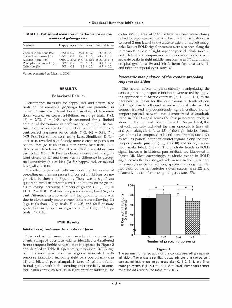
Limbic and paralimbic regions identified by separatelycontrasting happy and sad face events to neutral faceevents collapsed over trial types are shown in Figure 4and listed in Table IV. There was a lateralization of activa-tion for emotional face events in anterior insula cortex; sig-nificant BOLD signal increases were predominantly left lat-eralized for happy faces and right lateralized for sad faces.Happy faces also generated a cluster of activation in leftposterior MCC (area 320) about 15–20 mm dorsal to theMCC activation described earlier for the correct no-go ver-sus go events contrast. In contrast, sad faces activated abasolateral region of the left amygdala dorsomedial to theactivation seen in the no-go minus go events contrast.Figure 4B shows that all four of these regions alsodisplayed a nonsignificant activation for emotional faces ofthe opposite valence. Post hoc analyses revealed no differ-ences in these BOLD signal increases across trial types (ie,no-go vs. go events).
Interaction of emotional face valence and context
preceding response inhibition
Contrasting quadratic trends in the BOLD signal acrossthe four parametric levels separately for sad and happyno-go events versus neutral no-go events identified signifi-
cant interaction effects in the left insula cortex and rightinferior frontal gyrus right pars opercularis (area 44) andpars triangularis (area 45), as shown in Figure 5 anddetailed in Table V. Figure 5B illustrates that the quadratictrends in BOLD signal increases seen in the inferior frontalgyrus for happy faces and in the left insula cortex for sadfaces were inverted for neutral face events. These inferiorfrontal regions corresponded to those identified by thecontrasts for both the quadratic trends and no-go eventsminus go events collapsed over face valence. In contrast,the interaction effects in the left insula were contralateraland �9 mm dorsal to the insula activation identified bycontrasting sad to neutral face events collapsed over trialtype. No other regions showed a significant interactioneffect.
DISCUSSION
These results indicate that the behavioral encoding ofemotional stimuli to successfully guide response inhibitionin social contexts entailed the cooperation and interactionof previously identified neural substrates for response inhi-bition and emotional processing. Response inhibition cuedby emotionally salient stimuli activated the same prefron-tal inhibitory circuits [Rizzolatti et al., 2002], cingulate sub-strates for response selection [Picard and Strick, 2001], andsuperior parietal attention areas [Corbetta and Shulman,
TABLE II. Regional activation for the inhibition of responses to emotional faces collapsed over valence
Region Side BA
Talairach coordinates
Cluster size (mm3) tx y z
IFG pars triangularis R 45 40 30 8 304 4.75IFG pars triangularis L 45 240 20 12 1,458a 7.12Anterior insula cortex L – 234 18 3 5.87IFG pars opercularis R 44 42 7 29 2,062a 8.29Anterior insula cortex R – 30 15 24 5.53Anterior midcingulate cortex R 24c0/320 2 4 44 730 5.68Intraparietal sulcus area R 7 24 260 47 942 9.32Middle temporal gyrus R 37 46 261 10 5,810a 7.92Inferior occipital gyrus R 19 38 282 24 6.26Fusiform face area L 19 240 264 27 4,632a 10.91Inferior temporal gyrus L 37 246 255 27 8.21Amygdala L – 232 21 218 238 5.27
aThe volume consisted of two separate peaks.BA, Brodmann area; IFG, inferior frontal gyrus; L, left; R, right.
Figure 3.
(A) Regional activation generated by the parametric manipulation of the context preceding response inhibition shown on right and
left brain surfaces. The pars opercularis (Op), pars triangularis (Tri), and pars orbitalis (Orb) of the inferior frontal gyrus, inferior
(ITG) and superior temporal gyri (STG), temporoparietal junction (TPJ) and intraparietal sulcus area (SPL) demonstrated quadratic
trends in activation for correct no-go events after 0, 1 to 2, 3–4, and 5 or more go events. (B) The quadratic trends in BOLD signal
increases for no-go events across the four parametric levels are shown for left and right inferior frontal gyrus pars orbitalis. Error
bars denote the standard error of the mean.
r Emotional Response Inhibition r
r 7 r
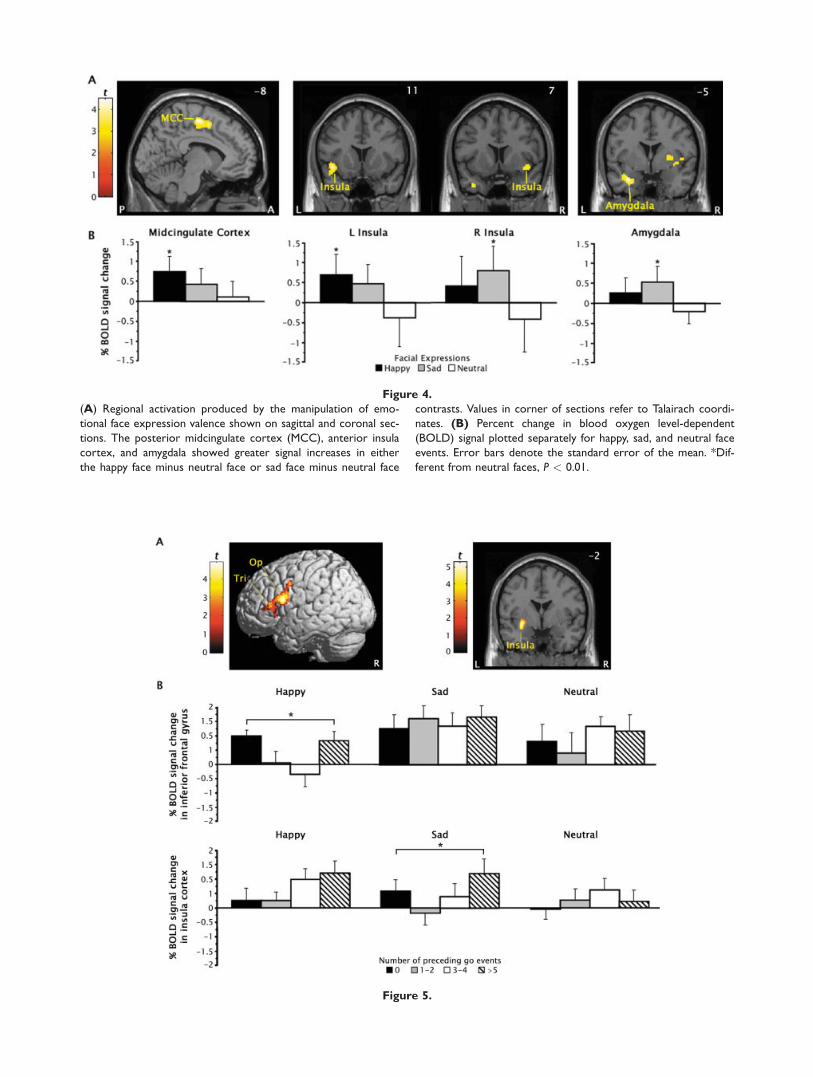
Figure 4.
(A) Regional activation produced by the manipulation of emo-
tional face expression valence shown on sagittal and coronal sec-
tions. The posterior midcingulate cortex (MCC), anterior insula
cortex, and amygdala showed greater signal increases in either
the happy face minus neutral face or sad face minus neutral face
contrasts. Values in corner of sections refer to Talairach coordi-
nates. (B) Percent change in blood oxygen level-dependent
(BOLD) signal plotted separately for happy, sad, and neutral face
events. Error bars denote the standard error of the mean. *Dif-
ferent from neutral faces, P < 0.01.
Figure 5.

2002] that have previously been linked to inhibition innonemotional contexts [Garavan et al., 2006; Simmondset al., 2008]. These regions may constitute a core networkthat mediates response inhibition across contexts and taskdemands. In addition, successful inhibition in the currentcontext required sensory processing of face stimuli by tem-poro-occipital association cortex [Kanwisher et al., 1997],as well as encoding of emotional cues from the context bythe amygdala and anterior paralimbic cortex [Dolan, 2007].This pattern of activation is broadly consistent with find-ings from previous studies [Elliott et al., 2000; Shafritzet al., 2006]. The independent manipulation of the contextpreceding response inhibition and emotional valence ofthe face stimuli in the current task provided additionalclues to the functional significance of these activations.
Manipulation of context preceding
response inhibition
Parametric manipulation of the context precedingresponse inhibition had similar effects on behavioral per-formance and activation in a right-lateralized frontoparie-tal network that is specialized to use contextual informa-tion in inhibitory control. Subjects had greater difficulty in-hibiting responses on no-go trials preceded by another no-go trial (ie, 0 go trials) and five or more go trials than no-go trials preceded by one to four go trials. In contrast,greater difficulty in inhibiting responses was only foundfollowing five or more go trials in a large sample of collegestudents [Schulz et al., 2007]. This discrepancy in the effectof preceding context on response inhibition may reflect ei-
ther the vast differences in the testing environmentbetween the MRI scanner in this study and the quiet roomused in the previous study or the difference in the size ofthe samples in the two studies. It is noteworthy that thetwo studies differed only in the inhibition of responses fol-lowing another no-go trial.Inverse context-related variations in the magnitude of
activation for no-go events were seen in several regionsengaged by emotional response inhibition, as well as innovel areas in pars orbitalis inferior frontal gyrus, righttemporoparietal junction, and left anterior temporal associ-ation cortex. Neural activation was greater when subjectsmade fewer correct inhibitions on no-go trials, specificallyon no-go trials that followed other no-go trials or five ormore go trials. These results are only partially consistentwith the findings of previous studies that used emotional[Elliott et al., 2000; Hare et al., 2005] and nonemotionalgo/no-go tasks [Durston et al., 2002]. The quadratic trendin inferior frontal opercular no-go activation in the presentstudy is analogous to a prior finding of linear increases inboth errors and frontal operculum activity across no-go tri-als preceded by one to five go trials [Durston et al., 2002].This task did not include no-go trials preceded by otherno-go trials. However, the present study did not replicatethe previous reports of activation in the inferior frontalpars orbitalis during the inhibition of responses to emo-tional cues [Elliott et al., 2000; Hare et al., 2005]. Rather,pars orbitalis activation was only detected when no-go tri-als were contrasted across the different contexts precedingno-go trials. None of these studies found variations in parsorbitalis activity as a function of stimulus valence. These
TABLE III. Regional activation generated by the parametric manipulation of the context preceding
response inhibition
Region Side BA
Talairach coordinates
Cluster size (mm3) tx y z
IFG pars orbitalis R 47 38 23 210 524 4.28IFG pars orbitalis L 47 242 25 25 216 3.51IFG pars triangularis R 45 48 20 14 1,438a 4.30IFG pars opercularis R 44 50 9 33 4.97Temporoparietal junction R 40 53 226 29 452 3.19Superior parietal lobule R 7 28 274 44 310 3.75Superior temporal gyrus L 22 253 9 29 604 4.17Inferior temporal gyrus L 37 251 247 28 478 3.84
aThe volume consisted of two separate peaks.BA, Brodmann area; IFG, inferior frontal gyrus; L, left; R, right.
Figure 5.
(A) Regional activation in the pars opercularis (Op) and pars tri-
angularis (Tri) of the right inferior frontal gyrus and the left
insula showed an interaction between the manipulation of the
context preceding response inhibition and the emotional face
expression valence. Values in corner of sections refer to Talair-
ach coordinates. (B) Quadratic trends in BOLD signal increases
for correct no-go events after 0, 1–2, 3–4, and 5 or more go
events were seen in the inferior frontal gyrus pars opercularis
for happy face but not for neutral faces (upper panel) and in the
left insula for sad but neutral faces (lower panel). Error bars
denote the standard error of the mean. *Different from neutral
faces, P < 0.01.
r Emotional Response Inhibition r
r 9 r

results suggest that the pars orbitalis may be involved inthe coding of the behavioral significance of emotionalstimuli.The inferior frontal gyrus pars orbitalis plays a critical
role as an interface between sensory events, motor func-tion, and contextual information in the frontoparietal sub-strate for inhibitory control [Sakagami and Pan, 2007]. Thepars orbitalis receives contextual input from inferotempo-ral cortex [Ungerleider et al., 1989] and incentive-valenceinput from orbitofrontal cortex and amygdala [Petridesand Pandya, 2002], and is unique among prefrontal areasin that it contains neurons that encode sensory cues thatsignal the inhibition of responses based on the behavioralsignificance of the cue stimulus [Sakagami et al., 2001].These neurons fire selectively for no-go cues in go/no-gotasks. This cue-selective activity precedes response execu-tion [Sakagami et al., 2001] and partially reflects interfer-ence from past stimulus-response associations [Lauwer-eyns et al., 2001]. Thus, no-go neurons convert sensoryand contextual inputs into behavioral codes to guide inhi-bition rather than actually inhibit responses [Sakagami andPan, 2007]. Regrettably, we were unable to determinewhether the variation in pars orbitalis no-go activationacross the contexts preceding inhibition reflected theencoding of these context differences or the differences inthe difficulty inhibiting responses across the contexts, asevidenced by the parallel variations in commission errors.
The pars orbitalis may influence response inhibitionthrough dense connections with the opercular and triangu-lar areas of inferior frontal gyrus [Petrides and Pandya,2002]. These inferior frontal regions correspond to Broca’sspeech motor area on the dominant side [Amunts et al.,1999]. However, evidence from neuroimaging studies sug-gest that the opercular and triangular areas could also beregarded as rostral extensions of ventral premotor cortex,involved in complex sensory guided motor acts, possiblyby storing motor representations of goal-directed handactions [Iacoboni and Wilson, 2006]. These ventral motorcortical areas can directly influence motor functionsthrough extensive connections with the primary motor cor-tex [Miyachi et al., 2005]. Meta-analyses of go/no-go andstop tasks have implicated these regions as the neuraleffectors for response inhibition [Garavan et al., 2006;Simmonds et al., 2008]. The variation in the quadratictrend in no-go activation in the pars opercularis and trian-gularis as a function of stimulus valence in the currentstudy suggests that these regions also encode the cognitive(ie, preceding events) and emotional or motivational con-texts of the stimulus [Watanabe and Sakagami, 2007].These variations may have reflected differences in the diffi-culty of inhibiting responses across emotional contexts,with the quadratic trends in no-go activation reflecting themarginally better inhibition of responses to happy facesthan neutral faces. Thus, these inferior frontal areas mayform one cortical entry point through which emotionalinputs influence motor functions.No-go output from the pars orbitalis may also exert top-
down inhibitory control over attention regulation and sen-sory processing [Sakagami and Pan, 2007]. Comparablecontext-dependent activations were found in right tempor-oparietal junction and superior parietal areas that havebeen implicated in target detection [Husain and Nachev,2007] and attention control [Corbetta and Shulman, 2002],respectively. The increased activation for no-go events pre-ceded by zero and five or more go events may reflectgreater salience [Husain and Nachev, 2007], top-down allo-cation of attention [Adler et al., 2001], and/or interferenceresulting in more errors [Mecklinger et al., 2003]. Elevatedtop-down attention for these events could also account forthe context-dependent pattern of activation in temporalsensory association cortex [Reddy et al., 2007].
TABLE V. Regional activation produced by the
interaction of the context preceding response inhibition
and emotional face expression valence
Contrast and region Side BA
Talairachcoordinates
Clustersize (mm3) tx y z
Happy vs. Neutralfaces
IFG pars opercularis R 44 51 14 16 1,588a 4.90IFG pars triangularis R 45 36 30 8 3.91Sad vs. Neutral facesInsula cortex L – 232 22 27 766 4.19
a The volume consisted of two separate peaks.BA, Brodmann area; IFG, inferior frontal gyrus; L, left; R, right.
TABLE IV. Regional activation produced by the manipulation of emotional face expression valence
Contrast and region Side BA
Talairach coordinates
Cluster size (mm3) tx y z
Happy vs. NeutralPosterior midcingulate cortex L 320 28 211 52 926 4.24Anterior insula cortex L – 243 11 27 302 3.72Sad vs. NeutralAnterior insula cortex R – 40 7 29 760 4.52Amygdala L – 226 25 222 350 4.32
BA, Brodmann area; IFG, inferior frontal gyrus; L, left; R, right.
r Schulz et al. r
r 10 r

Overall, the results indicate that emotionally guidedresponse inhibition involved a right frontoparietal networkthat used contextual cues to exert inhibitory control overperception, attention, and behavior. It is noteworthy thatdespite input from neural substrates of emotional process-ing, this network did not respond to emotional stimuli perse, but rather coded the behavioral significance of thesestimuli according to both the cognitive and emotional con-texts. However, these findings cannot clarify whether thecontext-dependent activation reflected some stimulus fea-ture (eg, salience) or increased interference for theseevents.
Manipulation of emotional face expression valence
Manipulation of the emotional valence of the face stim-uli used as trial cues in the current study identified a lim-bic and paralimbic brain circuit that integrates ongoingbehavior with emotion and internal states [Phillips et al.,2003]. Valence-dependent activation was seen in left amyg-dala, bilateral anterior insula cortex, and left posteriorMCC in the current study. The behavioral results suggestthat activity in this circuit may have biased performanceon the go/no-go task. The greater number of missedresponses to happy and sad faces than neutral faces sug-gest that stimulus valence had a detrimental effect onresponse execution processes. In contrast, stimulus valenceproduced a marginal improvement in response inhibition.However, there was no evidence of the previouslyreported difficulty with inhibiting responses to happyfaces when compared with sad faces [Schulz et al., 2007].This discrepancy may reflect the addition of neutral facesto the current study. Neutral expressions tend to be mis-takenly evaluated as happy or sad faces [Lee et al., 2008;Russell and Fehr, 1987]. This difficulty with face emotiondiscrimination may have been compounded by the use offaces with closed mouths in this study, which reduced theadvantage in detection and salience that smiling mouthsconfer to happy faces [Calvo and Nummenmaa, 2008].The pattern of valence-dependent activation is broadly
consistent with the results of previous studies that usedemotional go/no-go tasks [Elliott et al., 2000; Goldsteinet al., 2007; Hare et al., 2005; Shafritz et al., 2006]. The sig-nificant amygdala activation for sad but not happy faces inthe current study is consistent with a previous finding ofamygdala responses to fearful faces in a go/no-go task[Hare et al., 2005], and together with a prior report ofgreater amygdala activation for facial expressions of sur-prise in the context of sad than happy words [Kim et al.,2004], suggests that amygdala activity may code for stimu-lus valence, particularly negative valence [Straube et al.,2008]. However, accumulating evidence indicates that theamygdala activity reflects the representation of the saliencerather than valence of emotional stimuli [Gerber et al.,2008; Kim et al., 2004]. From this perspective, the currentfindings suggest that happy faces were less salient than
sad faces, which may also be attributable to the use offaces with closed mouths [Calvo and Nummenmaa, 2008].In turn, this amygdala activity may have influencedresponse inhibition indirectly through connections withthe inferior frontal gyrus [Petrides and Pandya, 2002] andthe anterior insula [Mesulam and Mufson, 1982].The valence-dependent activation of the anterior insula
cortex seen in the current study is consistent with themodel of this region as a cortical entry point throughwhich visceral, sensory, and autonomic inputs influenceemotional processing and motor functions [Augustine,1996]. The anterior insula is reciprocally connected withthe amygdala and receives primary olfactory, gustatory,and autonomic input, and contextual- and reward-relatedinformation from inferotemporal and orbitofrontal cortices[Mesulam and Mufson, 1982]. This region has been selec-tively activated by emotional stimuli during response inhi-bition [[Elliott et al., 2000; Shafritz et al., 2006] and othercognitive functions [Phan et al., 2002], but there is littleevidence of the lateralization of emotional processing byvalence found in this study [Wager et al., 2003]. The ante-rior insula is ideally positioned to integrate stimulus-evoked internal physiological states with input on stimuluscontext and reward value [Augustine, 1996], but valence-dependent variations in the quadratic trend in no-go acti-vation indicative of these integrative functions were local-ized more dorsally in the insula, more proximal to theopercular, premotor, and cingulate motor areas throughwhich the insula influences motor functions [Shi and Cas-sell, 1998]. These variations may have also reflected thedifferences in inhibiting responses across emotional con-texts. The quadratic trends in no-go activation may haverepresented the marginally better inhibition of responsesto sad faces than neutral faces.The conditional recruitment of the ACC found in this
study is among the most commonly reported findingamong studies that used emotional go/no-go tasks [Elliottet al., 2000; Goldstein et al., 2007; Hare et al., 2005; Shafritzet al., 2006] and other cognitive tasks with emotional stim-uli [Phan et al., 2002]. The inhibition of responses to emo-tional stimuli regardless of valence has been found to reli-ably engage a perigenual region of the ACC implicated inconflict monitoring and error processing [Bush et al.,2000], possibly reflecting the integration of emotional cuesinto the conflict resolution process [Cardinal et al., 2002].Most studies also reported greater ACC activation for neg-atively than positively valenced stimuli, but the localiza-tion of this activity varied from more emotional areas inthe subgenual ACC [Shafritz et al., 2006] to more cognitiveregions in the anterior MCC [Elliott et al., 2000; Goldsteinet al., 2007; Hare et al., 2005]. The current results providelittle help in disentangling the role of the ACC in emo-tional response inhibition. The valence-independent inhibi-tion of responses to emotional stimuli engaged the sameapproximate region of the anterior MCC that was selec-tively activated by sad stimuli in other studies [Elliottet al., 2000; Goldstein et al., 2007; Hare et al., 2005], possi-
r Emotional Response Inhibition r
r 11 r

bly reflecting conflicts in the selection of responses elicitedby cognitive and emotional cues [Picard and Strick, 2001].More incongruously, the current study localized valence-dependent activation for sad faces in cingulate motor areasin the posterior MCC [Picard and Strick, 2001]. The lack ofpreceding context effects on no-go activation in the ACCsuggests that this region subserves executive functionsother than response inhibition [Garavan et al., 2006], andthat the go/no-go task may not be the most ideal para-digm to study ACC function.In summary, the current results provide evidence that
the inhibition of responses to emotional facial expressionsrequires the cooperation and interaction of two dissociablebrain networks. The inferior frontal gyrus pars orbitalisformed the apex of a frontoparietal network that was spe-cialized to use contextual information to exert inhibitorycontrol over the motor, attention, and sensory functionsneeded to perform the task. A second circuit involving theamygdala and paralimbic cortex encoded the emotionalcues from the context. Regions in the two networksencoded different stimulus features as reflected in eithercontext-dependent or valence-dependent patterns of activa-tion. However, successful response inhibition also clearlyinvolved the interaction of the two networks, with va-lence-dependent variations in the quadratic trend in no-goactivation in the right inferior frontal gyrus and left poste-rior insula cortex identifying these regions as cortical entrypoints through which emotional inputs influence motorfunctions. Unfortunately, the analyses were unable to iden-tify the significance of MCC activations seen during emo-tional response inhibition.
REFERENCES
Adler CM, Sax KW, Holland SK, Schmithorst V, Rosenberg L,Strakowski SM (2001): Changes in neuronal activation withincreasing attention demand in healthy volunteers: An fMRIstudy. Synapse 42:266–272.
Amunts K, Schleicher A, Burgel U, Mohlberg H, Uylings HB,Zilles K (1999): Broca’s region revisited: Cytoarchitecture andintersubject variability. J Comp Neurol 412:319–341.
Augustine JR (1996): Circuitry and functional aspects of the insu-lar lobe in primates including humans. Brain Res Brain ResRev 22:229–244.
Buchsbaum BR, Greer S, Chang WL, Berman KF (2005): Meta-anal-ysis of neuroimaging studies of the Wisconsin card-sortingtask and component processes. Hum Brain Mapp 25:35–45.
Bush G, Luu P, Posner MI (2000): Cognitive and emotional influ-ences in anterior cingulate cortex. Trends Cogn Sci 4:215–222.
Calvo MG, Nummenmaa L (2008): Detection of emotional faces:Salient physical features guide effective visual search. J ExpPsychol Gen 137:471–494.
Cardinal RN, Parkinson JA, Hall J, Everitt BJ (2002): Emotion andmotivation: The role of the amygdala, ventral striatum, andprefrontal cortex. Neurosci Biobehav Rev 26:321–352.
Conners CK (1997): Conners’ Rating Scales—Revised, TechnicalManual. Toronto: Multi-Health Systems.
Corbetta M, Shulman GL (2002): Control of goal-directed and stim-ulus-driven attention in the brain. Nat Rev Neurosci 3:201–215.
Costafreda SG, Brammer MJ, David AS, Fu CH (2008): Predictorsof amygdala activation during the processing of emotionalstimuli: A meta-analysis of 385 PET and fMRI studies. BrainRes Rev 58:57–70.
Davidson RJ (2003): Seven sins in the study of emotion: Correc-tives from affective neuroscience. Brain Cogn 52:129–132.
Dolan RJ (2007): The human amygdala and orbital prefrontal cor-tex in behavioural regulation. Philos Trans R Soc Lond B BiolSci 362:787–799.
Durston S, Thomas KM, Worden MS, Yang Y, Casey BJ (2002):The effect of preceding context on inhibition: An event-relatedfMRI study. Neuroimage 16:449–453.
Elliott R, Rubinsztein JS, Sahakian BJ, Dolan RJ (2000): Selectiveattention to emotional stimuli in a verbal go/no-go task: AnfMRI study. Neuroreport 11:1739–1744.
Friston KJ, Fletcher P, Josephs O, Holmes A, Rugg MD, Turner R(1998): Event-related fMRI: characterizing differentialresponses. Neuroimage 7:30–40.
Garavan H, Hester R, Murphy K, Fassbender C, Kelly C (2006):Individual differences in the functional neuroanatomy of inhib-itory control. Brain Res 1105:130–142.
Gerber AJ, Posner J, Gorman D, Colibazzi T, Yu S, Wang Z, Kan-garlu A, Zhu H, Russell J, Peterson BS (2008): An affective cir-cumplex model of neural systems subserving valence, arousal,and cognitive overlay during the appraisal of emotional faces.Neuropsychologia 46:2129–2139.
Goldstein M, Brendel G, Tuescher O, Pan H, Epstein J, Beutel M,Yang Y, Thomas K, Levy K, Silverman M, Clarkin J, Posner M,Kernberg O, Stern E, Silbersweig D (2007): Neural substrates ofthe interaction of emotional stimulus processing and motor in-hibitory control: An emotional linguistic go/no-go fMRI study.Neuroimage 36:1026–1040.
Haberman J, Whitney D (2007): Rapid extraction of mean emotionand gender from sets of faces. Curr Biol 17:R751–R753.
Hampton AN, Adolphs R, Tyszka MJ, O’Doherty JP (2007):Contributions of the amygdala to reward expectancy andchoice signals in human prefrontal cortex. Neuron 55:545–555.
Hare TA, Tottenham N, Davidson MC, Glover GH, Casey BJ(2005): Contributions of amygdala and striatal activity in emo-tion regulation. Biol Psychiatry 57:624–632.
Hayasaka S, Phan KL, Liberzon I, Worsley KJ, Nichols TE (2004):Nonstationary cluster-size inference with random field andpermutation methods. Neuroimage 22:676–687.
Herwig U, Baumgartner T, Kaffenberger T, Bruhl A, Kottlow M,Schreiter-Gasser U, Abler B, Jancke L, Rufer M (2007): Modula-tion of anticipatory emotion and perception processing by cog-nitive control. Neuroimage 37:652–662.
Hoshi E, Tanji J (2004): Differential roles of neuronal activity inthe supplementary and presupplementary motor areas: Frominformation retrieval to motor planning and execution. J Neu-rophysiol 92:3482–3499.
Husain M, Nachev P (2007): Space and the parietal cortex. TrendsCogn Sci 11:30–36.
Iacoboni M, Wilson SM (2006): Beyond a single area: Motor con-trol and language within a neural architecture encompassingBroca’s area. Cortex 42:503–506.
Johansson K, Ronnberg J (1996): Speech gestures and facial expres-sion in speechreading. Scand J Psychol 37:132–139.
Johnstone T, Ores Walsh KS, Greischar LL, Alexander AL, Fox AS,Davidson RJ, Oakes TR (2006): Motion correction and the useof motion covariates in multiple-subject fMRI analysis. HumBrain Mapp 27:779–788.
r Schulz et al. r
r 12 r

Kanwisher N, McDermott J, Chun MM (1997): The fusiform facearea: A module in human extrastriate cortex specialized forface perception. J Neurosci 17:4302–4311.
Kim H, Somerville LH, Johnstone T, Polis S, Alexander AL, ShinLM, Whalen PJ (2004): Contextual modulation of amygdalaresponsivity to surprised faces. J Cogn Neurosci 16:1730–1745.
Lauwereyns J, Sakagami M, Tsutsui K, Kobayashi S, Koizumi M,Hikosaka O (2001): Responses to task-irrelevant visual featuresby primate prefrontal neurons. J Neurophysiol 86:2001–2010.
Lee E, Kang JI, Park IH, Kim JJ, An SK (2008): Is a neutral facereally evaluated as being emotionally neutral? Psychiatry Res157:77–85.
Mathews A, McLeod C (1994): Cognitive approaches to emotionand emotional disorders. Annu Rev Psychol 45:25–50.
Maxwell JS, Shackman AJ, Davidson RJ (2005): Unattended facialexpressions asymmetrically bias the concurrent processing ofnonemotional information. J Cogn Neurosci 17:1386–1395.
Mecklinger A, Weber K, Gunter TC, Engle RW (2003): Dissociablebrain mechanisms for inhibitory control: Effects of interferencecontent and working memory capacity. Brain Res Cogn BrainRes 18:26–38.
Mesulam MM, Mufson EJ (1982): Insula of the old world monkey.III: Efferent cortical output and comments on function. J CompNeurol 212:38–52.
Miyachi S, Lu X, Inoue S, Iwasaki T, Koike S, Nambu A, TakadaM (2005): Organization of multisynaptic inputs from prefrontalcortex to primary motor cortex as revealed by retrograde trans-neuronal transport of rabies virus. J Neurosci 25:2547–2556.
Mostofsky SH, Simmonds DJ (2008): Response inhibition andresponse selection: Two sides of the same coin. J Cogn Neuro-sci 20:751–761.
Mostofsky SH, Schafer JG, Abrams MT, Goldberg MC, FlowerAA, Boyce A, Courtney SM, Calhoun VD, Kraut MA, DencklaMBPekar JJ (2003): fMRI evidence that the neural basis ofresponse inhibition is task-dependent. Brain Res Cogn BrainRes 17:419–430.
Otta E, Lira BB, Delevati NM, Cesar OP, Pires CS (1994): Theeffect of smiling and of head tilting on person perception. JPsychol 128:323–331.
Paus T (2001): Primate anterior cingulate cortex: Where motor con-trol, drive and cognition interface. Nat Rev Neurosci 2:417–424.
Petrides M, Pandya DN (2002): Comparative cytoarchitectonicanalysis of the human and the macaque ventrolateral prefron-tal cortex and corticocortical connection patterns in the mon-key. Eur J Neurosci 16:291–310.
Phan KL, Wager T, Taylor SF, Liberzon I (2002): Functional neuro-anatomy of emotion: A meta-analysis of emotion activationstudies in PET and fMRI. Neuroimage 16:331–348.
Phelps EA, LeDoux JE (2005): Contributions of the amygdala toemotion processing: From animal models to human behavior.Neuron 48:175–187.
Phillips ML, Drevets WC, Rauch SL, Lane R (2003): Neurobiologyof emotion perception I: The neural basis of normal emotionperception. Biol Psychiatry 54:504–514.
Picard N, Strick PL (2001): Imaging the premotor areas. Curr OpinNeurobiol 11:663–672.
Price JL (2003): Comparative aspects of amygdala connectivity.Ann N Y Acad Sci 985:50–58.
Reddy L, Moradi F, Koch C (2007): Top-down biases win againstfocal attention in the fusiform face area. Neuroimage 38:730–739.
Rizzolatti G, Fogassi L, Gallese V (2002): Motor and cognitivefunctions of the ventral premotor cortex. Curr Opin Neurobiol12:149–154.
Russell JA, Fehr B (1987): Relativity in the perception of emotionin facial expressions. J Exp Psychol Gen 116:223–237.
Sakagami M, Pan X (2007): Functional role of the ventrolateral pre-frontal cortex in decision making. Curr Opin Neurobiol 17:228–233.
Sakagami M, Tsutsui K, Lauwereyns J, Koizumi M, Kobayashi S,Hikosaka O (2001): A code for behavioral inhibition on the ba-sis of color, but not motion, in ventrolateral prefrontal cortexof macaque monkey. J Neurosci 21:4801–4808.
Schulz KP, Fan J, Magidina O, Marks DJ, Hahn B, Halperin JM(2007): Does the emotional go/no-go task really measure be-havioral inhibition? Convergence with measures on a non-emo-tional analog. Arch Clin Neuropsychol 22:151–160.
Shafritz KM, Collins SH, Blumberg HP (2006): The interaction ofemotional and cognitive neural systems in emotionally guidedresponse inhibition. Neuroimage 31:468–475.
Shi CJ, Cassell MD (1998): Cortical, thalamic, and amygdaloid con-nections of the anterior and posterior insular cortices. J CompNeurol 399:440–468.
Simmonds DJ, Pekar JJ, Mostofsky SH (2008): Meta-analysis ofGo/No-go tasks demonstrating that fMRI activation associatedwith response inhibition is task-dependent. Neuropsychologia46:224–232.
Slotnick SD, Schacter DL (2004): A sensory signature that distin-guishes true from false memories. Nat Neurosci 7:664–672.
Steer RA, Ball R, Ranieri WF, Beck AT (1999): Dimensions of thebeck depression inventory-II in clinically depressed outpa-tients. J Clin Psychol 55:117–128.
Straube T, Pohlack S, Mentzel HJ, Miltner WH (2008): Differentialamygdala activation to negative and positive emotional pic-tures during an indirect task. Behav Brain Res 191:285–288.
Talairach J, Tournoux M (1988): Co-planar Stereotaxic Atlas of theHuman Brain. New York: Thieme Medical.
Ungerleider LG, Gaffan D, Pelak VS (1989): Projections from infe-rior temporal cortex to prefrontal cortex via the uncinate fasci-cle in rhesus monkeys. Exp Brain Res 76:473–484.
Vogt BA, Nimchinsky EA, Vogt LJ, Hof PR (1995): Human cingu-late cortex: Surface features, flat maps, and cytoarchitecture. JComp Neurol 359:490–506.
Wager TD, Phan KL, Liberzon I, Taylor SF (2003): Valence, gender,and lateralization of functional brain anatomy in emotion: A meta-analysis of findings from neuroimaging. Neuroimage 19:513–531.
Watanabe M, Sakagami M (2007): Integration of cognitive andmotivational context information in the primate prefrontal cor-tex. Cereb Cortex 17 (Suppl 1):i101–i109.
r Emotional Response Inhibition r
r 13 r


![Rydberg-valence interactions of CO, and spectroscopic evidence characterizing the C[sup ʹ] [sup 1]Σ[sup +] valence state](https://img.pdfslide.net/doc/110x75/634ccc7ec1597a4ca90a9a6e/rydberg-valence-interactions-of-co-and-spectroscopic-evidence-characterizing-the.jpg)


