Embed Size (px)
Citation preview

JOURNAL OF VIROLOGY, July 2006, p. 7089–7099 Vol. 80, No. 140022-538X/06/$08.00�0 doi:10.1128/JVI.02694-05Copyright © 2006, American Society for Microbiology. All Rights Reserved.
Distinct Roles for Nucleic Acid in In Vitro Assembly ofPurified Mason-Pfizer Monkey Virus CANC Proteins
Pavel Ulbrich,1 Sarka Haubova,1 Milan V. Nermut,2 Eric Hunter,3Michaela Rumlova,4,1* and Tomas Ruml1,4*
Department of Biochemistry and Microbiology, Institute of Chemical Technology, Technicka 3, 166 28 Prague, Czech Republic1;National Institute for Biological Standards and Control, South Mimms, Potters Bar, Herts, EN6 3QG, United Kingdom2;
Emory Vaccine Center, 954 Gatewood Road, Atlanta, Georgia3; and Department of Protein Biochemistry, Institute ofOrganic Chemistry and Biochemistry, Academy of Sciences of the Czech Republic, 166 10 Prague, Czech Republic4
Received 22 December 2005/Accepted 29 April 2006
In contrast to other retroviruses, Mason-Pfizer monkey virus (M-PMV) assembles immature capsids in thecytoplasm. We have compared the ability of minimal assembly-competent domains from M-PMV and humanimmunodeficiency virus type 1 (HIV-1) to assemble in vitro into virus-like particles in the presence and absenceof nucleic acids. A fusion protein comprised of the capsid and nucleocapsid domains of Gag (CANC) and itsN-terminally modified mutant (�ProCANC) were used to mimic the assembly of the viral core and immatureparticles, respectively. In contrast to HIV-1, where CANC assembled efficiently into cylindrical structures, thesame domains of M-PMV were assembly incompetent. The addition of RNA or oligonucleotides did notcomplement this defect. In contrast, the M-PMV �ProCANC molecule was able to assemble into sphericalparticles, while that of HIV-1 formed both spheres and cylinders. For M-PMV, the addition of purified RNAincreased the efficiency with which �ProCANC formed spherical particles both in terms of the overall amountand the numbers of completed spheres. The amount of RNA incorporated was determined, and for both rRNAand MS2-RNA, quantities similar to that of genomic RNA were encapsidated. Oligonucleotides also stimulatedassembly; however, they were incorporated into �ProCANC spherical particles in trace amounts that could notserve as a stoichiometric structural component for assembly. Thus, oligonucleotides may, through a transientinteraction, induce conformational changes that facilitate assembly, while longer RNAs appear to facilitate thecomplete assembly of spherical particles.
All retroviruses encode a single structural polyproteinprecursor, Gag, that directs the assembly of the immatureretroviral capsid and can direct the formation of retrovirus-like particles in mammalian cells and in bacteria. In additionto domains common to all retroviruses (matrix [MA], capsid[CA] and nucleocapsid [NC]), the Gag polyprotein of Mason-Pfizer monkey virus (M-PMV) also contains a phosphoprotein,pp16-18, an internal scaffold protein, p12, and a C-terminaldomain, p4.
The CA domain plays a key role in the assembly of bothimmature retroviral capsids and the mature cores of infectiousvirions. The CA protein of human immunodeficiency virus type1 (HIV-1) dimerizes in solution, while other CA proteins suchas those of Rous sarcoma virus (RSV), equine infectious ane-mia virus, and human T-cell leukemia virus type 1 are mono-meric in solution (5, 13, 27, 43, 44). Structural studies of ret-roviral CA proteins have shown that they consist of twostructural domains, the N-terminal assembly domain and theC-terminal dimerization domain (1, 4, 6, 13, 15, 20, 27, 31, 43,44, 57, 65, 76, 77). Several studies have focused on mutual
interactions between CA proteins; both C-terminal–C-termi-nal and N-terminal–C-terminal interaction seem to be involved(47, 48). In addition, a recent model suggests that interactionsmay occur between CA molecules by domain swapping, similarto those of mammalian SCAN domains (39, 45).
Several studies have shown that the RSV and HIV-1 CAproteins can assemble in vitro into tubular structures (23, 36)or conical particles resembling a mature core (23, 28, 36, 44).The latter protein also assembles into hollow cylinders at highsalt concentrations (22, 28, 36, 50), and structures assembledon lipid from a His-tagged HIV-1 CA protein show a cage-likelattice consisting of hexamers (2). CA proteins of several ret-roviruses form a �-hairpin stabilized by a salt bridge betweenthe N-terminal proline and conservative aspartates in positions50 to 57 of the protein, consistent with a molecular rearrange-ment of CA upon proteolytic maturation (26, 31, 37, 69). Ex-tension of the N terminus would therefore prevent the hairpinformation and direct assembly of spherical rather than tubularstructures (44, 61, 76). In concert with this, HIV-1 CA formsvariously sized spherical particles in the presence of four ormore flanking residues of MA, while HIV-1 CA particles lack-ing any MA residues are tubular (37, 76). Similarly, the C-terminal residues of the RSV p10 protein, which is located justN terminal of CA, differentiate between the formation of tubesor spheres in the assembly process of this virus (11, 41).
In agreement with these observations, we have shown pre-viously, using a bacterial expression/assembly system, that M-PMV CA with an intact N-terminal proline directs the assem-bly of sheets from a CANC fusion protein (69). Deletion of the
* Corresponding authors. Mailing address for Michaela Rumlova:Department of Protein Biochemistry, Institute of Organic Chemistryand Biochemistry, Academy of Sciences of the Czech Republic, Fle-mingovo n. 2, 166 10 Prague 6, Czech Republic. Phone:420-220183252. Fax: 420-220183556. E-mail: [email protected]. Mailing address for Tomas Ruml: Department ofBiochemistry and Microbiology, Institute of Chemical Technology,Technicka 3, 166 28 Prague, Czech Republic. Phone: 420-220443022.Fax: 420-220445140. E-mail: [email protected].
7089

proline or replacement with alanine converts assembly tospheres; this is consistent with a transition between the pre-cursor form of CA when the N-terminal proline is not availablefor interaction, allowing spherical immature capsids to formand the subsequent formation of a tubular core after its pro-teolytic release (69).
The role of RNA in the assembly of the retroviral immatureparticle remains ambiguous. It is clear that the NC protein iscrucial for specific incorporation of genomic RNA and canfacilitate Gag-Gag interactions. The majority of retroviral NCproteins contain two conserved zinc finger domains that areresponsible for genome encapsidation but dispensable for as-sembly (3, 7, 16, 19, 21, 33, 35, 55). Deletions of zinc fingerdomains as well as changes of zinc concentrations had no effecton assembly of RSV particles in vitro, but deletion of the basicresidues between them abrogated assembly (81, 83). Thesebasic residues contribute to RNA binding (17, 66) and arecritical for NC-RNA interactions (16, 18, 42, 64, 72). In vitrostudies indicate that the electrostatic interactions of nucleicacids with NC are required for efficient assembly (10, 11, 36,81), and mutation of basic residues results in assembly defectsand decreased processing of Gag (14). Moreover, RNA facil-itates the in vitro assembly of HIV-1 and RSV cores (11, 36),and RNase treatment of viral core preparations disrupts thecores (10, 12, 60). The fact that particles form when NC isreplaced by a protein interaction motif such as a leucine zippersuggests that RNA might act in part by facilitating Gag dimer-ization (1, 40, 82). While viral genomic RNA is not essentialfor particle assembly (32, 33, 35, 49, 52, 67), these particles docontain small cellular RNAs (34, 49, 51, 56). Although RSV,HIV-1, and M-PMV Gag have been reported to assemblewithout the addition of RNA, the presence of contaminatingnucleic acids was not fully excluded (11, 12, 46).
In this report, we reanalyze the role of RNA and oligonu-cleotides in the assembly of the M-PMV CANC fusion proteinand compare these properties to those of the HIV-1 CANCprotein. The results of these studies show that the M-PMVCANC protein, in contrast to that of HIV-1, is unable toassemble in vitro in the presence or absence of nucleic acid.Deletion of the N-terminal proline from M-PMV CANC, how-ever, results in the assembly of spherical particles in the ab-sence of any detectable nucleic acid. Addition of rRNA orMS2-RNA increased both the quantity and quality of the par-ticles and resulted in packaging of genome amounts of nucleicacid. In contrast, while oligonucleotides could also facilitateassembly, nonstoichiometric amounts were packaged, arguingfor a catalytic rather than structural role.
MATERIALS AND METHODS
Preparation of DNA constructs. Preparation of expression plasmids for�ProCANC and CANC of M-PMV was described previously (69). �ProCANCand CANC HIV-1 expression plasmids are based on the pET22b vector. Allcloning steps were carried out by established techniques that are describedelsewhere (71). The cloning strategies and details of the PCR primers can beobtained upon request from the authors. No mutations were introduced by thecloning strategy. The 5�- and 3�-terminal regions of the newly created plasmidswere verified by DNA sequencing. The correct sizes of all expressed proteinswere confirmed by sodium dodecyl sulfate-polyacrylamide gel electrophoresis(SDS-PAGE), and the N termini were verified by N-terminal sequencing.
Expression of M-PMV and HIV-1 genes. Luria-Bertani medium containingampicillin (final concentration of 100 �g/ml) was inoculated with Escherichia coliBL21(DE3) cells carrying the appropriate construct to achieve an optical density
at 590 nm of �0.1 and grown at 37°C. Expression was induced by the additionof isopropyl-�-D-thiogalactopyranoside to a final concentration of 0.4 mM,when the cells reached an optical density at 590 nm of �0.6 to 0.8. The cellswere harvested 4 h postinduction by low-speed centrifugation and were storedat �20°C.
Purification of M-PMV proteins CANC and �ProCANC. The bacterial pellet(from 1 liter of cell culture) was resuspended in 30 ml of buffer A (50 mMTris-HCl, 150 mM NaCl, 1 mM EDTA, pH 8.0) containing lysozyme (1 mg/ml),0.05% 2-mercaptoethanol, phenylmethylsulfonyl fluoride (PMSF) (100 �g/mlfinal concentration), and 1.2 ml of Complete (Roche) protease inhibitor mix(1 tablet per 2 ml of water), and the mixture was stirred at room temperature for30 min. The cells were sonicated on ice and then treated with sodium deoxy-cholate (0.1% final concentration) at 4°C for 30 min. The cell lysate was centri-fuged at 10,000 � g for 10 min at 4°C. The pellet was resuspended in 10 ml ofbuffer A containing 0.5% Triton X-100 and 1 M NaCl and then was centrifuged at10,000 � g for 10 min at 4°C. The pellet was resuspended in the same volume ofbuffer A containing 0.5% Triton X-100, 1.5 M NaCl, 0.05% 2-mercaptoethanoland centrifuged again as mentioned above. The supernatants containing CANCor �ProCANC were dialyzed against buffer Z (50 mM phosphate buffer, pH 7.5,containing 500 mM NaCl) overnight at 4°C. Dialyzed material was loaded on topof a Zn2�-chelating fast flow Sepharose chromatography column (volume of theresin was 5 ml; Amersham Pharmacia Biotech) equilibrated in buffer Z. Afterthree washing steps with 50 ml of buffer Z, the bound proteins were eluted with35 ml of 2 M NH4Cl in buffer Z. The fractions containing desired protein werecombined and dialyzed against buffer B (50 mM Tris-HCl, 100 mM NaCl, 0.01%2-mercaptoethanol, 1 �M ZnCl2, pH 7.5) overnight at 4°C. Dialyzed materialwas then loaded on the phosphocellulose column (Whatman). The purifiedproteins were eluted using a NaCl gradient (100 mM to 2 M NaCl in buffer B),and the fractions were analyzed by SDS-PAGE. The fractions containing therequired protein were combined, dialyzed overnight against 2 liters of buffer C(50 mM phosphate, 500 mM NaCl, 0.01% 2-mercaptoethanol, 1 �M ZnCl2, pH7.5), concentrated to 1 to 2 mg/ml by Millipore Centriplus membranes, aliquoted,and stored at �20°C. The protein was determined by SDS-PAGE (Fig. 1), andthe presence of nucleic acids was determined by spectrophotometry (A260 andA280) and by the Ribogreen Assay (Molecular Probes). To achieve the totalelimination of nucleic acids, the protocol was modified by NaCl addition (1 M)
FIG. 1. SDS-PAGE analysis of purified M-PMV and HIV-1 CANCand �ProCANC proteins. All proteins were occasionally partiallycleaved by bacterial proteases at positions that were identified close tothe viral protease cleavage site between CA and NC proteins. If pro-tease inhibitors were added, the fractions representing cleaved proteinwere minimal, thus not affecting the assembly process. M, molecularweight marker.
7090 ULBRICH ET AL. J. VIROL.

to the Zn Sepharose loading buffer. Thus, the nucleic acids were successfullyseparated from the protein during a few washing steps. Proteins were then elutedby pH gradient (pH 7.5 to 4.0) in 50 mM phosphate buffer containing 1 M NaCl.Another approach for elimination of nucleic acids involved the presence of 8 Murea in the Zn Sepharose loading buffer. Proteins were then eluted by pHgradient (pH 7.5 to 4.0) of phosphate buffer, dialyzed against decreased concen-trations of urea, and finally stored in the same buffer as that in the first-mentioned method. Both methods provided samples of the same purity.
Purification of HIV-1 CANC and �ProCANC proteins. We have used modi-fications of the methods published by Campbell and Vogt as well as Ma and Vogt(10, 54) for purification of HIV-1 proteins. Frozen bacterial pellets (obtainedfrom 1 liter of cell culture) were resuspended in 25 ml of buffer D (20 mMTris-HCl, pH 8, 0.5 M NaCl, 10% glycerol, 1 mM EDTA, 1 mM PMSF, 10 mMdithiothreitol [DTT], 1 mM Triton X-100) on ice. The cells were disrupted bysonication (four disruptions for 15 s each on ice). Insoluble debris and nucleicacids were removed by ultracentrifugation (Beckman TLA 100.3; 65,000 rpm,3 h, 4°C) after addition of 0.3% (wt/vol) polyethyleneimine. The protein wasprecipitated with 25% saturated ammonium sulfate. After 30 min at 4°C, theprecipitate was collected by centrifugation for 10 min at 12,000 � g and thenresuspended in buffer E (20 mM Tris-HCl, pH 8, 10 mM DTT, 1 mM PMSF, 0.1M NaCl, 50 �M ZnCl2) at 5 ml/liter of cell culture. Insoluble material wasremoved by centrifugation for 4 min at 6,000 � g, and the supernatant wasapplied on a DEAE-cellulose column. After washing with buffer E, theflowthrough and washing fractions were pooled and loaded onto a phosphocel-lulose (Whatman) column. The resin with bound protein was washed with bufferE and then with buffer E containing 0.3 M NaCl. The protein was eluted withbuffer E containing 0.5 M NaCl and then with the same buffer containing 1 MNaCl. Proteins were concentrated by ultrafiltration to 1 to 2 mg/ml, aliquoted,and stored at �20°C. The purity of proteins was determined by SDS-PAGE (Fig.1), and the contamination with nucleic acids was assessed by spectrophotometry(A260 and A280) and by Ribogreen Assay (Molecular Probes).
In vitro assembly of M-PMV and HIV-1 particles. An aliquot of 60 �g ofpurified CANC or �ProCANC was mixed with oligodeoxyribonucleotides (8-merGT8, 18-mer GT18, 22-mer GT22, and 22-mer GTAC22) or RNA (16S plus 32SE. coli rRNA or RNA of bacteriophage MS2). For RNA, a 10:1 (wt/wt) ratio ofprotein:RNA was prepared in a final volume of 100 �l; ratios of 100:1 and 60:1(wt/wt) were used for oligonucleotides, and the final reaction volume was also100 �l. The mixture was dialyzed against buffer containing 50 mM Tris-HCl, pH8, 100 mM NaCl, 1 �M ZnCl2 for 2 h at room temperature. The dialysate wasused for electron microscopy (EM) observation and for gradient ultracentrifu-gation. After ultracentrifugation on a linear sucrose gradient (10 to 60% [wt/wt],50,000 rpm, 40 min, 4°C; Beckman TLS55 rotor), a total of 11 fractions of 200 �leach were collected. Any pelleted material was also resuspended in 200 �l of theaforementioned buffer and analyzed (fraction no. 12). Fractions were analyzedby SDS-PAGE, incorporated nucleic acids were quantified, and fractions con-taining particles or aggregates were used for transmission electron microscopy.For assembly of CANC or �ProCANC in the absence of nucleic acids, the samedialysis conditions and analyses were employed.
Electron microscopy of negatively stained material. Particles formed duringassembly dialysis were negatively stained with 4% sodium silicotungstate (pH7.2) on carbon-coated grids and studied by transmission electron microscopyusing a JEOL JEM-1010 at magnifications ranging from �10,000 to �400,000 at80 kV.
Protein and nucleic acid quantification. Synthetic oligonucleotides labeled withfluorescein were used for in vitro assembly experiments. Fractions of the sucrosegradient (200 �l) used for analyzing assembly were placed in 96-well plates, andfluorescence was measured at 520 nm. The intensity of fluorescence was quantifiedusing an AIDA LAS1000 system and corrected for sucrose presence.
A Ribogreen Quantitation kit (Molecular Probes) was used to quantifyRNA according to the procedure recommended by the manufacturer. Sucrosegradient fractions were mixed with equal volumes of properly diluted Ribo-green reagent to a final volume of 200 �l in 96-well plates. The emission wasmeasured at 520 nm.
The amounts of CANC proteins were assessed by the Bradford and Biuretmethod, and their distribution in sucrose gradient fractions was determined bySDS-PAGE using the LAS1000 (Fujifilm, Japan, and Raytest, Germany) analysissystem and the AIDA computer program (Raytest, Germany).
RESULTS
Expression and purification of M-PMV proteins. We haveshown previously that, following expression in Escherichia coli,
the CANC fusion protein of Mason-Pfizer monkey virus couldassemble into sheets and that the mutant lacking the N-termi-nal proline, i.e., �ProCANC, could assemble mostly intospherical structures (62, 69).
To study assembly in an in vitro system, which makes itpossible to investigate the role of nucleic acids in this process,we have expressed both proteins in bacteria and optimizedmethods for their purification. Special attention was paid tothe elimination of contaminating bacterial nucleic acids in theprotein samples. In contrast to M-PMV Gag, which formsinsoluble inclusion bodies (46), both CANC and �ProCANCproducts were found in the supernatant fraction of the bacte-rial lysate. Affinity purification using the interaction of immo-bilized Zn2� on the chelating Sepharose with two zinc fingerswithin NC yielded �90% pure protein that was essentially freeof nucleic acids. Following further purification steps describedin Materials and Methods, both proteins were purified to anestimated purity of 95% and concentrated to about 1 to 2mg/ml (Fig. 1). The identity of purified proteins was confirmedby SDS-PAGE and by N-terminal sequencing, which also pro-vided evidence that the N-terminal methionine was efficientlyremoved from both products (more than 95%). This was es-pecially important for the CANC construct, since a residualinitiating methionine would prevent the formation of the Pro1-Asp50 (or Pro1-Asp57) salt bridge, resulting in a phenotypesimilar to that of the �ProCANC or Ala-ProCANC constructdescribed previously (69). In spite of using protease inhibitorsduring the purification process, both proteins were occasion-ally partially cleaved by bacterial proteases. According to thesizes of these minor products and N-terminal protein sequenc-ing, it was determined that multiple cleavage events occurrednear the cleavage site between CA and NC proteins.
Nucleic acids significantly promote the assembly of M-PMV�ProCANC particles but are dispensable as building compo-nents. (i) Assembly in the absence of nucleic acids. In order toinvestigate the effect of various oligodeoxyribonucleotides andRNA, the �ProCANC protein was purified as described inMaterials and Methods. To ensure that the nucleic acids wereefficiently eliminated from the protein, the purification step onphosphocellulose or use of high concentrations of NaCl orurea during purification were included. Two methods wereused to quantify potential contamination with nucleic acids, aspectrophotometric method measuring the A260/A280 ratio anda fluorescence method using the Ribogreen reagent (Molecu-lar Probes). The A260/A280 ratio of the purified �ProCANCprotein was below 0.69 for all preparations, which was compa-rable with preparations published for RSV Gag mutants usedfor similar purposes, where the spectrophotometrically deter-mined value of A260/A280 corresponding to 0.7 was reported asnegligible nucleic acid contamination (53, 81). By using theRibogreen solution, we confirmed that the presence of nucleicacid was below 0.1 ng of nucleic acid per 1 �g of purifiedprotein.
The purified nucleic acid-free �ProCANC protein assem-bled particles in vitro without the addition of nucleic acids tothe assembly mix, as documented both by the electrophoreticanalysis of fractions from gradient ultracentrifugation (Fig. 2,fractions 8 to 12) and by electron microscopy of negativelystained material (Fig. 3A), respectively. However, the effi-ciency was quite low, and a majority of the particles pelleted to
VOL. 80, 2006 NUCLEIC ACID INCORPORATION IN CANC PARTICLES 7091

the bottom of the gradient in interconnected clusters (Fig. 3A).Most of the particles showed aberrant morphology, and frag-ments of spherical shells or partially closed shells prevailed.The �ProCANC protein in the fractions corresponding to thepeak of particles did not contain any nucleic acids detectableby the methods used (detection limits of 250 ng per ml forA260/A280 and 1 ng per ml for fluorescent determination ofnucleic acids [data not shown]).
(ii) Assembly of M-PMV �ProCANC in the presence ofnucleic acids. To compare the effect of nucleic acids on theassembly of M-PMV oligodeoxyribonucleotides GT8-mer, GT18-mer, and GT22-mer, a self-complementary 22-mer, GTAC22,RNA of bacteriophage MS2 (3,569 nucleotides), and a mixtureof 16S and 23S rRNA of E. coli (�1,500 and 2,900 nucleotides,respectively) were included in the assembly mixtures. The oli-gonucleotides were selected based on previously publisheddata that defined the requirements for in vitro assembly of theRSV-truncated Gag (�MBD�PR) (53).
To investigate the homogeneity of the assembled particles,the material was centrifuged through a 10 to 60% sucrosegradient and the protein distribution in collected fractionswas analyzed by SDS-PAGE. As expected, the assembled�ProCANC nonenveloped particles were found in fractions
FIG. 2. SDS-PAGE analysis of assembled M-PMV proteins in su-crose gradient fractions. �ProCANC M-PMV protein (concentrationof 0.6 mg/ml, i.e., 17 �M) was assembled without addition of nucleicacids or with oligodeoxyribonucleotides GT8, GT18, GT22, and GTAC22at a ratio of 60:1 (wt/wt) or with RNA at a ratio of 10:1 (wt/wt). Thereaction mixtures were centrifuged to equilibrium through a 10 to 60%(wt/wt) sucrose gradient, and fractions were analyzed by SDS-PAGE.Fraction 12 represents aggregated material that was pelleted at thebottom of the tube. The sucrose densities of the fractions were asfollows: 1, 0.058 g/ml; 2, 0.069 g/ml; 3, 0.084 g/ml; 4, 1.10 g/ml; 5, 1.12g/ml; 6, 1.15 g/ml; 7, 1.18 g/ml; 8, 1.21 g/ml; 9, 1.23 g/ml; 10, 1.26 g/ml;and 11, 1.28 g/ml. NA, nucleic acid.
FIG. 3. Transmission EM images of negatively stained M-PMV particles assembled from �ProCANC protein. (A) Particles assembled from�ProCANC protein without addition of nucleic acids. In this case, about 80% represented fragments not fully closed and interconnected particles.(B) Particles assembled from �ProCANC protein and bacteriophage MS2 RNA at a ratio of 10:1 (wt/wt). (C) Particles assembled from �ProCANCprotein and 16S plus 32S rRNA of E. coli in a ratio of 10:1 (wt/wt). More than 90% of particles assembled in the presence of both MS2 RNA andrRNA were fully closed spheres. The average diameter of all assembled spheres was 75 nm 4 nm. Approximately 15%, 65%, and 75% of proteinwas efficiently assembled into particles in the absence of nucleic acid and in the presence of MS2 RNA and rRNA, respectively. The gross estimate(based on the number of molecules in the particle and the protein amount) is that 100% of assembled protein corresponds to 3 � 1011 particles.All assembly reactions were performed as mentioned in Materials and Methods. Bar, 100 nm.
7092 ULBRICH ET AL. J. VIROL.

corresponding to a density of approximately 1.2 to 1.3 g/ml(Fig. 2, fractions 7 to 11 from top of the gradient). In addition,all the samples contained a portion of free nonassembled pro-tein that remained on top of the gradient. In contrast to as-sembly in the absence of nucleic acid, much less of the proteinwas found as aggregates or particles connected with aggregatedmaterial that pelleted at the bottom of the tube.
The stimulatory effect of RNA on the �ProCANC assemblywas significant both in the presence of bacteriophage MS2
RNA and the mixture of E. coli 16S rRNA and 23S rRNA,since under these assembly conditions a majority of the proteinwas found in fractions of a density corresponding to properlyassembled particles (1.18 to 1.23 g/ml; Fig. 2 and 4A and B).Moreover, an electron microscopic analysis of these assembledstructures revealed a majority of uniform spherical particlesthat appeared for the most part to have completed assembly(Fig. 3B and C).
In order to quantify the amount of incorporated nucleic
FIG. 4. Protein and nucleic acid content of assembled M-PMV �ProCANC in sucrose gradient fractions. Average values from five independentmeasurements are shown. Fraction 11 represents aggregated (pelleted) material. Error bars indicate standard deviations. �ProCANC M-PMVprotein (60 �g) was assembled in the presence of DNA oligonucleotides or RNA in the ratios 60:1 and 10:1 (wt/wt), respectively. The reactionmixtures were centrifuged to equilibrium in a 10 to 60% (wt/wt) sucrose gradient, and amounts of protein and DNA were quantified (see Materialsand Methods). The sucrose densities of the fractions were as follows: 1, 0.058 g/ml; 2, 0.069 g/ml; 3, 0.084 g/ml; 4, 1.10 g/ml; 5, 1.12 g/ml; 6, 1.15g/ml; 7, 1.18 g/ml; 8, 1.21 g/ml; 9, 1.23 g/ml; 10, 1.26 g/ml; and 11, 1.28 g/ml. Shown are (A) RNA of bacteriophage MS2; (B) 16S plus 32S rRNAof E. coli; (C) GT8; (D) GT18; (E) GT22; (F) GTAC22; and (G) �ProCANC M-PMV protein assembled in the absence of nucleic acids. Greybars, protein; white bars, DNA oligonucleotides or RNA.
VOL. 80, 2006 NUCLEIC ACID INCORPORATION IN CANC PARTICLES 7093
acids into the assembled particles, the ratio of protein to nu-cleic acid was determined for all fractions collected from thesucrose gradient (Fig. 4). The amounts of incorporated de-oxyribonucleic acids were assessed by fluorescence for fluo-rescein-labeled oligonucleotides and by Ribogreen for RNA(Table 1). Quantification analyses were performed in five in-dependent experiments for each type of nucleic acid.
The amount of nucleic acids in one particle was deter-mined based on our calculations (see Discussion) that animmature particle consists on average of �1,500 molecules ofGag (�ProCANC in our case). Based on this, the �ProCANCparticles formed in the presence of MS2 RNA and rRNAsincorporated approximately 16 and 18 kb of RNA, respectively(Table 1). This corresponds well with the size of diploidgenomic M-PMV RNA, which is approximately 17 kb. Theuniformity of total length of incorporated RNA suggests aspatial control of incorporated genetic material by the virus-like particle.
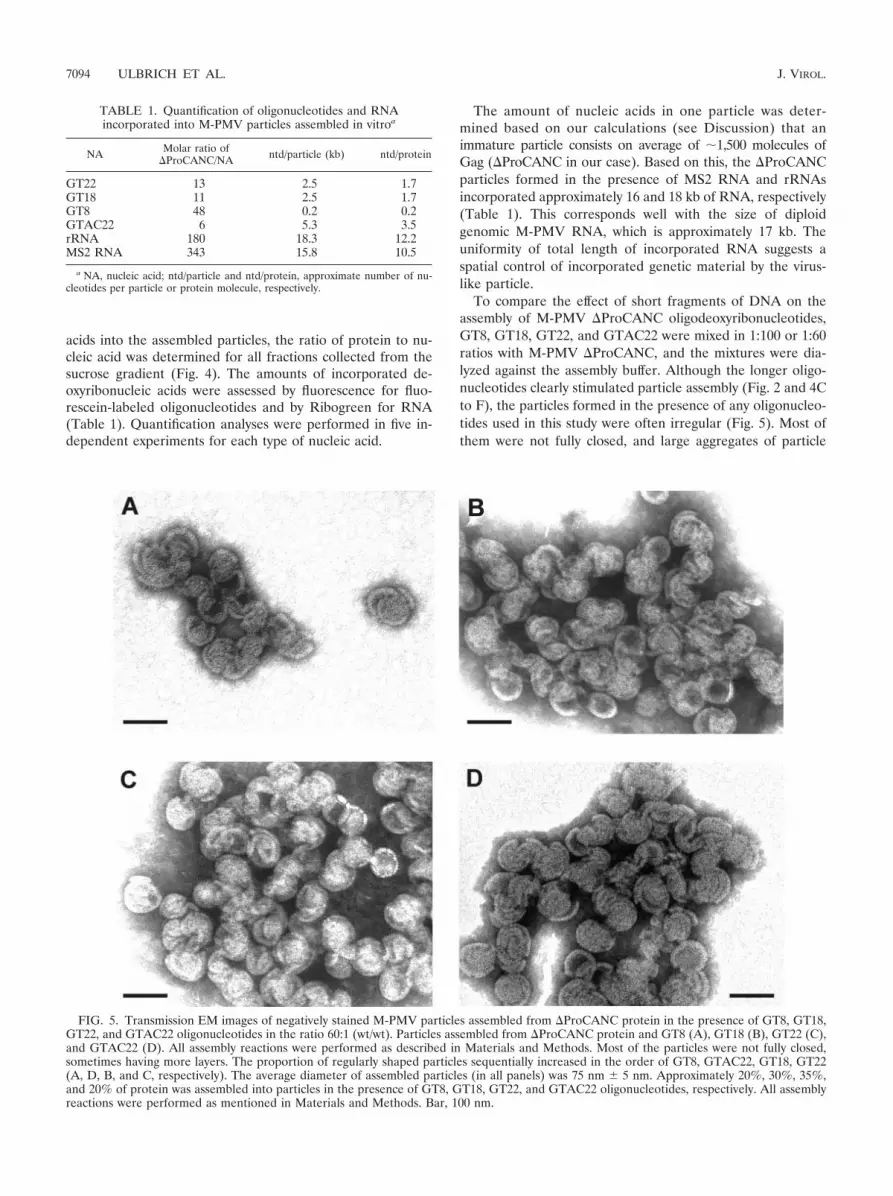
To compare the effect of short fragments of DNA on theassembly of M-PMV �ProCANC oligodeoxyribonucleotides,GT8, GT18, GT22, and GTAC22 were mixed in 1:100 or 1:60ratios with M-PMV �ProCANC, and the mixtures were dia-lyzed against the assembly buffer. Although the longer oligo-nucleotides clearly stimulated particle assembly (Fig. 2 and 4Cto F), the particles formed in the presence of any oligonucleo-tides used in this study were often irregular (Fig. 5). Most ofthem were not fully closed, and large aggregates of particle
FIG. 5. Transmission EM images of negatively stained M-PMV particles assembled from �ProCANC protein in the presence of GT8, GT18,GT22, and GTAC22 oligonucleotides in the ratio 60:1 (wt/wt). Particles assembled from �ProCANC protein and GT8 (A), GT18 (B), GT22 (C),and GTAC22 (D). All assembly reactions were performed as described in Materials and Methods. Most of the particles were not fully closed,sometimes having more layers. The proportion of regularly shaped particles sequentially increased in the order of GT8, GTAC22, GT18, GT22(A, D, B, and C, respectively). The average diameter of assembled particles (in all panels) was 75 nm 5 nm. Approximately 20%, 30%, 35%,and 20% of protein was assembled into particles in the presence of GT8, GT18, GT22, and GTAC22 oligonucleotides, respectively. All assemblyreactions were performed as mentioned in Materials and Methods. Bar, 100 nm.
TABLE 1. Quantification of oligonucleotides and RNAincorporated into M-PMV particles assembled in vitroa
NA Molar ratio of�ProCANC/NA ntd/particle (kb) ntd/protein
GT22 13 2.5 1.7GT18 11 2.5 1.7GT8 48 0.2 0.2GTAC22 6 5.3 3.5rRNA 180 18.3 12.2MS2 RNA 343 15.8 10.5
a NA, nucleic acid; ntd/particle and ntd/protein, approximate number of nu-cleotides per particle or protein molecule, respectively.
7094 ULBRICH ET AL. J. VIROL.

fragments were frequently found. We also observed a shift indensity of the protein within the sucrose gradient fractionstoward the lower densities in the case of GT8 oligonucleotidecompared to that of the other oligonucleotides used (Fig. 4).This is in agreement with higher proportions of fragments andincompletely closed particles that were observed by electronmicroscopy compared to the particles assembled in the pres-ence of GT18 and GT22 (Fig. 5). The amounts of incorporatedDNA, calculated as the length in kilobases per particle, rangedfrom 2.5 kb to 0.2 kb for GT22 and GT8, respectively (Table1), under conditions where excess DNA remained unincorpo-rated on top of the gradients. Approximately 5.3 kb of theGTAC22 oligonucleotide, which is expected to be doublestranded under the conditions of the experiment, was incorpo-rated in one particle. The fact that this number is double thatof the single-stranded GT22 (�2.5 kb) suggests that the DNAlength defines the efficiency of incorporation regardless of itssingle- or double-stranded form.
In order to investigate whether the low efficiency of DNAincorporation into the particles is caused by the limitation ofnucleic acid availability, the �ProCANC protein was titratedwith GT22 oligonucleotides. The protein was mixed with oli-gonucleotides in ratios of 200:1, 60:1, and 10:1 (wt/wt) anddialyzed in the assembly buffer for 2 to 3 h. The resultingmaterial was analyzed as in the previous experiments by SDS-PAGE and electron microscopy. It was found that varyingamounts of GT22 affected the yield of particles; however, it didnot affect the ratio of proteins to oligonucleotides in assembledparticles (data not shown).
(iii) Assembly of M-PMV CANC in vitro. In contrast to�ProCANC, the CANC protein formed no detectable struc-tures in vitro under these conditions. It should be noted herethat three independent purification methods were used (in-cluding two avoiding denaturation; see Materials and Meth-ods), and circular dichroism spectroscopy proved the signif-icant proportion of �-helical motifs comparable to M-PMV�ProCANC protein. The effect of pH (5 to 10), ionicstrength (0 to 2 M NaCl), Zn2� concentration (1 �M to 1mM), protein concentration (0.1 to 10 mg/ml), and assemblytemperature (4 to 42°C) was tested in order to optimizeassembly conditions, but no structures were observed forany combination of these conditions tested. Based on ourprevious data showing that the same construct forms sheetsin the cytoplasm of E. coli (69), we investigated whethersome cellular factor could promote the in vitro assembly ofM-PMV CANC. For this purpose we included E. coli cellextract in the CANC assembly mixture. Even under theseconditions, we observed only protein aggregates without anyevidence of organized structures (data not shown).
Since the results obtained with M-PMV CANC constructsvary from those described for a similar construct from RSVor HIV-1, we have used HIV-1 CANC and �ProCANC ascontrol proteins in order to determine if the assembly con-ditions used in our study would allow assembly of otherretroviral Gag molecules. Both sedimentation analysis andelectron microscopy were used to assess the efficiency ofassembly. Figure 6 indicates that for both HIV-1 CANC and�ProCANC, a majority of the protein that had assembledinto tubular particles in the presence or in the absence of
RNA sedimented at the bottom of the gradient. It is inter-esting that the HIV-1 �ProCANC protein assembles intotubular particles, since it lacks the ability to form the N-terminal proline-aspartate salt bridge that has been re-ported to be the driving force for tube formation (76). Thus,additional interactions appear to be involved in this process,as is the case for RSV, where CANC proteins with shortN-terminal extensions still assembled tubes and a stretch of25 amino acids was required for efficient assembly of spheres(41). Short tubular particles (of a length comparable tosphere diameter) banded together with the spherical parti-cles (Fig. 6, MS2 RNA, fractions 7 to 10). Under theseconditions, the HIV-1 CANC formed predominantly (ap-proximately 99%) tubes and only occasional core-like struc-tures in the presence and in the absence of RNA. Thediameter of the tubes was greater in the presence of RNAthan in the tubes assembled from pure protein itself (Fig. 7Aand B). Similarly, HIV-1 �ProCANC in the absence ofnucleic acid formed tubes (50 to 80%) and conical structuresresembling cores. The presence of RNA during the assemblyof HIV-1 �ProCANC resulted in the rare formation ofspherical particles (approximately 1%), with an average di-ameter of 80 to 85 nm, in addition to tubular structures andcores (10 to 40% of the total; Fig. 7C and D). The diametersof HIV-1 �ProCANC tubular structures were also greater in thepresence of RNA than in the absence of RNA. In the absence ofnucleic acid, the diameter of CANC HIV-1 tubes was 35 nm witha standard deviation of 4 nm, and that for �ProCANC HIV-1was 34 nm 3 nm. In the presence of RNA, the diameter ofCANC HIV-1 tubes was 46 nm 5 nm, which is in goodcorrelation with the data obtained for CANC HIV-1 tubularstructures previously presented by several authors reporting 44to 55 nm (8, 10, 36). �ProCANC HIV-1 formed tubes of adiameter 44 nm 4 nm. These measurements were performedmore than 100 times for each sample. All HIV-1 tubes dis-cussed in this study were of variable lengths, ranging from 80nm to 1,500 nm, which is in agreement with the published data(10).
FIG. 6. SDS-PAGE analysis of assembled HIV-1 proteins in su-crose gradient fractions. �ProCANC and CANC HIV proteins (con-centration of 0.6 mg/ml, i.e., 19 �M) were assembled without additionof nucleic acids or with MS2 phage RNA at a ratio of 10:1 (wt/wt). Thereaction mixtures were centrifuged to equilibrium through a 10 to 60%(wt/wt) sucrose gradient, and the fractions were analyzed by SDS-PAGE. Fraction 12 represents aggregated material that was pelleted atthe bottom of the tube. The sucrose densities of the fractions were asfollows: 1, 0.058 g/ml; 2, 0.069 g/ml; 3, 0.084 g/ml; 4, 1.10 g/ml; 5, 1.12g/ml; 6, 1.15 g/ml; 7, 1.18 g/ml; 8, 1.21 g/ml; 9, 1.23 g/ml; 10, 1.26 g/ml;and 11, 1.28 g/ml. NA, nucleic acid. (A) CANC HIV-1 protein;(B) �ProCANC HIV-1 protein.
VOL. 80, 2006 NUCLEIC ACID INCORPORATION IN CANC PARTICLES 7095

DISCUSSION
We have reported previously that a large portion of theM-PMV Gag polyprotein is dispensable for the bacterialassembly of immature capsid-like structures (69). As forother retroviruses, such as HIV-1 and RSV, the capsid (CA)and nucleocapsid (NC) domains were critical for particleformation in vitro. The presence of the N-terminal prolinein the CA domain was shown to play a critically importantrole for M-PMV CANC assembly in E. coli, where CANCassembled in sheets and �ProCANC mostly assembled intospheres (69). This observation was consistent with our pre-diction that CANC with a matured N terminus might rep-resent the assembly-competent material for mature coreformation. On the other hand, because of its inability toform the Pro1-Asp50 (or Asp57) salt bridge, the M-PMV�ProCANC N terminus should resemble that of CA in theGag precursor, which would then direct assembly into im-mature spherical particles.
In the present study, we have used an in vitro system to
investigate the requirements for efficient assembly of particlesfrom purified CANC and �ProCANC proteins and to comparethe differences in assembly of M-PMV and HIV-1. We havedemonstrated that the absence of N-terminal proline was es-sential for M-PMV CANC to assemble in vitro, in contrast tothe bacterial expression/assembly system, where this proteinassembled into planar structures. Purified CANC protein didnot assemble in vitro even under the most favorable conditionsfor �ProCANC assembly (100 mM NaCl, 50 mM Tris-HCl, 1�M ZnCl2, pH 8.0, in the presence of RNA). This suggests thatthe intracellular environment of E. coli facilitates the assemblyof CANC structures, perhaps by providing a cellular factor(s)necessary for correct protein folding and CANC-CANC inter-actions. In contrast, several research groups have shown thatHIV-1 CANC assembles in vitro into tubular structures (10, 22,36, 37). Similarly, we were able to show that the HIV-1 CANCprotein readily assembled into tubes under the same condi-tions where M-PMV CANC did not (Fig. 7). HIV-1 CANCassembled in the absence of RNA; however, RNA did increase
FIG. 7. Transmission EM images of negatively stained tubes assembled from HIV-1 CANC and �ProCANC proteins. (A) Tubes formed fromnucleic acid-free CANC. (B) Tubes assembled from CANC protein and bacteriophage MS2 RNA at a ratio of 10:1 (wt/wt). (C) Nucleic acid-free�ProCANC particles. (D) Particles assembled from �ProCANC and bacteriophage MS2 RNA at a ratio of 10:1 (wt/wt). All assembly reactionswere performed as mentioned in Materials and Methods. Predominantly tubes were observed for CANC both in the presence and the absence ofnucleic acids (no spheres were observed, and occasional cores did not exceed 1%). Nucleic acid-free �ProCANC particles assembled variablenumbers of core-like structures (�20 to 50%) versus tubes. Particles assembled from �ProCANC and bacteriophage MS2 RNA assembled amajority of tubes as well as about 1% spheres and �10 to 40% core-like structures. Approximately 70% and 80% of CANC protein was efficientlyassembled into particles without or in the presence of MS2 RNA, respectively. For �ProCANC, approximately 80% and 90% of protein wasefficiently assembled into particles without or with addition of MS2 RNA, respectively. Bar, 100 nm.
7096 ULBRICH ET AL. J. VIROL.

the efficiency of its assembly, and an increase in tube diameterwas also observed in the presence of nucleic acid. The inabilityof M-PMV CANC to form tubes may reflect some basic struc-tural difference in the CA proteins of HIV-1 and M-PMV,since purified M-PMV CA aggregated without any definedstructures in the presence of high salt (data not shown). Thiscontrasts with the results with purified HIV-1 CA, which haspreviously been shown to form tubes under high protein andsalt concentrations (22, 28, 29, 36, 50).
Unlike in RSV and HIV-1, the assembly of M-PMV �ProCANCwas independent of salt concentrations between 50 mM and200 mM, temperatures between 5°C and 37°C, and pHwithin the range of 5.5 to 9 (data not shown). PurifiedM-PMV �ProCANC formed spheres in a manner similar tothat of CANC from other retroviruses, where short exten-sions as an N terminus of MA were necessary for the as-sembly of spherical particles (44, 76).
The exact role of the nucleic acid interactions with Gagproteins during the assembly process is still not completelyunderstood. This study shows that assembly of �ProCANCoccurs even in the absence of nucleic acids, which is in agree-ment with previously published data for HIV-1 Gag (78, 79).Calculations based on experimental data show that the RSVNC binding site for nucleic acids spans eight nucleotides andtherefore a 16-mer should be sufficient for bridging togethertwo NC molecules (53, 54). HIV-1 and simian immunodefi-ciency virus NC were shown to have binding site sizes similar tothose of RSV NC (73, 74, 80). For HIV-1 it has been proposedthat binding of nucleic acid to NC concentrates the Gagpolyproteins and induces CA-CA interactions that lead subse-quently to the assembly of immature capsids. In this model,nucleic acids initially promote the interaction between car-boxy-terminal domains of the CA protein and a spacer peptidebetween CA and NC (38, 68). In the case of M-PMV, ananalogous spacer separating CA and NC has not been identi-fied, and data from the bacterial expression system show thatthe minimal requirement for assembly of a C-terminally trun-cated �ProCANC is the N-terminal 37 amino acids of NC (70).
The presence of residual nucleic acids in purified proteinsamples remains an issue in the experiments evaluating therole for nucleic acids in Gag assembly. In order to address thisquestion, we have used a Ribogreen nucleic acid quantitationkit in addition to the standard spectrophotometric measure-ment of the A260/A280 ratio. Both methods showed that con-tamination with nucleic acids of bacterial origin was less than10 ng of nucleic acid per assembly reaction (60 �g of protein ina total volume of 100 �l), which corresponds to the A260/A280
ratio of �0.7 that is considered negligible (53, 81). �ProCANCdeprived of nucleic acids assembled structures comparable tothose formed from the same protein in the presence of oligo-nucleotides (Fig. 3 and 5). However, the yield of particlesincreased with the length of oligonucleotides used (Fig. 4). Ourdata presented here suggest that oligonucleotides promote M-PMV �ProCANC assembly but are not incorporated in thestoichiometric amounts that would be necessary to serve asscaffolding units as proposed by Ma and Vogt (54). The lack ofa requirement for nucleic acid as the structural component ofthe capsid and the enhancement of assembly efficiency by oli-gonucleotides as short as GT8 could be described by a modelthat is consistent with two previously published observations.
First, the mechanism proposed by Ma and Vogt (54) originatesfrom the observation that GT16 promotes assembly of CANCdimers; however, the oligonucleotide per se is dispensable forthe dimer stability after its formation (54). Second, the oligo-nucleotides bound to the assembled particles are in dynamicequilibrium, as suggested by the exchange of bound nucleicacids for unbound ones (24). It therefore seems feasible thatnucleic acid binding promotes a cooperative structuralchange recruiting an additional interaction domain in M-PMV�ProCANC. Such a structural change could then induce therelease of nucleic acid, which would become available for an-other CANC interaction. This would explain how a low nucleicacid/protein molar ratio promotes assembly. In contrast to themodel proposed by Ma and Vogt (54), the role of oligonucle-otides would not be in linking two molecules of �ProCANCtogether but rather in promoting the conformational changeof �ProCANC monomers. The fact that the M-PMV NCstructured core domain contains two independently folded,rotationally uncorrelated globular domains connected by aseven-residue flexible linker (30) makes it likely that signif-icant changes in their orientation will be induced by nucleicacid binding. The loose binding of nucleic acids after suchchange would also allow its release from the core for reversetranscription.
The sufficient length of nucleic acid seems to play an impor-tant role in the assembly, as M13 phage DNA (our unpub-lished data) and the longer RNA of MS2 phage were bettercatalysts for particle assembly than DNA oligonucleotides, andits presence promoted formation of fully closed spherical par-ticles of uniform size (Fig. 3). This is in agreement with thefindings of Morikawa et al., who showed that RNA couldsupport the assembly of virus-like particles from isolatedHIV-1 Gag (58, 59). The M-PMV �ProCANC particles con-tained significantly larger amounts of RNA compared to any ofthe oligonucleotides in this study (Table 1). The calculationswere done for particles containing �1,500 �ProCANC mole-cules. Such an estimate, which is lower than recently publisheddata for HIV-1 (9), was based on our calculation of proteinsubunits in EM pictures (approximately 500 particles wereevaluated) and previously published data (25, 63, 75).
In summary, we have shown here that in vitro assembly ofM-PMV �ProCANC can be stimulated by both DNA oligo-nucleotides and nonviral RNA. These studies point to a modelin which nucleic acid, through a transient interaction with theNC domain, induces a conformational change that promotesassembly of Gag. Such a model may be broadly applicable toretroviruses that employ different morphogenetic pathways.
ACKNOWLEDGMENTS
This work was supported by Grant Agency of the Czech Republic203/03/P094, Grant Agency of the Academy of Sciences GrantA4055304, and research projects supported by the Czech Ministry ofEducation: 1M6837805002, Z 40550506, and MSM 6046137305.
REFERENCES
1. Accola, M. A., B. Strack, and G. Gottlinger. 2000. Efficient particle produc-tion by minimal Gag constructs which retain the carboxy-terminal domain ofhuman immunodeficiency virus type 1 capsid-p2 and a late assembly domain.J. Virol. 74:5395–5402.
2. Barklis, E., J. McDermott, S. Wilkens, S. Fuller, and D. Thompson. 1998.Organization of HIV-1 capsid proteins on a lipid monolayer. J. Biol. Chem.273:7177–7180.
VOL. 80, 2006 NUCLEIC ACID INCORPORATION IN CANC PARTICLES 7097

3. Berkowitz, R., J. Fisher, and S. P. Goff. 1996. RNA packaging. Curr. Top.Microbiol. Immunol. 214:177–218.
4. Berthtet-Colominas, C., S. Monaco, A. Novelli, G. Sibai, F. Mallet, and S.Cusack. 1999. Head-to-tail dimers and interdomain flexibility revealed by thecrystal structure of HIV-1 capsid protein (p24) complexed with monoclonalantibody Fab. EMBO J. 18:1124–1136.
5. Birkett, A. J., B. Yelamos, I. Rodriguez-Crespo, F. Gavilanes, and D. L.Peterson. 1997. Cloning, expression, purification and characterization of themajor core protein (p26) from equine infectious anemia virus. Biochim.Biophys. Acta 1339:62–72.
6. Borsetti, A., A. Ohagen, and H. G. Gottlinger. 1998. The C-terminal half ofthe human immunodeficiency virus type 1 Gag precursor is sufficient forefficient particle assembly. J. Virol. 72:9313–9317.
7. Bowles, N. E., P. Damay, and P.-F. Spahr. 1993. Effect of rearrangementsand duplications of the Cys-His motifs of Rous sarcoma virus nucleocapsidprotein. J. Virol. 67:623–631.
8. Briggs, J. A. G., T. Wilk, R. Welker, H. G. Krausslich, and S. D. Fuller. 2003.Structural organization of authentic, mature HIV-1 virions and cores.EMBO J. 22:1707–1715.
9. Briggs, J. A. G., M. N. Simon, I. Gross, H. G. Krausslich, S. D. Fuller, V. M.Vogt, and M. C. Johnson. 2004. The stoichiometry of Gag protein in HIV-1.Nat. Struct. Mol. Biol. 11:672–675.
10. Campbell, S., and V. M. Vogt. 1995. Self-assembly in vitro of purified CA-NCproteins from Rous sarcoma virus and human immunodeficiency virus type1. J. Virol. 69:6487–6497.
11. Campbell, S., and V. M. Vogt. 1997. In vitro assembly of virus-like particleswith Rous sarcoma virus Gag deletion mutants: identification of the p10domain as a morphological determinant in the formation of spherical par-ticles. J. Virol. 71:4425–4435.
12. Campbell, S., and A. Rein. 1999. In vitro assembly properties of humanimmunodeficiency virus type 1 Gag protein lacking the p6 domain. J. Virol.73:2270–2279.
13. Campos-Olivas, R., J. L. Newman, and M. F. Summers. 2000. Solutionstructure and dynamics of the Rous sarcoma virus capsid protein and com-parison with capsid proteins of other retroviruses. J. Mol. Biol. 296:633–649.
14. Cimarelli, A., and J. L. Darlix. 2002. Assembling the human immunodefi-ciency virus type 1. Cell. Mol. Life Sci. 59:1166–1184.
15. Cornilescu, C. C., F. Bouamr, X. Yao, C. Carter, and N. Tjandra. 2001.Structural analysis of the N-terminal domain of the human T-cell leukemiavirus capsid protein. J. Mol. Biol. 306:783–797.
16. Dannull, J., A. Surovoy, G. Jung, and K. Moelling. 1994. Specific binding ofHIV-1 nucleocapsid protein to PSI RNA in vitro requires N-terminal zincfinger and flanking basic amino acid residues. EMBO J. 13:1525–1533.
17. Darlix, J. L., M. Lapadat-Tapolsky, H. de Rocquigny, and B. P. Roques.1995. First glimpses at structure-function relationships of the nucleocapsidprotein of retroviruses. J. Mol. Biol. 254:523–537.
18. De Rocquigny, H., C. Gabus, A. Vincent, M. C. Fournie-Zaluski, B. Roques,and J. L. Darlix. 1992. Viral RNA annealing activities of human immuno-deficiency virus type 1 nucleocapsid protein require only peptide domainsoutside the zinc fingers. Proc. Natl. Acad. Sci. USA 89:6472–6476.
19. Dorfman, T., J. Luban, S. P. Goff, W. A. Haseltine, and H. G. Gottlinger.1993. Mapping of functionally important residues of a cysteine-histidine boxin the human immunodeficiency virus type 1 nucleocapsid protein. J. Virol.67:6159–6169.
20. Dorfman, T., A. Bukovsky, A. Ohagen, S. Hoglund, and H. G. Gottlinger.1994. Functional domains of the capsid protein of human immunodeficiencyvirus type 1. J. Virol. 68:8180–8187.
21. Dupraz, P., S. Oertle, C. Meric, P. Damay, and P.-F. Spahr. 1990. Pointmutations in the proximal Cys-His box of Rous sarcoma virus nucleocapsidprotein. J. Virol. 64:4978–4987.
22. Ehrlich, L. S., B. E. Agresta, and C. A. Carter. 1992. Assembly of recombi-nant human immunodeficiency virus type 1 capsid protein in vitro. J. Virol.66:4874–4883.
23. Ehrlich, L. S., T. Liu, S. Scarlata, B. Chu, and C. A. Carter. 2001. HIV-1capsid protein forms spherical (immature-like) and tubular (mature-like)particles in vitro: structure switching by pH-induced conformational changes.Biophys. J. 81:586–594.
24. Feng, Y. X., T. Li, S. Campbell, and A. Rein. 2002. Reversible binding ofrecombinant human immunodeficiency virus type 1 Gag protein to nucleicacids in virus-like particle assembly in vitro. J. Virol. 76:11757–11762.
25. Fuller, S. D., T. Wilk, B. D. Gowen, H. G. Krausslich, and V. M. Vogt. 1997.Cryo-electron microscopy reveals ordered domains in the immature HIV-1particle. Curr. Biol. 7:729–738.
26. Gamble, T. L., F. F. Vajdos, S. Yoo, D. K. Worthylake, M. Houseweart, W. I.Sundquist, and C. P. Hill. 1996. Crystal structure of human cyclophilin Abound to the amino-terminal domain of HIV-1 capsid. Cell 87:1285–1294.
27. Gamble, T. L., S. Yoo, F. F. Vajdos, U. K. von Schwedler, D. K. Worthylake,H. Wang, J. P. McCutcheon, W. I. Sundquist, and C. P. Hill. 1997. Structureof the carboxyl-terminal dimerization domain of the HIV-1 capsid protein.Science 278:849–853.
28. Ganser, B. K., S. Li, V. Y. Klishko, J. T. Finch, and W. I. Sundquist. 1999.
Assembly and analysis of conical models for the HIV-1 core. Science 283:80–83.
29. Ganser-Pornillos, B. K., U. K. von Schwedler, K. M. Stray, C. Aiken, andW. I. Sundquist. 2004. Assembly properties of the human immunodeficiencyvirus type 1 CA protein. J. Virol. 78:2545–2552.
30. Gao, Y., K. Kaluarachchi, and D. P. Giedroc. 1998. Solution structure andbackbone dynamics of Mason-Pfizer monkey virus, MPMV, nucleocapsidprotein. Prot. Sci. 7:2265–2280.
31. Gitti, R. K., B. M. Lee, J. Walker, M. F. Summers, S. Yoo, and W. I.Sundquist. 1996. Structure of the amino-terminal core domain of the HIV-1capsid protein. Science 273:231–235.
32. Gorelick, R. J., S. M. Nigida, L. O. Arthur, L. E. Henderson, and A. Rein.1988. Point mutants of Moloney murine leukemia virus that fail to packageviral RNA: evidence for specific RNA recognition by a “zinc finger-like”protein sequence. Proc. Natl. Acad. Sci. USA 85:8420–8424.
33. Gorelick, R. J., S. M. J. Nigida, J. W. J. Bess, L. O. Arthur, L. E. Henderson,and A. Rein. 1990. Noninfectious human immunodeficiency virus type 1mutants deficient in genomic RNA. J. Virol. 64:3207–3211.
34. Gorelick, R. J., S. M. Nigida, L. O. Arthur, L. E. Henderson, and A. Rein.1991. Role of nucleocapsid cysteine arrays in retroviral assembly and repli-cation: possible mechanisms in RNA encapsidation. In A. Kumar (ed.),Advances in molecular biology and targeted treatment for AIDS. PlenumPress, New York, N.Y.
35. Gorelick, R. J., D. J. Chabot, A. Rein, L. E. Henderson, and L. O. Arthur.1993. The two zinc fingers in the human immunodeficiency virus type 1nucleocapsid protein are not functionally equivalent. J. Virol. 67:4027–4036.
36. Gross, I., H. Hohenberg, and H. G. Krausslich. 1997. In vitro assemblyproperties of purified bacterially expressed capsid proteins of human immu-nodeficiency virus. Eur. J. Biochem. 249:592–600.
37. Gross, I., H. Hohenberg, C. Huckhagel, and H. G. Krausslich. 1998. N-terminal extension of human immunodeficiency virus capsid protein convertsthe in vitro assembly phenotype from tubular to spherical particles. J. Virol.72:4798–4810.
38. Gross, I., H. Hohenberg, T. Wilk, K. Wiegers, M. Grattinger, B. Muller, S.Fuller, and H. G. Krausslich. 2000. A conformational switch controllingHIV-1 morphogenesis. EMBO J. 19:103–113.
39. Ivanov, D., J. R. Stone, J. L. Maki, T. Collins, and G. Wagner. 2005. Mam-malian SCAN domain dimer is a domain-swapped homolog of the HIVcapsid C-terminal domain. Mol. Cell 17:137–143.
40. Johnson, M. C., H. M. Scobie, Y. M. Ma, and V. M. Vogt. 2002. Nucleicacid-independent retrovirus assembly can be driven by dimerization. J. Virol.76:11177–11185.
41. Joshi, S. M., and V. M. Vogt. 2000. Role of the Rous sarcoma virus p10domain in shape determination of Gag virus-like particles assembled in vitroand within Escherichia coli. J. Virol. 74:10260–10268.
42. Karpel, R. L., L. E. Henderson, and S. Oroszlan. 1987. Interactions ofretroviral structural proteins with single-stranded nucleic acids. J. Biol.Chem. 262:4961–4967.
43. Khorasanizadeh, S., R. Campos-Olivas, and M. F. Summers. 1999. Solutionstructure of the capsid protein from the human T-cell leukemia virus type-1.J. Mol. Biol. 291:491–505.
44. Kingston, R., E. Z. Eisenmesser, T. Fitzon-Ostendorp, G. W. Schatz, V. M.Vogt, C. B. Post, and M. G. Rossmann. 2000. Structure and self-associationof the Rous sarcoma virus capsid protein. Structure 8:617–628.
45. Kingston, R. L., and V. M. Vogt. 2005. Domain swapping and retroviralassembly. Mol. Cell 17:166–167.
46. Klikova, M., S. S. Rhee, E. Hunter, and T. Ruml. 1995. Efficient in vivo andin vitro assembly of retroviral capsids from Gag precursor proteins expressedin bacteria. J. Virol. 69:1093–1098.
47. Lanman, J., J. Sexton, M. Sakalian, and P. E. Prevelige, Jr. 2002. Kineticanalysis of the role of intersubunit interactions in human immunodeficiencyvirus type 1 capsid protein assembly in vitro. J. Virol. 76:6900–6908.
48. Lanman, J., T. T. Lam, M. R. Emmett, A. G. Marshall, M. Sakalian, andP. E. Prevelige, Jr. 2004. Key interactions in HIV-1 maturation identified byhydrogen-deuterium exchange. Nat. Struct. Mol. Biol. 11:676–677.
49. Levin, J. G., P. M. Grimley, J. M. Ramseur, and I. K. Berezesky. 1974.Deficiency of 60 to 70S RNA in murine leukemia virus particles assembledin cells treated with actinomycin D. J. Virol. 14:152–161.
50. Li, S., C. P. Hill, W. I. Sundquist, and J. T. Finch. 2000. Image reconstruc-tions of helical assemblies of the HIV-1 CA protein. Nature 407:409–413.
51. Linial, M., E. Medeiros, and W. S. Hayward. 1978. An avian oncovirusmutant (SE21Q1b) deficient in genomic RNA: biological and biochemicalcharacterization. Cell 15:1371–1381.
52. Linial, M. L., and A. D. Miller. 1990. Retroviral RNA packaging: sequencerequirements and implications. Curr. Top. Microbiol. 157:125–152.
53. Ma, Y. M., and V. M. Vogt. 2002. Rous sarcoma virus Gag protein-oligonu-cleotide interaction suggests a critical role for protein dimer formation inassembly. J. Virol. 76:5452–5462.
54. Ma, Y. M., and V. M. Vogt. 2004. Nucleic acid binding-induced Gag dimer-ization in the assembly of Rous sarcoma virus particles in vitro. J. Virol.78:52–60.
55. Meric, C., and P.-F. Spahr. 1986. Rous sarcoma virus nucleic acid-binding
7098 ULBRICH ET AL. J. VIROL.

protein p12 is necessary for viral 70S RNA dimer formation and packaging.J. Virol. 60:450–459.
56. Meric, C., and S. P. Goff. 1989. Characterization of Moloney murine leuke-mia virus mutants with single-amino-acid substitutions in the Cys-His box ofthe nucleocapsid protein. J. Virol. 63:1558–1568.
57. Momany, C., L. C. Kovari, A. J. Prongay, W. Keller, R. K. Gitti, B. M. Lee,A. E. Gorbalenya, L. Tong, J. McClure, L. S. Ehrlich, M. F. Summers, C.Carter, and M. G. Rossmann. 1996. Crystal structure of dimeric HIV-1capsid protein. Nat. Struct. Biol. 3:763–770.
58. Morikawa, Y., T. Goto, and K. Sano. 1999. In vitro assembly of humanimmunodeficiency virus type 1 Gag protein. J. Biol. Chem. 274:27997–28002.
59. Morikawa, Y., T. Goto, and F. Momose. 2004. Human immunodeficiencyvirus type 1 gag assembly through assembly intermediates. J. Biol. Chem.279:31964–31972.
60. Muriaux, D., J. Mirro, D. Harvin, and A. Rein. 2001. RNA is structuralelement in retrovirus particles. Proc. Natl. Acad. Sci. USA 98:5246–5251.
61. Nandhagopal, N., A. A. Simpson, M. C. Johnson, A. B. Francisco, G. W.Schatz, M. G. Rossman, and V. M. Vogt. 2004. Dimeric Rous sarcoma viruscapsid protein structure relevant to immature Gag assembly. J. Mol. Biol.335:275–282.
62. Nermut, V. N., P. Bron, D. Thomas, M. Rumlova, T. Ruml, and E. Hunter.2002. Molecular organization of Mason-Pfizer monkey virus capsids assem-bled from Gag polyprotein in Escherichia coli. J. Virol. 76:4321–4330.
63. Parker, S. D., J. S. Wall, and E. Hunter. 2001. Analysis of Mason-Pfizermonkey virus Gag particles by scanning transmission electron microscopy.J. Virol. 75:9543–9548.
64. Poon, D. T., J. Wu, and A. Aldovini. 1996. Charged amino acid residues ofhuman immunodeficiency virus type 1 nucleocapsid p7 protein involved inRNA packaging and infectivity. J. Virol. 70:6607–6616.
65. Reicin, A. S., S. Paik, R. D. Berkowitz, J. Luban, I. Lowy, and S. P. Goff.1995. Linker insertion mutations in the human immunodeficiency virus type1 gag gene: effects on virion particle assembly, release, and infectivity. J. Vi-rol. 69:642–650.
66. Rein, A. 1994. Retroviral RNA packaging: a review. Arch. Virol. Suppl.9:513–522.
67. Rein, A., D. P. Harvin, J. Mirro, S. M. Ernst, and R. J. Gorelick. 1994.Evidence that a central domain of nucleocapsid protein is required for RNApackaging in murine leukemia virus. J. Virol. 68:6124–6129.
68. Roldan, A., R. S. Russell, B. Marchand, M. Gotte, C. Liang, and M. A.Wainberg. 2004. In vitro identification and characterization of an early com-plex linking HIV-1 genomic RNA recognition and Pr55Gag multimerization.J. Biol. Chem. 279:39886–39894.
69. Rumlova-Klikova, M., E. Hunter, M. V. Nermut, I. Pichova, and T. Ruml.2000. Analysis of Mason-Pfizer monkey virus Gag domains required for
capsid assembly in bacteria: role of the N-terminal proline residue of CA indirecting particle shape. J. Virol. 74:8452–8459.
70. Rumlova, M., T. Ruml, J. Pohl, and I. Pichova. 2003. Specific in vitrocleavage of Mason-Pfizer monkey virus capsid protein: evidence for a po-tential role of retroviral protease in early stages of infection. Virology 310:310–318.
71. Sambrook, J., E. F. Fritsch, and T. Maniatis. 1989. Molecular cloning: alaboratory manual, 2nd ed. Cold Spring Harbor Laboratory Press, ColdSpring Harbor, N.Y.
72. Schmalzbauer, E., B. Strack, J. Dannull, S. Guehmann, and K. Moelling.1996. Mutations of basic amino acids of NCp7 of human immunodeficiencyvirus type 1 affect RNA binding in vitro. J. Virol. 70:771–777.
73. Urbaneja, M. A., B. P. Kane, D. G. Johnson, R. J. Gorelick, L. E. Henderson,and J. Casas-Finet. 1999. Binding properties of the human immunodefi-ciency virus type 1 nucleocapsid protein p7 to a model RNA: elucidation ofthe structural determinants for function. J. Mol. Biol. 287:59–75.
74. Urbaneja, M. A., C. F. McGrath, B. P. Kane, L. E. Henderson, and J.Casas-Finet. 2000. Nucleic acid binding properties of the simian immuno-deficiency virus nucleocapsid protein NCp8. J. Biol. Chem. 275:10394–10404.
75. Vogt, M. V., and M. Simon. 1999. Mass determination of Rous sarcoma virusvirions by scanning transmission electron microscopy. J. Virol. 73:7050–7055.
76. von Schwedler, U. K., T. L. Stemmler, V. Y. Klishko, S. Li, K. H. Albertine,D. R. Davis, and W. I. Sundquist. 1998. Proteolytic refolding of the HIV-1capsid protein amino-terminus facilitates viral core assembly. EMBO J.17:1555–1568.
77. von Schwedler, U. K., K. M. Stray, J. E. Garrus, and W. I. Sundquist. 2003.Functional surfaces of the human immunodeficiency virus type 1 capsidprotein. J. Virol. 77:5439–5450.
78. Wang, S. W., and A. Aldovini. 2002. RNA incorporation is critical for ret-roviral particle integrity after cell membrane assembly of Gag complexes.J. Virol. 76:11853–11865.
79. Wang, S. W., K. Noonan, and A. Aldovini. 2004. Nucleocapsid-RNA inter-actions are essential to structural stability but not to assembly of retroviruses.J. Virol. 78:716–723.
80. You, J. C., and C. S. McHenry. 1993. HIV nucleocapsid protein: expressionin Escherichia coli, purification, and characterization. J. Biol. Chem. 268:16519–16527.
81. Yu, F., S. M. Joshi, Y. M. Ma, R. L. Kingston, M. N. Simon, and V. M. Vogt.2001. Characterization of Rous sarcoma virus Gag particles assembled invitro. J. Virol. 75:2753–2764.
82. Zhang, Y., H. Qian, Z. Love, and E. Barklis. 1998. Analysis of the assemblyfunction of the human immunodeficiency virus type 1 Gag protein nucleo-capsid domain. J. Virol. 72:1782–1789.
83. Zlotnick, A. 2003. Are weak protein-protein interactions the general rule incapsid assembly? Virology 315:269–274.
VOL. 80, 2006 NUCLEIC ACID INCORPORATION IN CANC PARTICLES 7099