Embed Size (px)
Citation preview

�������� ����� ��
NMR structure of the N-terminal domain of capsid protein from the Mason-Pfizer monkey virus
Pavel Macek, Josef Chmelık, Ivana Krızova, Pavel Kaderavek, Petr Padrta,Lukas Zıdek, Marcela Wildova, Romana Hadravova, Radka Chaloupkova, IvaPichova, Tomas Ruml, Michaela Rumlova, Vladimır Sklenar
PII: S0022-2836(09)00736-0DOI: doi:10.1016/j.jmb.2009.06.029Reference: YJMBI 61521
To appear in: Journal of Molecular Biology
Received date: 7 April 2009Revised date: 8 June 2009Accepted date: 10 June 2009
Please cite this article as: Macek, P., Chmelık, J., Krızova, I., Kaderavek, P., Padrta,P., Zıdek, L., Wildova, M., Hadravova, R., Chaloupkova, R., Pichova, I., Ruml,T., Rumlova, M. & Sklenar, V., NMR structure of the N-terminal domain of cap-sid protein from the Mason-Pfizer monkey virus, Journal of Molecular Biology (2009),doi:10.1016/j.jmb.2009.06.029
This is a PDF file of an unedited manuscript that has been accepted for publication.As a service to our customers we are providing this early version of the manuscript.The manuscript will undergo copyediting, typesetting, and review of the resulting proofbefore it is published in its final form. Please note that during the production processerrors may be discovered which could affect the content, and all legal disclaimers thatapply to the journal pertain.

ACC
EPTE
D M
ANU
SCR
IPT
ACCEPTED MANUSCRIPT
NMR structure of the N-terminal domain of capsid protein from the Mason-Pfizer mon-
key virus
Pavel Macek†a,e, Josef Chmelík†a,e, Ivana Křížová†b, Pavel Kadeřáveka, Petr Padrtaa,
Lukáš Žídeka, Marcela Wildováb, Romana Hadravováb, Radka Chaloupkovác, Iva
Pichováb, Tomáš Rumld, Michaela Rumlováb*& Vladimír Sklenářa*
a National Centre for Biomolecular Research, Faculty of Science, Masaryk University,
Kotlářská 2, Brno, 611 37, Czech Republic
b Gilead Sciences & IOCB Research Centre, Institute of Organic Chemistry and Biochemistry,
Academy of Sciences of the Czech Republic, Flemingovo nám. 2, 166 10, Prague, Czech
Republic
c Loschmidt Laboratories, Institute of Experimental Biology & National Centre for
Biomolecular Research, Masaryk University, Kotlářská 2, Brno, 611 37, Czech Republic
d Institute of Chemical Technology, Technická 5, Praha, 166 28, Czech Republic
e Institute of Microbiology, Academy of Sciences of the Czech Republic, Prague, Czech
Republic
†All authors contributed equally.
Keywords
M-PMV, betaretroviruses, capsid protein, NMR structure; internal dynamics; salt-bridge.
*Corresponding authors: E-mail address:
Michaela Rumlová:[email protected]
Vladimír Sklenář: [email protected]
*
1

ACC
EPTE
D M
ANU
SCR
IPT
ACCEPTED MANUSCRIPT
Summary
The high resolution structure of the N-terminal domain (NTD) of the retroviral capsid protein
(CA) of Mason-Pfizer monkey virus (M-PMV), a member of Betaretroviruses family, has
been determined by NMR. The M-PMV NTD CA structure is similar to the other retroviral
capsid structures and is characterized by a six-α-helical bundle and an N-terminal β-hairpin,
stabilized by an interaction of highly conserved residues, Pro1 and Asp57. Since the role of
the β-hairpin has been shown to be critical for formation of infectious viral core, we also in-
vestigated the functional role of M-PMV β-hairpin in two mutants, i.e., ΔP1NTDCA, and
D57ANTDCA, where the salt bridge stabilizing the wild-type structure was disrupted. NMR
data obtained for these mutants were compared to that for the wild-type. The main structural
changes were observed within the β-hairpin structure, within helices 2, 3 and 5, and in the
loop connecting helices 2-3. This observation is supported by biochemical data showing a dif-
ferent cleavage pattern of the wild-type and the mutated capsid – nucleocapsid fusion protein
(CANC) by M-PMV protease. Despite these structural changes, the mutants with the disrup-
ted salt-bridge are still able to assemble into immature, spherical particles. This confirms that
the mutual interaction and topology within the β-hairpin and helix 3 might correlate with the
changes of interaction between immature and mature lattice.
Introduction
Mason-Pfizer monkey virus (M-PMV) belongs to a family of Betaretroviruses that is
characteristic by an assembly of immature viral particles in the cytoplasm prior to their
transport to the plasma membrane and budding from the infected cells. Similar to other
retroviruses, M-PMV is an enveloped virus with two copies of genomic RNA packed within a
mature core. In contrast to the C-type retroviruses, for which Human immunodeficiency virus
2

ACC
EPTE
D M
ANU
SCR
IPT
ACCEPTED MANUSCRIPT
(HIV-1) may serve as a prototype, assembly of the D-type Mason-Pfizer monkey virus is
driven by polymerization of Gag polyproteins within the cytoplasm of the infected cells.
Resulting immature spherical particles are transported and bud through the plasma membrane.
A viral protease cleaves the Gag polyprotein precursors, enabling formation of infectious
virions outside of the infected cell.
The mature, cylindrical core of M-PMV contains a major structural protein, the capsid
protein CA (p27). This protein is initially synthesized as a part of the Gag polyprotein
precursor, from which it is released by a proteolytic cleavage together with matrix protein MA
(p10), phosphoprotein (pp24), protein p12, nucleocapsid protein NC (p14), and protein p4.
Three of these structural proteins, namely MA, CA, and NC, are common to all retroviruses.
After the proteolytic cleavage, the proteins rearrange to form the mature, infectious virions.
The matrix protein remains associated with the viral membrane in mature virions. Recently, it
has been shown that the M-PMV matrix protein interacts with Tctex-1 and through the dynein
motor machinery mediates the transport to the cytoplasmic assembly site 1. The capsid protein
forms the protein core that surrounds the nucleocapsid-RNA complex together with the viral
enzymes, reverse transcriptase (RT) and integrase (IN). Although assembly of the immature
retrovirus particles occurs at different places, their shape is spherical regardless of the virus
type. In contrast, the shape of a mature retroviral core varies significantly among different
genera: cylindrical shape is observed in Betaretroviridae (M-PMV), conical in Lentiviridae
(HIV-1), and spherical in Alpharetroviridae, Gamma-retroviridae, Deltaretroviridae, and in
Spumaretroviridae. Although the retroviral capsid proteins do not exhibit significant sequence
similarity, the tertiary structures of all three-dimensional structures solved to date are
remarkably similar 2-11. Retroviral CA proteins are comprised of two structural domains, the
N-terminal assembly domain (NTD) and the C-terminal dimerization domain (CTD),
connected by short flexible linkers 5, 6, 12. Mutational analyses have demonstrated that
3

ACC
EPTE
D M
ANU
SCR
IPT
ACCEPTED MANUSCRIPT
changes in the NTD affect especially the mature core assembly, while mutations in the CTD
influence the formation of immature particles 13-15.
All structures of the N-terminal domains of retroviral capsid proteins of HIV-1 5, 6, 12,
Rous sarcoma virus (RSV) 2, 9, Human T-cell leukemia virus (HTLV) 3, 8, Equine infectious
anemia virus (EIAV) 7, N-tropic murine leukemia virus (N-MLV) 16, B-tropic murine leuk-
emia virus (B-MLV) 10 and Jaagsiekte sheep retrovirus (JSRV) 11 solved to date are composed
of six to seven α-helices with almost identical topology. In addition, the N-terminal regions
form well defined β-hairpin structures with exception of the NTD CA protein structures of
RSV and EIAV solved by NMR and X-ray, respectively, where the β-hairpin is missing. The
β-hairpins are formed only upon Gag polyprotein processing, during which the N-terminal
proline is released, and the structure is then stabilized by formation of a salt bridge between
the released proline and a highly conserved aspartate D51 in HIV-1 6, D52 in RSV 9, and D54
in HTLV-1 3, D54 in N-MLV 16. Point mutations throughout this region block the proteolytic
processing of HIV-1 14, Moloney murine leukemia virus (MoMLV) 17, and M-PMV 18, and af-
fect the virus core assembly and infectivity of HIV-1 14, 19-21 and M-PMV 18. Although the
mutation data suggests that the β-hairpin formation is important for viral maturation and prop-
er core assembly, its exact role remains unclear. Von Schwedler et al. 14 suggested that the
proteolytic refolding and formation of the β-hairpin creates a new CA-CA interface in the ma-
ture capsid core. This model is supported by the data obtained also for HIV-1 14, 15, 20 and M-
PMV 22, which indicates that the formation of the β-hairpin structure can shift the assembly
from immature, spherical particles, when the β-hairpin is absent, to mature, tubular cores,
when the β-hairpin is present. However, structuring of the β-hairpin is not crucial for the as-
sembly of immature particles since the deletion of the entire β-hairpin (Δ20CANC) does not
prevent virus assembly into the spherical particles (Rumlová, unpublished data). In their X-
ray crystal structure, Mortuza and colleagues first showed a hexameric arrangement of B-
4

ACC
EPTE
D M
ANU
SCR
IPT
ACCEPTED MANUSCRIPT
MLV NTD CA and suggested that six β-hairpins might form an extended interaction network
within the mature CA hexamer 16. Recently, using results of electron cryomicroscopy and im-
age analysis of hexameric arrays of full-length HIV-1 CA, Ganser-Pornillos and colleagues
proposed that the β-hairpin may stabilize assembly-competent conformations of important
residues in helix 1 and 2 of HIV-1 NTD CA and/or that β-hairpin formation disrupts interac-
tions that stabilize the immature lattice 23.
Here we present the NMR structure of NTD CA of a Betaretroviridae family member,
M-PMV, with a special focus on a missing piece in the mosaic of available information,
namely on comparison of the wild-type with the constructs where residues critical for the β-
hairpin formation were mutated. The structure of the wild-type NTD CA comprising six
helices and an N-terminal β-hairpin, stabilized by an interaction of Pro1 and Asp57, is very
similar to the other retroviral NTD CA structures. We also show the NMR data of two
mutants, ΔP1 NTD CA and D57A NTD CA. Both these mutations dramatically influenced the
structure within the region of β-hairpin and helices α3 and α5, which then became susceptible
to cleavage by M-PMV protease. The effect of mutations was investigated also in the CANC
fusion protein that was previously shown to be assembly competent in its wild-type form.
Despite the structural changes of the NTD CA, both mutations introduced into the CANC fu-
sion protein resulted in assembling spherical immature particles, suggesting that the exact
wild-type alike positions of helices α1-α3 and helix α5 of M-PMV CA is not crucial for their
assembly.
Results
Solution structure of M-PMV NTD CA
The three dimensional structure of the M-PMV NTD CA was calculated based on
NOE distances, backbone φ and ψ dihedral angles, hydrogen bonds, and residual dipolar
5

ACC
EPTE
D M
ANU
SCR
IPT
ACCEPTED MANUSCRIPT
couplings (RDCs) as described in Materials and Methods. Table 1 shows summary of the
restraints used for the structure calculation. The superposition of ten structures with the lowest
energy is shown in Figure 1 and their structure quality statistics is summarized in Table 1 (for
additional structural statistics see Table S1 in Appendix A). No distance and dihedral angle
restraints were violated among 50 structures with the lowest energy deposited in PDB (PDB
code 2kgf). The Q-factor for RDCs was 0.225. The axial component (normalized to the H-N
bond) and rhombicity of the RDC tensor optimized during the structure calculation were (-
8.73±0.19) Hz and 0.45±0.04, respectively. These values are close to those determined from
the RDC distribution.
Figure 1
Table 1
WHATIF Z-scores were superior to those calculated for NMR structures of related
retroviral capsid proteins (PDB codes 1G03, 1GWP), with the exception of RSV (PDB code
1D1D, Z-score of -2.7). Backbone torsion angles of five residues occasionally occurred in the
disallowed regions of the Ramachandran plot: Asp97 in four structures, Thr102 in two
structures, Arg14, Asp117, and Val137 in one structure. With the exception of Arg14, these
residues are located in the loop or terminal regions (see below). The negative WHATIF Z-
scores in Table 1 reflect somewhat lower quality of the refined structures with respect to the
reliable X-ray structures in the PDB database.
Figure 2
Figure 2 shows a ribbon diagram of the structure with the lowest energy. The M-PMV
NTD CA structure is characterized by a five-membered α-helical bundle with helical axes in
an antiparallel arrangement and by an N-terminal β-hairpin. Helices of the bundle are formed
by residues 21-33 (helix α1), 39-48 (helix α2), 55-65 (helix α3), 69-92 (helix α4), and 118-
132 (helix α6). Last two helices of the bundle are connected through a twenty-four residue
6

ACC
EPTE
D M
ANU
SCR
IPT
ACCEPTED MANUSCRIPT
chain including a partially ordered loop (residues 93-109) and a short α-helix (helix α5,
residues 110-114). Among the retroviral capsid proteins, this region is the least conserved and
significantly varies in length 9. The N-terminal β-hairpin, which strands are formed by
residues 2-7 and 11-16, is in a parallel arrangement to the axes of the α-helices. The
orientation of the β-hairpin with respect to the α-helical core is determined by 14 NOEs
defining distances between residues 2-52, 4-52, 2-53, 16-110, and 2-114 and positioning Pro1
near to Asp57. The close proximity of Pro1 and Asp57 indicates the salt bridge formation
between positively charged Pro1 and negatively charged Asp57.
Backbone 15N dynamics of M-PMV NTD CA
To assess dynamic properties of M-PMV NTD CA on the picosecond/nanosecond
timescale, the quantitative measurement of the longitudinal relaxation time, T1, the transverse
relaxation time, T2, and the steady-state {1H}-15N NOE enhancement (ssNOE) was performed.
Experimental values of T1, T2, and {1H}-15N NOE were obtained for 111 out of 135
protonated backbone 15N nuclei. Plots of T1, T2, and {1H}-15N NOE against residue number
are presented in Figure 3.
Figure 3
Relaxation times and steady state NOE enhancement are sensitive probes of backbone
N-H vector fluctuations 24. The ssNOE values lower than 0.65 indicate significant internal
motion. The average ssNOE of α-helical bundle residues (residues 19-132) was 0.78 ± 0.08.
Within the α-helical core, there were 3 residues with the ssNOE value lower than 0.65: Val48
(0.56), Asn51 (0.26), and Gln90 (0.56). The average ssNOE value of the C-terminus was 0.43
(residues 133-140) indicating increased mobility of the unstructured C-terminal tail. The
ssNOE values of residues of the β-hairpin decreased from the value 0.84 of the Val2 to 0.57
of the Ala12, and than increased again to 0.82 of the His15. It is in a good agreement with a
7

ACC
EPTE
D M
ANU
SCR
IPT
ACCEPTED MANUSCRIPT
model involving Pro1 bound to the Asp57 and β-hairpin with increased flexibility in the turn
region.
Relaxation data were interpreted in terms of overall tumbling, described by an
asymmetric diffusion tensor, and of internal motions, characterized by generalized order
parameter S2, internal correlation time, relaxation exchange rate Rex, order parameter for the
fast motion, and the internal correlation time for the slow motion, and of overall tumbling,
described by an asymmetric diffusion tensor. Its determined principal elements were 11.176
µs-1, 13.160 µs-1, and 16.088 µs-1. Residues exhibiting slow conformational exchange,
including Pro1, loop between the β-hairpin and helix α1 (residues 16-20), partially ordered
loop between helix α4 and helix α5 (residues 103-109 and 111), residues of helix α6 (119,
120, 122, 125), and residues 36, 57, 60, 70-72, 79, 91, 93, 94, were identified by analysis of
spectral densities 25. Average value of the generalized order parameter S2 was equal to 0.91 ±
0.13. Lower S2 values indicating higher flexibility were determined in helix α5, in loops and
in the C-terminal region, reflecting increased flexibility on the ns/ps timescale. All parameters
fitted in the analysis of the internal dynamics can be found in the Appendix A (Figure S7).
Values of the harmonic mean rotation correlation time predicted by the HydroNMR
calculation were compared with the experimental values, derived from the relaxation
measurements. In an ensemble of 50 refined structures with the lowest energy, the predicted
values were close to the experimental correlation time for the structures with the least
compact shape (Figure 1). An alternative explanation of the difference between predicted and
experimental rotation correlation time might be partial multimerization of the protein 26.
However, no evidence of oligomerization was observed in dilution experiments and
measurements of the 1H-15N HSQC spectra using 1.0mM and 0.05mM samples (Figure S1,
Appendix A)
8

ACC
EPTE
D M
ANU
SCR
IPT
ACCEPTED MANUSCRIPT
Salt bridge point mutations
As shown recently, destabilization of β-hairpin in M-PMV CA, by mutation either of
P1 or D57 (equivalent to D51 in HIV-1), has a dramatic effect on formation of the mature
core of the released virus and completely blocks its infectivity18.
To elucidate whether the mutations altered the structure of the CA NTD, the combined
chemical shift perturbations (CCSP) of backbone amide groups were monitored in NMR
spectra of a CA NTD mutant, with deletion of N-terminal proline (ΔP1 CA NTD), and of CA
NTD, containing D57A replacement (D57A CA NTD) (Figure S6, Appendix A). The 1H-15N
HSQC spectra of both mutants exhibited a large dispersion of chemical shifts in the proton
dimension, indicating that both mutants are well ordered (Figure S2, Appendix A).
Assignments of the 1HN, 15N, 13Cα, and 13Cβ nuclei were obtained from HNCACB and
CBCA(CO)NH spectra. Out of 135 possible 1HN and 15N resonances, 127 and 68 were
obtained for ΔP1 CA NTD and D57A CA NTD, respectively. In contrast to the wild-type CA
NTD (WT CA NTD), His15 and Phe19 of the ΔP1 mutant could not be assigned, while
Gly69, not assigned in the wild-type, was identified unambiguously. The Pro1 deletion
resulted in substantial peak displacement with CCSP higher than 0.1ppm for residues Trp52,
Thr54, Leu61, Met114, and Gln115 located in the loop between helices 2 and 3, in the helix 3,
and in the helix 5. Residues 18, 48, 56, 57, and 59 positioned within the loops between β-
hairpin and helix 1 and helixes 2 and 3, display CCSP between 0.06ppm and 0.1ppm. The
smallest peak displacement is exhibited by residues 70-110 (on average 0.01ppm) located in
the helix 4 and long partially ordered loop.
Differences between the D57A mutant and the WT CA NTD 1H-15N HSQC spectra
were more dramatic as 77 residues were not assigned: 2-27, 40, 45-54, 56-61, 68, 69, 90-96,
9

ACC
EPTE
D M
ANU
SCR
IPT
ACCEPTED MANUSCRIPT
100, 106, 110, 116, 117, and 119-128. The CCSP of the D57A mutant is higher than 0.1ppm
for 10 residues (29, 32, 62, 65, 112-115, 130, 132) whereas for 9 residues (30, 44, 63, 71, 74,
80, 82, 109, 129) the CCSP is in the 0.06ppm to 0.1ppm range.
A ribbon representation of the WT NTD CA color coded according to CCSP induced
by ΔP1 and D57A mutation is shown in Figures 4A and 4B for ΔP1 NTD CA and D57A CA
NTD, respectively. Residues without backbone amide group assignment in WT CA NTD and
proline residues are in grey color, while residues which could be assigned in the wild-type but
not a in particular mutant are colored by magenta. Residues with CCSP>0.1 are shown in red
and residues with CCSP<0.1 are colored using blue-green-red coding.
Figure 4
To assess ordering at the secondary structure level of the studied variants of NTD CA,
far-UV CD spectra were measured. All proteins showed one maximum at 195 nm and double
minima at 208 and 222 nm, which are typical of α-helical structures. The far-UV CD spectra
of measured proteins slightly differed in the Θ222/Θ208 ratio and also in the intensity suggesting
that the inserted mutations may have an effect on specific packing of amino acids in the
secondary structure of mutant variants, especially on an overall number of amino acids
contributing to the α-helical and β-sheet content (Figure S3, Appendix A).
Secondary structures of CA NTD mutants as well as WT CA NTD determined from
the CD spectra are listed in Table 2. The quantitative evaluation of a content of helical and β-
sheet parts of the protein structure revealed a small secondary structure variation among the
measured proteins. Proportion of the β-sheet is lower in the D57A CA NTD (11.0 %) and ΔP1
CA NTD (12.4%) in comparison to the WT CA NTD (14.7%), while somewhat higher helical
content was estimated for both mutants. Despite of the large CCSP values indicating
substantial variations of tertiary structure introduced by the mutations, the assigned chemical
shifts did not reveal any significant changes on the secondary structure level (see Figure S8 in
10

ACC
EPTE
D M
ANU
SCR
IPT
ACCEPTED MANUSCRIPT
Appendix A). Although the predicted content of helices from the chemical shifts is higher
than the estimation obtained by the analysis of CD spectra, both results showed that the
mutations did not disrupt the secondary structure of CA NTD.
The NMR data, showing the dramatic change of the overall structure of D57A CA NTD,
correlates with our finding that this mutation caused an improper processing of the Gag
polyprotein during virus maturation 18. This suggests that the D57A mutation might influence
not only the CA NTD structure itself but also folding of adjacent regions within the Gag
polyprotein. While the function of the N-terminal proline (P1) as a “shape determinant”
whose masking by a short extension redirects the assembly from spherical to tubular structure
is well known for HIV-1 14, 20, M-PMV 22 and RSV 27, the role of its interacting partner, i.e.,
aspartate residue was not studied in detail. To examine whether the D57 residue can affect the
shape of the M-PMV virus like particles (VLPs) similarly to ΔP1, D57A mutation was
introduced into M-PMV CANC fusion protein. Bacterial expression/assembly system and in
vitro assembly assay 28 were employed to study the effect of the introduced mutation. E. coli
BL21(DE3) cells were transformed with these constructs and the thin sections of the cells
expressing the ΔP1CANC or CANCD57A proteins were analyzed using electron microscopy
(EM). Structures closely resembling to immature (spherical) particles for both ΔP1CANC and
CANCD57A were observed within the bacterial intracytoplasmic inclusions (not shown). To
analyze the ability to assemble in vitro into organized structures, both ΔP1CANC and
CANCD57A proteins were purified as described in Material and Methods, mixed with nucleic
acid, and dialyzed against the assembly buffer. The negatively stained protein samples were
investigated by EM. As shown in Figure 5, the CANCD57A forms spherical particles similar
to those assembled from ΔP1CANC. This result indicates that blocking the salt bridge
formation by the D57A replacement produced particles with the same phenotype as particles
produced from the constructs in which the N-terminal proline was removed. A mutation of
11

ACC
EPTE
D M
ANU
SCR
IPT
ACCEPTED MANUSCRIPT
either of these amino acids switches both the bacterial and the in vitro assembly of CANC
fusion protein from tubular, mature like, to spherical, immature like particles.
Figure 5
We showed previously that M-PMV CANC protein is cleaved by M-PMV protease into
the CA and NC proteins, however, CA is further cleaved within the major homology region
(MHR) to yield 17 kDa cleavage product 29. To analyze whether the mutations which induce
structural changes of CA affect the processing pattern, in vitro cleavage of purified wild-type
CANC, ΔP1CANC and D57ACANC proteins with recombinant M-PMV protease was carried
out (Figure 6).
Figure 6
Products corresponding to a specific cleavage of the wild-type CANC between CA and NC as
well as additional protein representing the previously reported 17 kDa cleavage product were
observed at Western blots in all three samples (Figure 6, panel B). Unlike to the wild-type
CANC, another cleavage product of molecular weight 20kDa that was recognized by anti-CA
antibody was detected in both mutants, ΔP1CANC and D57ACANC. The products of the
CANC digestion were characterized by an N-terminal sequencing analysis. The 17kDa
cleavage products derived from all three fusion proteins, shared the N-terminal sequence of
CA, i.e., PVTET. Based on the molecular mass determined, this cleavage product was
attributed to the 1-158 fragment of CA, reported earlier 29. Edman degradation of the 20kDa
product however revealed the N-terminal sequence IVESV, corresponding to the 44-226
fragment of CA. The new cleavage site PYTLA43**44IVESV resides within the helix 2 of CA
NTD. The same cleavage pattern was also identified in the M-PMV released virus with P1A
and D57A mutations 18. These results clearly demonstrate that the helix 2 of ΔP1CANC and
D57ACANC mutants became more susceptible to the protease cleavage most likely due to
different packing of the helical bundle.
12

ACC
EPTE
D M
ANU
SCR
IPT
ACCEPTED MANUSCRIPT
Discussion
The superfamily of N-terminal domains of retroviral capsid proteins is an example of
highly conserved structures despite their low level of sequence homology (Figure S4,
Appendix A). Structure of the five-helical bundle is very similar in all retroviral CA NTDs
(Figure 7). The most significant differences occur in the cyclophilin binding loop, connecting
helix 4 to the C-terminal alpha-helix, and in the N-terminal β-hairpin.
Figure 7
The region spanning residues 85-93 of HIV-1 CA NTD is termed cyclophilin binding
loop according to its ability to bind cyclophilin A 5. The binding of CypA to this domain of
HIV-1 CA helps the virus to overcome the restriction by cellular protein TRIM5α. This
mechanism seems to be specific for HIV-1 and the CypA like binding domain varies among
the CA NTD structures in length and conformation. Therefore, the variations in this region of
the structural models presented in Figure 7 are likely to be related to real differences in the
physical behaviour of the molecules. M-PMV is not restricted by TRIM5α in its natural host
cells and it has been shown that M-PMV does not bind CypA 30. However, an interaction of
M-PMV and TRIM5α in New World monkey cells has been reported 31, proposing binding of
TRIM5α to the capsid protein. Compared to the other CA NTD structures, M-PMV CA NTD
has a significantly shorter region reminiscent of cyclophilin binding loop with a single regular
helical motif, while the corresponding regions of CA NTD structures of most of the other
retroviruses contain two short helices. However, analysis of the measured chemical shifts and
RDCs indicated signs of helical conformation for residues 105-107 and 103-105, respectively.
As the NMR relaxation data revealed an increased flexibility of residues 103-112, it is
possible, that the experimental restraints measured for this region represent an average value
for a broader ensemble of structures present at room temperature. Such restraints cannot
13
ACC
EPTE
D M
ANU
SCR
IPT
ACCEPTED MANUSCRIPT
confine a single physically meaningful conformation in the refinement. It may also explain
the lower backbone normality WHATIF score of associated residues (Figure S5, Appendix
A).
The N-terminal β-hairpin has different orientations in CA NTD structures in various
retroviruses shown in Figure 7. It should be noted that the position of the β-hairpin varies
significantly in the ensemble of 50 lowest-energy structures (Figure 1). The higher flexibility
detected in the connection between the β-hairpin and helix 1 and in the adjacent loop between
helices 2 and 3 suggests that the observed uncertainty in the hairpin orientation is not mere
consequence of missing experimental restraints but reflects real dynamics of the molecule. On
the other hand, hydrodynamic calculations indicate that the refined ensemble of structures
does not completely describe the actual range of conformations probed by the relaxation
measurements.
The position of the β-hairpin relative to the α-helical core is fixed by a salt bridge
between the N-terminal Pro1 and Asp57 (see Figure 8 for detail) and by the electrostatic
interactions of the β-hairpin residues (14,16) to the short helix α5 (111, 117). Similar
interactions between the highly conserved Pro1 and Asp at the beginning of helix three 32
were found in structures of HIV-1 5, 6, RSV 9, HTLV 3, B-MLV 10, and JSRV11.
Figure 8
It was suggested that processing of the Gag polyprotein triggers the β-hairpin folding
that is induced by a formation of a salt bridge. This structural rearrangement is believed to be
a morphological switch to formation of the mature capsid. To investigate the destabilization
of the β-hairpin upon the salt bridge disruption, the effect of P1 deletion and D57A
replacement on the backbone chemical shifts of amide groups of M-PMV NTD CA was
analyzed. Changes induced by the P1 deletion were localized predominantly in the vicinity of
residues involved in the β–hairpin formation and stabilization, but the loops connecting the
14

ACC
EPTE
D M
ANU
SCR
IPT
ACCEPTED MANUSCRIPT
helices 2-3, and the helices α3 and α5 were also affected. The changes in the D57A mutant
were more dramatic as a large portion of resonances except of the helix α4 was significantly
shifted. Many of the changes were scattered through the helices α1, α2, α3, α5, and α6 (Figure
4B). We can speculate that mutation of Asp57 destabilizes not only the β–hairpin but also
inter-helical interactions of helices α2, α3, and α5.
As mentioned above the structural changes both in ΔP1 and D57A mutants induced the
ability of purified CANC protein to assemble into spherical particles in vitro. This is
consistent with the concept that β-hairpin is required for the formation of mature core but it
prevents assembly of spherical particles. In contrast, the capability to form spheres remains
unchanged in both mutants, similarly to the situation where the N-terminus of CA is
covalently attached to the upstream sequence of the Gag polyproteins and thus incapable to
form β-hairpin 18. These results suggest that the N-terminal part of CA protein is an important
shape determinant and its correct folding is required for the assembly of immature particles.
Interestingly, deletion of the N-terminal 20 amino acids of M-PMV CANC did not abrogate
the in vitro assembly of spherical particles (unpublished results).
Both mutations ∆P1 and D57A influenced the virus maturation by improper
processing of structural Gag polyproteins. Besides the expected products, an internal cleavage
site, i.e., PYTLA43**44IVESV, within the helix 2 of CA, yielding a 20 kDa protein, was
detected after in vitro proteolytic cleavage with M-PMV protease in both ΔP1CANC and
D57ACANC mutants. Therefore it is highly probable that mutations ∆P1 and D57A that
destabilize the β-hairpin induce relaxation of the α-helical bundle that opens an access to this
cryptic cleavage site for promiscuous retroviral protease.
The effect of the salt bridge disruption by the mutation of Asp in the helix 3 was also
studied in HIV-1 (D51A) 14 and HTLV-1 (D54A) 33. In the latter work it was shown that the
structure of the D54A mutant is exclusively helical and in contrast to the HTLV-1 wild-type
15

ACC
EPTE
D M
ANU
SCR
IPT
ACCEPTED MANUSCRIPT
no β-hairpin was formed. Except of the Asp54, the largest structural changes were observed
for the N-terminal residues Pro1-Met17 that form the β-hairpin in the wild-type HTLV CA.
Smaller changes were detected for helix 1, the carboxyl terminus of helix 2, helix 5, and helix
6. These results correlate with our data on M-PMV specifically the decrease of the β-sheet
content in our CD measurements.
The structural changes induced by the D51A mutation in HIV-1 affect most residues
of the β-hairpin and helices 1, 2, 6, and the N-terminal parts of helices 3 and 7. The structural
changes connected with the D57A mutation in M-PMV closely resemble those in the D51A
mutation in HIV-1 as the positions of helices 5 and 6 in M-PMV NTD are similar to the CA
HIV-1 NTD CA helices 6 and 7. In HIV-1, the morphological switch from immature
(spherical) to mature (conical) particle is associated with conformational changes
accompanying the formation and stabilization of the β-hairpin within helix 3 and 6 of NTD
CA 34. The replacement of D51 in HIV-1 with glutamate, glutamine or asparagine, i.e., more
structurally related amino acids compared to alanine, partially restored the ability to assemble
both in vitro and intra-cellularly. However, the mutated virus was poorly released and was
noninfectious 19. This indicates indispensable and probably more complex role of this
aspartate residue than only the salt bridge formation. The aspartate replacement may also lead
to reduced association of RT with the more loosely packed cores as suggested by Tang et al.
35. The β-hairpin stabilization might be common in the retroviruses where morphological
switch, from spherical to conical or cylindrical particles occurs. This hypothesis correlates
with the finding that in HTLV-1, where such morphological transition does not occur, as both
immature and mature particles are spherical, these regions were only minimally perturbed by
the salt bridge disruption 33.
To summarize, we have determined the high resolution structure of the N-terminal do-
main of the retroviral capsid protein of Mason-Pfizer monkey virus, a member of Betaretro-
16

ACC
EPTE
D M
ANU
SCR
IPT
ACCEPTED MANUSCRIPT
virus family, using high resolution NMR spectroscopy. The M-PMV NTD CA solution struc-
ture is similar to other retroviral capsid structures solved to date and is characterized by a six-
α-helical bundle and an N-terminal β-hairpin. The structure shows that the N-terminal β-hair-
pin is stabilized by a salt bridge formation between Pro1 and Asp57. Orientation of the β-hair-
pin, almost parallel to the axis of a six-α-helical bundle, is determined by the electrostatic in-
teractions of its residues to the short helix α5. Besides structure, also dynamics of the N-ter-
minal domain was characterized using the 15N NMR relaxation data. The structure is relatively
rigid, with the exception of a partially flexible N-terminal β-hairpin and loops connecting
helices. Compared to the other CA NTD structures, a region reminiscent of the cyclophilin
binding loop is shorter, having only a single regular helical motif. In contrast, the correspond-
ing regions of CA NTD structures of most of the other retroviruses contain two short helices.
Residues in the range 102-113, where a second helical turn could be formed, show increased
flexibility indicating that the measured experimental restraints represent an average value for
a broader ensemble of structures present at room temperature. In addition, NMR spectra of
mutants ΔP1 NTD CA and D57A CA NTD showed that point mutations of the residues Pro1
and Asp57 dramatically affect the structure within the region of β-hairpin and helices α3 and
α5, and lead to destabilization of the β-hairpin. Nevertheless, both mutations when introduced
into the M-PMV capsid-nucleocapsid fusion protein did not block the assembly of spherical,
immature-like particles. Structural data also explained that the other putative interacting as-
partate in position 50 could not participate in formation of the β-hairpin due to its orientation
in the helix. This is consistent with the fact that in contrast to the D57A the D50A mutant
does not form spherical particles. Both ΔP1 and D57A mutations induced an additional cleav-
age by the M-PMV protease within helix 2 suggesting different packing of the helical bundle.
Materials and methods
17

ACC
EPTE
D M
ANU
SCR
IPT
ACCEPTED MANUSCRIPT
Bacterial constructs
All DNA manipulations were carried out by using standard subcloning techniques and
plasmid DNAs were propagated in E. coli DH5α. All newly created constructs were verified
by DNA sequencing. The expression vectors for NTDCA1-140 and ΔP1CANC were prepared as
described in 22, 36. Bacterial expression vectors for D57ACANC were prepared by
oligonucleotide-directed Pfu-mediated mutagenesis using CANCpET22b template,
constructed as described earlier 22. The DNA sequences coding ΔP1NTDCA1-140 and
D57ANTDCA1-140 were amplified by PCR and inserted into pET22b bacterial expression
vector (Novagen), between NdeI and XhoI sites.
Protein purification for NMR.
To define the N-terminal domain of the full length M-PMC capsid protein (1-226) a
limited trypsin digestion was performed. A fragment of molecular weight of 16.271Da and N-
terminus of PVTET, corresponding to the fragment CA1-149, was identified using MS and N-
terminal sequencing. After the initial NMR studies of CA1-149 a non-structured flanking 9
amino acids long C-terminal sequence was removed and the CA protein consisting of 1-140
amino acids was used for NMR measurement. The proteins NTDCA1-140, ΔP1NTDCA1-140 and
D57ANTDCA1-140 were expressed and purified as described earlier 36. Shortly, the proteins
were expressed at high levels in Escherichia coli and purified under native conditions by gel
chromatography. To achieve 13C and 15N isotopic enrichment the transformed bacterial cells
were grown in M9 minimal medium supplemented with 13C D-glucose and 15N ammonium
chloride.
NMR spectroscopy
18

ACC
EPTE
D M
ANU
SCR
IPT
ACCEPTED MANUSCRIPT
The NMR sample consisted of 1.0 mM 13C,15N-labeled M-PMV NTD CA in 50 mM
Tris buffer, pH 8.0, containing 150 mM NaCl, 0.25 mM TCEP, and 10% deuterium oxide.
The volume of the sample was 550 μl.
All NMR experiments were recorded at 295 K on Bruker Avance 600 MHz equipped
with a cryogenic 1H/13C/15N TCI probehead. Resonance assignment of the M-PMV CA NTD
was performed using standard triple-resonance and HCCH-TOCSY spectra 37, as reported
previously 36. The 15N-edited and 13C-edited NOESY-HSQC spectra with mixing time of
120 ms were recorded to obtain distance restraints.
The sample described above and a comparable sample aligned using the stretched
4.5% polyacrylamide gel were used for extraction of residual dipolar couplings (RDCs). The
IPAP [15N, 1H]-HSQC 38, HN[C]-S3E 39 and 2D version of the 13C' detected HCACO
experiment 40 with the IPAP procedure applied in the directly detected dimension 41, 42 were
used for the coupling constants measurements.
The 15N relaxation data were measured on uniformly 15N-only labeled sample.
Longitudinal relaxation time T1, transversal relaxation time T2, and heteronuclear steady-state
{1H}-15N NOE were obtained using standard experiments 43. The T1 and T2 relaxation delays
were sampled at 11*, 56, 134, 235*, 381*, 560*, 896, 1344* ms and 15*, 31, 62, 93*, 155*,
217*, 248*, 403 ms, respectively. Values marked with asterisk denote times for which
measurement was repeated. The heteronuclear {1H}-15N NOE spectra were acquired with 1H
saturation composed of train of sine-bell shaped pulses applied at 2,5 kHz field strength for
665 ms. The reference spectrum was obtained using the same conditions, but with the carrier
frequency set to 46 kHz off-resonance during saturation period. The relaxation delay was set
to 12 s. The rows of the {1H}-15N NOE and reference spectra were recorded in interleaved
manner.
19

ACC
EPTE
D M
ANU
SCR
IPT
ACCEPTED MANUSCRIPT
The combined chemical shift perturbation (CCSP) was calculated using the following
equation:
( ) ( )( ) ( ) ( )( )( )22 0.15421
wtNmutNwtHNmutHNcomb δδ+δδ=Δδ −−
where 0.154 is the weighting factor accounting for different sensitivity of 1H and 15N 44,
45. All spectra were processed using NMRPipe 46 and analyzed using Sparky (T.D. Goddard
and D.G. Kellner, UCSF, San Francisco, CA).
Distance, dihedral angle, and RDC restraints and structure calculation
Backbone φ and ψ torsion angles were derived from the amide H, N, C', Cα and Cβ
chemical shifts using the TALOS software 47 and used as restraints with a minimal error
± 20°. The CANDID 1.1 48 software package in concert with XPLOR-NIH 2.14 49, 50 was
used for the initial NOE assignment of unambiguously identified NOE cross-peaks.
Subsequently, the obtained unambiguous long-range restraints were used as input distances
for the ARIA 2.2/CNS 1.2 software 51-53 to obtain final NOE assignment with floating
chirality. During all calculations, the intra-helical and β-strand hydrogen-bonds were fixed by
constraining the O·· H distance within the 1.8 Å to 2.3 Å range in the regions identified as
regular secondary structure elements based on TALOS prediction and on long-range Hα-Hα
NOE's. From the measured RDCs, only those exhibiting the internal consistency within the
peptide plane 54 and/or in the regular helices 55 were used as structural restraints.
Structure refinement was performed with CNS version 1.2 using the RECOORD water
refinement protocol 56 and modified to include the TENSO module of CNS 57 for the RDCs
potential term treatment.
20

ACC
EPTE
D M
ANU
SCR
IPT
ACCEPTED MANUSCRIPT
NMR relaxation analysis
The backbone amide model-free dynamic parameters were derived using the Lipari-
Szabo approach 58, 59. The T1, T2 fitting and the model-free analysis was performed using
software Relax 60, 61. The average steady-state {1H}-15N NOE enhancement and associated
standard deviation were calculated from three independent measurements. The internuclear
distance of the backbone N-H pair was set to 1.02 Å and the nitrogen chemical shielding
anisotropy to -160 ppm. The overall tumbling was treated in two different ways, according to
the used analysis of the relaxation data (comparison with the hydrodynamic simulation and
estimation of parameters of internal motions). For the sake of comparison with the
hydrodynamic calculation, the apparent overall correlation time was fitted separately for each
residue. For the analysis of internal motions, a rotational diffusion tensor describing tumbling
of the whole molecule was applied. The elements of the rotational diffusion tensor were
determined using the following procedure. First, based on the analysis of spectral density
function values described by Krizova et al. 25, a set of residues not influenced by chemical
exchange and/or a large extent of internal dynamics was chosen. Second, the initial values of
the diffusion tensor were obtained by the program Tensor2 62. Several runs of the program
were performed and residues exhibiting the largest differences between the back calculated
and experimental ratio R2/R1 in the previous run were excluded from the next iteration until
the fitted values of the rotational diffusion tensor elements were accepted by the χ2 test. Third,
program Relax 60, 61 was run to optimize parameters of the internal motions (namely S2 and
τe) having the rotational diffusion tensor initially fixed. In the subsequent calculations, the
obtained parameters of the internal motions were kept constant and the rotational diffusion
tensor was optimized. Those two successive Relax runs were repeated until a convergence
was reached. The refined rotational diffusion tensor was then employed in a model-free
analysis of all residues for which reliable relaxation data were obtained. One of the five
21

ACC
EPTE
D M
ANU
SCR
IPT
ACCEPTED MANUSCRIPT
standard models of the internal motion was selected using the Akaike information criteria 60,
61.
Hydrodynamic calculations
The HYDRONMR 26, 63 was used for calculation of hydrodynamic parameters. Temperature
of 295K, viscosity of 9.55·10-4 kg·m-1·s-1, and atomic element radius of 3.2 Å were used in
the simulations. In order to minimize the error introduced by the beads position setting
algorithm, the lowest value minimum radius of beads in the shell, which does not exceed the
limiting number of 2000 beads in the shell, was determined (1.04 Å) and set in the
calculations. The uncertainty of the obtained data was assessed from 36 repeated simulations
for varied starting orientations of the molecule.
Circular Dichroism
Circular dichroism spectra were recorded at room temperature using a Jasco J-810
spectrometer (Jasco, Tokyo, Japan). Data were collected from 185 to 260 nm, at 100 nm/min,
1 s response time and 2 nm bandwidth using a 0.1 cm quartz cuvette containing studied
protein in 50 mM potassium phosphate buffer and 150mM KClO4 (pH=8.0). Each spectrum
shown is the average of ten individual scans and was corrected for absorbance caused by the
buffer. CD data were expressed in terms of the mean residue ellipticity (ΘMRE) using the
equation:
100obs WMRE
Θ MΘ =n c J⋅ ⋅
⋅ ⋅
where Θobs is the observed ellipticity in degrees, Mw is the protein molecular weight (15455.10
g/mol), n is number of residues (140), l is the cell path length (0.1 cm), c is the protein
22

ACC
EPTE
D M
ANU
SCR
IPT
ACCEPTED MANUSCRIPT
concentration (0.135 mg/ml and 0.138 mg/ml for D57A CA NTD; 0.138 mg/ml for WT CA
NTD; and 0.191 mg/ml and 0.109 mg/ml for ∆P1 CA NTD) and the factor 100 originates
from the conversion of the molecular weight to mg/dmol.
Secondary structure content was calculated from the spectra using Self Consistent
method Selcon3 64-66mplemented in the program DICROPROT (http://dicroprot-pbil.ibcp.fr) 67.
Bacterial expression and purification of the wt and mutant CANC
Bacterial expression and purification of recombinant proteins were carried out as
described previously 22 with some modifications. Bacterial pellet obtained from 400 ml of
Luria-Bertani medium harvested 4 hours post induction with 0.4mM IPTG was resuspended
in 12 ml of lysis buffer A (50mM Tris-HCl, 150mM NaCl, 1mM EDTA, pH 8) containing 0.1
% 2-mercaptoethanol, Pefablock (Roche) and cocktail of inhibitors (Sigma). Cell lysate was
stirred for 30 min in 4°C, sonicated on ice and treated with sodium deoxycholate (final
concentration of 0.1 %) in 4°C for 30 min. After centrifugation at 10,000xg for 15 min the
proteins were solubilized from the pellet by the addition of 10 ml of buffer A containing 0.5%
Triton X-100, 1M NaCl and centrifuged 15 min at 10,000xg. The pellet was solubilized again
in 10 ml of buffer A containing 0.1% Triton X-100, 0.1% 2-mercaptoethanol, 1 M NaCl and
centrifuged 15 min at 10.000xg. Proteins released into the supernatant were dialyzed against
buffer Z (50mM phosphate, 500mM NaCl, 0.05% 2-mercaptoethanol, pH 7.5) overnight at
4°C. Dialyzed material was loaded on the top of a Zn- chelating fast flow Sepharose
chromatography column equilibrated in buffer Z. The bound proteins were eluted with 2M
NH4Cl. The fractions containing desired proteins were dialyzed against the buffer H (10mM
phosphate, 0.15mM NaCl, pH 7.3) and loaded on the Heparin-Sepharose CL-6B column (15
ml). The bound proteins were eluted by a salt gradient (100mM to 2M), the fractions
containing the pure protein were pooled, concentrated to 1 to 2mg/ml and stored at -70°C.
23

ACC
EPTE
D M
ANU
SCR
IPT
ACCEPTED MANUSCRIPT
In vitro assembly
60 μg aliquot of purified ΔP1CANC and CANCD57A proteins was mixed with 6 μg
of oligodeoxyribonucleotides (76-mer) and dialyzed against the assembly buffer (50mM Tris-
HCl, 100mM NaCl, 1μM ZnCl2, pH 8) overnight at 4°C using 1kDa dialysis tube
(Spectrapor). Particles formed during assembly were negatively stained with 4% sodium
silicotungstate (pH 7.2) on carbon-coated grids and studied by transmission electron
microscopy JOEL JEM 120.
Proteolytic cleavage
Cleavage of CANC proteins with M-PMV protease was performed as published previously 29.
Accession numbers
Coordinates and NMR restraints were deposited in the Protein Data Bank with accession
number 2kgf. The 15N NMR relaxation data were deposited in Biological Magnetic
Resonance Bank with accession number 16234.
Acknowledgments
We thank Romana Cubínková for excellent technical assistance. The work was supported by
grants of the Ministry of Education, Youth and Sports of the Czech Republic
MSM0021622413 to J. Ch, R.Ch., and L.Ž, LC06030 to P.K. and V.S., LC545 to P.M.,
LC7017 to J.Ch, M6138896301, the grant of Grant Agency of the Czech Republic
204/09/1388 to MR, the research projects from the Ministry of Education of the Czech
Republic: 1M0508 and Z 40550506 and by a grant SCO/06/E001 (EUROCORES) from the
24

ACC
EPTE
D M
ANU
SCR
IPT
ACCEPTED MANUSCRIPT
Czech Science Foundation, grants of Czech Ministry of Education1M6837805002, MSM
6046137305 and grant CA 27834 from the National Institutes of Health and Z 4055905 I.P.
25

ACC
EPTE
D M
ANU
SCR
IPT
ACCEPTED MANUSCRIPT
Figure Legends.
Figure 1.
Stereo diagram showing backbone wire representations of 50 structures of M-PMV CA
NTD with the lowest energy. The structures are superimposed over the backbone atoms (Cα,
N, C') of secondary structure elements. The green to red gradient color coding of the
ensemble represents the RMSD of the experimental harmonic mean rotation correlation time
of individual structures vs. the values predicted by HydroNMR (green, RMSD=4.3 ns; red,
RMSD=12.9 ns).
Figure 2.
Stereo view of the ribbon representation of the M-PMV CA NTD lowest energy
structure. Secondary structures are depicted in cyan (β-hairpin), orange (helix α1), magenta
(helix α2), blue (helix α3), green (helix α4), yellow (helix α5), and red (helix α6).
Figure 3.
Dynamics of P-PMV CANT studied by 15N NMR relaxation. A) relaxation times T1; B)
relaxation times T2; C) steady-state {1H}–15N NOE; D) generalized order parameter S2
(crosses, residues without signs of conformational exchange; open circles, residues with order
parameter affected by conformational exchange); E) harmonic mean rotation correlation time
τm calculated from experimental T1, T2, and {1H}-15N NOE values (crosses) and predicted by
HydroNMR for 36 repeated simulations with varied starting orientations of the molecule
(cyan diamonds). Secondary structure elements are marked by color bars in the bottom panel
with the same color coding as in Figure 2.
26
ACC
EPTE
D M
ANU
SCR
IPT
ACCEPTED MANUSCRIPT
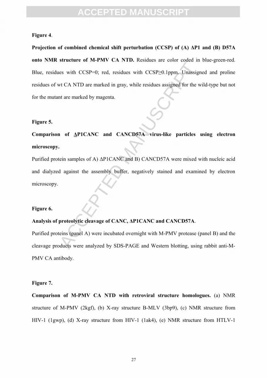
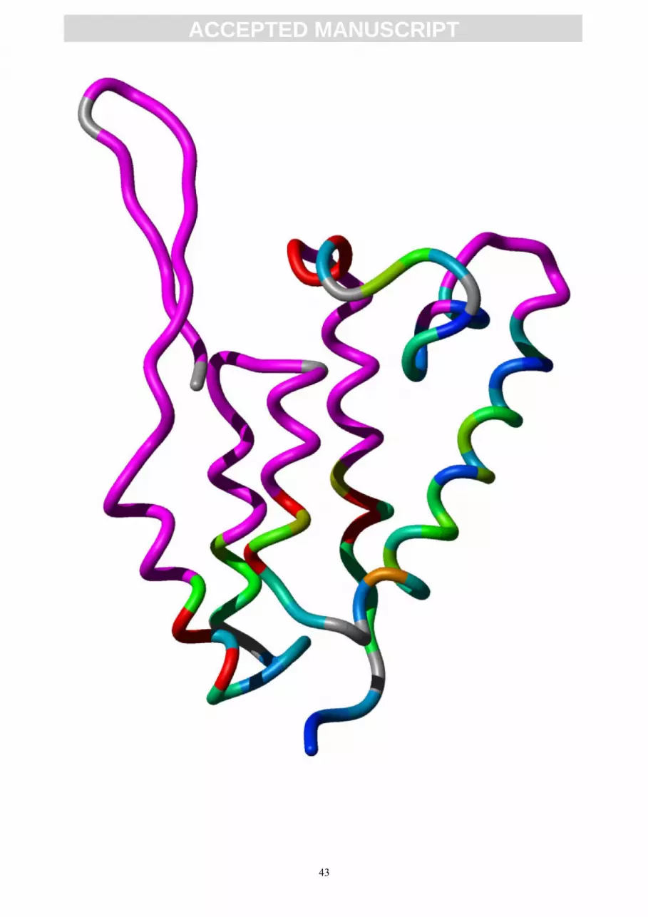
Figure 4.
Projection of combined chemical shift perturbation (CCSP) of (A) ΔP1 and (B) D57A
onto NMR structure of M-PMV CA NTD. Residues are color coded in blue-green-red.
Blue, residues with CCSP=0; red, residues with CCSP≥0.1ppm. Unassigned and proline
residues of wt CA NTD are marked in gray, while residues assigned for the wild-type but not
for the mutant are marked by magenta.
Figure 5.
Comparison of ΔP1CANC and CANCD57A virus-like particles using electron
microscopy.
Purified protein samples of A) ΔP1CANC and B) CANCD57A were mixed with nucleic acid
and dialyzed against the assembly buffer, negatively stained and examined by electron
microscopy.
Figure 6.
Analysis of proteolytic cleavage of CANC, ΔP1CANC and CANCD57A.
Purified proteins (panel A) were incubated overnight with M-PMV protease (panel B) and the
cleavage products were analyzed by SDS-PAGE and Western blotting, using rabbit anti-M-
PMV CA antibody.
Figure 7.
Comparison of M-PMV CA NTD with retroviral structure homologues. (a) NMR
structure of M-PMV (2kgf), (b) X-ray structure B-MLV (3bp9), (c) NMR structure from
HIV-1 (1gwp), (d) X-ray structure from HIV-1 (1ak4), (e) NMR structure from HTLV-1
27

ACC
EPTE
D M
ANU
SCR
IPT
ACCEPTED MANUSCRIPT
(1g03), (f) X-ray structure from RSV (1en9), (g) X-ray structure from JSRV (2v4x), (h) NMR
structure from RSV (1d1d), and (i) X-ray structure from EIAV (1eia).
Figure 8.
The detail of the electrostatic interactions between the β-hairpin (residues 1,14,16) and
the helices α3 (residue 57) and α5 (residues 111, 117). The interacting residues are shown
as stick models, while the secondary structure elements are colored as in Figure 2. The
surfaces of positively and negatively charged residues are colored in blue and red,
respectively, while hydrogen bonds are shown as dashed black lines.
28

ACC
EPTE
D M
ANU
SCR
IPT
ACCEPTED MANUSCRIPT
Reference List
1. Vlach, J., Lipov, J., Rumlova, M., Veverka, V., Lang, J., Srb, P., Knejzlik, Z., Pichova, I.,
Hunter, E., Hrabal, R. & Ruml, T. (2008). D-retrovirus morphogenetic switch driven by the
targeting signal accessibility to Tctex-1 of dynein. Proc. Natl. Acad. Sci. U. S. A 105, 10565-
10570.
2. Campos-Olivas, R., Newman, J. L. & Summers, M. F. (2000). Solution structure and
dynamics of the Rous sarcoma virus capsid protein and comparison with capsid proteins of
other retroviruses. Journal of Molecular Biology 296, 633-649.
3. Cornilescu, C. C., Bouamr, F., Yao, X., Carter, C. & Tjandra, N. (2001). Structural analysis
of the N-terminal domain of the human T-cell leukemia virus capsid protein. Journal of
Molecular Biology 306, 783-797.
4. Gamble, T. R., Yoo, S. H., Vajdos, F. F., vonSchwedler, U. K., Worthylake, D. K., Wang,
H., McCutcheon, J. P., Sundquist, W. I. & Hill, C. P. (1997). Structure of the carboxyl-
terminal dimerization domain of the HIV-1 capsid protein. Science 278, 849-853.
5. Gamble, T. R., Vajdos, F. F., Yoo, S., Worthylake, D. K., Houseweart, M., Sundquist, W. I.
& Hill, C. P. (1996). Crystal Structure of Human Cyclophilin A Bound to the Amino-
Terminal Domain of HIV-1 Capsid. Cell 87, 1285-1294.
6. Gitti, R. K., Lee, B. M., Walker, J., Summers, M. F., Yoo, S. & Sundquist, W. I. (1996).
Structure of the amino-terminal core domain of the HIV-1 capsid protein. Science 273, 231-
235.
7. Jin, Z., Jin, L., Peterson, D. L. & Lawson, C. L. (1999). Model for lentivirus capsid core
assembly based on crystal dimers of EIAV p26. Journal of Molecular Biology 286, 83-93.
29

ACC
EPTE
D M
ANU
SCR
IPT
ACCEPTED MANUSCRIPT
8. Khorasanizadeh, S., Campos-Olivas, R. & Summers, M. F. (1999). Solution Structure of
the Capsid Protein from the Human T-cell Leukemia Virus Type-I. Journal of Molecular
Biology 291, 491-505.
9. Kingston, R. L., Fitzon-Ostendorp, T., Eisenmesser, E. Z., Schatz, G. W., Vogt, V. M.,
Post, C. B. & Rossmann, M. G. (2000). Structure and self-association of the Rous sarcoma
virus capsid protein. Structure with Folding & Design 8, 617-628.
10. Mortuza, G. B., Dodding, M. P., Goldstone, D. C., Haire, L. F., Stoye, J. P. & Taylor, I.
A. (2008). Structure of B-MLV Capsid Amino-terminal Domain Reveals Key Features of
Viral Tropism, Gag Assembly and Core Formation. Journal of Molecular Biology 376, 1493-
1508.
11. Mortuza, G. B., Goldstone, D. C., Pashley, C., Haire, L. F., Palmarini, M., Taylor, W. R.,
Stoye, J. P. & Taylor, I. A. (2009). Structure of the capsid amino-terminal domain from the
betaretrovirus, jaagsiekte sheep retrovirus. J. Mol. Biol. 386, 1179-1192.
12. Momany, C., Kovari, L. C., Prongay, A. J., Keller, W., Gitti, R. K., Lee, B. M.,
Gorbalenya, A. E., Tong, L., McClure, J., Ehrlich, L. S., Summers, M. F., Carter, C. &
Rossmann, M. G. (1996). Crystal structure of dimeric HIV-1 capsid protein. Nat Struct Mol
Biol 3, 763-770.
13. Ganser-Pornillos, B. K., von Schwedler, U. K., Stray, K. M., Aiken, C. & Sundquist, W. I.
(2004). Assembly properties of the human immunodeficiency virus type 1 CA protein.
Journal of Virology 78, 2545-2552.
14. von Schwedler, U. K., Stemmler, T. L., Klishko, V. Y., Li, S., Albertine, K. H., Davis, D.
R. & Sundquist, W. I. (1998). Proteolytic refolding of the HIV-1 capsid protein amino-
terminus facilitates viral core assembly (vol 17, pg 1555, 1998). Embo Journal 19, 2391.
30

ACC
EPTE
D M
ANU
SCR
IPT
ACCEPTED MANUSCRIPT
15. von Schwedler, U. K., Stray, K. M., Garrus, J. E. & Sundquist, W. I. (2003). Functional
Surfaces of the Human Immunodeficiency Virus Type 1 Capsid Protein. J. Virol. 77, 5439-
5450.
16. Mortuza, G. B., Haire, L. F., Stevens, A., Smerdon, S. J., Stoye, J. P. & Taylor, I. A.
(2004). High-resolution structure of a retroviral capsid hexameric amino-terminal domain.
Nature 431, 481-485.
17. Auerbach, M. R., Brown, K. R. & Singh, I. R. (2007). Mutational Analysis of the N-
Terminal Domain of Moloney Murine Leukemia Virus Capsid Protein. J. Virol. 81, 12337-
12347.
18. Wildova, M., Hadravova, R., Stokrova, J., Krizova, I., Ruml, T., Hunter, E., Pichova, I. &
Rumlova, M. (2008). The effect of point mutations within the N-terminal domain of Mason-
Pfizer monkey virus capsid protein on virus core assembly and infectivity. Virology 380, 157-
163.
19. Abdurahman, S., Youssefi, M., Hoglund, S. & Vahlne, A. (2007). Characterization of the
invariable residue 51 mutations of human immunodeficiency virus type 1 capsid protein on in
vitro CA assembly and infectivity. Retrovirology 4, 69.
20. Gross, I., Hohenberg, H., Huckhagel, C. & Krausslich, H. G. (1998). N-terminal extension
of human immunodeficiency virus capsid protein converts the in vitro assembly phenotype
from tubular to spherical particles. Journal of Virology 72, 4798-4810.
21. Leschonsky, B., Ludwig, C., Bieler, K. & Wagner, R. (2007). Capsid stability and
replication of human immunodeficiency virus type 1 are influenced critically by charge and
size of Gag residue 183. J Gen Virol 88, 207-216.
22. Rumlova-Klikova, M., Hunter, E., Nermut, M. V., Pichova, I. & Ruml, T. (2000).
Analysis of Mason-Pfizer monkey virus gag domains required for capsid assembly in
31

ACC
EPTE
D M
ANU
SCR
IPT
ACCEPTED MANUSCRIPT
bacteria: Role of the N-terminal proline residue of CA in directing particle shape. Journal of
Virology 74, 8452-8459.
23. Ganser-Pornillos, B. K., Cheng, A. & Yeager, M. (2007). Structure of Full-Length HIV-1
CA: A Model for the Mature Capsid Lattice. Cell 131, 70-79.
24. Kay, L. E., Torchia, D. A. & Bax, A. (1989). Backbone Dynamics of Proteins As Studied
by N-15 Inverse Detected Heteronuclear Nmr-Spectroscopy - Application to Staphylococcal
Nuclease. Biochemistry 28, 8972-8979.
25. Krizova, H., Zidek, L., Stone, M. J., Novotny, M. V. & Sklenar, V. (2004). Temperature-
dependent spectral density analysis applied to monitoring backbone dynamics of major
urinary protein-I complexed with the pheromone 2-sec-butyl-4,5-dihydrothiazole. Journal of
Biomolecular NMR 28, 369-384.
26. Bernado, P., de la Torre, J. G. & Pons, M. (2002). Interpretation of N-15 NMR relaxation
data of globular proteins using hydrodynamic calculations with HYDRONMR. Journal of
Biomolecular NMR 23, 139-150.
27. Nandhagopal, N., Simpson, A. A., Johnson, M. C., Francisco, A. B., Schatz, G. W.,
Rossmann, M. G. & Vogt, V. M. (2004). Dimeric Rous Sarcoma Virus Capsid Protein
Structure Relevant to Immature Gag Assembly. Journal of Molecular Biology 335, 275-282.
28. Ulbrich, P., Haubova, S., Nermut, M. V., Hunter, E., Rumlova, M. & Ruml, T. (2006).
Distinct roles for nucleic acid in in vitro assembly of purified Mason-Pfizer monkey virus
CANC proteins. J. Virol. 80, 7089-7099.
29. Rumlova, M., Ruml, T., Pohl, J. & Pichova, I. (2003). Specific in vitro cleavage of
Mason-Pfizer monkey virus capsid protein: evidence for a potential role of retroviral protease
in early stages of infection. Virology 310, 310-318.
32

ACC
EPTE
D M
ANU
SCR
IPT
ACCEPTED MANUSCRIPT
30. Luban, J., Bossolt, K. L., Franke, E. K., Kalpana, G. V. & Goff, S. P. (1993). Human-
Immunodeficiency-Virus Type-1 Gag Protein Binds to Cyclophilin-A and Cyclophilin-B.
Cell 73, 1067-1078.
31. Diehl, W. E., Stansell, E., Kaiser, S. M., Emerman, M. & Hunter, E. (2008). Identification
of postentry restrictions to Mason-Pfizer monkey virus infection in New World monkey cells.
J. Virol. 82, 11140-11151.
32. Mcclure, M. A. (1991). Evolution of Retroposons by Acquisition Or Deletion of
Retrovirus-Like Genes. Molecular Biology and Evolution 8, 835-856.
33. Bouamr, F., Cornilescu, C. C., Goff, S. P., Tjandra, N. & Carter, C. A. (2005). Structural
and Dynamics Studies of the D54A Mutant of Human T Cell Leukemia Virus-1 Capsid
Protein. J. Biol. Chem. 280, 6792-6801.
34. Tang, C., Ndassa, Y. & Summers, M. F. (2002). Structure of the N-terminal 283-residue
fragment of the immature HIV-1 Gag polyprotein. Nat. Struct. Biol. 9, 537-543.
35. Tang, S., Murakami, T., Cheng, N., Steven, A. C., Freed, E. O. & Levin, J. G. (2003).
Human Immunodeficiency Virus Type 1 N-Terminal Capsid Mutants Containing Cores with
Abnormally High Levels of Capsid Protein and Virtually No Reverse Transcriptase. J. Virol.
77, 12592-12602.
36. Macek, P., Zidek, L., Rumlova, M., Pichova, I. & Sklenar, V. (2008). 1H, 13C, and 15N
resonance assignment of the N-terminal domain of Mason-Pfizer monkey virus capsid
protein, CANTD-140. Biomolecular NMR Assignments 2, 43-45.
37. Sattler, M., Schleucher, J. & Griesinger, C. (1999). Heteronuclear multidimensional NMR
experiments for the structure determination of proteins in solution employing pulsed field
gradients. Progress in Nuclear Magnetic Resonance Spectroscopy 34, 93-158.
38. Ottiger, M., Delaglio, F. & Bax, A. (1998). Measurement of J and dipolar couplings from
simplified two-dimensional NMR spectra. Journal of Magnetic Resonance 131, 373-378.
33

ACC
EPTE
D M
ANU
SCR
IPT
ACCEPTED MANUSCRIPT
39. Zidek, L., Wu, H. H., Feigon, J. & Sklenar, V. (2001). Measurement of small scalar and
dipolar couplings in purine and pyrimidine bases. Journal of Biomolecular NMR 21, 153-160.
40. Serber, Z., Richter, C., Moskau, D., Bohlen, J. M., Gerfin, T., Marek, D., Haberli, M.,
Baselgia, L., Laukien, F., Stern, A. S., Hoch, J. C. & Dotsch, V. (2000). New carbon-detected
protein NMR experiments using CryoProbes. Journal of the American Chemical Society 122,
3554-3555.
41. Bermel, W., Bertini, I., Duma, L., Felli, I. C., Emsley, L., Pierattelli, R. & Vasos, P. R.
(2005). Complete assignment of heteronuclear protein resonances by protonless NMR
spectroscopy. Angewandte Chemie-International Edition 44, 3089-3092.
42. Duma, L., Hediger, S., Lesage, A. & Emsley, L. (2003). Spin-state selection in solid-state
NMR. Journal of Magnetic Resonance 164, 187-195.
43. Farrow, N. A., Muhandiram, R., Singer, A. U., Pascal, S. M., Kay, C. M., Gish, G.,
Shoelson, S. E., Pawson, T., Formankay, J. D. & Kay, L. E. (1994). Backbone Dynamics of A
Free and A Phosphopeptide-Complexed Src Homology-2 Domain Studied by N-15 NMR
Relaxation. Biochemistry 33, 5984-6003.
44. Schumann,F.H.; Riepl,H.; Maurer,T.; Gronwald,W.; Neidig,K.P.; Kalbitzer,H.R. (2007).
Combined chemical shift changes and amino acid specific chemical shift mapping of protein-
protein interactions Journal of Biomolecular NMR 39, 275-289.
45. Mulder,F.A.; Schipper,D.; Bott,R.; Boelens,R. (1999). Altered flexibility in the substrate-
binding site of related native and engineered high-alkaline Bacillus subtilisins Journal of
Molecular Biology. 292, 111-123
46. Delaglio, F., Grzesiek, S., Vuister, G. W., Zhu, G., Pfeifer, J. & Bax, A. (1995). NMRPipe
- A Multidimensional Spectral Processing System Based on Unix Pipes. Journal of
Biomolecular NMR 6, 277-293.
34

ACC
EPTE
D M
ANU
SCR
IPT
ACCEPTED MANUSCRIPT
47. Cornilescu, G., Delaglio, F. & Bax, A. (1999). Protein backbone angle restraints from
searching a database for chemical shift and sequence homology. Journal of Biomolecular
NMR 13, 289-302.
48. Herrmann, T., Guntert, P. & Wuthrich, K. (2002). Protein NMR structure determination
with automated NOE assignment using the new software CANDID and the torsion angle
dynamics algorithm DYANA. Journal of Molecular Biology 319, 209-227.
49. Schwieters, C. D., Kuszewski, J. J., Tjandra, N. & Clore, G. M. (2003). The Xplor-NIH
NMR molecular structure determination package. Journal of Magnetic Resonance 160, 65-73.
50. Schwieters, C. D., Kuszewski, J. J. & Clore, G. M. (2006). Using Xplor-NIH for NMR
molecular structure determination. Progress in Nuclear Magnetic Resonance Spectroscopy
48, 47-62.
51. Brunger, A. T., Adams, P. D., Clore, G. M., Delano, W. L., Gros, P., Grosse-Kunstleve,
R. W., Jiang, J. S., Kuszewski, J., Nilges, M., Pannu, N. S., Read, R. J., Rice, L. M.,
Simonson, T. & Warren, G. L. (1998). Crystallography & NMR system: A new software suite
for macromolecular structure determination. Acta Crystallographica Section D-Biological
Crystallography 54, 905-921.
52. Brunger, A. T. (2007). Version 1.2 of the Crystallography and NMR system. Nat.
Protocols 2, 2728-2733.
53. Rieping, W., Habeck, M., Bardiaux, B., Bernard, A., Malliavin, T. E. & Nilges, M.
(2007). ARIA2: Automated NOE assignment and data integration in NMR structure
calculation. Bioinformatics 23, 381-382.
54. Zidek, L., Padrta, P., Chmelik, J. & Sklenar, V. (2003). Internal consistency of NMR data
obtained in partially aligned biomacromolecules. Journal of Magnetic Resonance 162, 385-
395.
35

ACC
EPTE
D M
ANU
SCR
IPT
ACCEPTED MANUSCRIPT
55. Mesleh, M. F. & Opella, S. J. (2003). Dipolar Waves as NMR maps of helices in proteins.
Journal of Magnetic Resonance 163, 288-299.
56. Nederveen, A. J., Doreleijers, J. F., Vranken, W., Miller, Z., Spronk, C. A. E. M.,
Nabuurs, S. B., Guntert, P., Livny, M., Markley, J. L., Nilges, M., Ulrich, E. L., Kaptein, R. &
Bonvin, A. M. J. J. (2005). RECOORD: A recalculated coordinate database of 500+proteins
from the PDB using restraints from the BioMagResBank. Proteins-Structure Function and
Bioinformatics 59, 662-672.
57. Sass, H. J., Musco, G., Stahl, S. J., Wingfield, P. T. & Grzesiek, S. (2001). An easy way
to include weak alignment constraints into NMR structure calculations. Journal of
Biomolecular NMR 21, 275-280.
58. Lipari, G. & Szabo, A. (1982). Model-Free Approach to the Interpretation of Nuclear
Magnetic-Resonance Relaxation in Macromolecules .1. Theory and Range of Validity.
Journal of the American Chemical Society 104, 4546-4559.
59. Lipari, G. & Szabo, A. (1982). Model-Free Approach to the Interpretation of Nuclear
Magnetic-Resonance Relaxation in Macromolecules .2. Analysis of Experimental Results.
Journal of the American Chemical Society 104, 4559-4570.
60. d'Auvergne, E. J. & Gooley, P. R. (2008). Optimisation of NMR dynamic models I.
Minimisation algorithms and their performance within the model-free and Brownian
rotational diffusion spaces. Journal of Biomolecular NMR 40, 107-119.
61. d'Auvergne, E. J. & Gooley, P. R. (2008). Optimisation of NMR dynamic models II. A
new methodology for the dual optimisation of the model-free parameters and the Brownian
rotational diffusion tensor. Journal of Biomolecular NMR 40, 121-133.
62. Dosset, P., Hus, J. C., Blackledge, M. & Marion, D. (2000). Efficient analysis of
macromolecular rotational diffusion from heteronuclear relaxation data. J. Biomol. NMR 16,
23-28.
36

ACC
EPTE
D M
ANU
SCR
IPT
ACCEPTED MANUSCRIPT
63. de la Torre, J. G., Huertas, M. L. & Carrasco, B. (2000). HYDRONMR: Prediction of
NMR relaxation of globular proteins from atomic-level structures and hydrodynamic
calculations. Journal of Magnetic Resonance 147, 138-146.
64. Sreerama, N., Venyaminov, S. Y. & Woody, R. W. (1999). Estimation of the number of
alpha-helical and beta-strand segments in proteins using circular dichroism spectroscopy.
Protein Sci. 8, 370-380.
65. Sreerama, N. & Woody, R. W. (2000). Estimation of protein secondary structure from
circular dichroism spectra: comparison of CONTIN, SELCON, and CDSSTR methods with
an expanded reference set. Anal. Biochem. 287, 252-260.
66. Sreerama, N., Venyaminov, S. Y. & Woody, R. W. (2000). Estimation of protein
secondary structure from circular dichroism spectra: inclusion of denatured proteins with
native proteins in the analysis. Anal. Biochem. 287, 243-251.
67. Deleage, G. & Geourjon, C. (1993). An interactive graphic program for calculating the
secondary structure content of proteins from circular dichroism spectrum. Comput. Appl.
Biosci. 9, 197-199.
37

ACC
EPTE
D M
ANU
SCR
IPT
ACCEPTED MANUSCRIPT
Table 1: Statistics of NMR structure determination for 10 lowest energy structures
Structural statistic Distance restraints 2246 (intraresidual/sequential/medium/long) (836/515/450/445) Hydrogen bonding restraints 124 Torsion Angle restraints (φ/ψ) 107/107 RDCs 130 (N(i)-HN(i)/HN(i)-C(i-1)/N(i)-C(i-1)) (73/31/26)Violations distance violations > 0.5Å 0 dihedral angle violations > 5° 0Pairwise Cartesian RMS deviation (Å) all heavy atoms 2.06 ± 0.25 backbone heavy atoms 1.64 ± 0.28PROCHECK Ramachandran assessment (%) most favoured region 86.8 ± 2.1 additionally allowed region 9.3 ± 1.4 generously allowed region 3.0 ± 0.6 disallowed region 0.8 ± 0.8Average rms deviation from current reliable structures (rms Z scores, null deviation=1) bond lengths 0.884 ± 0.028 bond angles 0.910 ± 0.029 improper dihedral distribution 1.310 ± 0.106Average deviation from current reliable structures (Z scores, null deviation=0) 2nd generation packing quality -1.056 ± 0.107 ramachandran plot appearance -4.818 ± 0.367 chi-1/chi-2 rotamer normality -4.266 ± 0.416
38

ACC
EPTE
D M
ANU
SCR
IPT
ACCEPTED MANUSCRIPT
Table 2: Prediction of protein secondary structure based in measured CD spectra
Helix (%) β-sheet (%) Turns (%) Others (%)WT CA NTD 35.3 14.7 14.7 37.2ΔP1 CA NTD 40.0 12.4 14.5 28.7D57A CA NTD 40.3 11.0 14.8 33.2
39

ACC
EPTE
D M
ANU
SCR
IPT
ACCEPTED MANUSCRIPT
40

ACC
EPTE
D M
ANU
SCR
IPT
ACCEPTED MANUSCRIPT
41

ACC
EPTE
D M
ANU
SCR
IPT
ACCEPTED MANUSCRIPT
42
ACC
EPTE
D M
ANU
SCR
IPT
ACCEPTED MANUSCRIPT
43

ACC
EPTE
D M
ANU
SCR
IPT
ACCEPTED MANUSCRIPT
44

ACC
EPTE
D M
ANU
SCR
IPT
ACCEPTED MANUSCRIPT
45

ACC
EPTE
D M
ANU
SCR
IPT
ACCEPTED MANUSCRIPT
46


![arXiv:2107.05446v3 [cs.LG] 17 Mar 2022 - Ian Mason](https://img.pdfslide.net/doc/110x75/6335b02ce687b37e4f05d60b/arxiv210705446v3-cslg-17-mar-2022-ian-mason.jpg)


